Embed Size (px)
Citation preview
1
Limited Accumulation of Damaged Proteins in L-Isoaspartyl (D-Aspartyl) O-Methyltransferase-deficient Mice*
Jonathan D. Lowenson‡, Edward Kim¶§, Stephen G. Young¶§, and
Steven Clarke‡
From the ‡Department of Chemistry and Biochemistry and the Molecular
Biology Institute, University of California, Los Angeles, California
90095-1569; the ¶Gladstone Institute of Cardiovascular Disease, San
Francisco, California 94141-9100; and the §Department Of Medicine and
Cardiovascular Research Institute, University of California, San
Francisco, California 94143
*This work was supported by NIH grants AG15451 and HL41633 (to S. G. Y),
NIH grants GM26020 and AG18000 (to S. C.), and by a grant from the University
of California Tobacco-Related Disease Research Program (to S. G. Y.).
‡To whom correspondence should be addressed. Tel.: 310 825 8754; Fax: 310
825 1968; E-mail: [email protected].
Corresponding Author: Steven Clarke640 Paul D. Boyer Hall611 Charles E. Young Drive EastLos Angeles, CA 90095-1570Tel.: 310-825-8754 Fax.: 310-825-1968E-mail: [email protected]
Running Title: Rescued Pcmt1 Knockout Mice
Copyright 2001 by The American Society for Biochemistry and Molecular Biology, Inc.
JBC Papers in Press. Published on March 7, 2001 as Manuscript M100987200
2
SUMMARY
L-Isoaspartyl (D-aspartyl) O-methyltransferase (PCMT1) can
initiate the conversion of damaged aspartyl and asparaginyl
residues to normal L-aspartyl residues. Mice lacking this enzyme
(Pcmt1-/- mice) have elevated levels of damaged residues and die at
a mean age of 42 days from massive tonic-clonic seizures. To
extend the lives of the knockout mice so that the long-term effects
of damaged residue accumulation could be investigated, we produced
transgenic mice with a mouse Pcmt1 cDNA under the control of a
neuron-specific promoter. Pcmt1 transgenic mice that were
homozygous for the endogenous Pcmt1 knockout mutation (“transgenic
Pcmt1-/- mice”) had brain PCMT1 activity levels that were 6.5–13%
those of wild-type mice, but had little or no activity in other
tissues. The transgenic Pcmt1-/- mice lived, on average, fivefold
longer than nontransgenic Pcmt1-/- mice, and accumulated only half
as many damaged aspartyl residues in their brain proteins. The
concentration of damaged residues in heart, testis, and brain
proteins in transgenic Pcmt1-/- mice initially increased with age
but unexpectedly reached a plateau by 100 days of age. Urine from
Pcmt1-/- mice contained increased amounts of peptides with damaged
aspartyl residues, apparently enough to account for proteins that
were not repaired intracellularly. In the absence of PCMT1,
proteolysis may limit the intracellular accumulation of damaged
proteins, but less efficiently than in wild-type mice having
PCMT1–mediated repair.
3
The spontaneous chemical modification of proteins by reaction with oxygen,
water, sugars, and other abundant metabolites is unavoidable. The accumulation
of such nonenzymatically altered proteins is associated with normal aging as
well as atherosclerosis, Alzheimer's disease, and diabetes (1-3). Organisms
have several strategies for dealing with damaged proteins, including
intracellular proteolysis mediated by proteosome and lysosomal action (4-6).
Some types of covalent damage, however, are simple enough to recognize and
repair directly (7). Enzymes such as cis-trans prolyl isomerase (8),
methionine sulfoxide reductase (9), and disulfide isomerase (8), can restore
activity to proteins that have been chemically altered.
We are interested in a common type of spontaneous protein damage in which
L-aspartyl and L-asparaginyl residues undergo an intramolecular reaction that
converts them to L-succinimidyl residues (10, 11). Nonenzymatic hydrolysis of
the succinimide ring readily occurs at either carbonyl to generate both normal
aspartyl residues and isoaspartyl residues, in which the peptide backbone
proceeds through the β-carbonyl rather than the α-carbonyl moiety (12). The
succinimide also racemizes more rapidly than do the open-chain forms, and
hydrolysis of the D-succinimide produces D-aspartyl and D-isoaspartyl residues
(12). Local protein structure causes some L-aspartyl and L-asparaginyl
residues to be especially prone to succinimide formation, and the presence of
damaged aspartyl residues at these sites can significantly alter the protein's
structure, function, and immunogenicity (10, 13).
To minimize the accumulation of damaged aspartyl residues in cellular
proteins, all mammalian tissues possess an L-isoaspartyl (D-aspartyl) O-
methyltransferase (E.C. 2.1.1.77; designated PCMT1 in mice)1 (14). This enzyme
uses S-adenosyl-L-methionine (AdoMet) to methylate L-isoaspartyl (and, less
4
efficiently, D-aspartyl) residues, but not normal L-aspartyl residues (15).
Nonenzymatic de-esterification of the methylated residues returns them to the
succinimide form much more rapidly than occurs in the absence of methylation,
resulting in the eventual conversion of most of the damaged residues to the
“repaired” L-aspartyl form (16, 17).
The physiological importance of this pathway remained unclear until Pcmt1
knockout (Pcmt1-/-) mice were created and found to display a distinctive
phenotype (18, 19). Pcmt1-/- mice have two- to sixfold higher levels of
damaged aspartyl residues in their brain, heart, liver, and erythrocytes than
are observed in wild-type tissues (18). Furthermore, Pcmt1-/- mice are smaller
than their Pcmt1+/- and Pcmt1+/+ littermates, undergo severe tonic-clonic
seizures, and die at an average age of 42 days (18, 19).
Electroencephalographic analysis shows that these mice suffer abnormal brain
activity about 50% of the time, not just during the tonic-clonic seizures (20).
Administration of the anti-seizure drug valproic acid enabled Pcmt1-/- mice to
attain the same size and weight as their wild-type littermates, suggesting that
the absence of the methyltransferase did not interfere directly with food
intake or metabolism (20). The combination of valproic acid and clonazepam
prolonged mean survival, but only by 36 days (20).
These results have raised new questions. Does the repair
methyltransferase, although expressed in all tissues, have particular
importance within the brain? Is it important in neurons or in nonneuronal cell
types? If the seizures in the Pcmt1-/- mice were prevented, would other organs
function abnormally as damaged aspartyl residues accumulated? In the current
study, we have approached these questions by producing transgenic mice that
express Pcmt1 solely within neurons.
5
EXPERIMENTAL PROCEDURES
Generation of Pcmt1 Transgenic Mice—A rat neuron-specific enolase (NSE)
promoter was used to direct the expression of mouse Pcmt1 cDNA in the brains of
transgenic mice. The methyltransferase coding sequence [including 119 base
pairs (bp) of 5'-noncoding and 777 bp of 3'-noncoding sequence] was obtained
from a 1580-bp murine testis cDNA clone (21) and was removed from plasmid
sequences with EcoRI. The proximal rat NSE promoter was isolated from the
plasmid NSE-APP695 (22) after digestion with HindIII. After overhangs were
filled with Klenow polymerase, the mouse Pcmt1 cDNA and the rat NSE vector were
ligated and the NSE-Pcmt1 transgenic construct isolated by digestion with SalI.
The transgene (2 ng/µl) was microinjected into F2 C57BL/6 × SJL fertilized
mouse eggs by standard techniques (23). From 37 microinjected eggs, 33 pups
were obtained, and four harbored the Pcmt1 transgene. These transgenic
founders were identified by PCR with primers corresponding to mouse Pcmt1 cDNA
sequences (5’–GCCAGCCACTCGGAGCTAATCC–3’ from exon 1 and
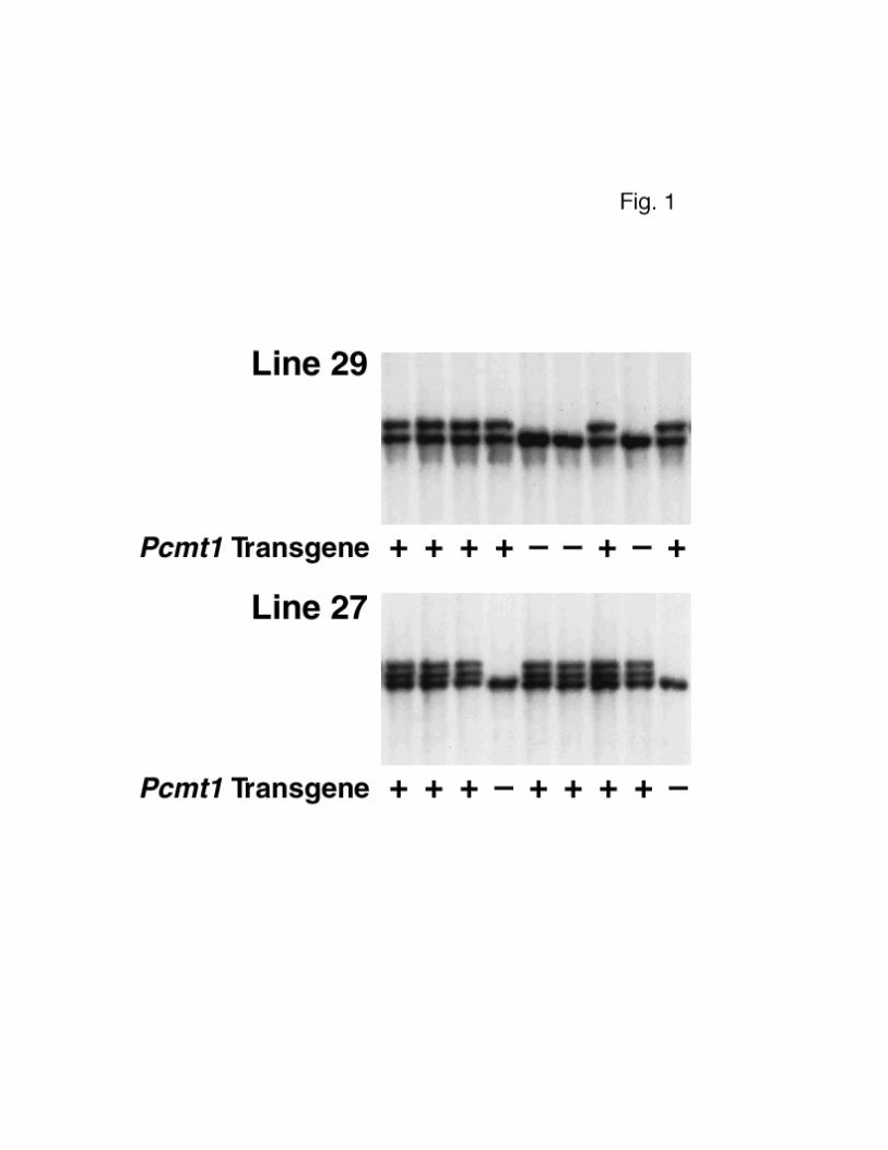
5’–CCACTATTTCCAACCATCCGTGC–3’ from exons 4 and 5). Southern blot analysis of
tail DNA confirmed the integration of the transgene (Fig. 1). Two of these
mice, founders 27 and 29, were bred with C57BL/6 × 129/SvJae mice that were
heterozygous for a knockout mutation in the endogenous Pcmt1 gene (18). DNA
samples from the tails of these mice were genotyped by PCR, both to detect the
transgene and to detect the knockout mutation. Mice that were heterozygous for
the knockout mutation and hemizygous for the transgene were selected for
breeding. All mice were weaned at 21 days of age, housed in a barrier facility
with a 12-h light/dark cycle, and fed a chow diet.
6
Preparation of Mouse Tissues—Brain, heart, liver, kidney, and testis were
removed immediately from sacrificed animals and placed in ice-cold buffer (5
ml/g wet weight) containing 250 mM sucrose, 10 mM Tris-HCl, 1 mM disodium EDTA,
pH 7.4, and the protease inhibitor phenylmethylsulfonylfluoride (25 µM). The
tissues were disrupted in a glass homogenization tube with a Teflon pestle
rotating at 310 rpm for four 10-s intervals. The homogenates were placed in
1.5–ml tubes and centrifuged at 20,800 × g for 10 min. The resulting
supernatant fractions contained both cytosolic proteins and microsomes and were
kept frozen until used.
Whole blood (100—200 µl) was taken from the tail or heart and combined
with 200 µl of 2 mg/ml disodium EDTA, 10 mM sodium phosphate, 137.9 mM sodium
chloride, pH 7.4. Erythrocytes were collected by centrifugation at 4000 × g
for 4 min and washed four times with 1 ml of the above buffer. Pelleted
erythrocytes were lysed in five volumes of 5 mM sodium phosphate, 1 mM disodium
EDTA, pH 7.4, and 25 µM phenylmethylsulfonylfluoride. The lysates were
centrifuged in 1.5–ml conical tubes at 20,800 × g for 10 min to remove the
membrane ghosts, and the supernatant fractions were stored at –20 °C.
Protein Assay—A modified Lowry assay was used to determine protein
concentrations in the extracts. Assays were done in duplicate with bovine
serum albumin as a standard (18).
Assay of L-Isoaspartyl (D-Aspartyl) O-Methyltransferase
Activity—Methyltransferase activity was assayed by its ability to transfer the
methyl group from S-adenosyl-L-methionine to ovalbumin. The supernatant
fraction from homogenized tissues (10-60 µg of brain, heart, or testis protein;
0.6-0.8 mg of erythrocyte protein) was incubated with 0.8 mg of ovalbumin
(Sigma, Grade V) in 0.2 M BisTris-HCl, pH 6.0, containing 10 µM [14C]AdoMet (53
7
mCi/mmol; Amersham) in final volume of 40 µl at 37 °C for 15 min. NaOH (80 µl
of a 200 mM solution) was added to stop the reaction and to hydrolyze the
[14C]methyl esters formed on ovalbumin to [14C]methanol. The reaction mixture
was immediately spotted onto an 8 × 2–cm piece of thick filter paper and
incubated above 5 ml of Safety-Solve scintillation fluid (RPI) in the neck of a
sealed 20–ml scintillation vial at room temperature for 2 h to allow
[14C]methanol to diffuse into the scintillation fluid. The filter paper was
removed, and the scintillation fluid counted for radioactivity. Incubations
containing S-adenosyl-L-[methyl-14C]methionine, ovalbumin, and buffer
constituted the “blank” for the assay; the radioactivity in these tubes
(typically <5% of the nonblank samples) was subtracted from total counts in the
determination of enzyme activity.
Quantitation of Damaged Aspartyl Residues—Cellular proteins were
incubated at 37 °C for 2 h with 0.8 µg of recombinant human L-isoaspartyl
methyltransferase (specific activity: 10,000 pmol methyl esters formed on
ovalbumin at 37 °C/min/mg protein) (24) in 0.2 M BisTris-HCl, pH 6.0, and 10 µM
[14C]AdoMet in a final volume of 40 µl. After base hydrolysis, [14C]methanol
production was measured as described above to quantitate L-isoaspartyl and
D–aspartyl methyl-accepting sites in cellular proteins. Incubations containing
[14C]AdoMet, recombinant methyltransferase, and buffer constituted the “blank”
for the assay; the radioactivity in these tubes was subtracted from each
sample's total counts. Each sample was assayed in duplicate or triplicate, and
the average value is reported.
Quantitation of Endogenously Methylated Damaged Aspartyl Residues—Damaged
residues that are already methylated within cells by the endogenous
methyltransferase and S-adenosyl-L-methionine are not measured in the assay
8
described above but can be quantified after mild base treatment. Protein
(8.3—9.6 mg) from homogenized Pcmt1-/- and Pcmt1+/+ brains was incubated in 20
µl of 75 mM potassium borate, pH 10.2. After times ranging from 5 s to 360
min, 10 µl of 500 mM BisTris-HCl, pH 5.7, was added to lower the pH to about 6.
Then, recombinant human methyltransferase (5 pmol/min) and S-adenosyl-L-
[methyl-14C]methionine (10 µM final concentration) in 10 µl of 150 mM
BisTris–HCl, pH 6.0, was added, and these reaction mixtures were incubated at
37 °C for 135 min. The reaction was stopped by freezing on dry ice, and then
the base-labile methyl esters were quantitated as described above.
Urine Collection and Analysis—Urine, freshly voided on Parafilm, was
collected with a pipette and stored frozen until used. Creatinine in the urine
was measured by a modified form of the procedure of Bosnes and Taussky (25).
An aliquot of each urine sample (0.3—1 µl) or standard creatinine (0—25 µg) was
diluted to 50 µl with water in duplicate tubes. Picric acid was added (25 µl
of a 40 mM solution), and the tubes were capped and incubated in a boiling
water bath for 45 min. After cooling to room temperature, 25 µl of 0.75 M NaOH
was added. Within 15 min, 90 µl of each sample was transferred to a
flat–welled microtiter plate (Costar) and absorbance was measured at 525 nm a
Beckman DU-600 plate reader. Damaged aspartyl residues in the urine were
assayed with recombinant human methyltransferase as described above.
Amino Acid Analysis—Free amino acids and isoaspartyl-containing
dipeptides in urine were derivatized with o-phthalaldehyde and
2–mercaptoethanol, separated by reverse-phase HPLC, and quantitated by
fluorescence as previously described (26). A precipitate that formed upon
mixing of the urine and derivatization reagent was removed by centrifugation at
20,800 × g for 3 min before HPLC injection. Urine that had been dried in 6 ×
9
50–mm glass tubes was hydrolyzed in vaporized hydrochloric acid in vacuo at 108
°C for 3 h with a PicoTag Work Station (Waters); amino acids in the
hydrolysates were quantitated as described above. The fluorescence color
constants for these derivatives were determined with amino acid and dipeptide
standards.
10
RESULTS
Expression of a Pcmt1 Transgene in Neurons Prolongs the Lives of Mice
Lacking Endogenous Pcmt1—We generated two Pcmt1 transgenic mouse lines, lines
27 and 29, in which the murine Pcmt1 methyltransferase cDNA was placed under
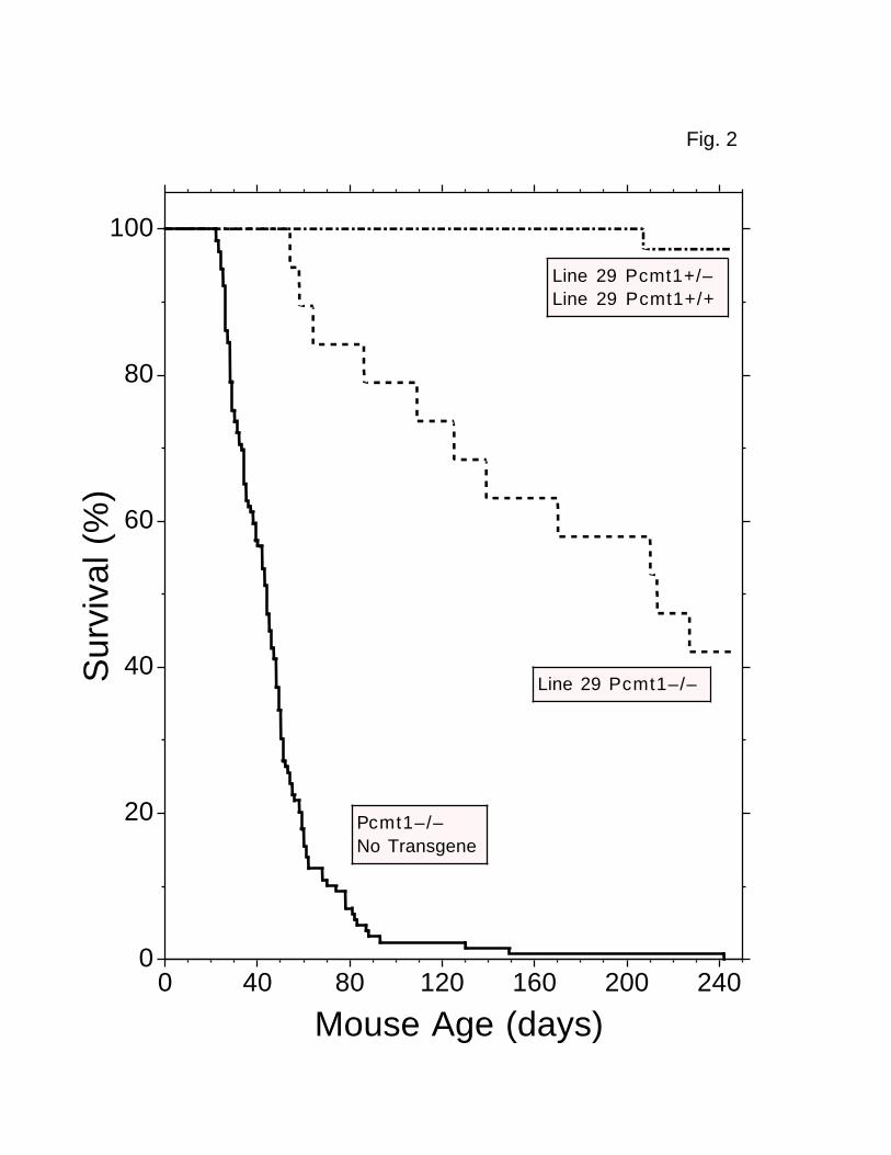
the control of a neuron-specific promoter. We then compared the survival of
“transgenic Pcmt1–/– mice” with that of “nontransgenic Pcmt1–/– mice.” Whereas
nontransgenic Pcmt1–/– (n = 129) mice died at a median age of 44 days (with
only one mouse living beyond 150 days), the transgenic Pcmt1–/– mice lived much
longer (Fig. 2). Of 11 line 27 transgenic Pcmt1–/– mice examined in this
study, six died between 30 and 90 days of age, but four lived from 549 to 757
days. Line 29 transgenic Pcmt1–/– mice (n = 19) died at a median age of 213
days (Fig. 2), and three lived more than 400 days. The nontransgenic Pcmt1–/–
mice began to die at about 21 days. In contrast, none of the line 29
transgenic Pcmt1–/– mice died at less than 52 days of age (Fig. 2).
Line 27 and most of the line 29 mice possessing one or two copies of the
endogenous Pcmt1 gene were indistinguishable from comparable nontransgenic mice
in size, weight, and behavior, although 15 of 94 line 29 mice ran rapidly in
circles. Line 27 and line 29 mice appeared to have normal physiological
functions and had unremarkable tissue histology. The transgenic Pcmt1-/- mice,
however, differed from the nontransgenic Pcmt1-/- animals in several ways.
First, although nontransgenic Pcmt1–/– mice weighed significantly less than
age- and sex-matched Pcmt1+/– and Pcmt1+/+ littermates (18), the weights of
transgenic Pcmt1–/– mice were identical to their Pcmt1+/– and Pcmt1+/+
littermates (data not shown). Second, due to their low-grade seizure activity
(e.g., facial grooming and myoclonic jerks), the nontransgenic Pcmt1-/- mice
11
could often be distinguished by observation from wild-type and heterozygous
littermates. In contrast, these abnormalities were not observed in the
transgenic Pcmt1-/- mice.2 Finally, nontransgenic Pcmt1-/- mice of either sex
never produced litters, even when housed with Pcmt1+/+ animals and given the
anti-seizure drugs valproic acid and clonazepam, and only a single mating had
been observed (20). Two line 27 Pcmt1-/- animals, however, produced two small
litters without the administration of any drug treatments, and 17 pairings
involving male and/or female line 29 Pcmt1-/- mice have produced three litters
(from two different Pcmt1-/- mothers).
Localization of Methyltransferase Activity in Tissues of Line 27 and Line
29 Transgenic Mice—The Pcmt1 transgene controlled by a neuron-specific promoter
appeared to rescue, at least partially, the early death phenotype seen in mice
lacking the endogenous methyltransferase. We next investigated where in the
mouse, and at what level, the transgene was being expressed by assaying
methyltransferase activity in various mouse tissues. As expected, transgenic
mice possessing one or two copies of the endogenous Pcmt1 gene expressed the
methyltransferase in all tissues assayed, including brain, heart, testes,
erythrocytes, liver, and kidney, at levels similar to those observed in
nontransgenic mice (Table I) (18). In contrast, transgenic Pcmt1-/- mice
expressed methyltransferase activity in the brain, but not in the other tissues
(Table I), and Western blot analysis detected PCMT1 protein only in brain
homogenates (data not shown), suggesting that the neuron-specific enolase
promoter was properly directing expression to neurons. However, the activity
of this transgene-derived methyltransferase in the brain was relatively low:
line 27 Pcmt1-/- brains had only 6.5% and line 29 Pcmt1-/- brains only 13% of
the PCMT1 activity observed in wild-type brains. The amount of activity in
12
brains from young (50 days) and older (370 days) transgenic Pcmt1-/- animals
was not significantly different (data not shown). Since about half of the
cells in the brain are not neurons (27), some reduction of methyltransferase
activity was expected in these brains. The low activity observed here,
however, suggested that even in neurons the expression of the transgene was
weaker than that of endogenous Pcmt1 in wild-type neurons. Relatively low
levels of neuronal expression from the neuron-specific enolase promoter have
been reported by other investigators (28, 29).
Accumulation of Damaged Aspartyl Residues in Brain Tissue—The finding
that transgenic mice expressing methyltransferase solely in neurons lived
longer than nontransgenic knockout mice led us to compare the accumulation of
damaged aspartyl residues in the brains of these animals. Recombinant human
methyltransferase was used to label these residues in cytosolic/microsomal
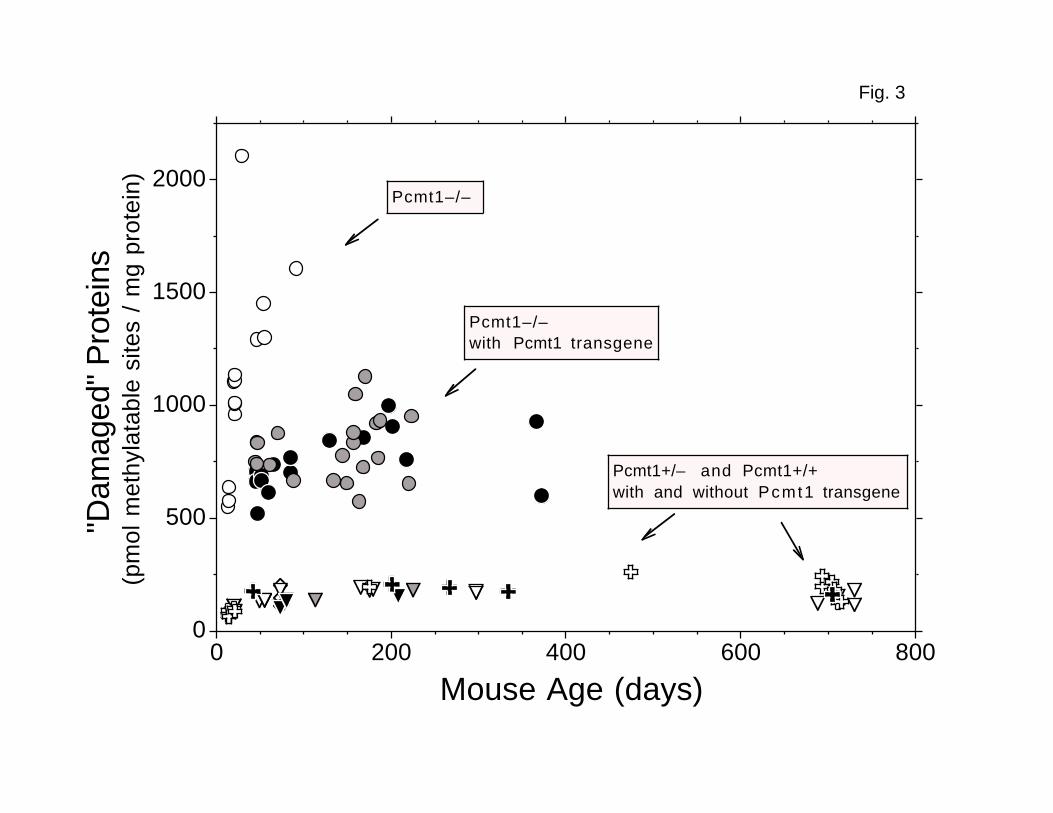
proteins with [14C]methyl groups from [14C]AdoMet in vitro. Examining 39
transgenic and 13 nontransgenic Pcmt1-/- mice, we found about 50% fewer damaged
aspartyl residues when the transgene was present, indicating that the
transgene-derived enzyme was repairing damaged neuronal proteins. However, the
transgenic Pcmt1-/- brains still had about 4.5-fold more damaged residues than
did Pcmt1+/- and Pcmt1+/+ brains (Fig. 3). These damaged residues could be
accumulating both in neurons, due to the relatively low methyltransferase
activity, and in glia, which should not express the transgene at all.
Examination of the levels of aspartyl damage in the brain with respect to
age revealed several interesting points. First, these levels increased with
age in young animals: 40-day-old and older mice had significantly more damaged
residues per mg of protein than did 13-21-day-old mice (P = 0.001 for Pcmt1-/-
mice; P = 10–8 for Pcmt1+/- and Pcmt1+/+ mice; Fig. 3). Second, 13-14 day–old
13
Pcmt1-/- animals already possessed about eightfold more damaged aspartyl
residues per mg protein than did age-matched Pcmt1+/- and Pcmt1+/+ animals
(Fig. 3). The difference in the amount of damage remained 8-11-fold as these
mice aged to 91 days, demonstrating that both nursing and weaned Pcmt1-/- mice
accumulate these residues. Finally, there was no significant increase in the
level of damaged aspartyl residues in transgenic Pcmt1-/- mice after about 100
days of age (Fig. 3). In addition, although line 29 Pcmt1-/- mice averaged
twice as much brain PCMT1 activity as did comparable line 27 mice, the plateau
level of damaged aspartyl residues in these two lines was not significantly
different. The quantity of damaged residues in Pcmt1+/- and Pcmt1+/+ mice
also attained an apparent steady state, though at a much lower level (Fig. 3).
Because damaged residues were arising continuously in the cellular proteins,
this stable level of damage probably represents a steady state between new
damage, methyltransferase-linked repair, protein turnover, and perhaps unknown
factors.
In control experiments, we asked whether the low level of damaged
residues measured in the assays of Pcmt1+/- and Pcmt1+/+ mice resulted in part
from the fact that some were already methylated by the endogenous enzyme. We
therefore incubated brain cytosolic proteins under basic conditions where L-
isoaspartyl α-methyl esters should hydrolyze within a few minutes to generate
L-isoaspartyl residues with about an 80% yield (30, 31). Analysis of these
samples with recombinant methyltransferase (data not shown) indicated that the
true number of L-isoaspartyl residues in the Pcmt1+/- and Pcmt1+/+ proteins
could be as much as 2.4-fold higher than the data shown in Fig. 3.3 Coupled
with the 8-11-fold higher levels actually observed, this result indicates that
the level of damaged aspartyl residues in Pcmt1-/- brain proteins was at least
14
fourfold higher than in heterozygous and wild-type brain proteins.
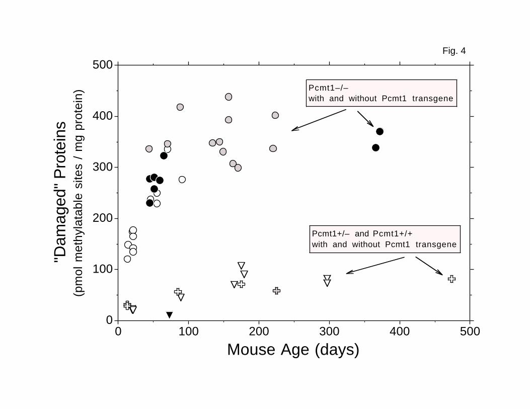
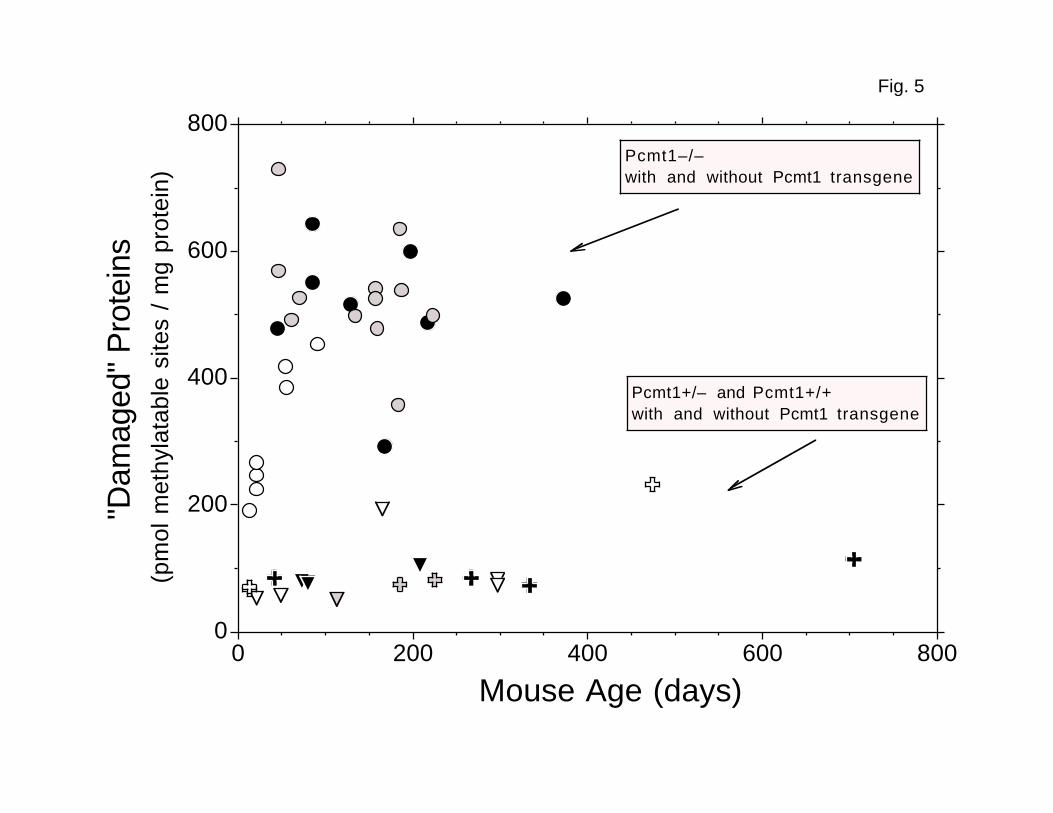
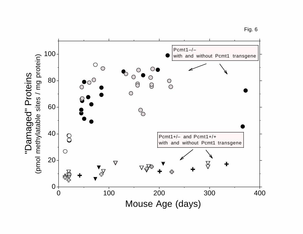
Limited Accumulation of Damaged Aspartyl Residues in Heart, Testes, and
Erythrocytes—The longer lifespan of the transgenic Pcmt1-/- mice enabled us to
investigate whether accumulation of damaged aspartyl residues continued in
tissues completely lacking methyltransferase-mediated repair as animals aged
beyond the limit set by the early deaths of the nontransgenic knockout mice.
In addition to heart (Fig. 4) and testis (Fig. 5), we also examined erythrocyte
cytosol as a control (Fig. 6). Because erythrocytes are normally removed from
the circulation after about 40 days (32), their average age in adult mice is
constant, and older proteins cannot accumulate. Unexpectedly, the accumulation
of damaged aspartyl residues in all of these tissues was quite similar. As in
brain, the number of methylatable residues per mg protein increased only in
animals younger than about 60 days and then leveled off as the mice got older
(Figs. 4–6). The apparent plateau levels of damaged aspartyl residues in
heart, testes, and erythrocytes from transgenic Pcmt1-/- mice averaged 4.5-,
4.7-, and 5.2-fold higher, respectively, than the average levels in tissues
from mice expressing the endogenous methyltransferase, very similar to the
4.5–fold difference observed in brain. However, the absolute plateau level of
damage in each tissue was significantly different, with brain having the
highest and erythrocytes having the lowest levels in both the presence and in
the absence of endogenous PCMT1 (Table II). As expected, the transgenic PCMT1
had no effect in tissues in which it is not expressed.
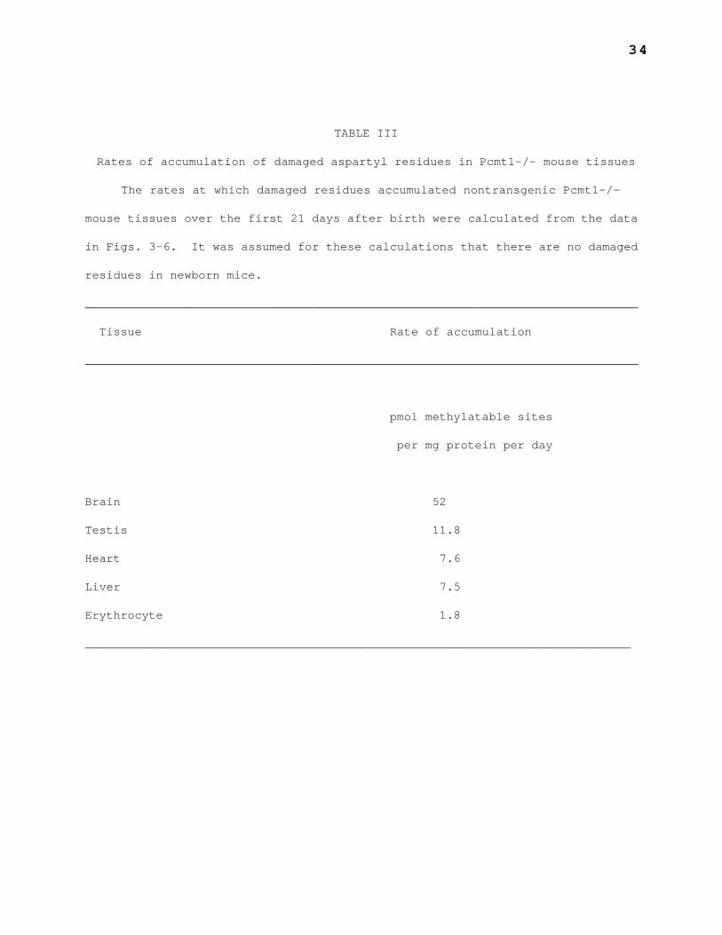
The rates at which damaged residues in cytosolic proteins accumulated in
young Pcmt1-/- mice have been calculated from the data in Figures 3-6. These
rates differ greatly between tissues, ranging from 51 pmol methylatable
residues/mg protein/day in brains to 1.8 pmol methylatable residues/mg
15
protein/day in erythrocytes (Table III). Because the rate at which aspartyl
and asparaginyl residues arise in proteins should not decrease during the life
of a mouse, we can use these rates to estimate the total number of damaged
residues arising in adult mice. Assuming that a mouse is 15% protein by
weight, and that half of this protein is intracellular, a 20 g mouse would have
1.5 g of intracellular protein. If the average rate of damaged residue
formation throughout the mouse is between 5 and 20 pmol/mg/day, as predicted
from the values in Table III, there should be 7.5 to 30 nmol of newly damaged
residues arising each day within the cells of an adult mouse.
Damaged Aspartyl Residues in Urine—The absence of increasing accumulation
of damaged aspartyl residues in the cytosolic proteins of adult mice can result
from repair of the damaged residues or from turnover of the proteins. Few
peptidases cleave isoaspartyl bonds, but proteolysis of the surrounding
residues create isoaspartyl-containing dipeptides and tripeptides that can be
excreted in the urine (33-36). Thus, if mice lacking endogenous PCMT1 do not
have another repair pathway, they might excrete the damaged residues that are
normally repaired in the cells of wild-type mice. To test this hypothesis, we
examined urine from Pcmt1-/- and Pcmt1+/+ mice in several ways.
First, we directly quantitated urinary dipeptide levels after
derivatization with o-phthalaldehyde and β-mercaptoethanol and separation by
reverse-phase HPLC. We did not detect significantly higher levels of
isoaspartyl-containing dipeptides in urine from Pcmt1 knockout mice (data not
shown). For example, isoaspartylglycine ("β-aspartylglycine"), the most
abundant of these dipeptides in human urine (35), was present at 28.2 pmol per
µg creatinine in urine from a 91–day–old Pcmt1-/- mouse and at 27.5 pmol/µg
creatinine in urine from a 40–day–old wild-type mouse. These results suggest
16
that much of this dipeptide might originate in dietary, extracellular, or
unrepairable intracellular proteins, rather than from the lack of repair in
Pcmt1-/- cells.
Second, peptides with an N-terminal isoaspartyl residue might accumulate
because the isopeptide bond is resistant to aminopeptidase activity. To
determine whether more of these peptides accumulated in Pcmt1-/- than in
Pcmt1+/+ mice, we compared the urinary levels of free amino acids and of amino
acids released by acid hydrolysis, which cleaves both aspartyl and isoaspartyl
bonds. Although wild-type urine contained 1.5-fold more total amino acid
residues (free and peptide bound) than did knockout mouse urine, both urine
samples contained eight- to ninefold more peptide-bound residues than free
residues (data not shown). This indicates that the knockout mice were not
excreting elevated levels of peptides or proteins relative to wild-type mice.
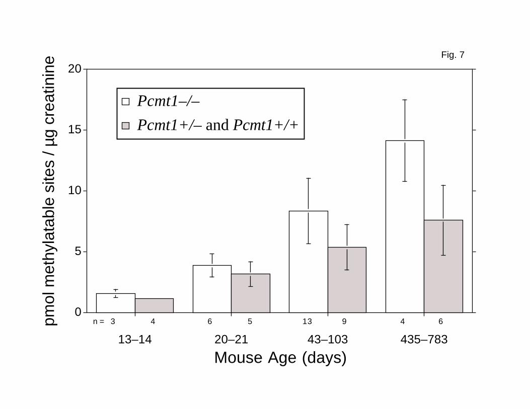
Finally, we used the recombinant L-isoaspartyl/D-aspartyl
methyltransferase as an analytical probe for L-isoaspartyl residues in peptides
large enough to be efficiently methylated (tetrapeptides and larger) (15).
Methylatable residues were found in all of the urine samples assayed, and the
level of damage increased with the age of the mouse (Fig. 7). We observed no
significant difference in the number of methylatable sites, relative to the
amount of creatinine, in urine from Pcmt1-/- animals and from Pcmt1-/+ and
Pcmt1+/+ controls in the 13-14- and 20-21-day-old groups (Fig. 7). However,
urine from older Pcmt1-/- mice contained almost twofold more methylatable sites
than urine from control animals (Fig. 7). Assuming that 435-783-day-old mice
weighing 20 g excrete 0.67 mg of creatinine per day (37), then Pcmt1-/- mice
excrete about 9.5 nmol and age-matched control animals about 5.1 nmol of
methylatable L-isoaspartyl residues per day. Thus, at least 4.4 nmol of
17
damaged residues that are repaired in wild-type mice are excreted each day by
knockout mice that are not accumulating additional damaged residues within
their cells. This daily excretion of damaged aspartyl residues in the urine
represents 15—67% of the total daily production of intracellular aspartyl
damage estimated above. This result indicates that proteolysis leading to
urinary excretion is a significant factor in limiting the accumulation of
damaged proteins in the absence, and perhaps as well in the presence, of the
repair methyltransferase.
18
DISCUSSION
We have been examining mice lacking PCMT1 to determine whether ineffective
repair of damaged aspartyl and asparaginyl residues contributes to disease or
to the deleterious effects of aging. We report here that damaged aspartyl
residues in Pcmt1-/- brain proteins from 13-14 day-old mice were already eight
times more abundant than those in aged-matched wild-type mice and accumulated
at a rate of about 52 pmol/mg protein/day until 20—21 days. Damaged residues
continued to accumulate but at a lower rate, averaging 9 pmol/mg protein/day
between days 21 and 55. The rate of accumulation appeared to decrease further
in the brains of older Pcmt1-/- mice, but few of these mice live beyond this
age. A similar pattern of damage accumulation has been recently observed by
Shimizu et al. in independently-derived Pcmt1-/- mice (38). In contrast to the
Pcmt1-/- mice, however, Pcmt1+/+ and Pcmt1+/- mice kept the level of damaged
residues very low for at least 2 years. Although they had only about half of
the methyltransferase activity, Pcmt1+/- mice survived as long as Pcmt1+/+
mice.
The high level of PCMT1 activity in wild-type brain, the fact that
damaged residues rapidly accumulate in the brain in the absence of the
methyltransferase, and the seizures in the Pcmt1-/- mice suggested that this
enzyme plays a critical role in normal brain function. PCMT1, however, is
expressed in all mammalian tissues and presumably is involved in repair
throughout the body. We were therefore also interested in examining what would
happen if tissues lacking this repair were allowed to accumulate higher levels
of damage over several years.
19
To answer these questions, we performed a “brain rescue” experiment by
creating mice with a Pcmt1 transgene under the control of a neuron-specific
promoter in a genetic background lacking the endogenous Pcmt1 gene. Here, we
obtained mice expressing the methyltransferase only in the brain. Although the
expression of the transgene-derived methyltransferase activity in the brains of
these mice was only 6.5—13% of the level in wild-type mice, the transgenic mice
lived much longer and accumulated only half the damaged residues found in
nontransgenic knockout animals. The success of this rescue experiment further
supported the importance of the methyltransferase in the brain.
We previously reported that mice lacking the repair methyltransferase are
more sensitive to the seizure–inducing drug metrazol than are wild-type mice
(18), and that the anti-seizure drugs valproic acid and clonazepam prolonged
the life span of these animals (20). It is likely that, by decreasing the
accumulation of damaged aspartyl residues, the transgene-derived
methyltransferase raised the seizure threshold of the rescued mice to an
intermediate level between the thresholds of nontransgenic Pcmt1-/- and
wild–type mice. Immunohistochemical studies revealed that the transgene-
derived methyltransferase was expressed largely in neurons, in both line 27 and
line 29. However, the pattern of neuronal expression was distinct in the two
lines (C. Farrar, E. Kim, S. Young, S. Clarke, and C. Houser, unpublished
data). These findings indicate that the expression of Pcmt1 in neurons (rather
than glia) is paramount in the prevention of the fatal seizure disorder. At
this point, however, we do not know whether the methyltransferase is critical
for the function of all neurons or only for certain types of neurons.
Because many of the transgenic Pcmt1-/- mice lived for several hundred
days, we were able to examine the long-term accumulation of damaged residues in
20
peripheral organs, where the methyltransferase is not expressed at all. We
found no apparent defects in these mice, and initial pathological studies were
uninformative. We found that damaged aspartyl residues accumulated in an
age–dependent manner only in relatively young mice and attained plateau levels
by 100 days of age. At first approximation, the rate at which damaged residues
arise in proteins remains constant with time; thus, the plateau levels of
damage in tissues of older mice must result from processes of repair and/or
turnover of these residues. We have been especially interested in the
situation in mouse tissues that lack the repair methyltransferase, where the
levels of damage are much greater but which still approach plateau values with
age. We suggest that enhanced proteolytic degradation may limit the
accumulation of proteins containing damaged aspartyl residues, especially in
methyltransferase-deficient tissues. Clear evidence has been presented for the
selective proteolytic degradation of spontaneously-damaged calmodulin in both
HeLa cells (6) and in Xenopus oocytes (39).
Isoaspartyl linkages themselves are generally not cleaved by mammalian
proteases (33, 34). We have thus investigated the possibility that proteolysis
of proteins containing damaged aspartyl residues in PCMT1-deficient mice may be
reflected in the increased urinary output of peptides containing L-isoaspartyl
residues. In fact, damaged aspartyl residues in proteins fed to rats are
excreted as isoaspartyl-containing dipeptides in the urine (40), and it has
been proposed that at least some endogenous isoaspartyl residues are dealt with
in a similar manner (41). Furthermore, an L-isoaspartyl residue that arises in
collagen has been found in human urine as part of an eight-residue peptide
(42). We found that urine from Pcmt1-/- mice contains more peptides with
damaged aspartyl residues than urine from Pcmt1+/+ mouse urine. Because PCMT1
21
repairs intracellular proteins (14), our results provide the first evidence
that damaged aspartyl residues that arise within cells can be excreted in the
urine.
Is the elevated level of altered aspartyl residues in the urine from
Pcmt1-/- mice enough to account for the observed steady state level of damaged
proteins in tissues despite continuing spontaneous generation of isoaspartyl
residues? From the rates of damage accumulation measured in tissues of young
Pcmt1-/- mice, we estimated that 7.5 to 30 nmol of newly damaged aspartyl
residues are generated each day. The daily excretion of damaged residues in
the urine of Pcmt1-/- mice was 4.4 nmol more than that in wild-type mice,
demonstrating that a significant fraction of the damage can be metabolized by
proteolysis. Additional isoaspartyl peptides may be present in the urine that
are not readily recognized by the methyltransferase (15), although we did not
detect increased levels of generally poorly-recognized dipeptides and N-
terminal L-isoaspartyl peptides.
Interestingly, the ability of the proteolytic pathway to stem the
accumulation of damaged aspartyl residues is apparently insufficient to prevent
seizures in animals lacking the repair methyltransferase. Thus, the repair
methyltransferase is needed (at least in the brain) to lower the level of
damaged residues beyond what the degradation mechanisms can accomplish.
Furthermore, it is possible that proteolysis of proteins that contain covalent
modifications important to learning and memory could have undesirable effects
in that their replacement proteins would not be appropriately modified (43).
Unlike the brain, other tissues appear to be able to function relatively
normally with higher levels of damaged aspartyl residues. The importance of
the methyltransferase in maintaining a lower steady–state level of damaged

22
aspartyl residues in these tissues remains to be determined. It is certainly
possible that mice reared under other conditions (e. g., outside of a vivarium)
would be more susceptible to pathologies in the absence of the repair
methyltransferase.
23
REFERENCES
1. Brownlee, M. (1995) Annu. Rev. Med. 46, 223-234
2. Lowenson, J. D., Clarke, S., and Roher, A. E. (1999) Methods Enzymol. 309,
89-105
3. Stadtman, E. R., and Levine, R. L. (2000) Ann. N. Y. Acad. Sci. U. S. A.
899, 191-208
4. Friguet, B., Bulteau, A. L., Chondrogianni, N., Conconi, M., and
Petropoulos, I. (2000) Ann. N. Y. Acad. Sci. 908, 143-154
5. Sano, H., Nagai, R., Matsumoto, K., and Horiuchi, S. (1999) Mech. Ageing
Dev. 107, 333-346
6. Tarcsa, E., Szymanska, G., Lecker, S., O'Connor, C. M., and Goldberg, A. L.
(2000) J. Biol. Chem. 275, 20295-20301
7. Visick, J. E. and Clarke, S. (1995) Mol. Microbiol. 16, 835-845.
8. Schiene, C., and Fischer, G. (2000) Curr. Opin. Struct. Biol. 10, 40-45
9. Moskovitz, J., Flescher, E., Berlett, B. S., Azare, J., Poston, J. M., and
Stadtman, E. R. (1998) Proc. Natl. Acad. Sci. U. S. A. 95, 14071-14075
10. Aswad, D. W., Paranandi, M. V., and Schurter, B.T. (2000) J. Pharmaceut.
Biomed. Anal. 21, 1129-1136
11. Volkin, D. B., Mach, H., and Middaugh, C. R. (1997) Mol. Biotechnol. 8,
105-122
12. Geiger, T., and Clarke, S. (1987) J. Biol. Chem. 262, 785–794
13. Mamula, M. J., Gee, R. J., Elliott, J. I., Sette, A., Southwood, S.,
Jones, P. J., and Blier, P. R. (1999) J. Biol. Chem. 274, 22321-22327
14. Clarke, S. (1999) in S-Adenosylmethionine-Dependent Methyltransferases:
Structures and Functions (Cheng, X, and Blumenthal, R. M., eds.) pp.
123-148, World Scientific Publishing, Singapore
15. Lowenson, J. D., and Clarke, S. (1995) in Deamidation and Isoaspartate
24
Formation in Peptides and Proteins (Aswad D, W., ed.) pp. 47–64, CRC
Press, Boca Raton, FL
16. McFadden, P. N., and Clarke, S. (1987) Proc. Natl. Acad. Sci. U. S. A.
84, 2595–2599
17. Johnson, B. A., Murray, E. D., Jr., Clarke, S., Glass, D. B., and Aswad,
D. W. (1987) J. Biol. Chem. 262, 5622-5629
18. Kim, E., Lowenson, J. D., MacLaren, D. C., Clarke, S., and Young, S. G.
(1997) Proc. Natl. Acad. Sci. U. S. A. 94, 6132–6137
19. Yamamoto, A., Takagi, H., Kitamura, D., Tatsuoka, H., Nakano, H., Kawano,
H., Kuroyanagi, H., Yahagi, Y., Kobayashi, S., Koizumi, K., Sakai, T.,
Saito, K., Chiba, T., Kawamura, K., Suzuki, K., Watanabe, T., Mori, H.,
and Shirasawa, T. (1998) J. Neurosci. 18, 2063-2074
20. Kim, E., Lowenson, J. D., Clarke, S., and Young, S. G. (1999) J. Biol.
Chem. 274, 20671-20678
21. Romanik, E. A., Ladino, C. A., Killoy, L. C., D'Ardenne, S. C., and
O'Connor, C. M. (1992) Gene 118, 217-222
22. Higgins, L. S., Catalano, R., Quon, D., and Cordell, B. (1993) Ann. N.
Y. Acad. Sci. 695, 224-227
23. Hogan, B., Beddington, R., Costantini, F., and Lacy. E. (1994) in
Manipulating the Mouse Embryo. A Laboratory Manual. 2nd Edition. Cold
Spring Harbor Laboratory Press, Plainview, New York
24. MacLaren, D. C., and Clarke, S. (1995) Protein Expr. Purif. 6, 99-108
25. Bonsnes, R. W., and Taussky, H.H. (1945) J. Biol. Chem. 109, 581-591
26. Gary, J. D., and Clarke, S. (1995) J. Biol. Chem. 270, 4076-4087
27. Kandel, E. R. (1991) in Principles Of Neural Science, Third Edition
(Kandel, E. R., Schwartz, J. H., and Jessell, T. M., eds.) pp. 22,
Appleton & Lange, Norwalk, CT
25
28. Forss-Petter, S., Danielson, P. E., Catsicas, S., Battenberg, E., Price,
J., Nerenberg, M., and Sutcliffe, J. G. (1990) Neuron 5, 187-197
29. Andra, K., Abramowski, D., Duke, M., Probst, A., Wiederhold, K. H., Burki,
K., Goedert, M., Sommer, B., and Staufenbiel, M. (1996) Neurobiol. Aging
17, 183-190
30. Stephenson, R. C., and Clarke, S. (1989) J. Biol. Chem. 264, 6164–6170
31. Patel, K. and Borchardt, R. T. (1990) Pharm. Res. 7, 703-711
32. Loeffler, M., Pantel, K., Wulff, H., and Wichmann, H. E. (1989) Cell
Tissue Kinet. 22, 13-30
33. Johnson, B. A., and Aswad, D. W. (1991) Biochemistry 29, 4373-4380
34. Haley, E. E., Corcoran, B. J., Dorer, F. E., and Buchanan, D.L. (1966)
Biochemistry 5, 3229-3235
35. Dorer, F. E., Haley, E. E., and Buchanan, D. L. (1966) Biochemistry 5,
3236-3240
36. Tanaka, T., and Nakajima, T. (1978) J. Biochem. (Tokyo) 84, 617-625
37. Crispens, C. G., Jr. (1975) in Handbook on the Laboratory Mouse. p. 131,
Charles C. Thomas, Springfield, IL
38. Shimizu, T., Watanabe, A., Ogawara, M., Mori, H., and Shirasawa, T. (2000)
Arch. Biochem. Biophys. 381, 225-234.
39. Szymanska, G., John D. Leszyk, J. D., and O'Connor, C. M. (1998) J. Biol.
Chem. 273, 28516-28523
40. Pisano, J. J., Prado, E., and Freedman, J. (1966) Arch. Biochem. Biophys.
117, 394-399
41. Buchanan, D. L., Haley, E. E., and Markiw, R. T. (1962) Biochemistry 1,
612-620
42. Rosenquist, C., Fledelius, C., Christgau, S., Pedersen, B. J., Bonde, M.,
Qvist, P., and Christiansen, C. (1998) Clin. Chem. 44, 2281-2289

26
43. Chain, D. G., Schwartz, J. H., and Hegde, A. N. (2000) Mol. Neurobiol.
20, 125-142
27
FOOTNOTES
1The abbreviations used are: PCMT1, L-isoaspartyl (D-aspartyl)
O–methyltransferase; AdoMet, S-adenosyl-L-methionine; [14C]AdoMet,
S–adenosyl[methyl-14C]-L-methionine; NSE, neuron-specific enolase; BisTris,
2,2-bis(hydroxymethyl)-2,2',2"-nitrilotriethanol.
2Nonfatal running/jumping seizures have been observed in three transgenic
Pcmt1+/- and Pcmt1+/+ mice, but never in nontransgenic animals.
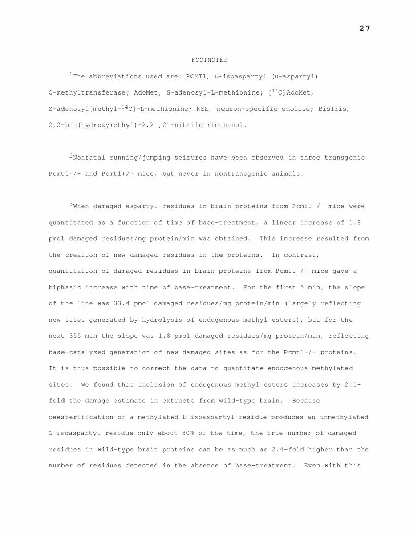
3When damaged aspartyl residues in brain proteins from Pcmt1-/- mice were
quantitated as a function of time of base-treatment, a linear increase of 1.8
pmol damaged residues/mg protein/min was obtained. This increase resulted from
the creation of new damaged residues in the proteins. In contrast,
quantitation of damaged residues in brain proteins from Pcmt1+/+ mice gave a
biphasic increase with time of base-treatment. For the first 5 min, the slope
of the line was 33.4 pmol damaged residues/mg protein/min (largely reflecting
new sites generated by hydrolysis of endogenous methyl esters), but for the
next 355 min the slope was 1.8 pmol damaged residues/mg protein/min, reflecting
base-catalyzed generation of new damaged sites as for the Pcmt1-/- proteins.
It is thus possible to correct the data to quantitate endogenous methylated
sites. We found that inclusion of endogenous methyl esters increases by 2.1-
fold the damage estimate in extracts from wild-type brain. Because
deesterification of a methylated L-isoaspartyl residue produces an unmethylated
L-isoaspartyl residue only about 80% of the time, the true number of damaged
residues in wild-type brain proteins can be as much as 2.4-fold higher than the
number of residues detected in the absence of base-treatment. Even with this

28
increase, however, there are still about fourfold more damaged residues in
Pcmt1-/- brain proteins than in wild-type brain proteins.
29
FIGURE LEGENDS
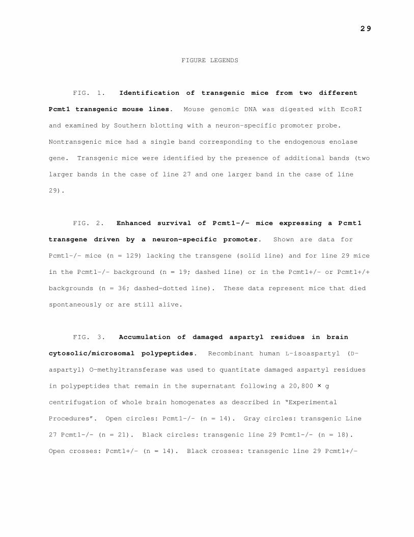
FIG. 1. Identification of transgenic mice from two different
Pcmt1 transgenic mouse lines. Mouse genomic DNA was digested with EcoRI
and examined by Southern blotting with a neuron-specific promoter probe.
Nontransgenic mice had a single band corresponding to the endogenous enolase
gene. Transgenic mice were identified by the presence of additional bands (two
larger bands in the case of line 27 and one larger band in the case of line
29).
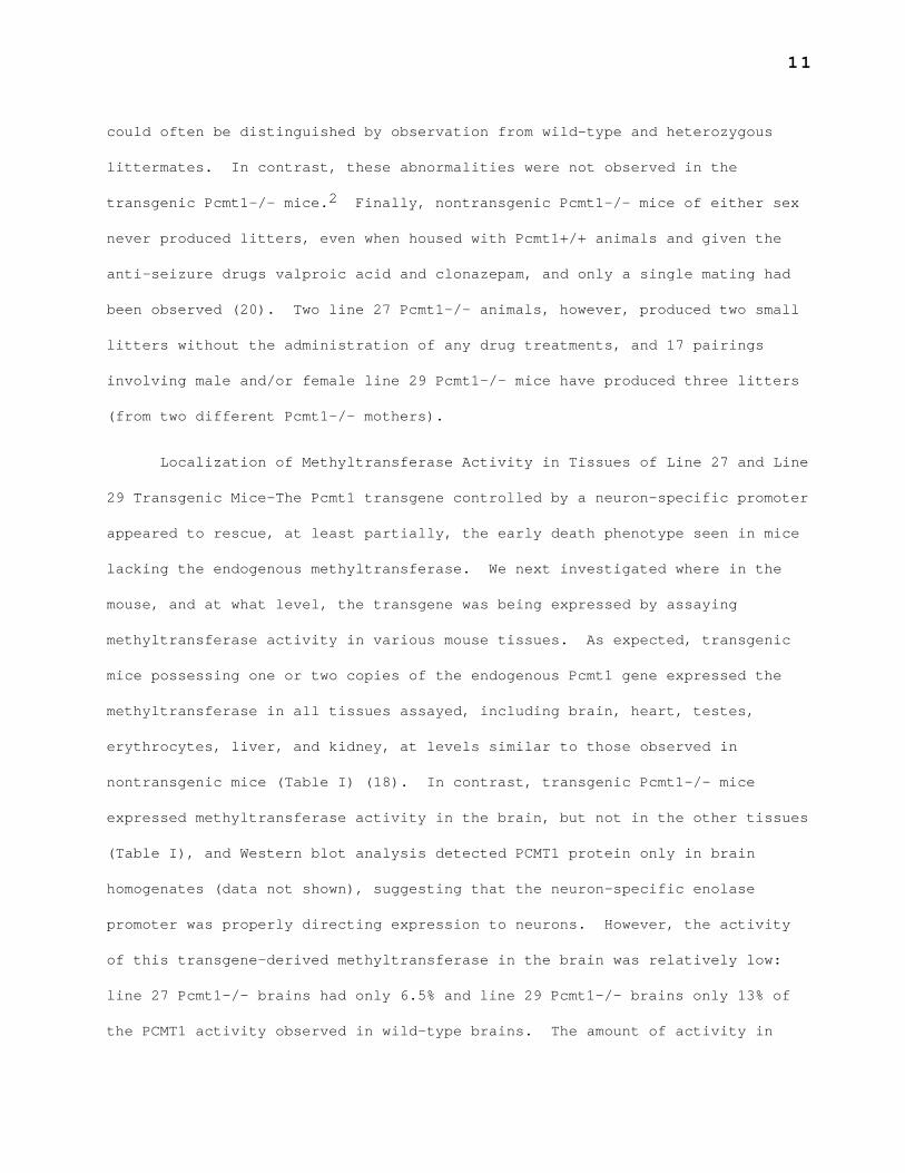
FIG. 2. Enhanced survival of Pcmt1-/- mice expressing a Pcmt1
transgene driven by a neuron-specific promoter. Shown are data for
Pcmt1-/- mice (n = 129) lacking the transgene (solid line) and for line 29 mice
in the Pcmt1-/- background (n = 19; dashed line) or in the Pcmt1+/- or Pcmt1+/+
backgrounds (n = 36; dashed-dotted line). These data represent mice that died
spontaneously or are still alive.
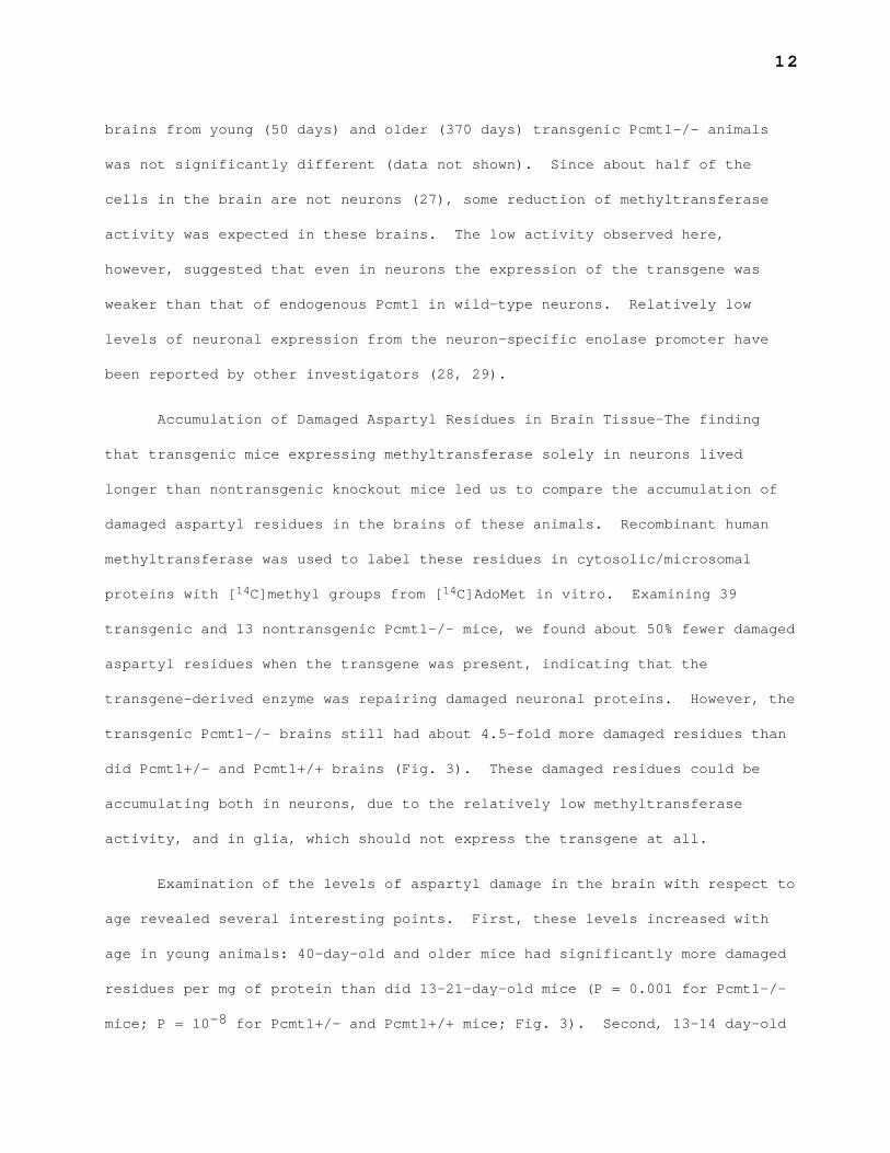
FIG. 3. Accumulation of damaged aspartyl residues in brain
cytosolic/microsomal polypeptides. Recombinant human L-isoaspartyl (D-
aspartyl) O-methyltransferase was used to quantitate damaged aspartyl residues
in polypeptides that remain in the supernatant following a 20,800 × g
centrifugation of whole brain homogenates as described in “Experimental
Procedures”. Open circles: Pcmt1-/- (n = 14). Gray circles: transgenic Line
27 Pcmt1-/- (n = 21). Black circles: transgenic line 29 Pcmt1-/- (n = 18).
Open crosses: Pcmt1+/- (n = 14). Black crosses: transgenic line 29 Pcmt1+/-
30
(n = 5). Open triangles: Pcmt1+/+ (n = 17). Open diamonds: Pcmt1+/- or
Pcmt1+/+ (n = 6). Gray triangles: transgenic Line 27 Pcmt1+/+ (n = 2). Black
triangles: transgenic Line 29 Pcmt1+/+ (n = 3).
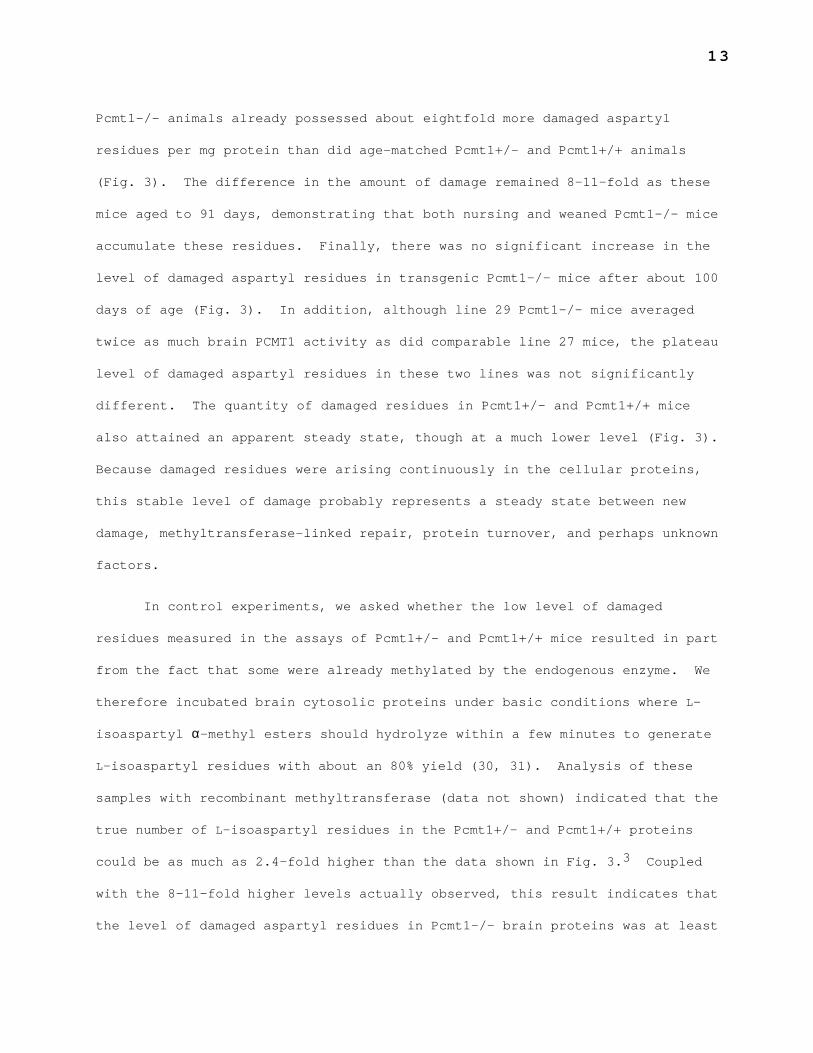
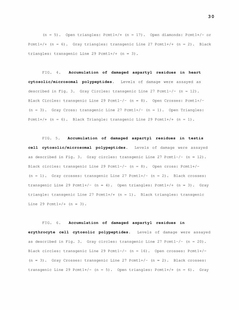
FIG. 4. Accumulation of damaged aspartyl residues in heart
cytosolic/microsomal polypeptides. Levels of damage were assayed as
described in Fig. 3. Gray Circles: transgenic Line 27 Pcmt1-/- (n = 12).
Black Circles: transgenic Line 29 Pcmt1-/- (n = 8). Open Crosses: Pcmt1+/-
(n = 3). Gray Cross: transgenic Line 27 Pcmt1+/- (n = 1). Open Triangles:
Pcmt1+/+ (n = 6). Black Triangle: transgenic Line 29 Pcmt1+/+ (n = 1).
FIG. 5. Accumulation of damaged aspartyl residues in testis
cell cytosolic/microsomal polypeptides. Levels of damage were assayed
as described in Fig. 3. Gray circles: transgenic Line 27 Pcmt1-/- (n = 12).
Black circles: transgenic Line 29 Pcmt1-/- (n = 8). Open cross: Pcmt1+/-
(n = 1). Gray crosses: transgenic Line 27 Pcmt1+/- (n = 2). Black crosses:
transgenic Line 29 Pcmt1+/- (n = 4). Open triangles: Pcmt1+/+ (n = 3). Gray
triangle: transgenic Line 27 Pcmt1+/+ (n = 1). Black triangles: transgenic
Line 29 Pcmt1+/+ (n = 3).
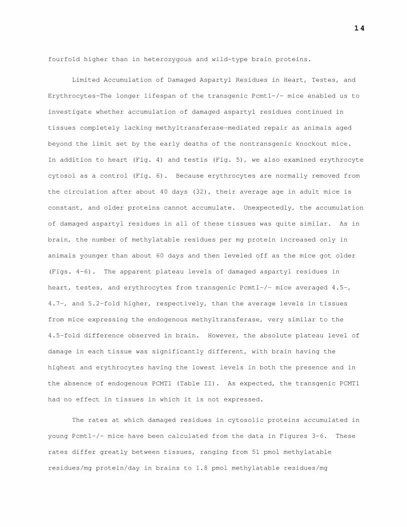
FIG. 6. Accumulation of damaged aspartyl residues in
erythrocyte cell cytosolic polypeptides. Levels of damage were assayed
as described in Fig. 3. Gray circles: transgenic Line 27 Pcmt1-/- (n = 20).
Black circles: transgenic Line 29 Pcmt1-/- (n = 16). Open crosses: Pcmt1+/-
(n = 3). Gray Crosses: transgenic Line 27 Pcmt1+/- (n = 2). Black crosses:
transgenic Line 29 Pcmt1+/- (n = 5). Open triangles: Pcmt1+/+ (n = 6). Gray
31
triangle: transgenic Line 27 Pcmt1+/+ (n = 1). Black triangles: transgenic
Line 29 Pcmt1+/+ (n = 3).
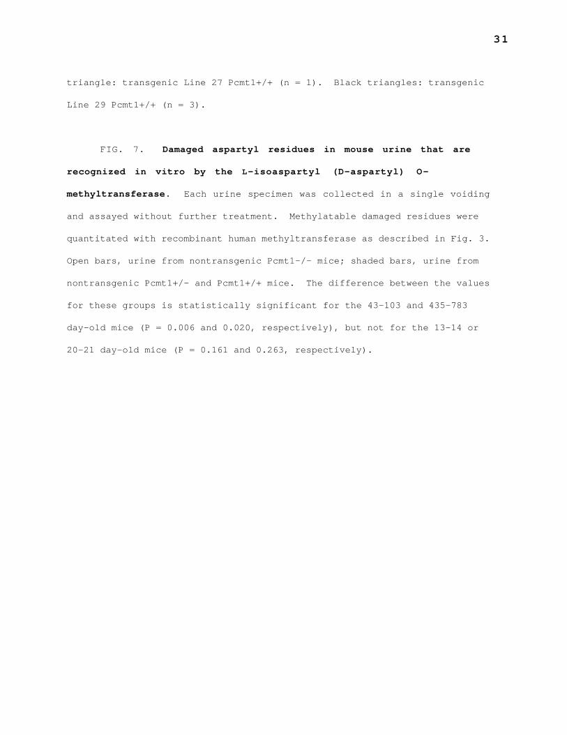
FIG. 7. Damaged aspartyl residues in mouse urine that are
recognized in vitro by the L-isoaspartyl (D-aspartyl) O-
methyltransferase. Each urine specimen was collected in a single voiding
and assayed without further treatment. Methylatable damaged residues were
quantitated with recombinant human methyltransferase as described in Fig. 3.
Open bars, urine from nontransgenic Pcmt1-/- mice; shaded bars, urine from
nontransgenic Pcmt1+/- and Pcmt1+/+ mice. The difference between the values
for these groups is statistically significant for the 43—103 and 435—783
day-old mice (P = 0.006 and 0.020, respectively), but not for the 13—14 or
20—21 day-old mice (P = 0.161 and 0.263, respectively).
32
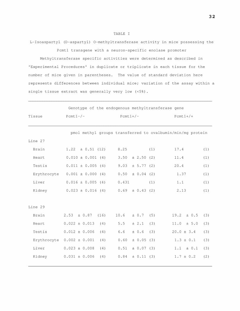
TABLE I
L-Isoaspartyl (D-aspartyl) O-methyltransferase activity in mice possessing the
Pcmt1 transgene with a neuron-specific enolase promoter
Methyltransferase specific activities were determined as described in
"Experimental Procedures" in duplicate or triplicate in each tissue for the
number of mice given in parentheses. The value of standard deviation here
represents differences between individual mice; variation of the assay within a
single tissue extract was generally very low (<5%).
______________________________________________________________________________
Genotype of the endogenous methyltransferase gene
Tissue Pcmt1-/- Pcmt1+/- Pcmt1+/+
______________________________________________________________________________
pmol methyl groups transferred to ovalbumin/min/mg protein
Line 27
Brain 1.22 ± 0.51 (12) 8.25 (1) 17.4 (1)
Heart 0.010 ± 0.001 (4) 3.50 ± 2.50 (2) 11.4 (1)
Testis 0.011 ± 0.005 (4) 9.03 ± 5.77 (2) 20.4 (1)
Erythrocyte 0.001 ± 0.000 (4) 0.50 ± 0.04 (2) 1.37 (1)
Liver 0.016 ± 0.005 (4) 0.431 (1) 1.1 (1)
Kidney 0.023 ± 0.016 (4) 0.69 ± 0.43 (2) 2.13 (1)
Line 29
Brain 2.53 ± 0.87 (16) 10.6 ± 0.7 (5) 19.2 ± 0.5 (3)
Heart 0.022 ± 0.013 (4) 5.5 ± 2.1 (3) 11.0 ± 5.0 (3)
Testis 0.012 ± 0.006 (4) 6.6 ± 0.6 (3) 20.0 ± 3.4 (3)
Erythrocyte 0.002 ± 0.001 (4) 0.60 ± 0.05 (3) 1.3 ± 0.1 (3)
Liver 0.023 ± 0.008 (4) 0.51 ± 0.07 (3) 1.1 ± 0.1 (3)
Kidney 0.031 ± 0.006 (4) 0.84 ± 0.11 (3) 1.7 ± 0.2 (2)
______________________________________________________________________________
33
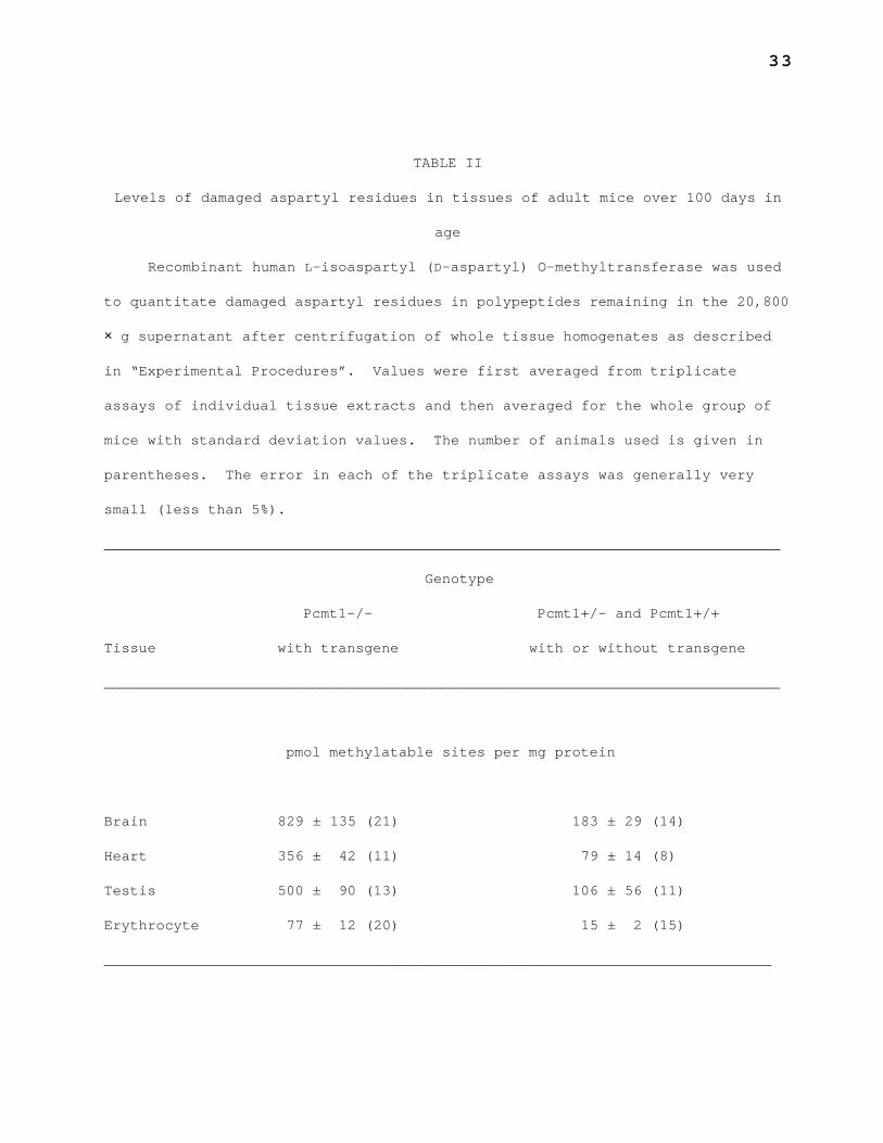
TABLE II
Levels of damaged aspartyl residues in tissues of adult mice over 100 days in
age
Recombinant human L-isoaspartyl (D-aspartyl) O-methyltransferase was used
to quantitate damaged aspartyl residues in polypeptides remaining in the 20,800
× g supernatant after centrifugation of whole tissue homogenates as described
in “Experimental Procedures”. Values were first averaged from triplicate
assays of individual tissue extracts and then averaged for the whole group of
mice with standard deviation values. The number of animals used is given in
parentheses. The error in each of the triplicate assays was generally very
small (less than 5%).
______________________________________________________________________________
Genotype
Pcmt1-/- Pcmt1+/- and Pcmt1+/+
Tissue with transgene with or without transgene
______________________________________________________________________________
pmol methylatable sites per mg protein
Brain 829 ± 135 (21) 183 ± 29 (14)
Heart 356 ± 42 (11) 79 ± 14 (8)
Testis 500 ± 90 (13) 106 ± 56 (11)
Erythrocyte 77 ± 12 (20) 15 ± 2 (15)
_____________________________________________________________________________
34
TABLE III
Rates of accumulation of damaged aspartyl residues in Pcmt1-/- mouse tissues
The rates at which damaged residues accumulated nontransgenic Pcmt1-/-
mouse tissues over the first 21 days after birth were calculated from the data
in Figs. 3-6. It was assumed for these calculations that there are no damaged
residues in newborn mice.
______________________________________________________________________________
Tissue Rate of accumulation
______________________________________________________________________________
pmol methylatable sites
per mg protein per day
Brain 52
Testis 11.8
Heart 7.6
Liver 7.5
Erythrocyte 1.8
_____________________________________________________________________________
0
20
40
60
80
100
Sur
viva
l (%
)
0 40 80 120 160 200 240
Mouse Age (days)
Pcmt1–/–No Transgene
Line 29 Pcmt1–/–
Line 29 Pcmt1+/–Line 29 Pcmt1+/+
Fig. 2
0
500
1000
1500
2000
(pm
ol m
ethy
lata
ble
site
s /
mg
prot
ein)
0 200 400 600 800
Mouse Age (days)
Pcmt1–/–
Pcmt1+/– and Pcmt1+/+with and without Pcmt1 transgene
"Dam
aged
" P
rote
ins
Pcmt1–/–with Pcmt1 transgene
Fig. 3
0
100
200
300
400
500"D
amag
ed"
Pro
tein
s
0 100 200 300 400 500
Mouse Age (days)
Fig. 4
(pm
ol m
ethy
lata
ble
site
s /
mg
prot
ein)Pcmt1–/–with and without Pcmt1 transgene
Pcmt1+/– and Pcmt1+/+with and without Pcmt1 transgene
0
200
400
600
800"D
amag
ed"
Pro
tein
s
0 200 400 600 800
Mouse Age (days)
Fig. 5
Pcmt1–/–with and without Pcmt1 transgene
Pcmt1+/– and Pcmt1+/+with and without Pcmt1 transgene
(pm
ol m
ethy
lata
ble
site
s /
mg
prot
ein)
0
20
40
60
80
100
"Dam
aged
" P
rote
ins
0 100 200 300 400
Mouse Age (days)
Fig. 6
(pm
ol m
ethy
lata
ble
site
s /
mg
prot
ein)
Pcmt1–/–with and without Pcmt1 transgene
Pcmt1+/– and Pcmt1+/+with and without Pcmt1 transgene
0
5
10
15
20
pmol
met
hyla
tabl
e si
tes
/ µg
crea
tinin
e
13–14 20–21 43–103 435–783
Mouse Age (days)
Pcmt1+/– and Pcmt1+/+
Pcmt1–/–
n = 3 4 96 5 13 64
Fig. 7