Embed Size (px)
Citation preview

Links between Aging and Proteostasis Decline in Saccharomyces cerevisiae
Veronica Andersson
AKADEMISK AVHANDLING
För filosofie doktorsexamen i Mikrobiologi, som med tillstånd från Naturvetenskapliga fakulteten kommer att offentligt försvaras fredagen den 28 mars
2014 kl. 9:00 i hörsal Arvid Carlsson, Academicum, Medicinaregatan 3, Göteborg
Fakultetsopponent: Professor S. Michal Jazwinski, School of Science and Engineering Tulane University, New Orleans, LA, USA
ISBN: 978-91-628-8958-6
http://hdl.handle.net/2077/34982

Cover picture:
Budscars of aged yeast cells visualized by WGA-staining together with Hsp104-GFP foci Original picture was taken by Sandra M Hill Edited by Veronica Andersson
© Veronica Andersson 2014
All rights reserved. No part of this publication may be reproduced or transmitted, in any form or by any means, without written permission.

Dad, this one’s for you


Abstract
Proteins are continuously synthesized and degraded to meet the demands of the cell. Hence, a proper balance between synthesis, folding, disaggregation and degradation is of essence to ensure cell survival. Disruption of any part of this proteostasis network may have severe consequences for cellular fitness and longevity. This thesis focuses on how proteostasis decline and aggregate-management is linked to aging in the yeast Saccharomyces cerevisiae and why aggregates accumulate in the yeast mother cells. The study revealed that three complementary processes are involved in this process; (i) mother cell-biased segregation of aggregated proteins during cytokinesis, (ii) - Text removed from public version - (iii) an age-related decline in proteasome function.
The first process explains why aggregates, once formed, by necessity must accumulate in aging mother cells. This asymmetric inheritance is the result of Hsp104-containing aggregates associating with the actin cytoskeleton, which effectively hinders movement of aggregates into the daughter cell. In addition, actin-tethered aggregates display retrograde transport towards the mother cell providing the daughter cell with the means of clearing itself of aggregates.
But why do aggregates form upon cellular aging? The data presented herein indicate that
- Text removed from public version -
The third process underlying aggregate accumulation includes a functional decline of the ubiquitin-proteasome-system, UPS. Experiments using a model in vivo UPS-substrate revealed that 26S proteasome function is greatly diminished in aged cells, an impediment which can be restored by elevating protein disaggregation. The data suggest that the accrual of aggregated proteins obstructs proper UPS function resulting in a negative proteostasis feedback loop.
Finally, - Text removed from public version - and proteasome function generated an extension in lifespan, enhanced disaggregation does not, suggesting that it may be more beneficial for the cell to prevent aggregates from forming than eliminating the ones already formed. Thus, aggregate-precursors may be the initial culprits in age-associated proteostasis decline.
Key words: Aging, Aggregates, Proteostasis, Proteasome, disaggregation, Peroxiredoxins, Asymmetric inheritance, S. cerevisiae

Abbreviations
AD Alzheimer´s disease AMP Adenosine 5’-monophosphate ATP Adenosine 5’-triphosphate cAMP Cyclic- AMP CR Caloric restriction Cys Cystein ERCs Extra chromosomal rDNA circles ETC Electron transport chain H2O2 Hydrogen peroxide HD Huntington’s disease Hsps Heat shock proteins IB Inclusion bodies IPOD Insoluble protein deposit JUNQ Juxtanuclear quality control MIM Mitochondrial inner membrane mtDNA Mitochondrial DNA NO· Nitric oxide radical O2·ˉ Superoxide anion radical ·OH Hydroxyl radical PD Parkinsson´s disease PIP Proteasome interacting protein PKA Protein kinase A PQC Protein quality control Prxs Peroxiredoxins
rDNA Ribosomal DNA ROS Reactive oxygen species SOD Superoxide dismutase SQC Spacial quality control TOR Target of rapamycin Trxs Thioredoxins Ub Ubiquitin UPS Ubiquitin proteasome system Δψm Mitochondrial membrane potential
List of Papers
This thesis is based on the following papers, which will be referred to in the text by their roman numeral;
I. Segregation of protein aggregates involves actin and the polarity machinery. Liu B, Larsson L, Franssens V, Hao X, Hill SM, Andersson V, Höglund D, Song J, Yang X, Öling D, Grantham J, Winderickx J, Nyström T Cell (2011) 147(5):959-61.
II. - Text removed from public version - Sarah Hanzén, Junsheng Yang*, Veronica Andersson*, Sara Zamarbide-Forés, Frederike Ewald, Lisa Malm, Benoît Biteau, Beidong Liu, Michel Toledano, Mikael Molin and Thomas Nyström Manuscript * contributed equally
III. Enhancing protein disaggregation restores proteasome activity in aged cells Veronica Andersson, Sarah Hanzén, Beidong Liu, Mikael Molin and Thomas Nyström. Aging (2013) 5, 802-812.

Index Aims of this thesis ......................................................................................... 10
Introduction .................................................................................................. 11
Why do we age? ..................................................................................................................... 11
Aging in Saccharomyces cerevisiae .......................................................................................... 13
Lifespan regulators in yeast .................................................................................................... 14
Sir2 ................................................................................................................................................. 14
Caloric restriction (CR) ................................................................................................................... 15
Protein kinase A signaling and RAS ............................................................................................... 16
TOR signaling ................................................................................................................................. 18
The retrograde response ............................................................................................................... 18
Proteostasis .................................................................................................. 20
The Hsp104 & Hsp70/40 chaperone system ................................................................................. 21
Protein degradation ................................................................................................................ 23
Autophagy ..................................................................................................................................... 23
The Lon protease ........................................................................................................................... 24
The proteasome ............................................................................................................................ 24
20S core particle ........................................................................................................................ 25
19S regulatory particle .............................................................................................................. 26
Other proteasome regulators ................................................................................................... 26
The Ubiquitin Proteasome System (UPS) .................................................................................. 26
Protein damage ............................................................................................. 28
Reactive Oxygen Species (ROS) ............................................................................................... 29
Antioxidants ........................................................................................................................... 30
Peroxiredoxins ............................................................................................................................... 30
Oxidative damage and protein modifications .......................................................................... 32
Aging and proteostasis decline ...................................................................... 34
The aging mitochondria .......................................................................................................... 35
The UPS and aging .................................................................................................................. 36
ATP levels – an issue for the proteasome? ................................................................................... 37
20S-mediated degradation of oxidized proteins ........................................................................... 37
Protein aggregation ................................................................................................................ 38
Inclusion bodies (IB) and spatial quality control (SQC) ................................................................. 39
Cell polarity and aggregate inheritance ........................................................................................ 40
Aggregation in neurodegenerative disorders ............................................................................... 41
Results and discussion ................................................................................... 44
Paper I.................................................................................................................................... 44
Movement of aggregates is retrograde-biased ............................................................................. 44
The polarisome is required for the active retention of Htt103Q aggregates during cytokinesis. 44
Main findings presented in paper I: ......................................................................................... 45
Paper II................................................................................................................................... 45
- Text removed from public version - ............................................................................................ 45
- Text removed from public version - ............................................................................................ 46
- Text removed from public version - ............................................................................................ 46
- Text removed from public version - ............................................................................................ 47
- Text removed from public version - ............................................................................................ 47
- Text removed from public version - ............................................................................................ 48
- Text removed from public version - ............................................................................................ 48
- Text removed from public version - ............................................................................................ 49
Main findings presented in paper II: ........................................................................................ 50
Paper III .................................................................................................................................. 51
Aged cells exhibit an impairment of the proteasome. .................................................................. 51
Lowering UPS activity is sufficient to induce aggregate formation in young cells. ....................... 51
Increasing proteasome function mitigates aggregate formation. ................................................ 51
The diminished proteasome function of aged cells can be restored by elevating protein disaggregation. .............................................................................................................................. 52
Enhancing disaggregation does not prolong lifespan in yeast. ..................................................... 53
Accumulation of aggregated proteins during aging might result in a negative proteostasis feedback loop. ............................................................................................................................... 54
Main findings presented in paper III: ....................................................................................... 55
Concluding Remarks ...................................................................................... 56
Acknowledgements ....................................................................................... 59
References .................................................................................................... 62
10
Aims of this thesis The main aim of this thesis was to elucidate a possible link between protein aggregation and aging in the yeast Saccharomyces cerevisiae. Specific Questions;
i) Why and how do protein aggregates accumulate in aging yeast mother cells? (paper I, II, III)
ii) - Text removed from public version - (paper II)
iii) Is UPS function declining with age in yeast (paper III)
iv) Do age-associated protein aggregates interfere with proper UPS function? (paper III)

11
Introduction In this section I will introduce the concept of aging and yeast as a model organism for aging research.
Why do we age? Aging can be defined as the progressive loss of function accompanied by decreasing fertility and increasing mortality with advancing age (Kirkwood & Austad, 2000). In 1959, Strehler defined four criteria that he considered had to be met, in order for an age-associated change to be considered a part of the basic aging process. These criteria are: i) universality – it should occur in all aged members of a species; ii) progressiveness – the process should be gradual and accumulative; iii) intrinsicality – genetic or internal disruptive events due to the design of the organism (must be due to endogenous, not extrinsic factors); iv) deleteriousness – age changes should result in a decreased survival capacity (Strehler, 1959). A question arising from this proposal is why aging occurs in the first place and how it evolved?
The evolutionary theories of aging attempt to answer this question and are based on Darwin’s theory of evolution by natural selection (Darwin, 1859). In natural environments, mortality is generally caused by extrinsic factors, such as predation, starvation, infection etc. and natural selection acts to increase the fitness and performance of the young ensuring reproductive success. The view of senescence as a process of evolution has been paradoxal, since natural selection supposedly promotes increased, not decreased, fitness and aging has therefore been considered just an inevitable and stochastic process of damage accumulation (Weismann, 1882). However, the greatly varying life-spans of different species as well as the existence of genetic mutations affecting the rate of aging gives support for a more complex process (Charlesworth, 2000).
Aging in the light of evolution was first approached by Weismann in the late 19th century, who developed the theory of programmed death (Ljubuncic & Reznick, 2009; Weismann, 1882). This theory suggested that aging evolved to benefit the species, not the individual, thus natural selection would eliminate individuals past their reproductive age, in an effort to minimize the competition for resources. In the endeavor to explain the process of programmed death, Weismann developed the idea of somatic cell division limitations, where lifespan is connected with the number

12
of somatic cell generations of an individual (Gavrilov & Gavrilova, 2002). Even though the concept of cell division limitations has become well established, now known as the Hayflick limit (Hayflick, 1965), the idea of aging as a programmed process, with “accelerated aging genes” is now receiving little support. Evidence against programmed death was proposed by Medawar, who observed the differences in lifespan due to extrinsic factor (Ljubuncic & Reznick, 2009), where animals tend to live longer if the extrinsic pressure (predation, starvation etc.) is low. In fact, Weismann had earlier discarded his theory of programmed death and formulated the germ-plasm theory which suggests that cells are divided into somatic and germ-line cells, where the germ cells transfer the hereditary material from generation to generation, while the soma ages due to lack of sufficient maintenance (Weismann et al, 1891). This theory is completely compatible with contemporary evolutionary theories of aging.
There are three major branches of the contemporary evolutionary theory of aging: the mutation accumulation (Medawar, 1952), the antagonistic pleiotropy (Williams, 1957), and the disposable soma (Kirkwood, 1977) theory. In 1952, Medawar proposed the mutation accumulation theory of aging where aging is considered an inevitable result of waning natural selection (Medawar, 1952). Here it is suggested that late-acting deleterious mutations will passively accumulate over time as these are not selected against, leading to an increased mortality rate. The antagonistic pleiotropy or “pay later” theory of aging suggests that late-acting deleterious genes may actually be favored by selection if they have beneficial effects at early age (Williams, 1957). Williams was also a pioneer in predicting the trade-off between reproduction and longevity, where he argued that senescence may be initiated when reproductive maturation is reached. In contrast to population genetic theories, Kirkwood (Kirkwood, 1977) developed the disposable soma theory of aging in terms of physiological “trade-offs”, stating that due to finite metabolic resources, the more an organism spends on reproduction, the less it can aliquot to somatic maintenance. It is therefore better, from an evolutionary stand-point, for an organism to invest more resources into reproduction than repair, since most naturally existing organisms die as a consequence of extrinsic hazards before intrinsic deterioration. It is argued that aging results from progressive accumulation of molecular and cellular damage due to evolved limitations for somatic maintenance and repair (Kirkwood, 2005; Kirkwood & Holliday, 1979). It is necessary to emphasize that the mutation accumulation, antagonistic pleiotropy and the disposable soma theories are considered not to be mutually exclusive possibilities (Charlesworth, 2000).

13
Aging in Saccharomyces cerevisiae Aging in the yeast Saccharomyces cerevisiae can be divided into two separate phenomena: chronological aging and replicative aging. Chronological aging, or stationary phase survival is defined as the time a yeast cell can remain viable in a non-dividing state, usually elicited by nutrient starvation (Muller et al, 1980). However, in the light of Strehler’s third criteria (Strehler, 1959) for aging (intrinsicality; as mentioned previously), one might argue that this is not a true aging phenomenon.
Yeast also undergo a finite number of divisions independent of its chronological age (Mortimer & Johnston, 1959). This replicative aging is defined as the number of times an individual yeast cell can divide prior to senescence and fulfills all criteria for aging proposed by Strehler (Strehler, 1959). Yeast has been used as a model organism for many years as it is easily manipulated and has a short life span, generation time and, accessible genetic libraries. Still, one might have concerns for using a unicellular organism to study aging since this system lacks many multi-cellular features, such as differentiation and cell-specialization (Gershon & Gershon, 2000). Nonetheless, numerous biological functions are conserved within eukaryotes including molecular mechanisms that lead to senescence and genetic regulation of aging, making aging research on yeast in many ways applicable to higher organisms (Barros et al, 2010).
Yeast replicate through asymmetric budding, where a mother cell gives rise to a smaller, easily distinguishable daughter cell. Over time, the mother cell starts to exhibit clear aging markers such as increased size, bud scar accumulation, slower cell cycle, sterility, DNA and nucleolar fragmentation, genetic instability, increased oxidative stress, mitochondrial damage, and accumulation of damaged and aggregated proteins (Aguilaniu et al, 2003; Erjavec et al, 2007; Laun et al, 2001; McMurray & Gottschling, 2003; Sinclair et al, 1998; Sinclair et al, 1997; Smeal et al, 1996; Woldringh et al, 1995). Remarkably, the daughter cells derived from such mothers show no aging markers. However, as the mother grows progressively older, asymmetric division seizes and the mother generates larger daughters with a compromised replicative potential (Kennedy et al, 1994). Jazwinski and colleagues suggested the existence of a diffusible cytoplasmic senescence factor which is thought to accumulate in the mother cell and displaying mother cell-biased segregation (Egilmez & Jazwinski, 1989). However, as the mother cell reaches terminal senescence, this factor is inherited by the daughter, which then experiences a reduced fitness similar to the mother (Egilmez & Jazwinski, 1989). Several such

14
aging factors might act in parallel and one of the first recognized was extra chromosomal rDNA circles (ERCs) (Sinclair & Guarente, 1997).
Mother-cell specific aging in yeast involves preferential accumulation of such ERCs as well as damaged and aggregated proteins during asymmetric division; in addition, the daughter cells can clear themselves from damage during the budding process through a polarisome-mediated retrograde transport (Erjavec et al, 2007; Liu et al, 2010). Cell polarity is a universal biological phenomenon and the asymmetric dispersion of age-associated damage and subsequent rejuvenation of the progeny has been compared with soma-like (mother-cell) and germ-like (rejuvenated daughter cell) lineages of higher organism in an attempt to avoid clonal senescence (Budovsky et al, 2011; Guarente, 2010; Liu et al, 2010). In further support for yeast as a useful model organism for aging research are the many evolutionary conserved longevity factors including the well-studied sirtuins (from the yeast gerontogene SIR2), caloric restriction (CR) and target of rapamycin (TOR) signaling which have been found to regulate life span in a variety of species from yeast to humans (Fontana et al, 2010; Guarente, 2005; Kaeberlein et al, 1999; Kennedy et al, 2007; Smith et al, 2008).
Lifespan regulators in yeast
Sir2
One of the first longevity factors found in yeast was the NAD+-dependent histone deacetylase Sir2 (silent information regulator 2) which is a conserved, key regulator of lifespan (Imai et al, 2000; Kaeberlein et al, 1999; Kennedy et al, 1995; Rogina & Helfand, 2004; Tissenbaum & Guarente, 2001). Sir2 was initially found to mediate silencing at the HML or HMR mating type loci where loss of silencing leads to simultaneous expression at both loci causing sterility in haploid strains (Kaeberlein et al, 1999; Rine & Herskowitz, 1987; Smeal et al, 1996). Sir2 also mediates silencing at telomeres and the ribosomal DNA (rDNA) loci, where loss of silencing causes telomere-shortening and the formation of extra chromosomal rDNA circles (ERCs), respectively. ERCs are formed by a Fob1-dependent excision of autonomously replicated 9,1kb 100-200 tandem repeats at the rDNA loci and repression of ERC formation requires Sir2. Sir2 is thought to be limiting during yeast aging since over-expressing SIR2 prolongs lifespan while sir2Δ cells have a reduced lifespan (Kaeberlein et al, 1999) and this has been correlated to the level of mother-cell specific ERC accumulation, a hallmark of yeast aging (Sinclair & Guarente, 1997). Moreover, Sir2 is also required for the retention of damaged and aggregated proteins in the mother

15
during budding, thus cells lacking Sir2 are unable to asymmetrically segregate these damaging agents and daughters are born “old” (Aguilaniu et al, 2003). Hence, sir2Δ cells exhibit many of the phenotypes seen in aging yeast, including sterility, accumulation of ERCs, and loss of polarity and asymmetry. Sir2 also plays a part in the caloric restriction (CR) and nutrient signaling-mediated life-span extension, which will be discussed below (Anderson et al, 2003; Guarente & Picard, 2005).
Although Sir2 has been recognized as a key regulator of yeast lifespan, many reports are conflicting with respect to the role of sirtuins in metazoan aging (Banerjee et al, 2012; Burnett et al, 2011; Sinclair & Verdin, 2012). Burnett et al. (2011) demonstrate that the previously reported effects of Sir2 in extending life-span in both Caenorhabditis elegans (Tissenbaum & Guarente, 2001) and Drosophila melanogaster (Rogina & Helfand, 2004) were due to second-site mutations and improper controls, respectively. In C. elegans, out-crossing the accompanied Dyf2 mutation from the Sir2 over-expressing strain abrogated life span extension. However, Banerjee et al. (2012) re-established the life-span extending function of Sir2 in Drosophila, when they found that Sir2 is required for life-span extension in response to CR and that tissue specific over-expression could increase longevity (Banerjee et al, 2012). Clearly, further studies are required to determine the importance of sirtuins for longevity in higher organisms.
Caloric restriction (CR) Caloric restriction (CR) involves the controlled reduction of calorie uptake without malnutrition. Already in 1935, CR was found to prolong lifespan in rats (McCay, 1935) and is currently known to increase life span in a variety of organisms, including yeast, worms, flies and mice (Gems & Partridge, 2008). CR was initially believed to promote longevity by a reduction of the metabolic rate based on Pearl’s “rate of living hypothesis” which suggests that lifespan is inversely proportional to its metabolic rate (Lakowski & Hekimi, 1998; Lee et al, 1999; Pearl, 1928; Ristow & Schmeisser, 2011). More recently it has been argued that CR, in fact leads to increased mitochondrial respiration (Barros et al, 2004; Hempenstall et al, 2012; Houthoofd et al, 2002; Lee et al, 1999; Lin et al, 2002; Schulz et al, 2007) though the exact mechanisms are still unclear and under debate. Some claim that CR extends lifespan through induction of stress response and redox-regulated signaling due to an adaptation to reactive oxygen species (ROS) caused by increased mitochondrial respiration, a process known as mitochondrial hormesis or mitohormesis (see later section) (Ristow & Schmeisser, 2011; Schulz et al, 2007; Sharma et al, 2011; Weinberger et al, 2010). Others suggest that CR leads to higher respiratory efficiency
16
and reduced stalling of the electron transport chain (ETC), leading to lower levels of ROS and hence, lifespan extension (Barros et al, 2004; Cerqueira & Kowaltowski, 2013; Gredilla et al, 2001). One conundrum lies in the still unresolved issues concerning ROS measurement in vivo and that not all ROS act in the same manner (Cerqueira & Kowaltowski, 2013; Murphy et al, 2011). Recently, it has been proposed that the levels of the nitric oxide radical (NO·) increase upon CR. At high concentrations, NO· is detrimental, inhibiting many components of the respiratory chain. However at lower concentrations, NO· has been shown to function as a signaling molecule, stimulating mitochondrial biogenesis, resulting in increased mitochondrial mass (Brown, 2003; Nisoli et al, 2003). As it has been reported that mitochondrial mass decreases with advancing age, the stimulation of mitochondrial generation through NO· by CR, could be a mechanism for CR-mediated life-span extension (Cerqueira & Kowaltowski, 2013). It should be noted, however, that CR has also been shown to prolong replicative lifespan in yeast lacking mitochondrial DNA, suggesting that CR can act independently of mitochondrial respiration (Kaeberlein et al, 2005a). It has also been suggested that CR can prolong lifespan through the activation of Sir2 (Anderson et al, 2003; Lin et al, 2000; Lin et al, 2004), but the mechanism behind have been debated. Lin et al. (2004) showed that in response to CR, yeast increases oxygen consumption and respiration, leading to a reduction in NADH levels, which acts as an inhibitor of Sir2, while Anderson et al. (2003) reported that CR activates Sir2 by reducing the levels of nicotinamide, another Sir2 inhibitor. However, whether Sir2 is required for lifespan extension by CR appears to be somewhat enigmatic, since CR has been reported to prolong lifespan in both a Sir2-dependent and Sir2-independent manner (Anderson et al, 2003; Kaeberlein & Powers, 2007; Lin et al, 2000; Lin et al, 2004). In contrast, Sir2 appears to be required for lifespan extension by CR in Drosophila (Banerjee et al, 2012). It is also suggested that CR promotes longevity, at least in part, by reducing other nutrient signaling pathways, such as Ras-cAMP-PKA, TOR (target of rapamycin) and IGF-1/insulin signaling, since CR does not prolong lifespan further when these pathway are impaired (Clancy et al, 2002; Kaeberlein et al, 2005b).
Protein kinase A signaling and RAS In response to glucose, the expression of approximately 40% of all genes in the yeast genome are either repressed or induced (Wang et al, 2004b), and most of the

17
glucose-induced signaling is regulated by the Ras-cAMP-PKA-pathway (Santangelo, 2006). In short, active (GTP-bound) Ras binds to adenylate cyclase (Cyr1) and stimulates production of cyclic-AMP (cAMP). The “second messenger” cAMP then activates protein kinase A (PKA) by binding to its inhibitor (Bcy1) (Santangelo, 2006). Yeast expresses two RAS-genes, RAS1 and RAS2, where the latter is expressed to a markedly higher degree (Mosch et al, 1999). Depending on the genetic background or the environmental conditions (i.e stress, nutrition) RAS2 overexpression could either be beneficial or detrimental to the cell. (Jazwinski, 1999). An overactive Ras pathway results in sensitivity to stress conditions and failure to arrest in the G1 phase of the cell cycle upon nutrient limitations. However, reduced activity causes enhanced stress resistance and constitutive expression of heat shock genes. Too little activity leads to inefficient cell-cycle progression and cell death (Thevelein, 1994). The RAS pathway is therefore believed to play a major role in allocating resources between growth and maintenance in response to nutritional changes (Jazwinski, 1999).
It has been reported that the expression of RAS2 decreases with age and that overexpressing RAS2 leads to both lifespan extension and postpones aging (Sun et al, 1994). Ras2 is also a modulator of the retrograde response in yeast, a response that is known to extend lifespan in response to mitochondrial dysfunction (Kirchman et al, 1999). However, RAS1 has been shown to limit lifespan in yeast, suggesting differential roles of the two genes (Sun et al, 1994). Even though overexpression of RAS2 extends lifespan, too much Ras-activity abolishes this effect, suggesting that there is a biphasic, or dose-dependent, effect on lifespan by RAS2 (Jazwinski, 1999). The RAS2val19 mutation, which generates an overactive Ras2, curtails yeast lifespan and leads to early senescence and apoptosis (Hlavata et al, 2008). Thus a hyperactive RAS-cAMP-PKA signaling pathway can be detrimental to the cell.
The role of Ras/PKA in regulating lifespan is strengthened by the fact that reduced signaling through PKA mimics the lifespan extension effect of low glucose concentrations during CR (Lin et al, 2000). Recent studies by Molin et al. (2011) also show that low PKA activity leads to increased H2O2 resistance and to a Tsa1-dependent lifespan extension in yeast (Molin et al, 2011). Many questions remain as to how, exactly, Ras/PKA interplay to regulate longevity.

18
TOR signaling TOR proteins are evolutionary conserved protein kinases that regulate a variety of cellular processes such as cell growth, ribosome biogenesis, autophagy, metabolism, stress response, and cell cycle-dependent polarization of the actin cytoskeleton, in response to nutrients (Kaeberlein et al, 2005b; Schmelzle & Hall, 2000). TOR activity has also emerged as a principal determinant of replicative aging in yeast. Reduced signaling through TOR has been shown to mediate lifespan extension by enhancing Sir2-dependent stabilization of rDNA, leading to lower levels of ERCs (Medvedik et al, 2007). Moreover, inhibiting TOR signaling also promotes longevity through a Sir2-independent mechanism resulting in the down-regulation of mRNA translation and ribosome biogenesis, and increased autophagy (Kaeberlein & Kennedy, 2011; Kaeberlein et al, 2005b). It has also been shown that the TOR signaling pathway communicates with several other known longevity pathways, including CR and IGF-1/insulin signaling (Johnson et al, 2013), and is involved in modulating the retrograde response (see below) (Liu et al, 2003b), demonstrating a complexity and interconnectivity between different longevity pathways.
The retrograde response The retrograde response was first described by Parikh et al. (Parikh et al, 1987) where they found that the nuclear genome could “sense” the state of the mitochondria. Loss of mitochondrial membrane potential (Δψm) (Miceli et al, 2011) and/or mtDNA (rho- or rho0) triggers the retrograde response in yeast, generating wide-ranging changes in nuclear gene expression (Butow & Avadhani, 2004; Parikh et al, 1987). These changes include elevated expression of genes involved in metabolic signaling, growth control, stress responses, and longevity (Jazwinski, 2005; Liu & Butow, 2006). Induction of the retrograde response leads to replicative lifespan extension and also postpones customary senescent phenotypes, such as increased generation time (Kirchman et al, 1999). The retrograde response and its effect on yeast life span in cells lacking mtDNA are modulated by Ras2 (see earlier section) (Kirchman et al, 1999; Sun et al, 1994). It has further been shown that the induction of the retrograde response and subsequent effect on lifespan, stands in relation to the severity of the mitochondrial impairment, hence the higher the induction, the greater the extension of lifespan (Jazwinski, 2000).
During aging, cells experience loss of Δψm, which activates the retrograde response (Jazwinski, 2005; Miceli et al, 2011). The retrograde response involves metabolic adaptation to mitochondrial respiratory-deficiency by activation of anapleurotic
19
pathways in a way to maintain cellular glutamate supplies (Butow & Avadhani, 2004). It is suggested that a progressive induction of the retrograde response mitigates the accumulating mitochondrial dysfunction seen during aging (Jazwinski, 2005).
A conundrum of the retrograde response is that in addition to extending lifespan, the retrograde response conversely induces genome instability by producing ERCs which are, as previously mentioned, a contributor to yeast aging (Borghouts et al, 2004). It is not fully understood how these two divergent roles of the retrograde response interplay to extend lifespan, but it has been suggested that the retrograde response partially ameliorates the damaging effects of ERCs (Jazwinski, 2005).

20
Proteostasis Protein homeostasis, or proteostasis, refers to the proper balance between synthesis, folding, trafficking, disaggregation and degradation of proteins. Thus, proteostasis involves a complex and integrated cellular network that governs the “life of proteins” from synthesis to proteolysis (Balch et al, 2008; Douglas & Dillin, 2010). In this section I will present the processes and components involved in proteostasis relevant for this thesis.
Figure 1. The “life of a protein”. After protein synthesis, a complex network of chaperones is involved in protein folding, refolding, disaggregation and degradation. Superfluous, misfolded or damaged proteins/polypeptides are degraded by the cells proteolytic machineries which include: the proteasome, vacuole (lysosome) and the Lon protease. The resulting amino acids can then be recycled (see text for more details)
21
Chaperones
Proper protein folding is an essential component of protein quality control (PQC) and proteostasis (Fig.1). Molecular chaperones and chaperonins are proteins that assist in the protein folding process. Chaperones bind to unfolded or misfolded proteins and facilitate protein remodeling without being a permanent component of the final complex (Ellis, 2006), while chaperonins function as protein-folding chambers (Koga et al, 2011). In addition to assisting de novo synthesized proteins, chaperones recognize and bind to exposed internal/hydrophobic residues of damaged and misfolded mature proteins to prevent and/or reverse unspecific protein-protein interactions. If the damage is irreparable, chaperones can facilitate the degradation of the substrate by the cellular proteolytic systems. Chaperones are also central for the assembly and disassembly of oligomeric complexes, translocation of proteins across cellular membranes, and regulated vesicular transport (Bukau & Horwich, 1998; Ellis, 2006; Hartl & Hayer-Hartl, 2002). Many chaperones are heat shock proteins (Hsps) in that they are induced upon heat and other cellular stressors when protein misfolding and unfolding increases.
The Hsp104 & Hsp70/40 chaperone system
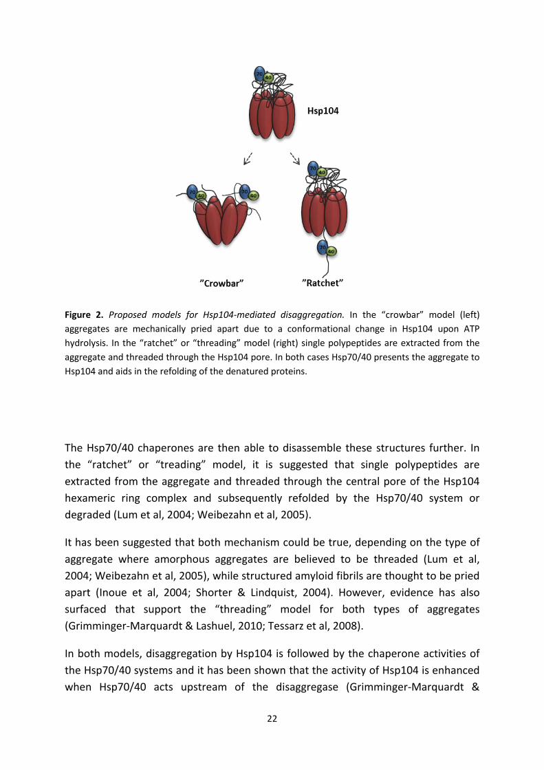
When the load of misfolded and unfolded proteins exceeds the cellular chaperone capacity, aggregates tend to form. Hsp104 is a hexameric AAA+ ATPase specializing in protein disaggregation and remodeling in yeast (Glover & Lindquist, 1998; Wendler et al, 2007). Hsp104 and its orthologs are present in fungi, plants and bacteria but no disaggregating chaperone similar to Hsp104 has been found in animals (Mosser et al, 2004). Hsp104 does not, unlike conventional chaperones, participate in the prevention of aggregate formation. However, after aggregates are formed due to e.g. oxidative stress or heat shock, Hsp104 collaborates with Hsp70 and Hsp40 in resolving aggregates to enable refolding or degradation by the proteolytic system (Glover & Lindquist, 1998). The cellular concentration of Hsp104 increases dramatically in response to stress (Bosl et al, 2006). The mechanism for how Hsp104 mediates its disaggregation functions has been debated, resulting in two different models: the “crowbar” and the “ratchet” model (Fig.2). The “crowbar” model was first described in (Glover & Lindquist, 1998), where they propose that Hsp104’s hexameric structure provides multiple sites for substrate (aggregate) binding after which an ATP dependent conformational change in Hsp104 alters the position of the binding sites thereby prying the aggregated proteins apart leading to partial disaggregation.
22
Figure 2. Proposed models for Hsp104-mediated disaggregation. In the “crowbar” model (left) aggregates are mechanically pried apart due to a conformational change in Hsp104 upon ATP hydrolysis. In the “ratchet” or “threading” model (right) single polypeptides are extracted from the aggregate and threaded through the Hsp104 pore. In both cases Hsp70/40 presents the aggregate to Hsp104 and aids in the refolding of the denatured proteins.
The Hsp70/40 chaperones are then able to disassemble these structures further. In the “ratchet” or “treading” model, it is suggested that single polypeptides are extracted from the aggregate and threaded through the central pore of the Hsp104 hexameric ring complex and subsequently refolded by the Hsp70/40 system or degraded (Lum et al, 2004; Weibezahn et al, 2005).
It has been suggested that both mechanism could be true, depending on the type of aggregate where amorphous aggregates are believed to be threaded (Lum et al, 2004; Weibezahn et al, 2005), while structured amyloid fibrils are thought to be pried apart (Inoue et al, 2004; Shorter & Lindquist, 2004). However, evidence has also surfaced that support the “threading” model for both types of aggregates (Grimminger-Marquardt & Lashuel, 2010; Tessarz et al, 2008).
In both models, disaggregation by Hsp104 is followed by the chaperone activities of the Hsp70/40 systems and it has been shown that the activity of Hsp104 is enhanced when Hsp70/40 acts upstream of the disaggregase (Grimminger-Marquardt &

23
Lashuel, 2010; Weibezahn et al, 2005). The disaggregating activity of Hsp104 in relation to aging and proteasome activity will be discussed further in the results section.
Protein degradation
Proteins are continuously synthesized and degraded to meet the demands of the cell; hence a proper protein turnover is crucial for cell survival. There are several intracellular protein degradation systems in eukaryotes. Proteasomal degradation and autophagy are responsible for the major part of intracellular protein elimination (Finley et al, 2012) while the Lon protease (Pim1 in S. cerevisiae) is the main protease for degradation of misfolded and oxidized proteins in the mitochondria (Fig.1) (Bota & Davies, 2002).
Autophagy
Autophagy refers to the degradation by the lysosome or vacuole (in yeast) of non-essential or damaged cellular constituents, aggregates and organelles (Lee et al, 2012; Reggiori & Klionsky, 2013). Autophagy can be divided into three different classes depending on the manner by which the substrate reaches the lysosome/vacuole: macroautophagy, microautophagy and chaperone-mediated autophagy (Vellai et al, 2009). Macroautophagy involves the formation of an autophagosome, a double, - or multiple membrane cytosolic vesicle (Klionsky et al, 2011). The autophagosome sequesters the cytoplasmic contents or organelle to be degraded and subsequently fuses with the lysosome/vacuole (Klionsky & Codogno, 2013). During microautophagy, the target is directly engulfed by the lysosome/vacuole and is ingested by membrane involution (Lee et al, 2012; Vellai et al, 2009). Chaperone-mediated autophagy is a strictly selective process involving chaperone recognition of a specific pentapeptide motif (KFERQ) and translocation of the chaperone-KFERQ-containing protein into the lysosome/vacuole through a specific receptor (the lysosome-associated membrane protein (LAMP)-2A) (Vellai et al, 2009).
Autophagy is the only known mechanism responsible for the removal of superfluous or dysfunctional mitochondria, also known as mitophagy (Lee et al, 2012). As cells age, there is a decline in autophagy function, and this has been correlated with the accumulation of dysfunctional mitochondria, increased oxidative stress and chronic pathologies including neurodegenerative disease (Lee et al, 2012).
24
The Lon protease The Lon protease (Pim1 in yeast) is an ATP-dependent, homo-oligomeric ring-shaped protease located in the mitochondrial matrix (Stahlberg et al, 1999). There are two major proteases in the mammalian mitochondrial matrix, ClpXP and Lon, whereas in yeast Pim1 (protease in mitochondria) acts alone as ClpP is absent (Van Dyck & Langer, 1999). The Lon protease is believed to be the main protease for degradation of oxidized and misfolded proteins in the mitochondria (Bota & Davies, 2002) and is imperative for mitochondrial function and homeostasis (Chondrogianni et al, 2012). The Lon protease is induced in response to a variety of stressors, including heat and H2O2 (Bender et al, 2010; Ngo & Davies, 2009; Van Dyck et al, 1994). It has been shown that cells lacking PIM1 have lower levels of ATP-dependent proteolysis, are respiratory deficient, sensitive to oxidative stress and have an increased accumulation of aggregated proteins (Suzuki et al, 1994; Van Dyck et al, 1994). Conversely, studies in P. anserina, reveal that overexpression of LON (PaLon) leads to lower levels of carbonylated proteins, enhanced resistance to oxidative stress and life-span extension (Luce & Osiewacz, 2009), suggesting a connection between the Lon protease and aging.
Recent studies demonstrating that Pim1 activity decreases with the age of yeast mother-cells further support a link between the Lon protease and aging (Erjavec et al, 2013). Deleting PIM1 leads to a reduced lifespan, increased accumulation of carbonylated and aggregated proteins in the cytosol, and a diminishes proteasome activity, suggesting interconnectivity between the different degradation pathways (Erjavec et al, 2013).
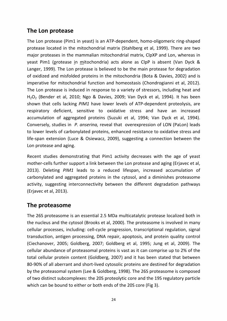
The proteasome The 26S proteasome is an essential 2.5 MDa multicatalytic protease localized both in the nucleus and the cytosol (Brooks et al, 2000). The proteasome is involved in many cellular processes, including: cell-cycle progression, transcriptional regulation, signal transduction, antigen processing, DNA repair, apoptosis, and protein quality control (Ciechanover, 2005; Goldberg, 2007; Goldberg et al, 1995; Jung et al, 2009). The cellular abundance of proteasomal proteins is vast as it can comprise up to 2% of the total cellular protein content (Goldberg, 2007) and it has been stated that between 80-90% of all aberrant and short-lived cytosolic proteins are destined for degradation by the proteasomal system (Lee & Goldberg, 1998). The 26S proteasome is composed of two distinct subcomplexes: the 20S proteolytic core and the 19S regulatory particle which can be bound to either or both ends of the 20S core (Fig 3).
25
Figure 3. Schematic image of the 26S proteasomal complex. The left image shows the arrangement of the outer α-rings with the N-terminal domains of α2, α3 and α4 creating a gate, blocking the entry into the catalytic core. The middle image represents the β-rings, where the proteolytically active β subunits (β1, β2 and β5) are shown in lighter green. The image to the right is a representation of the different parts of the 26S proteasome, the 19S regulatory particle in blue (base=light; lid=dark) and the 20S core.
20S core particle
The 20S proteasome is comprised of four stacked rings of seven subunits each, resulting in a barrel-shaped structure (Groll et al, 1997). The two identical outer rings are known as the α-rings (α1-7) and the inner rings are known as the β-rings (β1-7). The proteolytic sites are buried inside the 20S pore and entry is controlled by a gate formed by the N-terminal domains of the α2, α3 and α4 subunits of the outer rings (Fig. 3) (Groll et al, 1997; Jung et al, 2009). The inner β-rings, which harbor the proteolytic activity through the action of the N-terminal threonine residues of the β1, β2 and β5 subunits, give rise to six active sites (Fig.3). The peptide cleavage specificities of the three subunits differ, where the β1 subunits cleave after acidic amino acids (caspase-like activity), β2 after basic amino acids (trypsin-like activity) and β5 after hydrophobic or neutral amino acids (chymotrypsin-like activity) (Goldberg, 2007; Groll & Huber, 2004; Loidl et al, 1999). Mammalian cells have, in addition to the regular catalytic β-subunits, three βi subunits (β1i, β2i and β5i). Incorporation of the βi-subunits generates the 20Si or immunoproteasome, which is involved in antigen processing (Chondrogianni et al, 2012). Opening of the α-gate and subsequent entry into the catalytic core, is achieved through the binding of the 19S regulatory particle.

26
19S regulatory particle
The 19S regulator, also called PA700, consists of 19 subunits divided into a base and lid-like structure (Glickman et al, 1998; Jung et al, 2009). The base consists of six subunits with ATPase activity (Rpt1-Rpt6), arranged in a heteromeric ring complex associating with the core particle, and four non-ATPases (Rpn1, Rpn2, Rpn10 and Rpn13) (Finley et al, 2012). The base is believed to be involved in substrate recognition, ubiquitin binding, protein unfolding, and 20S gate opening (Braun et al, 1999; Hanna & Finley, 2007; Leggett et al, 2005). The lid contains the remaining nine subunits of the 19S regulatory particle (Rpn3, Rpn5-Rpn9, Rpn11, Rpn12 and Sem1) and is required for deubiquitination and degradation of ubiquitinated proteins (Glickman et al, 1998; Leggett et al, 2005).
Other proteasome regulators
There are other proteasome activators in addition to the 19S regulator. The best characterized is the PA28 regulator (11S; REG). Although not found in yeast, PA28 is an ATP-independent cytosolic regulator of 20S and appears in three isoforms: PA28α, β, and γ (Chondrogianni et al, 2012). PA28αβ is thought to be important in antigen processing (Kuehn & Dahlmann, 1997), while PA28γ is involved in cell-cycle regulation (Chen et al, 2007).
The yeast Blm10 (PA200 in mammalians) is a nuclear ATP- and ubiquitin-independent proteasome regulator which can assist in degrading peptides but not proteins (Savulescu & Glickman, 2011). PA200 is poorly conserved, sharing less than 20% identity between yeast and humans; however, they share key residues and structural motifs (Savulescu & Glickman, 2011). PA200 has been shown to be involved in DNA repair (Ustrell et al, 2002), and is required for mitochondrial function in yeast (Sadre-Bazzaz et al, 2010).
The Ubiquitin Proteasome System (UPS)
In order to maintain controlled protein degradation, there must be a way for the 26S proteasome to distinguish between proteins destined for degradation and proteins that are not. The ubiquitin system is responsible for recognizing proteins that are to be degraded and tagging them with a chain of ubiquitin, a small, essential protein found ubiquitously in all eukaryotic cells (Chondrogianni et al, 2012; Hershko & Ciechanover, 1998). Ubiquitination is a complex multistep process involving the action of three types of enzymes (Fig. 4): E1 (ubiquitin-activating enzyme), E2 (ubiquitin-conjugating enzyme) and E3 (ubiquitin ligase) (Hochstrasser, 1996).

27
Initially, E1 activates a ubiquitin-molecule (Ub) in an ATP-dependent manner and transfers it to E2, where E1 is subsequently released. A substrate specific E3-ligase recognizes and binds the target protein, the E2-Ub complex then binds the E3-substrate and finally, the ubiquitin-molecule is transferred to the substrate either directly or via the E3-ligase (Jung et al, 2009).
Figure 4. Ubiquitination. (1) Activation: a ubiquitin (ub) molecule (yellow) is activated by an E1 ubiquitin-activating enzyme (dark blue) in an ATP-dependent manner. (2) Conjugation: The E2 ubiquitin-conjugating enzyme (red) catalyses the transfer of ub from E1 to E2 and E1 is released. (3) Ligation: An E3 ubiquitin-ligase (green) recognizes a target protein, the E2-ub complex binds the E3-substrate. The E3-ligase either transiently binds ub (HECT-domain E3) or promotes the direct transfer of ub from E2 (RING-bearing E3) to the substrate (lighter blue). (4) Polyubiquitination: Subsequent rounds of ubiquitination, which can be facilitated by an E4 ubiquitin elongating factor (lighter green), are required for recognition by the 26S proteasome (Chondrogianni et al, 2012; Jung et al, 2009; Tu et al, 2007).
28
There are many different E3-ligases that bind defined motifs on target substrates. The target motif can for instance be a posttranslational modification (e.g. phosphorylation) or the N-terminal residue, a specific sequence or domain (e.g. a hydrophobic patch) of a protein that normally is not exposed (Ciechanover & Brundin, 2003). Consecutive rounds of ubiquitination are then required since the recognition and processing of the ubiquitinated target protein by the 26S proteasome is only achieved after a chain of at least four Ub-molecules is attached (Thrower et al, 2000). This Ub-chain elongation is occasionally assisted by an E4 ubiquitin elongation factor (Ufd2 in yeast) (Benirschke et al, 2010; Tu et al, 2007). After the 19S regulatory particle has recognized the ubiquitinated substrate, it is unfolded, deubiquitinated and translocated into the 20S catalytic core where it is hydrolyzed into peptides of an average length of 8-12 amino acids (Jung et al, 2009). Both the ubiquitin and amino acids are recycled for further use.
29
Protein damage Here I will give an introduction to how and why oxidative damage and protein modifications occur, as well as how they may be prevented. Emphasis will lie on components related to this thesis.
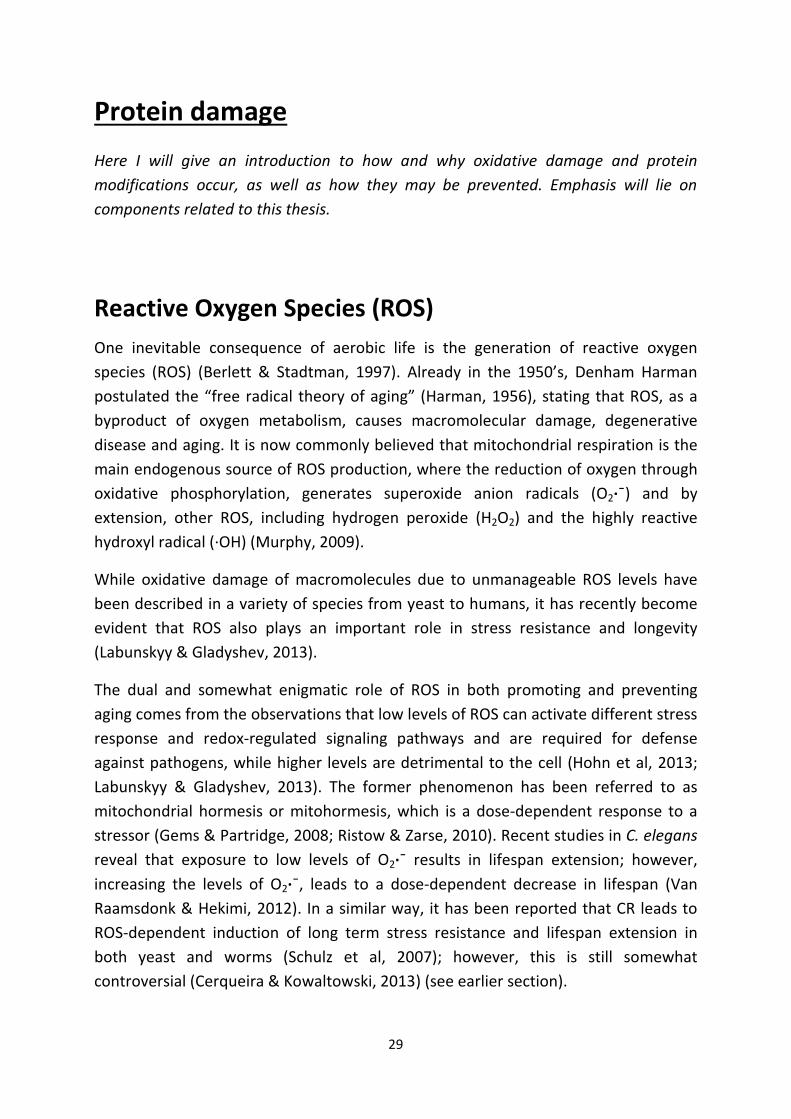
Reactive Oxygen Species (ROS) One inevitable consequence of aerobic life is the generation of reactive oxygen species (ROS) (Berlett & Stadtman, 1997). Already in the 1950’s, Denham Harman postulated the “free radical theory of aging” (Harman, 1956), stating that ROS, as a byproduct of oxygen metabolism, causes macromolecular damage, degenerative disease and aging. It is now commonly believed that mitochondrial respiration is the main endogenous source of ROS production, where the reduction of oxygen through oxidative phosphorylation, generates superoxide anion radicals (O2·ˉ) and by extension, other ROS, including hydrogen peroxide (H2O2) and the highly reactive hydroxyl radical (·OH) (Murphy, 2009).
While oxidative damage of macromolecules due to unmanageable ROS levels have been described in a variety of species from yeast to humans, it has recently become evident that ROS also plays an important role in stress resistance and longevity (Labunskyy & Gladyshev, 2013).
The dual and somewhat enigmatic role of ROS in both promoting and preventing aging comes from the observations that low levels of ROS can activate different stress response and redox-regulated signaling pathways and are required for defense against pathogens, while higher levels are detrimental to the cell (Hohn et al, 2013; Labunskyy & Gladyshev, 2013). The former phenomenon has been referred to as mitochondrial hormesis or mitohormesis, which is a dose-dependent response to a stressor (Gems & Partridge, 2008; Ristow & Zarse, 2010). Recent studies in C. elegans reveal that exposure to low levels of O2·ˉ results in lifespan extension; however, increasing the levels of O2·ˉ, leads to a dose-dependent decrease in lifespan (Van Raamsdonk & Hekimi, 2012). In a similar way, it has been reported that CR leads to ROS-dependent induction of long term stress resistance and lifespan extension in both yeast and worms (Schulz et al, 2007); however, this is still somewhat controversial (Cerqueira & Kowaltowski, 2013) (see earlier section).

30
Antioxidants To maintain a reduced intracellular redox environment, cells need to be equipped with antioxidant defense systems to cope with ROS. These antioxidants can be either protective enzymes that are upregulated as needed or non-enzymatic free radical scavengers (Morano et al, 2012). Antioxidant enzymes include: superoxide dismutases (SODs), catalases, methionine sulfoxide reductases (MSRs), thioredoxins and peroxiredoxins. If the amount of ROS produced exceeds the antioxidant capacity of the cell this will lead to oxidative stress (Morano et al, 2012), and in some conditions, shortened lifespan. It is therefore tempting to assume that the antioxidant capacity of the cell is directly correlated to its lifespan, this is, however, not necessarily true. Many contradictory reports regarding antioxidant’s effect on lifespan have emerged over the years. Some reports show that increasing the antioxidant capacity of the cell, e.g. overexpression of catalases (Schriner et al, 2005) and SODs (Curtis et al, 2007; Fabrizio et al, 2003; Parkes et al, 1998), can lead to extended lifespan, whereas, other studies show that increasing SODs have no effect (Doonan et al, 2008) or even shorten lifespan (Van Raamsdonk & Hekimi, 2009). One of the reasons for the negative effect of elevated antioxidant capacity could be the role for ROS in signaling, where excessive antioxidant scavenging would disrupt ROS homeostasis.
Peroxiredoxins Peroxiredoxins (Prxs), also called thioredoxin peroxidases, are ubiquitous thiol-specific antioxidant proteins found in yeast, plants and animals. They act as antioxidant scavengers, reducing and detoxifying H2O2 and a wide range of organic hydroperoxides (Nystrom et al, 2012; Wood et al, 2003). Prxs are efficient scavengers when H2O2 concentrations are low due to their high reactivity for the substrate, however when H2O2 increases, Prxs become saturated and catalases are more efficient (Seaver & Imlay, 2001). All Prxs contain at least one active-site peroxidatic cysteine (Cys) which is oxidized to a sulfenic acid by the peroxide substrate (Wood et al, 2003). Prxs can be divided into two classes based on the number of conserved Cys residues: 2-Cys Prxs contain both the N-terminal peroxidatic cysteine corresponding to Cys48 in yeast and a C-terminal resolving Cys corresponding to Cys171; 1-Cys Prxs only contain the N-terminal cysteine. The 2-Cys Prxs are subdivided into “typical” and “atypical” 2-Cys Prxs. “Typical” 2-Cys Prxs are homodimers, where the peroxidatic Cys condenses with the resolving Cys of the other subunit forming an intermolecular disulfide bond (Fig. 5), while the “atypical” 2-Cys is a monomer, forming a intramolecular disulfide bond between the peroxidatic and resolving Cys on the same

31
subunit, in response to peroxides. The disulfide bonds can then be reduced by thioredoxins (Trxs), making the Prxs available for another round of H2O2 reduction (D'Autreaux & Toledano, 2007). When peroxide concentrations are high the 2-Cys Prxs sulphenylated form is subject to further oxidation and inactivation by H2O2 (Yang et al, 2002) (Fig. 5). This can occur if the formation of disulfide bonds is insufficiently rapid and the catalytic cycle is momentarily stalled in the intermediary sulphenylated form (Hall et al, 2011).
Figure 5. “Typical” 2-Cys Prx catalytic cycle. The “typical” 2-Cys Prx is a homodimer which reduces H2O2 through the peroxidatic (Cys48) cysteine to a sulfenic acid (intermediary form) which condenses with the resolving (Cys171) cysteine on the other subunit to form a disulfide bond. This can subsequently be reduced by Trx in a NADPH-dependent manner. Prxs are subject to hyperoxidation by H2O2 to form sulfinic acid, inactivating the Prx peroxidase activity. The hyperoxidized Prx oligomerizes and has been shown to function as a molecular chaperone in the prevention of aggregated proteins. This hyperoxidized form can be reversibly reduced by Srx, indicating a regulatory process (D'Autreaux & Toledano, 2007; Nystrom et al, 2012; Wood et al, 2003).
32
The hyperoxidized, sulphinic acid form of the yeast Prx, Tsa1, oligomerizes and is suggested to function as a molecular chaperone (Jang et al, 2004). During normal cellular conditions, where the cell is exposed to fairly low concentrations of H2O2, 2-Cys Prxs exist in both the peroxidatic and chaperone form (Jang et al, 2004). However, during oxidative stress conditions, there is a switch towards the chaperone form where 2-Cys Prxs undergo conformational changes leading to high molecular weight complexes (Jang et al, 2004; Rhee & Woo, 2011). As certain levels of H2O2 are necessary for signal transduction, it has been proposed that the hyperoxidation and inactivation of Prxs also act as “floodgates” in a way to maintain sufficient basal H2O2
levels required for signaling (Wood et al, 2003). The dual role of Prxs was found when yeast expressing the non-peroxidatic “Cys-less” Tsa1 (C48S/C171S) could ameliorate the hypersensitivity to heat stress found in a tsa1/2 mutant strain (Jang et al, 2004). Thus, they reasoned that Tsa1 must have another function unrelated to its peroxidase acitivity. They also found that the peroxidatic Cystein Cys48 was essential for hyperoxidation and subsequent oligomerization in response to H2O2 stress in vivo; though, this was not the case during the in vitro heat stress experiments, suggesting two different pathways (Jang et al, 2004). These findings will be addressed further in the results and discussion section.
The hyperoxidized, sulphinylated form of Prxs can be reduced by Srxs (Srx1 in yeast) (Biteau et al, 2003), leading to dissociation into low molecular weight species and subsequently reactivation of its peroxidase function (Jang et al, 2004; Rhee & Woo, 2011). Recent studies by Molin et al. (2011) show that the low PKA activity seen during CR (glucose restriction) leads to a Tsa1-dependent lifespan extension in yeast. Low PKA generates increased levels of Srx1 which counteracts the H2O2-induced hyperoxidation and inactivation of the H2O2 scavanging peroxiredoxin Tsa1 leading to increased H2O2 resistance and lifespan extension (Molin et al, 2011).
Oxidative damage and protein modifications
Proteins are incessantly affected by various intrinsic and extrinsic factors. Under physiological conditions, approximately 30% of all newly synthesized proteins are misfolded and during oxidative conditions this number increases substantially (Chondrogianni et al, 2012). It has also been shown that severe oxidative stress can induce protein mistranslation (Ling & Soll, 2010) adding to the pool of misfolded proteins in the cell. Moreover, mistranslated proteins also have an increased
33
susceptibility to oxidative modifications caused by ROS (Ballesteros et al, 2001; Dukan et al, 2000). The observation that certain proteins have particular susceptibility to detrimental posttranslational modifications, suggests that protein damage is not an entirely random process. Recent work in human myoblasts, demonstrate that there is a selective oxidation of proteins even in the presence of severe peroxide stress (Baraibar et al, 2011). In addition, undifferentiated mouse embryonic stem (ES) cells show a preferential oxidation (carbonylation) of chaperones and cytoskeletal proteins (Hernebring et al, 2006) and selective oxidation has also been found during aging (Dukan & Nystrom, 1998; Reverter-Branchat et al, 2004).
There are several ways proteins can be oxidatively modified, either (i) directly through attacks of the peptide chain by ROS creating protein backbone damage or damage to specific amino acid side chains, or (ii) indirectly through the formation of protein glycation and lipid peroxidation products (Hohn et al, 2013). The two most commonly oxidized amino acids are cysteine and methionine, and they are also the only residues that can be reversibly reduced (Jung et al, 2009; Ugarte et al, 2010). In contrast, carbonylation is an irreparable damage involving the oxidation of lysine, arginine, proline and threonine residues (Hohn et al, 2013). Carbonylation is considered to be the most abundant irreversible oxidative modification of proteins (Stadtman & Levine, 2003). Depending on the severity of the oxidation, proteins can experience reduced activity or complete loss of function. Heavily oxidized proteins often have an altered tertiary structure, as do misfolded proteins, exposing hydrophobic residues normally buried inside the protein. In order to escape the aqueous environment of the cell, these unfolded, hydrophobic proteins tend to form unspecific protein-protein interactions, resulting in protein aggregation (Davies, 2001; Grune et al, 2004).

34
Aging and proteostasis decline Disruption of any part of the proteostasis network can have severe consequences for cellular fitness and longevity. Here an overview is given of how the decline in proteostasis maintenance is associated with aging and age-related diseases.
Figure 6. Schematic representation of proteostasis decline during oxidative stress and aging. Oxidative stress, as seen during aging, can affect many parts of the proteostasis network leading to protein mistranslation (1), increased susceptibility for oxidative modification and protein misfolding (2,3), protein aggregation (4) and inhibition of proteolysis (5,6). Disruption of any part of this network may lead to protestasis collaps and proteotoxicity (Ballesteros et al, 2001; Douglas & Dillin, 2010; Dukan et al, 2000; Ling & Soll, 2010). See text for more details.

35
The aging mitochondria
Oxidative damaged proteins accumulate during aging in almost every model system analyzed (Stadtman, 2006). As much as one in every third protein has been calculated to be modified by oxidative carbonylation in old individuals of animals (Stadtman & Levine, 2000). Mitochondria have been given a central role in this process of protein oxidation as they are both considered a major target for, and contributor of, oxidative damage (Boveris & Chance, 1973; Guarente, 2008; Murphy, 2009). The close proximity to the electron transport chain in the mitochondrial inner membrane (MIM), the main source of ROS production, causes many mitochondrial components to be vulnerable to oxidative damage, including: mitochondrial DNA (mtDNA), proteins and lipids (Harper et al, 1998; Shigenaga et al, 1994). Under physiological conditions, 1-2% of metabolized oxygen is converted to O2·ˉ and subsequently the relatively stable H2O2 (Boveris & Chance, 1973; Hohn et al, 2013), which can diffuse out of the mitochondria and into the cytosol and nucleus (Guarente, 2008). However, as cells age the amount of ROS produced increases. A progressive lipid peroxidatic damage of the MIM due to ROS causes increased proton leakage (Hagen et al, 1997; Harper et al, 1998; Shigenaga et al, 1994), and to compensate for the loss of membrane potential, aged cells increase the rate of oxygen consumption leading to diminished coupling efficiency and a greater production of ROS (Harper et al, 1998). In addition, mtDNA-damage by ROS, as seen in aged cells, causes reduced enzymatic activity of the electron transport chain, leading to electron-stalling at complexes I (NADH dehydrogenase) and III (cytochrome b-c1 complex) which results in further ROS production (Barja, 2007; Guarente, 2008; Jang & Remmen, 2009; Lenaz et al, 2000). Moreover, dysfunctional regulation of mitochondrial dynamics, causing enlarged or fragmented mitochondria, is reported to generate more oxidative stress during aging (Seo et al, 2010; Shigenaga et al, 1994). In addition to “self-inflicted” damage, recent studies in yeast demonstrate (Hughes & Gottschling, 2012) that a loss of vacuolar acidification causes mitochondrial dysfunction and fragmentation at an early onset of age. It has also been shown that mitochondria with higher O2·ˉ levels and lower redox potential are preferentially retained in the yeast mother cell during cytokinesis, leading to accumulation of dysfunctional mitochondria already early in life (McFaline-Figueroa et al, 2011). Conversely, it has been shown that counteracting mitochondrial fragmentation reduces the generation of H2O2, and increases fitness and lifespan in both the filamentous fungi P. anserina and in S. cerevisiase (Scheckhuber et al, 2007).
36
The UPS and aging In addition to the age-associated increase in ROS and subsequent oxidative damage, a decrease in proteasome activity and/or function with age has been reported in a variety of species, including: human muscle (Ferrington et al, 2005; Husom et al, 2004), lens (Viteri et al, 2004), lymphocytes (Carrard et al, 2003), epidermis (Bulteau et al, 2000), and fibroblasts (Sitte et al, 2000a; Sitte et al, 2000b). In addition, the heart (Bulteau et al, 2002), spinal cord (Keller et al, 2000) and brain (Zeng et al, 2005) of rats also show decreased proteasomal activity with age. There are many possible reasons for age-related proteasome dysfunction, such as altered assembly, expression, modification (Bulteau et al, 2000; Chondrogianni & Gonos, 2005; Chondrogianni et al, 2003; Ferrington et al, 2005; Keller et al, 2000; Vernace et al, 2007), or inactivation through association with protein aggregates (Bence et al, 2001; Sitte et al, 2000a). Carrard et al. (2003) noted that when comparing the proteasomal activity and abundance in peripheral blood lymphocytes from young and aged donors, the aged donors had a reduced proteasomal activity while the amount of assembled intracellular protesomes remained unchanged (Carrard et al, 2003). The severity of a protein’s oxidative modification/damage has also been shown to affect its susceptibility for proteasomal degradation (Jung et al, 2009). Low to moderate levels of protein oxidation gradually increases a proteins proteolytic susceptibility until it reaches a point where the oxidation level is too high, leading to a dramatic decline in proteasomal degradation and function (Jung et al, 2009). This biphasic response may contribute to the decline in proteasomal activity seen with age.
Studies on fibroblasts from healthy centenarians show that both proteasomal activity and the levels of oxidatively modified proteins were comparable to those found in young (Chondrogianni et al, 2000). In addition, the long-lived naked mole rat also exhibits elevated proteasome levels and activity (Perez et al, 2009). It has also been reported that increasing the amount of proteasomes, either by upregulating the catalytically active β-subunits of the 20S-core (Chondrogianni et al, 2005), overexpressing the 19S subunit Rpn11 (Tonoki et al, 2009), or by overexpressing the proteasomal assembly factor POMP (Ump1 in yeast) (Chen et al, 2006; Chondrogianni & Gonos, 2007), prolonges lifespan and increases degradation of oxidatively modified proteins. In C. elegans, ectopic expression of rpn6 is sufficient to provide proteotoxic stress resistance and extend lifespan (Vilchez et al, 2012). Moreover, recent studies in yeast show that stabilizing the UPS transcription factor Rpn4, by deletion of the E3-ligase and Rpn4 regulator Ubr2 (Ju et al, 2004; Wang et al, 2004a), result in increased proteasomal function and lifespan extension (Kruegel et al, 2011).

37
These results suggest a strong connection between proteasome function and proteostasis in modulating longevity which will be addressed further in later sections.
ATP levels – an issue for the proteasome? A major role for the mitochondria is the generation of ATP. However, during aging, as mitochondrial damage increases causing decreased mitochondrial membrane potential, ATP production is diminished (Baraibar et al, 2011; Shigenaga et al, 1994). Both assembly of, and degradation by, the 26S proteasome is ATP dependent (Liu et al, 2006) and it has been reported that at least 300 ATP molecules are hydrolyzed per protein degraded by the proteasome (Benaroudj et al, 2003). In addition, disassembly of the 19S regulatory particle and associated proteins from the 20S core also requires ATP hydrolysis (Babbitt et al, 2005). Studies in aged Drosophila melanogaster have shown a compromised 26S assembly which was correlated to a great reduction in ATP (Vernace et al, 2007). It has also been shown that the 26S proteasome is readily disassembled when isolated without ATP, but can be reassembled when ATP is once again present (Coux et al, 1996). Thus, maintaining sufficient cellular ATP concentrations appears to be of vital importance for 26S-mediated degradation and limitations in ATP concentrations could contribute to the age-associated decline in proteasome activity.
20S-mediated degradation of oxidized proteins There is some controversy in the literature with respect to whether the ATP-independent 20S proteasome can act alone in protein degradation. Pacifici et al. (1993) proposed that it would be beneficial for cells to have an ATP-independent degradation of oxidized proteins since this would rapidly remove damaged proteins and thereby prevent their accumulation (Pacifici et al, 1993). It has been shown that purified 20S proteasomes are able to degrade partially unfolded and oxidized proteins (Davies, 2001; Ferrington et al, 2001; Pacifici et al, 1989; Rivett, 1985). Possibly, this is due to hydrophobic residues exposed during unfolding, binding directly to the α-rings, thereby opening the 20S gate and allowing translocation to the catalytic core in an ATP/ubiquitin independent manner (Coux et al, 1996; Liu et al, 2003a). Studies in mammalian cells have shown that purified 20S proteasomes selectively degrade oxidatively-damaged over native proteins, while the 26S proteasome showed no preference (Davies, 2001; Grune et al, 2003). However, other studies suggest that the 20S proteasome alone is unable to degrade denatured proteins or cleave peptide (Kisselev & Goldberg, 2005).
38
Nevertheless, the 20S proteasome appears to be more resistant to oxidative stress than the 26S proteasome (Reinheckel et al, 1998). Recent studies show that in both yeast and humans, the 26S proteasome disassembles into free 19S and 20S complexes in response to increasing H2O2 concentrations (Wang et al, 2010). In yeast this dissociation is mediated by the proteasome-interacting protein (PIP), Ecm29, which during oxidative stress, is recruited to the 19S subcomplex and promotes disassembly of the 26S proteasome (Wang et al, 2010). It has previously been shown that yeast defective in 26S assembly is more resistant to peroxide stress and have an increased ability to degrade carbonylated proteins (Inai & Nishikimi, 2002). Consistent with these findings, it was also shown that ecm29 mutants are more sensitive to H2O2, adding further support for 20S core-mediated defense against oxidative stress (Wang et al, 2010) and degradation of oxidized proteins. Aiken et al proposed a model for oxidative stress-dependent regulation of the 26S proteasome (Aiken et al, 2011). They suggest that the 26S proteasome can degrade mildly oxidized proteins; however, in response to persistent or acute oxidative stress, the 26S proteasome temporarily disassembles causing accumulation of ubiquitinated proteins. When the stress is removed the 26S proteasome reassembles and the degradation of ubiquitinated proteins can recommence (Aiken et al, 2011).
Protein aggregation The importance of cellular proteostasis becomes evident in the light of protein aggregation. Aggregates are insoluble, ordered or amorphous, potentially cytotoxic structures that can disrupt membranes and inappropriately interact with cellular constituents (Hohn et al, 2013; Taylor & Dillin, 2011). Once the formation of aggregates has initiated, it is a cumulative process. The age-related increase in misfolded proteins together with diminished repair and/or removal capacity causes aggregation which, in turn, can serve as nucleation sites for more misfolded proteins (Squier, 2001).
Compelling evidence links UPS impairment to the accumulation of misfolded proteins and protein aggregates (Kaganovich et al, 2008; Lopez Salon et al, 2003; Tanaka et al, 2001). Aggregates are considered poor substrates for proteasomal degradation and due to the enormity of the deposit, it is unable to enter the 20S catalytic core (Hohn et al, 2013). It has even been suggested that aggregates could be directly inhibiting the proteasome by means of “clogging” (Bence et al, 2001), thereby adding to the diminished cellular protection, and further protein damage and aggregation (Squier, 2001). Another suggestion is that aggregates sequester components of the UPS, including ubiquitin, proteasomes and molecular chaperones, leading to depletion and
39
thus UPS impairment (Donaldson et al, 2003). These two models are not necessarily mutually exclusive (Bennett et al, 2005).
It has been shown that increasing the cells proteasomal capacity, through deletion of the E3-ligase (see earlier section) Ubr2, leads to improved clearance of toxic huntingtin fragments in yeast (Kruegel et al, 2011). Moreover, previous studies have shown that when inducing protein aggregation of model disease proteins/peptides such as huntingtin (Bence et al, 2001; Jana et al, 2001), α-synuclein (Outeiro & Lindquist, 2003), and amyloid-β (Shringarpure et al, 2000) UPS function is impaired. However, it has not been shown whether this is true for indigenous protein aggregates which accumulate during aging. This will be addressed further in the discussion.
Inclusion bodies (IBs) and spatial quality control (SQC) Inclusion bodies (IBs) are large species of sequestered aggregates and associated proteins that form in an organized process that seems to be conserved from yeast to humans (Kaganovich et al, 2008; Kopito, 2000). IBs have attracted much attention in regards to whether disease-associated IBs are protective or pathogenic forms of aggregated proteins (Bennett et al, 2005). Some suggest that IBs attract components of the UPS thereby sequestering vital components of the cells protein quality control machinery which are then unavailable to perform their duties elsewhere (Waelter et al, 2001). Others claim that sequestration of aggregated proteins in larger IB has a protective role where toxic misfolded protein species and aggregates are titrated away from sensitive cellular machineries (Ciechanover & Brundin, 2003; Kopito, 2000).
Even though studies report that disease-associated aggregates and aggregation intermediates have a negative effect on UPS function and activity, this impairment predominantly occurs prior to IB formation (Bennett et al, 2005). Inclusions have furthermore been shown to facilitate clearance of misfolded proteins (Taylor et al, 2003). Several studies in primary neuron models of Huntington’s disease (HD), show that cells that form IBs have increased UPS activity and experience less neuronal death than cells that do not form IBs (Arrasate et al, 2004; Mitra et al, 2009; Schipper-Krom et al, 2014). Moreover, it was shown that proteasomes associated to IBs in these cells were reversibly recruited and catalytically active (Schipper-Krom et al, 2014). These findings support the notion of IB formation as a protective cellular response.

40
Inclusions can, according to Kaganovich et al. (2008), be divided into two distinct spatial quality control (SQC) compartments depending on the sequestered proteins, ubiquitination and aggregation status. It is suggested that misfolded or aggregation-prone proteins are initially localized to a juxtanuclear compartment (JUNQ; juxtanuclear quality control) together with components of the UPS. This sequestration is believed to facilitate refolding and/or degradation of substrate proteins (Kaganovich et al, 2008). As the cellular load of misfolded or damaged proteins increase past the capacity of the PQC machinery, potentially toxic species are directed to an insoluble protein deposit (IPOD) adjacent to the vacuole. The aggregates localized to the IPOD compartment are suggested to be terminally sequestered as a means to avoid association with sensitive components of the UPS. It has also been shown that the IPOD co-localizes with an autophagic marker and a pre-autophagosomal structure (Kaganovich et al, 2008) and together with its close proximity to the vacuole could suggests a link between aggregated protein in the IPOD and autophagy.
An additional SQC structure, known as the aggresome, involves the retrograde transport of protein aggregates along microtubules into a large inclusion converging at the centrosome (spindle pole body in yeast) (Kopito, 2000; Wang et al, 2009). Compartmentalization of aggregates into aggresomes has been shown to mitigate proteotoxicity, supporting the protective role of IB in SQC (Kopito, 2000; Wang et al, 2009). However, the presence of an aggresome in yeast has been debated.
As most studies on IB are specifically related to disease-associated protein aggregation or model proteins designed to promote aggregation (Arrasate et al, 2004; Bennett et al, 2005; Kaganovich et al, 2008; Mitra et al, 2009; Wang et al, 2009) it remains unclear whether IB formation acts as an initial protective response for indigenous protein aggregates during aging.
Cell polarity and aggregate inheritance It has been suggested that cell polarity evolved very early and is a universal attribute of cellular organisms as a means to avoid clonal senescence (Macara & Mili, 2008). Polar segregation appears to be intimately linked to aging, as it aids in the rejuvenation of one sibling at the expense of the other. Even seemingly symmetrically dividing unicellular organisms, such as E. coli, experience polarity where aggregates preferentially accumulate in the sibling that retains the “old” pole after cell division (Lindner et al, 2008; Nystrom, 2007). In yeast, these processes include (i) asymmetric inheritance of ERCs, which are thought to attach to nuclear pore complexes that are
41
retained in the mother due to a septin-based diffusion barrier (Shcheprova et al, 2008), (ii) retention of dysfunctional mitochondria (McFaline-Figueroa et al, 2011; Vevea et al, 2014), as well as (iii) segregation and retrograde transport of damaged and aggregated proteins (Aguilaniu et al, 2003; Erjavec et al, 2007; Liu et al, 2010).
During cytokinesis in budding yeast, oxidatively damaged and aggregated proteins are unevenly distributed between the mother and daughter cell (Aguilaniu et al, 2003; Erjavec et al, 2007). It has been shown that this asymmetric inheritance is due to a Sir2-and Hsp104-dependent selective retention of damaged and aggregated proteins in the progenitor (Erjavec et al, 2007). This segregation of damage has been proposed to ensure the fitness of the progeny (Aguilaniu et al, 2003) and restrict senescence to one lineage (Macara & Mili, 2008). In addition, mathematical modelling suggest that cells that undergo asymmetric division and/or damage segregation can endure higher degrees of damage before entering clonal senescence (Erjavec et al, 2008). Recent studies demonstrate that segregation of aggregates during cell division require, in addition to Sir2 and Hsp104, the polarisome and myosin motor machinery (Liu et al, 2010). The same study revealed that daughter cells can rid themselves from aggregates while still attached to the mother by a polarisome-mediated retrograde transport of aggregates. The polarisome is a protein complex within the polarity cap, involved in determining cell polarity through assembly of actin filaments at the bud tip (Moseley & Goode, 2006). It has been suggested that actin cables act as scaffolds for large Hsp104-associated aggregates and that these tethered aggregates could be transported away from the bud tip through a Bni1-dependent nucleation of actin at the polarisome (Liu et al, 2010). Similar to aggresome formation, aggregates that translocate from the daughter to the mother were shown to coalesce into large inclusions. However, in the case of retrograde transport of aggregates, this process is dependent on actin filaments, not microtubles (Liu et al, 2010). Another study suggests that aggregate retention in the mother cell is merely due to limited mobility of the aggregate and the narrowness of the budneck, which increases the likelihood of aggregates remaining in the mother. This study also propose that movement of aggregates is due to un-directional random walk (Zhou et al, 2011). The confincting findings by Lui et al. (2010) and Zhou et al. (2011) set the basis for paper I and will be approached further in the discussion.
Aggregation in neurodegenerative disorders Neurodegenerative diseases can be defined as either hereditary or sporadic, where sporadic conditions often are associated with aging and oxidative protein modifications (Ross & Poirier, 2004). A common feature of many neurodegenerative

42
diseases is the abnormal accumulation and aggregation of disease-specific proteins (Takalo et al, 2013). Neurons are considered particularly vulnerable to diseases related to protein misfolding (Taylor & Dillin, 2011). The human brain accounts for approximately 20% of a body’s oxygen consumption while only comprising 2% of its mass (Halliwell, 2006). Meanwhile, the levels of antioxidants are fairly low leading to an increased risk for oxidative damage, and as neuronal cells are mainly post-mitotic, there is no way of titrating out potentially harmful aggregates (Halliwell, 2006). Protein aggregation is thus a hallmark of many age-associated diseases, including Alzheimer´s disease (AD), Parkinsson´s disease (PD) and Huntington’s disease (HD) (Ross & Poirier, 2004). It is, however, debated whether the accumulation of aggregates is a primary cause or a secondary consequence of the disease itself (Ciechanover & Brundin, 2003), though there is evidence conveying amyloid-β and polyglutamine aggregation as the cause of the neurotoxicity associated with AD and HD, respectively (Balch et al, 2008; Golde et al, 2006).
Moreover, aberrations of the UPS and lysosome-function/autophagy have been implicated in the pathogenesis of a range of neurodegenerative disorders (Ciechanover & Brundin, 2003). For instance, it has been reported that mutations in parkin, a gene coding for an E3 ubiquitin ligase involved in promoting UPS-mediated protein degradation, is the cause of almost half of early onset autosomal recessive forms of PD (Ciechanover & Brundin, 2003; Cuervo et al, 2010; Rana et al, 2013). Conversely, it was recently shown that overexpression of parkin leads to reduced aggregate formation and extended lifespan in flies (Rana et al, 2013).
Genetic dosage alterations could also increase the likelihood of aggregation (Ross & Poirier, 2004). For example, familial PD can be caused by a triplication at the α-synuclein locus (Singleton et al, 2003). Moreover, individuals with Down´s syndrome experience early onset of amyloid-β plaques and AD-symptoms due to an extra copy of the APP locus on chromosome 21 (Zigman & Lott, 2007).
There are several hypothesis as to how protein misfolding and aggregation are associated with neurodegeneration and apoptosis (Soto, 2003). These include: aggregate-activation of an apoptotic signaling pathway (NF-κB), recruitment of essential cellular factors (e.g. UPS components; see earlier sections), aggregate-mediated membrane disruption and depolarization, and the induction of oxidative stress (Soto, 2003).
Since proteotoxicity plays such a pivotal role in both aging and many age-associated diseases, why are the components involved in maintaining protein homeostasis not
43
constitutively expressed at high levels? In fact, theraputical proteostasis regulators that restore protein homeostasis have been used in clinical trials (Balch et al, 2008). In C. elegans-models of AD and HD, RNA interference (RNAi)-based proteostasis regulators that increase chaperone levels and disaggregase activity have been successful in delaying the onset of the gain-of-toxicity phenotype (Balch et al, 2008; Cohen et al, 2006). On the other hand, increasing chaperones and UPS components has been associated with tumorigenesis, which shows an inverse correlation to neurodegenerative diseases (Driver et al, 2012; Taylor & Dillin, 2011). In addition, UPS activity appears to be inversely correlated to fecundity, where studies in D. melanogaster show that mating results in an immediate diminished UPS activity and accelerated aging (Fredriksson et al, 2012). Thus, constitutively expressing proteostasis components would not be beneficial in an evolutionary standpoint as there appears to be a trade-off in resource allocation between somatic maintenance by the UPS and reproduction, in agreement with Kirkwood´s “disposable soma theory of aging” (Kirkwood, 1977).

44
Results and discussion
Paper I
Movement of aggregates is retrograde-biased Previous studies by Liu et al (2010) reveal that in addition to the actin- and Hsp104-dependent active retention of protein aggregates in the mother cell, daughter cells can also rid themselves from aggregates that have “leaked” into the bud, through a polarisome-mediated retrograde transport (Liu et al, 2010). However, a study by Zhou et al (2011) claims that the movement of aggregates is characterized by un-directional random walk. In paper I we further establish the existence of a retrograde transport by analyzing the movement of aggregates during 393 budding events. We found that 15.5% of budding events showed cross-compartment movement, and among these, 65.5% of were retrograde movement, while anterograde- and bi-direction movement was significantly less frequent (25.4% and 8.1% respectively). Moreover, using a mutant Hsp104Y662A-mCherry variant, defective in disaggregation, we saw that aggregates co-localize with Abp140-3GFP, which is known to bind actin cables. Abp140 has previously been used to visualize retrograde movement of actin cables (Yang & Pon, 2002). As the actin cytoskeleton does not diffuse freely, it is unlikely that aggregates associated with the actin cytoskeleton would do so. The association of Hsp104-containg aggregates with the actin cytoskeleton supports the notion of both actin-dependent retention and directional movement of aggregates.
The polarisome is required for the active retention of Htt103Q aggregates during cytokinesis. To further investigate the involvement of the polarisome in the segregation of aggregates during cytokinesis, we used the model disease protein Htt103Q-GFP as an aggregate reporter. Htt103Q is prone to aggregation without the disadvantages, such as a collapse in the actin cable network, accompanying heat-induced aggregation. Htt103Q-GFP aggregation was initially triggered in wt and bni1Δ cells after which cells were stained with concanavalin-A (con-A). The expression of Htt103Q-GFP was subsequently turned off, and cells were allowed to grow only enough to initiate the budding process. Aggregates were then monitored in Con-A stained mother cells, and

45
unstained daughter cells. We found that Htt103Q aggregates were retained in the mother in a factor-dependent manner and that this segregation of damage was greatly diminished in cells lacking Bni1. These data reveal that the polarisome is required for the segregation of both aggregation-prone disease proteins and heat-induced Hsp104-containing aggregates (Liu et al, 2010).
Main findings presented in paper I: • Hsp104-containing aggregates co-localize to the actin cytoskeleton. • Daughter cells have the ability to rid themselves of aggregates by retrograde
transport during cytokinesis. • Htt103Q-GFP aggregates are retained in the mother during cytokinesis in a
polarisome dependent manner.
Paper II
- Text removed from public version -

46
- Text removed from public version -
- Text removed from public version -

47
- Text removed from public version -
- Text removed from public version -

48
- Text removed from public version -
- Text removed from public version -

49
- Text removed from public version -
50
Main findings presented in paper II: - Text removed from public version -

51
Paper III
Aged cells exhibit an impairment of the proteasome. The aim of this paper was to elucidate whether the accumulation of aggregated proteins that occur during aging could be directly linked to diminished function of the UPS in S. cerevisiae. By using a cytosolic UPS substrate, ΔssCPY*-GFP, the in vivo proteasome function could be monitored. ΔssCPY*is a mutated and misfolded endoplasmatic reticulum (ER)-import-defective derivative of the carboxypeptidase Y (CPY, YscY) which is rapidly degraded by the UPS (Park et al, 2007). From a growing population of S. cerevisiae, cells at an average age of 13-15 generations were isolated and sorted with biotin and streptavidin-conjugated magnetic beads (Smeal et al, 1996). The dramatic increase in ΔssCPY*-GFP foci seen in the aged population indicate that the UPS-mediated PQC is diminished in these cells. The accumulation of aggregates is not just specific for the UPS substrate as visualized by using Hsp104-GFP, a reporter for indigenous protein aggregates.
Lowering UPS activity is sufficient to induce aggregate formation in young cells.
Lowering UPS activity, either through the addition of the potent proteasomal inhibitor MG132 or by using a UPS temperature sensitive (ts) strain (rpt4-145; 19S subunit), is sufficient to induce massive Hsp104-GFP foci formation in a growing population of predominantly young cells. These data demonstrate the necessity for an unperturbed proteasome activity in the prevention of protein aggregates in young cells. In order to study the requirement for a fully functional UPS in the clearance of aggregates once formed, we induced aggregate formation to the same extent in both wt and rpt4-145 cells and monitored aggregate clearance over time. Although the initial amount of foci were the same in the two strains, the UPS-defective strain failed to resolve the heat-induced aggregates. Hence, the data demonstrate that an intact UPS is imperative to the prevention and clearance of protein aggregates even in young cells.
Increasing proteasome function mitigates aggregate formation. To further investigate the correlation between aggregate formation and UPS function, we tested if increasing proteasome activity would alleviate the formation of

52
Hsp104-GFP foci. It has previously been shown that deleting the E3 ubiquitin ligase UBR2 leads to increased proteasome capacity through stabilization of the proteasome subunit transcription factor Rpn4 (Kruegel et al, 2011). We therefore used a ubr2Δ strain to increase UPS activity and subsequently studied foci formation, in young cells after peroxide (H2O2) stress and in unstressed aged cells. We once again confirmed the requirement for the UPS in the prevention of aggregates since boosting proteasome production mitigates aggregate formation in both cases. In addition, the amount of large inclusions was markedly reduced in aged cells devoid of Ubr2.
The data suggest that the UPS may be limiting for proper PQC in aged cells, perhaps due to an overload of misfolded and damaged proteins overwhelming the proteasomal system leading to aggregate formation and accumulation.
The diminished proteasome function of aged cells can be restored by elevating protein disaggregation. To test whether proteasomal activity is diminished in aged cells, we used a ubiquitinated β-galactosidase (ub-Pro-βgal) fusion protein which functions as an inducible in vivo 26S proteasome substrate (Bachmair et al, 1986). Young and aged (13-15 generations) cells expressing ub-Pro-βgal were isolated and, the degradation of β-gal was monitored over time. The data demonstrate that while ub-Pro-βgal was efficiently degraded in young cells, the substrate was stable in aged cells. Moreover, the inability of aged cells to degrade ub-Pro-βgal was not due to less proteasomes or a diminished degradation capacity shown from western/native-gel analysis and in vitro proteasome activity assay. These data indicate that although the proteasomes of aged cells appear to have the potential to function properly, they cannot do so in vivo.
Previous studies have shown a link between diminished UPS function and the accumulation of protein aggregates using model disease proteins and peptides (Bence et al, 2001; Jana et al, 2001; Outeiro & Lindquist, 2003; Shringarpure et al, 2000). It has thus been suggested that these aggregates may interfere with proper UPS function through direct interactions (Bence et al, 2001). However, this has never been evidently shown.
If protein aggregates are “clogging” the proteasome, making it unavailable to perform its PQC functions, or if components of the UPS are being sequestered in aggregates, then perhaps reducing the aggregation load in the cell could reverse this impediment. We were, therefore, curious as to whether increasing disaggregation

53
could rescue UPS function. We constituently overproduced Hsp104 to levels three times higher than wt by introducing a genomic HSP104 controlled by the strong GPD promoter. The accumulation of both indigenous protein aggregates and ΔssCPY*-GFP foci was markedly reduced in aged cells when overproducing Hsp104. Remarkably, we also demonstrate that overproduction of Hsp104 greatly restored the diminished in vivo proteasome function in aged cells, seen by an increased degradation of the UPS substrate ub-Pro-βgal and this was not due to an increase in the levels of proteasomal subunits or in vitro proteasome capacity.
Enhancing disaggregation does not prolong lifespan in yeast. Recent studies have shown that overproducing proteasomes leads to lifespan extension in S. cerevisiae (Kruegel et al, 2011). Hence, we wanted to know if the restored UPS function seen by enhancing protein disaggregation would result in a similar phenotype. Interestingly, overproduction of Hsp104 did not lead to a significant increase in lifespan. The reason for the lack of effect on longevity could be that the proteasome activity is merely partially restored to that of young cells when overproducing Hsp104 and not elevated to the same extent as in the Ubr2 deficient cells. When overproducing Hsp104, there is no significant increase in proteasomal capacity in either young or aged cells. On the other hand, cells devoid of Ubr2 exhibit both an enhanced in vitro proteasome capacity (Kruegel et al, 2011) and in vivo activity as seen by an unmeasurably rapid degradation of ub-Pro-βgal in both young and aged cells (paper III).
Moreover, while enhancing protein disaggregation reduces the amount of aggregates seen both in young (stressed) and aged cells and rescues UPS degradation in aged cells to levels close to that of young cells, the means as to how this phenotype is achieved is different from that of deleting UBR2. Hsp104 is a disaggregating enzyme, which, unlike many other chaperones, does not prevent the accumulation of damaged and aggregated proteins (Glover & Lindquist, 1998). Enhancing proteasome production, on the other hand, will most likely eliminate damaged or misfolded proteins before aggregation. In a similar way, the effect on aggregation was different when young cells were stressed by H2O2 or heat. The aggregate-ameliorating effect seen by increasing proteasome activity (ubr2Δ) was more pronounced during peroxide stress when damage occurs at a more gradual rate than during heat stress whereupon aggregation is immediate. Contrariwise, overproduction of Hsp104 had a greater effect on heat-induced aggregation than during peroxide stress (data not shown). Therefore it appears that in order to prolong lifespan, it may be of greater advantage for the cell to prevent the formation of aggregates by increasing

54
proteasome activity and thus removing potential aggregate-precursors than to rid themselves of aggregates once formed.
Accumulation of aggregated proteins during aging might result in a negative proteostasis feedback loop. Considering the data reported in Paper III we propose a model as to how aggregated proteins could interfere with the proper UPS function and thus spiral to proteostasis collapse (Fig. 8). When the load of damaged proteins is low, i.e. in young cells, the UPS aids in maintaining proteostasis by degrading native proteins that are no longer required (e.g. cell cycle regulators, transcription factors etc; see introduction for details) together with misfolded and otherwise damaged proteins. However, as cells age the load of misfolded and/or damaged proteins increase, exceeding the proteasomal degradation capacity of the cell.
Figure 8. Model depicting how aggregates can cause a negative proteostasis feedback loop. As cells age there is an increasing abundance of misfolded and/or damaged proteins. If the load exceeds the proteasomal and/or chaperone capacity of the cell protein aggregates tend to form which subsequently may interfere with proper UPS function resulting in a negative feedback loop. We demonstrate two ways by which age-associated accrual of aggregated proteins can be counteracted, either by increasing proteasome abundance through the deletion of UBR2 or by enhancing protein disaggregation through overproduction of Hsp104. [PN= native protein; PD= damaged protein; Pagg= aggregated protein]

55
The presence of these damaged proteins increase the tendency for potentially cytotoxic protein-protein interactions and aggregation (Davies, 2001). The disaggregase Hsp104 can together with the Hsp70/40 chaperoning system resolve these aggregates for refolding or degradation (Glover & Lindquist, 1998). Nevertheless, as the accumulation of protein aggregates grows too severe, surpassing the cells disaggregating capacity, these aggregates may interfere with proper UPS function creating a negative feedback loop (Fig. 8). In paper III we demonstrate two ways to counteract the age-associated accumulation of aggregated proteins; (i) by increasing the levels of active proteasomes, and (ii) by enhancing protein disaggregation.
What the data does not entail is whether aggregates are physically “clogging” and thereby directly inhibiting the proteasomes in aged cells or if active proteasomes and/or other UPS components are sequestered at sites of aggregation. In both cases, disaggregation would free constituents that have been obstructed and/or incapacitated but not irrevocably impaired. Much like recent studies using HD-associated protein aggregates, which reveal that proteasomes that are localized to polyglutamine initiated IB are still active and are merely reversible recruited and not permanently sequestered (Schipper-Krom et al, 2014). In a similar way we show that in aged cells, the UPS is not irreparably damaged only hindered by protein aggregates.
Main findings presented in paper III: • There is an inverse correlation between UPS function and aggregate formation. • The UPS of aged cells are incapacitated by aggregates. • Disaggregation restores the diminished UPS function of aged cells.
56
Concluding Remarks
Yeast cytokinesis encompasses a polarity-driven mechanism to ensure lineage-specific rejuvenation. The polarity machinery prevents aging factors, such as ERCs, dysfunctional mitochondria, damaged and aggregated proteins from passing over to the progenitor during cytokinesis and provides the ability of daughter cells to rid themselves from damage (Aguilaniu et al, 2003; Erjavec et al, 2007; Macara & Mili, 2008; McFaline-Figueroa et al, 2011). We show in paper I that Hsp104-containing aggregates associate with the actin cytoskeleton, and are thus prevented from entering the budding daughter cell. In addition, aggregates may display retrograde movement with actin cables towards the mother thus generating a cellular “junk yard”. We also show that the formation of IPOD/JUNQ inclusion bodies (Spokoini et al, 2012) is not a prerequisite for the asymmetric inheritance of misfolded proteins since the Huntingtin 103Q protein that forms multiple small aggregates rather than IPOD/JUNQ inclusions is retained in the mother cell during cytokinesis in a polarisome-dependent manner. The end result of this mother cell-biased segregation is that protein aggregates, by necessity, accumulate in mother cells during repeated cell divisions.
But why do aggregates form in aging cells in the first place? In paper II we demonstrate
- text removed from public version-
The age-associated increase in oxidative stress and subsequent protein oxidation, misfolding, and aggregation together with the active retention of oxidized (carbonylated) proteins and aggregates (paper I) might overwhelm the PQC-machinery in mother cells, - text removed from public version- (paper II) and the UPS (paper III).
In paper III we show that the UPS is inhibited by protein aggregates formed upon aging leading to a negative proteostasis feedback loop. The inhibitory effect of aggregates can be mitigated by overproducing the disaggregase Hsp104. The fact that overproducing the disaggregase Hsp104 can partially restore the in vivo proteasome activity of aged cells, but is not sufficient to prolong lifespan suggests that

57
aggregation may be consequence, not a cause of aging. Several studies report that the sequestration of aggregate intermediates and misfolded proteins into IB act to protect the cells form toxic agents interfering with cellular functions (Ciechanover & Brundin, 2003; Kopito, 2000). Thus, aggregate-precursors may be the initial culprits in age-associated proteostasis decline. Disaggregation by Hsp104 may lead to the release of these cytotoxic protein-species thereby masking the beneficial effects of restoring proteasome function in aged cells (paper III) and leading to a “status quo” with regards to lifespan extension.
Consequently, it may be more beneficial for the cell to prevent aggregates from forming than eliminating the ones already formed. Increasing proteasome activity through the deletion of UBR2 appears to reduce the formation of aggregates while overproducing Hsp104 increases their removal (paper III). In contrast Hsp104-overproducing cells, cells devoid of Ubr2 have an extended replicative lifespan (Kruegel et al, 2011).
- text removed from public version-
Decreasing the amount of damaging agents and/or increasing the removal of aggregate-precursors may therefore lead to enhanced cellular fitness and postpone the effects of aging.
On another note, the lack of effect on longevity when overproducing Hsp104 could be due to the existence of differentially toxic aggregates.
- text removed from public version-

58
Although approached separately, it is important to note that the above-mentioned arguments are not necessarily mutually exclusive.
The combined studies of paper I, II and III presented in thesis highlight the importance of proper PQC in aggregate prevention and management establishing a link between protein aggregation and aging in S. cerevisiae. Moreover, it raises the question of whether - text removed from public version- the UPS and the disaggregation machinery, as part of the proteostasis network, can be targeted in therapeutic gerontology to delay the onset of neurodegeneration.

59
Acknowledgements
During my time as a PhD, I’ve learned to truly love (and sometimes hate...) the roller coaster that is scientific research. I have also had the pleasure of meeting many interesting, knowledgeable, inspiring, challenging, and amazing people. This section is dedicated to all of you who have, in any way, enriched my life and made this thesis possible…
… Special thanks to
My supervisor Thomas Nyström – Thank you for giving me this amazing opportunity and for your extraordinary ability to raise my spirits during times of despair, while keeping me grounded when I get too excited.
Anne Farewell – Your enthusiasm towards microbiology convinced me to choose this path. Thanks for great lunchtime talks, Thanksgiving dinners and murder mysteries.
Mikael Molin – For always taking the time to explain any scientific questions I might have had and for all your constructive feedback.
Lisa Larsson Berglund– You are a true friend! Before Switzerland, you were “the girl in the lab coat”, now you are the first person I call (sometimes even before Ulrik...) when I have exciting news or when I need a shoulder to cry on. You have a great sense of humor, relaxed attitude and surprising (!) party skills (upp till bevis!). You are also amazingly encouraging and accepting and I’m going to hate not having you around every day!
Laurence Nachin – You are such a great friend and I hope you find your way back to GBG soon and that we continue spending time together during many summers to come.
Sarah Hanzén, Sandra Malmgren Hill, Anna Jörhov, Rebecca Lindkvist – My new ”posse”... After the old gang left, I though the lab would be “all work and no play” – but then you guys showed up and brightened up the TN group. You are so considerate, kind, fun and AWSOME, lecturing me when I need to slow down and cheering me up when I’m feeling blue. Thank You!
Bertil Gummesson – We first met during Mol. Micro and I loved you from the start. You’re fun, fierce and absolutely fabulous!

60
Malin Hernebring – It might be hard to find two people more unalike, but they say that opposites attract and in our case I believe it’s true. You are a trustworthy friend, and I can always count on you to give well thought-through advice.
Beidong Liu – For all your help and your friendly nature, for gladly picking out strains from the freezer and for doing it again when I forget to freeze the ones I already received... XinXin Hao – You are probably the kindest person I know and I really enjoy our talks...good luck with everything! Åsa Fredriksson – My protein extraction guru! Thanks for all your knowledgeable advice concerning buffers etc. David Öling – The only person (?) who can match my party skills. Kristian Kvint – For great lunchtime conversations, even if you haven’t always enjoyed the topic... Junsheng Yang – For always being kind and helpful Marija Cvijovic – My office-roomie with mad mathematical and goulash-making skills. Lisa Magnusson – For your extremely positive nature Antonio Caballero-Reyes – For all the great times we spent when you were in the lab and for sharing a dish of alligator the first time we met. Elin Krogh Johansson – For your friendly way despite your lack of judgment concerning hockey teams... Örjan Persson and Annabelle Forsmark – For your fabulous sense of humor Ana Ugidos – for being such a great roomie Stephanie Spikings-Nordby – For introducing me to the fabulous world of Members.com Xiaoxue Yang and Song Jia – For your contagious smiles Katarina Vielfort – For your easy-going nature and for sharing my interest in baking Frederik Eisele – For always being so nice and for supplying me with CPY* Anna Maria Eisele – My latest and last office-roomie in the TN-lab Friederike Ewald – For your super-enthusiasm. To the new guys from Vlaanderen – Ken Peeters and Tom Lebacq and to all the new members of the lab that I unfortunately haven’t had the pleasure of getting to know since I’ve been locked up in my office lately: Chikako Asami, Zhaolei, Lihua Chen, Qian Jiu.
I also want to thank all students that have passed through the lab at some point.
In addition to the past and present members of the TN-group, I would also like to give a warm thanks to all the technical and administrative staff that have, in a very hands-on way, made thesis possible: Ann, Helena, Susanne, Marianne – You have been amazing in handling my “somewhat” problematic time reports (sorry…) Agneta – for helping me with travel reports and answering any questions I might have about almost anything. Ellinor – For nice corridor-chats and for ordering all the stuff I need in the lab. Lars and Bruno – For helping me, and calming me down when I crashed my computer.

61
Special and loving thanks to my family:
My sister Linda, and Axel, Arvid, Ida, Isak and my brother Michael
The Andersson family: Gunvår, Ragnar (who sadly is no longer with us), Rolf, Inger, Scott, John, Lynn
My mother Eva and my father Åke – It is of great comfort and joy to have you nearby, I can always count on you – for ANYTHING. Thanks for all the good time we spend together, for always taking such good care of our girls and for believing in me.
My wonderful daughters Elle and Svea – You put all things into perspective. If I never accomplish anything else in my life, at least I know that I helped make the world a brighter place bringing you two into it. I love you more than words can say!
Ulrik – My love, My life, My superhero! Thank you for being such a loving, caring and patient person. For accepting me as I am, despite my frequent tantrums (even more so near the end of this writing period...) For gracefully taking on the task as “stay-at-home-dad” these past 8 months. I know it hasn’t always been easy and without your support, this would not have been possible. For this, and many other reasons…
I Love U

62
References
Aguilaniu H, Gustafsson L, Rigoulet M, Nystrom T (2003) Asymmetric inheritance of oxidatively damaged proteins during cytokinesis. Science 299: 1751-1753
Aiken CT, Kaake RM, Wang X, Huang L (2011) Oxidative stress-mediated regulation of proteasome complexes. Molecular & cellular proteomics : MCP 10: R110 006924
Anderson RM, Latorre-Esteves M, Neves AR, Lavu S, Medvedik O, Taylor C, Howitz KT, Santos H, Sinclair DA (2003) Yeast life-span extension by calorie restriction is independent of NAD fluctuation. Science 302: 2124-2126
Arrasate M, Mitra S, Schweitzer ES, Segal MR, Finkbeiner S (2004) Inclusion body formation reduces levels of mutant huntingtin and the risk of neuronal death. Nature 431: 805-810
Babbitt SE, Kiss A, Deffenbaugh AE, Chang YH, Bailly E, Erdjument-Bromage H, Tempst P, Buranda T, Sklar LA, Baumler J, Gogol E, Skowyra D (2005) ATP hydrolysis-dependent disassembly of the 26S proteasome is part of the catalytic cycle. Cell 121: 553-565
Bachmair A, Finley D, Varshavsky A (1986) In vivo half-life of a protein is a function of its amino-terminal residue. Science 234: 179-186
Balch WE, Morimoto RI, Dillin A, Kelly JW (2008) Adapting proteostasis for disease intervention. Science 319: 916-919
Ballesteros M, Fredriksson A, Henriksson J, Nystrom T (2001) Bacterial senescence: protein oxidation in non-proliferating cells is dictated by the accuracy of the ribosomes. The EMBO journal 20: 5280-5289
Banerjee KK, Ayyub C, Ali SZ, Mandot V, Prasad NG, Kolthur-Seetharam U (2012) dSir2 in the adult fat body, but not in muscles, regulates life span in a diet-dependent manner. Cell reports 2: 1485-1491
Baraibar MA, Hyzewicz J, Rogowska-Wrzesinska A, Ladouce R, Roepstorff P, Mouly V, Friguet B (2011) Oxidative stress-induced proteome alterations target different cellular pathways in human myoblasts. Free radical biology & medicine 51: 1522-1532
Barja G (2007) Mitochondrial oxygen consumption and reactive oxygen species production are independently modulated: implications for aging studies. Rejuvenation research 10: 215-224

63
Barros MH, Bandy B, Tahara EB, Kowaltowski AJ (2004) Higher respiratory activity decreases mitochondrial reactive oxygen release and increases life span in Saccharomyces cerevisiae. The Journal of biological chemistry 279: 49883-49888
Barros MH, da Cunha FM, Oliveira GA, Tahara EB, Kowaltowski AJ (2010) Yeast as a model to study mitochondrial mechanisms in ageing. Mechanisms of ageing and development 131: 494-502
Benaroudj N, Zwickl P, Seemuller E, Baumeister W, Goldberg AL (2003) ATP hydrolysis by the proteasome regulatory complex PAN serves multiple functions in protein degradation. Molecular cell 11: 69-78
Bence NF, Sampat RM, Kopito RR (2001) Impairment of the ubiquitin-proteasome system by protein aggregation. Science 292: 1552-1555
Bender T, Leidhold C, Ruppert T, Franken S, Voos W (2010) The role of protein quality control in mitochondrial protein homeostasis under oxidative stress. Proteomics 10: 1426-1443
Benirschke RC, Thompson JR, Nomine Y, Wasielewski E, Juranic N, Macura S, Hatakeyama S, Nakayama KI, Botuyan MV, Mer G (2010) Molecular basis for the association of human E4B U box ubiquitin ligase with E2-conjugating enzymes UbcH5c and Ubc4. Structure 18: 955-965
Bennett EJ, Bence NF, Jayakumar R, Kopito RR (2005) Global impairment of the ubiquitin-proteasome system by nuclear or cytoplasmic protein aggregates precedes inclusion body formation. Molecular cell 17: 351-365
Berlett BS, Stadtman ER (1997) Protein oxidation in aging, disease, and oxidative stress. The Journal of biological chemistry 272: 20313-20316
Biteau B, Labarre J, Toledano MB (2003) ATP-dependent reduction of cysteine-sulphinic acid by S. cerevisiae sulphiredoxin. Nature 425: 980-984
Borghouts C, Benguria A, Wawryn J, Jazwinski SM (2004) Rtg2 protein links metabolism and genome stability in yeast longevity. Genetics 166: 765-777
Bosl B, Grimminger V, Walter S (2006) The molecular chaperone Hsp104--a molecular machine for protein disaggregation. Journal of structural biology 156: 139-148
Bota DA, Davies KJ (2002) Lon protease preferentially degrades oxidized mitochondrial aconitase by an ATP-stimulated mechanism. Nature cell biology 4: 674-680
Boveris A, Chance B (1973) The mitochondrial generation of hydrogen peroxide. General properties and effect of hyperbaric oxygen. The Biochemical journal 134: 707-716

64
Braun BC, Glickman M, Kraft R, Dahlmann B, Kloetzel PM, Finley D, Schmidt M (1999) The base of the proteasome regulatory particle exhibits chaperone-like activity. Nature cell biology 1: 221-226
Brooks P, Fuertes G, Murray RZ, Bose S, Knecht E, Rechsteiner MC, Hendil KB, Tanaka K, Dyson J, Rivett J (2000) Subcellular localization of proteasomes and their regulatory complexes in mammalian cells. The Biochemical journal 346 Pt 1: 155-161
Brown GC (2003) NO says yes to mitochondria. Science 299: 838-839
Budovsky A, Fraifeld VE, Aronov S (2011) Linking cell polarity, aging and rejuvenation. Biogerontology 12: 167-175
Bukau B, Horwich AL (1998) The Hsp70 and Hsp60 chaperone machines. Cell 92: 351-366
Bulteau AL, Petropoulos I, Friguet B (2000) Age-related alterations of proteasome structure and function in aging epidermis. Experimental gerontology 35: 767-777
Bulteau AL, Szweda LI, Friguet B (2002) Age-dependent declines in proteasome activity in the heart. Archives of biochemistry and biophysics 397: 298-304
Burnett C, Valentini S, Cabreiro F, Goss M, Somogyvari M, Piper MD, Hoddinott M, Sutphin GL, Leko V, McElwee JJ, Vazquez-Manrique RP, Orfila AM, Ackerman D, Au C, Vinti G, Riesen M, Howard K, Neri C, Bedalov A, Kaeberlein M, Soti C, Partridge L, Gems D (2011) Absence of effects of Sir2 overexpression on lifespan in C. elegans and Drosophila. Nature 477: 482-485
Butow RA, Avadhani NG (2004) Mitochondrial signaling: the retrograde response. Molecular cell 14: 1-15
Carrard G, Dieu M, Raes M, Toussaint O, Friguet B (2003) Impact of ageing on proteasome structure and function in human lymphocytes. The international journal of biochemistry & cell biology 35: 728-739
Cerqueira FM, Kowaltowski AJ (2013) Mitochondrial metabolism in aging: effect of dietary interventions. Ageing research reviews 12: 22-28
Charlesworth B (2000) Fisher, Medawar, Hamilton and the evolution of aging. Genetics 156: 927-931
Chen Q, Thorpe J, Dohmen JR, Li F, Keller JN (2006) Ump1 extends yeast lifespan and enhances viability during oxidative stress: central role for the proteasome? Free radical biology & medicine 40: 120-126

65
Chen X, Barton LF, Chi Y, Clurman BE, Roberts JM (2007) Ubiquitin-independent degradation of cell-cycle inhibitors by the REGgamma proteasome. Molecular cell 26: 843-852
Chondrogianni N, Gonos ES (2005) Proteasome dysfunction in mammalian aging: steps and factors involved. Experimental gerontology 40: 931-938
Chondrogianni N, Gonos ES (2007) Overexpression of hUMP1/POMP proteasome accessory protein enhances proteasome-mediated antioxidant defence. Experimental gerontology 42: 899-903
Chondrogianni N, Petropoulos I, Franceschi C, Friguet B, Gonos ES (2000) Fibroblast cultures from healthy centenarians have an active proteasome. Experimental gerontology 35: 721-728
Chondrogianni N, Petropoulos I, Grimm S, Georgila K, Catalgol B, Friguet B, Grune T, Gonos ES (2012) Protein damage, repair and proteolysis. Molecular aspects of medicine
Chondrogianni N, Stratford FL, Trougakos IP, Friguet B, Rivett AJ, Gonos ES (2003) Central role of the proteasome in senescence and survival of human fibroblasts: induction of a senescence-like phenotype upon its inhibition and resistance to stress upon its activation. The Journal of biological chemistry 278: 28026-28037
Chondrogianni N, Tzavelas C, Pemberton AJ, Nezis IP, Rivett AJ, Gonos ES (2005) Overexpression of proteasome beta5 assembled subunit increases the amount of proteasome and confers ameliorated response to oxidative stress and higher survival rates. The Journal of biological chemistry 280: 11840-11850
Ciechanover A (2005) Intracellular protein degradation: from a vague idea thru the lysosome and the ubiquitin-proteasome system and onto human diseases and drug targeting. Cell death and differentiation 12: 1178-1190
Ciechanover A, Brundin P (2003) The ubiquitin proteasome system in neurodegenerative diseases: sometimes the chicken, sometimes the egg. Neuron 40: 427-446
Clancy DJ, Gems D, Hafen E, Leevers SJ, Partridge L (2002) Dietary restriction in long-lived dwarf flies. Science 296: 319
Cohen E, Bieschke J, Perciavalle RM, Kelly JW, Dillin A (2006) Opposing activities protect against age-onset proteotoxicity. Science 313: 1604-1610
Coux O, Tanaka K, Goldberg AL (1996) Structure and functions of the 20S and 26S proteasomes. Annual review of biochemistry 65: 801-847
Cuervo AM, Wong ES, Martinez-Vicente M (2010) Protein degradation, aggregation, and misfolding. Movement disorders : official journal of the Movement Disorder Society 25 Suppl 1: S49-54

66
Curtis C, Landis GN, Folk D, Wehr NB, Hoe N, Waskar M, Abdueva D, Skvortsov D, Ford D, Luu A, Badrinath A, Levine RL, Bradley TJ, Tavare S, Tower J (2007) Transcriptional profiling of MnSOD-mediated lifespan extension in Drosophila reveals a species-general network of aging and metabolic genes. Genome biology 8: R262
D'Autreaux B, Toledano MB (2007) ROS as signalling molecules: mechanisms that generate specificity in ROS homeostasis. Nature reviews Molecular cell biology 8: 813-824
Darwin C (1859) On the Origin of Species by Means of Natural Selection, or, The Preservation of Favoured Races in the Struggle for Life, London: J. Murray.
Davies KJ (2001) Degradation of oxidized proteins by the 20S proteasome. Biochimie 83: 301-310
Donaldson KM, Li W, Ching KA, Batalov S, Tsai CC, Joazeiro CA (2003) Ubiquitin-mediated sequestration of normal cellular proteins into polyglutamine aggregates. Proceedings of the National Academy of Sciences of the United States of America 100: 8892-8897
Doonan R, McElwee JJ, Matthijssens F, Walker GA, Houthoofd K, Back P, Matscheski A, Vanfleteren JR, Gems D (2008) Against the oxidative damage theory of aging: superoxide dismutases protect against oxidative stress but have little or no effect on life span in Caenorhabditis elegans. Genes & development 22: 3236-3241
Douglas PM, Dillin A (2010) Protein homeostasis and aging in neurodegeneration. The Journal of cell biology 190: 719-729
Driver JA, Beiser A, Au R, Kreger BE, Splansky GL, Kurth T, Kiel DP, Lu KP, Seshadri S, Wolf PA (2012) Inverse association between cancer and Alzheimer's disease: results from the Framingham Heart Study. British medical journal 344
Dukan S, Farewell A, Ballesteros M, Taddei F, Radman M, Nystrom T (2000) Protein oxidation in response to increased transcriptional or translational errors. Proceedings of the National Academy of Sciences of the United States of America 97: 5746-5749
Dukan S, Nystrom T (1998) Bacterial senescence: stasis results in increased and differential oxidation of cytoplasmic proteins leading to developmental induction of the heat shock regulon. Genes & development 12: 3431-3441
Egilmez NK, Jazwinski SM (1989) Evidence for the involvement of a cytoplasmic factor in the aging of the yeast Saccharomyces cerevisiae. Journal of bacteriology 171: 37-42
Ellis RJ (2006) Molecular chaperones: assisting assembly in addition to folding. Trends in biochemical sciences 31: 395-401
67
Erjavec N, Bayot A, Gareil M, Camougrand N, Nystrom T, Friguet B, Bulteau AL (2013) Deletion of the mitochondrial Pim1/Lon protease in yeast results in accelerated aging and impairment of the proteasome. Free radical biology & medicine 56: 9-16
Erjavec N, Cvijovic M, Klipp E, Nystrom T (2008) Selective benefits of damage partitioning in unicellular systems and its effects on aging. Proceedings of the National Academy of Sciences of the United States of America 105: 18764-18769
Erjavec N, Larsson L, Grantham J, Nystrom T (2007) Accelerated aging and failure to segregate damaged proteins in Sir2 mutants can be suppressed by overproducing the protein aggregation-remodeling factor Hsp104p. Genes & development 21: 2410-2421
Fabrizio P, Liou LL, Moy VN, Diaspro A, Valentine JS, Gralla EB, Longo VD (2003) SOD2 functions downstream of Sch9 to extend longevity in yeast. Genetics 163: 35-46
Ferrington DA, Husom AD, Thompson LV (2005) Altered proteasome structure, function, and oxidation in aged muscle. FASEB journal : official publication of the Federation of American Societies for Experimental Biology 19: 644-646
Ferrington DA, Sun H, Murray KK, Costa J, Williams TD, Bigelow DJ, Squier TC (2001) Selective degradation of oxidized calmodulin by the 20 S proteasome. The Journal of biological chemistry 276: 937-943
Finley D, Ulrich HD, Sommer T, Kaiser P (2012) The ubiquitin-proteasome system of Saccharomyces cerevisiae. Genetics 192: 319-360
Fontana L, Partridge L, Longo VD (2010) Extending healthy life span--from yeast to humans. Science 328: 321-326
Fredriksson A, Johansson Krogh E, Hernebring M, Pettersson E, Javadi A, Almstedt A, Nystrom T (2012) Effects of aging and reproduction on protein quality control in soma and gametes of Drosophila melanogaster. Aging cell 11: 634-643
Gavrilov LA, Gavrilova NS (2002) Evolutionary theories of aging and longevity. TheScientificWorldJournal 2: 339-356
Gems D, Partridge L (2008) Stress-response hormesis and aging: "that which does not kill us makes us stronger". Cell metabolism 7: 200-203
Gershon H, Gershon D (2000) The budding yeast, Saccharomyces cerevisiae, as a model for aging research: a critical review. Mechanisms of ageing and development 120: 1-22

68
Glickman MH, Rubin DM, Coux O, Wefes I, Pfeifer G, Cjeka Z, Baumeister W, Fried VA, Finley D (1998) A subcomplex of the proteasome regulatory particle required for ubiquitin-conjugate degradation and related to the COP9-signalosome and eIF3. Cell 94: 615-623
Glover JR, Lindquist S (1998) Hsp104, Hsp70, and Hsp40: a novel chaperone system that rescues previously aggregated proteins. Cell 94: 73-82
Goldberg AL (2007) Functions of the proteasome: from protein degradation and immune surveillance to cancer therapy. Biochemical Society transactions 35: 12-17
Goldberg AL, Gaczynska M, Grant E, Michalek M, Rock KL (1995) Functions of the proteasome in antigen presentation. Cold Spring Harbor symposia on quantitative biology 60: 479-490
Golde TE, Dickson D, Hutton M (2006) Filling the gaps in the abeta cascade hypothesis of Alzheimer's disease. Current Alzheimer research 3: 421-430
Gredilla R, Sanz A, Lopez-Torres M, Barja G (2001) Caloric restriction decreases mitochondrial free radical generation at complex I and lowers oxidative damage to mitochondrial DNA in the rat heart. FASEB journal : official publication of the Federation of American Societies for Experimental Biology 15: 1589-1591
Grimminger-Marquardt V, Lashuel HA (2010) Structure and function of the molecular chaperone Hsp104 from yeast. Biopolymers 93: 252-276
Groll M, Ditzel L, Lowe J, Stock D, Bochtler M, Bartunik HD, Huber R (1997) Structure of 20S proteasome from yeast at 2.4 A resolution. Nature 386: 463-471
Groll M, Huber R (2004) Inhibitors of the eukaryotic 20S proteasome core particle: a structural approach. Biochimica et biophysica acta 1695: 33-44
Grune T, Jung T, Merker K, Davies KJ (2004) Decreased proteolysis caused by protein aggregates, inclusion bodies, plaques, lipofuscin, ceroid, and 'aggresomes' during oxidative stress, aging, and disease. The international journal of biochemistry & cell biology 36: 2519-2530
Grune T, Merker K, Sandig G, Davies KJ (2003) Selective degradation of oxidatively modified protein substrates by the proteasome. Biochemical and biophysical research communications 305: 709-718
Guarente L (2005) Calorie restriction and SIR2 genes--towards a mechanism. Mechanisms of ageing and development 126: 923-928
Guarente L (2008) Mitochondria--a nexus for aging, calorie restriction, and sirtuins? Cell 132: 171-176
69
Guarente L (2010) Forever young. Cell 140: 176-178
Guarente L, Picard F (2005) Calorie restriction--the SIR2 connection. Cell 120: 473-482
Hagen TM, Yowe DL, Bartholomew JC, Wehr CM, Do KL, Park JY, Ames BN (1997) Mitochondrial decay in hepatocytes from old rats: membrane potential declines, heterogeneity and oxidants increase. Proceedings of the National Academy of Sciences of the United States of America 94: 3064-3069
Hall A, Nelson K, Poole LB, Karplus PA (2011) Structure-based insights into the catalytic power and conformational dexterity of peroxiredoxins. Antioxidants & redox signaling 15: 795-815
Halliwell B (2006) Oxidative stress and neurodegeneration: where are we now? Journal of neurochemistry 97: 1634-1658
Hanna J, Finley D (2007) A proteasome for all occasions. FEBS letters 581: 2854-2861
Harman D (1956) Aging: a theory based on free radical and radiation chemistry. Journal of gerontology 11: 298-300
Harper ME, Monemdjou S, Ramsey JJ, Weindruch R (1998) Age-related increase in mitochondrial proton leak and decrease in ATP turnover reactions in mouse hepatocytes. The American journal of physiology 275: E197-206
Hartl FU, Hayer-Hartl M (2002) Molecular chaperones in the cytosol: from nascent chain to folded protein. Science 295: 1852-1858
Hayflick L (1965) The Limited in Vitro Lifetime of Human Diploid Cell Strains. Experimental cell research 37: 614-636
Hempenstall S, Page MM, Wallen KR, Selman C (2012) Dietary restriction increases skeletal muscle mitochondrial respiration but not mitochondrial content in C57BL/6 mice. Mechanisms of ageing and development 133: 37-45
Hernebring M, Brolen G, Aguilaniu H, Semb H, Nystrom T (2006) Elimination of damaged proteins during differentiation of embryonic stem cells. Proceedings of the National Academy of Sciences of the United States of America 103: 7700-7705
Hershko A, Ciechanover A (1998) The ubiquitin system. Annual review of biochemistry 67: 425-479

70
Hlavata L, Nachin L, Jezek P, Nystrom T (2008) Elevated Ras/protein kinase A activity in Saccharomyces cerevisiae reduces proliferation rate and lifespan by two different reactive oxygen species-dependent routes. Aging cell 7: 148-157
Hochstrasser M (1996) Ubiquitin-dependent protein degradation. Annual review of genetics 30: 405-439
Hohn A, Konig J, Grune T (2013) Protein oxidation in aging and the removal of oxidized proteins. Journal of proteomics
Houthoofd K, Braeckman BP, Lenaerts I, Brys K, De Vreese A, Van Eygen S, Vanfleteren JR (2002) Axenic growth up-regulates mass-specific metabolic rate, stress resistance, and extends life span in Caenorhabditis elegans. Experimental gerontology 37: 1371-1378
Hughes AL, Gottschling DE (2012) An early age increase in vacuolar pH limits mitochondrial function and lifespan in yeast. Nature 492: 261-265
Husom AD, Peters EA, Kolling EA, Fugere NA, Thompson LV, Ferrington DA (2004) Altered proteasome function and subunit composition in aged muscle. Archives of biochemistry and biophysics 421: 67-76
Imai S, Armstrong CM, Kaeberlein M, Guarente L (2000) Transcriptional silencing and longevity protein Sir2 is an NAD-dependent histone deacetylase. Nature 403: 795-800
Inai Y, Nishikimi M (2002) Increased degradation of oxidized proteins in yeast defective in 26 S proteasome assembly. Archives of biochemistry and biophysics 404: 279-284
Inoue Y, Taguchi H, Kishimoto A, Yoshida M (2004) Hsp104 binds to yeast Sup35 prion fiber but needs other factor(s) to sever it. The Journal of biological chemistry 279: 52319-52323
Jana NR, Zemskov EA, Wang G, Nukina N (2001) Altered proteasomal function due to the expression of polyglutamine-expanded truncated N-terminal huntingtin induces apoptosis by caspase activation through mitochondrial cytochrome c release. Human molecular genetics 10: 1049-1059
Jang HH, Lee KO, Chi YH, Jung BG, Park SK, Park JH, Lee JR, Lee SS, Moon JC, Yun JW, Choi YO, Kim WY, Kang JS, Cheong GW, Yun DJ, Rhee SG, Cho MJ, Lee SY (2004) Two enzymes in one; two yeast peroxiredoxins display oxidative stress-dependent switching from a peroxidase to a molecular chaperone function. Cell 117: 625-635
Jang YC, Remmen VH (2009) The mitochondrial theory of aging: insight from transgenic and knockout mouse models. Experimental gerontology 44: 256-260
71
Jazwinski SM (1999) Longevity, genes, and aging: a view provided by a genetic model system. Experimental gerontology 34: 1-6
Jazwinski SM (2000) Metabolic control and gene dysregulation in yeast aging. Annals of the New York Academy of Sciences 908: 21-30
Jazwinski SM (2005) The retrograde response links metabolism with stress responses, chromatin-dependent gene activation, and genome stability in yeast aging. Gene 354: 22-27
Johnson SC, Rabinovitch PS, Kaeberlein M (2013) mTOR is a key modulator of ageing and age-related disease. Nature 493: 338-345
Ju D, Wang L, Mao X, Xie Y (2004) Homeostatic regulation of the proteasome via an Rpn4-dependent feedback circuit. Biochemical and biophysical research communications 321: 51-57
Jung T, Catalgol B, Grune T (2009) The proteasomal system. Molecular aspects of medicine 30: 191-296
Kaeberlein M, Hu D, Kerr EO, Tsuchiya M, Westman EA, Dang N, Fields S, Kennedy BK (2005a) Increased life span due to calorie restriction in respiratory-deficient yeast. PLoS genetics 1: e69
Kaeberlein M, Kennedy BK (2011) Hot topics in aging research: protein translation and TOR signaling, 2010. Aging cell 10: 185-190
Kaeberlein M, McVey M, Guarente L (1999) The SIR2/3/4 complex and SIR2 alone promote longevity in Saccharomyces cerevisiae by two different mechanisms. Genes & development 13: 2570-2580
Kaeberlein M, Powers RW, 3rd (2007) Sir2 and calorie restriction in yeast: a skeptical perspective. Ageing research reviews 6: 128-140
Kaeberlein M, Powers RW, 3rd, Steffen KK, Westman EA, Hu D, Dang N, Kerr EO, Kirkland KT, Fields S, Kennedy BK (2005b) Regulation of yeast replicative life span by TOR and Sch9 in response to nutrients. Science 310: 1193-1196
Kaganovich D, Kopito R, Frydman J (2008) Misfolded proteins partition between two distinct quality control compartments. Nature 454: 1088-1095
Keller JN, Huang FF, Markesbery WR (2000) Decreased levels of proteasome activity and proteasome expression in aging spinal cord. Neuroscience 98: 149-156
Kennedy BK, Austriaco NR, Jr., Guarente L (1994) Daughter cells of Saccharomyces cerevisiae from old mothers display a reduced life span. The Journal of cell biology 127: 1985-1993
72
Kennedy BK, Austriaco NR, Jr., Zhang J, Guarente L (1995) Mutation in the silencing gene SIR4 can delay aging in S. cerevisiae. Cell 80: 485-496
Kennedy BK, Steffen KK, Kaeberlein M (2007) Ruminations on dietary restriction and aging. Cellular and molecular life sciences : CMLS 64: 1323-1328
Kirchman PA, Kim S, Lai CY, Jazwinski SM (1999) Interorganelle signaling is a determinant of longevity in Saccharomyces cerevisiae. Genetics 152: 179-190
Kirkwood TB (1977) Evolution of ageing. Nature 270: 301-304
Kirkwood TB (2005) Understanding the odd science of aging. Cell 120: 437-447
Kirkwood TB, Austad SN (2000) Why do we age? Nature 408: 233-238
Kirkwood TB, Holliday R (1979) The evolution of ageing and longevity. Proc R Soc Lond B Biol Sci 205: 531-546
Kisselev AF, Goldberg AL (2005) Monitoring activity and inhibition of 26S proteasomes with fluorogenic peptide substrates. Methods in enzymology 398: 364-378
Klionsky DJ, Baehrecke EH, Brumell JH, Chu CT, Codogno P, Cuervo AM, Debnath J, Deretic V, Elazar Z, Eskelinen EL, Finkbeiner S, Fueyo-Margareto J, Gewirtz D, Jaattela M, Kroemer G, Levine B, Melia TJ, Mizushima N, Rubinsztein DC, Simonsen A, Thorburn A, Thumm M, Tooze SA (2011) A comprehensive glossary of autophagy-related molecules and processes (2nd edition). Autophagy 7: 1273-1294
Klionsky DJ, Codogno P (2013) The mechanism and physiological function of macroautophagy. Journal of innate immunity 5: 427-433
Koga H, Kaushik S, Cuervo AM (2011) Protein homeostasis and aging: The importance of exquisite quality control. Ageing research reviews 10: 205-215
Kopito RR (2000) Aggresomes, inclusion bodies and protein aggregation. Trends in cell biology 10: 524-530
Kruegel U, Robison B, Dange T, Kahlert G, Delaney JR, Kotireddy S, Tsuchiya M, Tsuchiyama S, Murakami CJ, Schleit J, Sutphin G, Carr D, Tar K, Dittmar G, Kaeberlein M, Kennedy BK, Schmidt M (2011) Elevated proteasome capacity extends replicative lifespan in Saccharomyces cerevisiae. PLoS genetics 7: e1002253

73
Kuehn L, Dahlmann B (1997) Structural and functional properties of proteasome activator PA28. Molecular biology reports 24: 89-93
Labunskyy VM, Gladyshev VN (2013) Role of Reactive Oxygen Species-Mediated Signaling in Aging. Antioxidants & redox signaling 19: 1362-1372
Lakowski B, Hekimi S (1998) The genetics of caloric restriction in Caenorhabditis elegans. Proceedings of the National Academy of Sciences of the United States of America 95: 13091-13096
Laun P, Pichova A, Madeo F, Fuchs J, Ellinger A, Kohlwein S, Dawes I, Frohlich KU, Breitenbach M (2001) Aged mother cells of Saccharomyces cerevisiae show markers of oxidative stress and apoptosis. Molecular microbiology 39: 1166-1173
Lee CK, Klopp RG, Weindruch R, Prolla TA (1999) Gene expression profile of aging and its retardation by caloric restriction. Science 285: 1390-1393
Lee DH, Goldberg AL (1998) Proteasome inhibitors: valuable new tools for cell biologists. Trends in cell biology 8: 397-403
Lee J, Giordano S, Zhang J (2012) Autophagy, mitochondria and oxidative stress: cross-talk and redox signalling. The Biochemical journal 441: 523-540
Leggett DS, Glickman MH, Finley D (2005) Purification of proteasomes, proteasome subcomplexes, and proteasome-associated proteins from budding yeast. Methods Mol Biol 301: 57-70
Lenaz G, D'Aurelio M, Merlo Pich M, Genova ML, Ventura B, Bovina C, Formiggini G, Parenti Castelli G (2000) Mitochondrial bioenergetics in aging. Biochimica et biophysica acta 1459: 397-404
Lin SJ, Defossez PA, Guarente L (2000) Requirement of NAD and SIR2 for life-span extension by calorie restriction in Saccharomyces cerevisiae. Science 289: 2126-2128
Lin SJ, Ford E, Haigis M, Liszt G, Guarente L (2004) Calorie restriction extends yeast life span by lowering the level of NADH. Genes & development 18: 12-16
Lin SJ, Kaeberlein M, Andalis AA, Sturtz LA, Defossez PA, Culotta VC, Fink GR, Guarente L (2002) Calorie restriction extends Saccharomyces cerevisiae lifespan by increasing respiration. Nature 418: 344-348
Lindner AB, Madden R, Demarez A, Stewart EJ, Taddei F (2008) Asymmetric segregation of protein aggregates is associated with cellular aging and rejuvenation. Proceedings of the National Academy of Sciences of the United States of America 105: 3076-3081
74
Ling J, Soll D (2010) Severe oxidative stress induces protein mistranslation through impairment of an aminoacyl-tRNA synthetase editing site. Proceedings of the National Academy of Sciences of the United States of America 107: 4028-4033
Liu B, Larsson L, Caballero A, Hao X, Oling D, Grantham J, Nystrom T (2010) The polarisome is required for segregation and retrograde transport of protein aggregates. Cell 140: 257-267
Liu CW, Corboy MJ, DeMartino GN, Thomas PJ (2003a) Endoproteolytic activity of the proteasome. Science 299: 408-411
Liu CW, Li X, Thompson D, Wooding K, Chang TL, Tang Z, Yu H, Thomas PJ, DeMartino GN (2006) ATP binding and ATP hydrolysis play distinct roles in the function of 26S proteasome. Molecular cell 24: 39-50
Liu Z, Butow RA (2006) Mitochondrial retrograde signaling. Annual review of genetics 40: 159-185
Liu Z, Sekito T, Spirek M, Thornton J, Butow RA (2003b) Retrograde signaling is regulated by the dynamic interaction between Rtg2p and Mks1p. Molecular cell 12: 401-411
Ljubuncic P, Reznick AZ (2009) The evolutionary theories of aging revisited--a mini-review. Gerontology 55: 205-216
Loidl G, Groll M, Musiol HJ, Huber R, Moroder L (1999) Bivalency as a principle for proteasome inhibition. Proceedings of the National Academy of Sciences of the United States of America 96: 5418-5422
Lopez Salon M, Pasquini L, Besio Moreno M, Pasquini JM, Soto E (2003) Relationship between beta-amyloid degradation and the 26S proteasome in neural cells. Experimental neurology 180: 131-143
Luce K, Osiewacz HD (2009) Increasing organismal healthspan by enhancing mitochondrial protein quality control. Nature cell biology 11: 852-U168
Lum R, Tkach JM, Vierling E, Glover JR (2004) Evidence for an unfolding/threading mechanism for protein disaggregation by Saccharomyces cerevisiae Hsp104. The Journal of biological chemistry 279: 29139-29146
Macara IG, Mili S (2008) Polarity and differential inheritance--universal attributes of life? Cell 135: 801-812
McCay CM (1935) The effect of retarded growth upon the length of life span and upon the ultimate body size. journal of Nutrition 10: 63-79

75
McFaline-Figueroa JR, Vevea J, Swayne TC, Zhou C, Liu C, Leung G, Boldogh IR, Pon LA (2011) Mitochondrial quality control during inheritance is associated with lifespan and mother-daughter age asymmetry in budding yeast. Aging cell 10: 885-895
McMurray MA, Gottschling DE (2003) An age-induced switch to a hyper-recombinational state. Science 301: 1908-1911
Medawar P (1952) An Unsolved Problem of Biology, London: Lewis.
Medvedik O, Lamming DW, Kim KD, Sinclair DA (2007) MSN2 and MSN4 link calorie restriction and TOR to sirtuin-mediated lifespan extension in Saccharomyces cerevisiae. PLoS biology 5: e261
Miceli MV, Jiang JC, Tiwari A, Rodriguez-Quinones JF, Jazwinski SM (2011) Loss of mitochondrial membrane potential triggers the retrograde response extending yeast replicative lifespan. Frontiers in genetics 2: 102
Mitra S, Tsvetkov AS, Finkbeiner S (2009) Single neuron ubiquitin-proteasome dynamics accompanying inclusion body formation in huntington disease. The Journal of biological chemistry 284: 4398-4403
Molin M, Yang J, Hanzen S, Toledano MB, Labarre J, Nystrom T (2011) Life span extension and H(2)O(2) resistance elicited by caloric restriction require the peroxiredoxin Tsa1 in Saccharomyces cerevisiae. Molecular cell 43: 823-833
Morano KA, Grant CM, Moye-Rowley WS (2012) The response to heat shock and oxidative stress in Saccharomyces cerevisiae. Genetics 190: 1157-1195
Mortimer RK, Johnston JR (1959) Life span of individual yeast cells. Nature 183: 1751-1752
Mosch HU, Kubler E, Krappmann S, Fink GR, Braus GH (1999) Crosstalk between the Ras2p-controlled mitogen-activated protein kinase and cAMP pathways during invasive growth of Saccharomyces cerevisiae. Molecular biology of the cell 10: 1325-1335
Moseley JB, Goode BL (2006) The yeast actin cytoskeleton: from cellular function to biochemical mechanism. Microbiology and molecular biology reviews : MMBR 70: 605-645
Mosser DD, Ho S, Glover JR (2004) Saccharomyces cerevisiae Hsp104 enhances the chaperone capacity of human cells and inhibits heat stress-induced proapoptotic signaling. Biochemistry 43: 8107-8115
Muller I, Zimmermann M, Becker D, Flomer M (1980) Calendar life span versus budding life span of Saccharomyces cerevisiae. Mechanisms of ageing and development 12: 47-52

76
Murphy MP (2009) How mitochondria produce reactive oxygen species. The Biochemical journal 417: 1-13
Murphy MP, Holmgren A, Larsson NG, Halliwell B, Chang CJ, Kalyanaraman B, Rhee SG, Thornalley PJ, Partridge L, Gems D, Nystrom T, Belousov V, Schumacker PT, Winterbourn CC (2011) Unraveling the biological roles of reactive oxygen species. Cell metabolism 13: 361-366
Ngo JK, Davies KJA (2009) Mitochondrial Lon protease is a human stress protein. Free Radical Bio Med 46: 1042-1048
Nisoli E, Clementi E, Paolucci C, Cozzi V, Tonello C, Sciorati C, Bracale R, Valerio A, Francolini M, Moncada S, Carruba MO (2003) Mitochondrial biogenesis in mammals: the role of endogenous nitric oxide. Science 299: 896-899
Nystrom T (2007) A bacterial kind of aging. PLoS genetics 3: e224
Nystrom T, Yang J, Molin M (2012) Peroxiredoxins, gerontogenes linking aging to genome instability and cancer. Genes & development 26: 2001-2008
Outeiro TF, Lindquist S (2003) Yeast cells provide insight into alpha-synuclein biology and pathobiology. Science 302: 1772-1775
Pacifici RE, Kono Y, Davies KJ (1993) Hydrophobicity as the signal for selective degradation of hydroxyl radical-modified hemoglobin by the multicatalytic proteinase complex, proteasome. The Journal of biological chemistry 268: 15405-15411
Pacifici RE, Salo DC, Davies KJ (1989) Macroxyproteinase (M.O.P.): a 670 kDa proteinase complex that degrades oxidatively denatured proteins in red blood cells. Free radical biology & medicine 7: 521-536
Parikh VS, Morgan MM, Scott R, Clements LS, Butow RA (1987) The mitochondrial genotype can influence nuclear gene expression in yeast. Science 235: 576-580
Park SH, Bolender N, Eisele F, Kostova Z, Takeuchi J, Coffino P, Wolf DH (2007) The cytoplasmic Hsp70 chaperone machinery subjects misfolded and endoplasmic reticulum import-incompetent proteins to degradation via the ubiquitin-proteasome system. Molecular biology of the cell 18: 153-165
Parkes TL, Elia AJ, Dickinson D, Hilliker AJ, Phillips JP, Boulianne GL (1998) Extension of Drosophila lifespan by overexpression of human SOD1 in motorneurons. Nature genetics 19: 171-174
Pearl R (1928) The Rate of Living: being an account of some experimental studies on the biology of life duration, New York: Knopf.

77
Perez VI, Buffenstein R, Masamsetti V, Leonard S, Salmon AB, Mele J, Andziak B, Yang T, Edrey Y, Friguet B, Ward W, Richardson A, Chaudhuri A (2009) Protein stability and resistance to oxidative stress are determinants of longevity in the longest-living rodent, the naked mole-rat. Proceedings of the National Academy of Sciences of the United States of America 106: 3059-3064
Rana A, Rera M, Walker DW (2013) Parkin overexpression during aging reduces proteotoxicity, alters mitochondrial dynamics, and extends lifespan. Proceedings of the National Academy of Sciences of the United States of America 110: 8638-8643
Reggiori F, Klionsky DJ (2013) Autophagic processes in yeast: mechanism, machinery and regulation. Genetics 194: 341-361
Reinheckel T, Sitte N, Ullrich O, Kuckelkorn U, Davies KJ, Grune T (1998) Comparative resistance of the 20S and 26S proteasome to oxidative stress. The Biochemical journal 335 ( Pt 3): 637-642
Reverter-Branchat G, Cabiscol E, Tamarit J, Ros J (2004) Oxidative damage to specific proteins in replicative and chronological-aged Saccharomyces cerevisiae: common targets and prevention by calorie restriction. The Journal of biological chemistry 279: 31983-31989
Rhee SG, Woo HA (2011) Multiple functions of peroxiredoxins: peroxidases, sensors and regulators of the intracellular messenger H(2)O(2), and protein chaperones. Antioxidants & redox signaling 15: 781-794
Rine J, Herskowitz I (1987) Four genes responsible for a position effect on expression from HML and HMR in Saccharomyces cerevisiae. Genetics 116: 9-22
Ristow M, Schmeisser S (2011) Extending life span by increasing oxidative stress. Free radical biology & medicine 51: 327-336
Ristow M, Zarse K (2010) How increased oxidative stress promotes longevity and metabolic health: The concept of mitochondrial hormesis (mitohormesis). Experimental gerontology 45: 410-418
Rivett AJ (1985) Preferential degradation of the oxidatively modified form of glutamine synthetase by intracellular mammalian proteases. The Journal of biological chemistry 260: 300-305
Rogina B, Helfand SL (2004) Sir2 mediates longevity in the fly through a pathway related to calorie restriction. Proceedings of the National Academy of Sciences of the United States of America 101: 15998-16003
Ross CA, Poirier MA (2004) Protein aggregation and neurodegenerative disease. Nature medicine 10 Suppl: S10-17

78
Sadre-Bazzaz K, Whitby FG, Robinson H, Formosa T, Hill CP (2010) Structure of a Blm10 complex reveals common mechanisms for proteasome binding and gate opening. Molecular cell 37: 728-735
Santangelo GM (2006) Glucose signaling in Saccharomyces cerevisiae. Microbiology and molecular biology reviews : MMBR 70: 253-282
Savulescu AF, Glickman MH (2011) Proteasome Activator 200: The HEAT is on ... Molecular & Cellular Proteomics 10
Scheckhuber CQ, Erjavec N, Tinazli A, Hamann A, Nystrom T, Osiewacz HD (2007) Reducing mitochondrial fission results in increased life span and fitness of two fungal ageing models. Nature cell biology 9: 99-105
Schipper-Krom S, Juenemann K, Jansen AH, Wiemhoefer A, van den Nieuwendijk R, Smith DL, Hink MA, Bates GP, Overkleeft H, Ovaa H, Reits E (2014) Dynamic recruitment of active proteasomes into polyglutamine initiated inclusion bodies. FEBS letters 588: 151-159
Schmelzle T, Hall MN (2000) TOR, a central controller of cell growth. Cell 103: 253-262
Schriner SE, Linford NJ, Martin GM, Treuting P, Ogburn CE, Emond M, Coskun PE, Ladiges W, Wolf N, Van Remmen H, Wallace DC, Rabinovitch PS (2005) Extension of murine life span by overexpression of catalase targeted to mitochondria. Science 308: 1909-1911
Schulz TJ, Zarse K, Voigt A, Urban N, Birringer M, Ristow M (2007) Glucose restriction extends Caenorhabditis elegans life span by inducing mitochondrial respiration and increasing oxidative stress. Cell metabolism 6: 280-293
Seaver LC, Imlay JA (2001) Alkyl hydroperoxide reductase is the primary scavenger of endogenous hydrogen peroxide in Escherichia coli. Journal of bacteriology 183: 7173-7181
Seo AY, Joseph AM, Dutta D, Hwang JC, Aris JP, Leeuwenburgh C (2010) New insights into the role of mitochondria in aging: mitochondrial dynamics and more. Journal of cell science 123: 2533-2542
Sharma PK, Agrawal V, Roy N (2011) Mitochondria-mediated hormetic response in life span extension of calorie-restricted Saccharomyces cerevisiae. Age (Dordr) 33: 143-154
Shcheprova Z, Baldi S, Frei SB, Gonnet G, Barral Y (2008) A mechanism for asymmetric segregation of age during yeast budding. Nature 454: 728-734
Shigenaga MK, Hagen TM, Ames BN (1994) Oxidative damage and mitochondrial decay in aging. Proceedings of the National Academy of Sciences of the United States of America 91: 10771-10778
79
Shorter J, Lindquist S (2004) Hsp104 catalyzes formation and elimination of self-replicating Sup35 prion conformers. Science 304: 1793-1797
Shringarpure R, Grune T, Sitte N, Davies KJA (2000) 4-hydroxynonenal-modified amyloid-beta peptide inhibits the proteasome: possible importance in Alzheimer's disease. Cellular and Molecular Life Sciences 57: 1802-1809
Sinclair D, Mills K, Guarente L (1998) Aging in Saccharomyces cerevisiae. Annual review of microbiology 52: 533-560
Sinclair D, Verdin E (2012) The longevity of sirtuins. Cell reports 2: 1473-1474
Sinclair DA, Guarente L (1997) Extrachromosomal rDNA circles--a cause of aging in yeast. Cell 91: 1033-1042
Sinclair DA, Mills K, Guarente L (1997) Accelerated aging and nucleolar fragmentation in yeast sgs1 mutants. Science 277: 1313-1316
Singleton AB, Farrer M, Johnson J, Singleton A, Hague S, Kachergus J, Hulihan M, Peuralinna T, Dutra A, Nussbaum R, Lincoln S, Crawley A, Hanson M, Maraganore D, Adler C, Cookson MR, Muenter M, Baptista M, Miller D, Blancato J, Hardy J, Gwinn-Hardy K (2003) alpha-Synuclein locus triplication causes Parkinson's disease. Science 302: 841
Sitte N, Huber M, Grune T, Ladhoff A, Doecke WD, Von Zglinicki T, Davies KJ (2000a) Proteasome inhibition by lipofuscin/ceroid during postmitotic aging of fibroblasts. FASEB journal : official publication of the Federation of American Societies for Experimental Biology 14: 1490-1498
Sitte N, Merker K, von Zglinicki T, Grune T (2000b) Protein oxidation and degradation during proliferative senescence of human MRC-5 fibroblasts. Free radical biology & medicine 28: 701-708
Smeal T, Claus J, Kennedy B, Cole F, Guarente L (1996) Loss of transcriptional silencing causes sterility in old mother cells of S. cerevisiae. Cell 84: 633-642
Smith ED, Tsuchiya M, Fox LA, Dang N, Hu D, Kerr EO, Johnston ED, Tchao BN, Pak DN, Welton KL, Promislow DE, Thomas JH, Kaeberlein M, Kennedy BK (2008) Quantitative evidence for conserved longevity pathways between divergent eukaryotic species. Genome research 18: 564-570
Soto C (2003) Unfolding the role of protein misfolding in neurodegenerative diseases. Nature reviews Neuroscience 4: 49-60
Spokoini R, Moldavski O, Nahmias Y, England JL, Schuldiner M, Kaganovich D (2012) Confinement to organelle-associated inclusion structures mediates asymmetric inheritance of aggregated protein in budding yeast. Cell reports 2: 738-747

80
Squier TC (2001) Oxidative stress and protein aggregation during biological aging. Experimental gerontology 36: 1539-1550
Stadtman ER (2006) Protein oxidation and aging. Free radical research 40: 1250-1258
Stadtman ER, Levine RL (2000) Protein oxidation. Annals of the New York Academy of Sciences 899: 191-208
Stadtman ER, Levine RL (2003) Free radical-mediated oxidation of free amino acids and amino acid residues in proteins. Amino acids 25: 207-218
Stahlberg H, Kutejova E, Suda K, Wolpensinger B, Lustig A, Schatz G, Engel A, Suzuki CK (1999) Mitochondrial Lon of Saccharomyces cerevisiae is a ring-shaped protease with seven flexible subunits. Proceedings of the National Academy of Sciences of the United States of America 96: 6787-6790
Strehler BL (1959) Origin and Comparison of the Effects of Time and High-Energy Radiations on Living Systems. Q Rev Biol 34: 117-142
Sun J, Kale SP, Childress AM, Pinswasdi C, Jazwinski SM (1994) Divergent roles of RAS1 and RAS2 in yeast longevity. The Journal of biological chemistry 269: 18638-18645
Suzuki CK, Suda K, Wang N, Schatz G (1994) Requirement for the yeast gene LON in intramitochondrial proteolysis and maintenance of respiration. Science 264: 273-276
Takalo M, Salminen A, Soininen H, Hiltunen M, Haapasalo A (2013) Protein aggregation and degradation mechanisms in neurodegenerative diseases. American journal of neurodegenerative disease 2: 1-14
Tanaka Y, Engelender S, Igarashi S, Rao RK, Wanner T, Tanzi RE, Sawa A, V LD, Dawson TM, Ross CA (2001) Inducible expression of mutant alpha-synuclein decreases proteasome activity and increases sensitivity to mitochondria-dependent apoptosis. Human molecular genetics 10: 919-926
Taylor JP, Tanaka F, Robitschek J, Sandoval CM, Taye A, Markovic-Plese S, Fischbeck KH (2003) Aggresomes protect cells by enhancing the degradation of toxic polyglutamine-containing protein. Human molecular genetics 12: 749-757
Taylor RC, Dillin A (2011) Aging as an event of proteostasis collapse. Cold Spring Harbor perspectives in biology 3

81
Tessarz P, Mogk A, Bukau B (2008) Substrate threading through the central pore of the Hsp104 chaperone as a common mechanism for protein disaggregation and prion propagation. Molecular microbiology 68: 87-97
Thevelein JM (1994) Signal transduction in yeast. Yeast 10: 1753-1790
Thrower JS, Hoffman L, Rechsteiner M, Pickart CM (2000) Recognition of the polyubiquitin proteolytic signal. The EMBO journal 19: 94-102
Tissenbaum HA, Guarente L (2001) Increased dosage of a sir-2 gene extends lifespan in Caenorhabditis elegans. Nature 410: 227-230
Tonoki A, Kuranaga E, Tomioka T, Hamazaki J, Murata S, Tanaka K, Miura M (2009) Genetic evidence linking age-dependent attenuation of the 26S proteasome with the aging process. Molecular and cellular biology 29: 1095-1106
Tu D, Li W, Ye Y, Brunger AT (2007) Structure and function of the yeast U-box-containing ubiquitin ligase Ufd2p. Proceedings of the National Academy of Sciences of the United States of America 104: 15599-15606
Ugarte N, Petropoulos I, Friguet B (2010) Oxidized mitochondrial protein degradation and repair in aging and oxidative stress. Antioxidants & redox signaling 13: 539-549
Ustrell V, Hoffman L, Pratt G, Rechsteiner M (2002) PA200, a nuclear proteasome activator involved in DNA repair. Embo Journal 21: 3516-3525
Waelter S, Boeddrich A, Lurz R, Scherzinger E, Lueder G, Lehrach H, Wanker EE (2001) Accumulation of mutant huntingtin fragments in aggresome-like inclusion bodies as a result of insufficient protein degradation. Molecular biology of the cell 12: 1393-1407
Van Dyck L, Langer T (1999) ATP-dependent proteases controlling mitochondrial function in the yeast Saccharomyces cerevisiae. Cellular and molecular life sciences : CMLS 56: 825-842
Van Dyck L, Pearce DA, Sherman F (1994) PIM1 encodes a mitochondrial ATP-dependent protease that is required for mitochondrial function in the yeast Saccharomyces cerevisiae. The Journal of biological chemistry 269: 238-242
Van Raamsdonk JM, Hekimi S (2009) Deletion of the mitochondrial superoxide dismutase sod-2 extends lifespan in Caenorhabditis elegans. PLoS genetics 5: e1000361
Van Raamsdonk JM, Hekimi S (2012) Superoxide dismutase is dispensable for normal animal lifespan. Proceedings of the National Academy of Sciences of the United States of America 109: 5785-5790

82
Wang L, Mao X, Ju D, Xie Y (2004a) Rpn4 is a physiological substrate of the Ubr2 ubiquitin ligase. The Journal of biological chemistry 279: 55218-55223
Wang X, Yen J, Kaiser P, Huang L (2010) Regulation of the 26S proteasome complex during oxidative stress. Science signaling 3: ra88
Wang Y, Meriin AB, Zaarur N, Romanova NV, Chernoff YO, Costello CE, Sherman MY (2009) Abnormal proteins can form aggresome in yeast: aggresome-targeting signals and components of the machinery. FASEB journal : official publication of the Federation of American Societies for Experimental Biology 23: 451-463
Wang Y, Pierce M, Schneper L, Guldal CG, Zhang X, Tavazoie S, Broach JR (2004b) Ras and Gpa2 mediate one branch of a redundant glucose signaling pathway in yeast. PLoS biology 2: E128
Weibezahn J, Schlieker C, Tessarz P, Mogk A, Bukau B (2005) Novel insights into the mechanism of chaperone-assisted protein disaggregation. Biological chemistry 386: 739-744
Weinberger M, Mesquita A, Caroll T, Marks L, Yang H, Zhang Z, Ludovico P, Burhans WC (2010) Growth signaling promotes chronological aging in budding yeast by inducing superoxide anions that inhibit quiescence. Aging 2: 709-726
Weismann A (1882) Ueber die Dauer des Lebens, ein Vortrag, Jena,: G. Fischer.
Weismann A, Poulton EB, Schönland S, Shipley AE (1891) Essays upon heredity and kindred biological problems, 2d edn. Oxford,: Clarendon press.
Vellai T, Takaks-Vellai K, Sass M, Klionsky DJ (2009) The regulation of aging: does autophagy underlie longevity? Trends in cell biology 19: 487-494
Wendler P, Shorter J, Plisson C, Cashikar AG, Lindquist S, Saibil HR (2007) Atypical AAA+ subunit packing creates an expanded cavity for disaggregation by the protein-remodeling factor Hsp104. Cell 131: 1366-1377
Vernace VA, Arnaud L, Schmidt-Glenewinkel T, Figueiredo-Pereira ME (2007) Aging perturbs 26S proteasome assembly in Drosophila melanogaster. FASEB journal : official publication of the Federation of American Societies for Experimental Biology 21: 2672-2682
Vevea JD, Swayne TC, Boldogh IR, Pon LA (2014) Inheritance of the fittest mitochondria in yeast. Trends in cell biology 24: 53-60
Vilchez D, Morantte I, Liu Z, Douglas PM, Merkwirth C, Rodrigues AP, Manning G, Dillin A (2012) RPN-6 determines C. elegans longevity under proteotoxic stress conditions. Nature 489: 263-268

83
Williams GC (1957) Pleiotropy, Natural-Selection, and the Evolution of Senescence. Evolution 11: 398-411
Viteri G, Carrard G, Birlouez-Aragon I, Silva E, Friguet B (2004) Age-dependent protein modifications and declining proteasome activity in the human lens. Archives of biochemistry and biophysics 427: 197-203
Woldringh CL, Fluiter K, Huls PG (1995) Production of senescent cells of Saccharomyces cerevisiae by centrifugal elutriation. Yeast 11: 361-369
Wood ZA, Schroder E, Robin Harris J, Poole LB (2003) Structure, mechanism and regulation of peroxiredoxins. Trends in biochemical sciences 28: 32-40
Yang HC, Pon LA (2002) Actin cable dynamics in budding yeast. Proceedings of the National Academy of Sciences of the United States of America 99: 751-756
Yang KS, Kang SW, Woo HA, Hwang SC, Chae HZ, Kim K, Rhee SG (2002) Inactivation of human peroxiredoxin I during catalysis as the result of the oxidation of the catalytic site cysteine to cysteine-sulfinic acid. The Journal of biological chemistry 277: 38029-38036
Zeng BY, Medhurst AD, Jackson M, Rose S, Jenner P (2005) Proteasomal activity in brain differs between species and brain regions and changes with age. Mechanisms of ageing and development 126: 760-766
Zhou C, Slaughter BD, Unruh JR, Eldakak A, Rubinstein B, Li R (2011) Motility and segregation of Hsp104-associated protein aggregates in budding yeast. Cell 147: 1186-1196
Zigman WB, Lott IT (2007) Alzheimer's disease in Down syndrome: neurobiology and risk. Mental retardation and developmental disabilities research reviews 13: 237-246