Embed Size (px)
Citation preview

RESEARCH ARTICLE
Lipid droplet dynamics during Schizosaccharomyces pombesporulation and their role in spore survivalHui-Ju Yang1 Hiroko Osakada2 Tomoko Kojidani23 Tokuko Haraguchi12 and Yasushi Hiraoka12
ABSTRACTUpon nitrogen starvation the fission yeast Schizosaccharomycespombe forms dormant spores however the mechanisms by which aspore sustains life without access to exogenous nutrients remainunclear Lipid droplets are reservoirs of neutral lipids that act asimportant cellular energy resources Using live-cell imaging analysiswe found that the lipid droplets of mother cells redistribute to theirnascent spores Notably this process was actin polymerization-dependent and facilitated by the leading edge proteins of theforespore membrane Spores lacking triacylglycerol synthesiswhich is essential for lipid droplet formation failed to germinateOur results suggest that the lipid droplets are important for thesustenance of life in spores
KEY WORDS Actin Forespore membrane Lipid droplet Septationinitiation network Spore Germination
INTRODUCTIONSpore formation represents a fungal survival strategy underunfavorable conditions Lipid droplets (LDs) are implicated infungal spore development (Fan et al 2015 Lin et al 2013 Renet al 2014) however the precise role of LDs in spore developmentremains elusive An LD is a membrane monolayer organelle that isprimarily comprised of the neutral lipids triacylglycerols (TAGs)and sterol esters (Thiam et al 2013) LDs play a role in diversebiological pathways involved in the supply of lipids for membranesynthesis energy production and formation of lipophilic molecules(Blom et al 2011 Dichlberger et al 2013 Pol et al 2014Rambold et al 2015 Shpilka et al 2015) and interact with variousother organelles to exert specific functions (Gao and Goodman2014) To elucidate the role of LDs in spore development anunderstanding of the dynamic movements of these organellesduring sporulation is requiredThe fission yeast Schizosaccharomyces pombe undergoes
sporulation when deprived of nitrogen sources Upon induction ofsporulation the yeast enters meiosis to generate four haploid nucleiin an ascus These haploid nuclei are packaged into four ascosporescapable of survival under nutrient-limited conditions Sporepackaging begins with the assembly of the forespore membrane
(FSM) which will subsequently be utilized as the spore plasmamembrane and is assembled via fusion of the membrane vesicles atthe spindle pole body (SPB) during meiosis II (Ikemoto et al 2000Nakase et al 2008) A proportion of the membrane vesicles arisefrom robust endocytosis of the ascus plasma membrane(Kashiwazaki et al 2011) The endocytic membrane vesiclestransport cargo including the SNARE protein Psy1 to the meioticSPB (Nakamura et al 2008 2001) Vesicle tethering at the SPB isfacilitated by the Rab GDPGTP exchange factor Spo13 localized atthe cytoplasmic plaque of the meiotic SPB (Yang and Neiman2010) and these vesicles subsequently fuse with each other to formthe FSM through SNARE complex formation (Maeda et al 2009Nakamura et al 2005 Neiman 1998 Yang et al 2008)
The opening of a growing FSM is decorated with the leading edgeproteins (LEPs) which assemble into ring structures at the leadingedge and guide the FSM along the nuclear envelope (Moreno-Borchart et al 2001 Neiman 2011) In S pombe the LEP rings arecomprised ofMeu14 actin andMcp4 (Ohtaka et al 2007 Okuzakiet al 2003 Yan and Balasubramanian 2012) Following capture ofthe nucleus by the FSM constriction of the LEP rings facilitatesFSM closure (Diamond et al 2008 Yan and Balasubramanian2012)
FSM closure is a process equivalent to cytokinesis separating theascus cytoplasm from the spore cytoplasm The septation initiationnetwork (SIN) which regulates cytokinesis modulates sporulation inS pombe (Goyal et al 2011 Krapp et al 2006) and a kinasecascade that occurs during SIN signaling ultimately activates thenuclear Dbf2-related (NDR) kinases (Rhind and Russell 2012)Notably a strain harboring a deletion of the gene encoding themeiosis-specific NDR kinase Mug27 (mug27Δ) produced FSMs thatwere small in size and frequently failed to enclose the nucleus duringspore formation (Ohtaka et al 2008 Perez-Hidalgo et al 2008Yan et al 2008) Moreover meiotic actin ring constriction inNDR-kinasemutants show slow kinetics (Yan and Balasubramanian2012) indicating that SIN signaling regulates FSM closure
In this study we examined the dynamics of LDs in sporulatingcells of S pombe LDs were actively transported to forespores andmost LD-depleted spores were incapable of germination
RESULTS AND DISCUSSIONLDs form clusters during meiosis II and partition intoforesporesTo elucidate the mechanism by which spores acquire LDs weobserved living sporulating cells expressing Ptl2-GFP Ptl2 a TAGlipase of S pombe (Yazawa et al 2012) localizes to the LDs(Fig S1) the average number of LDs labeled by Ptl2-GFP in asporulating cell was 25 LDs showed dynamic movements duringsporulation scattering in the cytoplasm during meiosis I (Fig S20ndash18 min) but clustering around the twodivided nuclei just before theonset of meiosis II (Fig S2 36ndash42 min) Clustering of LDs occurredin proximity to the site of initiation of FSM assembly in meiosis IIReceived 12 October 2016 Accepted 20 December 2016
1Graduate School of Frontier Biosciences Osaka University Suita Japan2Advance ICT Research Institute Kobe National Institute of Information andCommunications Technology Kobe Japan 3Japan Womenrsquos University TokyoJapan
Author for correspondence (hiraokafbsosaka-uacjp)
YH 0000-0001-9407-8228
This is an Open Access article distributed under the terms of the Creative Commons AttributionLicense (httpcreativecommonsorglicensesby30) which permits unrestricted usedistribution and reproduction in any medium provided that the original work is properly attributed
217
copy 2017 Published by The Company of Biologists Ltd | Biology Open (2017) 6 217-222 doi101242bio022384
BiologyOpen
by guest on May 12 2018httpbiobiologistsorgDownloaded from
(Fig 1A arrows) However in the spo13Δ mutant LDs clusteredefficiently at the nucleus without FSM assembly (Fig S3) indicatingthat LD clustering occurs independent of FSM assembly As the FSMgrew into a crescent-shaped structure in anaphase II the LD clustersfurther partitioned into each of the four FSMs (Fig 1A 12ndash24 minarrowheads) Continuous extension of the FSM eventually enclosedthe LDs within the forespore (Fig 1A 24ndash60 min)
LDs were found in close proximity to the leading edges of theFSM during FSM extension (Fig 1A 12 min arrowheads)Consistently when the cell undergoing FSM extension wasfurther subjected to electron microscopy (EM) (Fig 1BC) LDswere often observed near the leading edge of each FSM (Fig 1Darrows) FSM leading edges are decorated by the three LEP ringsthe Meu14 ring located at the ascus cytoplasmic side of the FSMleading edge the Mcp4 ring at the future spore cytoplasmic sideand the meiotic actin ring situated between the Meu14 ring and theMcp4 ring (Fig 1E) (Ohtaka et al 2007) Co-localization analysisrevealed that LDs closely associate with the Mcp4 ring but localizebehind the meiotic actin ring and the Meu14 ring (Fig 1FndashH)indicating that the LDs were located at the future spore cytoplasm
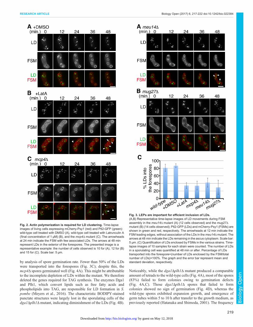
LEPs facilitate efficient inclusion of LDs by the FSMWe next examined whether the LEPs play a role in LD movementThe meiotic actin ring was dissembled by treating the sporulatingcells with the actin polymerization inhibitor Latrunculin A Whilemost LDs clustered at the FSM initiation sites in the control cells(Fig 2A 0 min) the LDs in the Latrunculin A-treated cellsremained scattered upon initiation of FSM assembly (Fig 2B0 min) suggesting that actin polymerization is required for LDclustering at the FSM assembly site Furthermore the FSM leadingedge in the Latrunculin A-treated cells was associated with few orno LDs (Fig 2B 24 min arrowheads) resulting in inefficientinclusion of LDs by FSMs in these cells (Fig 2B 48 min arrows)As in the Latrunculin A-treated cells LDs failed to cluster well at theFSM initiation site and numerous LDs were excluded from thespore cytoplasm in the mcp4Δ mutant (Fig 2C 48 min arrows)The similarity in the phenotype of Latrunculin A-treated cells andmcp4Δ cells is consistent with a previous study reporting that Mcp4is involved in F-actin positioning (Ohtaka et al 2007)
Meanwhile depletion of theMeu14 ring had little effect on initialclustering of LDs (Fig 3A 0 min) In contrast with the wild-typecells (Fig 1A 12 min arrowheads) the meu14Δ mutant exhibitedpoor association of LD clusters with the FSM leading edges(Fig 3A 12 min arrowheads) As a result LDs were not enclosedby the FSM instead remaining in the ascus cytoplasm in themeu14Δ mutant (Fig 3A 48 min arrows)
We propose that LD transport into spores involves two steps firstactin polymerization is required for LD clustering at the FSMassembly site second the LEP rings facilitate efficient inclusion ofLDs by the FSM A previous study demonstrated that Mug27regulates constriction of the meiotic actin rings without affectingtheir assembly (Yan and Balasubramanian 2012) Accordingly weexamined LDmovements in themug27Δmutant In agreement withour hypothesis initial clustering of LDs was normal in the mug27Δmutant (Fig 3B 0 min) however most of the LDs were stillexcluded from the forespores (Fig 3B 48 min arrows) Thereforefewer LDs were enclosed by FSMs in the LEP-disruption mutantsthan in the wild-type cells (Fig 3C) indicating that LEP ringsmediate LD transport to the forespores
LDs are required for spore germination and spore wallintegrityIn addition to enclosing LDs with low efficiency relative to the wildtype the FSMs of meu14Δ and mug27Δ mutants exhibit abnormalformation and frequently fail to engulf the spore nucleus (Ohtakaet al 2008 Okuzaki et al 2003) making it complex to verify therequirement of LDs for spore survival By contrast in the presentstudy the mcp4Δ mutant formed four spores per ascus (tetrads) asfrequently as wild-type cells (Fig 4A) We assayed spore survival
Fig 1 Dynamics of LDs during spore formation (A) Representative time-lapse images of the FSMs engulfing the LDs observed in living S pombe cellsexpressing the FSM marker mCherry-Psy1 and the LD marker Ptl2-GFP(11 cells observed) FSM and LDs are shown in red and green respectivelyLDs appeared as distinct focal structures FSM initiation (detected asaggregation of the mCherry-Psy1 fluorescence signals in the cytoplasm) wasdesignated as 0 min The arrows at 0 min indicate clustering of LDs near theFSM initiation site The arrowheads at 12 min indicate four LD clusters near theFSM leading edges The arrowheads at 24 min indicate inclusion of the LDs byFSM extension Scale bar 5 microm (B) Fluorescence images of FSM extensionCells expressing mCherry-Psy1 were fixed for EM imaging (see Materials andMethods) Thenumbers 1ndash4 represent the four FSMs Scale bar 2 microm (C) TEMimage of the same cell shown in (B) Scale bar 2 microm (D) Magnified TEMimages of the cell depicted in (C) Each of the numbered images corresponds tothe numbered FSMs in (B) The red arrowheads indicate the FSM The cyanarrows indicate LDs which appear as white matter when visualized by TEM Nnucleus M mitochondria Scale bar 500 nm (E) Localization of the LEP ringsat the FSM leading edge N nucleus (F-H) Co-localization of LDs and LEPsImmediately before imaging the fluorescent dye BODIPY493503 or BODIPYTR was added to the sporulation medium containing cells expressing Mcp4-mCherry Meu14-GFP or LifeAct-GFP The BODIPY dyes stain LDs LifeAct-GFP binds to actin filaments to allow visualization of the meiotic actin ring (Yanand Balasubramanian 2012) Scale bar 5 microm
218
RESEARCH ARTICLE Biology Open (2017) 6 217-222 doi101242bio022384
BiologyOpen
by guest on May 12 2018httpbiobiologistsorgDownloaded from
by analysis of spore germination rate Fewer than 50 of the LDswere transported into the forespores (Fig 3C) despite this themcp4Δ spores germinated well (Fig 4A) This might be attributableto the incomplete depletion of LDs within the mutant We thereforedeleted the genes required for TAG synthesis The enzymes Dga1and Plh1 which convert lipids such as free fatty acids andphospholipids into TAG are responsible for LD formation in Spombe (Meyers et al 2016) The characteristic BODIPY-stainedpunctate structures were largely lost in the sporulating cells of thedga1Δplh1Δmutant indicating diminishment of the LDs (Fig 4B)
Noticeably while the dga1Δplh1Δ mutant produced a comparableamount of tetrads to the wild-type cells (Fig 4A) most of the spores(83) failed to form colonies owing to germination defects(Fig 4AC) Those dga1Δplh1Δ spores that failed to formcolonies showed no sign of germination (Fig 4D) whereas thewild-type spores exhibited expansion growth and emergence ofgerm tubes within 5 to 10 h after transfer to the growth medium aspreviously reported (Hatanaka and Shimoda 2001) The frequency
Fig 3 LEPs are important for efficient inclusion of LDs(AB) Representative time-lapse images of LD movements during FSMassembly in the meu14Δ mutant (A) (12 cells observed) and the mug27Δmutant (B) (14 cells observed) Ptl2-GFP (LDs) andmCherry-Psy1 (FSMs) areshown in green and red respectively The arrowheads at 12 min indicate theFSM leading edges without association of the LDs in themeu14Δmutant Thearrows at 48 min indicate the LDs remaining in the ascus cytoplasm Scale bar5 microm (C) Quantification of LDs enclosed by FSMs in the various strains Time-lapse images of 10 samples for each strain were counted The number of LDsin a sporulating cell was quantified at 48 min or after Percentage of LDstransported into the forespore=(number of LDs enclosed by the FSMtotalnumber of LDs)times100 The graph and the error bar represent mean andstandard deviation respectively
Fig 2 Actin polymerization is required for LD clustering Time-lapseimages of living cells expressing mCherry-Psy1 (red) and Ptl2-GFP (green)wild-type cell treated with DMSO (A) wild-type cell treated with Latrunculin A(final concentration of 1 microM) (B) and the mcp4Δ mutant (C) The arrowheadsat 24 min indicate the FSM with few associated LDs The arrows at 48 minrepresent LDs in the exterior of the forespores The presented image is arepresentative example the number of cells observed is 10 for (A) 12 for (B)and 15 for (C) Scale bar 5 microm
219
RESEARCH ARTICLE Biology Open (2017) 6 217-222 doi101242bio022384
BiologyOpen
by guest on May 12 2018httpbiobiologistsorgDownloaded from
of spore germination was only 17 (Fig 4A) intriguinglyhowever the frequency of tetrads containing four viable sporeswas strikingly higher (5 2 out of 42 asci) than that predicted byrandom distribution of viable spores in an ascus (0080174=00008) This result of non-random distribution indicatesthat spores in each ascus share the same fate Thus it is likely that
the viability of spores in the absence of TAG synthesis ismetabolically determined during meiosis and sporulationAlthough the viability of spores produced by the dga1Δplh1Δmutant largely decreased during sporulation it further decreasedgradually when maintained in the absence of TAG synthesis(Fig 4E) In contrast the spores of wild-type cells retained high
Fig 4 LDs are important for spore germination and spore wall integrity (A) Frequency of tetrad formation and spore germination in the different strainsTetrad formation frequency=(number of asci with four sporesnumber of total zygotes)times100 At least 600 tetrads were scored for each strain After 3-5 days ofsporulation the rate of spore germination of each strain was determined by assessing spore colony formation through tetrad analysis (42 tetrads were dissectedfor each strain) Spores that failed to form colonies were further confirmed for germination using a dissecting microscope Germination frequency=(number ofgerminated sporesnumber of total spores)times100 A lys1+-integrating plasmid carrying dga1+ or plh1+ was used to restore the germination efficiency of thedga1Δplh1Δmutant (B) The dga1Δplh1Δmutant possessed few LDs The fluorescent dye BODIPY was used for LD labeling The white dashed line outlines thesporulating cell Scale bar 5 microm (C) Examples of spore colony formation in the dga1Δplh1Δ mutant (D) Representative morphological changes of sporegermination over time in the wild-type or the dga1Δplh1Δ spore Scale bar 6 microm (E) The germination defect of the dga1Δplh1Δ spores was time-dependent Thecells were subjected to sporulation on anME plate for 2 4 8 or 16 days At each time point spore germination was assayed by tetrad dissection on the YES plate(three independent experiments per strain 14 tetrads were dissected per experiment) Germination frequency=(number of germinated sporesnumber of totalspores)times100 The graph and the error bar represent mean and standard deviation respectively (F) The spore wall was improperly assembled in thedga1Δplh1Δmutant Isp3-GFP was used to visualize the outermost layer of the spore wall Isp3-GFP fluorescence signals were evenly distributed on the surfaceof the wild-type ascospores whereas Isp3-GFP exhibited aggregate formation and uneven decoration of the ascospores of the dga1Δplh1Δ mutantScale bar 5 microm
220
RESEARCH ARTICLE Biology Open (2017) 6 217-222 doi101242bio022384
BiologyOpen
by guest on May 12 2018httpbiobiologistsorgDownloaded from
viability in sporulation medium for 16 days (Fig 4E) These resultsindicate that TAG plays a necessary role in spore survival understarvation conditionsThe LD-deficient mutant not only exhibits defects in spore
germination but also in spore wall integrity (Fig 4F) Spore walldeposition after FSM assembly confers resistance to spores againstvarious stresses (Coluccio et al 2008 Fukunishi et al 2014) Theoutermost layer of the S pombe spore wall comprises a protein layercomposed of Isp3 which is highly palmitoylated (Fukunishi et al2014 Zhang et al 2013) This Isp3 coating was defective in thespores of the dga1Δplh1Δ strain (Fig 4F) raising the possibility thatTAGs mediate the characterized lipid-modification of Isp3 Theseresults indicate that LDs are important for spore germination andspore wall integrityLDs are crucial for the survival of starved cells (Rambold et al
2015 Shpilka et al 2015) Our study revealed that LDs are activelytransported to nascent spores and that dga1Δplh1Δ spores bearingfew LDs barely germinate These data indicate that LDs representan important cellular energy source for spores under starvationconditions Alternatively apoptosis may be induced in dga1Δplh1Δspores as a result of their failure to transform diacylglycerol intoTAG (Zhang et al 2003) Further studies will clarify themechanisms by which LDs support spore survival
MATERIALS AND METHODSYeast strains and cultureThe S pombe strains used in this study are listed in Table S1 All strains weregrown on yeast extract with supplements (YES) plates at 30degC as describedby Moreno et al (1991) To induce sporulation freshly cultured cells werecollected in nitrogen-free Edinburgh minimal medium (Moreno et al 1991)supplemented with adenine uracil histidine lysine and leucine (EMM-N+5S) at a density of 109 cellsml Cells were then transferred to malt extract(ME) plates to allow sporulation at 26degC
Gene disruption was performed using a polymerase chain reaction (PCR)-based strategy (Baumlhler et al 1998) The PCR primers used in these analysesare listed in Table S2 For deletion of themeu14+ gene DNA fragments withhomology to the target gene locus were amplified using the primersHJO423 HJO424 HJO425 and HJO426 whereas DNA fragments for thedeletion of the dga1+ gene were amplified using the primers HJO684HJO685 HJO686 and HJO687 The plh1+ gene was replaced with the drugresistance gene module kanMX6 using the plasmid pFA6a-kanMX6 andprimers HJO689 HJO690 HJO691 and HJO692 The mcp4Δ mug27Δand spo13Δ strains were derived from strains FY16412 FY17842 andFY12290 respectively (obtained from the Yeast Genetic Resource Center ofJapan) (Nakase et al 2008 Ohtaka et al 2007 2008)
To fluorescently label Ptl2 Isp3 or Mcp4 a two-step PCR methodintroducing the chromosomal GFP or mCherry tag was used (Hayashi et al2009) To visualize the FSM integrating plasmids carrying mCherry-psy1+
were introduced into the cells as described in Chikashige et al (2006) GFP-taggedMeu14 or LifeAct was expressed from the lys1+-integrating plasmid
Live-cell imaging of sporulating cellsAfter overnight incubation on ME plates cells were re-suspended in EMM-N+5Smedium To disperse sporulating cells suspensions were subjected bybrief sonication (Handy Sonic Tomy Seiko Tokyo Japan) 20 microl of the cellsuspension was then dropped onto lectin (02 mgml Sigma-AldrichTokyo Japan)-coated 35-mm glass-bottomed culture dishes (MatTekAshland MA USA) to immobilize cells (Asakawa and Hiraoka 2009) Forimaging Latrunculin A-treated cells Latrunculin A (Thermo FisherScientific Tokyo Japan) was added at a final concentration of 1 microM priorto cell immobilization Cells undergoing sporulation were selected for live-cell imaging
A DeltaVision microscope equipped with a CoolSNAP HQ2 charge-coupled device (GE Healthcare Tokyo Japan) was used for imageacquisition Optical section images were acquired at 05-microm focus intervals
using an oil-immersion 60times objective lens (PlanApoN60x OSC NA14Olympus Tokyo Japan) Images were processed using the de-noisingalgorithm (Boulanger et al 2009) and by constrained iterativedeconvolution (Agard et al 1989)
EM imagingCells were induced to sporulate on ME plates overnight and aliquoted inmonolayers on lectin-coated glass-bottomed culture dishes with addressinggrids (grid size 50 microm ibid Bremen Germany) Cells were fixed with 2glutaraldehyde (Polysciences Inc Warrington PA USA) in 01 Mphosphate buffer (pH 72) for 2 h at 4degC Optical section images (02-micromintervals) of a cell of interest were obtained using the Olympus objectivelens as described above EM observation was performed as describedpreviously (Asakawa et al 2010) Briefly cells were post-fixed with a 12KMnO4 solution overnight at 4degC and embedded in Epon812 The epoxyblock containing the same cells observed by fluorescence microscopy wastrimmed according to the location on the coverslip Serial sections with80-nm thickness were stained with 4 uranyl acetate and a commercialready-to-use solution of lead citrate (Sigma-Aldrich St Louis MO USA)and analyzed using a JEM1400 transmission electron microscope (JEOLTokyo Japan) Adobe Photoshop CS4 (ver1101) was used for imageprocessing
AcknowledgementsWe thank Aaron Neiman (Stony Brook University) for his valuable discussions andthe Yeast Genetic Resource Center of Japan for providing yeast strains
Competing interestsThe authors declare no competing or financial interests
Author contributionsH-JY TH and YH conceived designed and interpreted experiments H-JYHO and TK performed the experiments and analyzed the data H-JY TH andYH wrote the manuscript which was approved by all authors
FundingThis work was supported by Japan Society for the Promotion of Science (JSPS)KAKENHI grant numbers JP13F03384 to H-JY JP25116006 to TH andJP26116511 JP16H01309 JP26251037 to YH
Supplementary informationSupplementary information available online athttpbiobiologistsorglookupdoi101242bio022384supplemental
ReferencesAgard D A Hiraoka Y Shaw P and Sedat J W (1989) Fluorescence
microscopy in three dimensions Methods Cell Biol 30 353-377Asakawa H and Hiraoka Y (2009) Live-cell fluorescence imaging of meiotic
chromosome dynamics in Schizosaccharomyces pombeMethodsMol Biol 55853-64
Asakawa H Kojidani T Mori C Osakada H Sato M Ding D-Q HiraokaY and Haraguchi T (2010) Virtual breakdown of the nuclear envelope in fissionyeast meiosis Curr Biol 20 1919-1925
Bahler J Wu J-Q Longtine M S Shah N G McKenzie A III SteeverA B Wach A Philippsen P and Pringle J R (1998) Heterologous modulesfor efficient and versatile PCR-based gene targeting in Schizosaccharomycespombe Yeast 14 943-951
Blom T Somerharju P and Ikonen E (2011) Synthesis and biosynthetictrafficking of membrane lipids Cold Spring Harb Perspect Biol 3 a004713
Boulanger J Kervrann C andBouthemy P (2009) A simulation and estimationframework for intracellular dynamics and trafficking in video-microscopy andfluorescence imagery Med Image Anal 13 132-142
Chikashige Y Tsutsumi C Yamane M Okamasa K Haraguchi T andHiraoka Y (2006) Meiotic proteins Bqt1 and Bqt2 tether telomeres to form thebouquet arrangement of chromosomes Cell 125 59-69
Coluccio A E Rodriguez R K Kernan M J and Neiman A M (2008) Theyeast spore wall enables spores to survive passage through the digestive tract ofDrosophila PLoS ONE 3 e2873
Diamond A E Park J-S Inoue I Tachikawa H and Neiman A M (2008)The anaphase promoting complex targeting subunit Ama1 links meiotic exit tocytokinesis during sporulation in Saccharomyces cerevisiae Mol Biol Cell 20134-145
221
RESEARCH ARTICLE Biology Open (2017) 6 217-222 doi101242bio022384
BiologyOpen
by guest on May 12 2018httpbiobiologistsorgDownloaded from
Dichlberger A Kovanen P T and Schneider W J (2013) Mast cells from lipiddroplets to lipid mediators Clin Sci 125 121-130
Fan Y Ortiz-Urquiza A Garrett T Pei Y and Keyhani N O (2015)Involvement of a caleosin in lipid storage spore dispersal and virulence in theentomopathogenic filamentous fungus Beauveria bassiana Environ Microbiol17 4600-4614
Fukunishi K Miyakubi K Hatanaka M Otsuru N Hirata A Shimoda Cand Nakamura T (2014) The fission yeast spore is coated by a proteinaceoussurface layer comprising mainly Isp3 Mol Biol Cell 25 1549-1559
Gao Q and Goodman J M (2014) The lipid droplet-a well-connected organelleFront Cell Dev Biol 3 49
Goyal A Takaine M Simanis V and Nakano K (2011) Dividing the spoils ofgrowth and the cell cycle The fission yeast as amodel for the study of cytokinesisCytoskeleton (Hoboken) 68 69-88
Hatanaka M and Shimoda C (2001) The cyclic AMPPKA signal pathway isrequired for initiation of spore germination inSchizosaccharomyces pombeYeast18 207-217
Hayashi A Ding D-Q Tsutsumi C Chikashige Y Masuda H HaraguchiT and Hiraoka Y (2009) Localization of gene products using a chromosomallytagged GFP-fusion library in the fission yeast Schizosaccharomyces pombeGenes Cells 14 217-225
Ikemoto S Nakamura T Kubo M and Shimoda C (2000) S pombesporulation-specific coiled-coil protein Spo15p is localized to the spindle polebody and essential for its modification J Cell Sci 113 545-554
Kashiwazaki J Yamasaki Y Itadani A Teraguchi E Maeda Y ShimodaC and Nakamura T (2011) Endocytosis is essential for dynamic translocationof a syntaxin 1 orthologue during fission yeast meiosis Mol Biol Cell 223658-3670
Krapp A Collin P Cokoja A Dischinger S Cano E and Simanis V (2006)The Schizosaccharomyces pombe septation initiation network (SIN) is requiredfor spore formation in meiosis J Cell Sci 119 2882-2891
Lin C P-C Kim C Smith S O and Neiman A M (2013) A highly redundantgene network controls assembly of the outer spore wall in S cerevisiae PLoSGenet 9 e1003700
Maeda Y Kashiwazaki J Shimoda C and Nakamura T (2009) TheSchizosaccharomyces pombe syntaxin 1 homolog Psy1 is essential in thedevelopment of the forespore membrane Biosci Biotechnol Biochem 73339-345
Meyers A del Rio Z P Beaver R A Morris R M Weiskittel T M AlshibliA K Mannik J Morrell-Falvey J and Dalhaimer P (2016) Lipid dropletsform from distinct regions of the cell in the fission yeast Schizosaccharomycespombe Traffic 17 657-669
Moreno-Borchart A C Strasser K Finkbeiner M G Shevchenko AShevchenko A and Knop M (2001) Prospore membrane formation linked tothe leading edge protein (LEP) coat assembly EMBO J 20 6946-6957
Moreno S Klar A and Nurse P (1991) Molecular genetic analysis of fissionyeast Schizosaccharomyces pombe Methods Enzymol 194 795-823
Nakamura T Nakamura-Kubo M Hirata A and Shimoda C (2001) TheSchizosaccharomyces pombe spo3+ gene is required for assembly of theforespore membrane and genetically interacts with psy1(+)-encoding syntax in-like protein Mol Biol Cell 12 3955-3972
Nakamura T Kashiwazaki J and Shimoda C (2005) A fission yeast SNAP-25homologue SpSec9 is essential for cytokinesis and sporulation Cell StructFunct 30 15-24
Nakamura T Asakawa H Nakase Y Kashiwazaki J Hiraoka Y andShimoda C (2008) Live observation of forespore membrane formation in fissionyeast Mol Biol Cell 19 3544-3553
Nakase Y Nakamura-Kubo M Ye Y Hirata A Shimoda C and NakamuraT (2008) Meiotic spindle pole bodies acquire the ability to assemble the sporeplasma membrane by sequential recruitment of sporulation-specific componentsin fission yeast Mol Biol Cell 19 2476-2487
Neiman A M (1998) Prospore membrane formation defines a developmentallyregulated branch of the secretory pathway in yeast J Cell Biol 140 29-37
Neiman A M (2011) Sporulation in the budding yeast Saccharomyces cerevisiaeGenetics 189 737-765
Ohtaka A Okuzaki D Saito T T andNojima H (2007) Mcp4 ameiotic coiled-coil protein plays a role in F-actin positioning during Schizosaccharomycespombe meiosis Eukaryot Cell 6 971-983
Ohtaka A Okuzaki D andNojima H (2008) Mug27 is ameiosis-specific proteinkinase that functions in fission yeast meiosis II and sporulation J Cell Sci 1211547-1558
Okuzaki D SatakeW Hirata A andNojima H (2003) Fission yeastmeu14+ isrequired for proper nuclear division and accurate forespore membrane formationduring meiosis II J Cell Sci 116 2721-2735
Perez-Hidalgo L Rozalen A E Martin-Castellanos C andMoreno S (2008)Slk1 is a meiosis-specific Sid2-related kinase that coordinates meiotic nucleardivision with growth of the forespore membrane J Cell Sci 121 1383-1392
Pol A Gross S P and Parton R G (2014) Biogenesis of the multifunctionallipid droplet lipids proteins and sites J Cell Biol 204 635-646
Rambold A S Cohen S and Lippincott-Schwartz J (2015) Fatty acidtrafficking in starved cells regulation by lipid droplet lipolysis autophagy andmitochondrial fusion dynamics Dev Cell 32 678-692
Ren J Lin C P-C Pathak M C Temple B R S Nile A H Mousley C JDuncan M C Eckert D M Leiker T J Ivanova P T et al (2014) Aphosphatidylinositol transfer protein integrates phosphoinositide signaling withlipid droplet metabolism to regulate a developmental program of nutrient stress-induced membrane biogenesis Mol Biol Cell 25 712-727
Rhind N and Russell P (2012) Signaling pathways that regulate cell divisionCold Spring Harb Perspect Biol 4 a005942
Shpilka T Welter E Borovsky N Amar N Mari M Reggiori F and ElazarZ (2015) Lipid droplets and their component triglycerides and steryl estersregulate autophagosome biogenesis EMBO J 34 2117-2131
Thiam A R Farese R V Jr and Walther T C (2013) The biophysics and cellbiology of lipid droplets Nat Rev Mol Cell Biol 14 775-786
Yan H and Balasubramanian M K (2012) Meiotic actin rings are essential forproper sporulation in fission yeast J Cell Sci 125 1429-1439
Yan H Ge W Chew T G Chow J Y McCollum D Neiman A M andBalasubramanian M K (2008) The meiosis-specific Sid2p-related proteinSlk1p regulates forespore membrane assembly in fission yeastMol Biol Cell 193676-3690
Yang H-J and Neiman A M (2010) A guaninine nucleotide exchange factor is acomponent of the meiotic spindle pole body in Schizosaccharomyces pombeMol Biol Cell 21 1272-1281
Yang H-J Nakanishi H Liu S McNew J A and Neiman A M (2008)Binding interactions control SNARE specificity in vivo J Cell Biol 1831089-1100
Yazawa H Kumagai H and Uemura H (2012) Characterization of triglyceridelipase genes of fission yeast Schizosaccharomyces pombe Appl MicrobiolBiotechnol 96 981-991
Zhang Q Chieu H K Low C P Zhang S Heng C K and Yang H (2003)Schizosaccharomyces pombe cells deficient in triacylglycerols synthesis undergoapoptosis upon entry into the stationary phase J Biol Chem 278 47145-47155
Zhang M M Wu P-Y J Kelly F D Nurse P and Hang H C (2013)Quantitative control of protein S-palmitoylation regulates meiotic entry in fissionyeast PLoS Biol 11 e1001597-e1001597
222
RESEARCH ARTICLE Biology Open (2017) 6 217-222 doi101242bio022384
BiologyOpen
by guest on May 12 2018httpbiobiologistsorgDownloaded from

(Fig 1A arrows) However in the spo13Δ mutant LDs clusteredefficiently at the nucleus without FSM assembly (Fig S3) indicatingthat LD clustering occurs independent of FSM assembly As the FSMgrew into a crescent-shaped structure in anaphase II the LD clustersfurther partitioned into each of the four FSMs (Fig 1A 12ndash24 minarrowheads) Continuous extension of the FSM eventually enclosedthe LDs within the forespore (Fig 1A 24ndash60 min)
LDs were found in close proximity to the leading edges of theFSM during FSM extension (Fig 1A 12 min arrowheads)Consistently when the cell undergoing FSM extension wasfurther subjected to electron microscopy (EM) (Fig 1BC) LDswere often observed near the leading edge of each FSM (Fig 1Darrows) FSM leading edges are decorated by the three LEP ringsthe Meu14 ring located at the ascus cytoplasmic side of the FSMleading edge the Mcp4 ring at the future spore cytoplasmic sideand the meiotic actin ring situated between the Meu14 ring and theMcp4 ring (Fig 1E) (Ohtaka et al 2007) Co-localization analysisrevealed that LDs closely associate with the Mcp4 ring but localizebehind the meiotic actin ring and the Meu14 ring (Fig 1FndashH)indicating that the LDs were located at the future spore cytoplasm
LEPs facilitate efficient inclusion of LDs by the FSMWe next examined whether the LEPs play a role in LD movementThe meiotic actin ring was dissembled by treating the sporulatingcells with the actin polymerization inhibitor Latrunculin A Whilemost LDs clustered at the FSM initiation sites in the control cells(Fig 2A 0 min) the LDs in the Latrunculin A-treated cellsremained scattered upon initiation of FSM assembly (Fig 2B0 min) suggesting that actin polymerization is required for LDclustering at the FSM assembly site Furthermore the FSM leadingedge in the Latrunculin A-treated cells was associated with few orno LDs (Fig 2B 24 min arrowheads) resulting in inefficientinclusion of LDs by FSMs in these cells (Fig 2B 48 min arrows)As in the Latrunculin A-treated cells LDs failed to cluster well at theFSM initiation site and numerous LDs were excluded from thespore cytoplasm in the mcp4Δ mutant (Fig 2C 48 min arrows)The similarity in the phenotype of Latrunculin A-treated cells andmcp4Δ cells is consistent with a previous study reporting that Mcp4is involved in F-actin positioning (Ohtaka et al 2007)
Meanwhile depletion of theMeu14 ring had little effect on initialclustering of LDs (Fig 3A 0 min) In contrast with the wild-typecells (Fig 1A 12 min arrowheads) the meu14Δ mutant exhibitedpoor association of LD clusters with the FSM leading edges(Fig 3A 12 min arrowheads) As a result LDs were not enclosedby the FSM instead remaining in the ascus cytoplasm in themeu14Δ mutant (Fig 3A 48 min arrows)
We propose that LD transport into spores involves two steps firstactin polymerization is required for LD clustering at the FSMassembly site second the LEP rings facilitate efficient inclusion ofLDs by the FSM A previous study demonstrated that Mug27regulates constriction of the meiotic actin rings without affectingtheir assembly (Yan and Balasubramanian 2012) Accordingly weexamined LDmovements in themug27Δmutant In agreement withour hypothesis initial clustering of LDs was normal in the mug27Δmutant (Fig 3B 0 min) however most of the LDs were stillexcluded from the forespores (Fig 3B 48 min arrows) Thereforefewer LDs were enclosed by FSMs in the LEP-disruption mutantsthan in the wild-type cells (Fig 3C) indicating that LEP ringsmediate LD transport to the forespores
LDs are required for spore germination and spore wallintegrityIn addition to enclosing LDs with low efficiency relative to the wildtype the FSMs of meu14Δ and mug27Δ mutants exhibit abnormalformation and frequently fail to engulf the spore nucleus (Ohtakaet al 2008 Okuzaki et al 2003) making it complex to verify therequirement of LDs for spore survival By contrast in the presentstudy the mcp4Δ mutant formed four spores per ascus (tetrads) asfrequently as wild-type cells (Fig 4A) We assayed spore survival
Fig 1 Dynamics of LDs during spore formation (A) Representative time-lapse images of the FSMs engulfing the LDs observed in living S pombe cellsexpressing the FSM marker mCherry-Psy1 and the LD marker Ptl2-GFP(11 cells observed) FSM and LDs are shown in red and green respectivelyLDs appeared as distinct focal structures FSM initiation (detected asaggregation of the mCherry-Psy1 fluorescence signals in the cytoplasm) wasdesignated as 0 min The arrows at 0 min indicate clustering of LDs near theFSM initiation site The arrowheads at 12 min indicate four LD clusters near theFSM leading edges The arrowheads at 24 min indicate inclusion of the LDs byFSM extension Scale bar 5 microm (B) Fluorescence images of FSM extensionCells expressing mCherry-Psy1 were fixed for EM imaging (see Materials andMethods) Thenumbers 1ndash4 represent the four FSMs Scale bar 2 microm (C) TEMimage of the same cell shown in (B) Scale bar 2 microm (D) Magnified TEMimages of the cell depicted in (C) Each of the numbered images corresponds tothe numbered FSMs in (B) The red arrowheads indicate the FSM The cyanarrows indicate LDs which appear as white matter when visualized by TEM Nnucleus M mitochondria Scale bar 500 nm (E) Localization of the LEP ringsat the FSM leading edge N nucleus (F-H) Co-localization of LDs and LEPsImmediately before imaging the fluorescent dye BODIPY493503 or BODIPYTR was added to the sporulation medium containing cells expressing Mcp4-mCherry Meu14-GFP or LifeAct-GFP The BODIPY dyes stain LDs LifeAct-GFP binds to actin filaments to allow visualization of the meiotic actin ring (Yanand Balasubramanian 2012) Scale bar 5 microm
218
RESEARCH ARTICLE Biology Open (2017) 6 217-222 doi101242bio022384
BiologyOpen
by guest on May 12 2018httpbiobiologistsorgDownloaded from
by analysis of spore germination rate Fewer than 50 of the LDswere transported into the forespores (Fig 3C) despite this themcp4Δ spores germinated well (Fig 4A) This might be attributableto the incomplete depletion of LDs within the mutant We thereforedeleted the genes required for TAG synthesis The enzymes Dga1and Plh1 which convert lipids such as free fatty acids andphospholipids into TAG are responsible for LD formation in Spombe (Meyers et al 2016) The characteristic BODIPY-stainedpunctate structures were largely lost in the sporulating cells of thedga1Δplh1Δmutant indicating diminishment of the LDs (Fig 4B)
Noticeably while the dga1Δplh1Δ mutant produced a comparableamount of tetrads to the wild-type cells (Fig 4A) most of the spores(83) failed to form colonies owing to germination defects(Fig 4AC) Those dga1Δplh1Δ spores that failed to formcolonies showed no sign of germination (Fig 4D) whereas thewild-type spores exhibited expansion growth and emergence ofgerm tubes within 5 to 10 h after transfer to the growth medium aspreviously reported (Hatanaka and Shimoda 2001) The frequency
Fig 3 LEPs are important for efficient inclusion of LDs(AB) Representative time-lapse images of LD movements during FSMassembly in the meu14Δ mutant (A) (12 cells observed) and the mug27Δmutant (B) (14 cells observed) Ptl2-GFP (LDs) andmCherry-Psy1 (FSMs) areshown in green and red respectively The arrowheads at 12 min indicate theFSM leading edges without association of the LDs in themeu14Δmutant Thearrows at 48 min indicate the LDs remaining in the ascus cytoplasm Scale bar5 microm (C) Quantification of LDs enclosed by FSMs in the various strains Time-lapse images of 10 samples for each strain were counted The number of LDsin a sporulating cell was quantified at 48 min or after Percentage of LDstransported into the forespore=(number of LDs enclosed by the FSMtotalnumber of LDs)times100 The graph and the error bar represent mean andstandard deviation respectively
Fig 2 Actin polymerization is required for LD clustering Time-lapseimages of living cells expressing mCherry-Psy1 (red) and Ptl2-GFP (green)wild-type cell treated with DMSO (A) wild-type cell treated with Latrunculin A(final concentration of 1 microM) (B) and the mcp4Δ mutant (C) The arrowheadsat 24 min indicate the FSM with few associated LDs The arrows at 48 minrepresent LDs in the exterior of the forespores The presented image is arepresentative example the number of cells observed is 10 for (A) 12 for (B)and 15 for (C) Scale bar 5 microm
219
RESEARCH ARTICLE Biology Open (2017) 6 217-222 doi101242bio022384
BiologyOpen
by guest on May 12 2018httpbiobiologistsorgDownloaded from
of spore germination was only 17 (Fig 4A) intriguinglyhowever the frequency of tetrads containing four viable sporeswas strikingly higher (5 2 out of 42 asci) than that predicted byrandom distribution of viable spores in an ascus (0080174=00008) This result of non-random distribution indicatesthat spores in each ascus share the same fate Thus it is likely that
the viability of spores in the absence of TAG synthesis ismetabolically determined during meiosis and sporulationAlthough the viability of spores produced by the dga1Δplh1Δmutant largely decreased during sporulation it further decreasedgradually when maintained in the absence of TAG synthesis(Fig 4E) In contrast the spores of wild-type cells retained high
Fig 4 LDs are important for spore germination and spore wall integrity (A) Frequency of tetrad formation and spore germination in the different strainsTetrad formation frequency=(number of asci with four sporesnumber of total zygotes)times100 At least 600 tetrads were scored for each strain After 3-5 days ofsporulation the rate of spore germination of each strain was determined by assessing spore colony formation through tetrad analysis (42 tetrads were dissectedfor each strain) Spores that failed to form colonies were further confirmed for germination using a dissecting microscope Germination frequency=(number ofgerminated sporesnumber of total spores)times100 A lys1+-integrating plasmid carrying dga1+ or plh1+ was used to restore the germination efficiency of thedga1Δplh1Δmutant (B) The dga1Δplh1Δmutant possessed few LDs The fluorescent dye BODIPY was used for LD labeling The white dashed line outlines thesporulating cell Scale bar 5 microm (C) Examples of spore colony formation in the dga1Δplh1Δ mutant (D) Representative morphological changes of sporegermination over time in the wild-type or the dga1Δplh1Δ spore Scale bar 6 microm (E) The germination defect of the dga1Δplh1Δ spores was time-dependent Thecells were subjected to sporulation on anME plate for 2 4 8 or 16 days At each time point spore germination was assayed by tetrad dissection on the YES plate(three independent experiments per strain 14 tetrads were dissected per experiment) Germination frequency=(number of germinated sporesnumber of totalspores)times100 The graph and the error bar represent mean and standard deviation respectively (F) The spore wall was improperly assembled in thedga1Δplh1Δmutant Isp3-GFP was used to visualize the outermost layer of the spore wall Isp3-GFP fluorescence signals were evenly distributed on the surfaceof the wild-type ascospores whereas Isp3-GFP exhibited aggregate formation and uneven decoration of the ascospores of the dga1Δplh1Δ mutantScale bar 5 microm
220
RESEARCH ARTICLE Biology Open (2017) 6 217-222 doi101242bio022384
BiologyOpen
by guest on May 12 2018httpbiobiologistsorgDownloaded from
viability in sporulation medium for 16 days (Fig 4E) These resultsindicate that TAG plays a necessary role in spore survival understarvation conditionsThe LD-deficient mutant not only exhibits defects in spore
germination but also in spore wall integrity (Fig 4F) Spore walldeposition after FSM assembly confers resistance to spores againstvarious stresses (Coluccio et al 2008 Fukunishi et al 2014) Theoutermost layer of the S pombe spore wall comprises a protein layercomposed of Isp3 which is highly palmitoylated (Fukunishi et al2014 Zhang et al 2013) This Isp3 coating was defective in thespores of the dga1Δplh1Δ strain (Fig 4F) raising the possibility thatTAGs mediate the characterized lipid-modification of Isp3 Theseresults indicate that LDs are important for spore germination andspore wall integrityLDs are crucial for the survival of starved cells (Rambold et al
2015 Shpilka et al 2015) Our study revealed that LDs are activelytransported to nascent spores and that dga1Δplh1Δ spores bearingfew LDs barely germinate These data indicate that LDs representan important cellular energy source for spores under starvationconditions Alternatively apoptosis may be induced in dga1Δplh1Δspores as a result of their failure to transform diacylglycerol intoTAG (Zhang et al 2003) Further studies will clarify themechanisms by which LDs support spore survival
MATERIALS AND METHODSYeast strains and cultureThe S pombe strains used in this study are listed in Table S1 All strains weregrown on yeast extract with supplements (YES) plates at 30degC as describedby Moreno et al (1991) To induce sporulation freshly cultured cells werecollected in nitrogen-free Edinburgh minimal medium (Moreno et al 1991)supplemented with adenine uracil histidine lysine and leucine (EMM-N+5S) at a density of 109 cellsml Cells were then transferred to malt extract(ME) plates to allow sporulation at 26degC
Gene disruption was performed using a polymerase chain reaction (PCR)-based strategy (Baumlhler et al 1998) The PCR primers used in these analysesare listed in Table S2 For deletion of themeu14+ gene DNA fragments withhomology to the target gene locus were amplified using the primersHJO423 HJO424 HJO425 and HJO426 whereas DNA fragments for thedeletion of the dga1+ gene were amplified using the primers HJO684HJO685 HJO686 and HJO687 The plh1+ gene was replaced with the drugresistance gene module kanMX6 using the plasmid pFA6a-kanMX6 andprimers HJO689 HJO690 HJO691 and HJO692 The mcp4Δ mug27Δand spo13Δ strains were derived from strains FY16412 FY17842 andFY12290 respectively (obtained from the Yeast Genetic Resource Center ofJapan) (Nakase et al 2008 Ohtaka et al 2007 2008)
To fluorescently label Ptl2 Isp3 or Mcp4 a two-step PCR methodintroducing the chromosomal GFP or mCherry tag was used (Hayashi et al2009) To visualize the FSM integrating plasmids carrying mCherry-psy1+
were introduced into the cells as described in Chikashige et al (2006) GFP-taggedMeu14 or LifeAct was expressed from the lys1+-integrating plasmid
Live-cell imaging of sporulating cellsAfter overnight incubation on ME plates cells were re-suspended in EMM-N+5Smedium To disperse sporulating cells suspensions were subjected bybrief sonication (Handy Sonic Tomy Seiko Tokyo Japan) 20 microl of the cellsuspension was then dropped onto lectin (02 mgml Sigma-AldrichTokyo Japan)-coated 35-mm glass-bottomed culture dishes (MatTekAshland MA USA) to immobilize cells (Asakawa and Hiraoka 2009) Forimaging Latrunculin A-treated cells Latrunculin A (Thermo FisherScientific Tokyo Japan) was added at a final concentration of 1 microM priorto cell immobilization Cells undergoing sporulation were selected for live-cell imaging
A DeltaVision microscope equipped with a CoolSNAP HQ2 charge-coupled device (GE Healthcare Tokyo Japan) was used for imageacquisition Optical section images were acquired at 05-microm focus intervals
using an oil-immersion 60times objective lens (PlanApoN60x OSC NA14Olympus Tokyo Japan) Images were processed using the de-noisingalgorithm (Boulanger et al 2009) and by constrained iterativedeconvolution (Agard et al 1989)
EM imagingCells were induced to sporulate on ME plates overnight and aliquoted inmonolayers on lectin-coated glass-bottomed culture dishes with addressinggrids (grid size 50 microm ibid Bremen Germany) Cells were fixed with 2glutaraldehyde (Polysciences Inc Warrington PA USA) in 01 Mphosphate buffer (pH 72) for 2 h at 4degC Optical section images (02-micromintervals) of a cell of interest were obtained using the Olympus objectivelens as described above EM observation was performed as describedpreviously (Asakawa et al 2010) Briefly cells were post-fixed with a 12KMnO4 solution overnight at 4degC and embedded in Epon812 The epoxyblock containing the same cells observed by fluorescence microscopy wastrimmed according to the location on the coverslip Serial sections with80-nm thickness were stained with 4 uranyl acetate and a commercialready-to-use solution of lead citrate (Sigma-Aldrich St Louis MO USA)and analyzed using a JEM1400 transmission electron microscope (JEOLTokyo Japan) Adobe Photoshop CS4 (ver1101) was used for imageprocessing
AcknowledgementsWe thank Aaron Neiman (Stony Brook University) for his valuable discussions andthe Yeast Genetic Resource Center of Japan for providing yeast strains
Competing interestsThe authors declare no competing or financial interests
Author contributionsH-JY TH and YH conceived designed and interpreted experiments H-JYHO and TK performed the experiments and analyzed the data H-JY TH andYH wrote the manuscript which was approved by all authors
FundingThis work was supported by Japan Society for the Promotion of Science (JSPS)KAKENHI grant numbers JP13F03384 to H-JY JP25116006 to TH andJP26116511 JP16H01309 JP26251037 to YH
Supplementary informationSupplementary information available online athttpbiobiologistsorglookupdoi101242bio022384supplemental
ReferencesAgard D A Hiraoka Y Shaw P and Sedat J W (1989) Fluorescence
microscopy in three dimensions Methods Cell Biol 30 353-377Asakawa H and Hiraoka Y (2009) Live-cell fluorescence imaging of meiotic
chromosome dynamics in Schizosaccharomyces pombeMethodsMol Biol 55853-64
Asakawa H Kojidani T Mori C Osakada H Sato M Ding D-Q HiraokaY and Haraguchi T (2010) Virtual breakdown of the nuclear envelope in fissionyeast meiosis Curr Biol 20 1919-1925
Bahler J Wu J-Q Longtine M S Shah N G McKenzie A III SteeverA B Wach A Philippsen P and Pringle J R (1998) Heterologous modulesfor efficient and versatile PCR-based gene targeting in Schizosaccharomycespombe Yeast 14 943-951
Blom T Somerharju P and Ikonen E (2011) Synthesis and biosynthetictrafficking of membrane lipids Cold Spring Harb Perspect Biol 3 a004713
Boulanger J Kervrann C andBouthemy P (2009) A simulation and estimationframework for intracellular dynamics and trafficking in video-microscopy andfluorescence imagery Med Image Anal 13 132-142
Chikashige Y Tsutsumi C Yamane M Okamasa K Haraguchi T andHiraoka Y (2006) Meiotic proteins Bqt1 and Bqt2 tether telomeres to form thebouquet arrangement of chromosomes Cell 125 59-69
Coluccio A E Rodriguez R K Kernan M J and Neiman A M (2008) Theyeast spore wall enables spores to survive passage through the digestive tract ofDrosophila PLoS ONE 3 e2873
Diamond A E Park J-S Inoue I Tachikawa H and Neiman A M (2008)The anaphase promoting complex targeting subunit Ama1 links meiotic exit tocytokinesis during sporulation in Saccharomyces cerevisiae Mol Biol Cell 20134-145
221
RESEARCH ARTICLE Biology Open (2017) 6 217-222 doi101242bio022384
BiologyOpen
by guest on May 12 2018httpbiobiologistsorgDownloaded from
Dichlberger A Kovanen P T and Schneider W J (2013) Mast cells from lipiddroplets to lipid mediators Clin Sci 125 121-130
Fan Y Ortiz-Urquiza A Garrett T Pei Y and Keyhani N O (2015)Involvement of a caleosin in lipid storage spore dispersal and virulence in theentomopathogenic filamentous fungus Beauveria bassiana Environ Microbiol17 4600-4614
Fukunishi K Miyakubi K Hatanaka M Otsuru N Hirata A Shimoda Cand Nakamura T (2014) The fission yeast spore is coated by a proteinaceoussurface layer comprising mainly Isp3 Mol Biol Cell 25 1549-1559
Gao Q and Goodman J M (2014) The lipid droplet-a well-connected organelleFront Cell Dev Biol 3 49
Goyal A Takaine M Simanis V and Nakano K (2011) Dividing the spoils ofgrowth and the cell cycle The fission yeast as amodel for the study of cytokinesisCytoskeleton (Hoboken) 68 69-88
Hatanaka M and Shimoda C (2001) The cyclic AMPPKA signal pathway isrequired for initiation of spore germination inSchizosaccharomyces pombeYeast18 207-217
Hayashi A Ding D-Q Tsutsumi C Chikashige Y Masuda H HaraguchiT and Hiraoka Y (2009) Localization of gene products using a chromosomallytagged GFP-fusion library in the fission yeast Schizosaccharomyces pombeGenes Cells 14 217-225
Ikemoto S Nakamura T Kubo M and Shimoda C (2000) S pombesporulation-specific coiled-coil protein Spo15p is localized to the spindle polebody and essential for its modification J Cell Sci 113 545-554
Kashiwazaki J Yamasaki Y Itadani A Teraguchi E Maeda Y ShimodaC and Nakamura T (2011) Endocytosis is essential for dynamic translocationof a syntaxin 1 orthologue during fission yeast meiosis Mol Biol Cell 223658-3670
Krapp A Collin P Cokoja A Dischinger S Cano E and Simanis V (2006)The Schizosaccharomyces pombe septation initiation network (SIN) is requiredfor spore formation in meiosis J Cell Sci 119 2882-2891
Lin C P-C Kim C Smith S O and Neiman A M (2013) A highly redundantgene network controls assembly of the outer spore wall in S cerevisiae PLoSGenet 9 e1003700
Maeda Y Kashiwazaki J Shimoda C and Nakamura T (2009) TheSchizosaccharomyces pombe syntaxin 1 homolog Psy1 is essential in thedevelopment of the forespore membrane Biosci Biotechnol Biochem 73339-345
Meyers A del Rio Z P Beaver R A Morris R M Weiskittel T M AlshibliA K Mannik J Morrell-Falvey J and Dalhaimer P (2016) Lipid dropletsform from distinct regions of the cell in the fission yeast Schizosaccharomycespombe Traffic 17 657-669
Moreno-Borchart A C Strasser K Finkbeiner M G Shevchenko AShevchenko A and Knop M (2001) Prospore membrane formation linked tothe leading edge protein (LEP) coat assembly EMBO J 20 6946-6957
Moreno S Klar A and Nurse P (1991) Molecular genetic analysis of fissionyeast Schizosaccharomyces pombe Methods Enzymol 194 795-823
Nakamura T Nakamura-Kubo M Hirata A and Shimoda C (2001) TheSchizosaccharomyces pombe spo3+ gene is required for assembly of theforespore membrane and genetically interacts with psy1(+)-encoding syntax in-like protein Mol Biol Cell 12 3955-3972
Nakamura T Kashiwazaki J and Shimoda C (2005) A fission yeast SNAP-25homologue SpSec9 is essential for cytokinesis and sporulation Cell StructFunct 30 15-24
Nakamura T Asakawa H Nakase Y Kashiwazaki J Hiraoka Y andShimoda C (2008) Live observation of forespore membrane formation in fissionyeast Mol Biol Cell 19 3544-3553
Nakase Y Nakamura-Kubo M Ye Y Hirata A Shimoda C and NakamuraT (2008) Meiotic spindle pole bodies acquire the ability to assemble the sporeplasma membrane by sequential recruitment of sporulation-specific componentsin fission yeast Mol Biol Cell 19 2476-2487
Neiman A M (1998) Prospore membrane formation defines a developmentallyregulated branch of the secretory pathway in yeast J Cell Biol 140 29-37
Neiman A M (2011) Sporulation in the budding yeast Saccharomyces cerevisiaeGenetics 189 737-765
Ohtaka A Okuzaki D Saito T T andNojima H (2007) Mcp4 ameiotic coiled-coil protein plays a role in F-actin positioning during Schizosaccharomycespombe meiosis Eukaryot Cell 6 971-983
Ohtaka A Okuzaki D andNojima H (2008) Mug27 is ameiosis-specific proteinkinase that functions in fission yeast meiosis II and sporulation J Cell Sci 1211547-1558
Okuzaki D SatakeW Hirata A andNojima H (2003) Fission yeastmeu14+ isrequired for proper nuclear division and accurate forespore membrane formationduring meiosis II J Cell Sci 116 2721-2735
Perez-Hidalgo L Rozalen A E Martin-Castellanos C andMoreno S (2008)Slk1 is a meiosis-specific Sid2-related kinase that coordinates meiotic nucleardivision with growth of the forespore membrane J Cell Sci 121 1383-1392
Pol A Gross S P and Parton R G (2014) Biogenesis of the multifunctionallipid droplet lipids proteins and sites J Cell Biol 204 635-646
Rambold A S Cohen S and Lippincott-Schwartz J (2015) Fatty acidtrafficking in starved cells regulation by lipid droplet lipolysis autophagy andmitochondrial fusion dynamics Dev Cell 32 678-692
Ren J Lin C P-C Pathak M C Temple B R S Nile A H Mousley C JDuncan M C Eckert D M Leiker T J Ivanova P T et al (2014) Aphosphatidylinositol transfer protein integrates phosphoinositide signaling withlipid droplet metabolism to regulate a developmental program of nutrient stress-induced membrane biogenesis Mol Biol Cell 25 712-727
Rhind N and Russell P (2012) Signaling pathways that regulate cell divisionCold Spring Harb Perspect Biol 4 a005942
Shpilka T Welter E Borovsky N Amar N Mari M Reggiori F and ElazarZ (2015) Lipid droplets and their component triglycerides and steryl estersregulate autophagosome biogenesis EMBO J 34 2117-2131
Thiam A R Farese R V Jr and Walther T C (2013) The biophysics and cellbiology of lipid droplets Nat Rev Mol Cell Biol 14 775-786
Yan H and Balasubramanian M K (2012) Meiotic actin rings are essential forproper sporulation in fission yeast J Cell Sci 125 1429-1439
Yan H Ge W Chew T G Chow J Y McCollum D Neiman A M andBalasubramanian M K (2008) The meiosis-specific Sid2p-related proteinSlk1p regulates forespore membrane assembly in fission yeastMol Biol Cell 193676-3690
Yang H-J and Neiman A M (2010) A guaninine nucleotide exchange factor is acomponent of the meiotic spindle pole body in Schizosaccharomyces pombeMol Biol Cell 21 1272-1281
Yang H-J Nakanishi H Liu S McNew J A and Neiman A M (2008)Binding interactions control SNARE specificity in vivo J Cell Biol 1831089-1100
Yazawa H Kumagai H and Uemura H (2012) Characterization of triglyceridelipase genes of fission yeast Schizosaccharomyces pombe Appl MicrobiolBiotechnol 96 981-991
Zhang Q Chieu H K Low C P Zhang S Heng C K and Yang H (2003)Schizosaccharomyces pombe cells deficient in triacylglycerols synthesis undergoapoptosis upon entry into the stationary phase J Biol Chem 278 47145-47155
Zhang M M Wu P-Y J Kelly F D Nurse P and Hang H C (2013)Quantitative control of protein S-palmitoylation regulates meiotic entry in fissionyeast PLoS Biol 11 e1001597-e1001597
222
RESEARCH ARTICLE Biology Open (2017) 6 217-222 doi101242bio022384
BiologyOpen
by guest on May 12 2018httpbiobiologistsorgDownloaded from
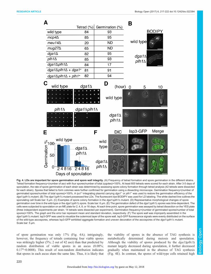