Embed Size (px)
Citation preview

Prof. Lipid Res. Vol. 27, pp. 199-270, 1988 0163-7827/88/$0.00 + 0.50 Printed in Great Britain. All rights reserved © 1988 Pergamon Press pie
LIPIDS AND THYROID HORMONES
FREDERIC L. HOCH*
Departments of Internal Medicine and Biological Chemistry, The University of Michigan Medical School, Ann Arbor, Michigan 48109, U.S.A.
C O N T E N T S ABBREVIATIONS
I. Tin,on3 HORMONE MECHANISMS A. Calorigenic and anabolic effects B. Thyroid hormone entry into cells
II. LIPID METAeOLISM A. Cell lipid composition B. Fatty acid synthesis
1. Protein phosphorylation-dephosphorylation 2. Gene expression
C. NADPH generation and oxidation 1. Cytoplasm: NADP+--*NADPH 2. Mitochondria: NADP + + NADH- ,NADPH + NAD + 3. Microsomes: NADPH-*NADP +
D. Fatty acid activation E. Glycerolipid synthesis F. Lipolysis G. Ketogenesis H. Cholesterol metabolism
I. Intracellular cholesterol metabolism 2. Cholesterol transport
I. Fatty acid desaturation 1. Anuran metamorphosis
J. Fatty acid oxidation K. Prostanoid synthesis L. Sulfolipid synthesis M. Summary
III. CELL MEMBRANE AND ASSOCIATED SYSTEMS A. Lipid composition and physical properties B. cAMP synthesis and hydrolysis
I. Adenylate cyclase 2. Cyclic nucleotide phosphodiesterases
C. Ca2+-ATPases D. Na+K+-ATPase E. Plasma Membrane Permeability
1. Na +, K+-Cotransporters 2. Na+-H + Antiporter
IV. MITOCHONDRIAL OXIDATIVE PHO6PHORYLATION A. State 4 respiration in liver mitochondria B. State 3 respiration in liver mitochondria
1. ADP/ATP carrier 2. Substrate carriers 3. Cytochrome bc~ segment 4. Cytochrome oxidase 5. P, transporter 6. Substrate dehydrogenases 7. ATP-synthetase 8. Ca2+-transporter 9. Pyridine nucleotide transhydrogenase
10. Repetitive additions of ADP C. Liver mitochondria of fetal and neonatal rats D. Heart and brain mitochondria E. Essential fatty acid deficiency
V. PEP.SPECTIVE ACKNOWLEI~Emwrs RW'V.~NC~S
199 200 200 201 202 202 205 206 208 210 210 211 211 211 213 216 217 217 217 220 221 223 224 227 227 227 228 228 229 229 232 233 235 239 239 240 241 245 248 249 250 250 251 251 251 252 252 253 254 254 255 257 259 259 259
A B B R E V I A T I O N S T3--triiodothyronine T4----thyroxine
EFA--essential fatty acids FFA--free fatty acids
*Mailing address: 3455 Woodland Road, Ann Arbor, MI 48104, U.S.A.
199

200 F.L. Hoch
UFA--unsaturated fatty acyls MUFA--monounsaturated fatty acyls PUFA--polyunsaturated fatty acyls
HMGR--~-hydroxy-fl -methylglutaryI-CoA rcductase CPT---carnitine palmitoyl transferas¢
TH, ~TH--pyridine nucleotid¢ transhydrogenase, energy-independent or -dependent UI--unsaturation index [Y~(% fatty acyl group x number of unsaturated bonds)]
[~]----concentration of ~P
I. THYROID HORMONE MECHANISMS
The availability of thyroid hormones affects reactions in almost all pathways of lipid metabolism, as has been documented in previous reviews. 23'226'231'233'477't'41 Changes in lipid metabolism are only a portion of the diverse, pleiotypic effects of altered thyroid states on cell metabolism. Attempts have been made to reduce this diversity into one or a few common initial events. Such an event should include the specific binding of L-triiodothyronine (LT3) or thyroxine (LT4) at a cellular locus that exists only in thyroid-sensitive cells. Integrated overviews of thyroid regulation have been based on the consequences of receptor occupancy only in nuclei, with subsequent gene expres- sion; 125'45~-4~'599 in mitochondria, with subsequent modulation of cell energy metabo- lism; 2:6'2aL233 in both nuclei and mitochondria; 577 and in nuclei, mitochondria, plasma membranes, and perhaps cytosolfl 2 Evidence is reviewed here that thyroid hormones act at nuclear receptors to express genes for a few lipogenic enzymes and perhaps their modifiers, and that ensuing changes in membrane lipid compositions mediate processes that underlie part of the pleiotypy. Lipids of membranes are considered both as regulating membrane-dependency that modulates protein enzyme-, transporter- or receptor-mediated activities, and as permitting unmediated cation leakage. Pertinent thyroid hormone mechanisms are first examined in general terms, then empirical observations of thyroid influence over lipid composition, and mechanisms whereby thyroid deficiency or ad- ministration regulates reactions in lipid metabolism. Effects of hormone-dependent lipid metabolism on membrane receptors, transporters, and enzymes are described in Sections III and IV.
A. Calorigenic and Anabolic Effects
Like other hormones, T3 and T4 are necessary intercellular messengers that coordinate cellular metabolic adaptations 447 to a constantly changing external and internal environment. Three general coordinative roles can be distinguished for thyroid hormones. First, thyroid accelerates, and thyroid deficiency slows, catabolic, exergonic oxidative reactions in most cells. Thus, an appropriate level of thyroid hormone is necessary for an optimal rate of free energy liberation. Energy utilization by the ubiquitous Na+K+-ATPase is also thyroid-dependent, and is proposed to determine calorigenic rates (see Section III.D). Thyroid up-regulates lipid catabolism. Lipolysis of stored triglycerides and oxidation of fatty acids support increased calorigenesis. Second, thyroid hormones simultaneously exert major anabolic effects; lipogenesis is stimulated, which seems parad- oxical for the calorigenic role of the hormones. In thyrotoxic subjects, the energetically wasteful combination of lipid synthesis and oxidation has been thought to act as a futile cycle to promote heat production. However, since hypothyroidism slows both synthesis and oxidation, normal T3 and T4 concentrations must also stimulate both processes constructively. This is seen in the requirement for thyroid hormones for growth and development in the mammalian fetus and neonate. I propose that thyroid-stimulated fatty acid synthesis and desaturations regulate mitochondrial oxidations via altered membrane fatty acyl compositions. Third, thyroid hormones have a permissive function, being necessary for and/or synergistic with actions of other hormones, e.g. lipolytic hormones, insulin, cortisol (in increasing mRNA synthesis), and testosterone (in inducing liver ct2v- globulin). The increase in scope of controls through permissive actions contributes to the synchronization of cell metabolism for whole-body adaptations, and is discussed in the next two sections.

Lipids and thyroid hormones 201
What organismic purpose is served when thyroid hormones stimulate both lipogenesis and fatty acid oxidation? Two are discussed in subsequent sections. One is calorigenic, in a futile cycle. The other is regulatory, the control of concentrations of specific metabolites through differential effects on both synthesis and further degradation. Some of these metabolites are effectors and also expand the hormone signal, e.g. cholesterol, cAMP, and membrane fatty acyl groups.
B. Thyroid Hormone Entry into Cells
Thyroid hormones act by binding to stereospecific, high-affinity, limited-capacity protein sites that impart a message when occupied that in turn alters cell metabolism. Sites with such properties are 'receptors', although that term is often used for just binding sites. Reasons to believe that the lipophilic T3 or T4 molecules themselves do not alter mitochondrial membrane permeability are discussed in Section IV. Thyroid hormone receptors exist in plasma membranes, 455 cytosol, 26 endoplasmic reticulum, 84'85 mito- chondrial inner membranes (Ref. 577, but see Ref. 189), and nuclei. Injected labeled hormone reaches all these sites within minutes (see Ref. 116). Receptor occupancy promotes gene expression in nuclei (see Section II.D), possibly oxidative phosphorylation in mitochondria (see Section IV), T3 and 2-deoxyglucose uptake, and CaE+-ATPase activity in selected plasma membranes (see Section III.E.1), and T3 transport in cytosol.
Several sets of thyroid hormone-binding 'receptors' are described. 'The' nuclear receptor in all vertebrates that circulate T3 is a 50.5 kDa protein, and its occupancy initiates thyroid action. 454 A second 'receptor' is a common, abundant, LT3-binding protein of molecular mass ~55 kDa that is found in membranes of the endoplasmic reticulum and their continuations into the nuclear envelope in all lines of cultured cells examined, s4'85 This protein is present in all tissues of an adult monkey, even those not thyroid-responsive, unlike the nuclear 50.5 kDa receptor. In cultured cells of rat pituitary tumor (GH3), 48-hr incubation with T3 decreases the number of 55 kDa protein molecules by specifically accelerating their degradation, but without changing their initial rate of synthesis or the amount of p55-coding mRNA. 44~ Thus, T3 regulates at a post-translational level. A similar mechanism of down-regulation of the 50.5 kDa nuclear receptor operates in GHI cells after incubation with T3 for 24 hr, but on briefer exposure T3 inhibits synthesis. TM
Since T3 does not down-regulate liver T3-receptors in vivo, 52~ the susceptibility of the 55 kDa protein to T3 control does not necessarily make it a 'receptor'. The sequence of the 55 kDa protein is not similar to that of human plasma thyroid-binding prealbumin or globulin. 526'628 However, a brief note says that the coding region of the 55 kDa protein has an 85% and 98% sequence homology to the endoplasmic reticulum enzymes protein disulfide isomerase and the fl-subunit of prolyl 4-hydroxylase, respectively. 85 It is not stated that the 55 kDa protein has these enzymatic activities, but the fl-subunit has the same disulfide isomerase activity in vitro as the disulfide isomerase itself, and monoclonal antibody to this subunit inhibits isomerase activityY 6 The fl-subunit is present in many cells in great excess over the active hydroxylase ~tfl-tetramers.
A third 'receptor' group comprises nuclear proteins, encoded by the c-erb-A gene, that are present in small amounts in many adult and embryonic cells. They have the binding properties of the nuclear T3-receptor although their functional capacities are not as yet described; weights are 46, 52, and 55 kDa; sequences of two cellular c-erb-A proteins are dissimilar to that of the nuclear 55 kDa protein. 526'628 A viral counterpart encoded by the v-erb-A oncogene does not bind T3 similarly. A high degree of sequence homology between c-erb-A protein and human glucocorticoid- and estradiol-receptor proteins suggests that all three may have evolved from a primordial receptor gene. 63° All have a cysteine-rich region thought to bind DNA; the protein disulfide isomerase that resembles the nuclear 55 kDa protein also has a cysteine-rich sequence in the active site. 127
A fourth set of 'receptor' proteins with high affinity for thyroid hormones is identified in nuclei of tissues that do not contain the nuclear 55 kDa protein and do not respond to administered hormone by increasing their respiration or lipogenesis. One is in the adult
JPLR 27/~-D

202 F .L . Hoch
rat brain but not in liver, and is proposed to be one of many receptors that account for the diversity of thyroid hormone effects. ~°5 Another is in testis. 29a These binders await certification as receptors. Are the criteria for defining a receptor molecule by its binding properties to thyroid hormones sufficiently rigorous to exclude other lipophilic proteins found in membranes? A similar question applies to the identification of the mitochondrial T3-receptor as the ADP/ATP carrier (see Section IV.B.1).
Thyroid entry into cells is apparently membrane-mediated, being saturable, specific, and sensitive to temperature and to protein- and lipid-modifications? 92 Plasma membrane receptors may be involved in T3 entry into cells. Triiodothyronine enters the cell across the plasma membrane via an energy-dependent transporter system. ~4'3~'49~ Extranuclear proteins bind 96% of hepatocyte T3; the minor fraction of T3 in hepatic nuclei plays a major role in promoting gene expression of lipogenic enzymes. In a rat pituitary tumor line, cytosol proteins compete less and nuclei contain 44% of cell T3.156 Recent evidence shows that another energy-dependent, stereospecific system carries LT3 across the hepatic cytosol to the nuclear binding sites; ~'45~ it accounts for the in situ nuclear LT3 content being 100 times greater than would be predicted from affinity constants of isolated nuclei or solubilized receptors. Free LT3 does bind to its nuclear receptor, however, unlike the steroids which must complex with a cytoplasmic receptor before they bind and act on nuclear components. The absence of reports that incubation of nuclei with T3 in vitro stimulates nuclear RNA polymerases therefore remains puzzling--unless it is an LT3-protein complex that actually binds to nuclear receptors. A T3-binding protein in rat kidney cytosol mediates T3-uptake by kidney mitochondria, perhaps by donating its T3 to a T3-receptor in the mitochondrial outer membrane, which is transported by other T3-carrier proteins in the intermembrane space, to a T3-receptor in the inner membrane? 4
Transport of LT3 across the cytosol seems to be tissue-specific. The T3-pump in the plasma membrane or the cytosol is invoked as the source of a highly selective differential effect of a T3-analogue on heart and liver. To measure the contribution of the pump to the selectivity, Underwood et al. 6~4 compare binding and displacement of LT3 and analogues in vivo and in vitro (to eliminate operation of the pump). As biologic responses to nuclear-receptor-binding, they assay mitochondrial glycerol-3-phosphate dehy- drogenase and depression of plasma [cholesterol] in hypothyroid rats 48 hr after one dose of LT3 or analogue. The Y-pyridazinone-3,5-dibromothyronine (SK&F-94901) is highly selective for liver: it has 18 % of the inducing potency of LT3 in liver (0.1% in heart), liver binds it in vivo 50% as well as LT3 (heart, 1.3%), and liver nuclei bind it in vitro 0.9% as well as LT3 (heart, 2%). LT3 and SK&F-94901 depress plasma [cholesterol] effectively and equally (see Section II.H). This selective access to liver nuclei is proposed to reside in the hormone-concentrating mechanisms, presumably in the energy-dependent plasma membrane and/or cytosol hormone-transporters---where hepatic cytosol may contain more SK&F-94901 than heart cytosol either because hepatocytes accumulate it more actively or cardiomyocytes exclude it. DT4 has no selective potency, and induces glycerol-3-phosphate dehydrogenase in either heart or liver about 10% as effectively as LT3, as might be expected from the relative binding of DT4 and LT3 by the nuclear receptor.
II . L I P I D M E T A B O L I S M
A. Cell Lipid Composition
Thyroid state influences cell and plasma lipid composition, but some reports are contradictory, probably because of differences in degree and duration of abnormal thyroid levels, and the many other effectors of lipid composition. Lipids in plasma of hyperthyroid rats, as compared with normals, show the expected decreases in free and esterified cholesterol concentrations, but also increases in [~PL] and in the PC/PL ratio, m Erythrocyte membranes do not equilibrate lipid content with plasma, as is shown by their greater cholesterol content and normal PC/PL ratio, although PL/protein also increases;

Lipids and thyroid hormones 203
the PL contents of i 8: l, 18: 2 and 20: 4 acyls remain unchanged. In livers of rats, either hypo= or hyperthyroidism halves total lipid content, while hypothyroidism depresses cholesterol/protein by 20%; 396 Ellefson and Mason 13° find TG content low in hypothyroids, PL contents high in hyperthyroids; Tata 596'59s'5~ finds diminished PL/protein ratios in livers of hyperthyroid rats.
In hepatic microsomes Faas and Carter t4°:41 report that hypo- or hyperthyroidism leaves unaltered the contents of PL, TG and cholesterol, and the distribution of the different PL classes. On the other hand, Ruggiero et al. 5~° see that hyperthyroidism decreases microsomal PL and cholesterol contents.
Hyperthyroidism induced with our original enormous doses of LT4, 40 #g/g/day 246 and given for 6 days, increases the PL content of rat liver mitochondria but does not change the proportions of the PL classes (although no CL fraction was distinguished), whilst hypothyroidism does not change either in the studies by Nelson and Cornatzer: 24 In liver mitochondria of our hypothyroid rats, we find normal amounts of total lipids, PL and NL per g protein and increased CL/PL ratios. 2~ Studies combining measurements of CL and cytochrome a contents per g protein in whole cell homogenates and in purified mito- chondria, with stereological micrographic analyses of liver cells and mitochondria, have compared hypothyroids and normals, and used a prolonged T4-treatment (doses every other day for 21 days). 277 Hypothyroidism selectively decreases, and T4 increases, the relative area of mitochondrial inner membranes, but the ratios of mitochondrial/liver protein and mitochondrial/cell volume do not change. In hypothyroid cells and mito- chondria, CL and cytochrome a contents per g protein decrease 20-40% below normal levels; T4, 0.1/~g/g, restores normal values. When normal rats receive 1.2/zg/g T4, CL and cytochrome a contents increase to 20-40% above normal.
Mitochondrial membranes also proliferate in thyrotoxic rat hearts. ~ Taken together with data showing that cell ~PL contents are 27% lower in hypothyroids than in euthyroids, 596 and our data exhibiting high CL/PL ratios in hypothyroids, it would appear that hypothyroidism, or this prolonged T4-regimen, alters amounts of i~PL per g protein more severely than amounts of CL. Thyroid state affects the CL contents of individual liver mitochondria rather than the number of mitochondria per cell. Reasons to think that the CL/PL ratio of such an altered mitochondrion regulates its inner membrane properties are discussed in Section IV.
Fatty acyl compositions of PL are more consistently found to be thyroid-dependent, although details vary. Some of the effects of thyroid state on PL fatty acyl composition result from changes in the fatty acyl proportions in one or more PL classes, others from a shift in the proportions of PL classes that have distinctive fatty acyl compositions. 24s For example, CL contains much more 18:2 acyls and less other PUFA than PC, PE, or PI) 53 Liver PL from hypothyroid rats, as compared with euthyroids, have slightly lowered 18:2 and 20:4 contents, and vigorous LT4 treatment (2/~g/g/day x 14 days) of either hypo- thyroid or normal rats increases percentage contents of 20:4 while decreasing percentage contents of 18: 2, indicating that T4 activates the 'degradation' of hepatic unsaturated fatty acyls. 396'47~ In other studies, ~3° liver phospho!ipids in hypothyroid rats have contents of 16:0, 18:1 and 18:2 that are 80% above normal levels, and 20:4 contents that are 70°/0 below normal, and what might be a 20: 3 acyl appears; in thyrotoxic rats, 18: 1 and 18: 2 contents increase by 90%, and 20:4 decreases 25%. Since the hormone augments adipose tissue 16:0 and 18:1 contents, Ellefson and MasonS3° propose that the hormone stimulates fatty acid de novo synthesis, fatty acid mobilization from fat-~liver, and desaturations of saturated and unsaturated fatty acids. In hypothyroid hamsters, liver lipids are slightly depleted in 18: 2; in thyrotoxic hamsters, 18: 2 and 20: 4 decrease more severely while 18 : 1 content increases. ~ The results of Peifer 47~ on thyroid effects are more difficult to fit into the pattern because the rats were fed diets containing 1°/0 18:2oJ6, 1%o 18:3co3, and 8% saturated fatty acids, and made hyperthyroid by inclusions of dessicated thyroid powder for 2 months. In these hyperthyroids, liver lipids were 40°/0 below untreated 'controls' in percentage contents of 18:2 and 22:6, while in heart lipids, 18:2 percentage was halved and 22:6 percentage was 2.6-times elevated. This diet would produce EFA-deficiency as

204 F . L . Hoch
well as the ready replacement of cardiac co 6-PUFA by co 3-PUFA noted by Gudbjarnason e t al.; ~96 the hormone was thought to accelerate the desaturative biosynthesis of both 22:6 and 20:4 from their dietary precursors.
Since cell PL are almost all in membranes, it is to be expected that organdie and cell membranes reflect the PL fatty acyl composition changes. Liver mitochondrial fatty acyl profiles in thyrotoxic rats have slightly lower 18:2 and 33% lower 20:4 percentages. 47s Heart mitochondria from hypothyroid rats and hamsters show no fatty acyl changes, while those in thyrotoxics have slightly lowered 18: 2; 468'575 this is discussed further in Section IV. Table 1 compares total lipid contents of fatty acyl groups in liver mitochondria prepared from fasted control or hypothyroid rats, or from rats of either group killed after injection(s) of LT3. 235'236 In our studies, hypothyroidism elevates mitochondrial contents of 18:2, 18:3co6, and 20:3co6 fatty acyls and depletes 20:4 acyls. One LT3-injection into hypothyroid rats rapidly substitutes saturated acyls for a part of the unsaturated fatty acyls. The hormone depresses mitochondrial 18:2, 20:4 and 22:6 fatty acyl contents to as low as 50% of the levels in untreated hypothyroids by 1-2.5 hr. The 35%-increased contents of 16:0 and 18:0 acyls replace the polyenes, and the UI diminishes to 71% of that in untreated hypothyroids and to 62% of that in the euthyroid controls. Only weeks-long EFA-deficiency (see Section IV.E), or a few hours of feeding carbohydrate to previously starved euthyroid rats (see Section II.B), depletes liver mitochondrial co 6-PUFA to this extent. Three-day LT3-treatment of control rats also raises liver mitochondrial 18: 0 content but, in addition, depletes 18:2 and 20:4 acyls.
Changes in oxidative phosphorylation at 25°C accompany the early depressions of mitochondrial PUFA contents within 3 hr after hypothyroid rats are injected with LT3 (see Section IV.A), as well as during the more chronic depletions of co6-PUFA in EFA- deficiency (see Section IV.E), but do not appear to have been examined during carbo- hydrate refeeding. Altered thermotropic properties of partial reactions also occur in liver and heart mitochondria when thyroid status is changed (see Section IV). These findings indicate that the changes in mitochondrial fatty acyl composition are great enough to have functional consequences.
Liver microsomes from hypothyroid rats have abnormal fatty acyl compositions similar to those in their mitochondria, but these have no effect on A9-desaturase activity. 243
TABLE 1. Fatty Acyl Compositions of Liver Mitochondria of Hypothyroid and Control Rats, and Changes after LT3 Injection(s)*
Hypothyroids + LT3 (hours after injection)
Fatty Controls Hypothyroids Controls acyls (6) (9) 0.5 (2) 1.0-2.5 (8) 4.0 (3) + LT3 (4)
16:0 12.6 16.0 b 16.2 21.2 E 21.1 16.7 18:0 17.3 17.2 18.5 23.4 D 16.5 24.6 E 18:1 17.9 18.2 14.3 19.6 21.7 15.1 18:2 18.7 24.1 b 22.9 17.6 E 18.8 12.5 A 18 : 3 0.3 1.3 c 1.2 1.5 1.8 2.5 20:3 1.0 2.0 d 2.9 1.4 1.8 0.7 20:4 23.5 16.7 b 16.5 9.4 c 9.6 16.9 E 22:6 4.6 3.2 2.6 1.5 a 3.1 4.7
Unsat. Index 189 167 166 I 18 B 136 162 "co6 131 122 126 81 ° 84 E 105 "093 29 26 21 17 ° 22 36
*Fatty acyl percentage contents are shown as means; for 55 values, the coefficient of variation is 16.6% _+ 1.9% SE. The number of experiments, each on one rat, is in parentheses. The unsaturation index (U.I. = Y~[%fatty acyl group u number of unsaturated bonds]) is shown for all unsaturated fatty acyl groups and separately for to6- and co3-acyls. Some hypothyroid rats were injected with LT3 ( + LT3) intraperitoneally, 1 #g/g, and killed at the times indicated; some control rats were similarly injected with LT3, 1 #g/g/day for 3 days, and killed on the 4th day (controls +LT3). Student's t test was used for group comparisons', p < ~).001; ha0.005; ~:0.01; riD0.025; or'0.05; lower case symbols denote p values for comparisons of Hypothyroids vs. Controls, upper case symbols for comparisons of Hypothyroids + LT3 vs. Hypothyroids and Controls + LT3 vs. Controls.

Lipids and thyroid hormones 205
LT3-injection changes microsomal composition slightly later and less extensively than in mitochondria. The proportion of 20:4 acyls falls at 2.5hr and reaches 75% of the hypothyroid level (49% of the control level) at 4.0 hr. With the accompanying decrease in 22:6 acyl percentage, the unsaturation index falls to 89% of the hypothyroid value at 4 hr. Three daily injections of LT3 into control rats have little effect on microsomal fatty acyl composition, other than to increase 18:0 content and decrease the ~06-unsaturation index.
In liver mitochondria of hypothyroid rats, it is the phosphatidylcholines and phos- phatidylethanolamines that contain excess 18 : 2 fatty acyls and are depleted in 20: 4 acyls, while the CLs have normal contents of 18:2 but the ratio CL/PL is increased. 248 Hepatic nuclea~ 9 phospholipids prepared from hypothyroid rats show the same abnormal fatty acid composition as mitochondria (Table 1) and microsomes, suggesting a general defect in unsaturated fatty acid metabolism. Because laboratory diets for rats contain no arachidonic acid, the desaturative biosynthesis of 20: 4 fatty acyls from dietary 18: 2 seems at fault. Although fatty acyl-CoA A6-desaturase activity is cited as 'rate-limiting' in this conversion, 56 the appearance of 20:3096 acyls suggests a more limiting defect in the A5-desaturase in hypothyroidism. Thyroid regulation of desaturase activities is discussed below (see Section II.I).
B. Fatty Acid Synthesis
Thyroid status is one of the regulators of de novo fatty acid synthesis. In general, rapid (0-24 hr), reversible modifications of existing enzymes regulate fatty acid synthesis by several mechanisms. 621 Allosteric effectors signal intracellular conditions, and cyclic nucleotide- or Ca2+-mediated phosphorylation/dephosphorylation regulates under hor- monal control for whole-organism needs. The acetyl-CoA carboxylase, 445 which is said to be 'rate-limiting' under most circumstances, is rapidly activated by citrate and inactivated by long-chain fatty acyl-CoA, but no certain thyroid influence via these effectors is established. Phosphorylation inactivates, and dephosphorylation reactivates, the acetyl- CoA carboxylase and fatty acid synthetase enzymes. An appropriate stimulus starts synthesis of some lipogenic enzymes even more quickly than the actions of rapid effectors or covalent modifiers. Synthesis rather than degradation is stated to regulate the activity of the fatty acid synthetase, 5 the malic enzyme,177 the glucose-6-phosphate dehydrogenase, and the 6-phosphogluconate dehydrogenase, through the selective expression of genes and the nuclear synthesis or supression of mRNAs (see Section II.B.2).
Hypothyroidism in developing or adult rats depresses hepatic fatty acid synthetase activity by 50%, and diminishes but does not abolish carbohydrate-induced activation of fatty acid synthetase. 336'62° Cultures of hepatocytes obtained from hypothyroid rats synthesize fatty acids from acetate half as fast as nonnals. 173 Incubation of these hepatocytes with ~ 1 nM LT3 stimulates synthesis in 2 hr and triples the rate by 4 hr independently of protein synthesis, since cycloheximide fails to inhibit. Greater LT3 concentrations accelerate fatty acid synthesis in hepatocytes from normal rats 37% in 4 hr. LT4-injection of normal rats doubles the amount of hepatic citrate lyase and triples that of malic enzyme in 7.5 hr, 17° and increases fatty acid synthetase and acetyl-CoA carboxylase activities more slowly. 366
Starvation, 7,s,17°.6~ diabetes, 1°5 or hypophysectomy 324a decrease hepatic fatty acid syn- thesis more severely (to < 10% of normal), as well as activities of fatty acid synthetase, acetyl-CoA carboxylase, malic enzyme, citrate lyase, and glucose-6-phosphate dehy- drogenase. In starved or diabetic rats, the liver contains considerable amounts of immunoreactive fatty acid synthetase that is enzymatically inactive. Carbohydrate fed to fasted rats 64s increases fatty acid synthetase in two phases. Within 1 hr, additional inactive fatty acid synthetase is synthesized. After 3 hr, fatty acid synthetase activity begins to increase, in parallel with incorporation of labeled 4'-phosphopantotheine into fatty acid synthetase apoenzyme, forming holoenzyme. We do not know what makes existing apoenzyme take up its prosthetic group. Refeeding accelerates synthesis of malic enzyme

206 F.L. Hoch
and citrate lyase in 6 hrJ 7° When rats are refed with carbohydrates for 15 days, depressed activities of hepatocyte cytosol acetyl-CoA carboxylase, fatty acid synthetase, malic enzyme, citrate lyase, and glucose-6-phosphate dehydrogenase coordinately rise to levels many times normalJ 7° This coordination prevents the increased flux of metabolites from accumulating behind each relatively slowed step. Supplementation of fat-free diet with 18:2 or 20:4 (but not 16:0 or 18:1) dampens the induction of all these enzymes; as discussed under Section II.I, major regulatory properties are also ascribed to dietary linoleate through its potent inhibition of liver fatty acid synthetase and the fatty acyl-CoA A9-desaturase. 279 Thyroid state controls the hepatic uptake and esterification of administered 18:209 6, thereby further regulating lipogenic enzymes. The suppressive effect of fat-feeding on the carbohydrate-inductions of acetyl-CoA carboxylase, fatty acid synthetase, and malic enzyme diminishes with aging of rats, and is relieved by T3- treatment, more in young rats than in old. 163 Thyroid hormones and refeeding fasted rats with carbohydrates exert synergistic effects on lipogenic enzyme gene expression (see Section II.B.2).
Effects of one T3 injection on hepatic fatty acid synthetase and acetyl-CoA carboxylase have been measured in hypophysectomized 32~ or diabetic ~°5 rats. T3 given to hypophy- sectomized rats does not increase fatty acid synthetase activity from 4--12 hr later, but Kumar et al. 32~ did not measure short-term effects on fatty acid synthetase amount or synthesis. Long-term, T3 activates fatty acid synthetase and acetyl-CoA carboxylase only after 24 hr, to reach maximal activity at 3-4 days; injecting T3 twice over 7 days accelerates activities of fatty acid synthetase and acetyl-CoA carboxylase 15- to 20-fold, and synthesis of fatty acid synthetase ~ 10-fold. A different biphasic pattern is seen after diabetic rats are injected with T3.1°5 In 4-12hr, activities of fatty acid synthetase and acetyl-CoA carboxylase rise slowly, together with conversion of apoenzyme--,holoenzyme and accu- mulation of immnoreactive fatty acid synthetase; however, cycloheximide or actinomycin D does not block the increase in activities or the parallel holoenzyme formation. In a second stage from 24-72 hr after T3 injection, a more rapid increase in activities and fatty acid synthetase antibody titer occurs that is inhibited by blockage of transcription or translation and so represents net protein synthesis. Comparable long-term T3 treatment of diabetic animals stimulates hepatic fatty acid synthesis from all precursors. 5~ Here, insulin does not mediate T3 effects. Insulin given to diabetic rats acts more quickly than T3, and increases fatty acid synthetase activity 5-fold in 6 hr, 20-fold in 12 hr (see Kumar et al. 324~ for Refs). In contrast, in avian liver explant culture, insulin induces a 5-fold increase in fatty acid synthetase; T3 o r hydrocortisone potentiate the induction, individ- ually 2-fold, together 4-fold; without insulin, they are ineffective. ~5°':85 Apparently, these cultures require insulin, and T3 or hydrocortisone play supportive roles in induction of fatty acid synthetase.
1. Protein Phosphorylat ion-Dephosphorylat ion
As noted above, cAMP-dependent and -independent protein kinases inhibit rate- limiting enzymes of fatty acid synthesis. In vitro, sequential additions of protein kinases and phosphatases interconvert active and inactive forms of an enzyme. 2~4'4~ For example, in vitro dephosphorylation activates pigeon liver fatty acid synthetase 15-fold, and phosphorylation inactivates to the same degree. ¢7 Usual procedures for fractionation indicate that under normal physiologic conditions 90% of a lipogenic liver enzyme, /~-hydroxy-p-methylglutaryl-CoA reductase (HMGR, see Section II.H), exists i n an inactive phosphorylated form. ~9 In this case, decapitating conscious rats is shown to result in almost complete phosphorylation of enzyme in seconds. Using anesthetized fed rats, and cold-clamped liver to eliminate the delay during centrifugal fractionations, HMGR is 80% active. Such procedures may well be critical in correlating the receptor-mediated thyroid-state effects on other lipogenic enzymes as well.
Thyroid treatment induces several protein kinases and increases the in vivo phos- phorylation of endogenous proteins. In the absence of valid examinations of phos-

Lipids and thyroid hormones 207
phorylation states of lipogenic enzymes, evidence for direct connections with thyroid levels remains circumstantial--we discern motive, means, and opportunity but have no witness to dephosphorylation. In the myocardium, 2 hr after T3-injection, the kinase activity of nuclear non-histone proteins increases for each substrate tested (histones, casein, phos- vitin). TM Activity doubles after 3 daily T3-injections but even continued treatment for more than 7 days does not prevent the return of kinase activities to control levels. T3-treatment also activates two isozymes of a myocardial cytosol cAMP-dependent protein kinase I in 2 hr; detectable rises in [cAMP] and [cGMP] start at 4 hr. ¢ The significance of these early kinase activations or inductions for lipogenesis is uncertain, since the heart has little lipogenic enzyme apparatus.
In rat liver, T3-treatment produces stimulations analogous to those in heart, but more slowly. When hepatocytes from fetal rats are incubated with T3 for 3 days (but not for 5 hr) increased activity of nuclear protein kinases and susceptibility of chromosomal non-histone proteins to phosphorylation are observedfl 9 Nuclear non-histone proteins extracted from livers of rats after 2 daily injections of T3 are increased in phosphorylative actions on casein and phosvitin by 60-90%; partially purified fractions of nuclear extracts obtained after 8 daily injections are 4 times more active than those from control rats . 324,5°1
These T3-induced fractions, as compared with controls, have different pI values, are more heat-labile, are less sensitive to inhibition by a sulfhydryl reagent, and have greater autophosphorylative activity. 324 Thus T3 seems to be evoking gene expression of 'new' protein kinases. Since these kinases, like those in heart, disappear even with continued T3-treatment, a down-regulation may decrease the number of nuclear receptors TM or LT3 access to the receptors.
In livers of hypothyroid rats, phosphorus-content of ribosomal proteins, and activities and cAMP-binding of soluble protein-kinases, are below euthyroid levels. ~°° Nuclear chromatin non-histone proteins autophosphorylate at a subnormal level, except for one large fraction that is much more active than normal? 4 Hypothyroidism decreases both nuclear and cytosolic casein kinase activity and T3-injection restores these activities in 1 5 hr. Protamine kinase in the cytosol is more than normally active, and decreases after T3-treatment. 5°t Thus, thyroid state appears to modulate specific protein kinases, and hypothyroidism even activates some nuclear kinase(s) while inactivating others. Further characterization of thyroid-responsive cytosolic protein kinases in livers of hypothyroid ra ts 416'469'47° shows that the cAMP-independent histone kinases are normally active while the casein kinases have half-normal activity. T4-treatment restores one fraction of the cAMP-dependent casein kinases to normal levels in 2-5 days, 469'47° and these are re- sponsible for the phosphorylation of the cytosolic proteins. 416 In addition, T3-adminis- tration to hypothyroid rats for 3 days activates a cytosolic cAMP-dependent protamine kinase. 416 Lag periods of days must be compared with the few hours it takes for T3 to activate hepatic lipogenic enzymes that are well documented to be activated when dephosphorylated. Thus, in vivo studies on protein phosphorylation in hypothyroid rats fasted-and-refed-glucose and injected with 32P i and T3 before killing, 93 are instructive because fasted/refed animals normally dephosphorylate hepatic cytosolic lipogenic enzymes. Before the hypothyroid rats receive T3, nuclear proteins incorporate injected 32p at 15-35% below normal levels--so hypothyroidism suppresses nuclear protein phos- phorylation. T3-injection progressively increases 3:p-incorporation to reach significant levels 6 hr later. Cytosol protein phosphorylation increases even more slowly. Again, the lag time in the nucleus seems too great to account for the 40 min that it takes T3 to activate the nuclear DNA-dependent RNA polymerase II (Ref. 286; but see Ref. 608). This is curious because both the hepatic polymerases I and II are activated by either cAMP-dependent or -independent protein kinases and inactivated by phosphoprotein phosphatases. 3m9
Phosphoprotein phosphatases are of particular interest when one regards the number of enzyme reactions in the pathways of lipid metabolism (and of carbohydrate metabolism, for that matter) that are activated both by T3-treatment and by protein de- phosphorylation. 2H'm'267'319 No protein phosphatase has as yet been connected with thyroid
208 F .L . Hoch
state. However, some of these phosphatases are indirectly activated through the phos- phorylation of phosphatase-inhibitor proteins by either cAMP-dependent or -independent protein kinases, 265 and thyroid state regulates activity of protein kinases of either persuasion, as well as [cAMP] (see Section Ill.B).
Covalent enzyme modifications by phosphorylation/dephosphorylation mechan- isms 2tl'2m2 may also account for some hormonal and dietary interactions with T3 effects. A preliminary note reports that starvation (which was taken as a glucagon signal) decreases the degree of phosphorylation of several histones and non-histones in isolated nuclei incubated with [~,-32p]ATP, and refeeding carbohydrate (taken as an insulin signal) rephosphorylates nuclear non-histones. 3~ In contrast, insulin promotes dephosphorylation in hepatic cytoplasm. Dephosphorylation appears to regulate important cytosol enzymes in the pathways that convert glucose-6-phosphate to cytoplasmic acetyl-CoA, viz. phos- phofructokinase 2, pyruvate kinase (a cAMP-dependent protein kinase inactivates), and pyruvate dehydrogenase. Citrate lyase is subject to reversible phosphorylation, but its activity remains unaltered. As is discussed elsewhere in this review, conversion of acetyl-CoA to lipids also involves enzymes activated by dephosphorylation, 3m9 insulin, or thyroid hormone; these include acetyl-CoA carboxylase, glycerol-3-phosphate acyltrans- ferase (inactivated by a cAMP-kinase), HMG-CoA reductase, cholesterol-7~-hydroxylase, cholesterol ester-hydrolase (activated by a cAMP-kinase), and possibly fatty acyl-CoA A9-desaturase.
2. Gene Expression
As noted in the preceding section, T3 or T4 increase amounts and synthetic rates of several enzymes involved in fatty acid synthesis, in some cases after activating inactive enzyme. The rapid progress of studies on the T3-induction of such enzymes, and its relationship to the mechanisms whereby dietary carbohydrate induces lipogenic enzyme activities in the livers of starved animals, ~ has been updated; 3st'451-454 the details of information transfer are beyond the scope of this review. The enzymes most studied are fatty acid synthetase, acetyl-CoA carboxylase, citrate lyase, malic enzyme, glucose-6- phosphate dehydrogenase, and 6-phosphogluconate dehydrogenase. Additional rapidly induced lipogenic enzymes are the mitochondrial glycerol-3-phosphate dehydrogenase (see Section II.E) and the fatty acyl-CoA A9-desaturase (see Section II.I). The mechanisms of the synergism between carbohydrate-feeding and T3 treatment do not involve a carbohydrate-induced increase in nuclear T3-uptake per dosage, or the number and affinity of the T3-receptor sites. Further indication that insulin is not necessary for the T3-effect lies in observations that fructose-feeding restores the depressed T3-induction of hepatic malic enzyme in diabetic rats, since fructose is metabolized in the absence of insulin (see Kumar et al. 32~ for Refs). It is therefore postulated that some intracellular product of glucose metabolism induces malic enzyme, and that T3 acts as a constant multiplier of that signal in the nucleus. The nature of this metabolite is not known, but it appears to be of mitochondrial origin, since dichloroacetate promotes its action: dichloroacetate activates the pyruvate dehydrogenase by inhibiting the dehydrogenase kinase in the dehydrogenase complex.
When injected in rico, the hormone augments or suppresses 21 out of 250 separated liver mRNAs. A T3-induced increase in pituitary growth hormone level effects the changes in 7 of these 21 mRNAs. Carbohydrate-feeding also changes 8 of these 21, and in the same direction as T3. A dose of T3 large enough to convert receptor occupancy from the hypothyroid to the hyperthyroid level augments 4 of the mRNAs in carbohydrate-fed animals, from 4-fold to 13-fold. 376 Three of them are mostly induced in the range of T3-occupancy from euthyroid to hyperthyroid, as is malic enzyme-mRNA, mRNAst 4 (see below), and activity of the lipogenic enzymes. Only 1 mRNA is induced at 4 hr (the earliest time of examination), the other 3 at > 8 hr. Obviously, only a few more proteins are involved in these rapid inductions than the half-dozen or so lipogenic enzymes studied, but not nearly enough proteins to account for the great diversity of thyroid effects.
Lipids and thyroid hormones 209
And the 11 hr of T3 residence in the liver 537 is too short to account for additional T3-receptor-mediated, long-term syntheses.
Several liver mRNAs respond very rapidly to T3. T3 and carbohydrate-feeding each start hepatic malic enzyme synthesis in about 2 hr, so neither works by stimulating the other. The amount of malic enzyme-mRNA translated correlates with the increase in malic enzyme synthesis, so synthesis and/or processing of the mRNA are primary events. These times are still longer than the 20-60 min after T3 injection reported for generation of a hepatic mRNA dubbed mRNAs,4. A heteronuclear precursor RNA for mRNAsl4 is said to appear as early as 10 min after a dose of T3 that saturates liver T3-receptors. 42° The proportion of basal, unstimulated [mRNAs14] to total mRNA contents is high in lipogenic tissues: 10 times greater in adipose cells than in livers of males, and 3 times more in livers of females (7 times if they are lactating) than in males. Hypothyroidism drops the mRNAst4 content in fat by 80%. 287 T3 raises [mRNAs14] in lipogenic cells, to a varying degree. At 24 hr, a receptor-saturating dose of T3 increases mRNAsj4 in fat cells twice as much as in liver cells. 287
Non-lipogenic tissues have much less mRNAs,4: brain contains about 6% as much as male liver, kidney, heart and spleen < 1%; mRNAst 4 in these tissues does not increase after T3 administration. Thus, the presence of T3 receptors in kidney and heart is not sufficient for T3 to evoke this mRNA or its product. The translation product of mRNAsl 4, a cytoplasmic polypeptide of 17 kDa, does not appear to be a component of enzymes or transporters involved in fatty acid metabolism that have registered sequences. From its temporal primacy after T3 treatment, from the involvement of the T3-induced carbo- hydrate signal with protein phosphorylation, from the early T3-activation of existing inactive lipogenic enzymes, and from the known T3-induction of nuclear protein kinases, 93 one can not resist the speculation that this is a subunit of such a kinase (or a phosphoprotein phosphatase). This, despite the fact that so far T3 induces known kinases much too slowly, and has no demonstrated effects on phosphoprotein phosphatases.
Adipocyte nuclear receptors bind 0.4 ng LT3/mg DNA 71 as compared with 0.6 ng/mg DNA in liver nuclei. However, there is more disagreement as to the effects of thyroid state on enzymes of fatty acid synthesis and NADP ÷ reduction in adipose tissues. In hyperthyroid rats, Roncari and Murthy 499 find a 40% decrease in the in vivo synthesis of fatty acids, activities of fatty acid synthetase and acetyl-CoA carboxylase, and total fatty acid synthetase; corresponding values in liver are + 40% to + 90%. Contrarily, apparently equivalent LT4 or LT3 dose/time schedules increase fatty acid synthetase activity by 50--70% in white fat ~'5'38° and rat neonate brown fat. 2°5 But Diamant et al) ~5 find hyperthyroidism does not increase malic enzyme activity while it augments glucose-6- phosphate dehydrogenase and 6-phosphogluconate dehydrogenase activities, whereas Mariash et al. 38° see a rise in malic enzyme activity and no changes in these dehydrogenases. In adipose tissue from hypothyroid rats, activities of fatty acid synthetase, malic enzyme, glucose-6-phosphate dehydrogenase, and 6-phosphogluconate dehydrogenase are reported to be depressed 40% to 80% below normal levels) s° In apparently similar adipose tissue, acetyl-CoA carboxylase and glucose-6-phosphate dehydrogenase V values are ,-~ 2.5 times normal, and the specific activities of fatty acid synthetase and 6-phosphogluconate dehydrogenase are 50o to 80% above normal levels; in the report of Correze eta/., 97
T3-injection decreases glucose-6-phosphate dehydrogenase activity to near normal levels in 2-5 hr. This rapid inactivation is not attributable to any known activation of specific proteolysis, and the glucose-6-phosphate dehydrogenase is reported to be not subject to phosphorylation by cAMP-dependent protein kinases. 267 Since responses of lipogenic enzymes expected from adipocyte LT3-receptor occupancy and subsequent gene expres- sion are not observed by all investigators, it would appear that other regulatory mechanisms intervene, perhaps including altered states of phosphorylation.
The situation is somewhat cleared by studies of cultured precursors of adi- pocytes, in which effects of several hormones on lipogenic enzymes can be distinguished (see Ref. 634a). Thyroid hormones, insulin, growth hormone, or glucocorticoids induce adipogenic activities in clonal lines obtained from fibroblast-like cells. As measured by lipid

210 F.L. Hoch
synthesis and glycerol-3-phosphate dehydrogenase activity, T3 and T4 have no effects on primary cultures derived from adipose tissues, but insulin and glucocorticoids are required for conversion to adipocytes. T3 is essential for adipose differentiation of ob 17 cells ~9 and is active in preadipocytes kept in serum-free medium, but has no effect on rat preadipocytes under the culture conditions of Wiederer and L6ffler? 3~ Incubation of ob17 cells with 1.5 nM T3 for 12 days first activates existing fatty acid synthetase, later increases the amount of fatty acid synthetase, and subsequently leads to accumulation of inactive enzyme that is still immunoreactive. 169
Triiodothyronine induces lipogenic enzymes interactively with a number of hormones, including estrogens and luteinizing hormone (see discussion above, of the high basal [mRNAs~4] in females), growth hormone, glucocorticoids, and insulin. Insulin interactions are the most extensively studied. In all lines of adipocyte precursor cells, insulin induces a coordinate increase of acetyl-CoA carboxylase, fatty acid synthetase, malic enzyme, and citrate lyase. Insulin incubation with extracts of adipocyte plasma membrane normally promotes a kinase-mediated phosphorylation of Tyr in the insulin fl-subunit receptor; in extracts from hypothyroid rats, this autophosphorylation response to insulin increases 2- to 3-fold, and T3-treatment of these hypothyroid rats depresses the insulin action to normal levels in 24 hr. 99 Such a mechanism would seem to mediate an insulin-T3 antagonism, while covalent modifications might mediate collaboration.
One may ask what physiological purpose is served when calorigenic and anabolic hormones like T3 and T4 act so immediately on nuclear receptors to express genes for just a few lipogenic enzymes that mediate fatty acid and cholesterol (see Section II.H) synthesis, NADP + reduction, and perhaps fatty acyl-CoA desaturation (see Section II.I).
Elongation of palmitoyl-CoA and general fatty acid chain elongation by malonyl-CoA in liver microsomes accelerates up to 3-fold in rats pretreated with thyroid hormones for 21 days, and slows by 30-40% in rats made hypothyroid. 3°°'336 The competing malonyl- CoA decarboxylase remains unchanged by LT3-treatment, and decreases slightly in hypothyroidism. Fatty acyl chain elongation by acetyl-CoA condensation in mitochondria is not affected by thyroid state, 336 despite the thyroid-responsiveness of the carnitine palmitoyltransferase in the outer face of the inner membrane (see Section II.J), which is thought to intromit acyl-CoA for elongationfl
Brains of adult animals readily incorporate labeled acetate from the blood and synthesize fatty acids that appear in PL, but the slow turnover contrasts with that of other tissues. The acetyl-CoA carboxylase and the fatty acid synthetase of brain are similar to those of other tissues, except that they have longer biologic half-lives, and do not respond to dietary changes. 6~ Hypothyroidism, when induced from birth, depresses fatty acid synthetase activity in brain as well as in liver; fatty acid chain elongation in brain microsomes and mitochondria is also reducedJ 93a Daily T3 treatment of normal rats starting at 1 day of age increases brain microsomal chain elongation by malonyl-CoA 2.5-fold by day 6, to synthesize those saturated, long-chain fatty acids characteristic of myelin lipids. Thyroid treatment of normal adult animals does not affect brain fatty acid synthetase activity in doses that stimulate the liver synthetase. ~2°
C. N A D P H Generation and Oxidation
1. Cytoplasm: N A D P + ~ N A D P H
Thyroid inductions of hepatic cytosol enzymes that reduce NADP + to NADPH include malic enzyme, glucose-6-phosphate dehydrogenase, and 6-phosphogluconate dehy- drogenase, which are discussed in Section II.B.2. Fasting/refeeding does not induce the last two enzymes in hypophysectomized rats; partial return of adaptation to refeeding occurs after 10-14 days of treatment with T3 (25%), hydrocortisone, or growth hormone; hydrocortisone + T3 (or growth hormone) restores induction 50%, and all three produce a supernormal response. ~°2'6°3

Lipids and thyroid hormones 211
2. Mitochondria: N A D P + + N A D H - - * N A D P H + N A D +
Mitochondrial energy-dependent pyridine nucleotide transhydrogenase is thought to play a role in lipid syntheses by mediating the transfer of reducing equivalents from mitochondrial NADH to NADPH, whence the citrate shuttle transfers the equivalents to the cytoplasm to regenerate NADPH--a role analogous to other thyroid-dependent cyto- plasmic generators of NADPH (see above). The transhydrogenase also provides mito- chondrial NADPH required in the oxidation of unsaturated fatty acids, 55 and so contributes to /~-oxidation. Among the auxiliary enzymes that metabolize the unusual A3-c/s-enoyl-CoA or A2-trans-A4-cis-dienoyl-CoA esters to intermediates accessible to the normal /~-oxidation enzymes are a 2-enoyl-CoA reductase and a 2,4-dienoyl-CoA 4-reductase, both of which use NADPH to reduce double bonds.
The hypothyroid state increases energy-dependent transhydrogenase activity but not energy-independent activity. ~36,137 In intact liver mitochondria, accelerated reduction of NADP + during the State 3--. State 4 transition provides an indirect measurement. 232,234 In submitochondrial particles where the enzyme is on the outer surface, manipulation of substrate concentrations provides measurements of kinetics) 36'~37 There the V of the ATP-supported and the succinate-supported activities is doubled in hypothyroid prepara- tions, while the K= values remain unchanged. Since the kinetics of the energy-independent mode of the enzyme are unchanged, the amount of enzyme would not appear to have increased. Because these particles oxidize succinate at normal rates, the coupling between energy source and transhydrogenation is thought to be increased in hypo- thyroidism. Within 3 days after injecting hypothyroid rats with LT4 or LT3, energy- dependent activities decreased to the lower, normal levels. Possible mechanisms that involve membrane lipids in the selective thyroid-dependency of the energy-linked transhydrogenase are discussed in Section IV.B.9.
3. Microsomes: N A D P H ~ N A D P +
Hyperthyroidism increases, and hypothyroidism decreases, activities of flavoprotein components of hepatic microsomal NADPH-oxidizing systems (NADPH-cytochrome c reductase and NADPH diaphorase). 29s'58s A small dose of LT3, given to hypothyroid rats, corrects activities within 20hr. 5ss Hypothyroidism concomitantly increases amounts of component hemoproteins (cytochrome b5 and P450). Some of these NADPH-dependent systems are involved in steroid metabolism (see Section II.H. 1). Flavoprotein components of NADH-cytochrome c reductase and NADH diaphorase are said to increase in activity, but we noted no change in NADH-cytochrome c reductase activity in our hypothyroid rats. 243
D. Fatty Acid Activation
Long-chain fatty acyl-CoA synthetases (A in Fig. 1) in liver exist mostly in endoplasmic reticulum and in outer membrane of mitochondria, and much less in peroxisomes. Microsomal and mitochondrial enzymes are not identical. By their location and perhaps by their specificity, they segregate fatty acyls into a pool for glycerolipid synthesis (and/or chain elongation and desaturation) and a pool for oxidation, as has been shown for the two different acyl-CoA synthetases of the yeast Candida lipolytica. 445 Injected labeled 18:2 is conserved from oxidation in vivo, 9x3~4 although isolated heart mitochondria oxidize 18: 2 faster than 16:0. 57 Preferential esterification of 18:2 with CoA by the microsomal synthetase would direct 18:2 into giycerolipids or further desaturation, and away from mitochondrial oxidation. Trigiyceride and PL synthesis in hcpatocytes have different specificities for fatty acids. 75 Trigiycerides and diacylgiycerides incorporate all fatty acids indiscriminately, in a pattern that reflects the composition of the suspending medium. Phospholipids, especially PE and PI, incorporate more 20: 4 than do trigiycerides when the medium contains 18: 2 and 20: 4.
Hepatic esterification of free fatty acids by long-chain acyl-CoA synthetase (A in
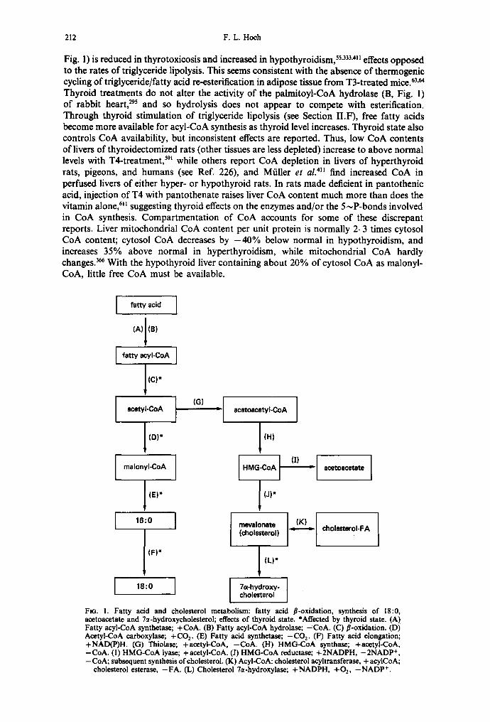
212 F.L. Hoch
Fig. 1) is reduced in thyrotoxicosis and increased in hypothyroidism, 5s,333,4ml effects opposed to the rates of triglyceride lipolysis. This seems consistent with the absence of thermogenic cycling of triglyceride/fatty acid re-esterification in adipose tissue from T3-treated mice. 63'~ Thyroid treatments do not alter the activity of the palmitoyl-CoA hydrolase (B, Fig. 1) of rabbit heart, 295 and so hydrolysis does not appear to compete with esterification. Through thyroid stimulation of triglyceride lipolysis (see Section II.F), free fatty acids become more available for acyl-CoA synthesis as thyroid level increases. Thyroid state also controls CoA availability, but inconsistent effects are reported. Thus, low CoA contents of livers of thyroidectomized rats (other tissues are less depleted) increase to above normal levels with T4-treatment, 591 while others report CoA depletion in livers of hyperthyroid rats, pigeons, and humans (see Ref. 226), and Mfiller e t ai . 41m find increased CoA in pcrfused livers of either hyper- or hypothyroid rats. In rats made deficient in pantothenic acid, injection of T4 with pantothenate raises liver CoA content much more than does the vitamin alone, 6H suggesting thyroid effects on the enzymes and/or the 5,,-P-bonds involved in CoA synthesis. Compartmentation of CoA accounts for some of these discrepant reports. Liver mitochondrial CoA content per unit protein is normally 2-3 times cytosol CoA content; cytosol CoA decreases by - 4 0 % below normal in hypothyroidism, and increases 35% above normal in hyperthyroidism, while mitochondrial CoA hardly changes. 3~ With the hypothyroid liver containing about 20% of cytosol CoA as malonyl- CoA, little free CoA must be available.
I fatty acid I (A)I(B)
I I l lC)*
acetyl-CoA t
I (D)*
I malonyI-CoA I
l IE)*
I 16:0 I
I (F)*
I
(G) ' [ acetoacetyl-CoA
J Ill)
I
J (J)*
mevaJonate (cholesterol)
(L)"
7,~-hydroxy- cholesterol
OK, _-[ I i acetoacetate I
(K) = cholesteroI-FA
FIG. 1. Fatty acid and cholesterol metabolism: fatty acid fl-oxidation, synthesis of 18:0, acctoacctate and 7a-hydroxycholcsterol; effects of thyroid state. *Affected by thyroid state. (A) Fatty acyl-CoA synthetase; +CoA. (B) Fatty acyl-CoA hydrolase; -CoA. (C) fl-oxidation. (D) Acetyl-CoA carboxylas¢; +CO 2. (E) Fatty acid synthetas¢; -CO 2. (F) Fatty acid elongation; +NAD(P)H. (G) Thiolas¢; +acetyi-CoA, -CoA. (H) HMG-CoA synthas¢; +acctyl-CoA, -CoA. (1) HMG-CoA lyas¢; +acetyl-CoA. (J) HMG-CoA reductase; +2NADPH, -2NADP +, -CoA; subsequent synthesis of cholesterol. (K) AcyI-CoA: cholesterol acyltransferase, + acylCoA;
cholesterol esteras¢, -FA. (L) Cholesterol 7a-hydroxylase; +NADPH, +02, -NADP +.
Lipids and thyroid hormones
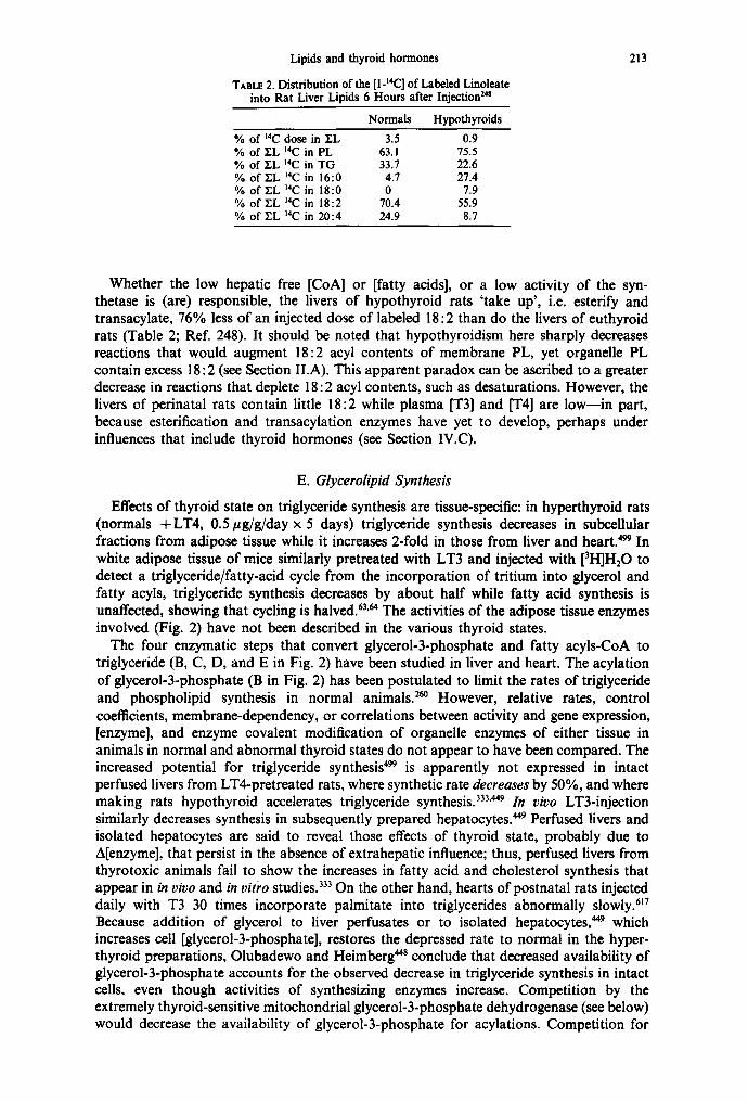
TABt~ 2. Distribution of the [1-t4C] of Labeled Linoleate into Rat Liver Lipids 6 Hours after Injection ~s
Normals Hypothyroids % of ~4C dose in EL 3.5 0.9 % of YL 14C in PL 63.1 75.5 % of Y-L m4C in TG 33.7 22.6 % of YL 14C in 16:0 4.7 27.4 % of Y-L 14C in 18:0 0 7.9 % of Y~L 14C in 18:2 70.4 55.9 % of YL 14C in 20:4 24.9 8.7
213
Whether the low hepatic free [CoA] or [fatty acids], or a low activity of the syn- thetase is (are) responsible, the livers of hypothyroid rats 'take up', i.e. esterify and transacylate, 76% less of an injected dose of labeled 18:2 than do the livers of euthyroid rats (Table 2; Ref. 248). It should be noted that hypothyroidism here sharply decreases reactions that would augment 18:2 acyl contents of membrane PL, yet organdie PL contain excess 18:2 (see Section II.A). This apparent paradox can be ascribed to a greater decrease in reactions that deplete 18:2 acyl contents, such as desaturations. However, the livers of perinatal rats contain little 18:2 while plasma [T3] and IT4] are low--in part, because esterification and transacylation enzymes have yet to develop, perhaps under influences that include thyroid hormones (see Section IV.C).
E. Glycerolipid Synthesis
Effects of thyroid state on triglyceride synthesis are tissue-specific: in hyperthyroid rats (normals +LT4, 0.5/zg/g/day × 5 days) triglyceride synthesis decreases in subcellular fractions from adipose tissue while it increases 2-fold in those from liver and heart: 99 In white adipose tissue of mice similarly pretreated with LT3 and injected with [3H]H20 to detect a triglyceride/fatty-acid cycle from the incorporation of tritium into glycerol and fatty acyls, triglyceride synthesis decreases by about half while fatty acid synthesis is unaffected, showing that cycling is halved. 63.64 The activities of the adipose tissue enzymes involved (Fig. 2) have not been described in the various thyroid states.
The four enzymatic steps that convert glycerol-3-phosphate and fatty acyls-CoA to triglyceride (B, C, D, and E in Fig. 2) have been studied in liver and heart. The acylation of glycerol-3-phosphate (B in Fig. 2) has been postulated to limit the rates of triglyceride and phospholipid synthesis in normal animals. 26° However, relative rates, control coefficients, membrane-dependency, or correlations between activity and gene expression, [enzyme], and enzyme covalent modification of organelle enzymes of either tissue in animals in normal and abnormal thyroid states do not appear to have been compared. The increased potential for triglyceride synthesis 499 is apparently not expressed in intact perfused livers from LT4-pretreated rats, where synthetic rate decreases by 50%, and where making rats hypothyroid accelerates triglyceride synthesis. 333'449 In vivo LT3-injection similarly decreases synthesis in subsequently prepared hepatocytes. 449 Perfused livers and isolated hepatocytes are said to reveal those effects of thyroid state, probably due to A[enzyme], that persist in the absence of extrahepatic influence; thus, perfused livers from thyrotoxic animals fail to show the increases in fatty acid and cholesterol synthesis that appear in in vivo and in vitro studies. 333 On the other hand, hearts of postnatal rats injected daily with T3 30 times incorporate palmitate into triglycerides abnormally slowly: 17 Because addition of glycerol to liver perfusates or to isolated hepatocytes, ~9 which increases cell [glycerol-3-phosphate], restores the depressed rate to normal in the hyper- thyroid preparations, Olubadewo and Heimberg 44s conclude that decreased availability of glycerol-3-phosphate accounts for the observed decrease in triglyceride synthesis in intact cells, even though activities of synthesizing enzymes increase. Competition by the extremely thyroid-sensitive mitochondrial glycerol-3-phosphate dehydrogenase (see below) would decrease the availability of glycerol-3-phosphate for acylations. Competition for

214 F.L. Hoch
glycerol-3-P
J 'IB)*
1-acylglycerol-3-P
[ ~ (A)* : I dihydroxyacetone-P J
i lK)
1-acyldihydroxyacetone-P
(C)*
I ] (D)*
,., ,o, I (E)*
[ triacylgiycerol ]
I (F)* " I CDP-diacylglycerol I
(G)*
I cerdiolipin
FIG. 2. Effects of thyroid state on some steps in the conversion of glycerol-3-phosphate to triglycerides and phosphoglycerides. P = phosphate; *affected by thyroid state, others not tested. (A) Glycerol-3-phosphatE dehydrogenase; +NAD(H) or FAD(H2). (B) Acyl-CoA:glycerol-3- phosphate acyltransferase; +acyI-CoA, -CoA. (C) l-Acylglycerol-3-phosphate acyltransferase; +acyl-CoA, -CoA. (D) Phosphatidate phosphohydrolase; +H20, -P i . (E) Diacylglycerol acyltransferase; + acyl-CoA, - CoA. (F) CTP-phosphatidate cytidyltransfErase; +CTP, - PP~, ((3) Includes phosphatidylglycerophosphate synthEtase (+glycerol-3-P, -CMP), phosphatidyl- glycerophosphatE phosphohydrolase (+H20, -Pi) , and CDP-diglyceride:ph0sphatidylglyceroi phosphatidyltransferasE (+ CDP-diacylglycerol, -CMP). (I-I) CDP-choline pathway to PC syn- thesis, which includes choline kinase, phosphocholine-cytidyl transfErase, and phosphocholine diacylglycerol transferase; +cholinE, +ATP, +CTP, -ADP, -PPi, -CMP. (J) Trans- mEthylation pathway to PC synthesis, which includes methyltransferase, + S-adenosylmethionine, -S-adenosylhomocysteine. Phospholipase A activities (not shown) can produce FFA at steps B
and C. For more complete diagrams of pathways, see REfs 60, 388.
fatty acyls-CoA by the thyroid-sensitive mitochondria! CPTi (see Section II.J) would similarly decrease availability of fatty acyls.
Different glycerol-3-phosphate dehydrogenases (A, in Fig. 2) in cytoplasm and in the outer surface of the inner mitochondrial membrane act in a shuttle of substrates that carry reducing equivalents from cytoplasmic NADH to mitochondrial FADH2. Activity of the NAD+-dependent cytoplasmic enzyme is normally 10 times greater in liver than in heart, and does not change with thyroid state. 341"~ The FAD-dependent mitochondrial enzyme is normally 40% more active in heart than in liver. The liver mitochondrial FAD-enzyme responds so robustly to thyroid state that it is used as a verifying marker. Hypothyroidism decreases both liver and heart activity by -75%, and depresses translocation of reducing equivalents into liver mitochondria. 629 Thyroid treatment of hypothyroid rats (LT3, ,-, 3 #g/g 3 days before killing) or normal rats (thyroid-feeding for 10 days) increases liver mitochondrial activity 20-fold and heart mitochondrial activity 3-fold. PAGE and histochemical identification suggested that the induction does not involve gene expres- sion. 6°7 Because treatment with transcription- or translation-blockers inhibits the rise in liver mitochondrial activity, Lee and Miller ~5 concluded that the hormone promotes the supply of mRNA for synthesis of the enzyme. Through the use of polyclonal antibodies to pig brain glycerol-3-phosphate dehydrogenase that react with the rat liver enzyme, Taylor and Ragan 6°' show that T3-treatment of rats concomitantly increases the activity and amount of liver mitochondrial enzyme, and the amount of liver mRNA that codes for the enzyme.

Lipids and thyroid hormones 215
In livers of rats treated with LT3 for 4 days, microsomal acyl-CoA: glycerol-3-phosphate acyltransferase (B in Fig. 2) activity decreases but mitochondrial activity remains unaffected) °4 This leaves the increased biosynthesis of triglycerides observed by Roncari and Murthy 499 unaccounted for, if step B has a high control strength. Hypothyroidism accelerates liver mitochondrial activity 38%, but not the microsomal enzyme. I know of no studies on effects of thyroid on liver enzymes C and D (Fig. 2). Liver microsomal diacylglycerol acyltransferase (E, Fig. 2) activity is thyroid-dependent, being + 60% above normal in thyrotoxics and - 4 0 % below normal in hypothyroids. 647 Heart enzymes have been studied more completely. In hearts of rabbits injected with LT3 for 6-10 days, both mitochondria and microsomes form diacylglycerol-3-phosphate from glycerol-3:phosphate and 16:0 fatty acid (steps B, C in Fig. 2, and A in Fig. 1) 4-5 times faster than normally? 9s T3-treatment accelerates the acyl-CoA:monoacylglycerol-3-phosphate acyltransferase (C, Fig. 2) reaction more than the acyl-CoA:glycerol-3-phosphate acyltransferase (B in Fig. 2) reaction. 294'295 Further conversion of the diacylglycerol-3-phosphate to neutral glycerides or phosphoglycerides depends on the activities of three sets of reactions that compete for phosphatidate: conversion to triglyceridcs via D, E; to PC and PE Via D, H; and to cardiolipins via F, G. Phosphatidate phosphohydrolase (D, Fig. 2) activity increases in thyrotoxic rabbit heart microsomes and lysosomes, ~'~95 promoting diglyceride syn- thesis. Membrane-bound (but not solubilized) phosphohydrolase responds to T3, sug- gesting thyroid-mediated changes in membrane-dependency. But T3 does not alter heart mitochondrial or microsomal diacylglycerol acyltransferase (E, Fig. 2) activity, ~9s unlike its stimulation of liver enzyme. The increased activities of most of the cardiac triglyceride synthesizing enzymes in cell-free fractions seem consistent with a reported increase in triglyceride content in hyperthyroid guinea pig hearts. 58 However, no myocardial trigly- cerides accumulate in hyperthyroid rabbits, 295 and triglyceride content decreases in hearts of postnatal rats made hyperthyroid. 617
Thyroid status relates directly to rates of phospholipid synthesis in organdie mem- branes, and more consistently than to rates of triglyceride synthesis. Hyperthyroidism increases the incorporation of 32P i injected in vivo into all individual PL of rat liver and kidney mitochondria and into PC and PE of heart, while hypothyroidism decreases incorporation into most of the PL in the mitochondria of liver and kidney but does not affect those of heart. 424'596'598'599 During the first 6 hr after injecting chickens with T3, 32P i
enters liver PC and PE and then the other PL abnormally rapidly, m Hearts of thyrotoxic rats, as compared with those of controls, incorporate more administered fatty acid into PC, PE, and CL; 617 with the increased formation of diacylglycerol, this suggests that thyroid stimulates pathways to PL synthesis (H, Fig. 2) more than the synthesis of triglycerides (E, Fig. 2).
The major metabolic route to phosphatidylcholine synthesis is from choline--,phos- phocholine--.CDP-choline + 1,2-diacylglycerol; these steps are included under H in Fig. 2. This pathway is thyroid-regulated, from studies on the incorporation of injected labeled choline into the PC of liver organdies. In liver mitochondria of hypothyroid rats, uptake decreases to half-normal, in thyrotoxic rats it increases 2- to 3-fold? 93 In livers of hypothyroid rats that receive one injection of LT3, uptake increases after 10 hr and progresses to 2-fold by 48 hr; choline incorporation into membranes is in the order microsomes > mitochondria > nuclei. 596,598'599 Recognized thyroid-sensitive steps are the phosphocholine (and -ethanolamine and ceramide) diacylglycerol transferases in liver microsomes. LT3-feeding of rats increases phosphocholine diacylglycerol transferase activity 40%, but induction of hypothyroidism has no effect. 647 In hypothyroid chicks, activities decline to levels that are 10-15% of controls; LT4-treatment of these chicks restores all three transferases to normal activity in 36--48 hr) 6s Cycloheximide given to the hypothyroid chicks restores all activities even faster, in 24hr, and does not alter the LT4 effect. This suggests that blocking synthesis of some protein (a protein kinase or phosphatase?) allows full activity of existing transferase enzymes. The phosphocholine-cytidyl transferase enzyme is inhibited by cAMP-dependent protein kinases) °~ In contrast to these enzymes, activity of a liver microsomal acyI-CoA: 1-acyl-
216 F.L. Hoch
glycerol-3-phosphocholine acyltransferase increases in hypothyroidism and decreases in hyperthyroidism. 104
The minor path for the hepatic synthesis of PC, with a rate-limiting step at the first methylation of PE by methyltransferase I and S-adenosylmethionine in plasma membranes and microsomes (J, Fig. 2), is augmented in the hearts of hyperthyroid rats (see Ref. 101). In vivo incorporation of (methyl-~4C)methionine into PC and lysoPC is maximally accelerated 5.5 hr after T3-injection. 3s~ These methyltransferases are activated by cAMP- dependent protein kinases, reciprocally with the inhibition of the CDP-choline pathway.~°~ Because PC synthesized via transmethylation contains more unsaturated fatty acyl groups than PC synthesized via CDP-choline transfer, a differential sensitivity of these paths to thyroid levels may produce some of the observed alterations in PC unsaturation, ~48 and perhaps modulate receptor responses) °~
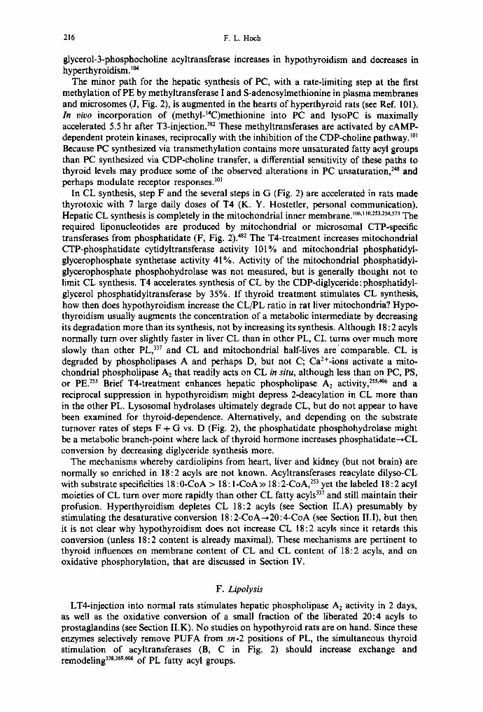
In CL synthesis, step F and the several steps in G (Fig. 2) are accelerated in rats made thyrotoxic with 7 large daily doses of T4 (K. Y. Hostetler, personal communication). Hepatic CL synthesis is completely in the mitochondrial inner membrane) °6:1°'253'254,573 The required liponucleotides are produced by mitochondrial or microsomal CTP-specific transferases from phosphatidate (F, Fig. 2). 482 The T4-treatment increases mitochondrial CTP-phosphatidate cytidyltransferase activity 101% and mitochondrial phosphatidyl- glycerophosphate synthetase activity 41%. Activity of the mitochondrial phosphatidyl- glycerophosphate phosphohydrolase was not measured, but is generally thought not to limit CL synthesis. T4 accelerates synthesis of CL by the CDP-diglyceride:phosphatidyl- glycerol phosphatidyltransferase by 35%. If thyroid treatment stimulates CL synthesis, how then does hypothyroidism increase the CL/PL ratio in rat liver mitochondria? Hypo- thyroidism usually augments the concentration of a metabolic intermediate by decreasing its degradation more than its synthesis, not by increasing its synthesis. Although 18: 2 acyls normally turn over slightly faster in liver CL than in other PL, CL turns over much more slowly than other PL, 337 and CL and mitochondrial half-lives arecomparable. CL is degraded by phospholipases A and perhaps D, but not C; Ca2÷-ions activate a mito- chondrial phospholipase A2 that readily acts on CL in situ, although less than on PC, PS, or PE? 53 Brief T4-treatment enhances hepatic phospholipase As activity, 255'~ and a reciprocal suppression in hypothyroidism might depress 2-deacylation in CL more than in the other PL. Lysosomal hydrolases ultimately degrade CL, but do not appear to have been examined for thyroid-dependence. Alternatively, and depending on the substrate turnover rates of steps F + G vs. D (Fig. 2), the phosphatidate phosphohydrolase might be a metabolic branch-point where lack of thyroid hormone increases phosphatidate~CL conversion by decreasing diglyceride synthesis more.
The mechanisms whereby cardiolipins from heart, liver and kidney (but not brain) are normally so enriched in 18:2 acyls are not known. Acyltransferases reacylate dilyso-CL with substrate specificities 18: 0-CoA > 18: 1-CoA >> 18 : 2-CoA, 253 yet the labeled 18: 2 acyl moieties of CL turn over more rapidly than other CL fatty acyls 337 and still maintain their profusion. Hyperthyroidism depletes CL 18:2 acyls (see Section II.A) presumably by stimulating the desaturative conversion 18:2-CoA~20:4-CoA (see Section II.I), but then it is not clear why hypothyroidism does not increase CL 18:2 acyls since it retards this conversion (unless 18:2 content is already maximal). These mechanisms are pertinent to thyroid influences on membrane content of CL and CL content of 18:2 acyls, and on oxidative phosphorylation, that are discussed in Section IV.
F. Lipolysis
LT4-injection into normal rats stimulates hepatic phospholipase A2 activity in 2 days, as well as the oxidative conversion of a small fraction of the liberated 20:4 acyls to prostaglandins (see Section II.K). No studies on hypothyroid rats are on hand. Since these enzymes selectively remove PUFA from sn-2 positions of PL, the simultaneous thyroid stimulation of acyltransferases (B, C in Fig. 2) should increase exchange and remodeling 338'369"~ of PL fatty acyl groups.

Lipids and thyroid hormones 217
Adequate levels oi" thyroid hormone are permissive for epinephrine-induced but not basal unstimulated lipolysis of triglycerides in adipose tissue; n4'576 hyperthyroidism triples both basal and epinephrine-stimulated lipolysis; one injection of T3 into a euthyroid rat has no effect at 3 hr, but 4 injections over 12hr maximize the lipolytic response to epinephrine, n2 Thyroid exerts these effects via the responsiveness of lipolysis to [epi- nephrine]: hypothyroidism lowers sensitivity to the point that maximal lipolytic rates require 3 times more epinephrine than do euthyroid preparations, but the maximae are similar; 16 hr after T3-injection of hypothyroid rats, epinephrine-sensitivity improves. ~76 Thus, hypothyroidism does not appear to reduce the amount or capacity of lipolytic apparatus, although the amount of hormone-sensitive lipase does not appear to have been measured directly. Since the lipolytic effects of other hormones (e.g. norepinephrine, ACTH, TSH, glucagon, vasopressin) also depend on the thyroid state, it seems that thyroid regulates through effects on [cAMP], the phosphorylation state of the hormone-sensitive lipase (see Section II.F and III.B), and/or the properties of cell membrane receptors (see Section III).
G. Ketogenesis
Ketogenesis from 18: 1 oxidation (steps A, C, G, H and I in Fig. 1) increases in perfused livers 333'4n and in hepatocytes 44s from LT3-pretreated rats. Livers from hypothyroid rats either make more ketone bodies but oxidize less 18:1 to CO2, 233 or show no change from normal? I~ The HMG-CoA pool destined for ketogenesis is mitochondrial, that for cholesterogenesis cytosolic, and the HMG-CoA synthases are compartmented ac- cordingly. The mitochondrial HMG-CoA synthase may be 'rate-limiting' for ketogenesis (see Ref. 650), but data are not available on its activity and that of the HMG-CoA lyase in different thyroid states.
H. Cholesterol Metabolism
A variety of interacting physiological stimuli, including thyroid state, regulate choles- terol metabolism. 6s'394'42s Thyroid-sensitive sites in cholesterol metabolism include steps in intraceUular synthesis, conversions to other sterols, cholesterol processing at cell mem- branes, and cholesterol transport.
1. Intracellular Cholesterol Metabolism
The rate of cholesterol synthesis from acetate (but not from mevalonate) and the turnover of cholesterol relate directly to thyroid levels. 122 The hepatic microsomal ~-hydroxy-fl-methylglutaryl-coenzyme A reductase (HMGR; J in Fig. 1) is said to be 'rate-limiting' for acetyl-CoA-*cholesterol. This enzyme is highly regulated, by hormones, and by cholesterol and its precursors and metabolites. Cholesterol or its oxidized products decrease cholesterol synthesis by suppressing transcription of the HMGR gene; LDL, mevalonate, 25-hydroxycholesterol, and phosphorylation of H M G R accelerate HMGR degradation. In thyrotoxic rats, HMGR, acetate thiokinase, and HMG-condensing enzyme activities all increase over normal rates; in hypothyroids only H M G R activity decreases, and to ver~, low levels, t9° Experiments on the effects of thyroid treatment under different conditions give complementary information on the time course of the activation and later induction of HMGR. Perfusion of isolated livers of hypothyroid rats with T3 activates H M G R within 30 min, 5-fold at 120 min and 10-fold at 360 min) 9° One injection of T3 into hypothyroid rats induces HMGR: activity is unchanged when the earliest time of measurement is 12 hr, and begins to rise only at 36 hr, to reach a maximum at 48 hr. 19v
Thyroid treatment induces H M G R even more strikingly in hypophysectomized rats, but as slowly as in hypothyroid rats? 29 The liver microsomal HMGR activity in untreated animals is depressed more than the 50% seen in hypothyroids, to barely detectable levels. A dose of LT3 that saturates 90% of liver nuclear T3-receptors increases activity only after
JPLR 27/3--E

218 F .L . Hoch
36 hr, and maximally at 48-72 hr; HMGR activity in rough microsomes rises to 4 times normal, and acetate is converted to cholesterol at 10 times the normal rate. Under these circumstances, thyroid hormone induces HMGR only in liver and adrenal, and not in kidney, brain, or testis. T3-treatment of normal rats does not activate. Inhibitor studies indicate that these relatively slow inductions in hypophys~tomized rats involve new mRNA synthesis, m'429 Forty-eight to 72 hr after T3-injection, reductase mRNA appears together with a 97 kDa protein that reacts with antisera to pure HMGR, and reductase activity accelerates many-fold. 52° An HMGR-immunoreactive, 35 kDa protein is present in untreated hypophysectomized rats and is not augmented after T3-treatment. The lag period of 36 hr practically eliminates the possibility that T3-occupancy of nuclear receptors is directly promoting HMGR gene expression (see Section II.B.2), and indicates that the increased activity at 3 hr of liver perfusion is an activation of existing enzyme. It seems likely, then, that synthesis of new HMGR is mediated through secondary alterations after T3 enters the cell. Zammit and Easom 652 propose that covalent modification of HMGR mediates A[HMGR]: phosphorylated enzyme is inactive and is proteolytically degraded faster than the active dephosphorylated form. ~7 This would explain the sequence activation~persistence but not activation~synthesis.
Injection of a glucocorticoid blocks the T3-promoted increase in activity TM and the appearance of 97 kDa active HMGR, and halves the amount of new reductase mRNA; 52° perhaps the absence of ACTH in hypophysectomized rats permits T3-induction to exceed that in hypothyroid rats. Feeding mevinolin or cholestyramine to these animals increases HMGR activity to levels only 10-40% of those reached after T3-injection. It is concluded that T3 is not absolutely required, and may induce HMGR indirectly, perhaps via oxysterol metabolites of mevalonate. Thyroid also activates HMGR in concert with insulin or fasting/refeeding. In hypophysectomized diabetic rats, the activity that insulin induces at 2 hr, and that T3 induces at 54 hr, are each blocked by administration of glucagon or glucocorticoids334--perhaps indicating that cAMP-mediated protein-phosphorylation blocks both enzyme activation and gene expression. These studies may be pertinent to possible thyroid effects through [cAMP] regulation (see Section III.B).
Most investigators agree that dephosphorylation activates, and phosphorylation deacti- vates, the HMGR in situ in microsomes. 29'2~a6s'652 Some find that phosphorylation reversibly inactivates purified, electrophoreticaUy homogeneous HMGR, 427'432 others that it does not. 42s Ness et al. 42s'43°'43~ suggest that a mevalonate kinase, present in preparations of HMGR kinase, accounts for effects attributed to the HMGR kinase, and that the allosteric kinetics of NADPH as cofactor for the double reduction of HMGCoA may mediate regulatory effects. Thyroid state controls cytoplasmic [NADPH] (see Section II.C). The significance of HMGR regulation by such covalent modification has been questioned because the enzyme was found to be >90% phosphorylated and inactive under most conditions, including those favoring cholesterol synthesis (see Ref. 652). However, with appropriate rapid methods (using cold-clamped livers from anesthetized fed rats), 80% of HMGR is in dephosphorylated, active form, and insulin-state in vivo acutely regulates HMGR phosphorylation. Analogous specific effects of thyroid-state on HMGR phos- phorylation await similar examination, even though T3-administration takes days to detectably augment protein kinase activities and a general phosphorylation of endogenous proteins (see Section II.B.2). Thyroid also regulates the adenylate kinase and thereby [cAMP] (see Section III.B. 1), which may in turn modulate protein kinase or phosphatase activities.
The HMGR is membrane-dependent. Arrbenius profiles of its activity inflect, when measured in microsomes, 4°1'472'm'56~ and in a partly purified preparation. 427 Alterations of endoplasmic reticulum fatty acyl and cholesterol composition, produced by diet, or by inducing diabetes (thyroid effects have not been examined), shift the transition tem- peratures and alter E, values. 562~ Compared with rats fed saturated fatty acids, the feeding of 18:2 fatty acid doubles the 18:2 content of the endoplasmic reticulum membrane, decreases HMGR activity at 37°C by 70%, and doubles acyl-CoA:cholesterol acyl- transferase activity (Ref. 401; K in Fig. 1). Adding cholesterol to either the saturated fat

Lipids and thyroid hormones 219
or the 18:2 diet decreases HMGR activity by 90%. It has been suggested that membrane cholesterol rather than fatty acyls modulates HMGR activity, because the increased E, above the transition temperature indicates decreased fluidity? °~ The opposing effects on acyl-CoA:cholesterol acyltransferase activity are ascribed to its location in the rough endoplasmic reticulum, while the HMGR is in the smooth endoplasmic reticulum-- although cholesterol accumulation does not seem to have been so localized. Studies comparing normal and leukemic lymphocytes 4n also conclude that endoplasmic reticulum membrane cholesterol regulates HMGR activity. If the general inhibition of lipogenesis by high contents of 18:2 acyls in endoplasmic reticulum 279'4~3 includes inhibition of cholesterol synthesis, T3-injection could produce early activation of HMGR through the observed rapid depletion of ER 18:2 acyls, in a period when cholesterol/fatty acyl ratios stay constant. 236'24s
Cholesterol 7~-hydroxylase (L in Fig. 1), an NADPH-dependent cytochrome P450 (perhaps a specific P450) of the endoplasmic reticulum, catalyzes 'the rate-limiting, committal step' in hepatic bile acid biosynthesis, and is thus a major disposal path for cholesterol. The enzyme is a regulatory site: it has a short half-life (2-4 hr) and a diur- nal rhythm, and is rapidly activated by feeding carbohydrate to starved animals. Almost all physiological regulators, including thyroid hormones, act on the 7~-hy- droxylase in coordination with, and in the same direction as, their actions on the HMGR, the acyl-CoA:cholesterol acyltransferase, and the cholesterol ester-hydrolase. Hypo- thyroidism decreases activity. 5s2 Thyroid treatment activates in hypophysectomized rats, and so acts independently of those hormones evoked by pituitary trophins. However, thyroid state also modulates other regulators of the hepatic cholesterol 7~-hydroxylase.
The cholesterol 70e-hydroxylase is more sensitive to in vivo thyroid levels than is the HMGR. 592 In fasted euthyroid rats, refeeding with glucose activates cholesterol synthesis within 3 hr, and increases 70~-hydroxylation starting 1 hr laterfl 3 When rats are made increasingly hypothyroid with graded doses of propylthiouracil, and then fasted and refed, the lowest dose decreases stimulation of 7~-hydroxylase activity, but only the highest dose decreases activation of acetyl-CoA conversion to fatty acids and to cholesterol. Con- versely, making rats hyperthyroid with graded doses of T4 increases glucose-induced 7~-hydroxylase activity at the lowest T4-dose, cholesterol synthesis at the highest. Further, glucose stimulates 7~-hydroxylation more rapidly in hyperthyroid than in hypothyroid preparations, but activates cholesterol synthesis at about the same time in both.
The mechanisms of the early regulations are consistent with an activation by de- phosphorylation, except for the adrenal cortex cholesterol esterase which is inactivated thereby, and the liver enzyme which does not appear to be affected at all. However, the 7~-hydroxylase has only been studied through activity changes in microsomal preparations incubated with protein kinase + MgATP, or protein phosphatase. ~Ts'33L524 No measure- ments of the phosphorylation stoichiometry of the purified enzyme are on hand, so the 7~-hydroxylase does not as yet meet the criteria of Krebs and Beavo 319 for an enzyme regulated by phos/dephos. Nor have the rapid assays used by Zammit and Easom 652 on the HMGR been applied to this enzyme.
The details of the modulation of the cholesterol 7~-hydroxylase differ from those of the HMG-CoA reductase in several ways. Insulin administration, perhaps by way of a specific phosphoprotein phosphatase, activates the reductase but not the hydroxylase; hydro- cortisone activates the hydroxylase but deactivates the reductase. Possibly the different mechanisms are involved in what have seemed to be 'biphasic actions' of the administered thyroid hormones 226 on cholesterol levels in cells and blood, in which low T3-dosage raises cholesterol levels and high T3-dosage lowers them.
Thyroid hormones also promote steps in the synthesis of bile acids from 7~-hydroxy- cholesterol. 332"4~4 In rats, but not in humans, the hormone increases [chenodeoxycholic acid] at the expense of [cholic acid], apparently by activating the microsomal (but not the mitochondrial) 26-hydroxylase, and deactivating the 12~-hydroxylase. Hypothyroidism effects the opposite changes. These modulations are not general to cytochrome P450s, since

220 F.L. Hoeh
thyroid treatment increases, and hypothyroidism decreases, total liver P450. Such select- ivity also appears in the undisturbed ability of the hypothyroid rat to induce specific hepatic P450s after treatment with methylcholanthrene, u3 phenobarbitone and other xenobiotics. ~1
Thyroid state affects steroid kinetics in the whole body. In thyrotoxic humans, sec- retory rates of the C-21 steroids cortisol and aldosterone are augmented, as is their metabolic clearance which includes a shorter half-life and increased turnover. Is4 Hypo- thyroidism exhibits opposite effects. Contrarily, thyrotoxicosis depresses metabolic clear- ance and secretory rates of the C-19 and C-18 steroids testosterone and estradiol, while hypothyroidism increases them. These phenomena seem to be caused in part by the thyroid-dependence of specific, high-affinity, binding proteins in plasma of males and females. The [free steroid] levels in plasma are not changed, nor are symptoms of steroid excess or deficiency obvious. Thyroid levels do affect cellular metabolism however. Thyroid increases aromatase activity. An increase in hepatic reduction of C------C bonds in ring A is attributed to an increase in [NADPH] (see Section II.C) rather than to induction of the A4-corticosteroid hydrogenase. 645 Thyroid also accelerates 5~-reductions and 17~-hydroxysteroid dehydrogenations. 184 Sex difference regulates thyroid effects on pro- gesterone metabolism in hepatic microsomes. 298 In males, progesterone hydroxylation is normally faster, and A4-reduction slower, than in females. T4-treatment decreases hydroxylation and increases A4-reduction in males but not in females.
Intracellularly, the lysosomal acid cholesterol esterase activity in liver and adipose tissue (but not in heart) is thyroid-dependent: ~47 hypothyroidism decreases activity, T3-treatment for 3 days increases it.
2. Cholesterol Transport
Thyroid hormones reduce plasma [cholesterol] at least in part through increased hepatic catabolism of cholesterol. 397 The hypocholesterolemic effects of thyroid hormones offer a therapeutic opportunity for prevention and even reversal of atherosclerosis. However, the strong inotropic and chronotropic cardiac effects of LT4 and LT3 preclude their use where most needed, in coronary atherosclerosis. DT4 lowers [cholesterol] and stimulates heart action less than LT4 but still formidably. A recently described T3-analogue, a synthetic thyromimetic SK&F-94901, lowers plasma [cholesterol] in hypothyroid rats as effectively as LT3 by acting on liver cholesterol metabolism but has a strikingly lesser cardiac chronotropic effect; its mechanism of action is described in Section II.B.
Thyroid hormones participate in the regulation of several steps in the pathways described by Bilheimer 3s as transporting exogenous or endogenous cholesterol, and those of reverse transport. In the endogenous transport, hypothyroidism decreases the expres- sion of hepatic plasma membrane LDL-receptors, and the decreased LDL degradation contributes to the observed hypercholesterolemia; 53° T3 treatment restores receptor- mediated catabolism of LDL. 74 In the reverse pathway, the PC-cholesterol acyltransferase, which is thought to esterify cholesterol in plasma, is poorly secreted from hepatocytes of hypothyroid rats, further contributing to the increased [cholesterol]. 2°9'493 In the exogenous path, it has been proposed that the intestine plays a major role in cholesterol metabolism. 3s7 Thyroid state-altered rates of absorption, synthesis and excretion are said to be offset by compensatory changes in cholesterol transformation, to maintain the cholesterol balance. These workers disagree with the usually accepted idea that continuous thyroid treatment decreases blood cholesterol concentration by stimulating cholesterol degradation and elimination more than cholesterol synthesis. If this were so, they point out, such treatment should progressively deplete body cholesterol--but measured body cholesterol content stays constant in hyperthyroidism. They show that hyperthyroidism depresses the coefficient of intestinal absorption of cholesterol (the amount of dietary cholesterol absorbed/ingested), which promotes biosynthesis and results in large increases in fecal excretion of cholesterol and products. Conversely, hypothyroidism increases the absorp- tion coefficient, decreases fecal excretion, and thereby depresses biosynthesis. No mech-

Lipids and thyroid hormones 221
anisms for this homeostasis are specified, but presumably they would involve effector actions of cholesterol and metabolites on synthesis and oxidations.
Thyroid dysfunction is associated with altered compositions and concentrations of all major plasma lipoprotein classes. 2°9,3°5 The lipoprotein lipase that heparin releases into blood from extra-hepatic capillary endothelium is involved in all three pathways of plasma cholesterol metabolism. Its activity decreases in severe hypothyroidism and normalizes after hormone treatment. Conflicting results are reported in thyrotoxicosis, but Hansson et al. 2°9 find no change in human plasma activity, where plasma [cholesterol] decreases 20% through depressed [LDL] and total [HDL]; it is not clear what lipoprotein lipase hydrolysis of glycerides and the subsequent concentration of cholesterol into IDL particles contribute to plasma [cholesterol]. The role of the hepatic cell membrane lipase in plasma lipoprotein metabolism is also not well understood, but heparin releases less than normal lipase activity into the blood stream in hypothyroid subjects and more than normal in euthyroids treated with T3. 2°9'263 Feeding hypothyroid rats a diet enriched in 18:2 and 20:4 restores levels of heparin-released hepatic lipase in a week, suggesting that depression of liver cell membrane fluidity or co 6-PUFA content is responsible for defective binding and/or content of this enzyme. 263
I. Fatty Acid Desaturation
Oxygen-dependent liver microsomal fatty acyl-CoA desaturases specific for introducing a double bond into the A9 or A6 positions (and probably the A5 and A4 as well) share an electron-donating transport system that consists of NADH-cytochrome b 5 reductase and cytochrome bs. In microsomes from hypothyroid rats, the V a n d / ~ for the reductase, measured with cytochrome c as electron acceptor, are at normal levels. 243 Cytochrome b5 content is 50% above normal in microsomes from hypothyroid rats, 30% below normal in those from thyrotoxic animals, and is transiently decreased hours after hypothyroids are injected with T3 (Table 3). However, the thyroid dependency of the desaturase system resides in the desaturase proteins, as shown by spectrophotometric assays of the rates of reoxidation of reduced b5 in the presence of the acyl-CoA substrate. 4s°'4s7'4ss Apparent first-order rate constants, k, are calculated from initial rates; k + is the rate constant attributable to electron transport through the substrate, and k - is the cytochrome b5 autoxidation rate constant observed in the absence of substrate (Table 3). In hypo- thyroidism, k- is at normal levels; T3 injected into hypothyroid rats progressively raises k - over 4 hr; in T3-injected normal (thyrotoxic) rats k - is high.
TABLE 3. Fatty Acyl-CoA Desaturase Activities, Cytochrome bs Autoxidation Rates and Contents in Rat Liver Microsomes, and Changes after LT3 Injection(s)*
Hypothyroids + LT3 (hours after injection)
Controls Hypothyroids Controls (6--8) (6-10) 0.5 (2) 1.0 (6) 2.5 (5) 4.0 (6) + LT3 (8)
A9-Desaturase: kw~ (rain-i) 2.7 3.1 3.7 4.9 5.6 8.9 ̂ 10.2 A
K m (/~M) 2.0 1.6 1.9 0.7 1.2 3.3 5.3 A6-Desaturase: kwx (rain -t ) 4.5 2.2 c 6.1 4.4 c 6.0 A 3.5 2.8
Km (IZM) 17.9 3.6 9.0 10.2 9.7 5.1 2.6 A5-Desaturase: V (pmol/min/mg) 12,8 7.0 26.0
Kra (/zM) 0.15 0.06 0.14 Autoxidation: k - (rain- t) 1.9 1.6 2.2 2.4 a 2.4 e 2.9 e 4.2 a Cyt. b s (nmol/mg) 0.13 0.20" 0.20 0.17 0.14 e 0.18 0.09 e
*See Table 1 for details, and the text for definitions of kinetic parameters. Only means are shown; for 42 values, the coefficient of variation is 21.1% + 2.1% SE. For group comparisons: p < ~'0.001; be0.01; ~"0.05; lower case symbols denote p values for comparisons of Hypothyroids vs. Controls, upper case symbols for comparisons of Hypothyroids + LT3 vs. Hypothyroids and Controls + LT3 vs. Controls.
222 F.L. Hoch
The extrapolated maximal desaturation rate constant, km~ = k + at [S] = oo, and Km for the A9-desaturase (stearoyl-CoA, 37°C) in fasted hypothyroid rats is the same as in fasted euthyroid rats; k + is 4 times normal in fasted thyrotoxic rats (Table 3). In hypothyroid animals a fasting/refeeding-with-carbohyrate cycle does not increase the A9-dcsaturase k +, whereas in normal rats it raises k + strikingly. 243'4ss One LT3 injection increases the A9-desaturase k + in fasting hypothyroids linearly over 4 hr (Table 3; r = 0.972) and (not shown) to 5-fold at 17hr. The T3-induced changes in microsomal PL fatty acyl composition 2~ do not appear to mediate the increased k +, as judged from Arrhenius profiles. 243 Brain A9-desaturase activity decreases from birth, to reach 10% of that in the fetus at maturity; fasting/refecding does not consistently induce the adult enzyme, s"
The purpose of the thyroid dependency of this enzyme is likely connected with the thyroid dependency of the enzymes of de novo fatty acid synthesis, which also are activated 1-4 hr after LT3 injection in hypothyroid rats, and are subsequently synthesized (see Section II.B). In view of the activations of acetyl-CoA carboxylase and the fatty acid synthetase by dephosphorylation, it is therefore of some interest that 2 hr incubation of Tetrahymena pyriformis with/~-adrenergic agonists increases [cAMP] together with fatty acid synthesis and the activities of the microsomal A9- and A 12-desaturases. 6'3 The dietarily 'induced' A9 enzyme in rat liver microsomes is partly and progressively inhibited by a protein kinase + ATP (unpublished data), further suggesting that the A9-desaturase acts in concert with the fatty acid synthesizing process. Unlike the cytosolic fatty acid synthesizing enzymes, the A9-desaturase has not yet been resolved from other microsomal polyl~ptides for study of its phosphorylation state or for immunoassay.
Three metabolic roles can be distinguished for the A9-desaturase in converting 18:0-CoA~18:I-CoA. Derived (as well as dietary) 18:l-acyls are normally a major membrane component. Oleoyl-CoA becomes a precursor for PUFA in membranes, mainly 20:3co9 produced by the A6- and A5-desaturases, only when co6-EFA intake is deficient: the A9 enzyme is the first step in producing unsaturated fatty acids from endogenous sources when necessary. The A9-desaturase is proposed as a major locus where dietary linoleate down-regulates hepatic lipogenesisy 9 18:2 also selectively damps the enzymes of de novo fatty acid synthesis, m While dietary carbohydrate rapidly induces the enzyme, dietary 18:2 inhibits the A9-desaturase even more effectively and rapidly than it inhibits the fatty acid synthetase, the A9 enzyme having the much shorter biological half-life of 3-4 hr. Insulin induces Ag-desaturase activity when incubated for 48 hr with hepatocytes. 2s° In diabetic rats, activity stops in microsomes of liver and fat; fasting/refeeding induces the enzyme only 24 hr after insulin treatmenU 6s Interestingly, thyroid treatment of diabetic rats induces the A9-desaturase and fatty acid synthesis independently of insulin. ~75
Liver microsomal A6-, A5- and A4-acyl-CoA desaturases, 56'27s,57° and separate micro- somal desaturases that act on the corresponding PL-acyls rather than acyl-CoAs as substrates, ~6 synthesize PUFA for membrane components (mainly 20:4<o6 and 22:6co3 in rat organelles) and as precursors of prostanoids. In hypothyroids, the acyl-CoA A6- and A5-desaturase km,~ values are about half-normal (p > 0.05 for the A5- V); values for KI are normal (p > 0.05 for the low A6-desaturase Kin) (Table 3). Injecting hypothyroid rats with LT3 transiently increases the A6-desaturase km~ about 3-fold in 2.5 hr. Thyrotoxicosis doubles the A5-desaturase kmax, and leaves the A6-desaturase kmax unaltered. The high A6-desaturase activity in the thyroid-sensitive brain of fetal rats persists to 20 clays after birth, as the brain becomes less responsive to thyroid, 94 in consistence with the behavior of the hepatic enzyme.
The thyroid-dependence of the hepatic A6- and A5-desaturases seems to account for the 18:2 acyl accumulation in organelle membranes of hypothyroid rats, and depletion in thyrotoxics. ~4°'14~'236'239'242'262 It accounts as well for decreased ability of the livers of hypothyroid rats to convert administered [1-14C]18:2 fatty acid to 20:4 fatty acyls (Table 2). Livers of diabetic rats show a similar defect, ~58 but no insulin-independent T3-induction of the A6- and A5-desaturases has been described that parallels the induction of the A9-desaturase. m The transience of the restoration of A6-desaturase activity after hormone injection (Table 3) suggests either enzyme turnover or successive activation and
Lipids and thyroid hormones 223
deactivation. Despite the restored A6-desaturation activity, which should improve the 18:2--,20:4 conversion (unless the AS-desaturase proves to be unrestored by T3), T3-administration counter-correctively depresses PUFA contents in mitochondrial and microsomal lipids even below the abnormally low levels in untreated hypothyroid rats (Ref. 248; Table 1). The most likely concomitant T3-induced mechanism to remove PUFA is massive and rapid substitution by newly synthesized saturated and mono-unsaturated fatty acyls (see Section II.B.1), like that after fasting/refeeding. 7̀ s Phospholipase A2 action (see Section II.F) may remove PUFA.
1. Anuran Metamorphosis
Thyroid-induced metamorphosis involves early general increases in hepatic protein synthesis, and phospholipid synthesis increases even earlier--presumably to provide membranes to support the increased translation of newly expressed mRNA. 5~599 We have recently found that thyroid control over PUFA composition is also involved very early in amphibian metamorphosis.
Tadpoles are naturally athyroid. Lipids of tadpoles and frogs have distinctive fatty acyl unsaturation profiles. In the livers of Rana pipiens, Xenopus laevis and Rana catesbeiana tadpoles the major unsaturated fatty acyls are monoenes and the polyene acyls of the dietarily essential fatty acids 18:2to6 and 18:3to3. 23a'24~ Frog livers contain less monoenes and more PUFA products of A6- and A5-desaturations of the essential fatty acids. Heart lipids from R. pipiens tadpoles or frogs have higher proportions of biodesaturated polyene acyls than do their respective liver lipids. Liver lipids of R. catesbeiana tadpoles contain 67% neutral lipids, frog liver lipids contain 75% polar lipids. Both neutral and polar lipids from the tadpoles are higher in monoenes than are the corresponding lipids from frogs. We do not know the relative amounts or the fatty acyl compositions of the PL subclasses. Tadpole livers metabolize intraperitoneally injected [1-14C]18:2to6 unexpectedly vigoro- usly, considering how few derivatives accumulate in their lipids. In 18 hr, 20% of the counts are incorporated into liver lipids, of which 40% is in polar lipids. Frogs incorporate 10% of the carbon counts into liver lipids, of which 90% is in polar lipids. Of the total counts in either tadpoles or frogs, 37% is in biosynthesized co6-PUFA derivatives. This degree of metabolism may be compared with that in livers of rats 6 hr after receiving labeled 18:2 (Table 2); normal rats incorporate 3.5% of injected label and convert 25% of it to the 20:4 derivative; hypothyroids incorporate 0.9% of label and convert only 8.7% to 20:4.
Thyroid hormone specifically induces Anuran metamorphosis) 98 During the natural development of tadpoles, the proportions of liver lipid mono-unsaturated acyls decrease and those of polyene acyls increase at the stages when plasma thyroxine levels rise 5s8 and urea synthesis begins, m6 as shown in Fig. 3. The changes in unsaturation pattern follow the natural surge of circulating [T4] at least as closely as does the appearance of urea. Thyroid-induced metamorphosis is accompanied by similar lipid alterations. Immersing tadpoles in 1 #M T3 for 48 hr converts their liver lipid profile to or beyond the polyene pattern typical of frogs. Injecting tadpoles once with T3, 0.2 #g/g, produces similar but lesser and transient changes over 5 days. Injecting frogs with T3, 1 #g/g, decreases liver monoene contents transiently over 2 days and in addition partially depletes to 6-PUFA--as it does more rapidly in hypothyroid rat liver organelle membranes (Table 1). When tadpoles are injected with T3 at time = 0 and with labeled 18:2 at 18 hr, the following changes occur. The ratio of liver PL to total lipids increases to reach 76% by the 8th day after T3 injection. The PL incorporate more of the ~4C, the neutral lipids, less. The proportion of label in desaturation products of 18:2 in the PL increases linearly over the 8 days to become 2.5 times greater than in untreated tadpoles or in frogs. Similar T3 treatment changes tadpole liver endoplasmic reticulum from simple vesicular membranes to more complex lamellar s t r u c t u r e s . ~7
We conclude that, in tadpole livers, fatty acyl unsaturation patterns reflect a high content of neutral lipids that are repleted with monoene acyl groups. In frogs, unsaturation

224 F. L. Hoch
200-
150~
50"
Told
~FA
;o 1's ~0 is Frog Tadpole Stage
Fio. 3. Developmental changes in the overall unsaturation index and in the contributions of polyunsaturated and mono-unsaturated fatty aeyls in the total liver lipids of livers of R. catesbeiana tadpoles; and comparisons with values in frogs. Untreated tadpoles were observed from Stage 10 to Stage 24. Values of unsaturation indices calculated from measured fatty acyl compositions are shown for the sum of all fatty acyls (Total), for the sum of the polyunsaturated fatty acyls (PUFA) and for the sum of the mono-unsaturated fatty acyls (MUFA). The dashed lines indicate values for plasma thyroxine concentrations (1"4, in ng/40 ml) taken from Ref. 588 and for hepatic urea
concentrations (Urea, in #g/0.4 g) taken from Ref. 16.
comprises both the high polyene content characteristic of their PL, and a greater proportion of PL in their liver lipids. Both natural and T3-induced metamorphosis involve a shift from the tadpole lipid pattern toward the frog pattern. The shift is early enough to serve as a biochemical marker for metamorphosis, like urea synthesis. Tadpole livers convert 18:2 to co6-PUFA derivatives as well as frog livers do, so thyroid hormone is not necessary for the desaturations involved, but perhaps is necessary for the synthesis of PL that retain the PUFA produced. T3 injection also increases desaturations in tadpoles, indicating that T3 is sufficient but not necessary to stimulate Anuran hepatic A6- and A5-desaturases.
J. Fatty Acid Oxidation
Fatty acid oxidation rate is proportional to the thyroid state.' Mechanisms that regulate the partition of fatty acids between oxidation and esterification in liver are discussed by Zammit? 5° The major site of control of fatty acid oxidation itself is in the mechanisms that transport cytoplasmic fatty acyl-CoA, with the acylcarnitine as an intermediate, to the fatty acyl-CoA dehydrogenase in the mitochondrial matrix adjacent to the electron- transport chain. Cytoplasmic acyl-CoA--+ acylcarnitine conversion is 'rate-limiting' for long-chain fatty acid oxidation in isolated mitochondria, '59'392 because the transferase system in intact mitochondria is partly latent. Thyroid hormones increase hepatic oxidation of 18:1 by accelerating entry of 18: 1-CoA into the mat r ix : "
Carnitine acyltransferases, as exemplified by the carnitine palmitoyltransferase (CPT), are the most important, although the translocase may also regulate. The CPT enzyme(s) are highly regulated 36's4 by positive and negative effectors; by covalent modification (phosphorylation activates2'°); by substrate and product; 55'4~'-463 and by vicinal phos- pholipids in the inner or outer layers of the mitochondrial inner membrane. *sl'~2'~3,rs°'rs3 Kinetic measurements are a dubious basis for determining mechanisms of thyroid control over CPT, because the amphipathic substrates and products form micelles readily and may act non-specifically as detergents, s9
The outer enzyme, CPTo, in intact liver mitochondria normally has about 10-20% of the total CPT activity found in detergent-lysed mitochondria, depending on the detergent (Refs 32, 53; see Table 4); CPT o activity in the intact membrane has been estimated as 0.2% of inner enzyme, CPT i, ac t iv i ty? CPT i activity is expressed fully by gentle disruption of the membranes, but loses appreciable activity when solubilized by more drastic extractions, suggesting that its active configuration requires lipid.

Lipids and thyroid hormones 225
TABLE 4. The Effects of Thyroid State (Fed Rats) on 16:0 Oxidation (nmol/min/mg protein; 37°C) in Isolated Hepatoeytes; and on CPTo and Total CPT Activity, and by Difference, cirri (nmol 16:0-Carnitine Formed/min/mg
protein; 30°C) in Liver Mitoehondria; from Refs 571,572
System Activity Normal Hypothyroid Hypo + LT3
Hepatoeytes Oxidation 1.5 0.3 2.6 Mitoehondria CPTo 3.8 1.7 6.7
Total CPT 33.8 29.6 31.5 A -- Cirri 30.0 27.9 24.8
Whether CPTo and CPT~ are two proteins with different primary structures and lipid affinities, or one protein in the different lipid environments of the inner and outer layers of the mitochondrial inner membrane, is crucial for determining thyroid control mech- anisms. CPTo and CPT~ are said to be the same enzyme by the criteria of their physical properties (molecular weights, solubilities, electro-mobilities), and kinetics (e.g. Km values of membrane-free enzymes for their several substrates), 53'12s and their immunologic identity. 32'5° Evidence presented for liver mitochondrial CPTo and CPTi being differ- ent enzymes, or more ambiguously, different 'forms of CPT', includes the following. CPTo and CPTi have different susceptibilities to inhibition by malonyl-CoA, thought by some to reside in a regulatory subunit; 651 and different Mr values (CPTo = 63 kDa, CPTi = 67 kDa). 491 Polyclonal antibodies raised against a mono-disperse, PL-free CPT o prepared from beef liver mitochondria uncontaminated with peroxisomes 49°'491 (which contain molecularly distinct short- and medium-chain acyltransferases aS) and against a purified CPTi, do not cross-react by Western blot or by inhibiting activity? 91 Although these experiments are thought to demonstrate that CPTo and CPTi differ in physical properties rather than in membrane environment, in earlier work antibodies against a purified homogeneous CPTo purified from calf liver mitochondria 313 inhibited CPTo, but those from 1 out of 3 rabbits also inhibited CPTi.65'314 Taken together with observations that urea or guanidine convert CPTo~CPTi, Kopec and Fritz 314 thought CPTi is a 'transformed product' of CPTo. Since it appears possible that even immuno-reactivity is modified by membrane environment, yet more specific molecular properties such as primary structure may be needed to determine when gene expression of CPT~ or CPTo, and/or membrane lipid changes, regulate CPT activities.
Hypothyroidism halves the activity of the CPTo in isolated rat liver mitochondria, and reduces 14CO 2 production from 14C-palmitoyl-CoA in isolated hepatocytes by 80% (Table 4). T3-treatment restores CPTo activity and 16:0 oxidation even beyond normal levels (?thyrotoxic). Total CPT activity, in lysed mitochondria, remains similarly high in all three groups; by difference, C P T i in hypothyroids is at about the control activity, but in T3-treated animals is rather lower. The approximate constancy of the total CPT activity speaks for there being one enzyme that thyroid treatment converts from inactive CPT~ to active CPTo form, thereby increasing the oxidation of 16:0. Given the uncertainties of these activity measurements, additional quantitation of thyroid effects on amounts of each 'form' are needed, as well as more direct determination as to whether or not thyroid induces only the CPTo form.
It is also possible that thyroid state regulates CPTo activity in the intact cell indirectly, through [malonyl-CoA] and CPT sensitivity to inhibition by malonyl-CoA. In perfused livers, hypothyroidism decreases [malonyl-CoA] and hyperthyroidism does not affect [malonyl-CoA], so Miiller e t al . 4H doubt its regulatory role in thyroid effects. However, in freeze-clamped livers, hypothyroidism doubles [malonyl-CoA] while hyperthyroidism decreases it slightly, 365 effects consistent with a regulatory role. Malonyl-CoA inhibits the increased activity of CPTo that follows T3-treatment (Table 4) less than normally, 55 accenting the decreased concentration. The diminution of malonyl-CoA inhibition of fatty acid oxidation is thought to permit a futile, calorigenic cycle of simultaneously accelerated synthesis and oxidation. The CPTo in hypothyroid rats (Table 4) retains normal sensitivity to the high [malonyl-CoA], and so is depressed.
226 F .L . Hoch
When liver mitochondria from normal rats are preincubated in KCl-media, malonyl- CoA does not inhibit; mitochondria from hypothyroid rats lose sensitivity more slowly and those from hyperthyroid rats, faster. Therefore, CPTo is thought to be regulated by components of membranes that respond to physiological stimuli. 3j'46m'65m'653 Malonyl-CoA acts on membrane-bound CPTo like a classical negative allosteric effector, 36 but does not inhibit after the membrane is exposed to phospholipase C (which itself activates CPTo643), or after CPTo is solubilized. Malonyl-CoA inhibition of CPTo depends on high membrane fluidity: in liver mitochondria (especially from fed rats) kept at 25-37°C (but not at <25°C), slow conformation changes make CPTo less sensitive to malonyl-CoA and increase CPTo activity. 6~1
Another mechanism may involve regulation of CPTo through phosphorylation status. Thyroid state modifies the effects of starvation. Fasting increases oxidation 25-50% in mitochondria from euthyroid rats, 300-400% in hypothyroids, and not at all in thyrotoxics (where it is already increased). Starvation normally augments both CPTo and (acyl)carni- tine translocase activities, and hepatic fatty acid oxidation; cycloheximide blocks the increase in CPTo activity, indicating that starvation induces microsomal synthesis of new protein(s). 519 The new protein might be either CPTo or a protein phosphatase or kinase, since starvation signals protein phosphorylation, and phosphorylation may activate CPT. 2j0
(Acyl)carnitine translocase mediates a 1 : 1 exchange of carnitine and an acylcarnitine. At 37°C, the exchange rate appears to be much greater than the capacity for//-oxidation. However, at low tissue [acylcarnitine], the translocase may come to share control over oxidation with the CPTo. 55 Urinary excretion of carnitine, and serum carnitine concen- tration, are proportional to serum IT4] in hypo- and hyperthyroid subjects , 375 but there are no measurements that show cell carnitine availability limits either the translocase or CPT activity. It is not known if thyroid state affects translocase activity, but thyroid regulation of mitochondrial inner membrane CL content and composition offers a possible mechanism to explain some effects attributed to membrane environment. The purified translocase contains bound CL, 149 and reconstitution of the solubilized (acyl)carnitine translocase requires cardiolipins: 43 Adriamycin, which binds CL specifically (see Section IV.B.5), inhibits both native and reconstituted translocase activity. 4~° Since the purified CPT is also activated by CL, Noel and Pande ~.3 propose that CL provides orienting binding sites for the translocase and the transferase, and thereby facilitates the flow of acylcarnitines from the outer to the inner layers of the membrane.
The asymmetry of CL distribution, 75-90% being in the inner layer of the inner membrane of mitochondria (see Ref. 106), resembles that of the activities of liver CPTo and CPT~ (Table 4). If CL does bind CPT firmly in vivo, and if the distinction between CPTo and CPT~ reflects only their membrane environment, the paucity of CL in the outer face may account for the relatively small fraction of CPT in the outer face. Although effects of thyroid state on the sidedness of CL in hepatic mitochondrial inner membranes have not been reported, hypothyroidism increases the proportion of liver mitochondrial CL/YPL by 72% while maintaining the high 18:2 content of the CL. 2gs The physical properties of CL favor the excess CL being in the inner layer (see Section IV); less transferase would then exist in the CPTo form (Table 4). Thyrotoxicosis also increases liver mitochondrial CL/PL 34%, but the CL is depleted in 18:2 by 63%. 5~m If this abnormal CL does not bind CPT fully, the fraction in the CPTo form would increase.
In liver mitochondria of 1-5 day old pigs, CPTo = C P T i .37 In liver mitochondria of neonatal rats none of the CPT is in latent form, 17 CL/PL is low, and the CL 18:2 content is halved, as compared with adults. In adult heart mitochondria, LT3 (0.33 #g/g/day x 2 days) given to hypothyroid rats does not change the activity of CPTo, although it increases liver mitochondrial CPTo (Ref. 55; Table 4). Neither does such short-term thyroid hormone treatment of normal rats (LT3, 1 #g/g/day x 3 days) change heart mitochondrial 18:2 content or CL/PL ratio; 237 we did not examine CL/PL values in heart mitochondria of T3-treated hypothyroid rats. The apparent disparity in T3 effects on heart and liver may reflect only that heart responses require greater dose-time schedules, as is explicated in

Lipids and thyroid hormones 227
Section IV.D. Thyroid feeding for 6 days accelerates palmitate oxidation in perfused hearts of dogs; 174 and LT4-injection, ,,,0.25/ag/g/day x 10 days, more than doubles CPT activity in guinea pig heart homogenates, ss The latter workers attribute the increase in fatty acid oxidation to the high plasma [FFA], and the doubled heart [acylcarnitine] and [total carnitine], after thyroid pretreatment.
In mitochondria of mature brain, the activity of CPTo plus CPTi is about 25% of that in liver;//-oxidation is much slower, and neither CPT o o r C P T i is rate-limiting. 41 Neither thyroid-treatment nor starvation increases activities in these I o w - C L 632 mitochondria. The higher CPTo activity in fetal brain decreases 50% after weaning, as what small amounts of CL there are in the adult brain begin to appear (see Section IV.D).
K. Prostanoid Synthesis
Brief treatment of rats with LT4, 0.2#g/g/day x 2 days, increases prostaglandin synthetase activity by 179'/o in kidney and 300% in lung; at the same time, it inhibits renal PGF2~ metabolism by 40% in pathways that include conversions by 15-hydroxyprosta- glandin dehydrogenase and prostaglandin A13-reductase, and prostaglandin 9-hydroxy- dehydrogenase. ~55'~ The prostaglandin synthetase of spleen microsomes, as measured by the conversion 20:4--*PGE2, also increases in hyperthyroidism but is unaffected by hypothyroidism; however, concentrations of indomethacin or phenylbutazone that inhibit PGE2 formation about 50% in preparations from normal and hyperthyroid animals inhibit only about 20% in hypothyroids. ~s5 It seems unlikely that T3 stimulates conversion of enough hepatic microsomal 20:4 acyl groups to prostaglandins to contribute to the observed rapid depletion of 20:4 (see Section II.A).
L. Sulfolipid Synthesis
Brain myelination and maturation require adequate thyroid hormone levels. *z Hypo- thyroidism in the neonate retards accumulation of brain cholesterol, cerebrosides, and sulfatides. T3-treatment at 18 days after birth does not replenish the very low amounts of these lipids, although T3 given much earlier does stimulate sulfatide synthesis. 62s The defect in sulfatide synthesis is caused by depressed synthesis of 3'-phosphoadenosine-5'- phosphosulfate and/or the subsequent NADPH-dependent (and thereby thyroid-depen- dent?; see Section II.C) reduction of the sulfate of this donor to SOl-. Thyroid effects have been studied in myelin-synthesizing glial cells grown in culture, free of contamination with neurones (which complicate interpretations by producing nerve growth factors in response to T3). 3~5 Growth in T3-supplemented media for 3-5 days stimulates [35S]sulfate incorpo- ration into sulfatides and sulfogalactosyl dialkylglycerols, as well as synthesis of fatty acids and cholesterol, and activity of 2',3'-cyclic-nucleotide 3'-phosphodiesterase. These cultures progressively lose their sensitivity to T3, rather like neonatal rats lose brain responses to developmental thyroid effects (see Section IV.D).
M. Summary
The pervasiveness of thyroid-state influence on enzymatic steps in lipid metabolism is striking. Especially in the branching pathways of fatty acid and cholesterol metabolism (Fig. 1) and conversions of glycerol-3-phosphate to triglycerides and phosphoglycerides (Fig. 2), as well as in fatty acid desaturations, almost all steps are thyroid-sensitive. Several points can be made: (i) the conversions of carbohydrate to lipids show an analogous prevalence of steps sensitive to insulin, and to protein phosphorylation. 2~2 Observed effects of thyroid hormones on protein phosphorylations are suggestive, but no such rigorous correlation has been made with the enzymes involved in lipid conversions. (ii) Thyroid levels do not exert their influence at a single 'rate-controlling' enzyme in a lipid pathway, but on successive enzymes. To regulate the evolution of the lipid product, the hormone must act differentially on each step, and several steps may contribute to rate-control. Such

228 F.L. Hoch
analysis of thyroid controls has been done on the steps of mitochondrial oxidative phosphorylation (see Section IV). (iii) Thyroid stimulation of gene expression can account for only a few of the many sensitive loci in lipid metabolism. In the succeeding sections, mechanisms are discussed whereby thyroid-effected changes in lipid metabolism may amplify signals of T3-receptor occupancy to modulate membrane-dependent catalysts, transporters, and receptors.
III. CELL MEMBRANE AND ASSOCIATED SYSTEMS
Plasma membranes contain a number of enzymes, transporters and receptors that are thyroid-responsive. Such sensitivity may be mediated by mechanisms of gene expression, covalent modifications of the proteins, and lipid-mediated membrane-dependency of the proteins, and ion leaks.
A. Lipid Composition and Physical Properties
All the thyroid-sensitive active proteins in the plasma membrane are known to change activity when membrane lipid composition is manipulated. However, I find no reports on fatty acyl and cholesterol compositions of purifed plasma membranes from animals in different thyroid states, so causal connections with altered activities remain uncertain. Inferences from coincidence and circumstantial evidence should be guarded, especially because the plasma membrane differs in several important respects from organelle membranes. Lipid composition of purified hepatocyte plasma membranes (and erythrocyte membranes) is quite different from that of the thyroid-responsive membranes of the endoplasmic reticulum, mitochondria, and nuclei (plasma membranes are usually sedimen- ted with nuclei as heavy fractions). The unsaturation index of the YPL of hepatocyte plasma membrane ranges from 703°~'5s~ to 130, 423 as compared with about 190 in the organelle membranes, and 215 in heart sarcolemma. 2 Twenty percent of the liver plasma membrane and erythrocyte membrane PL are sphingomyelins, with their characteristically high contents of saturated long chain fatty acyls, and UI ,,,45, which lowers overall unsaturation. Molar ratios o f free cholesterol/PL contents in plasma membranes are 0.35-0.6, and in mitochondria and rough endoplasmic reticulum, 0.03-0.05. ~32'565'5s1'6~5 Some investigators take the presence of cholesterol in mitochondria to denote con- tamination with heavy fractions, but Rogers et al. 4n find that cholesterol-feeding increases mitochondrial cholesterol content in rat liver (but not in other tissues), depresses State 3 respiration, and desensitizes the ATP-synthetase toward oligomycin inhibition.
An acute lipogenic stimulus--feeding carbohydrate for a few hours to a starved rat--does not deplete hepatocyte plasma membrane 18: 2 acyls and actually increases 20: 4 acyl contents (in organelle membranes, it extensively replaces both 18:2 and 20:4 with newly synthesized MUFA and saturated fatty acyls), but decreases the cholesterol/PL ratio by -24%. 58~ More prolonged EFA-deficiency, in Ehrlich cells grown in EFA-deficient mice, does severely deplete plasma membrane co-6-PUFA (18:2 by -80%, 20:4 by -45%) and replaces them with biosynthesized co9-PUFA (18:1, 20:3, 22:3), but the cholesterol/PL ratio and the proportions of PL classes remain at control levels. 3°s'Ssl Thyroid effects on plasma membrane lipid contents need measuring in preparations certified to be minimally contaminated with organelle membranes--it seems possible that plasma membrane and organelle membrane fatty acyl compositions do not respond similarly to thyroid state.
However, thyroid state or dietary lipids affect plasma membrane general physical properties, and enzyme and transporter activities, in a manner consistent with mediation through lipid changes. Plasma membrane lipids are known to modify the mechanism of a number of receptors. TM Arrhenius plots of the motion parameter of 12-nitroxide stearic acid probes in isolated plasma membranes of Ehrlich tumor cells exhibit transitions at 31 °C and 19°C when prepared from control-fed mice, and at 31.5°C and 24.5°C when prepared from EFA-deficient animals. 3°8 The order parameter at 37°C is greater in the EFA-deficient

Lipids and thyroid hormones 229
membranes. These changes are attributed to the decrease in unsaturation at the co6-position, where larger kinks in the fatty acyl chains disturb the packing order especially toward the center of the bilayer. In contrast, when fatty acyls of the plasma membranes of murine T lymphocyte tumor cells in culture are substituted by less or more unsaturated fatty acyls, the temperature-dependence of order parameters and rotational correlation times of both 5- and 12-nitroxide probes remain normal (although enzyme activities change; see below). ~1 It should be noted that the UI of the Ehrlich tumor cells in animals on normal diets is 140-170, higher than the UI of liver plasma membranes, and that tumor cell lipid metabolism and membrane composition differ from those of normal cells, sIT In thyrotoxic rats, liver plasma membrane preparations are abnormally rigid and ordered (as estimated from polarization of fluorescent cis- and trans-parinaric acid probes), and lose their reactivity to added insulin or glucagon. TM Plasma membranes from hypothyroid rats are normally ordered, but T3 added in vitro increases their rigidity. Arrhenius plots of fluorescence polarization of these probes in plasma membranes from thyrotoxic rats inflect at 25°C, in hypothyroids, near 18°C. Such abnormalities seem consistent with excess thyroid levels decreasing fatty acyl unsaturation, and emphasize the need for lipid analysis since thyroid-treatment usually increases unsaturation in organelle membranes.
B. cAMP Synthesis and Hydrolysis
The concentration of cAMP in contact with its target enzymes mediates thyroid state regulation in several cases, notably the hormone-sensitive lipase. Hypothyroidism de- creases the rise of [cAMP] in adipocytes after application of lipolytic hormones (cate- cholamines, glucagon, ACTH). 97'98'616 The mechanisms include simultaneous impairment of synthesis and enhancement of degradation of cAMP, decreased cAMP-dependent protein kinase activity, and decreased phosphorylation of the lipase that lowers its activity. Hyperthyroidism increases adipocyte [cAMP] rise effected by lipolytic agents, and thereby increases lipolysis. Whatever the mechanisms, thyroid control over [cAMP] appears to be analogous to that over hepatic [cholesterol] in that the hormone differentially regulates their synthesis and (more acutely) their degradation, and both cAMP and cholesterol are effectors in a variety of enzyme and transporter processes.
1. Adenylate Cyclase
Thyroid hormone regulation of the adenylate cyclase system in plasma membranes is important in lipid metabolism because cAMP mediates reversible phosphorylation of many enzymes in these metabolic pathways, and because thyroid state regulates the adenylate cyclase system at several membrane-dependent components that respond to lipid changes. Many regulators, together with the thyroid state, are tissue-specific in controlling the multicomponent adenylate cyclase system. Some thyroid effects are primary in the sense that they occur during in vitro incubation with plasma membranes. Some are exerted secondarily and result in permissive effects of thyroid state on such regulators of lipid metabolism as catecholamines, 579 glucagon and insulin.
Some thyroid effects may be mediated by plasma membrane PL. Adenylate cyclase activity and its coupling to hormone receptors require plasma membrane PL. Removal of acidic PL (PI, PS) but not neutral PL (PC, PE, Sph) by phospholipase C inactivates the cyclase and ablates its activation by epinephrine or glucagon) 49-351,497,5°9 Addition of PS (and to a lesser extent PE or Sph, but not PC or CL) reconstitutes the solubilized, inactive cat heart cyclase. TM In rat liver plasma membranes, EFA-deficiency depresses glucagon- stimulated adenylate cyclase activity, 363 but in titrations with A[dietary linoleate], contents of co6-PUFA in phosphatidylcholines correlate inversely with activity; 423 it is not clear to what degree fatty acyl dependency of the adenylate cyclase or the glucagon-receptor systems is being measured. Repletion of the plasma membrane phospholipids of a T-lymphocyte tumor cell or LM cell line with saturated fatty acyls suppresses basal and

230 F.L. Hoch
PGE2-sensitive adenylatc cyclase (and Na+K+-ATPase, see Section III.D) activity, while repletion with 18: 1 or 18: 2 fatty acyls enhances activity, both compared with cells grown with minimal amounts of 18:2. m'4s~ The constancy of overall membrane lipid fluidity parameters during these substitutions suggests a more specific interaction between the PL fatty acyls and the enzyme system. In contrast to the thymocyte and fibroblast cultures, when plasma membrane fluidity of turkey erythrocytes is progressively increased by insertion of c/s-vaccenic acid (18: leo 7), the rate constant for activation of the cyclasc by epinephrine increases linearly with membrane fluidity (measured by fluorescence polar- ization of a probe), indicating that coupling between the epinephrine receptor and the cyclase depends on the lipidsfl °s.494 The maximal activity of the catalytic unit itself, when stimulated by several activators, also increases with lipid fluidity. In contrast, adenosine activates the cyclase in these erythrocytcs independently of membrane fluidity, probably at a site on a G-protein.
Either hyper- or hypothyroidism in vivo or incubation with hormone in vitro can modulate adenylatc cyclase activity in broken-cell preparations. 53s Adcnylate cyclasc activity is twice-normal in minces of fat pads from hyperthyroid rats, and half-normal in hypothyroids. 322 Epinephrine stimulates lipolysis, when added to adipose tissue of hyperthyroid rats, to maximal levels 2-3 times those reached in tissue from euthyroid rats. Even high [epinephrine] fails to activate the cyclase in fat cells from hypothyroid rats; T4-trcatment restores the response in 48 hr.
In vivo mechanisms that mediate such thyroid effects in adipose and other tissues include changes in: numbers and affinity of fl-adrcncrgic receptors in plasma membranes; coupling between receptors and cyclase components; and function and amounts of regulatory and catalytic cyclase components. 579 In vivo thyroid effects differ in heart, adipose tissue, lung, renal medulla, and nucleated crythrocytes from those in liver. 377 Hypothyroidism de- creases, and hyperthyroidism increases, the number of fl-adrencrgic receptors in heart, ~ fa t , 377 turkey crythrocytes, and rat reticulocytes. In hyperthyroid animals, the cardiac receptors have abnormally high affinity for fl-adrenergic agonists. Since blood and tissue [catecholaminc] arc in the normal range in thyrotoxicosis, the many cardiovascular signs seem to arise from hypersensitivity to catecholamines. However, see Rcf. 349 for the opposing view that the thyrotoxic hearts of several species arc not hypersensitive to norcpincphrinc, based mainly on their mechanical function.
The rat liver shows effects opposite to those in the tissues just discussed. Perhaps this specificity is accounted for by observations that, in normal rats, a-adrcnoceptors mediate activation of hepatic glycogen phosphorylase by catecholamines, in other species, //-receptors (see Rcf. 485). Hypothyroidism increases total cyclase activity in rat hepatic plasma membranes by 30-45%, 2°°'2~ and the number of//-adrenergic receptors by 2- to 3-fold. 579 Hyperthyroidism decreases the number of liver//-adrenoceptors, cyclase activity, and response of cyclase to //-agonists, 579 or does not change cyclase activity and the //-receptor response. 2s4 The cyclasc deficiencies in hyperthyroidism arc not associated with a decrease in amount of G-protein, but with lesser effectiveness of the G-proteins in reconstituting adcnylate cyclase in other membranes, and/or with decreases in function of the catalytic cyclase unit (see Rcf. 579 for additional references).
In vivo effects on the permanent coupling between //-adrenergic receptors and the catalytic subunit of adenylate cyclase through the regulatory G-proteins are also tissue- specific. Hypothyroidism decreases the amount of G-protein in rat reticulocyte plasma membranes together with the number of//-receptors; both defects are tentatively ascribed to decreased protein synthesis. 579 Some of the effects of hypothyroidism on the adipocyte adenylate cyclase system are apparently mediated by the GTP-dependent regulatory proteins. 378 The (measured) amount and the activity of the inhibitory protein G i increases while the amount of the stimulatory protein Gs is little changed. Thereby the inhibitory regulation by GTP, adenosine (which inhibits in fat cells), and other metabolites is enhanced by up to 50 times. At the same time, cyclase activity and response to activators are dampened, probably through a change in a / / - or ~-subunit common to Gi and Gs. It is unusual for the hypothyroid state to evoke an increase in the amount of a protein,

Lipids and thyroid hormones 231
because hypothyroidism diminishes gene expression; the mRNAs for the subunit pcptides will be interesting to sec.
Thyroid state not only modifies /~-receptor-mediated lipolytic and positive cardiac chronotropic and inotropic effects, but also what appear to be ~-receptor-mediated effects on these parameters. Thus, norepincphrine (a mixed ~t-fl agonist) elicits no lipolytic response in adipocytes from hypothyroid humans although those from normals arc quite responsive; addition of an ~t-receptor antagonist normalizes the lipolytic action, and hypothyroidism augments an ~-response which inhibits the adenylate cyclase, j91''92'5°2'5°3 Chronotropic dose-effects of norcpinephrine are subnormal in isolated atria of hearts from thyrotoxic rats but normal in those from hypothyroid animals, while dose-effects of epinephrine decrease in hypothyroids and remain normal in thyrotoxics. 5s3 As measured by inotropic actions, atria from hypothyroids have suprasensitive oc-receptors while their fl-receptors are 10-times less sensitive than normals, from measured potency ratios of agonists, and the new susceptibility to inhibition by an ~-receptor blocking agent. 3°,327-329 The sensitivities revert to or beyond control levels after hypothyroid animals receive 7 daily T4- or T3-injections, the only dose schedule examined. Alpha-agonists also make aortae from hypothyroid rabbits contract more forcibly than normals) 92 Hypothyroidism also augments the ~-adrenoceptor-mediated rise in [cAMP] in electrically driven contracting rat heart muscle, and reciprocally depresses the fl-receptor-mediated rise; T4-treatment of hypothyroid rats restores thc normal pattern in 8 days. 326 As in measurements of total cyclase activity and number of adrenergic receptors, liver cells show the opposite pattern: in hepatocytes from hypothyroid rats ~-receptor mediation falls and fl-receptor mediation increases reciprocally. ~5
Evidence is presented that the reciprocity of ~ and/3 effects reflects interconversion of myocardial c¢- and fl-adrenoceptors through an allosteric change in one common precursor adrenoceptor. 327-329 The fl-receptors convert to oc-receptors when the temperature of isolated hearts of frogs or rats is lowered to < 17°C. 329 Others 7° confirm that a f l - ~ transition occurs on cooling isolated frog hearts, but from the effects of irreversible antagonists conclude that separate pools of fl- and ~-precursor receptors are recruited at the different temperatures. Since these studies on isolated hearts seem to preclude adaptations through hormone actions, it seems likely that temperature-sensitive plasma membrane fluidity is involved, since either cold or hypothyroidism decreases fluidity in organelle membranes, and perhaps also in myocardial plasma membranes. It is claimed that the observed effects of thyroid status on adrenergic sensitivities are not reflected in the number of fl- and ~-receptors. As measured by the binding of 3H-dihydroalprenolol at 37°C and 3H-dihydroergocryptine at 25°C to estimate the number of fl- and ~-receptors, respectively, crude preparations of heart plasma membranes from hypothyroid rats have only 20% as many ~-receptors and 70% as many//-receptors as normals. 87 Thyrotoxicosis halves the number of ~-receptors and triples the fl-receptors, while keeping the total number about constant. If hypothyroidism converts fl to ~, the total/3 + ~ should remain constant, but the unexplained measurements of receptors at different temperatures would seem to vitiate such a totalling, from the data of Kunos et al. 326-329 Besides, the shift from fl- to ~-receptor-induced rise in [cAMP] requires that the myocardium be contracting. 326
Thermotropic properties of receptor-coupled adcnylate cyclase activity in washed membrane particles of livers from normal rats may pertain to the above studies. Arrhenius plots of basal activity and activity stimulated in vitro by F- or PGE~ are linear, but plots of activity stimulated by epinephrine or glucagon are inflected. 32° The plots of hormone- activated rates are superimposable and unusual: both transition temperatures, Tt, are at 32°C, whereas those secn with lipid-sensitive membrane-dependent enzymes are usually between 17°C and 22°C; ~9 E, at > T~ is double the E, at < Tt, while the opposite is usually seen in membrane-dependent enzymes where decreased fluidity at the low temperatures increases the damping effects on activity. Because either saturating or lower [epinephrine] and [glucagon] produce identical Arrhenius profiles, Kreiner eta/. 32° think it unlikely that temperature changes alter receptor count, but rather that they act on a protein component common to the hormone-stimulated system but not to the F-- or the PGEl-stimulated

232 F.L. Hoch
systems. One wonders if these observations are explained by an increased number of ~-receptors in hearts at the higher temperatures, and one hopes for direct measurements of thermotropic effects on adrenoceptors in these liver membranes.
Glucagon-receptors also respond to changes in thyroid status. Again there are appar- ently conflicting findings. In liver cell plasma membranes (frozen and thawed) from hypothyroid rats, Sperling et al. 569 report a 50% decrease in the number of glucagon- receptors; receptors have normal affinity for glucagon in vitro and bind less glucagon than normals at all [glucagon]; increased [glucagon] is necessary to produce a maximal increase in [cAMP], and the maximal [cAMP] produced is also half the maximum in euthyroid preparations. In intact hepatocytes from hypothyroid and euthyroid rats, Malbon et al. 377
find that glucagon raises [cAMP] identically. When liver cell plasma membranes are prepared from rats treated with small doses of LT4, 0.05/zg/g/day × 14 days, glucagon- receptors and [cAMP] responses are all normal. 569 But in fat cells from rats treated about 10 times more vigorously with LT3, ~0.1/~g/g/day x 8 days, the number of glucagon- receptors and the response of [cAMP] to glucagon are more than twice-normal. 374 It is difficult to say whether a lesser degree of hyperthyroidism or a greater responsiveness of fat cells contributes to the latter disparity, and whether plasma membrane integrity is involved in the former.
In vitro incubation of rat fat cells, 322 thymocyte plasma membranes, 53s or cat myo- cardium membrane preparations, 352 with I nM to 1 #M LT3 stimulates cyclase activity.* Maximal increases occur at the/ZM concentrations, and as rapidly as 3-5 min. 352'53s From congener efficacies and the effects of transcription blockers, T3 receptors (perhaps in plasma membranes) and protein synthesis mediate these increases in adenylate cyclase activity, except in cat heart. 352 These in vitro actions eliminate possible mediation by receptors for other activators and indicate that thyroid hormones can activate indepen- dently. Thyroid hormones in vitro can also regulate the receptors for other hormones that are coupled to the adenylate cyclase system. Incubation of rat heart ventricle slices (but not isolated membranes) with LT3 quadruples the number of/~-adrenergic receptors at 1.5-2 hr with no immediate further increase; 15 pM LT3 gives half-maximal stimulation, 0.1 nM LT3 saturates /~-agonist binding. 3°3 Synthesis of protein is not involved. A 15-hr incubation increases the number of/~-receptors another 4 times, which does involve synthesis of new protein. Both the early and the later effects apear to be mediated by specific LT3 receptors. The rapid augmentation of cell surface ~-adrenoceptors is attribu- ted to an in vitro effect of T3 that somehow promotes their insertion from an existing cytosolic receptor pool. The increased number of ~-receptors should increase [cAMP], if they are efficiently coupled to the adenylate cyclase, and phosphodiesterase activities are low.
2. Cyclic Nucleot ide Phosphodiesterases
These diesterases are not in the plasma membrane but in the particulate membrane fractions that sediment at > 100,000 x g (microsomes), and in the supernatants. They are included here because there is evidence that thyroid hormone regulation of low-Kin cAMP phosphodiesterase activity complements and even predominates over thyroid regulation of adenylate cyclase activity in control of [cAMP]. Studies on thyroid effects on this heterogeneous group of enzymes are complicated by the existence of multiple enzyme 'forms', as shown by diesterase elution patterns during various chromatographic pro- cedures, by their solubilities and kinetic properties, and by their specificities for substrate,
*Not so long ago, in vitro effects of thyroid hormones on oxidative phosphorylation were transferred from a 'physiologic' to a 'pharmacologic' category because they required micromolar concentrations. ~7 In Ref. 538, the i nM ['1"3] needed for significant activation, and the 1 #M needed for maximal activation, of adenylate cyclase are recognized to be well in excess of the ~ 10 pM [free T3] available in vivo; Segal et al. s38 note that in 1985 the frequent need for 'supraphysiologic' concentrations of hormones to induce an/n vitro effect that parallels their in vivo action is well known. They give as an example that most tissues of the rat require 0.1-100/~ M [epinephrine] to activate adenylate cyclase, whereas [epinephrine] in rat plasma is ~ 1 riM.

Lipids and thyroid hormones 233
effector, and antibodies. 2s Some of the forms may be convertible by solubilization from their loci in membranes, and by changes in phosphorylation status: insulin- and cAMP- dependent phosphorylation doubles the V of a phosphodiesterase in liver membranes.
Membrane-binding seems crucial for exhibiting phosphodiesterase thyroid-dependence, at least in fat cells. Hypothyroidism increases the activity and the V of membrane- bound, but not of soluble or solubilized, low-Kin cAMP phosphodiesterase in rat adipocytes 12.96'9s'ls6 and in liver. 2°°'4°7 Five days of T3-treatment of hypothyroids decreases fat cell phosphodiesterase V by - 4 0 % . Adipocyte membranes contain only 10-20% of the total phosphodiesterase activity, and the 2- to 3-fold increase in activity, which is seen when hypothyroid preparations are disrupted in isotonic (but not hypotonic) buffers, occurs in a resolvable component uniquely sensitive to complete inhibition by excess cGMP; solubilization decreases the amount of this component. ~86 In 3T3-L1 adipocytes in culture, in contrast, both soluble and membrane-bound phosphodiesterase activity in- creases in cells in a low-T3 medium, and decreases in cells in a hyperthyroid (,-, 0.1 nM T3) medium, as compared with cells in a medium containing serum from a euthyroid c a l f . t29
Since epinephrine stimulates lipolysis in fat pads from hypothyroid rats hardly at all (c.f. ~ 2.5 times in euthyroids), and since theophylline-inhibition of phosphodiesterase restores the epinephrine stimulation in hypothyroid preparations, several workers propose that thyroid (and insulin) regulation of cAMP-dependent lipolysis in adipocytes is mediated by cGMP and the phosphodiesterase.
Destruction of membrane-dependence in hypotonic media seems to account for several reports that thyroid status does not alter low-K~ cAMP phosphodiesterase activity. In the liver, Morgan et al. 4°7 s e e no increase in V of a phosphodiesterase that is solubilized from membranes of hypothyroid rats (although hyperthyroidism decreases V); and the phos- phodiesterase activity of hypotonic homogenates of hepatocytes T M and of fat cells 322 from T3-injected rats is at control levels. Thymocyte phosphodiesterase activity is nil _ T3 in vitro) 3s Fat pads minced in Krebs-Ringer-5% albumin show similar phosphodiesterase activities in euthyroid and hyperthyroid rats. 322 Cat heart preparations may be an exception; when homogenized in isotonic buffer, their phosphodiesterase activity remains unchanged after incubation with 5/~M T3 although adenylate cyclase is activated, 352 and Levy et al ) 52a find normal phosphodiesterase activity in sedimented membrane particles (medium isotonic) prepared from cats treated with T3 for up to 21 days. It should be noted that cats are unique among mammals in lacking a hepatic A6-desaturase to convert 1 8:2-COA to 20:4-CoA, 495a and that there is no information on their cardiac membranes in different thyroid states. Thus, in hyperthyroid rat heart membranes prepared in isotonic buffer, cAMP phosphodiesterase activity is below normal levels. 61° I find no investigations of the membrane-dependence of the cGMP-sensitive form of phosphodiesterase in adipocytes, or the possible effects of the known changes in hepatic microsome fatty acyl unsaturation in hypothyroidism 24s on phosphodiesterase activity.
C. Ca2+-A TPases
Thyroid levels affect the amount of intracellular Ca :+ ions. Thyroid treatment markedly increases accumulation of Ca in liver and uterus, less so in skeletal muscle, ~42 and stimulates both efflux and influx of Ca 2+ in liver in vitro without increasing binding or protein synthesis. 624 Hypothyroidism depletes a slow-turnover pool of intracellular Ca 2+ in isolated hepatocytes while leaving a fast-turnover pool unchanged; 48 hr after T3-treatment the more inert pool remains unrestored although respiration increases. 5°3 These observations reflect the actions of plasma membrane Ca2+-ATPase that partly dephosphorylates MgATP 2- (Ca2+/Mg2+-dependent ATPase) to pump Ca from the cell. The concentration of free Ca 2+ ions in cytoplasm ([free Ca2+]cyto) is the physiologically important effector. Calcium ion pumps located in several membranes normally maintain a low [free Ca2+]cyto against a 104 times greater [free Ca 2+] in serum--a higher gradient across the plasma membrane than that of any other ion) s3 The Ca2+-ATPase of plasma membranes is supplemented by another Ca2+-ATPase in endoplasmic reticulum and by Ca2+-carriers in
JPLR 27/3--F

234 F .L . Hoch
the mitochondrial inner membrane. The sarcoplasmic reticulum Ca2+-ATPase pumps cytoplasmic C a 2+ ions into the reticulum lumen where they are sequestered by binding to proteins, and the mitochondrial systems segregate Ca 2+ in the matrix. Thus, [free Cae+]cyto depends on three separate membrane systems. In resting non-muscle cells the plasma membrane Ca-ATPase and the mitochondrial Ca2+-carriers predominate. Thyroid state modulates all these mechanisms; effects on the mitochondrial carriers are discussed in Section IV.B.8. In addition, Ca 2+ efltux is stimulated by high extracellular [Na +] via a plasma membrane Ca2+-Na + exchange carrier; in this way, changes in rates of the thyroid- sensitive plasma membrane Na+K+-ATPase (see Section III.D) and Na+-H+-antiporter (see Section III.E.2) can affect Ca 2+ transports.
Ca 2+ efflux across plasma membranes is best studied in red blood cell ghosts, in the absence of organelle membranes, protein synthesis, and nuclear actions. In hypothyroid rats, activity of erythrocyte membrane Ca2+-ATPase (at an unspecified temperature) is twice that in membranes from euthyroids. 's5 Because acceleration is confined to the calmodulin-dependent fraction of total activity and depends on iso-osmotic preparation of the membranes, thyroid deficiency is thought to increase the association of activated Ca-calmodulin complex with the membrane rather than to induce Ca2+-ATPase (by some mechanism unusual to hypothyroidism). These findings, if applicable to other plasma membranes, are inconsistent with observed depletion of cell Ca in hypothyroidism) °2 In contrast to an inverse relationship between activity and thyroid level, incubation of human erythrocyte membranes with 0.1 nM T4 triples activity, and with 0.1 nM T3, almost doubles activity measured at 37°C. '°9 Because these preparations do not convert T4 to T3, this is a direct effect of T4, and one probably not mediated by known cell membrane T3-receptors. Rat erythrocyte Ca2+-ATPase does not respond to these low T3 and 3"4 concentrations) °9 but incubation with 1 nM T3 or 100 nM T4 affects activity in a manner that depends on whether the rats have been fed a diet that contains corn oil or lard--and therefore that probably depends on membrane fatty acyl composition? 64 The ratios 20:4co6/18:2co6 and 18:1c~9/18:2c~6 are 46% and 131% higher, respectively, in mem- branes from lard-fed rats than in membranes from corn oil-fed animals. T3, and less so T4, inhibits Ca2+-ATPase activity in ghosts from the lard-fed group, but activates Ca2+-ATPase in ghosts from the corn oil group. Only T3 and T4 among thyroid analogues are active, so these effects might be receptor-mediated.
Activity of endoplasmic reticulum Ca 2+-ATPase is directly proportional to thyroid state, at least when measured at 25°C in the specialized sarcoplasmic reticulum of muscle cells. Sarcoplasmic reticulum of heart 3~'5s5 and skeletal muscle '5' of thyrotoxic rats and rabbits accumulates Ca 2+ ions and has a Ca2+-ATPase activity twice as fast as preparations from euthyroid controls, but stores normal amounts of Ca 2+. In rats, these T4 effects depend on protein synthesis, and are associated with increased passive permeability of the membranes to Ca 2+ ions, increased sarcoplasmic reticulum phospholipid contents, and depleted 18:2 and 20:4 acyl contents that are replaced with 16:0 and 18:0 fatty acyls. 3~ In hypothyroid animals, Ca 2+ uptake and Ca2+-ATPase activity measured at 25°C are 40-70% below normal levels) 46'5s5 Treating hypothyroid rats (newborns from hypothyroid mothers) with T3 restores skeletal muscle membrane Ca 2+ uptake slowly, by 50% in 5 days and 100% in 10 days, which suggests that synthesis of both protein and phospholipid are involved. 's6 Partly contrasting results are reported in thyrotoxic dogs, where cardiac sarcoplasmic reticulum accumulates and exchanges Ca 2+ at 37°C faster than control preparations, while Ca2+-ATPase activity at that temperature is at normal levels and activity of the 'basic' Mg2+-ATPase is depressed. 42' The Mg2+-ATPase is not a sarco- plasmic reticulum enzyme but is contributed by contaminating plasma membranes and mitochondria. ~¢ Without rigorous comparisons, it is not possible to decide whether the thyroid-insensitivity of the Ca2+-ATPase in a report by Naylor et al. 42' arises from differences in species or in temperature of measurement. However, temperature affects myocardial contractility differently in normal and abnormal thyroid conditions, and at least part of these contractile defects are attributed to altered in s i tu behavior of sarcoplasmic reticulum Ca2+-ATPase, through control of [free Ca2+]cyto. '°'~5''42~

Lipids and thyroid hormones 235
Isometrically contracting heart muscles from hyperthyroid cats, as compared with euthyroids, shorten faster, develop tension faster, and remain contracted for a shorter time. ~ Muscles from hypothyroid cats show opposite changes. These abnormalities occur in isolated muscles kept at temperatures between 21°C and 32°C, but the degr¢~ of difference at any one temperature varies with thyroid state--that is, thyroid levels regulate the thermotropic properties of contractility parameters. Since such behavior characterizes membrane-dependent processes, the molecular mechanisms are relevant here. Thyrotoxic hearts hypertrophy and hypothyroid hearts atrophy. Amounts of sarcotubular membrane per unit of myofibrillar volume remain constant while mitochondrial membranes increase in thyrotoxic rats. ~° Actomyosin proliferates in thyrotoxic hearts and changes qual- itatively, the proportion of the Vt isomyosin rising to 90%, as compared with 12% Vt and 88% V3 in euthyroids.t3s In hypothyroids, only V3 heavy-chain myosin mRNA is expressed; V~ mRNA appears 4 hr after such rats receive T3, and becomes 90% of total myosin mRNA at 72hr. Thyrotoxic rabbit hearts at 21°C contract with decreased efficiency, measured as heat production per J'(tension-time), which is equivalent to the ratio 'energy (ATP) utilized during cross-bridge cycling' per unit of work done.t° Most of the defect is ascribed to an increase in Vl, which cycles with a faster rate-limiting step and has higher myosin-ATPase activity than V3. The remainder is attributed to the enhanced rates of depression of [free Ca2+]cyto and Ca 2+ release by sarcoplasmic reticulum membranes during depolarization. In view of observations that absence of Vt isomyosin accounts for 60% of the abnormal contractility in pressure-overload hypertrophy, t° presumably the major depletion of V~ contributes comparably to the defects in hypothyroidism.
No studies of thermotropic properties of V 3 or Vl isomyosins seem to be available. However, the sarcoplasmic reticulum Ca2+-ATPase depends on lipids. Delipidated Ca2+-ATPase is reactivated by either phospholipids or hydrocarbon anionic detergents, TM
but requires phospholipids to transport Ca2+) s6 Cholesterol enrichment in liposomes also increases Ca2+-ATPase activity. 373 The degree of fatty acyl unsaturation plays no im- portant role in reactivation at room temperature, 3s6 although it does alter Tt and Ea values in the inflected Arrhenius plots of activity. 225'635 Experiments are still needed to decide if changes in sarcoplasmic reticulum lipids actually mediate part of the thyroid effects on Ca2+-ATPase activity.
D. Na +K+-ATPase
The Na+K+-ATPase protein complex is integral to cell membranes of vertebrates (except dog erythrocytes), where it maintains low [Na +] and high [K +] inside the cell ([Na+]i and [K+]i). Na+K+-ATPase is highly regulated. Highest activities are found in renal tubules and neural tissues, cells meant to exchange ions. Activity and regulation are, not unexpectedly, lipid-dependent, and in thyroid-sensitive tissues, thyroid-dependent. A major calorigenic role is proposed for this enzyme-transporter system, so its general relationship to lipids is relevant.
Several lines of evidence clarify Na + K+-ATPase membrane-dependency. Delipidation of plasma membranes with phospholipase A inactivates Na+K+-ATPase, but phospholipase C is relatively ineffective, 78"595 which indicates that PL fatty acyls play a role in Na+K+-ATPase activity. Low concentrations of non-specific lipid-solubilizing detergents such as dodecyl sulfate and deoxycholate actually increase Na+K+-ATPase activity, whereas higher concentrations decrease activity. The activation may expose enzyme active sites otherwise covered by lipids, since the more lipid-soluble a cardiac glycoside is, the better it binds to rat brain Na+K+-ATPase. 4 Heart sarcolemma Na+K+-ATPase seems to lack this lipid barrier, and binds the glycoside ouabain with extremely low affinity and dissociates from bound ouabain very rapidly; the enzyme is in lipids with a UI about twice that in other plasma membranes, which may contribute to its unusual behavior. Recon- stitutions of delipidated Na+K+-ATPase in liposomes of acidic PL (PS or PG are those naturally present in plasma membranes) are most effective in restoring normal levels of activity, sensitivity to inhibition by ouabain, and biphasic Arrhenius plots of activity. 79

236 F.L. Hoch
Although PS seems so specific as to indicate it is essential for activity in brain or rabbit kidney enzyme, 7s'3°7'595 1:1 PC + PE and dioleoylphosphatidylglycerol are also effective with renal enzyme. 3°7 A 1981 review 535 concludes that activity requires no one PL polar head-group, and that the PL fatty acyls non-specifically provide a degree of lipid fluidity essential for Na+K+-ATPase activity, but the following evidence indicates that fatty acyls play a more specific role.
Fatty acyl composition of cell membrane phospholipids, when altered by diet, affects Na+K+-ATPase specific activity, the amount of Na+K+-ATPase, and Na+K+-ATPase membrane-dependency as shown by Arrhenius profiles and cooperativity of effectors. Chronic dietary deficiency of o~6-fatty acids partly replaces plasma membrane o~6-PUFA with c09-PUFA, and increases Na+K+-ATPase activity and amounts--surprisingly, considering the paucity of o~6-PUFA in normal plasma membrane phospholipids and in the PS of plasma membranes. 632 The number of Na+K+-ATPase molecules increases in liver and kidney particulate fractions. 355'356 Activity increases in plasma membranes of hepatocytes, 58~ brain synaptosomes, 596 heart sarcolemma, 2 and in Ehrlich ascites tumor cells grown in mice so fed. 565 Plasma membranes of cultured murine T-lymphocyte tumor EL4 cells incorporate saturated or unsaturated (18 : 1 or 18:2) fatty acids from their growth media to the extent of 20-50% of total PL fatty acyls. 48~ Neither saturated or unsaturated fatty acids affect ouabain-sensitive Na+K+-ATPase specific activity at 37°C but the unsaturates decrease ouabain-binding or fluidity calculated from ESR of substituted 18:0-derivative probes. The 'molecular activity' ([ouabain-sensitive Na+K+-ATPase] [bound ouabain] -~) is about 8 times higher in UFA-media, which is thought to represent the exposure of more existing Na+K+-ATPase ouabain-binding sites through direct molecular interactions between Na+K+-ATPase and unsaturated fatty acyl moieties. In Arrhenius plots of Ehrlich cell plasma membrane Na+K+-ATPase activity, the transition temperature is ~ 14°C in EFA-deficiency (c.f. 22°C in controls); at temperatures > 14°C, Na+K+-ATPase activity is greater than control activity while Ea is unchanged; at temperatures < 14°C, Ea is twice the control value, 565 indicating that the Na+K+-ATPase is in a less fluid lipid environment. The fatty acyl chain order parameters of plasma membranes from a hamster ovary cell line can be manipulated to change Na+K+-ATPase activity 10-fold without altering the number of ATPase molecules, 556 further indication that fluidity and activity are related. In heart sarcolemma from EFA-deficient rats, Na+K+-ATPase activity is greater than control levels at all temperatures below the transition temperature but the transition temperature is unchanged? Arrhenius profiles show that membrane lipids of rat brain interact progressively differently with Na+K+-ATPase during the postnatal phase of brain development, 425 a period when brain cell respiration loses thyroid-dependence.
The PL fatty acyl composition of rat erythrocyte membranes resembles that of hepatocyte plasma membranes. The effects of EFA-deficiency on the Na+K+-ATPase of erythrocyte membranes have been examined through alterations in responses to Na+K+-ATPase effectors as measured by n, the Hill coefficient, as an index of degree of cooperativity (see Section IV.A). 147 Since the specific activity of Na+K+-ATPase as measured in vitro depends strongly on the history and current state of the enzyme's exposure to effectors, 4 these studies seem pertinent to considerations of thyroid regulation of Na+K+-ATPase. In red cells from rats fed the usual corn oil-supplemented diet, n = 2.9 for Na+-activation of the ATPase; Na+-activation would need to be a high power term of [Na +] for amplification 527-m of the relatively small A[Na+]i that regulates Na+K+-ATPase activity. Red cells from rats fed a fat-free diet show decreased n values for Na+K÷-ATPase activation by Na + or K + and for Na+K÷-ATPase inhibition by F-. As the dietarily manipulated ratio of erythrocyte lipid unsaturated/saturated fatty acyls is increased, n decreases. Cholesterol supplementation of the corn oil-containing diet increases n for F--inhibition to 3.6. Thus, decreased membrane fluidity seems to increase the degree of cooperativity of this inhibitor, through the value of cell [Na÷] n.
Evidence that activity and amount of Na÷K÷-ATPase mediates thyroid-induced calorigenesis includes observed correlations between rates and ouabain-sensitivity of

Lipids and thyroid hormones 237
oxygen consumption, Na+K+-ATPase specific activity, Na+K+-ATPase amount, cell ['Na +] and [K+], tissue thyroid-sensitivity, and time-courses after administration of T3. t25,2~9'269'27°'s~ For example, ouabain inhibits 30--40% of the respiration of liver or kidney slices from euthyroid rats, .5'269'633 as well as the relatively depressed respiration in slices from hypothyroid rats; 269 ouabain inhibits the stimulated respiration after T3-pretreatment (3 doses over 6 days) to about the levels in rats not treated with T3; and Na+K+-ATPase activity in homogenates is roughly proportional to slice respiration rates. 269 Assumptions involved in concluding that Na+K+-ATPase mediates normal and pathological thyroid regulation of respiration include: (i) ouabain specifically binds and completely inhibits Na+K+-ATPase; (ii) ouabain-blockage of the Na+-pump affects no respiration-linked system other than Na+K+-ATPase; and (iii) mitochondrial respiratory control mechanisms respond 'normally' to A[ADP] after the Na+-pump is inhibited by ouabain. These assumptions were critically reviewed in 1976225a and have been further challenged since.
(i) Ouabain-specificity: although ouabain did not directly inhibit respiration of isolated mitochondria from kidney, ~ it inhibited respiration of brain mitochondria by up to 420/0. 634 Less direct effects were seen in heart mitochondria prepared from guinea pigs pretreated with toxic doses of ouabain, where ADP did not fully accelerate respiration, ~ perhaps because of interfering cation recompartmentation, as discussed by Gerrais et a l . t~ These findings throw some doubt on the conclusion that ouabain-inhibition reveals a 40% Na+K+-ATPase contribution to thyroid respiratory stimulation. Moreover, tissue slices introduce artifacts into measurements of Na+K+-ATPase activity and amounts, 42 and quantitative correlation between Na+K+-ATPase activity and amount is not valid when respiration and ouabain-binding are measured in different ionic media? Some of the problems inherent in the use of slices are avoided in intact cell preparations, but there the Na+K+-ATPase contribution to thyroid calorigenesis is minimal or negligible: < 15% in striated muscles, 42,9' and 5% in perfused liver 27,~ and in isolated fat cells. '43 Further, in broken-cell preparations of rat heart and skeletal muscle, 2°,~s rabbit renal tubules, ~a and tissues of genetically obese ob/ob mice, 646 in vivo T3-pretreatment fails to induce Na+K+-ATPase. In the rabbits, T3-receptors are shown to be present and renal glycerol- 3-phosphate dehydrogenase and hepatic Na+K+-ATPase are concomitantly accelerated. 543 The ob/ob mice lack a thyroid-responsive ouabain-sensitive Na + K +-ATPase although they have other thyroid-responsive systems, and York et al. ~ discuss the possible contribution of the lack of Na+K+-ATPase-mediated energy consumption to accumulation of trigly- cerides that characterizes these animals. Hypothyroidism decreases ouabain-binding in the muscle membrane preparations; T3-treatment of either hypothyroid or euthyroid rats (0.5/~g/g on days 1, 3, and 5, killed on day 6) raises the ouabain-titer by markedly increasing the affinity of the Na+K+-ATPase for ouabain2°.~S--which is more consistent with T3-alteration of membrane lipids than with synthesis of Na+K+-ATPase.
(ii) Ouabain-blockage of the Na +-pump decreases cytosol [K +] and [ADP], and increases [Na +] and [Pi]. It is the decrease in [ADP] that is postulated to account for the ouabain-inhibition of respiration. Additions of K + sufficient to prevent most of the decrease in [K +] in ouabain-bathed slices did not affect ouabain-insensitive respira- tion. 269'27° However, through effects on proton entry into mitochondria, the mitochondrial inner membrane Na+/H + antiport would slow respiration when cytosol [Na +] increases, and the Pi/OH- antiport (which is equivalent to a Pi/H + symport) would stimulate respiration when [Pi] increases.
(iii) Mitochondrial respiratory control: the premise that cytosol [ADP] always controls mitochondrial respiration in hypothyroid and hyperthyroid cells is overtaken by recent advances in understanding of control sensitivities in oxidative phosphorylation and the effects thereon of thyroid state changes (see Section IV.B.1). Briefly, [ADP] controls the increase in respiration during the State 4--*State 3 transition only at intermediate rates of respiration, and not at all in State 3; the adenine nucleotide translocase contributes 29% of the control over State 3 respiration in mitochondria from normal rats, and only 19%

238 F.L. Hoch
in thyrotoxic rats. Why State 4 increases in thyrotoxicosis is discussed at length in Section IV.A. Furthermore, effects of altered Na+K+-ATPase activity on respiration must be measured in whole cells to preserve the existing cytosol [ADP]. But substrate efficacies in increasing cell respiration greatly modify mitochondrial State 4--,State 3 transitions: glucose, which is conventionally used with whole-cell preparations, provides relatively little (24%) oxidative support for the Na+K+-ATPase, while glucose plus fatty acids provides the most support (68%). 213 All this makes uncertain which control-contributing step (see Section IV.A) might link Na+K+-ATPase activity to respiration.
Ouabain-binding, immunoassay, and incorporation of [35S]methionine or [y-32p]ATP into electrophoretically resolved ~t- and fl-subunits ~°3'357-359 have been used to show that T3-treatment of hypothyroid or euthyroid rats increases the number of Na+K+-ATPase molecules per mg protein in cells or membrane preparations. In vivo T3-treatment of hypothyroid rats also increases the amount of a mRNA for the ~t-subunit of Na+K+-ATPase (mRNA~) after 40-72 hr (not detectable at 5 hr but detectable at 24 hr in kidney and heart126.2u.39°,a91--this does not seem consistent with findings that T3 fails to increase the V=~ of Na+K+-ATPase? °'us Comparable lengths of time are required in a variety o f cultured cells for 10-100 nM T3 or 100 n i 1"4 to increase Na+K+-ATPase activity, mRNA~, or transmembrane resting potential, or to decrease cell [Na+]. 21's0'271,306 Na+K+-ATPase in different tissues may be genetically heterogeneous) 44'145'39~'532'59° Thus, although immunoassay and ouabain-binding show similar numbers of Na+K+-ATPase molecules in erythrocytes and myotubes, they show a 25-fold discrepancy in fibroblasts and some other cells, and ouabain and antibody do not compete for binding. ~'t45 Only one or very few genes code for Na+K+-ATPase but mRNA~ levels vary in different tissues, suggesting control at transcriptional or post-transcriptional processes) 33 Although trans- lation-blockers prevent the induction (increased ouabain-binding) of the Na+K+-ATPase in HeLa cells grown in low [K +] medium, a transcription-blocker permits ouabain-binding to increase but decreases K+-influx activity. 47 The ,t- and fl-subunits are initially inserted into endoplasmic reticulum membranes, ~26'2u the fl at the start of its synthesis, the ,t as late as 40%-syntbesized, depending on unspecified properties of the membrane) 66 Normally, about half the newly synthesized Na+K+-ATPase later appears in the plasma membrane while the rest accumulates intracellularly. 594 Thyroid hormones coordinately induce both 0t and fl subunits. 35s'a59 Brain contains distinct ~t and ,t + forms; 53a'59° in developing brain, thyroid treatment hastens the appearance of both ,t (also found in kidney) and ~ + (the axolemmal form), while hypothyroidism blunts these increases. 39j'Sa2
The slowness of these thyroid inductions, which begin at 24-48 hr after T3 adminis- tration, is worth noting. The much earlier inductions of enzymes of lipogenesis and fatty acyl desaturation (see Section III.B. 1) are not, from evidence available, directly connected with changes in the membrane-dependency of Na+K+-ATPase activity, although sur- rounding membrane lipids do affect activity (see above). Arrhenius plots of activity in myocardial microsomes from control or T3-treated rats each inflect at 24°C and have similar calculated values of Ea, while Na+K+-ATPase activity is about 35% higher in the hyperthyroid preparations at temperatures from 13°C to 40°C. 473 Further, in crude homogenates of liver or kidney, the Ea of Na+K+-ATPase activity and of Na+K+-ATPase-dependent respiratory rates (S = glucose) between 31°C and 40°C are unchanged in hyperthyroidism although Na+K+-ATPase and respiration are acceler- ated. ~a Thus, one may conclude either that hyperthyroidism does not change the local lipid environment of the Na + K +-ATPase in the plasma membrane (Ref. 473; see Section III.A), or that any change that hyperthyroidism does effect in plasma membrane lipids does not alter Na+K+-ATPase thermotropic responses--perhaps in analogy with the failure of documented lipid changes in the mitochondrial inner membrane to alter those of the normally lipid dependent pyridine nucleotide transhydrogenase (see Section IV.B.9). What, then, might be the relationship between the T3-induced expression of genes, first and very early on those for a few lipogenic enzymes, and, later, those for the subunits of cell membrane Na+K+-ATPase?

Lipids and thyroid hormones 239
E. Plasma Membrane Permeability
Evidence now accumulates that T3-treatment first increases plasma membrane passive permeability to Na + and K +, and that increased cytosol [Na +] or decreased [H +] then mediates Na÷K+-ATPase induction. High demands for Na+-transport generally induce Na+K+-ATPase, and are striking in renal tubules, especially those of the remaining kidney after uninephrectomyfl 9 The leakiness of cell membranes to Na + and K + correlates with the number of Na+K+-ATPase molecules in the membrane, and inhibition of Na+K+-ATPase leads to synthesis of more Na+K+-ATPase (see Ref. 225a). Dystrophic hamsters have sarcolemmae in heart and skeletal muscle that leak K + and have very high Na+K÷-ATPase activity. In HeLa cells grown in low [K +] medium to induce the Na+K÷-ATPase, [Na+]i increases to a maximum at 8 hr and then starts to fall as ouabain- binding rises. 47 An experimental doubling of passive Na+-influx into neuroblastoma cells more promptly increases Na+K+-pump activity that keeps [Na+]i constant. 4°3
Although thyroid hormones increase the activity of Na+K+-ATPase measured in isolated plasma membrane preparations, in intact cells the passive permeability of the plasma membrane to Na+-influx and K+-etflux, rather than the Na+K+-ATPase capacity, limits any sustained increase in active pumping of Na + and K + . 13'14'154'202-204'403 Thyroid hormones (0.2-1 #M T3 or T4) (see footnote on page 232) applied in vitro increase, within the first hour, passive Na+-transport in the isolated bladder of the toad, t87 and passive K ÷ permeability in frog skeletal muscle, m Thyroid pretreatment in vivo stimulates perme- ability first. In diaphragm, and in heart and liver slices, passive permeability was initially rated a secondary target because thyroid treatment seemed to stimulate Na÷K+-ATPase activity without changing cell [Na ÷] or [K+]Y ° However, T3-administration consisting of 3 doses of 0.5 #g/g given to normal rats over 6 days increases net passive flux of K ÷ and Na ÷ in perfused isolated livers TM and in liver slices, 2°3 and in isolated skeletal muscle. 14'2°2 Very small doses (0.01 #g/g/day x 14-16 days) given to hypothyroid rats increase K+-efflux but leave Na+K÷-ATPase activity unchanged in liver slices ~ and renal proximal tubules, 73 whereas 0.5 #g/g/day for 3-7 days first augments K+-efflux, then stimulates Na÷K÷-ATPase activity. Thus, increased passive mono-cation fluxes, rather than T3-occupancy of nuclear receptors, more immediately mediate thyroid activation of Na+K+-ATPase. 2°:-~ Thyroid effects on passive Na+-influx in intact tissues tend to be obscured by the difficulty of differentiating [Na+]i from extracellular [Na+]. The use of thyroid-sensitive cultured hepatocytes ts8 appears to circumvent this problem. Exposure to 10 nM T3, renewed daily for 3 days, increases both passive Na+-influx and K+-efflux. 272 The time course of such T3 actions is not yet available, but changes appear in days rather than in minutes.
Na ÷ and K + can move passively across cell membranes by unmediated diffusion across the membrane lipids or by protein-mediated mechanisms. Non-mediated passive diffusion of Na ÷ and K ÷ across the phospholipids of the plasma membrane are discussed by Else and Hulbert TM as regulating calorigenesis in resting cells and being under the control of thyroid hormone levels. Membrane fatty acyl composition regulates cation leakage: Na ÷ and K + diffuse across artificial PL membranes prepared from lipids of normal rats (E,--- 15-17 kcal/mol), and more readily across membranes from EFA-deficient animals (Ea--4 kcal/mol). 4°s'~5 Mediated transport has been studied more extensively.
1. Na +, K÷.Cotransporters
The Na÷-K÷-CI - cotransporter in the cultured hepatocytes is eliminated as a mediator in T3-stimulation of passive K+-influx because T3-treatment does not change its inhibition by furosemidefl 2 The glucose-Na + symporter in epithelial cells of small intestine and renal proximal tubules simulates a Na+-leak, and glucose is the conventional energy source for whole-cell preparations. The intestinal hexose-Na + symporter promptly activates the Na÷K÷-ATPase to maintain [Na+]i homeostasis. ~61 However, neither hypo- nor hyper- thyroidism changes Na+-dependent glucose uptake by renal tubule preparations. ~
240 F.L. Hoch
T3-incubation accelerates the insulin-dependent 2-deoxy-D-glucose transporter in cultured chick, embryo heart ce l l s 539 but the expected concomitant entry of K + would presumably decrease Na+K+-ATPase activity. Since blockage of synthesis of DNA, RNA, or proteins fails to affect T3-stimulation of deoxyglucose uptake in these cells: 41 and the in vitro effectiveness of thyroid hormone analogues ranks almost similar to their efficacy in other in vivo assays, T3-receptors in the cell membrane are thought to mediate this glucose transport. Rat liver plasma membranes contain such specific, high-affinity sites 479 but analogous sites do not appear to be connected with glucose-Na + entry into renal proximal tubules. T3-incubation with rat thymocytes first raises [cAMP] transiently, then increases 2-deoxyglucose uptake, and both effects require extracellular Ca2+. ~ Epinephrine acts similarly but more rapidly, although no Ca 2+ is required. Since alprenolol blocks the T3-effects as well as the epinephrine-effects, T3 seems to be acting here via/~-adrenergic receptors.
2. Na +-H + Antiporter
The plasma membrane Na+-H ÷ antiporter is thyroid-responsive. This antiporter has been recently recognized to be ubiquitous in plasma membranes of nucleated mammalian cells, where it exchanges Na ÷ for H ÷ one-for-one, electroneutrally, to contribute to cytoplasm pH (pHi) homeostasis (reviewed in Ref. 311). Cytoplasm [H ÷] is increased by mitochondrial and other oxidations. Na+-H ÷ antiporter senses pHi, perhaps through a site on its cytoplasmic face that binds H ÷ and thereupon activates exchange) 93 In effect, this transporter has a steep v vs. pHi profile. Proton-efflux is driven in many cells by the pre-existing energy of the concentration gradient of [Na+]-outside > [Na+]i, which has, in turn, been generated by the Na÷K+-ATPase at the expense of ATP hydrolysis. The Na+-H + antiporter in brush border plasma membranes of epithelial cells of renal proximal tubules depends on thyroid status. 5~8 The antiporter couples Na+-influx down the [Na+]-gradient (lumen~cell) with secretion o f H + against the [I-I+]-gradient (cell~lumen). The basolateral plasma membrane of these renal cells contains a (thyroid-sensitive) Na+K+-ATPase that pumps Na + out of the cytoplasm into the blood stream while returning K + from blood to cytoplasm. Initial rates of Na÷-H + antiporter activity in brush border membrane preparations from hypothyroid rats are half those in euthyroid controls; and V is twice normal in membranes from hypothyroid rats made hyperthyroid by thyroid treatment for 3 weeks. At least 24 hr is required for these effects of thyroid state change to appear. Na+-H + antiporter activity correlates highly with log[T4] in serum, under these conditions. Normally, increase in cytosol [H +] is a positive cooperative activator of Na÷-H + antiporter, with a Hill coefficient, n = 1.43; this is a relatively low n to amplify the signal from observed ApHi values of 0.1-0.6, that correspond to a A[H ÷] of 1.26--4, during metabolic and developmental transitions in a variety of cells. Thyroid treatment affects neither n nor [H+]05. From these findings, Sacktor and Kinsella sts suggest that thyroid hormone either increases the number of Na+-H + antiporter per unit membrane or increases the exchange activity of existing Na+-H + antiporter. It is not yet clear what the relative rates and thyroid sensitivities are of the Na+-H + antiporter that puts Na + into the cytosol and the Na+K+-ATPase that pumps Na + out. Differential thyroid effects on cytosol [Na +] may resemble the classical balancing of hepatic [cholesterol] through thyroid regulation of both synthesis and further metabolism.
Amiloride (a specific Na+-H + antiporter inhibitor) blocks > 80% of the entry of Na + into either control or T3-incubated (for 72 hr) cultured liver cells, indicating that most of the Na + enters via the Na+-H + antiporter with or without added T3. 272 However, T3 stimulates the remaining <20% of Na+-entry (Table IV in Ref. 272), by +86% with an active Na+-H + antiporter and by + 56% with inactive Na+-H + antiporter. It is not clear if T3 up-regulates this minor Na+-leakage path early enough and sufficiently to account for the later stimulation of Na+K+-ATPase specific activity or synthesis, or if this leakage is mediated or unmediated.
In considering how thyroid hormones may effect gene expression through alterations in

Lipids and thyroid hormones 241
membrane permeability to cations, it is of interest that hypothyroidism produces striking renal atrophy, and hyperthyroidism, hypertrophy. Thyroid state is only one of several regulators of the Na+-H + antiporter: among other activators are second messengers, growth-promoting factors, mitogens, phorbol diesters, insulin, glucocorticoids, PGE2. Several of these agents, as well as normal fertilization of sea urchin eggs, exert a common effect in raising pHi, which is thought to mediate activation of DNA synthesis. 72
Reports that T3-treatment leaves the membrane-dependency of the Na+K+-ATPase unchanged 473 suggest that, if the hormone induces changes in plasma membrane lipids, they might affect the cation-leakage (via the Na+-H + antiporter and possibly across the PL as well) that initiates activation of the Na+K+-ATPase. The steps in a sequence of mech- anisms, A[T3, T4]~ Alipid composition ~increased C + permeability ~ A[C + ]i--*activation of gene expression--,translation of the C+-pump protein, would merit measurements of the control strengths, Ci (see Section IV.B). The step(s) A[C+]i--*gene expression is of great interest for the mechanisms of a number of instances where thyroid hormone first promotes activation of existing enzyme (e.g. by covalent modification; see Section II.B. 1), then synthesis of that enzyme. 235'236 Evidence discussed above indicates that C + = H+: the cell membrane system that exchanges Na + and H + is thyroid-responsive, and thyroid treatment decreases [H+]i, which in turn may stimulate transcription and synthesis of Na+K+-ATPase. Thyroid regulation of cell H +-circuits is discussed in the next section, on oxidative phosphorylation.
IV. MITOCHONDRIAL OXIDATIVE PHOSPHORYLATION
Thyroid-state dependent changes in membrane lipid composition are likely to alter function in the inner membrane of mitochondria, which functions so completely through its properties as a barrier and as a matrix for deeply embedded transporter and enzyme proteins. Changes in inner membrane lipid composition brought about by a variety of means correlate with defects in oxidative phosphorylation. 92'24°'37°'393'489 Three examples can be given: (i) ethanol-feeding is of particular interest, in view of current observations that propylthiouracil, given in dosage used to convert human hyperthyroid subjects to euthyroid status, protects their livers against the lethal toxic effects of chronic excessive ethanol intake? ~ Ethanol feeding produces the following changes in rats. It brings about a 'hyperthyroid' metabolic pattern in liver, 27a but the mitochondrial dysfunction comprises decreases in both State 4 and State 3 respiration. It raises 18:2 and depresses 20:4 acyl proportions in liver mitochondrial YPL, PC, and PE, 157'395 but specifically lowers CL/PL ratios by 33% in a report by Miceli and Ferrell, a95 and depletes 18:2 content in CL by 25%. 626 It alters thermotropic properties of State 4 respiration, 5°S's°7 and decreases activities of the microsomal fatty acyl-CoA A5- and A6-desaturases? 26 However, for an opposing view, see Ref. 182. In contrast, hypothyroidism raises CL/PL and the CL contains normally high amounts of 18:2 acyls; 2¢ one wonders if this is connected with the therapeutic effects of induced hypothyroidism. (ii) Adaptation to a cold environment is accompanied by changes in mitochondrial fatty acyl composi t ion, 37°,468,47s,4sg,575 and fails in hypothyroid rats. ~5 (iii) The most straight-forward demonstration of the degree to which mitochondrial o~ 6-PUFA distributions must be altered to affect coupling and the proton leak should come from experiments that involve dietarily induced 18:2co6-deficiency and refeeding. Contradictory reports, and the importance of estimating the roles of these fatty acyls in thyroid mechanisms, make it advisable to examine effects of EFA-deficiency on oxidative phosphorylation here (see Section IV.E).
The degree of respiratory stimulation after thyroid treatment in vivo, and of respiratory depression in hypothyroidism, has been correlated with the amount of nuclear T3-receptors in different tissues. 453'455 As shown in Table 5, spleen, testis, and brain from euthyroid rats have relatively few T3-receptors, as compared with liver, or on an absolute basis. Slices from those tissues (in which one cannot define respiration as being in State 4 or State 3) respire at about the rates of the thyroid-responsive tissues like liver, kidney, and heart, but T3-treatment does not stimulate their respiration, nor does hypothyroidism

242 F.L. Hoch
TABLE 5. T3-Rcceptors, Respiration of Slices, and Mitochondrial Cardiolipins (CL) in Various Tissues Respiration (#1 O2/mg wet weight/hr) Mitochondrial C L d
T3-receptors T issue (normalized a) Eu b A% Eu + T4 H c A% H + T3 %CL/PL % 18 : 2/CL Liver 1.0 1.29 + 62.4 1.01 + 63.8 14.8 59.4 Kidney 1.47 3.62 + 37.8 3.11 9.0 60.5 Heart 0.45 0.76 + 24.2 0.47 + 200 20.0 74.8 Sk. muscle 0.57 +28.6 0.45 +47.7 12.5 23.3 Spleen 0.18 1.45 + 10.8 1.43 + 1.6 7.3 48.6 Testis 0.01 0.61 - 3.6 0.63 - 5.7 0.15 13.2 Brain 0.24 1.01 + 4.3 0.93 + 2.4 1.6 I 0.3 ang T3 bound/mg DNA, normalized to liver. 453 b(Ref. 183). C(Refs 24, 25, 312). d(Refs 253, 632).
depress it. Both receptor count and respiratory response to altered thyroid levels correlate as well with the relative amounts of tissue cardiolipins (CL/PL ratio) and with the 18 : 2 acyl content of the CL. Thus, testis and brain PL include very little CL, and their CLs contain relatively little 18:2, as compared with the 10-20% CL/PL ratios in liver, kidney, and heart, and the 60-75% 18:2 contents in those CLs (Table 5). Spleen is intermediate, with a 7.3% CL/PL ratio and 50% 18:2 in the CL. In contrast, heart PL combine the highest CL proportion with the highest percentage of 18: 2 in the CL, and heart respiration responds to T3 treatment of hypothyroid rats most robustly despite a relatively low T3-receptor content. These data seem consistent with the 18:2 acyls of CL mediating effects of T3-receptor occupancy on tissue respiration. Respiratory responses to thyroid levels may depend on (mitochondrial) amount and 18: 2 contents of CL, which may in turn depend on T3-receptor count, by way of T3-regulation of lipogenic enzymes.
Table 6 compares effects of thyroid levels on State 4 and State 3 respiration in rat heart mitochondria, with mitochondrial 18:2 content and CL/PL ratios. Hypothyroidism depresses respiration in either State, depending on the temperature of measurement. At 25°C, hyperthyroidism accelerates either respiratory State, depending on the thyroid dose x time treatment schedule. 474 In heart mitochondria, hypothyroidism leaves CL/PL ratios unchanged while 18:2 in CL decreases 22%; 237 in liver mitochondria, hypothy- roidism increases the ratio CL/PL by 72% but leaves the 18:2 content in CL unchanged. 24s In liver mitochondria of thyrotoxic rats (normals injected with T3, 0.3/zg/g/day x 3 days), CL/PL increases by 34% and CL 18:2 content decreases by 63%. 5~' In our studies on thyrotoxics, T3 (1/~g/g/day x 3 days) decreases liver mitochondrial 18:2 in PL by 26% (Table 1); in heart mitochondria, that dose does not alter CL/PL or 18:2 per- centage in CL or PL. 237 Thus, heart and liver mitochondrial CL compositions respond to thyroid levels differently; heart and liver mitochondrial functions differ, as is discussed in Section IV.D.
Membrane PL regulate a number of oxidative enzymes and related anion-carriers. 335'523 Evidence from delipidation and reconstitution experiments also assigns a special role to cardiolipins in mediating thyroid effects on oxidative phosphorylation component pro- teins, as will be detailed. Cardiolipins are necessary for full reconstitution of several steps in oxidative phosphorylation but they are not sufficient without other PL. Cardiolipins bind some membrane proteins strongly and activate, or inhibit and make activity 'latent'. Distinctive properties of cardiolipins include the following. They contain 18:2 as about 55% (liver mitochondria) to 80% (heart mitochondria) of total fatty acyls, as compared to less than 15% in PCs and less than 10% in PEs. ~°6:53'632 Cardiolipins are almost all in mitochondria, where they comprise 10% to 20% of the Y.PL. The inner face of the inner membrane contains about 90% of the CLs of mitochondria, 65% of the PEs and 50% of the PCs) °6 Hence, in heart mitochondria, the CL should be 40% of the inner face PL, and account for 85% of the inner face 18:2 acyls; in liver mitochondria, CL should be 20% of inner face PL and contain 60% of the 18:2. Cardiolipins have high activities in translocating cations across an organic phase separating two aqueous phases. 612 Their extended hydrophilic group with its four long-chain hydrophobic moieties creates regions of small curvature in the membrane; 394 perhaps that is why cardiolipins find their way to

TAB
LE 6
. H
eart
Mit
ocho
ndri
a in
Hyp
o- a
nd H
yper
thyr
oid
Rat
s: O
xida
tive
Pho
spho
ryla
fion
, %
18:
2 C
onte
nt,
and
the
Rat
io C
L/P
L;
Eff
ects
of
Die
ts D
efic
ient
in
18:
2; A
ccom
pany
ing
18:2
Con
tent
s in
Liv
er M
itoc
hond
ria
are
(Par
enth
esiz
ed)
Oxi
dati
ve p
hosp
hory
lati
on
Mit
ocho
ndri
al 1
8: 2
AD
P
% 1
8:2
% C
L
Sta
te
Sta
te
Tre
atm
ent
(day
s)
Sub
stra
te (
T °)
4
3 0
(Ref
.)
PL
, :EL
P
L
(Ref
.)
Hyp
othy
roid
G
luta
mat
e (3
2 °)
- 4
- 2
0 (5
50)
+ 36
(+ 3
6)
- 6
(+ 7
2)
(550
) +3
" (575)
H +
T30
.25/
~g/
g (+
1)
Glu
tam
ate
(32 °
) +
70
+
51
0 (5
50)
+43
(5
50)
C+
T3
1/
Ag/
g/D
(x
3)
Glu
tam
ate
(25 °
*)
0 -3
2
0 (2
37)
+4
(-2
6)
-4(+
34
) b
(511
,550
) H
ypot
hyro
id
Glu
t/M
al (
35 °)
-
36
- 20
-
1 (4
42)
C +
T4
0.35
#g/
g/D
(x
13)
+
8
+1
0
-9
- 13
c( -
17)
(575
) C
+ T
3 0.
1/~g
/g/D
(x
10)
~
KG
luta
rate
(25
° )
-9
-18
-4
7
(474
) 0.
5/4g
/g/D
( x
18)
-12
+
29
-2
0.
5/~g
/g/D
(x
44)
+
43
+1
6
-69
-4
8a(
-36
) (4
71)
C +
Sat
urat
ed F
A (
x 5
) -2
1(-7
8)
(7, 8
) ( x
19)
-4
8(-
93
) C
+ F
aste
d/re
fed
( x 2
) -4
0(-
99
)
(All
dat
a ar
e gi
ven
as p
erce
ntag
e ch
ange
s vs
. co
ntro
ls.)
*M
itoc
hond
ria
assa
yed
wit
hin
30 ra
in i
n ic
ed s
uspe
nsio
n; C
+ T
3 gr
oup
lost
Sta
te 3
and
Sta
te 4
res
pira
tion
on
stan
ding
abo
ut 4
-7 t
imes
fas
ter
than
con
trol
hea
rt
mit
ocho
ndri
a. "
Hyp
othy
roid
rat
s =
cont
rol
rats
fed
pro
pylt
hiou
raci
l fo
r 10
day
s. 57
5 bC
+ T
3 =
cont
rol
rats
+ T
3, 0
.3/~
g/g/
day
(x 3
) in
crea
sed
CL
/PL
by
34%
, bu
t th
e 18
:2 i
n C
L d
ecre
ased
by
67%
. 5"
q-Ie
art
mit
ocho
ndri
a: c
ontr
ols
+T
4
l/~
g/g/
day
(x 1
0);
live
r m
itoc
hond
ria:
2/~
g/g/
day
(×8
)Y 5
dC
ontr
ols
+T
4,
0.2/
~g/g
/day
( x
60)
. 471
O-
,,<
m.
¢3.
O

244 F.L. Hoch
the inner surface of liposomes formed from mixtures of PL, 41° and why CL digestion but not PE or PC digestion correlates with the disruption of inner membrane structure) s
Most workers agree that thyrotoxicosis accelerates, and hypothyroidism slows, mito- chondrial State 4 respiration measured in vitro and its expression in vivo, the BMR. Three daily T4-injections given to normal rats accelerate liver mitochondrial State 4 respiration at 30°C 30-50%, and decrease State 3 respiration slightly, with glutamate, oe-ketoglutarate, or fl-hydroxybutyrate as substrate. 245'2~s Observations on State 3 respiration conflict: some find that thyroid state has no effect, 245'246 and others that it alters State 3 in the same direction as State 4, 6m'62'551'552 depending on the substrate; 229 several report uncoupling of phosphorylation after high-dosage thyroid treatment in vivo, 245.246'3s4"3s5 and others find normal P/O ratios after lower doses. ~°'~ Recent non-invasive in situ measurements show decreased mitochondrial P/O ratios in hearts of rats made hyperthyroid with small doses of T4. 3°4
Despite the fact that thyroid treatment promotes gene expression and late synthesis of mitochondrial cytochromes, 51'292'5°°'53m'597'~ liver mitochondria from our normal and hypo- thyroid rats, and rats treated for short periods with hormone, show no major changes in either the relative amounts of proteins as measured by SDS-PAGE; s~'s3 in the amounts of cytochromes measured spectroscopically; 5~'6~'62 or in protein/lipid ratios. 24s Taken together with the multiplicity of thyroid-dependent membrane enzymes and transporters, and the small number of genes expressed after T3-treatment, these observations indicate to me the existence of one basic causal phenomenon, to others, multiple action loci. 412'577 That phenomenon is temperature-sensitive.
Effects of altered lipid compositions on membrane proteins have been estimated through measurements of physical properties, and through effects of temperature and cooperativity of effectors on protein-mediated or lipid-mediated rate processes. Kinetic measurements have the advantage of measuring specific biological function. Since membrane lipids sense temperature changes, Arrhenius plots demonstrate how membrane enzymes and trans- porters respond to their lipid environment. 393'4s9 Arrhenius profiles of State 4 respiratory rates in mitochondria from rat heart are normally linear from 10°C to 37°C whereas those of State 3 inflect at ~ 25°C 2~ as do State 3 plots with liver mitochondria. 3°2'347 Studies on State 4 respiration of liver mitochondria, especially in simple media, are difficult to find. However, it appears that State 3 respiration in this temperature range proceeds under at least two sets of mechanisms in either liver or heart mitochondria. Thyroid hormone deficiency in rats alters the inflected Arrhenius plot profiles obtained from liver or heart mitochondrial State 3 respiration, s~-s3'237'262'55° respiration-supported ATP-syn- thetase, sl's2 adenine nucleotide translocations, 23~ and calcium ion transport (J. Peabody and F. L. Hoch, unpublished data). Hypothyroidism usually lowers activities and makes the Arrhenius profiles more linear at temperatures between 10°C and 37°C. In some cases the hypothyroid and euthyroid plots intersect, making the temperature of measurement crucial for demonstrating hormone effect. Thus, observations that LT4-feeding does not change some mitochondrial systems measured at 37°C have been taken to indicate that concomitant thyroid responses at <37°C are artifacts, lms'l~9
Cooperativity of lipids and effectors on kinetics of membrane-dependent systems is also thyroid-dependent. Changes in the Hill coefficients of activations or inhibitions of allosteric membrane-bound enzymes denote a regulation of the enzyme through changes in membrane lipid unsaturation, effected by feeding rats EFA-deficient diets. 147 The Hill number is said to detect changes in Ea of ,,,0.75 kcal/mol as compared to ,-~3 kcal/mol needed to shift the corresponding break in the Arrhenius profile. Wallach 62a points out that, in a lattice mosaic model of biomembranes, protein-lipid subunits show positive cooperative interactions, and gel-,liquid-crystal phase transitions can be effected either by raising temperature or by changing the ionic composition of the aqueous environment at constant temperature. The latter is the important phenomenon in membranes of homeo- thermic animal cells. The fact that a temperature change induces a lipid phase transition that alters function of a membrane enzyme or transporter, implies that ionic or effector changes would alter function similarly. An abnormal Arrhenius profile of a transporter

Lipids and thyroid hormones 245
caused by redistribution of membrane fatty acyls implies that the transporter will also respond abnormally in vivo to effectors. In poikilotherms, Arrhenius plots may directly reflect adaptations through altered membrane lipids.
If some thyroid effects are mediated through interactions of membrane lipids and proteins, altered interactions should be measurable in situ in biomembranes by spec- troscopic methods. Here, apparently incompatible findings need conciliation for inter- preting thyroid mechanisms. By 2H-NMR, all the PL form a homogeneous phase: no lipid shell persists long enough to allow a stoichiometric complex. 77'562 Intrinsic membrane proteins enveloped by the hydrophilic parts of amphiphilic lipids appear to respond only to the average properties of the PL, presumably at the level of amino acid side chain molecular rotation and translation, although Smith and Oldfield s62 do not rule out specific interactions that are beyond current detection limits. Such methods do not appear to have been applied to possible thyroid influences on protein-lipid interactions as yet. On the other hand, membrane PL do affect membrane proteins, and all PL do not affect proteins similarly. In model membranes, PLs are not 'specifically' required for activity since detergents also activate, which is valuable information for understanding molecular mechanisms. However, membrane proteins do not see detergents in the mitochondrion, they see the natural amphiphiles PC, PE, CL, PS, PI, and perhaps some lyso-PLs. Evidence exists that proteins respond to changes in the proportions and fatty acyl compositions of these PLs--a biological selectivity if not a chemical specificity. And some thyroid- responsive proteins are difficult to delipidate, they retain stoichiometric amounts of CL, and CL effectively reconstitutes function after they are vigorously delipidated (see below).
A. State 4 Respiration in Liver Mitochondria
Recent insights into proton circuits, 364'39s-4°°'436'437 control mechanisms of oxidative phosphorylation, and their dependence upon membrane lipids explain some apparent discrepancies in studies on thyroid regulation. State 4 and State 3 respiration are differently controlled: State 4 depends on the barrier properties of membrane phospholipids. State 3 depends on several transporter proteins that participate in rate regulation, according to experiments (see Section B below) based on theoretical formulations of Refs 220, 221,289, 290 and 527-529. The regulatory contributions of these transporters are tissue specific and strongly affected by the rate of respiration, the extra- or intramitochondrial location of the ADP-generating process, and the nature of their lipid matrix in the membrane. Thus, the respiratory control ratio, State 3/State 4, is hybrid, and State 4 respiratory rate is a more valid index of intactness of the inner membrane. 436
Shears and Bronk m'552 show that LT4-treatment of normal rats, 8 #g/g body weight injected sc 24 hr before killing, accelerates State 4 respiration and increases Ap across the liver mitochondrial membrane, mainly by increasing A~(+ 13 mV) rather than ApH (+ 3 mV) (Expt. 1, Table 7). The State 3 rate increases by 36%; because the accompanying Ap (113 mV; not shown) remains unchanged, the hormone was thought to accelerate State 3 and State 4 by different mechanisms, m'552 In hypothyroid rats killed 24hr after
TABLE 7. Rat Liver Mitochondrial State 4 and State 3 Respiration (ng atom O/min/mg); A~, ApH, Ap (mV); Effects of LT4-injection, and the Presence of 'Decoupling' Agents
State 4 State 3
Resp. A~, ApH Ap Resp. (Ref.) 1. Controls (succinate 25 °) 20 181 14 195 104 (551,552)
+LT4 (8 #g/g, 24h) 30 194 17 211 141 2. Controls (glutamate 30 ° ) 14 158 51 209 (584)
+ Bilirubin (24 #M) 41 164 65 229 3. Controls (succinate 37 °) 80 155 32 190 176 (506)
+ 16:0 (10 nmol/mg) 175 160 20 180 175 Controls (succinate 25 °) 40 190 120 + 18:1 (20 nmol/mg) 180 190 180

246 F.L. Hoch
T3-injection, A~b increases + 7 m V . 6Is However, no reports of which I am aware compare Ap in liver mitochondria from euthyroid and hypothyroid rats, or from hypothyroids subjected to lesser dose × time treatments; and extrapolation from effects of thyroid on euthyroid rats is risky. Further, interpreting these data in terms of delocalized chem- iosmosis poses a dilemma: if T4-injection acts as a protonophore, State 4 would accelerate but Ap would fall; if T4 decreases the leak, Ap will be maintained or increase but then State 4 should decrease.
Shears 55m proposes that thyroid injection of normal rats might increase proton current non-ohmically under State 4 conditions by two effects: an increase in Ap (presumably from electron-transport stimulation) enough to break down the proton resistivity of the thyroid-state-altered membrane lipids. The necessity for such postulates under State 3 conditions is disputed by Brand and Murphy? ~ A non-ohmic, exponential relation- ship between Jx+ and Ap in rat liver mitochondria does become important at values of Ap ~-200mV 323,435,456 and pertains particularly to State 4 respiration, where Ap is around 200mV: State 4 would thus be highly sensitive to changes in Ap. This exponential relationship is seen not only in intact mitochondria but also in protein-free liposomes made from mitochondrial l ip ids 323 and in bilayer membranes consisting of pure phospholipids.201.439, 5°4
The great disparity between induced changes in electron transport and A/2H+ is dismissed by Rottenberg and Hashimoto ~ in their own experiments and those of Nicholls 435 and Pietrobon and Caplan 47s as artifactual because it occurs at 25°C but not at 37°C. However, this thermosensitivity suggests that the fatty acyl chains of the membrane PL are involved. Saturated fatty acyl chains pack closely in membranes; unsaturated acyls kink and do not pack as closely, which permits water intercalation. Water molecules within the hydro- phobic domain appear to conduct protons across pure PL membranes. 52'2°~ Unsaturation at the co6-position disturbs packing order mostly at the center of the bilayer. 56,3°s Thus oJ6-unsaturated fatty acyls may play a special role in proton conductance. The observed redistribution of mitochondrial inner membrane co6-PUFA contents (see Section II.A) may mediate at least some thyroid effects on proton flux in State 4 respiration.
Although proton current in State 4 is dissipated through the permeation of H + through membrane PL, mediated cation/H + antiport and anion/H + symport also transport protons under usual conditions of assay. 39~-~° Recommended and standard procedures 76,~35 measure State 4 in the presence of Pi and Mg 2+ ions that are necessary only for subsequent assay of State 3 but also contribute to carrier-mediated proton transport in State 4. State 4 respiration may well provide energy for such ion-pumping in vivo. However, adding mitochondria last to reaction mixtures designed for measurement of proton leaks through the PL of the membrane significantly depresses State 4 respiration because no unnecessary ions (e.g. K +, Na +, Mg 2+, Pi) are present and inhibitors are added to block ion transporters and the ATP-synthetase. "'67'323'438 Measurements under such conditions would help to decide whether changes in membrane phospholipid fatty acyl groups and/or proton-cotransport proteins mediate thyroid-state regulation of the proton leak and State 4 respiration. In particular, while a substrate cartier must support State 4 respiration, in effect, the carriers for tricarboxylates, glutamate, pyruvate, and dicarboxylates (see Ref. 51) introduce H + into the matrix; the (acyl)carnitine + carrier seems free from this objection. Besides, fatty acids are a more physiological substrate for mitochondrial oxidations; the succinate carrier is not required because succinate is generated and oxidized intramitochondrially.~°7
An alternative interpretation of the effects of thryoid state involves the existence of localized intramembrane proton circuits. 637 In a mosaic chemiosmosis scheme, an intra- membrane route coexists in parallel with the external proton circuit to regulate competition between energy-dependent processes . 459'631 Decouplers are thought to introduce the intra- membrane protons to transmembrane leakage routes. T4-injection (Expt. 1, Table 7) acts much like a decoupler does in vitro. Among the decouplers are free fatty acids 5°6 and bilirubin 584 (see Expts 2 and 3, Table 7). Chronic ethanol-feeding (which changes mitochondrial fatty acyl composition) also decouples. 5°7 Decouplers accelerate State 4

Lipids and thyroid hormones 247
respiration but do not decrease Ap in concentrations that uncouple; they produce loose coupling, 342 as does thyroid treatment. 22~'24~ They uncouple most in systems that require a high Ap (reverse electron flow), less in those with an intermediate energy demand (ATP-synthetase), and minimally in those with low energy load (pyridine nucleotide transhydrogenase). 5°5'~ Decouplers act in at least stoichiometric quantities; the amount of free fatty acids required for maximal uncoupling far exceeds the content of proton pumps (1 nmol/mg protein), and the amount of bilirubin needed is even greater. Protonophores act in catalytic quantity. 438 Normal rat liver mitochondria contain little T4 or T3, about 0.4pmol/mg protein; in hypothyroidism, hormone content decreases by 80%; after hormone injection, in hypothyroids, the mitochondrial hormone increases 100-fold in 2min, and in euthyroids 550-fold by 3 hr. 116'22s These data speak strongly against the normal amount of hormone molecules acting directly as decouplers, although hormone accumulation after thyroid injection in euthyroid rats does approach stoichiometric values. On the other hand, thyroid treatment redistributes the phospholipid fatty acyl composition in bulk, and the fatty acyl chains seem eligible as decouplers.
Proton leakage under State 4 conditions has been quantitated in terms of the amount of added FCCP that is equivalent to the leak at [FCCP]=0) 94 In normal liver mitochondria in a reaction mixture containing succinate/malate and the traditional irrelevant ionic ingredients (K +, Mg 2+, Pi, and even 1 mM ATP) at 25°C, the extrapolated value is about 12 pmol FCCP per mg protein (State 4, Table 8). Liver mitochondria from hypothyroid rats leak protons under these State 4 conditions abnormally slowly; one injection of LT3, 0.4/~g/g body weight, partly restores the leak in 24 hr (more completely when measured by State 4 respiration). Verhoeven e t al. 61s recognize that their uncoupler titrations do not reveal the process responsible for proton leakage that is affected by the hormone, other than that the presence of oligomycin eliminates participation by the ATPase. The rate of State 4 respiration itself is more informative on mechanism.
From separate experiments, State 4 respiration is, in general, inversely proportional to the percentage of 18:2co6 fatty acyls in membrane phospholipids, s~-83 but does not correlate with contents of 20:4co6 or 22:6co 3 or the overall UI (membrane fluidity). To determine if a rigorous correlation exists, concomitant measurements of State 4 respiration under appropriate conditions, and fatty acyl compositions, are still needed, especially as functions of dose x time after thyroid treatment of euthyroid and hypothyroid animals. Such a correlation casts 18:2 as a 'proton-plug' that decreases transmembrane proton leakage; and/or as a 'coupler' that conducts intramembrane protons from the redox pump directly to the ATP-syntbetase but away from the leak that connects intramembrane protons with the bulk phases; or as a positive effector for the redox pump that increases Ap, particularly at the cytochrome bc~ segment (Table 8). These considerations imply that thyroid state regulates both the incorporation of 18:2 into liver PL (Table 2) and the desaturations of 18:2 to 20:4 (see Section II.I) to adjust the inner membrane content of 18:2 fatty acyls, and thereby maintains efficient proton transport for oxidative phos- phorylation. Desaturation of linoleoyl acyl groups would then supply 20: 4 for prostanoid
TABLE 8. Liver Mitochondria: The Effects of Thyroid State on the Proton Leak During State 4 Respiration, on the Distribution of Control Strengths (Ci) during State 3 Respiration, and on Respiration Rates (Succinate/Malate, 25-26°C); From
Refs 194, 618 Step Normal Hypo H + T
State 4
State 3
Proton leak (pmol FCCP/mg) 11.8 4.5 5.8 (ng ion H+/min/mg) * 318 198 276
Respiration (ng atom 0/min/mg) 305 227 338 ADP/ATP carder (C~) 0.29 0.18 0.27 Dicarboxylate carrier (Ci) 0.33 0.21 0.34 Cytochrome bc I (C) 0.03 0.21 0.14 Cytochrome oxidase (Ci) 0.17 0.14 0.18 Proton leak (Ci) 0.04 Z(C~) 0.86 0.74 0.93
*Calculated from State 4 respiration (not shown), and H+/O = 6.
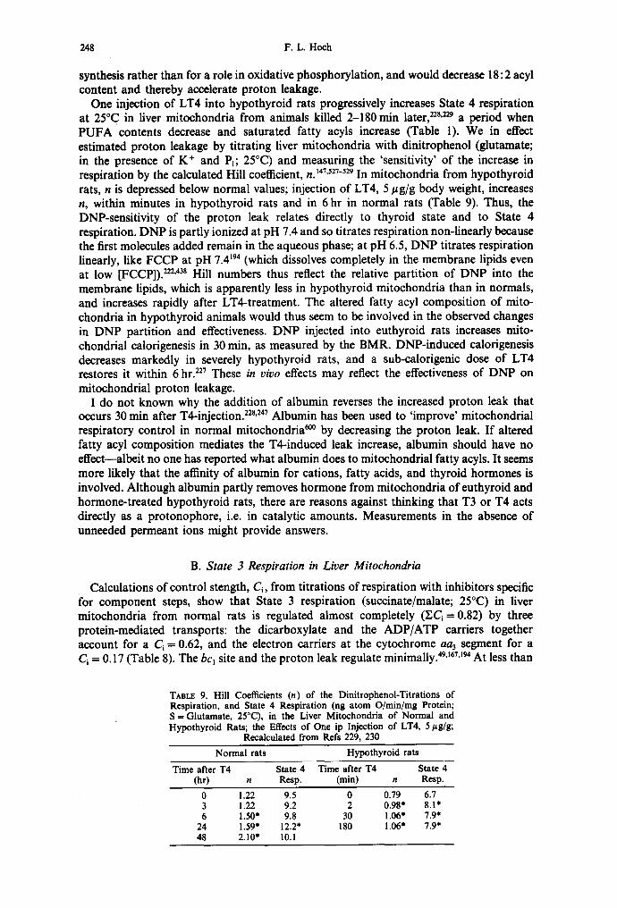
248 F .L . Hoch
synthesis rather than for a role in oxidative phosphorylation, and would decrease 18:2 acyl content and thereby accelerate proton leakage.
One injection of LT4 into hypothyroid rats progressively increases State 4 respiration at 25°C in liver mitochondria from animals killed 2-180min later, 22s'229 a period when PUFA contents decrease and saturated fatty acyls increase (Table 1). We in effect estimated proton leakage by titrating liver mitochondria with dinitrophenol (glutamate; in the presence of K + and P~; 25°C) and measuring the 'sensitivity' of the increase in respiration by the calculated Hill coefficient, n. 147,527-529 In mitochondria from hypothyroid rats, n is depressed below normal values; injection of LT4, 5 ~g/g body weight, increases n, within minutes in hypothyroid rats and in 6 hr in normal rats (Table 9). Thus, the DNP-sensitivity of the proton leak relates directly to thyroid state and to State 4 respiration. DNP is partly ionized at pH 7.4 and so titrates respiration non-linearly because the first molecules added remain in the aqueous phase; at pH 6.5, DNP titrates respiration linearly, like FCCP at pH 7.4194 (which dissolves completely in the membrane lipids even at low [FCCP]). 222'43s Hill numbers thus reflect the relative partition of DNP into the membrane lipids, which is apparently less in hypothyroid mitochondria than in normals, and increases rapidly after LT4-treatment. The altered fatty acyl composition of mito- chondria in hypothyroid animals would thus seem to be involved in the observed changes in DNP partition and effectiveness. DNP injected into euthyroid rats increases mito- chondrial calorigenesis in 30 min, as measured by the BMR. DNP-induced calorigenesis decreases markedly in severely hypothyroid rats, and a sub-calorigenic dose of LT4 restores it within 6 hr. 227 These in vivo effects may reflect the effectiveness of DNP on mitochondrial proton leakage.
I do not known why the addition of albumin reverses the increased proton leak that occurs 30 min after T4-injection. 22s'247 Albumin has been used to 'improve' mitochondrial respiratory control in normal mitochondria ~ by decreasing the proton leak. If altered fatty acyl composition mediates the T4-induced leak increase, albumin should have no effect--albeit no one has reported what albumin does to mitochondrial fatty acyls. It seems more likely that the affinity of albumin for cations, fatty acids, and thyroid hormones is involved. Although albumin partly removes hormone from mitochondria of euthyroid and hormone-treated hypothyroid rats, there are reasons against thinking that T3 or T4 acts directly as a protonophore, i.e. in catalytic amounts. Measurements in the absence of unneeded permeant ions might provide answers.
B. State 3 Respiration in Liver Mitochondria
Calculations of control stength, Ci, from titrations of respiration with inhibitors specific for component steps, show that State 3 respiration (succinate/malate; 25°C) in liver mitochondria from normal rats is regulated almost completely (Y.Ci = 0.82) by three protein-mediated transports: the dicarboxylate and the ADP/ATP carriers together account for a Ci = 0.62, and the electron carriers at the cytochrome aa3 segment for a Ci = 0.17 (Table 8). The bc~ site and the proton leak regulate minimally. 49'167'~94 At less than
TABLE 9. Hill Coefficients (n) of the Dinitrophenol-Titrations of Respiration, and State 4 Respiration (ng atom O/min/mg Protein; S = Glutamate, 25°C), in the Liver Mitochondria of Normal and Hypothyroid Rats; the Effects of One ip Injection of LT4, 5/~g/g;
Recalculated from Refs 229, 230 Normal rats Hypothyroid rats
Time after T4 State 4 Time after T4 State 4 (hr) n Resp. (rain) n Resp.
0 1.22 9.5 0 0.79 6.7 3 1.22 9.2 2 0.98* 8.1" 6 1.50" 9.8 30 1.06* 7.9*
24 1.59' 12.2" 180 1.06" 7.9* 48 2.10" 10.1

Lipids and thyroid hormones 249
maximal respiratory stimulation by external ADP, the ATP/ADP carrier Ci increases up to about 0.5 and that for the dicarboxylate carrier decreases to 0.2, 49'~67 but the effects of thyroid state have been reported only for maximal State 3 respiration. In State 3 the Pi carrier Ci and the ATP synthetase 49 Ci = 0. A much different C~ distribution regulates State 3 in other than adult rat liver mitochondria (see Sections IV.C and D).
Hypothyroidism decreases the rate of State 3 respiration while markedly increasing the C~ of the cytochrome bc~ segment at the expense of the control strengths of the adenine nucleotide translocase and the succinate carrier: electron transport through bc~ is slowed relative to the capacities of the two carriers that normally regulate most (Table 8). The YC~ = 0.74; among the steps not measured are the succinate dehydrogenase, the P~ transporter, the various cation transporters, and the proton leak. The proton leak does not seem a good candidate for gaining in control over State 3, since it is markedly depressed in hypothyroidism.
LT3-injection restores the State 3 rate and the C~ values toward control levels, although the bc~ segment still contributes more than normally. Conspicuously unresponsive to thyroid state is the minor C~ of the cytochrome oxidase step. The dose of T3 used does not uncouple, and does not increase proton leakage to become a significant regulator of State 3 respiration. We still do not know what doses of thyroid hormone large enough to uncouple do to mitochondrial lipid composition, but dietary deficiency of 18:2 fatty acid, especially when accompanied by feeding of saturated or co 3-unsaturated fatty acids, severely depletes mitochondrial 18:2 fatty acyls, and uncouples, albeit inconsistently (see Section IV.E).
The energy-dependent fluorescence yield of 1-anilino-8-naphthalene under State 3 conditions (succinate, 30°C) decreases in liver mitochondria from hypophysectomized rats and is restored after LT4-treatment. 372 Since this probe presumably reports on the polar-nonpolar interface region of the inner membrane, these data indicate that thyroid affects State 3 through membrane phospholipids. Thyroid-responsive liver mitochondrial proteins that regulate State 3 respiration involve cardiolipins: the transporters of (1) ADP/ATP, (2) substrates, and electrons at (3) the cytochrome bc~ segment. (4) The cytochrome aa3 step also regulates and is CL-dependent, but not thyroid-responsive. Other proteins that may not regulate respiration in liver mitochondria (although some regulate in mitochondria of neonatal liver and heart; see Section IV.C and D), but also involve CL include (5) the P~ carrier, (6) substrate dehydrogenases, (7) the ATP synthetase, (8) the Ca 2+ transporter, and (9) the pyridine nucleotide transhydrogenase.
1. ADP/A TP Carrier
The observed slowing in adenine nucleotide translocation in mitochondria from livers of hypothyroid rats ~a is claimed to depress ATP synthesis 578 because the translocator is 'rate-limiting') °s Indeed, the ADP/ATP carrier contributes to respiratory regulation in euthyroid rat hepatocytes suspended in ionic buffer, Ca 2+ ions, bovine serum albumin, and lactate + pyruvate, at 37°C.122a Eighty percent of the respiration is mitochondrial, and 70% of that represents State 3 respiration, for which the Ci = 0.26 (c.f. 0.29 in isolated mitochondria, Table 8). Hyperthyroidism is thought to accelerate the ADP/ATP carrier in vivo, from studies on perfused rat livers where cytoplasm ATP/ADP ratios are twice normal even though ATP is more rapidly used for enhanced urea and glucose synthesis, and Na+-pumping. ~2 These claims are contradicted by the observed decrease of the Ci in hypothyroids and the restoration of the Ci after T3-treatment (Table 8; see also Ref. 51). Claims that the ADP/ATP carrier of adult beef heart mitochondria is the LT3-receptor that regulates respiration 578 are countered by additional facts: heart mitochondrial respiration is not regulated under any experimental conditions by this carrier, H7 and no information is available that thyroid state affects the heart translocase; the moles of thyroid hormone bound 57s and found H6 in liver mitochondria are at least 3 orders of magnitude less than the ,-, 1 nmol of carrier per mg protein; 45 the putative similarity between amino acid compositions (not sequences), and perhaps LT3-binding, of the carrier and the
JPLR 2 7 / ~

250 F.L. Hoeh
receptor may be no more than is to be expected from two lipophilic membrane proteins; and by other considerations. 51'~°'6~9
The abnormal thermotropic properties of the ADP/ATP carrier in liver mitochondria from hypothyroid rats 2~ suggest that altered fatty acyl composition might also change carrier regulation by other effectors. However, in normal mitochondria the Ca = 0.29 at 25°C (Table 8) and 0.31 at 37°C; 251 in hypothyroid mitochondria, Ci = 0.18 at 25°C, 0.17 at 37°C. Brief LT3-pretreatment of hypothyroid rats leaves the Ci at 37°C unchanged but restores the State 3 rate. 25~ Temperature in this range does not appear to affect the relative sensitivity of the carrier, but more rigorous experiments are still needed.
CL binding is involved in activity, given that the CL on the ADP/ATP carrier is the negatively charged target for cationic activators, as Kr/imer et al) ~s propose. The ADP/ATP translocase loses 50% of its activity when liver mitochondria are depleted of only 1% of their total PL by a pancreatic phospholipase A with substrate specificity CL > PE > PC, while Crotalus adamanteus venom phospholipase A, with substrate specificity PC = PE>>CL, must remove 8 times more PL to inhibit 50%. 5¢ Although incorporation of the delipidated beef heart translocase protein in CL, PC or PE vesicles activates equally, 3~7 CL binds to the isolated carrier with the unusually high molar stoichiometry of 6: 1 (c.f. 2-3: 1 in the cytochrome oxidase complex), and with great affinity that produces strong CL immobilization, inferred from 31p NMR. In contrast, PC and PE are not immobilized on the protein and are easily removed; these findings indicate a specific CL-protein interaction. 34 The increased mitochondrial CL/PL ratio in the hypothyroid liver may account for the abnormal thermotropic properties of the translocase, and the maintenance of high activity (decrease in Ci) relative to the other carriers that regulate State 3 respiration.
2. Substrate Carriers
Among mitochondrial substrate carriers, the 18: l-carnitine translocase has been shown to be completely CL-dependent (see Section II.J). Cardiolipins are unique in being the only membrane PL that keeps the monocarboxylate, 4~s dicarboxylate, 296 tricarboxylate, 5s° ~-ketoglutarate, 419 and aspartate/glutamate ~6 carriers active during purification and reactivates them after isolation. Only the dicarboxylate carrier has been examined and shown to be thyroid-sensitive (Table 8).
3. Cytochrome bcl Segment
Surface spectrophotometry of intact perfused rat liver 2~6 and studies on isolated mitochondria ~ are taken to show that cytochrome b is normally a control point in electron transport, although the very low Ci for the bc~ segment (Table 8) says it is not. In hypothyroid rats, when the liver is perfused with fructose to decrease electron flow to the State 4 level by depleting P~ and deaminating adenine nucleotides, respiratory inhibition and the degree of cytochrome b oxidation are greater than normal, 216 which does agree with the significant Ci. In hyperthyroid rat liver, the inhibition of respiration and oxidation of cytochrome b that is seen in normals does not occur. Hassinen et aL 2t6 conclude that thyroxine loosens the coupling between phosphorylation and electron transport; con- versely, hypothyroidism increases coupling in situ. In isolated rat liver mitochondria, hyperthyroidism increases the degree of reduction of cytochromes b, c and c~ during State 3 respiration (succinate, 37°C), while in State 4 cytochrome b is more oxidized and cytochromes c and c~ are more reduced. 62"252 Mitochondrial ubiquinone content increases 75%, and ubiquinone becomes more reduced; over this long treatment period, LT4 also increases concentrations of b, c and aa3. The hormone is thought to regulate State 3 respiration mainly through the bc~ complex, and State 4 by increasing proton permeability, 2s2 as had been shown by control strength analyses. 6~s
The cytochrome bc, segment is lipid dependent. The extremely hydrophobic apo- cytochrome b is encoded by mitochondrial DNA and synthesized in the mitochondrion, 559

Lipids and thyroid hormones 251
so the nascent protein assumes mature conformation at least near, if not in contact with, mitochondrial CL. Arrhenius profiles of the rate of electron transfer through bc~ are inflected, suggesting either a conformation change in a membrane component or merely a change in the 'rate,limiting' step. 536 Various PL reactivate phospholipid-depleted ubiquinol: cytochrome c reductase, which contains 80% of the mitochondrial cytochrome b, but only in the presence of CL: CL binds, PL activate; 59'~62 NADH: cytochrome c reductase prepared from beef heart mitochondria is also CL-dependent, as judged from its inhibition by adriamycin derivatives. 179-~8~ The 90%-delipidated, inactive succinate: cytochrome c reductase is reactivated by adding first ubiquinone, then asolectin. ~9 The residual lipids are 65% CL, 20% PC and 10% PE, so the 'non-specificity' of the asolectin (which itself contains 10% of total lipid P as CL 4~8) activation involves significant amounts of CL.
4. Cytochrome Oxidase
Cytochrome oxidase of bovine heart mitochondria copurifies with CL. Of the 50 lipid-binding sites on the 200 kDa complex, 2-3 molecules of tightly bound CL are specifically required for catalytic activity of an active four-subunit fragment. Other membrane PL have a different, non-specific, activating and dispersive role, that is duplicated by detergents such as lysoPC and Tween-20.16°'~6~'496 Based on these findings, cardiolipins are described as a prosthetic group of cytochrome oxidase involved in the cyclic transport of monovalent cations, although H + ions are omitted from the list. t6° However, endogenous CL can be lowered to 0.3-0.4 moles/mol complex by replacement with dimyristoyl-PC while full oxidative activity is retained. 4s4 EPR spectroscopy of bound spin-labeled CL shows that CL binds to cytochrome oxidase > 5 times more strongly than this PC, but none of the binding sites on the enzyme complex are highly specific for CL. This lack of specificity may explain why cytochrome oxidase contribution to regulation of State 3 respiration in liver mitochondria is impervious to abnormal thyroid states.
5. Pi Transporter
Pa transporter protein purified from pig or cow heart mitochondria loses activity when CL is removed; only CL protects activity during solubilization. 43 CL reactivates the soluble protein, egg yolk PL activates but less. Protein-PC vesicles are inactive unless CL is added, then adriamycin inhibits. 4°9'4~° Adriamycin, an antineoplastic anthracycline amino- glycoside, forms stable intercalation complexes, probably with phosphodiester bridges of DNA and RNA (whereby it inhibits DNA replication and RNA transcription) and with the oriented phosphoester groups on the membrane-surface CL (whereby it disrupts mitochondrial structure, promotes lipid peroxidation, and inhibits oxidative phos- phorylation)J8 °'4t5 In addition, adriamycin inhibits some lipogenic enzymes (isocitrate (NADP) dehydrogenase, 6-phosphogluconate dehydrogenase, and malic enzyme) by interaction with -SH groups. 142 Adriamycin is thought to inhibit the Pi-transporter by making CL inaccessible. 15 These observations suggest absolute CL-dependency. However, the Pi carrier does not regulate heart mitochondrial State 3 respiration, and there is no information on thyroid effects in heart or liver mitochondria. In yeast mitochondria respiring in State 3, the Pa carrier Ca = 0.4-0.6, the cytochrome aa3 Ca = 0.5-0.6; 389 the CL/PL ratio is 0.15, but no 18: 2 acyls are present. 2~5 Oddly enough, 40 #M LT4 accelerates yeast respiration (glucose; 37°C). 434
6. Substrate Dehydrogenases
These can apparently contribute little to the overall control of State 3 respiration in liver mitochondria of euthyroid rats (~< 0.14) or hormone-treated hypothyroid rats (~< 0.07), but they might contribute some of the 0.26 of unmeasured Ca in mitochondria from hypothyroids (Table 8). The soluble dehydrogenases that act on isocitrate, glutamate or

252 F.L. Hoeh
malate, as well as the intrinsic membrane-bound succinate or glycerol-3-phosphate dehydrogenases, increase in thyrotoxicosis and decrease in hypothyroidism. 29t The membrane-bound p-hydroxybutyrate dehydrogenase is an exception, in that thyro- toxicosis decreases its activity while hypothyroidism affects it but little. Brand and Murphy 5] believe that thyroid treatment increases de novo synthesis or alters lipid environment, or both, to increase substrate dehydrogenase activities, and that the increases in subsequent electron-transporters are less important in accelerating electron flux.
Specific PL-binding and specific PL-reactivation of a membrane-dependent enzyme are clearly distinguished in studies on the soluble apoenzyme of the D-/~-hydroxybutyrate dehydrogenase (NAD ÷) prepared from rat liver or bovine heart mitochondria. The apoenzyme specifically requires PC for activity, even when in aqueous solution. 59"t52,165'2ss'~ However, although CL (or PE) itself does not reactivate, in the presence of CL 2-5 mol of pure PC per mol of enzyme half-reactivates, as compared with 60 mol PC/mol enzyme in the absence of CL. Although the apoenzyme interacts with every pure PL tested, it interacts most strongly with films containing CL. Berrez et al. 33 propose that CL molecules on the inner face govern the binding to the inner membrane. The ~-hydroxybutyrate dehydrogenase is normally 'crypticized' and mostly inactive in intact mitochondria, and full expression of its activity in vitro requires membrane lysis. The similar latency of the mitochondrial inner face carnitine palmitoyltransferase CP%, and the dependency of that enzyme on CL (see Section II.J), suggest a common regulatory role for CL. Thyroid- induced depression of the/~-hydroxybutyrate dehydrogenase has also been attributed to the concomitant decrease in [NAD+], to which this enzyme is sensitive. TM
Activity and amount of membrane-bound glycerol-3-phosphate dehydrogenase are remarkably thyroid-dependent (see Section II.E), but little information is on hand as to its lipid-dependency. Succinate dehydrogenase is about 30% latent in situ, and is regulated by the availability of the lipid-soluble ubiquinone and by membrane fluidity. 22
Phospholipid-depleted NADH:ubiquinone reductase, which contains the NADH dehydrogenase (FMN, FeS), requires CL specifically, PL non-specifically. The NADH dehydrogenase of intact mitochondria is progressively solubilized in correlation with the course of CL hydrolysis by phospholipase A, but not with PC or PE hydrolysis, implicating CL in its binding into the inner membrane. ~8
7. ATP-syn the tase
ATP-synthetase is thyroid- and CL-dependent. In inner membrane vesicles from liver mitochondria of hypothyroid rats, the rate of the reaction ADP + Pi--*ATP is depressed at 30°C as a reflection of the linearization of the normally inflected Arrhenius profile. 81-83'242 At 30°C, respiratory rate and flavin and cytochrome contents are all normal but the ATP- synthetase, acting independently of the ADP/ATP carrier, shows a decreased V and K~ for ADP; titration of phosphorylation rate with oligomycin reveals a diminished apparent number of phosphorylating sites, enough to account for most of the phosphorylation defect. In normal inner membranes, the progressive decrease in ATPase activity during incubation with ascorbate or cysteine, which peroxidize PL and fatty acyls, correlates uniquely with the disappearance of 18:2 from CL. 362'525 Reconstitution of the ATPase of heart mitochondria (where its Ci is significant) requires lipids for the complete, oligomycin- sensitive, amphipathic Fj F0 assembly. CL, in some reports, 102.589 is the most effective of the naturally available PL, although lyso-PC is more effective. A highly purified, active ATPase that retains oligomycin-sensitivity contains 2 mol of CL per mol, and lesser amounts of PC and PE. t23 The cardiac ATP synthetase is thyroid dependent in situ and in vivo (see Section IV.D).
8. Ca2+-transporter
The Ca2+-transporter, partially purified from cow heart submitochondrial particles, takes up very little Ca 2+ when reconstituted in liposomes made of 1 : 1 PC + PE, but increases

Lipids and thyroid hormones 253
uptake linearly with [CL] added during reconstitutions, to about 12-fold at [CL] ffi 10 mol%) 2° Liver mitochondria of hypothyroid rats pump Ca 2+ ions (30°C) into the matrix at rates 40% less than normals when the energy source is added ATP, but at near normal rates ( - 7 % ) even when supported by slowed substrate oxidation. 242'433 Arrhenius profiles of such ATP-driven (but not substrate-driven) transport are abnormal (J. Peabody and F. L. Hoch, unpublished data). Again these data indicate that lipid-sensitive utilization of proton flux decreases when protons emanate from the H+-ATPase and is normal (or even increased) when protons originate from substrate oxidation. When liver mitoehondria from hypothyroid rats are incubated for 1 min with T3 at 37°C, respiration in State 4 and State 3 remains unchanged, but in the presence of Ca 2+ ions, increases in [T3] accelerate State 3 linearly. 223 At 1/~M [T3], the V for Ca2+-uptake and the rate constant for Ca2+-efflux are increased maximally, suggesting that the hormone acts directly on the inner membrane to increase Ca 2÷ ion mobility. Thyroid-state control over Ca 2+ re- moval from cytoplasm is more extensively studied in myocardial sarcoplasmic reticulum (see Section Ill.C).
9. Pyridine Nucleotide Transhydrogenase
As discussed in Section II.C.2, hypothyroidism doubles the V of the ~ TH reaction but not the TH reaction in liver mitochondrial inner membrane vesicles, and T3-treatment restores normal low values of V. In intact liver mitochondria, the normal steady state of substrate-induced delay in reduction of intrinsic pyridine nucleotides during the State 3~State 4 transition, caused by the rate-controlling ~ T H reaction, is much accelerated in hypothyroid mitochondria. 232'234 Oligomycin similarly accelerates reduction in mito- chondria for normal rats, TM by blocking proton egress through the ATP-synthetase and thereby stimulating TH activity even in non-phosphorylating submitochondrial par- ticles. 343 Thus, the increased proportion of inner membrane 18:2 acyls in hypothyroidism may conduct protons to the ~ TH and away from the ATP-synthetase.
In addition to this unusual increase in rate, the TH differs from the transporters in liver mitochondria in lipid dependence and in sensitivity to a decoupler. Energy mode of the TH reactions determines lipid dependence. 5t2-5~6 The energy-independent reaction is much less inhibited by Crotalus terrificus phospholipase A2 (which is specific for PC and PE but hydrolyses little CU s) than is the ~ T H reaction in submitochondrial particles 33°,367 and membrane particles of Escherichia coli WS1.557 Liposomes formed from any of several PL reconstitute purified transhydrogenase from beef heart submitochondrial panicles to highest TH activity; low [CL] reconstitutes most effectively, but high [CL] inhibits. 5~4 PC is specific for the ~ TH mode, and small amounts of CL inhibit. Arrhenius profiles for ~ T H activity, measured in everted vesicles prepared from liver mitochondria of hypothyroid rats before or after T-treatment, maintain normal values for Ea and Tt (transition temperature). 136:37 Since one protein catalyzes both TH and ~ T H reac- tions, these observations suggest that the lipids exert their effects via the proton leak and Ap (as they do to regulate State 4 respiration) rather than by binding to the TH protein (as they may bind to the transporters that regulate State 3 respiration).
As noted, the 'decoupler' action of palmitate at 25°C does not inhibit energy-dependent TH activities in rat liver submitochondrial particles while it inhibits succinate-supported reverse electron-flow and increases State 4 respiration and ATPase activity. 5°6 If, as Rottenberg and Hashimoto 5°5'5°6 propose, the ATPase membrane-associated F0-complex serves as a 'proton capacitor' that is intermediate between oxidation and phosphorylation, and that is discharged by free fatty acids, then the lack of effect of palmitate on ~ TH activities indicates that a different proton pathway couples ~ T H to oxidation and the ATPase. If, as is discussed above, proton coupling between oxidation and phosphorylation involves 18:2 fatty acyls and CL, then the proton coupling to the ~ T H does not appear to involve 18:2 and CL the same way, or at all.

254 F.L. Hoch
10. Repetitive Additions of ADP
Recent measurements of respiration of liver mitochondria from hypothyroid rats show rates of State 4 and State 3 (succinate, 26°C) that are at control levels, but both decrease progressively as ADP is added repetitively, while rates are maintained in controls, m Hypothyroid mitochondria consume less O z during State 4--,3--,4 transitions. Treatment of hypothyroid rats with small doses of T4, 0.02/zg/g/day x 21 days, reverses these abnormalities. Objections to these data include the presentation of only derivative traces, d[O2]/dt, rather than absolute rates, so that 02 used during proton leakage is measured indiscriminately with 02 used during phosphorylation; and the absence of added Mg 2+ ions to support maximal State 3 respiration. 5s2 Since Ap is abnormally high in State 4 in mitochondria of thyrotoxics (Table 7; Ap data are needed in mitochondria from hypo- thyroid rats), the absence of differences in State 4 rates in the data of Ezawa et al. 139 seems important. To simulate conditions in vivo, they added pulses of ADP successively. Cycling of ADP additions is recommended to 'improve' rates of State 3 respiration in normal rat liver mitochondria, a7~ but it beclouds physiological control mechanisms. Success- ive additions of ADP increase respiration in State 4 (i.e. increase proton leakage) and State 3 (i.e. redistribute Ca contributions) progressively but do not change respiratory control ratios much. Data similarly disparate with respiration before and after single additions of ADP come from ADP-cycling with liver mitochondria from EFA-deficient rats (see Section IV.E). The mechanisms do not appear to have been studied. However, the ADP/ATP carrier is electrogenic and moves protons across the inner membrane, not as part of the mechanism, but electrophoretically. 339 Thus, if this carrier is involved in the respiratory effects of ADP-cycling, it may be by way of its introducing additional proton circuits. Further, multiple additions of neutralized ADP solutions introduce Na + or K + ions, to contribute to proton movements.
C. Liver Mitochondria of Fetal and Neonatal Rats
Plasma IT4] and [T3] of newborn rats rise 7- to 8-fold and linearly from day 2 to day 21, 86,622 as shown in Table 10. Accompanying changes in liver mitochondrial function and lipid composition should be informative on physiological and developmental effects of the hormone, as opposed to effects of hormone injections. Reports on mitochondrial respiration during the fetal and postnatal periods conflict. Some investigators find that fetal mitochondria, as compared with those from adult rats, respire (succinate, 30°C) more slowly in State 3 but not in State 4.11'19'206'417 This respiratory defect is ascribed to the low contents of adenine nucleotides, which are thought to be due to low ADP/ATP carrier activityfl 7 Since this carrier exchanges ADP or ATP one-for-one, some other explanation seems necessary. The proton leak in mitochondria of newborns is said to be equivalent to 7 pmol of FCCP per mg protein, and in adults, to 21.5 pmol/mg, but the titration curves
TAnLE 10. Neonatal Liver Mitochondria: Plasma [T4] and IT3]; Mitochondrial Respiration (Succinate, 25-30°C) and Carbamoyl-P Synthetase Content; Liver Phospholipid Composition; and Developmental
Changes Days after birth
I-2 4-5 9-10 15-21 (Refs)
Plasma [T4]: (~g/dl) 0.6 1.1 2.5 3.0 (86, 622) Plasma IT3]: (ng/dl) 10 20 42 79 (86, 622)
Respiration: State 4 (ng atom O/min/mg) 53 30 26 25 (402, 480) State 3 (ng atom O/rain/rag) 159 150 143 150 (402, 480)
Carbamoyl-P synthetase: (% ~ polypeptides) 12 16 20 20 (402)
Liver PL: 18:2/PL (%) 5.8 5.5 10.6 (546) 18:2/CL (%) 43 47 45 78 (402, 640) CL/PL (%) 6.0 8.8 9.9 12.2 (402)

Lipids and thyroid hormones 255
are not linear for the newborns, and the respiratory rates at [FCCP] -- 0 (i.e. State 4) are similar to those in adults) 9 Comparison of methods for isolating mitochondria shows that the presence of bovine serum albumin greatly improves the poor recovery of fetal (but not adult) mitochondria over that in conventional media containing sucrose-EDTA, ~° which suggests that conventional methods selectively damage fetal mitochondria. With albumin or mannitol in the isolating media, mitochondria prepared by these methods from livers of 1-day old rats leak protons (State 4, succinate, 25-30°C) 70-100% faster than do mitochondria from adult rats and are permeable to low molecular-weight solutes, while State 3 rates are at adult levels 4°2'4s° (Table 10). State 4 respiration halves between day 2 and 9, while State 3 respiration stays constant. In spite of this leakiness, such fetal mitochondria phosphorylate ADP efficiently ~° and the ADP/O ratio stays at 2.0 after birth. 4°2 Thus, the proton leak correlates inversely with the increasing plasma IT3] and [T4]--tbe opposite of the direct correlation seen when adult thyroid state is altered. In newborns, the ATP synthetase (Ci =0.6), the Pi carrier (Ci~<0.13), the dicarboxylate carrier (Ca = 0.12) and the ADP/ATP carrier (Ca = 0.15) regulate State 3; .9 the first two do not regulate in adult liver mitochondria.
Liver phospholipids of 1-day rats contain 5.8% 18:2, which doubles between days 5 and 15:46 The 18:2 deficit in neonates reflects in part the amount and composition of mitochondria: the 18:2 content of CL is 26% lower in fetal livers than in maternal livers ~ and rises 31% between days 5 and 15; ~6 the low ratio of CL/gPL in the newborn liver further contributes to the low 18: 2 contents in liver lipids, and increases linearly from day 1 to 154°2 (see Ref. 106). However, because mitochondrial CL/mg protein does not change, it appears that more mitochondria are made during early developmentY 6 Thus, the amounts of 18:2 acyls and of CL correlate inversely with the proton leak, as is seen in adults. Relative amounts of the mitochondrial 165 kDa carbamoyl-phosphate synthetase increase 67% from days 1-10; 9°'~2 this enzyme is induced during the postnatal rise in plasma thyroid hormones much as it is during spontaneous Anuran metamorphosis, ~6'5s7 and so serves as a marker for gene expression induced by spontaneously rising thyroid hormone levels. Other enzymes are also induced in this period: activities of fl-hydroxy- butyrate, succinate, glutamate, malate, and NADH dehydogenases increase 4- to 20-fold in homogenates, less in mitochondria (further showing mitochondrial replication). ~;76'627 Mitochondrial glycerol-3-phosphate dehydrogenase activity does not increase, ~ indicating that thyroid hormones are not omnipotent in inducing hepatic enzymes during postnatal development.
Thyroid levels do not affect liver PL contents of 18:2 acyls and CL/PL ratios in newborns like they do in adults. The effect of thyroid in newborns may be more on the incorporation of 18:2 into liver PL and especially CL (Table 2) than on the depletion of 18:2 acyls by further desaturation as it is in adults. Fetal storage of maternal 18:2 is limited, and CL 18:2 is low. 64° EFA-deficiency may produce the rapid mitochondrial proton leak (see Section IV.E) and subsequent suckling may supply 18:2 while 18:2-incorporating enzymes develop under thyroid and insulin ~ss influences. Interestingly, the 18: 2 content of PC and PE fractions of fetal livers are at adult levels, but their contents of 16:1 and 18:1 are 2.5 to 4 times above contents in the dam? 75 The greater contribution of mono-unsaturates to overall unsaturation resembles that seen in tadpole livers as compared with frog livers, and decreases as rapidly after injecting the tadpole with LT3 as the induction of the enzymes synthesizing urea (Ref. 238; see Section II.I).
D. Heart and Brain Mitochondria
Thyrotoxicosis has been said to make heart mitochondria inefficient more readily than liver mitochondria, ~9 but evidence to the contrary is in Table 6. The decreased economy of isometric contraction observed in rabbits treated with LT4, 0.2 #g/g/day x 14 days, is not related to decreased mitochondrial resynthesis of ATP. 9''° Calorimetry shows that the ratio [recovery heat liberated during diastole]/[initial heat liberated during contraction and

256 F.L. Hoch
relaxation] decreases, and this is mostly accounted for by an abnormal accumulation of the VI myosin isoprotein, which develops isometric force less efficiently for structural reasons. On the other hand, saturation transfer 31P-NMR studies demonstrate that perfused hearts of euthyroid rats injected with LT4, 0.35 #g/g/day x 10--14 days, respire more rapidly but do not increase ATP synthesis flux: 3°4 the ATP synthetase is thyroid- dependent in situ and in vivo. These findings confirm that excess thyroid hormone uncouples when measured by 'non-invasive' methods, as had been suggested in hyper- thyroid muscle, liver and kidney by decreased 32P-uptake into ATP in vivo TM (see Ref. 226). In contrast, 3~P-NMR measurements on hearts of hypothyroid rats show a slowed ATP synthesis that is accounted for by decreased respiratory rate, not uncoupling. ~*Ta
Isolated heart mitochondria require a greater LT3 or LT4 dose-time treatment than do liver mitochondria to achieve comparable increases in State 4 respiration 474 and to deplete 18:2 (Table 6). Heart mitochondria from our thyrotoxic rats, prepared by treating normal animals rather briefly with LT3, 1 #g/g/day for 3 days, are not uncoupled or loose-coupled, contain normal amounts of 18:2 and CL in EL, but lose State 3 respiration on standing at 4°C for a reason not revealed by the lipid analyses d o n e . 237 Such increased 'fragility' might be connected with the mitochondrial swelling that occurs in hypotonic sucrose solutions together with activation of endogenous phospholipase and hydrolysis of CL and PC; 297 o r with effects like those in glucagon-treated rats, where liver mitochondria tend to lose PC, PE, and CL during preparation? 55 It takes 44-60 daily hormone injections to produce heart mitochondria that are uncoupled in vitro and half-depleted of their 18:2 acyls (Table 6).
In hypothyroid rats, heart mitochondrial State 4 respiration (glutamate) is at normal levels at 32°C) 5° Injecting hypothyroid rats once with LT3, 0.25/zg/g, increases State 4 rate by 80% in 1 day and maintains an elevated rate for at least 3 days. State 3 respiration responds to altered temperatures abnormally: rates are below normal only when measured at temperatures between 20°C and 34°C, reflecting the fact that Arrhenius profiles of hypothyroid preparations are normal below about 18°C, but show an abnormally high Ea above that temperature. The mitochondrial YL and PL contents are normal, as are the proportions of PC, PE, PS, and CL (c.f. the 72% increase in CL/PL in liver mitochondria). The 18:2 content in hypothyroid PL is 35.5% (normals, 26.8%), due to rises in 18:2 content of PC and PE, but not in CL.
Heart and liver mitochondria have different lipid compositions, carrier protein contents, and regulatory mechanisms, n7'~6 which may account for the differences in thyroid effects. Although the heart has less apparatus for lipid syntheses, heart mitochondria contain twice as much CL per ~PL as liver mitochondria, and the CL contains about 50% more 18:2. Fasting/refeeding depletes the 18:2 acyls in heart mitochondria much less than in liver mitochondria 7'8 (Table 5). Heart mitochondria contain twice as much ATP synthetase and 4 times as much ADP/ATP carrier. Their State 3 respiration (pyruvate/malate, 25°C) is regulated by the ATP synthetase ( C i = 0.46) and the NADH dehydrogenase (Ci = 0.37), and not at all by the ADP/ATP carrier"7--all in contrast to the roles of these component steps in liver mitochondria (Table 8).
If mitochondrial 18:2 acyls and/or CL mediate some thyroid actions, as they may in liver mitochondria and less in heart mitochondria, then brain mitochondria offer another system to test the generality. Mature brain mitochondrial function is completely insensitive to thyroid status.564 Brain mitochondrial CL/PL ratios are 2-5 %, and CL 18:2 acyl content is only 10%; PC contains no 18:2, PE contains 1% 18:2. 635 Fasting/refeeding fails to increase the activities of lipogenic enzymes in brain (see Section II). Essential fatty acid (EFA) deficiency depletes tissue 18: 2 in the order liver > heart > brain, and depletes brain 20:4 little, if at all. Thus, the insensitivity of brain mitochondria to thyroid status is in contrast to the low sensitivity of heart mitochondria, which contain lots of CL with lots of 18:2 in it, both of which are hard to deplete. However, mitochondria in immature brain are quite thyroid-responsive, 564 but contain no CL: as in neonatal liver mitochondria, the thyroid-induced development of an adult complement of enzymes and transporters seems to predominate over thyroid effects mediated by membrane lipids.

Lipids and thyroid hormones 257
E. Essential Fatty Acid Deficiency
Essential fatty acid metabolism and the thyroid state of animals are related: the BMR increases within 1-2 weeks (the earliest sign) in animals on a fat-free diet, and feeding small amounts of methyl linoleate corrects it within days; 3~°'63° the hypermetabolism is not accompanied by thyroid hypersecretion; 4°8 dietary linoleic acid protects against experi- mental thyrotoxicity; TM and Klein and Johnson 31° noted that fat deficiency and hyper- thyroidism each uncouple oxidative phosphorylation in a mechanism that might involve mitochondrial unsaturated fatty acids.
Linoleic acid, 18:2~o6, is the essential fatty acid in vertebrates because they lack a A12-desaturase; its 20:4w6 derivative is absent from most lab chow diets for rats. Considerable evidence contradicts the view 2°7 that co6-PUFA are essential as precursors of prostanoids but not for the function of any membrane. Daily EFA requirements are orders of magnitude greater than production of prostaglandins; prostaglandin synthetase inhibitors do not induce EFA-deficiency; administration of PG does not relieve all the symptoms of EFA-deficiency; 249'25° those symptoms are not usually associated with actions of PG; 394'495 columbinic acid cures EFA-deficiency but is not a PG precursor and even inhibits the primary enzyme for PG synthesis. Columbinic acyls replace 20:4 acyls in tissues, and restore the decreased rate of ATP synthesis in heart mitochondria 256'25s but State 4 respiration rates are not mentioned.
Problems with the measurements of mitochondrial function in EFA-deficiency include the following. Different essential fatty acids have different functionsfl 5 Individual tissues and their individual PL fatty acyls respond differently to EFA-deficiency and replace- ment. 4~2 Different temperatures of measurement may produce conflicting results, since membrane PUFA distribution affects Arrhenius profiles. 4s9 Respiratory control ratios are sometimes presented without mention of the rate of State 4 respiration whereby to estimate the proton leak. Respiratory rates or control ratios presented after ADP-cycling "393'5°s are similar in controls and EFA-deficient animals; see Section IV.B.10 for objections to this procedure.
Protein-free vesicles formed from natural purified PCs are as impermeable to trapped Na +4°5 and K +465 as are erythrocyte membranes, since Ea for diffusion is 15-17 kcal/g ion in all. Phosphatidylcholines purified from livers, kidneys and hearts of rats fed a fat-free diet for months are severely depleted of 18:2 ( - 8 2 % ) and 20:4 ( - 9 0 % ) acyls, and form vesicles that leak Na +, the Ea being 4.5 kcal/g ion--about the value for free ion diffusion in water. Proton diffusion in such membranes, as well as Ap and proton leakage rates in mitochondria of EFA-deficient rats, have not, to my knowledge, been measured as yet.
Several dietary regimens to induce EFA-deficiency deplete mitochondrial 18:2 (especially in CL) and 20:4 acyls (Table 11). Feeding completely saturated fatty acids accentuates 18:2co6 fatty acid deficiency. Feeding diets rich in saturated fatty acids decreases the ratio of co 6/oJ 3 PUFA in PL of mitochondria from rat liver, heart, or kidney, but leaves the ratio of saturated/unsaturated fatty acyls unchanged, suggesting a 'homeo- static' mechanism to Gibson et a1.172 Such homeostasis, like the increased co 9-unsaturation, may provide membrane fluidity for some nonspecific systems and prevent lethal outcome of EFA-deficiency. But only sufficient co6-PUFA prevent appearance of abnormal mitochondrial oxidative phosphorylation and membrane properties.
When State 4 respiration is reported, EFA-deficiency almost always accelerates State 4. State 3 respiration and respiratory control ratios almost always decrease. Phosphorylative efficiency usually decreases only slightly, indicating that EFA-deficiency produces loose coupling a42 or decoupling. 5°6 Alfin-Slater and Aftergood 6 concluded that defective oxidative phosphorylation is an early lesion in EFA-deficiency. One group 2s3'574'636 reports no abnormalities in mitochondrial oxidative phosphorylation, but shows only respiratory control ratios and no absolute values for State 4 or State 3 respiratory rates. Mitochondrial co6-PUFA contents in these experiments are greatly decreased, and in correlation the lipid domains are tighter packed and allow less motional freedom of spin labels and a slower frequency of volume oscillations during energy-dependent ion transport. 636 Some research-
JPLR 27/3~H
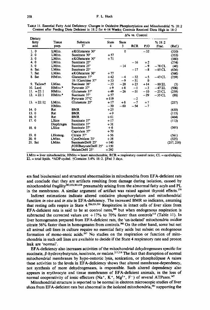
258 F . L . Hoch
TABLE 11. Essential Fatty Acid Deficiency: Changes in Oxidative Phosphorylat ion and Mitochondrial % 18:2 Content after Feeding Diets Deficient in 18:2 for 4-14 Weeks; Controls Received Diets High in 18:2
A% vs. Control Dietary
fatty Tissue Substrate State State % 18: 2 acid prep. T ° 4 3 R C R P/O Frae. (Ref.)
1. 0 LMito. , ,KGlutarate 30 ° 2. 0 LMito. Succinate 30 ° + 5 7 3. 0 LMito. aKGlu ta ra te 30 ° +71 4. 0 LMito. Succinate 25 ° 5. 0 LMito. Succinate 25 ° 6. Sat LMito. Succinate 25 ° 7. Sat LMito. , ,KGlutarate 300 +77 8. Sat HMito. Glutamate 37 ° +42
18: ICarnitine 37 ° +33 9. Tal lowt LMito. Succinate 26 ° - 3 5
10. Lard HMito.* Pyruvate 37 ° + 9 11. +22 :1 HMito. Glutamate 37 ° + 4 9 12. +22 :1 HMito.* Succinate 37 ° +57
Pyruvate 37°C + 119 13. +22:1:~ LMito. Glutamate 25 ° + 17
HMito. - 30 14. 0 Rat B M R +25 15. 0 Rat B M R + 9 16. 0 Rat B M R +61 17. 0 LSlice Succinate 370 + 17
Diaphragm Succinate 37 ° + 31 18. 0 LSlice Succinate 37 ° + 6
Caprylate 37 ° + 7 0 19. 0 LHomog. Citrate 37 ° + 5 6 20. 0 LMito. CytoOxidase 25 ° +38 21. Sat LMito. SuccinateDeH 25 ° + 140
f lOHButyrateDeH 25 ° + 150 MalateDeH 25 ° +292
0 - 3 2 (310) (353) (56O)
- 16 + 2 (274) - 14 - 9 - 7 8 CL (40)
- 17 - 8 - 8 5 CL (636) (560)
- 4 - 3 2 - 5 - 4 3 CL (259) - 9 - 3 1 0
- 2 0 +23 + 1 4 - 8 0 5ZL (3) + 8 - I - 3 - 4 7 YL (508)
- 2 6 - 5 1 - lO - 2 3 CL (259) - 29 - 35 CL (88)
+ 8 - 6 0
--2 - -7 + 7
--54 --7 (257)
(630) (113) (464) (113)
(561)
(561) (325)
(217,218)
LMito = liver mitochondria; HMito -- heart mitochondria; R C R -- respiratory control ratio; CL = cardiolipins; YL = total lipids. *ADP-cycled. "t'Contains 3.4% 18:2. :~Fed 3 days.
ers find biochemical and structural abnormalities in mitochondria from EFA-deficient rats and conclude that they are artifacts resulting from damage during isolation, caused by mitochondrial fragility 2s2'353'56~'636 presumably arising from the abnormal fatty acyls and PL in the membranes. A similar argument of artifact was raised against thyroid e f f ec t s . 379
Indirect estimations indicate altered oxidative phosphorylation and mitochondrial function in vivo and in situ in EFA-deficiency. The increased BMR so indicates, assuming that resting cells respire in State 4, 226~23~'55~ Respiration in intact cells of liver slices from EFA-deficient rats is said to be at control rates, ~ but when endogenous respiration is subtracted the corrected values are + 17% to 70% faster than controls H3 (Table 1 1). In liver homogenates prepared from EFA-deficient rats, the 'un-isolated' mitochondria oxidize citrate 56% faster than in homogenates from controls. ~ On the other hand, some but not all animal cell lines in culture require no essential fatty acids but subsist on endogenous formation of mono-enoic acids? 67 No studies on the respiration or function of mito- chondria in such cell lines are available to decide if the State 4 respiratory rate and proton leak are 'normal'.
EFA-deficiency also increases activities of the mitochondrial dehydrogenases specific for succinate, fl-hydroxybutyrate, isocitrate, or malate. 2~7'21s The fact that disruption of normal mitochondrial membranes by hypo-osmotic lysis, sonication, or phospholipase A raises these activities to the levels in EFA-deficiency shows that altered membrane-dependency, not synthesis of more dehydrogenases, is responsible. Such altered dependency also appears in erythrocyte and tissue membranes of EFA-deficient animals, in the loss of normal cooperativity of ionic effectors (Na +, K +, Mg 2+, F-) of several ATPases? 47
Mitochondrial structure is reported to be normal in electron microscopic studies of liver slices from EFA-deficient rats but abnormal in the isolated mitochondria, ss~ supporting the

Lipids and thyroid hormones 259
argument for artifact. However, other electron microscope studies on tissue slices find major structural alterations in such mitochondria in situ. ss3"5~4"6~'639
The importance, if not the essentiality, of 18:2 and its derivative fatty acyls in mitochondrial membrane function receives support from the available data on EFA- deficient animals. Crucial experimentation, such as determination of Ci values in State 3 and proton leakage in State 4 are, to my knowledge, still not reported.
V. P E R S P E C T I V E
Evidence is presented here, some of it circumstantial or incomplete, that thyroid hormone occupancy of specific receptors sets off a train of amplifying and diversifying mechanisms. A suggested sequence starts with the expression and modification of a few lipogenic enzymes; proceeds through changes in membrane phospholipid fatty acyl and cholesterol contents; with subsequent modulation of enzyme, transporter, and receptor systems, as well as unmediated membrane permeability to mono-cations; that in turn regulate concentrations of multipotent intracellular effectors. These systems include: cell membrane adenylate cyclase and intracellular cyclic nucleotide phosphodiesterases, and cell and organelle membrane Ca2+-pumps, that regulate [cAMP] and thereby protein kinases and phosphoprotein phosphatases; cell membrane Na +, K+-ATPase and Na+-H+-antiporter, that regulate intracellular [H +] and thereby gene expression and protein synthesis; phospholipases A and prostaglandin synthetases; mitochondrial inner membrane permeability and transporters that regulate [ATP] and [cations]. The reduction of the diversity of thyroid hormone effects to such amplifying systems is a continuing effort.
Acknowledgements--This work was supported in part by a grant from the American Heart Association of Michigan.
(Received 22 December 1987)
R E F E R E N C E S
1. ABELIN, I. and KOgSTL~NER, P. Biochem. Z. 198, 19-46 (1928). 2. ABEYWARDENA, M. Y., McMtmcHm, E. J., RUSSELL, G. R., SAWYER, W. H. and CHARNOCK, J. S.
Biochim. Biophys. Acta 776, 48-59 (1984). 3. ABUmI~LEH, N. M. and ELSON, C. E. Lipida 15, 918-924, 925-931 (1980). 4. AKEnA, T., CHOI, Y. R. and YAMAMOTO, S. In Na, K-ATPase. Structure and Kinetics, pp. 405-420
(SKou, J. C. and NORBY, J. G., eds) Academic Press, New York, 1979. 5. ALBERTS, A. W. and GnJmNSPAN, M. D. In New Compr. Biochem. 7 (Fatty Acid Metabolism and Its
Regulation), pp. 29-58 (NuMA, S., ed.), Elsevier, Amsterdam, 1984. 6. ALFIN-SLATER, R. B. and AFTEgC, OOD, L. Physiol. Rev. 48, 758-784 (1968). 7. ALLMANN, D. W. and GImON, D. M. J. Lipid Res. 6, 51-62 (1965). 8. ALLMANN, D. W., HUBBARD, D. D. and GIBSON, D. M. J. Lipid Res. 6, 63--74 (1965). 9. ALl'EaT, N. R. and MULIERI, L. A. Basic Res. CardioL 72, 153-159 (1977).
10. ALPERT, N. R. and MuLmm, L. A. Fed. Proc. 45, 2597-2600 (1986). 11. APRILLE, J. R. and ASIIiAKIS, G. K. Arch. Biochem. Biophys. 201, 564-575 (1980). 12. ARI~TRONG, K. J. and STOUFFER, J. E. J. Biol. Chem. 249, 4226-4231 (1974). 13. ASANO, Y. Experientia (Suppl.)32, 199-203 (1978). 14. ASANO, Y., LIBERMAN, U. A. and EDELMAN I. S. J. Clin. Invest. 57, 368-379 (1976). 15. ASI~NI, Y. and CATRAVAS, G. N. Anal. Biochem. 109, 55--62 (1980). 16. ASHLEY, H., KATTL P. and FRIEDEN, E. Dev. Biol. 17, 293-307 (1968). 17. AUGENFELD, A. and FRITZ, I. B. Can. J. Biochem. 48, 288-294 (1970). 18. AWASTHI, Y. C., BEREZNEY, R., RUZICKA, F. J. and CRANE, F. L. Biochim. Biophys. Acta 189, 457--460
(1969). 19. BAOGETTO, L., GAUTHERON, D. and GODINOT, C. Arch. Biochem. Biophys. 232, 670-678 (1984). 20. BANERIEE, S. P. and SHARI~tA, V. K. Br. J. Pharmacol. 65, 615-621 (1979). 21. BANNETT, R. R., SAMI)SON, S. R. and SHAINBERG, A. Brain Res. 294, 75-82 (1984). 22. B A l l , O, M. C., RIAL, E., GtmTUB^~', J. I. G., GO~, F. M. and M^CARULLA, J. M. In Dev. Bioenerg.
Biomemb. 6 (Structure and Function of Membrane Proteins), pp. 335-338 (QUAGLIARmLLO, E. and PALMIERI, F., ods) Elsevier, Amsterdam, 1983.
23. BAV, KER, S. B. Physiol. Rev. 31, 205-243 (1951). 24. BARKER, S. B. Proc. Soc. Exp. Biol. Med. 90, 109-111 (1955). 25. BARKER, S. B. and KLITGAARD, H. M. Am. J. Physiol. 170, 81-86 (1952). 26. BARSANO, C. P. and DEGROOT, L. In Molecular Basis of Thyroid Hormone Action, pp. 134-177
(OPPENHEIMER, J. H. and SAmmLS, H. H., eds) Academic Press, New York, 1983.

260 F .L . Hoch
27. BARite, P. D. and SESTOV'r, L. Biochim. Biophys. Acta 633, 56-67 (1980). 28. BEAvo, J. A., H~S~N, R. S., HAPJZlSOH, S. A., HURWITZ, R. L., MARlaNS, T. J. and MuMnY, M. C.
Mol. Cell. Endocrinol. 28, 387-410 (1982). 29. BEO, Z. H., STOHIK, J. A. and BREWER, H. B., JR. Proc. Natl. Acad. Sci. U.S.A. 75, 3678-3682 (1978). 29a. BENBROOK, D. and I~AHL, M. Science 238, 788-791 (1987). 30. BEVY, B. G., KuNos, G. and NICKERSOH, M. Br. J. Pharmacol. 51, 253-257 (1974). 31. BERGSOn, S., LUHD, H., POISSOH, J.-P., B~M~, J., DAv~-VAN THIE~H, W. and DAvxs, E. J.
Biochim. Biophys. Acta 876, 551-558 (1986). 32. BERGSTROM, J. D. and R~ITZ, R. C. Arch. Biochem. Biophys. 204, 71-79 (1980). 33. BERREZ, J. M., P^TTUS, F. and LATRU~, N. Arch. Biochem. Biophys. 243, 62-69 (1985). 34. BE'~R, K. and KLIHGENBERG, M. Biochemistry 24, 3821-3826 (1985). 35. B~E~, L. L., EMAUS, R., VALK~R, K. and FAgRELL, S. Fed. Proc. 41, 2858-2862 (1982). 36. B1EB~, L. L. and FIOL, C. J. Biochem. Soc. Trans. 14, 674-676 (1986). 37. B~BER, L. L., MARKWELL, M. A. K., BLAIn, M. and HELMRATH, T. A. Biochim. Biophys. Acta 326,
145-154 (1973). 38. BILI-IE1M~, D. W. Am. J. Cardiol. 57, 7H-15H (1986). 39. BING, R. J. Am. J. Med. 30, 679-691 (1961). 40. BIRAN, L. A., BARTLEY, W., CARTER, C. W. and RENS~W, A. Biochem. J. 94, 247-251 (1965). 41. BIRD, M. I., MUNDAY, L. A., SAGGEnSON, E. D. and CLARK, J. B. Biochem. J. 226, 323-330 (1985). 42. BlgON, R., BURGER, A., C-'m~T, A., CLAUSEN, T. and DUBOIS-FERR~P.JE, R. J. Physiol. (London)297,
47--60 (1979). 43. BISACCIA, F., TOMMASlNO, M. and PALMIERZ, F. In. Dev. Bioenerg. Biomemb. 6 (Structure and Function
of Membrane Proteins), pp. 339-346, (QUAGUARIELLO, E. and PALMmRI, F., eds) Elsevier, Amsterdam, 1983.
44. BisHoP, P. D. and ATKINSOH, D. E. Arch. Biochem. Biophys. 230, 335-344 (1984). 45. BLOCK, M. R., POUGEOIS, R. and VIGHAIS, P. V. FEBS Lett. 117, 335-340 (1980). 46. BLOND, D. M. and WHITTAM, R. Biochem. J. 92, 158-167 (1964). 47. BOARDMAN, L., HUETT, M., LAMB, J. F., NEWTON, J. P. and POLSON, J. M. J. Physiol. (London) 241,
771-794 (1974). 48. BOGDANOVSKY-SEQUEVAL, D., RAYMO~rDJ~AH, M., BACH~rER, L., K~IP, B. and SC~PIRA, G. Life Sci.
27, 387-394 (1980). 49. BOH~HSACK, R., KOSTER, U. and LETKO, G. Biochim. Biophys. Acta 680, 271-280 (1982). 50. BRADY, P. S. and BRADY, L. J. Fed. Proc. 46, 2005 (1987). 51. BRAND, M. D. and MURPHY, M. P. Biol. Rev. 62, 141-193 (1987). 52. BRAU~LL, J. Biochemistry 26, 2848-2855 (1987). 53. BREMER, J. Biochim. Biophys. Acta 665, 628-631 (1981). 54. BREMER, J. Physiol. Rev. 63, 1420-1480 (1983). 55. BlaMER, J. and OSMUND~N, H. In New Compr. Biochem. 7, (Fatty Acid Metabolism and Its Regulation),
pp. 113-154 (NUMA, S., ed.) Elsevier, Amsterdam, 1984. 56. BRENNER, R. R. Prog. Lipid Res. 23, 69-96 (1984). 57. BR~LER, R. and FRIEDBERG, S. J. Arch. Biochem. Biophys. 104, 427-432 (1964). 58. B~SSLER, R. and WITTELS, B. J. Clin. Invest. 45, 1326-1333 (1966). 59. BP,~RLEY, G. P., MEROLA, A. J. and FLFJSCHER, S. Biochim. Biophys. Acta 64, 218-228 (1962). 60. BRIHDI~Y, D. N. and STURTOH, R. G. In New Compr. Biochem. 4 (Phospholipids), pp. 179-213
(HAW~ORN~, J. N. and ANSELL, G. B., eds) Elsevier, Amsterdam, 1982. 61. BRONg, J. R. Science 141, 816-818 (1963). 62. BROHK, J. R. Science 153, 638-639 (1966). 63. BROOKS, B., ARCH, J. R. S. and NEWSHOLME, E. A. FEBS Lett. 146, 327-330 (1982). 64. BROOKS, B., ARCH, J. R. S. and NEWSHOLME, E. A. Biosci. Rept. 3, 263-267 (1983). 65. BROSNAN, J. T., KOPE¢, B. and FRITZ, I. B. J. Biol. Chem. 248, 4075-4082 (1973). 66. BROWN, G. C. and BRAND, M. D. Biochem. J. 225, 399-405 (1985). 67. BROWN, G. C. and BRAND, M. D. Biochem. J. 234, 75-81 (1986). 68. BROWN, M. S. and GOLDSTE~N, J. L. Science 232, 34-47 (1986). 69. Buco~4o, R. A., SPAHN, J. F., JR., POOL, P. E., SON~C~NBL~CK, E. H. and BIZAUNWALD, E. J. Clin. Invest.
I0, 1669-1682 (1967). 70. BUCKt~Y, G. A. and JORDAN, C. C. Br. J. Pharmacol. 38, 394-398 (1970). 71. BUERG~, U. and ABBUEHL, U. Metabolism 33, 326-328 (1984). 72. BUSA, W. B. Curr. Top. Membr. Transp. 26, 291-305 (1986). 73. CAPASSO, G., LIH, J.-T., DE SAwro, N. G. and KIH~, R. Pfliigers Arch. 403, 90-96 (1985). 74. CHA~T, A., B~RMAN, E. L. and ALBERS, J. J. J. Clin. Endocrinol. Metab. 48, 887-889 (1979). 75. CHAMBAZ, J., GUILLOUZO, A., CARDOT, P., PEPIN, D. and BEREZIAT, G. Biochim. Biophys. Acta 878,
310-319 (1986). 76. C-'ttSNC~, B. and W~LUAMS, G. R. J. Biol. Chem. 217, 409-424 (1955). 77. CHAPMAn, D., C_~M~z-FEgNA.~DEZ, J. C. and Go~, F. M. Trends Biochem. Sci. 7, 67-70 (1982). 78. C~RNOCK, J. S., ALM~DA, A. F. and To, R. Arch. Biochem. Biophys. 167, 480-487 (1975). 79. CHArlOCK, J. S. and BASHFORD, C. L. Mol. Pharmacol. 11, 766-774 (1975). 80. CHAUDHURY, S., ISMA~L-BE~GI, F., G I ~ , G., LEWHSO~, R. and EDELMAN, I. S. Mol. Endocrinol. 1, 83-89
(1987). 81. CI-mH, Y.-D. I. Fed. Proc. 34, 314 (1975). 82. CREw, Y.-D. I. and HOCH, F. L. Arch. Biochem. Biophys. 172, 741-744 (1976). 83. CHIN, Y.-D. I. and HOCH, F. L. Arch. Biochem. Biophys. 181, 470-483 (1977). 84. CnENG, S.-Y., GONG, Q.-H., PARK~SON, C., ROmHSON, E. A., APPELLA, E., MERLINO, G. T. and
PASTAH, I. J. Biol. Chem. 262, 11221-11227 (1987).

Lipids and thyroid hormones 261
85. C'~NG, S.-Y., HASUMVRA, S., WILLINGHAM, M. C. and PASTAN, I. Proc. NatL Acad. Sci. U.S.A. 83, 947-951 (1986).
86. C--'H~ON, R. G., KAPLAN, M. M. and LARSEN, P. R. Endocrinology 106, 1405-1409 (1980). 87. C1ARALD[, T. and MARINETTI, G. V. Biochem. Biophys. Res. Commun. 74, 984-991 (1977). 88. CLANmNIN, M. T. J. Nutr. 108, 273-281 (1978). 89. CLARKE, P. R. H. and BIEBEIt, L. L. J. Biol. Chem. 256, 9869-9873 (1981). 90. CLARKE, S. J. Biol. Chem. 251, 950-961 (1976). 91. CLAUSEN, T. Physiol. Rev. 66, 542-580 (1986). 92. CLFJAN, S., COLLIPP, P. J. and MADDAIAH, V. T. Arch. Biochem. Biophys. 203, 744-752 (1980). 93. COLEONI, A. H. and DI~GROOT, L. J. Endocrinology 106, 1103-1107 (1980). 94. COOK, H. W. and SPliCE, M. W. Biochim. Biophys. Acta 369, 129-141 (1974). 95. COOTS, R. H. J. Lipid Res. 5, 473-476 (1964). 96. CORKEZE, C., AUCLAIR, R. and NtmEz, J. Mol. Cell. Endocrinol. 5, 67-79 (1976). 97. CORREZE, C., BERmCHE, S., TAMAYO, L. and NtmEZ, J. Fur. J. Biochem. 122, 387-392 (1982). 98. CORREZ£, C., LAUDAT, M. H., LAUDAT, P. and NUNEZ, J. Mol. Cell. Endocrinol. 1, 309-327 (1974). 99. CORKEZE, C., PIERRE, M., THIBOUT, H. and TORU-DELBAUF~, D. Biochem. Biophys. Res. Commun. 126,
1061-1068 (1985). 100. CORREZE, C., PIN'ELL, P. and NUNEZ, J. FEBS Lett. 23, 87-91 (1972). 101. CREWS, F. T. In Phospholipids and Cellular Regulation, Vol. 1, pp. 131-158, (Kuo, J. F., ¢d.) CRC Press,
Boca Raton, FL, 1985. 102. Ct~NINGHAM, C. C. and GEORGE, D. T. J. Biol. Chem. 250, 2036-2044 (1975). 103. CURFMAN, G. D., CROWLEY, T. J. and SMITH, T. W. J. Clin. Invest. 59, 586-590 (1977). 104. DANG, A. Q., FAAS, F. H. and CARTER, W. J. Lipids 20, 897-902 (1985). 105. DAS, D. K. Arch. Biochem. Biophys. 203, 25-36 (1980). 106. DAUM, G. Biochim. Biophys. Acta 822, 1-42 (1985). 107. DAVIS, E. J. and DAVIS-VAN THIEN~N, W. I, A. Arch. Biochem. Biophys. 233, 573-581 (1984). 108. DAvis, E. J. and LUM~NG, L. J. Biol. Chem. 250, 2275-2282 (1975). 109. DAVIS, P. J. and BLAS, S. D. Biochem. Biophys. Res. Commun. 99, 1073-1080 (1981). 110. DAVIDSON, J. B. and STANACEV, N. Z. Biochem. Biophys. Res. Commun. 42, 1191-1199 (1971). I l l . DEAN, W. L. and TANFORD, C. Biochemistry 17, 1638-1690 (1978). 112. DF.soNs, A. F. and SCHWARTZ, I. L. J. Lipid Res. 2, 86-89 (1961). 113. DEMARTINIS, F. D. and PASCHKIS, K. E. Am. J. Physiol. 199, 879-882 (1960). 114. DEYKIN, D. and VAUGHAN, M. J. Lipid Res. 4, 200-203 (1963). 115. DIAMANT, S., GORIN, E. and SHAFRm, E. Eur. J. Biochem. 26, 553-559 (1972). 116. DILLON, R. S. and HOCH, F. L. Biochem. Med. 1, 219-229 (1967). 117. DOUSSmRE, J., LIGETL E., BRANDOLIN, G. and VIGNAIS, P. V. Biochim. Biophys. Acta 766, 492-500 (1984). 118. Dow, D. S. Biochemistry 6, 2915-2922 (1967). 119. Dow, D. S. (1967) Biochemistry 6, 3350-3355 (1967). 120. DUBINSKY, W., KANDRACH, A. and RACKER, E. In Membrane Bioenergetics, pp. 267-280 (L~, C.-P.,
SeHATZ, G. and ERNSTER, L., eds) Addison-Wesley, Reading, MA, 1979. 121. DUGAN, R. E., NESS, G. C., LAKSHMANAN, M. R., NEPOKROEI~, C. M. and PORTEg, J. W. Arch. Biochem.
Biophys. 161, 499-504 (1974). 122. DUGAN, R. E. and PORTER, J. W. In Biochemical Actions of Hormones, Vol. 4, pp. 197-247 (LITWACK,
G., ed.) Academic Press, New York, 1977. 122a. DUSZYNSKI, J., GROEN, A. K., WANDERS, R. J. A., VERVOORN, R. C. and TA6ER, J. M. FEBS Left.
146, 262-266 (1982). 123. Ent£, K. S. and CUN~NGHAM, C. C. Fed. Proc. 46, 1966 (1987). 124. ECKEL, J., RAo, G. S., RAo, M. L. and BI~UER, H. Biochem. J. 182, 473-491 (1979). 125. EDELMAN, I. S. N. Engl. J. Med. 290, 1303-1308 (1974). 126. EDELMAN, I. S., PSY~LEY, T. H. and HIATr, A. In The Sodium Pump: Proc. 4th Intl. Conf. on Na,
K-ATPase, Aug. 1984, Cambridge. pp. 153-159, The Company of Biologists, Cambridge, UK, 1985. 127. EDMA~, J. C., ELLIS, L., BLACHER, R. W., ROTH, R. A. and RUTTER, W. J. Nature 317, 267-270 (1985). 128. EDWARDS, Y. N., CHASE, J. F. A., EDWARDS, M. R. and Tusas, P. K. Fur. J. Biochem. 46, 209-215
(1974). 129. ELKS, M. L. and MANGIELLO~ V. C. Endocrinology 117, 947-953 (1985). 130. ELLEFSON, R. D. and MASON, H. L. Endocrinology 75, 179-186 (1964). 131. ELm, P. L. and HULBERT, A. J. Am. J. Physiol. 253, Rl-R7 (1987). 132. EM~LOT, P., BOS, C. J. and VAN HOEVEN, R. P. Meth. Enzymol. 31, 75-90 (1974). 133. ENGELHARD, V. H., ESKO, J. D., STORM, D. R. and GLASER, M. Proc. Natl. Acad. Sci. U.S.A. 73,
4482-4486 (1976). 134. ERSHOFF, B. H. J. Nutr. 39, 259-281 (1949). 135. ESTAnROOK, R. W. Meth. Enzymol. 10, 41-47 (1967). 136. EVANS, T. C. Ph.D. Thesis, Department of Biological Chemistry, The University of Michigan, Ann Arbor,
MI, 1976. 137. EVANS, T. C. and HOCH, F. L. Biochem. Biophys. Res. Commun. 69, 635-640 (1976). 138. EVERETT, A. W., UMEDA, P. K., SIN-HA, A. M., RAB1NOWITZ, M. and ZAK, R. Fed. Proc. 45, 2568-2572
(1986). 139. EzAwA, I., YA~J, IOTO, M., KIMURA, S. and OOATA, E. Eur. J. Biochem. 141, 9-13 (1984). 140. FA~, F. H. and CARIT.IL W. J. Biochem. J. 193, 845-852 (1981). 141. FAAS, F. H. and CARTER, W. J. Biochem. J. 207, 29-35 (1982). 142. FAB~.~AT, I., SATRfJSTEGUI, J. and MACHADO, A. Biochem. Med. 32, 289-295 (1984). 143. FAIN, J. N. and RO~NTHAL, J. W. Endocrinology 89, 1205-1211 (1971). 144. FAMSROUGH, D. M. Cold Spring Harbor Syrup. Quant. Biol. 48, OPt. 1), 297-304 (1983).

262 F.L. Hoch
145. F.'~qeROUGH, D. M. and BA'n~, E. K. J. Biol. Chem. 258, 3926-3935 (1983). 146. F^t, mUl,.G, B. L. J. Clin. Invest. 47, 2499-2506 (1968). 147. FAR~, R. N., BLOj, a., MORERO, R. D., SINERIZ, F. arid TRUCCO, R. E. Biochim. Biophys. Acta 415,
231-251 (1975). 148. FERNXh'DEZ, J. L., ROSF..MBLATT, M. and HIDALGO, C. Biochim. Biophys. Acta ~99, 552-568 (1980). 149. FIOL, C. J. and Bmev.g, L. L. J. Biol. Chem. 259, 13084-13088 (1984). 150. FISCHER, P. W. F. and GOODmDGE, A. G. Arch. Biochem. Biophys. 190, 334-355 (1978). 151. FITTS, R. H., WINDER, W. W., BROOKE, M. H., ~ , K. K. and HOLLOSZY, J. O. Am. J. Physiol.
238, C15-C20 (1980). 152. FLEXSCI-mR, S., McIwrY~, J. O., CmJRCHZLL, P., FLEER, E., and MAURER, A. In Dev. Bioenerg. Biomemb.
6 (Structure and Function of Membrane Proteins), pp. 283-300 (QUA(3LIARmI.LO, E. and PALI~OERI, F., eds) Elsevier, Amsterdam, 1983.
153. FLI~SCHF.R, S. and ROUSER, G. J. Am. Oil Chem. Soc. 42, 588--607 (1965). 154. FOLrOS, M. and SESTOV'r, L. J. Physiol. (London) 269, 407-419 (1977). 155. F~ZONE, J., NATAtJS, T., NEGRO, F., CURZIO, M., TOgRmLLI, M. V. and SENA, L. M. Boll. Soc. llal.
Biol. Sper. 57, 604-609 (1981). 156. FPOSAKE, H. C., MOOP,.~iAN, A. D., SCHWARTZ, H. L. and OPPENHEIMER, J. H. Mol. Cell. Endocrinol.
44, 25-35 (1986). 157. FREqCH, S. W., IRRIG, T. J., SHAW, G. P., TAN^gT,, T. T. and NORUM, M. L. Res. Commun. Chem.
Pathol. Pharmacol. 2, 567-585 (1971). 158. FgU~DMANN, N., GELLHORN, A. and BENJAMIN, W. Israel J. Med. Sci. 2, 677-682 (1966). 159. FmTZ, I. B. Physiol. Rev. 41, 52-129 (1961). 160. FaY, M. and GREEN, D. E. Proc. Natl. Acad. Sci. U.S.A. 77, 6391-6395 (1980). 161. FRY, M. and GREEN, D. E. Biochem. Biophys. Res. Commun. 93, 1238-1246 (1980). 162. FaY, M. and GIt~N, D. E. J. Biol. Chem. 256, 1874-1880 (1981). 163. FUKUDA, H., K A T S ~ X , A. and IRITANI, N. Biochim. Biophys. Acta 921, 43-49 (1987). 164. GxLo, M. G., U~^TES, L. E. and FMUAS, R. N. J. Biol. Chem. 256, 7113-7114 (1981). 165. GAZZOTrl, P., BOOK, H.-G. and FLEXSCtmR, S. J. Biol. Chem. 250, 5782-5790 (1975). 166. Gl~'lqO, K., MEYER, D. I., PACCOLAT, M.-P., KRAEI~NeOI-IL, J.-P. and ROSSiER, B. C. J. Biol. Chem.
260, 5154-5160 (1985). 167. GELLERICH, F. N., BO~NSACK, R. and KUNZ, W. Biochim. Biophys. Acta 722, 381-391 (1983). 168. GELLHORN, A. and BENJA~flN, W. Biochim. Biophys. Acta 84, 167-175 (1964). 168a. GERVAkS, A., BUSCH, U., WOOD, J. M. and SCHWARTZ, A. J. Mol. Cell. Cardiol. 10, 1003-1015 (1978). 169. GlO.Rm-Cmtu, J., BISMUTH, J., LLSSITZKY, S. and TogR~s^NI, J. Biochim. Biophys. Acta 750, 282-290
(1983). 170. Glesos, D. M., LYoNs, R. T., SCOTT, D. F. and MUTO, Y. Adv. Enzyme Reg. 10, 187-204 (1972). 171. GIeSON, K., TlCHO~¢KY, L. and KRUH, J. Mol. Cell. Biochem. 9, 79-83 (1975). 172. GIBSON, R. A., McMURCm~, E. J., CHARNOCg, J. S. and Kh~.EeO~, G. M. Lipids 19, 942-951 (1984). 173. GNO~a, G. V., GE~.EN, M. J. H., BIJt~WLD, C., QUAGL~JELI, O, E. and VAN DEN B~gGH, S. G.
Biochem. Biophys. Res. Commun. 128, 525-530 (1985). 174. GOLD, M., SCOTT, J. C. and SPITZER, J. J. Am. J. Physiol. 213, 239-244 (1967). 175. GOMPL:RTZ, D. and GRI~h'BAUM, A. L. Biochim. Biophys. Acta 116, 441-459 (1966). 176. GOODlvlAN, H. M. and BRAY, G. A. Am. J. Physiol. 210, 1053-1058 (1966). 177. GOOVgIIX3E, A. G. In Molecular Basis of Thyroid Hormone Action, pp. 246-265. (Op~rm~lur~g, J. H.
and S~vn~t,s, H. H., eds) Academic Press, New York, 1983. 178. GOODWlS, C. D., COOPER, B. W. and MARC_,OLtS, S. J. Biol. Chem. 257, 4469-4472 (1982). 179. GOOP.MAGh~GH, E., BRASSEUR, R. and RUYSSCR~RT, J. M. Biochem. Biophys. Res. Commun. 104, 314-320
(1982). 180. GOORMXGI-rrlGH, E., CHATELAIN, P., CASl~RS, J. and RUYSSCHAERT, J. M. Biochim. Biophys. Acta 597,
1-14 (1980). 181. GOORIVlAGHTI(3H, E., HUAgT, P., BRASSlSURI R. and RUYSSCI-IAERT, J. M. Biochim. Biophys. Acta 878, 83-94
(1986). 182. GORDOn, E. R., ROCHMAN, J., ARAI, M. and Dze~, C. S. Science 216, 1319-1321 (1982). 183. GORDON, E. S. and HE~NG, A. E. Endocrinology 34, 353-360 (1944). 184. GORDON, G. G. and Sou'm~N, A. L. Bull. N.Y. Acad. Med. 53, 241-259 (1977). 185. GoswA~n, A. and ROS~Ne~gG, I. N. Endocrinology 108, 1105-1108 (1981). 186. GoswA~n, A. and ROSENeEgG, I. N. J. Biol. Chem. 260, 82-85 (1985). 187. GREEN, K. and M^yrY, A. J. J. Endocrinol. 28, 205-211 (1964). 188. GgEGG, V. A. and ED~I.~N, I. S. Biochim. Biophys. Acta 887, 319-322 (1986). 189. GgEIF, R. L. and St~3x~, D. Endocrinology 103, 1899-1902 (1978). 190. GRIES, F. A., MATSCm~S~Y, F. and Wi~LAND, O. Biochim. Biophys. Acta 56, 615-617 (1962). 191. GRILL, V. and ROSENQVIST, U. Acta Med. Scand. 194, 129-133 (1973). 192. GRILL, V. and ROSENQVlST, U. Acta Endocrinol. 78, 39-43 (1975). 193. GgXNS'rE~N, S., CottoN, S., GO~rz, J. D., ROTRS~S, A., MELLORS, A. and GELFAND, E. W. Curt. Top.
Membr. Transp. 26, 115-134 (1986). 193a. GmPpo, J. and MENrdSS, J. H. Pediatr. Res. 5, 466-471 (1971). 194. GROES, A. K., WA~DEgS, R. J. A., Wr.s~rdaoF~, H. V., vA~ OER MEER, R. and TAGER, J. A.
J. Biol. Chem. 257, 2754-2757 (1982). 195. Ggossm, J. J. Cell. Physiol. 88, 117-126 (1976). 196. GUDeJARNASON, S., OS~SDOT'rm, G., OLAFSDOTrIR, H. and OLAFSSON, G. Recent Adv. Stud. Card.
Struct. Metab. 6, 193-203 (1975). 197. GUDER, W., NOLI~, I. and WmLAND, O. Eur. J. Biochem. 4, 273-278 (1968). 198. GUDEa~XTSC~, J. F. Arch. Entwicklungsmech. Org. 35, 457-483 (1912).

Lipids and thyroid hormones 263
199. GUGUL~-GUILLOUZO, C., TICHONICKY, L., GI.+JSE, D. and K~UI-l, J. Biol. Cell 44, 101-109 (1982). 200. GUMAA, K. A., HOTHF.~^LL J. S., Gm~S^UM, A. L. and McLEAN, P. FEBS Lett. gO, 45-48 (1977). 201. GUTK~Cl-rr, J. J. Membr. Biol. 82, 105-112 0984). 202. H~SER, R. S. and LOEB, J. N. Endocrinology 111, 1217-1223 (1982). 203. H,~SER, R. S. and LoEa, J. N. Endocrinology 115, 291-297 0984). 204. H~sr~, R. S. and LOI~B, J. N. Endocrinology 118, 207-211 (1986). 205. HAHN, P. and HASSANALI, S. Biol. Neonate 41, 1-7 0982). 206. HALLMAN, M. Biochim. Biophys. Acta 253, 360-372 (1971). 207. HANSON, H. S. Trends Biochem. Sci. 11, 263-265 0986). 208. H^~SKI, E., RIMON, G., BRAUN, S. and L~TZKI, A. Dec. Cell Biol. 4, 213-216 (1979). 209. H^NssoN, P., VALDEMAZSSON, S. and NILSSON-EHLE, P. Horm. Metab. Res. 15, 449-452 (1983). 210. H ~ , ~ o , Y., ~ W A G I , A., KO~XMA, H., SUZUKI, M., HASmMOTO, T. and SmGETA, Y. FEBS Lett. 188,
267-271 (1985). 211. H~D~, D. G. In Mol. Asp. Cell. Reg. 1 (Recently Discovered Systems of Enzyme Regulation by Reversible
Phosphorylation), pp. 33-62 (Com~N, P., ed.) Elsevier, Amsterdam, 1980. 212. HARDllZ, (3. Trends Biochem. Sci. 6, 75-77 (1981). 213. HARRIS, S. I., BALASAN, R. S., BARRETT, L. and MANDEL, L. J. J. Biol. Chem. 256, 10319-10328 (1981). 214. HAsmzuME, K., KOnAYASm, M., M~AMOTO, T. and YA~[AUCm, K. Endocrinology 119, 1063-1070 (1986). 215. HASLAM, J. M., PROUDLOCK, J. W. and LINNANE, A. W. Bioenergetics. 2, 351-370 (1971). 216. HASSINEN, I. E., YUKAH~, R. H. and KAH6NlZN, M. T. Arch. Biochem. Biophys. 147, 255-261 (1971). 217. HAYASHIDA, T. and PORTMAN, O. W. Arch. Biochem. Biophys. 91, 206-209 (1960). 218. HAYASHIDA, T. and PORTMAN, O. W. J. Nutr. 81, 103-109 (1963). 219. HEGYVARY, C. Res. Commun. Chem. Pathol. Pharmacol. 17, 689-702 (1977). 220. HEtNSaCH, R. and RAPAPORT, T. A. Fur. J. Biochem. 42, 97-105 (1974). 221. HEXNRICH, R. and RAPOPORT, S. M. Biochem. Soc. Trans. 11, 31-35 (1983). 222. HEI~oz~, H. C. Biochim. Biophys. Acta 63, 46-54 (1962). 223. HERD, P. A. Arch. Biochem. Biophys. 188, 220--225 (1978). 224. HIATT, A., McDONOUGH, A. A. and EDELMAN, I. S. J. Biol. Chem. 7,59, 2629-2635 (1984). 225. HmALC._,O, C., IK~OTO, N. and GF.RO~LY, J. J. Biol. Chem. 251, 4224-4232 (1976). 225a. HIM~S-HAoEN, J. Ann. Rev. Physiol. 38, 315-351 (1976). 226. HOCH, F. L. Physiol. Rec. 42, 605-671 (1962). 227. HOCH, F. L. Endocrinology 77, 991-998 (1965). 228. HOCH, F. L. Proc. Natl. Acad. Sci. U.S.A. 58, 506-512 (1967). 229. HOCH, F. L. Arch. Biochem. Biophys. 124, 238-247 (1968). 230. HOCH, F. L. Arch. Biochem. Biophys. 124, 248-257 (1968). 231. HOCH, F. L. Energy Transformations in Mammals: Regulatory Mechanisms, In Physiological Chemistry,
Vol. 3, 217 pp. (MASORO, E. J. cd.) Saunders, Philadelphia, PA, 1971. 232. HOCH, F. L. Arch. Biochem. Biophys. 150, 810-811 (1972). 233. HOCH, F. L. In Handbook of Physiology, Section 7, Endocrinology, Vol. 3, Thyroid, pp. 391-411
( G R ~ , M. A. and SOLOMON, D. H., eds) American Physiological Society, Washington, DC, 1974. 234. HOCH, F. L. J. Bioenerg. Biomembr. 8, 223-238 (1976). 234a. HOCH, F. L. Arch. Biochem. Biophys. 178, 535-545 (1977). 235. HOCH, F. L. In Endocrinology 1980, pp. 409-412 ( ~ N G S , I. A., FUNDER, J. W. and MENDELSOHN,
F. A. O., cds) Elsevier, Amsterdam, 1980. 236. HOCH, F. L. Prog. Lipid Res. 20, 225-228 (1982). 237. HOCH, F. L. J. Mol. Cell. Cardiol. 14, 81-86 (1982). 238. HOCH, F. L. In Phylogenic Aspects of Thyroid Hormone Actions, 19th Gunma Symposium on Endocrinology,
pp. 139-149 (Institute of Endocrinology, Gunma University, cds.) Center for Acadomic Publications Japan, Tokyo, Japan, 1982.
239. HOCH, F. L. In The Roots of Modern Biochemistry. Fritz Lipmann's Squiggle and its Consequences, pp. 645-655 (KL~,AU~, H., vow D6HR~N, H. and JAE~CK~, L. cd.) De Gruyter, Berlin, 1988.
240. HOCH, F. L. and CHAWS, T. R. Fed. Proc. 41, 968 (1982). 241. HOCH, F. L. and CHAVlS, T. R. Fed. Proc. 42, 325 (1983). 242. HOCH, F. L., CHEN, Y.-D. I., EVANS, T. C., SHAW, M. J. and NL~'MARK, M. A. In Proc. 7th Intl. Thyroid
Conf., Boston 1975, pp. 347-351 (ROBBINS, J. and BRAVERMAN, L. E., eds) Excerpta Medica, Amsterdam, 1976.
243. HOCH, F. L., I~Pmlu~, J. W. and ERNSTER, L. Eur. J. Biochem. 109, 301-306 (1980). 244. HocI~, F. L. and H6LmR, B. J. Bioenerg. Biomembr. 13, 25-35 (1981). 245. HOCH, F. L. and L~PMANN, F. Fed. Proc. 12, 218 (1953). 246. HOCH, F. L. and L~PMANN, F. Proc. Natl. Acad. Sci. U.S.A. 40, 909-921 (1954). 247. HOCH, F. L. and MOTTA, M. V. Proc. Natl. Acad. Sci. U.S.A. 59, 118-122 (1968). 248. HOCH, F. L., SUBRAMANIAN, C., DHOPESHWARKAR, G. A. and MEAD, J. F. Lipida 6, 328-335 (1981). 249. HOLMAN, R. T. Prog. Chem. Fats Other Lipids 9, 275-348 (1971). 250. HOLMAN, R. T., JOHNSON, S. B. and HATCH, T. F. Am. J. Clin. Nutr. 35, 617-623 (1982). 251. HOLNmS, M., CRr~SVO-AR~AS, A. and MOWBRAY, J: FEBS Lett. 177, 231-235 (1984). 252. HORRUM, M. A., Ton~s, R. B. and ECKLUND, R. E. Biochem. Biophys. Res. Comman. 138, 381-386 (1986). 253. HOSTL~TLVat, K. Y. In New Compr. Biochem. 4 (Phospholipida), pp. 215-261 (HAWTHO~bm, J. N. and
ANSELL, G. B., eds) Elsevier, Amsterdam, 1982. 254. HOSTL~rL~R , K. Y., VAN DEN BOSCH, H. and VAN D ~ N , L. L. M. Biochim. Biophys. Acta 239, 113-119
(1971). 255. HOULT, J. R. S. and MOORE, P. K. Br. J. Pharmacoi. 62, 416P (1978). 256. HOUTSMULLr~, U. M. T. Prog. Lipid Res. 20, 889-896 (1982).

264 F .L . Hoeh
257. HotrrSWOLL~, U. M. T., STRUUK, C. B. and VAN DER B~K, A. Biochim. Biophys. Acta 218, 564-566 (1970).
258. HOUTSMULLER, U. M. T., VAN DER BEEK, A. and ZAALBERG, J. Lipids 4, 571-574 (1969). 259. HSU, C. M. L. and KUMtcmROW, F. A. Lipids 12, 486-494 (1977). 260. HOBSCHER, G. In Lipid Metabolism, pp. 280-370 (WAKIL, S. J., ed.) Academic Press, New York, 1970. 261. HUDSON, R. L. and SCHULTZ, S. G. Science 224, 1237-1239 (1984). 262. HULn~RT, A. J., AUGF.E, M. L. and RAISON, J. K. Biochim. Biophys. Acta 455, 597--601 (1976). 263. HOI.SMANN, W. C., OERLEMANS, M. C. and GEELHOED-MIERAS, M. M. Biochem. Biophys. ICes. Commun.
79, 784-788 (1977). 264. HUNTER, C. F. and RODWELL, V. W. J. Lipid Res. 21, 399-405 (1980). 265. INGEeRITSEN, T. S. and COHEN, P. Science 221, 331-338 (1983). 266. INGEBRITSEN, T. S., GEELEN, M. J. H., PARKER, R. A., EYENSON, K. J. and GmsoN, D. M. J. Biol. Chem.
254, 9986-9989 (1979). 267. INGEBRITSEN, T. S. and GIBSON, D. M. In Mol. Asp. Cell Reg. 1 (Recently Discovered Systems of Enzyme
Regulation by Reversible Phosphorylation), pp. 63-94 (COHEN, P., od.) Elsevier, Amsterdam, 1980. 268. INGEnRI'rSEN, T. S., LEE, H.-S., PARKER, R. A. and GmSON, D. M. Biochem. Biophys. Res. Commun. gl,
1268-1277 (1978). 269. ISMAIL-BEIGI, F. and EDELMAN, I. S. J. Gen. Physiol. 57, 711-722 (1971). 270. ISMAIL-BEIGI, F. and EDELMAN, I. S. Am. J. Physiol. 225, 1172-1177 (1973). , 271. ISMAIL-BEIGI, F., GICK, G., PRF.SSLEY, T. A., CHAUDHURY, S. and EDELMAN, I. S. Program 61st Meeting,
Am. Thyroid Assoc., Sept. 10-13. 1986, Phoenix, AZ, P. T54, 1986. 272. ISMAIL-BEIGI, F., HABER, R. S. and LOEn, J. N. Endocrinology 119, 2527-2536 (1986). 273. ISRAEL, Y., VIDELA, L. and B~RNSq'EIN, J. Fed. Proc. 34, 2052-2059 (1975). 274. ITO, T. and JOHNSON, R. M. J. Biol. Chem. 239, 3201-3208 (1964). 275. JAHN, A., JOPPE, U., WINKLER, L. and GOETZE, E. Acta Biol. Med. Ger. 19, 231-239 (1967). 276. JAKOVC1C, S., HADDOCK, J., GETZ, G. S., RAmNOWITZ, M. and SWIFT, H. Biochem. J. 121, 341-347 (1971). 277. JAKOVClC, S., SWIFT, H. H., GROSS, N. J. and RAmNOWITZ, M. J. Cell Biol. 77, 887-901 (1978). 278. JEFFCOAT, R. Essays Biochem. 15, 1-36 (1979). 279. J~FCOAT, R. and JAMES, A. T. In New Compr. Biochem. 7 (Fatty Acid Metabolism and Its Regulation),
pp. 85-112 (NffMA, S., ed.) Elsevier, Amsterdam, 1984. 280. JEFFCOAT, R., ROBERTS, P. A., ORMF.SI-mR, J. A. and JAMES, A. T. Fur. J. Biochem. 101, 439-445 (1979). 281. JOHNSON, P. C., POSEY, A. F., PATRICK, D. R. and CAPUTTO, R. Am. J. Physiol. 192, 279-282 (1958). 282. JOHNSON, R. M. J. Nutr. 81, 411-414 (1963). 283. JOHNSON, R. R., BOUCHARD, P., TINOCO, J. and LYM^N, R. L. Biochem. J. 105, 343-350 (1967). 284. JONES, J. K., ISMAIL-BEIGI, F. and EDELMAN, I. S. J. Clin. Invest. 51, 2498-2501 (1972). 285. JOsm, V. C. and ARANDA, L. P. J. Biol. Chem. 254, 11779-11782 (1979). 286. JOTHY, S., BILODEAU, J. L., CHAMPSAUR, H. and SIMPKINS, H. Biochem. J. 150, 133-135 (1975). 287. JUMP, D. B. and OPPENHEIMER, J. H. Endocrinology 117, 2259-2266 (1985). 288. JURTSmJK, P., SEKUZU, I. and GREEN, D. E. Biochem. Biophys. Res. Commun. 6, 71"75 (1961). 289. KAC~R, H. Biochem. Soc. Trans. 11, 35-40 (1983). 290. KACSER, H. and BURNS, J. A. In Rate Control of Biological Processes, pp. 65-104 (DAVIES, D. D., ed.)
Cambridge University Press, London, 1973. 291. KADENBACH, B. Biochem. Z. 344, 49-75 (1966). 292. KADENeAeH, B., MENDS, P., KOLBE, H. V. J., STIPANI, I. and PALMmRI, F. FEBS Lett. 139, 109-112
(1982). 293. KAISER, W. and B~'GRAYE, F. L. Fur. J. Biochem. 11, 93-96 (1969). 294. KAI(O, K. J. and LIU, M. S. FEBS Lett. 39, 243-246 (1974). 295. KAKO, K. J. and PA'r'~RSON, S. D. Biochem. J. 152, 313-323 (1975). 296. KAPLAN, R. S. and PEDERSEN, P. L. J. Biol. Chem. 260, 10293-10298 (1985). 297. KARGAPOLOV, A. V. Biokhimiya 44, 293-296 (1979); CA 90, 163410f (1980). 298. KATO, R., TAKAHASm, A. and OMoRI, Y. J. Biochem. (Tokyo) 68, 603-612 (1970). 299. KATZ, A. I. and EPSTEIN, F. H. J. Clin. Invest. 46, 1999-2011 (1967). 300. KAWASHIMA, Y. and KOZUKA, H. Biochim. Biophys. Acta 834, 118-123 (1985). 301. KEENAN, T. W. and MORRO, D. J. Biochemistry 9, 19-25 (1970). 302. KEMP, A., JR., GROOT, G. S. P. and REITSMA, H. J. Biochim. Biophys. Acta 180, 28--34 (1969). 303. KEMPSON, S., MARINETTI, G. V. and SHAW, A. Biochim. Biophys. Acta 540, 320-329 (1978). 304. KEOGH, J. M., MATTHEWS, P. M., SEYMOUR, A.-M. and RADDA, G. K. Adv. Myocardiol. 6, 299-309 (1985). 305. KEYES, W. G., WILcox, H. G. and HEIMnERG, M. Metabolism 30, 135-146 (1981). 306. KIM, D. and SMITH, T. W. J. Clin. Invest. 74, 1481-1488 (1984). 307. KIMJSLBERG, H. K. and PAPAHADJOPOULOS, D. J. Biol. Chem. 249, 1071-1080 (1974). 308. KING, M. E., ST^WNS, B. W. and SP~.CTOR, A. A. Biochemistry 16, 5280-5285 (1977). 309. KINSELLA, J. L. and SACKTOR, B. Proc. Natl. Acad. Sci. U.S.A. 82, 3606-3610 (1985). 310. KLEIN, P. D. and JoHNsoN, R. M. J. Biol. Chem. 211, 103-110 (1954). 311. KLEINZlSLLER, A. and BRONNER, F. (eds) Na+-H + Exchange, lntracellular pH, and Cell Function
(ARONSON, P. S. and BORON, W. R., guest eds). Curr. Topics Membranes Transport 26, 315 pp., Academic Press, New York, 1986.
312. KLITGAARD, H. M. Endocrinology 78, 642-644 (1966). 313. KoPI.~c, B. and FRITZ, I. B. Can. J. Biochem. 8, 941-948 (1971). 314. KopBc, B. and FRITZ, I. B. J. Biol. Chem. 248, 4069-4074 (1973). 315. KOPER, J. W., HOEeEN, R. C., HOCtiSTENBAeH, F. M. H., VAN GOLDE, L. M. G. and LOpEs-CAP.DOZO,
M. Biochim. Biophys. Acta 887, 327-334 (1986). 316. IOAMER, R. FEBS Lett. 176, 351-354 (1984). 317. KRgMER, R. and KLINGENBERG, M. FEBS Lett. 119, 257-260 (1980).

Lipids and thyroid hormones 265
318. ~ , R., MAVR, U., HEBEROEa, C. and TSOMPANIDOU, S. Biochim. Biophys. Acta 855, 201-210 (1986). 319. IO, FJ)S, E. G. and BEAVO, J. A. Ann. Rev. Biochem. 48, 923-959 (1979). 320. KaUm~R, P. W., KEmNS, J. J. and BI~NSKV, M. W. Proc. Natl. Acad. Sci. U.S.A. 70, 1785-1789 (1973). 321. KRENN1NG, E. P., DOCTER, R., BERNARD, B., VISSER, T. and HENNEMAN, G. Biochim. Biophys. Acts 676,
314-320 (1981). 322. KRISHNA, G., HYNm, S. and BRODm, B. B. Proc. Natl. Acad. Sci. U.S.A. 59, 884-889 (1968). 323. KRlSHNAMOORTHY, G. and HINKLE, P. C. Biochemistry 23, 1640-1645 (1984). 324. KRUH, J. and TICHONICKY, L. Eur. J. Biochem. 62, 109-115 (1976). 324a. KUMAR, S., DAS, D. K., DORFMAN, A. E. and ASATO, N. Arch. Biochem. Biophys. 178, 507-516 (1977). 325. KUNI~L, H. O. and WILLIAMS, J. N., JR. J. Biol. Chem. 189, 755-761 (1951). 326. KUNOS, G., MuccI, L. and JAEGER, V. Life Sci. 19, 1597-1602 (1976). 327. KUNOS, G. and SZENTIV~N~'I, M. Nature 217, 1077-1078 (1968). 328. KUNOS, G., VERMES-KUNOS, I. and NICKERSON, M. Nature 250, 779-781 (1974). 329. KUNOS, G., YONG, M. S. and NICKERSON, M. Nature 241, 119-120 (1973). 330. KUPPaYANOV, V. V. and LUZIKOV, V. N. FEBS Lett. 45, 267-270 (1974). 331. KWOK, C. T., BURNETT, W. and HARDIE, I. R. J. Lipid Res. 22, 570-579, 580-589 (1981). 332. KWOK, C. T., PILLA¥, S. P. and HARDIn, I. R. In Cholesterol 7c(-hydroxylase (7~-Monooxygenase)
(FEAts, R. and SABINE, J. R., eds), in CRC Series in Enzyme Biology, pp. 89-102 (SAmNE, J. R., ed.) CRC Press, Boca Raton, FL, 1986.
333. LAKER, M. E. and MAVES, P. A. Biochem. J. 196, 247-255 (1981). 334. LAKSFIMANAN, M. R., DUGAN, R. E., NEPOKROEFF, C. M., NESS, G. C. and PORTER, J. W. Arch. Biochem.
Biophys. 168, 89-95 (1975). 335. LAMBETH, J. D. In Phospholipids and Cellular Regulation, Vol. 2, pp. 189-228 (Kuo, J. F., ed.)
CRC Press, Boca Raton, FL, 1985. 336. LANDRISCINA, C., GNONI, G. V. and QUAGLIARIELLO, E. Eur. J. Biochem. 71, 135-143 (1976). 337. LANDRISC1NA, C., MEGLI, F. M. and QUAGL~mLLO, E. Lipids 11, 61-66 (1976). 338. LANDS, W. E. M. Biochem. Soc. Trans. 8, 25-27 (1980). 339. LANotm, K., MIZANI, S. M. and KLINOENBERO, M. J. Biol. Chem. 253, 191-198 (1978). 340. LANOUE, K. F. and SCHOOLWERTH, A. C. Metab. Transp. Mammalian Mitochon. Bioenerg. 9, 221-268
(1984). 341. LARDY, H. A., LEE, Y.-P. and TAKEMORI, A. Ann. N.Y. Acad. Sci. 86, 506-511 (1960). 342. LARDY, H. A. and WELLMAN, H. J. Biol. Chem. 195, 215-224 (1952). 343. LEE, C.-P. and ERNSTER, L. Biochim. Biophys. Acta 81, 187-190 (1964). 344. LEE, H.-S. and GIBSON, D. M. Hanguk Nonghwa Hakhoe Chi 23, 23-30 (1980); C. A. 93, 237567y (1981). 345. LEE, K.-L. and MILLER, O. N. Arch. Biochem. Biophys. 120, 638-645 (1967). 346. LEE, K. S., SCHWARTZ, A. and BURSTEIN, R. J. Pharmacol. Exp. Therap. 129, 123-127 (1960). 347. LEE, M. P. and GEAR, A. R. L. J. Biol. Chem. 249, 7541-7549 (1974). 348. LEE, Y.-P. and LARD¥, H. A. J. Biol. Chem. 240, 1427-1436 (1965). 349. LEvEY, G. S. Am. J. Med. 50, 413-420 (1971). 350. LEVEY, G. S. Biochem. Biophys. Res. Commun. 43, 108-113 (1971). 351. LEvEY, G. S. Ann. N.Y. Acad. Sci. 185, 449-457 (1971). 352. LEv~, G. S. and EPSTEIN, S. E. Biochem. Biophys. Res. Commun. 33, 990-995 (1968). 352a. LEVEv, G. S., SKELTON, C. L. and EPSTEIN, S. E. Endocrinology 85, 1004-1009 (1969). 353. LEWN, E., JOHNSON, R. M. and ALPERT, S. J. Biol. Chem. 220, 15-21 (1957). 354. LIMAS, C. J. Am. J. Physiol. 253, H745-H752 (1978). 355. LIN, M. H. and AKERA, T. J. Biol. Chem. 253, 723-726 (1978). 356. LIN, M. H., ROMSOS, D. R., AKEaA, T. and LEVEILLE, G. A. Experientia 35, 735-736 (1979). 357. Lo, C.-S., AUGUST, T. R., LmERMAN, U. A. and EDELMAN, I. S. J. Biol. Chem. 251, 7826-7833 (1976). 358. Lo, C.-S. and EDELMAN, I. S. J. Biol. Chem. 251, 7834-7840 (1976). 359. Lo, C.-S. and Lo, T. N. J. Biol. Chem. 255, 2131-2136 (1980). 360. LOEB, H. G. and BURR, G. O. J. Nutr. 33, 541-551 (1947). 361. LOH, H. H. and LAW, P. Y. Ann. Rev. Pharmacol. Toxicol. 20, 201-234 (1980). 362. L6PEz-MORATALLA, N., SEGOVIA, J. L. and SANTIAGO, E. Rev. Espan. Fisiol. 29, 329-334 (1973). 363. Louis, S. L., BRIVIO-HAUOLAND, R. P. and WILLIAMS, M. A. J. Supramol. Struct. 4, 487-496 (1976). 364. LowE, A. G. and JONES, M. N. Trends Biochem. Sci. 9, 11-12 (1984). 365. LUND, H. Biochim. Biophys. Acts 918, 67-75 (1987). 366. LUND, H., STAKKESTAD, J. A. and SIO.EDE, S. Biochim. Biophys. Acta 876, 685-687 (1986). 367. LUZIKOV, V. N., KUPRIYANOV, V. V. and MAKHLlS, T. A. J. Bioenerg. 4, 521-532 (1973). 368. LYMAN, E. M., DOVE, J. L. and SRIBNEY, M. Can. J. Biochem. 54, 15-21 (1976). 369. LYNCH, D. V. and THOMPSON, G. A. JR. Trends Biochem. Sci. 9, 442-445 (1984). 370. LYONS, J. M. Cryobiology 9, 341-350 (1972). 371. LYONS, J. M., RAISON, J. K. and KUMAMOTO, J. Meth. Enzymol. 32B, 258-262 (1974). 372. MADDAIAH, V. Y., CLEJAN, S., PALEKAR, A. G. and COLLIPP, P. J. Arch. Biochem. Biophys. 210, 666-677
(1981). 373. MADDEN, T. D.. CHAPMAN, D. and QUINN) P. J. Nature 279, 538-540 (1979). 374. MAD~N, S. N. and So~rE, O. Nature 262, 793-795 (1976). 375. MAEBASm, M., KAWAMURA, N., S^TO, M., IMAMURA, A., YOSI-nNAGA, K. and SuzuKI, M. Metab. Clin.
Exp. 26, 351-356, 357-361 (1977). 376. MAGNUSON, M. A., DOZIN, B. and NIKODEM, V. M. J. Biol. Chem. 260, 5906-5912 (1985). 376a. MALl)ON, C. C. and GR~NBER(3, M. L. J. Clin. Invest. 69, 414-426 (1982). 377. MALBON, C. C., LI, S. and FAIN, J. N. J. Biol. Chem. 253, 8820-8825 (1978). 378. MALBON, C. C., RAPIF.JKO, P. J. and MANGANO, T. J. J. Biol. Chem. 260, 2558-2564 (1985). 379. MALEY, G. F. Am. J. Physiol. 188, 35-39 (1957).
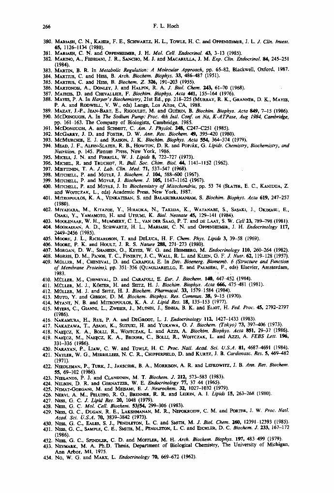
266 F .L . Hoch
380. MAR~.sn, C. N., ~ , F. E., SCmVXRTZ, H. L., TOWLE, H. C. and OPPENHEIMER, J. L. J. Clin. Incest. 65, 1126-1134 (1980).
381. MAm,~I-I, C. N. and O p p ~ m . . ~ , J. H. Mol. Cell. Endocrinol. 43, 3-13 (1985). 382. MAR1NO, A., FEDRIANI, J. R., S^NCHO, M. J. and MACARULt.A, J. M. Exp. Clin. Endocrinol. 84, 245-251
(1984). 383. MARTIN, B. R. In Metabolic Regulation: A Molecular Approach, pp. 65-82, BlackweU, Oxford, 1987. 384. MARTIUS, C. and HESS, B. Arch. Biochem. Biophys. 33, 486-487 (1951). 385. MARTIUS, C. and HESS, B. Biochem. Z. 326, 191-203 (1955). 386. MARTONOSl, A., DONLEY, J. and HALPIN, R. A. J. Biol. Chem. 243, 61-70 (1968). 387. MATH~, D. and ~ALLIER, F. Biochim. Biophys. Acta 441, 155-164 (1976). 388. MAYES, P. A. In Harper's Biochemistry, 21st Ed., pp. 218-225 (MURRAY, R. K., GRAN~R, D. K., MAYF.$,
P. A. and RODWELL, V. W., eds) Lange, Los Altos, CA, 1988. 389. MAZAT, J.-P., Jr~N-BART, E., RIGOULFr, M. and GUdruN, B. Biochim. Biophys. Acta 849, 7-15 0986). 390. McDONoUGH, A. In The Sodium Pump: Proc. 4th Intl. Conf. on Na, K-ATPase, Aug 1984, Cambridge,
pp. 161-163. The Company of Biologists, Cambridge, 1985. 391. McDoNouGn, A. and ScnmTr, C. Am. J. Physiol. 248, C247--C251 (1985). 392. McGARRy, J. D. and FOSTER, D. W. Ann. Rev. Biochem. 49, 395-420 0980). 393. McMURcmI~, E. J. and RAISON, J. K. Biochim. Biophys. Acta 554, 364-374 0979). 394. MEAD, J. F., ALFIN-SLATER, R. B., HOWTON, D. R. and PoPJ,~r, G. Lipids. Chemistry, Biochemistry, and
Nutrition, p. 145. Plenum Press, New York, 1986. 395. MICELI, J. N. and FER~LL, W. J. Lipids 8, 722-727 (1973). 396. MICHEL, R. and TRUCHOT, R. Bull. Soc. Chim. Biol. 44, 1141-1152 (1962). 397. MIElrr l~, T. A. J. Lab. Clin. Med. 71, 537-547 0968). 398. MITCHELL, P. and MOYLE, J. Biochem. J. 104, 588-600 (1967). 399. MITCHELL, P. and MOYLE, J. Biochem. J. 105, I147-I162 (1967). 400. MITCHELL, P. and MOYLE, J. In Biochemistry of Mitochondria, pp. 53-74 (SLATER, E. C., KANIUGA, Z.
and WOJTCZAK, L., eds) Academic Press, New York, 1987. 401. MITROPOUt~3S, K. A., VENKATESA~, S. and BALASUBRAMANIAM, S. Biochim. Biophys. Acta 619, 247-257
(1980). 402. MIVAnARA, M., KITAZO~, Y., HIRAOKA, N., TAr, EDA, K., WATANAnE, S., SASAKI, J., OKIMASU, E.,
OSAKI, Y., YAMAMOTO, H. and UTSUMI, K. Biol. Neonate 45, 129-141 (1984). 403. MOOLF.nAAR, W. H., MUMMERY, C. L., VAN DER SAA(3, P. T. and DE LAAT, S. W. Cell 23, 789-798 (1981). 404. MOORAOtAN, A. D., SCHWARTZ, H. L., MARIASH, C. N. and OPl~t,n-mlMER, J. H. Endocrinology 117,
2449-2456 0985). 405. MOORE, J. L., P, JCHARDSON, T. and D~LUCA, H. F. Chem. Phys. Lipids 3, 39-58 (1969). 406. MOORE, P. K. and HOULT, J. R. S. Nature 288, 271-273 0980). 407. MORGAN, D. W., SnAHr~N, O., KEYES, W. G. and HEIMm~6, M. Endocrinology l l0, 260-264 (1982). 408. MORRIS, D. M., PANOS, T. C., FItCERrt, J. C., WALL, R. L. and KLEIn, G. F. J. Nutr. 62, 119-128 (1957). 409. MOLLER, M., CHENEVAL, D. and CARAFOLI, E. In Dev. Bioenerg. Biomemb. 6 (Structure and Function
of Membrane Proteins), pp. 351-356 (QUAGLIAR~LLO, E. and PALMIFJU, F., eds) Elsevier, Amsterdam, 1983.
410. Mi~LLER, M., CHENEVAL, D. and CARAFOLI, E. Eur. J. Biochem. 140, 447-452 (1984). 411. MOLLER, M. J., KOSTER, H. and SEITZ, H. J. Biochim. Biophys. Acta 666, 475-481 (1981). 412. MOLLER, M. J. and SFJTZ, H. J. Biochem. Pharmacol. 33, 1579-1584 (1984). 413. MUTO, Y. and GI~O~, D. M. Biochem. Biophys. Res. Commun. 38, 9-15 (1970). 414. MYANT, N. B. and MITROPOULOS, K. A. J. Lipid Res. 18, 135-153 (1977). 415. MYERS, C., GIAWIql, L., ZWEIF.R, J., MUINDI, J., SINHA, B. K. and ELIOT, H. Fed. Proc. 45, 2792-2797
(1986). 416. NAKAMURA, H., RUE, P. A. and DEGROOT, L. J. Endocrinology 112, 1427-1433 (1983). 417. NAKAZAWA, T., ASAMI, K., SUZUKI, H. and YUKAWA, O. J. Biochem. (Tokyo) 73, 397-406 (1973). 418. NAJ~cz, K. A., BOLLI, R., WOJTCZAK, L. and Azzl, A. Biochim. Biophys. Acta 851, 29-37 (1986). 419. NA~CZ, M., Nat~cz, K, A., BROGER, C., BOLU, R., WO/TCZAK, L. and AZZl, A. FEBS Lett. 196,
331-336 (1986). 420. NARAYAN, P., LL~W, C. W. and TOWLE, H. C. Proc. Natl. Acad. Sci. U.S.A. 81, 4687-4691 (1984). 421. NAYLER, W. G., MERRILLEES, N. C. R., CHIPPERFIELD, D. and KURTZ, J. B. Cardiovasc. Res. 5, 469~82
(1971). 422. N~DLEMAN, P., TURK, J., JAKSCmK, B. A., MOt~ISON, A. R. and LI~EKOWiTZ, J. B. Ann. Rev. Biochem.
55, 69-102 (1986). 423. NI~LANDS, P. J. and CLANDINI~, M. T. Biochem. J. 212, 573-583 (1983). 424. NELSON, D. R. and CORNATZER, W. E. Endocrinology 77, 37-44 (1965). 425. NEMAT-GORGANI, M. and MEIST~MI, E. J. Neurochem. 32, 1027-1032 (1979). 426. NF~W, A. M., PELUFFO, R. O., BRENNER, R. R. and LUKIN, A. I. Lipids 15, 263-268 (1980). 427. N~ss, G. C. J. Lipid Res. 20, 1048 (1979). 428. Nws, G. C. Mol. Cell. Biochem. 53/54, 299-306 (1983). 429. NESS, G. C., DUGAN, R. E., LAKSIIMANAN, M. R., NI~POKROEFF, C. M. and POR~R, J. W. Proc. Natl.
Acad. Sci. U.S.A. 70, 3839-3842 (1973). 430. NESS, G. C., EA~s, S. J., PEND~N, L. C. and SMITH, M. J. Biol. Chem. 260, 12391-12393 (1985). 431. N~SS, G. C., SAMPLE, C. E., SMITH, M., PENDLETON, L. C. and EICHLER, D. C. Biochem. J. 233, 167-172
(1986). 432. N~ss, G. C., SmNDLF.R, C. D. and MOFFI.ER, M. H. Arch. Biochem. Biophys. 197, 483-499 (1979). 433. N ~ K , M. A. Ph.D. Thesis, Department of Biological Chemistry, The University of Michigan,
Ann Arbor, MI, 1975. 434. NG, W. G. and MARX, L. Endocrinology 70, 669-672 (1962).

Lipids and thyroid hormones 267
435. NICHOLLS, D. G. Eur. J. Biochem. 50, 305-315 (1974). 436. NICHOLI.S, D. G. Bioenergetics. An Introduction to the Chemiosmotic Theory, 190 pp., Academic Press,
New York, 1982. 437. NICtIOLLS, D. G. In New Compr. Biochem. 9 (Bioenergetics), pp. 29--48 (ERNSTER, L., ed.)
Elsevier, Amsterdam, 1984. 438. NICtlOLLS, P. and WF.~R, C. E. Arch. Biochem. Biophys. 151, 206-215 (1972). 439. NICHOLS, J. W. and DEAMr~, D. W. Proc. Natl Acad. Sci. U.S.A. 77, 2038-2042 (1980). 440. NIEMI~.YER, H., CRANE, R. K., KEh'NI~Y, E. P. and LIPMANN, F. Bol. Soc. Biol. Santiago, Chile 10, 54-58
(1953). 441. NmWL, J. G. and Rom~'~ON, N. Biochem. Soc. Trans. 4, 930-931 (1976). 442. Nlsmrd, K., ERECIIqSKX, M., WILSON, D. F. and COOPER, S. Am. J. Physiol. 235, C212-C219 (1978). 443. NO~, H. and PA~E, S. V. Eur. J. Biachem. 150, 99-102 (1986). 444. NORDSTROM, J. L., RODWELL, V. W. and MITSCm~LEN, J. J. J. Biol. Chem. 252, 8924-8934 (1977). 445. NUM,',, S. and TA.NABE, T. In New Compr. Bioehem. 7 (Fatty Acid Metabolism and Its Regulation),
pp. 1-28 (NuMA., S., ed.) Elsevier, Amsterdam, 1984. 446. N u t , z, J. Mol. Cell. Endocrinol. 37, 125-132 (1984). 446a. O~TA, T., IOTAOAWA, S., GONG, Q.-H., PASTAN, I. and C--~NG, S.-Y. J. Biol. Chem. 263, 782-785 (1988). 447. Ocl~, R. S., HANSON, R. W. and HA.LL, J., (eds) Metabolic Regulation, 244 pp., Elsevier, Amsterdam,
1985. 448. OLUg,.DEWO, J. O. and HEIMBERG, M. Biochem. Biophys. Res. Commun. 132, 1001-1007 (1985). 449. OLUBADEWO, J. O., WILCOX, H. G. and HEIMeERG, M. J. Biol. Chem. 259, 8857-8862 (1984). 450. OMURA, T. and SATO, R. J. Biol. Chem. 239, 2370-2378 (1964). 451. OPPlS~naVJMER, J. H. Science 203, 971-979 (1979). 452. OPPENHEn~sR, J. H. Ann. Int. Med. 102, 374-384 (1985). 453. OPPENHEI~SR, J. H. and SAMUEl.S, H. H., (eds) Molecular Basis of Thyroid Hormone Action, 498 pp.,
Academic Press, New York, 1983. 454. OPPENHEIMER, J. H., SCHWARTZ, H. L., MARIASH, C. N., KINLAW, W. B., WONG, N. C. W. and
FRr.ArJZ, H. C. Endocrine Rev. 0, 288-308 (1987). 455. OPPEm-I~M~, J. H., SCHWARTZ, H. L. and SURKS, M. I. Endocrinology 95, 897-903 (1974). 455a. O ~ o o , H., BLAKE, J. E., B~a~lS, L. M., COMFrON, K. V. and I s ~ , Y. New Engl. J. Med. 317,
1421-1427 (1987). 456. O'SlmA, P. S., l~tm_~lN-TI-mL~, S. and AzzI, A. Biochem. J. 220, 795-801 (1984). 457. OSmNO, N., Iloa, Y. and SAXO, R. J. Biochem. (Tokyo) 69, 155-167 (1971). 458. OSmNO, N. and SATO, R. Arch. Biochem. Biophys. 149, 369-377 (1972). 459. P~AN, E. and Rma'~BERO, H. Eur. J. Biochem. 40, 431-437 (1973). 460. PAGE, E. and MCALLISTVat, L. P. Am. J. Cardiol. 31, 172-181 (1973). 461. PA~E, S. and MuRxI~, M. Fed. Proc. 46, 1180 (1987). 462. PA~E, S. V., M U R ~ , M. S. R. and NOi~L, H. Biochim. Biophys. Acta 877, 223-230 (1986). 463. PA.NDE, S. V. and PARwN, R. In Carnitine Biosynthesis, Metabolism, and Functions, pp. 143-157
(FrmNr~L, R. A. and McGARRY, J. D., eds) Academic Press, New York, 1980. 464. PANOS, T. C. and FI~,nERXY, J. C. J. Nutr. 54, 315-331 (1954). 465. PAPAHADJOI'OUI.OS, D. and WAT~NS, J. C. Biochim. Biophys. Acta 135, 639-652 (1967). 466. PARK, C. E., MARAI, E. and M o o ~ , S. Biachim. Biophys. Acta 270, 50-59 (1972). 467. PARK£R, R. A., MILLER, S. J. and GmsoN, D. M. Biochem. Biophys. Res. Commun. 125, 629-635 (1984). 468. PATTON, J. F. and PLAXNER, W. S. Proc. Soc. Exp. Biol. Med. 137, 196-201 (1971). 469. PA.vvovIc-Hotn~NAC, M., D~BAUF~, D. and O~AYON, R. Mol. Cell. Endocrinol. 12, 255-265 (1978). 470. PAVLOVIc-Hotn~NAC, M., DELBAUV~, D. and O~YON, R. Proc. FEBS Meet. 1978. 54 (Cyclic Nucleotides
Protein Phosphorylation Cell Regulation), pp. 263-272 (1979). 471. PETER, J. J. J. Lipid Res. 9, 193-199 (1968). 472. PmLIPVOT, J. R., COOPER, A. G. and WALLA.CH, D. F. H. Proc. Natl. Acad. Sci. U.S.A. 74, 956-960
(1977). 473. PmLIV'SON, K. D. and EDEL~N, I. S. Am. J. Physiol. 232, C202-C206 (1977). 474. PIATh~K-LEL~SSEN, D. A. and LEUNLSSEN, R. L. A. Circ. Res. 25, 171-181 (1969). 475. PIL~gOnON, D. and CAPLAr~, S. R. Biochemistry 24, 5764-5776 (1985). 476. PlHLAJAN1EMI, T., I-IELAAKOSgd, T., TASAh'EN, K., MYLLYL.~, R., HUEITALA, M.-L., KOlVU, J. and
IOVImKKO, K. I. EMBO J. 6, 643-649 (1987). 477. PITT-RIW_.RS, R. and "rATA, J. R. In The Thyroid Hormones, pp. 120-123, Pergamon, New York, 1959. 478. PLATNER, W. S., PATNA.YAK, B. C. and ~ , R. R. J. Proc. Soc. Exp. Biol. Med. 140, 857-861 (1972). 479. I~IAM, N. B. and GOLDF~h'E, I. D. Biochem. Biophys. Res. Commun. 79, 166-172 (1977). 480. POLLA.K, J. K. Biochem. J. 150, 477-488 (1975). 481. POON, R., PdCRARDS, J. M. and CLARK, W. R. Biochim. Biophys. Acta 649, 58-66 (1981). 482. POORTHUIS, B. J. H. M. and HOSTI~rLER, K. Y. Biochim. Biophys. Acta 431, 408-415 (1976). 483. PORTNAY, G. I., MCCLENDON, F. D., BUSH, J. E., BRAVER~,N, L. E. and BAalOR, B. M. Biochem. Biophys.
Res. Commun. 55, 17-21 (1973). 484. POW'~LL, G. L., KNOWL~S, P. F. and MARSH, D. Biochim. Biaphys. Acta 816, 191-194 (1985). 485. Pg~IgS~xm, H. G. and Ktmos, G. Life Sci. 24, 35-42 (1979). 486. PUOH, E. L. and KATES, M. Lipids 14, 159-165 (1979). 487. QURESm, A. A., JENIK, R. A., KIM, M., LOR~ITZO, F. A. and PORTER, J. W. Biochem. Biophys. Res.
Commun. 66, 344-351 (1975). 488. RAmMIFAR, M. and ISMAIL-BEIGI, F. Mol. Cell. Endocrinol. 6, 327-331 (1977). 489. RAISON, J. K. Bioenergetics 4, 285-309 (1973). 490. RAMSAY, R. R. Fed. Proc. 46, 1013 (1987). 491. RAMSAY, R. R., DERRICK, J. P., FRn~ND, A. S. and TueBs, P. K. Biochem. J. 244, 271-278 (1987).

268 F.L. Hoch
492. RAO, G. S. Mol. Cell. Endocrinol. 21, 97-108 (1981). 493. RJDGWAY, N. D. and DOLPHIN, P. J. J. Lipid Res. 26, 1300-1313 (1985). 494. RIMON, G., HANSKI, E., BRAUN, S. and L~vrrzKi, A. Nature 276, 394-396 (1978). 495. ~ , J. P. W. and FRANK~L, T. L. Br. Med. Bull. 37, 59-64 (1981). 495a. I~WRS, J. P. W., SINCLAm, A. J. and CRAWFORD, M. A. Nature 258, 171-173 (1975). 496. ROmNSON, N. C., STRY, F. and T^LBERT, L. Biochemistry 19, 3656-3661 (1980). 497. RODBELL, M. and JONES, A. B. J. Biol. Chem. 241, 140-142 (1966). 498. Roosts, K. S., HI~INS, E. S. and GRO6A~, W. MCL. J. Nutr. II0, 248-254 (1980). 499. RONCARI, D. A. K. and MURTHY, V. K. J. Biol. Chem. 250, 4134-4138 (1975). 500. ROODYN, D. B., FREEMAN, K. B. and TATA, J. R. Biochem. J. 94, 628-641 (1965). 501. ROQUF~, M., TmARD, A., DEGROOT, L. J. and RAPAUD, J. Endocrinology I00, 967-973 (1977). 502. ROSENQ~qST, U. Acta Med. Scand. 192, 353-359 (1972). 503. ROSENQWST, U. Mol. Cell. Endocrinol. 12, 111-117 (1978). 504. ROSSlONOL, M., THOMAS, P. and GRIG~ON, C. Biochim. Biophys. Acta 684, 195-199 (1982). 505. ROTTENBERG, H. Mol. Cell Biol. 4, 47-83 (1985). 506. ROTTENBERG, H. and HASHIMOTO, K. Biochemistry 25, 1747-1753 (1986). 507. ROTT~NnERG, H., ROBERTSON, D. E. and RUBIN, E. Biochim. Biophys. Acta 809, I-I0 (1985). 508. ROYCe, S. M. and HOLMES, R. P. Biochim. Biophys. Acta 792, 371-375 (1984). 509. RUBALCAVA, B. and RODBELL, M. J. Biol. Chem. 248, 3831-3837 (1973). 510. RUGGIERO, F. M., LANDRISCINA, C., GNONI, G. V. and QUAGLIARmLtO, E. Horm. Metabol. Res. 16, 37-40
(1984). 511. Ruc_~.lto, F. M., LANDRISCINA, C., GNOHI, G. V. and QUAOUAmELLO, E. Lipids 19, 171-178 (1984). 512. RYDSrRtSM, J. Biochim. Biophys. Acta 463, 155-184 (1977). 513. RYDSrR6M, J. J. Biol. Chem. 25,1, 8611-8619 (1979). 514. RYDSTR6M, J. and FLEISCH~, S. Meth. Enzymol. 50, 811-816 (1979). 515. RYDSTROM, J., HOEK, J. B., ERICSOr~, B. G. and HUNDAL, T. Biochim. Biophys. Acta 430, 419-425 (1976). 516. RYDSTR6M, J., KAm~a~., N. and RACKER, E. Biochem. Biophys. Res. Commun. 67, 831-839 (1975). 517. SAmNE, J. R. Trends Biochem. Sci. 8, 234-236 (1983). 518. SACKTOR, B. and KXNSELLA, J. L. Curr. Topics Membr. Transp. 26, 223-244 (1986). 519. SAC_~ERSON, E. D., BraD, M. I., CARPEN~R, C. A., WIrCrF.R, K. A. and WRiGrrr, J. J. Biochem. J. 224,
201-206 (1984). 520. SAMPLE, C. E., I~NDLETON, L. C. and NF.SS, G. C. Biochemistry 26, 727-731 (1987). 521. SAm~LS, H. H., P~L~t,~N, A. J., RAACKA, B. M. and S'rANLEY, F. In Molecular Basis of Thyroid Hormone
Action, pp. 100-139 (OPPENrmI~tER, J. H. and SAmJELS, H. H., ecls) Academic Press, New York, 1983. 522. SANCHO, M. J., MAC~ULLA, J. M., SEC_,OVlA, J. L. and GONZALEZ, C. An. Quire. 75, 413-415 (1979). 523. SAr~Dmt~L~rt~, H., JR. Biochim. Biophys. Acta 515, 209-213 (1978). 524. SANGnv~, A., GRAssy, E., WARTY, V., D~VEN, W., Wl~ar, C. and Lr.STrat, R. Biochem. Biophys. Res.
Commun. 103, 886-892 (1981). 525. SANTt~O, E., I.,61~z-MoRATALLA, N. and S~OWA, J. L. Biochem. Biophys. Res. Commun. 53, 439--445
(1973). 526. SAP, J., Mt~oz, A., DAre, t, K., GOLDBm~, Y., GnVSD~L, J., LEUrZ, A., BEU~, H. and
VENNSTR6~t, B. Nature 3/,4, 635-640 (1986). 527. SAVA~F.AU, M. A. Arch. Biochem. Biophys. 145, 612--621 (1971). 528. SAVAOr.AU, M. A. Curr. Topics Cell. Reg. 6, 63-130 (1972). 529. SAvA~r.~U, M. A. Biomed. Biochim. Acta 44, 839-844 (1985). 530. Scmomt:rrTot.o, L., TR~ZZ~, E., ROMA, P. and COTAPANO, A. L. Atherosclerosis 59, 329-333 (1986). 531. SC~PULLA, R. C., KIL~, M. C. and S¢~PULLA, K. M. J. Biol. Chem. 261, 4660--4662 (1986). 532. ScnmrT, C. and McDoNou6H, A. Fed. Proc. 46, 361 (1987). 533. SCHNEIDER, J. W., MERCER, R. W., CAPLAN, M., EMA~a~'EL, J. R., Swr.ADr~rr~, K. J., BENZ, E. J., JR and
LEVENSON, R. Proc. Natl. Acad. Sci. U.S.A. 82, 6357-6361 (1985). 534. SClmOF.DER, F. J. Membr. BioL 68, 1-10 (1982). 535. SCrlt~I~IANS ST~KnOWr~, F. M. A. H. and BONT~G, S. L. Physiol. Rev. 61, 1-76 (1981). 536. SCHtrtrR~ANS STEKHOVEN, F. M. A. H., SAN~, B. P. and SANADi, D. R. Biochim. Biophys. Acta 226, 20-32
(1971). 537. SCHWARTZ, H. L., T~NCE, D., OI'PE~a-lm~n~R, J. H., J~AN~, N. S. and JOMP, D. B. Endocrinology 113,
1236-1243 (1983). 538. SEOAL, J., BUCKLEY, C. and IN,BAR, S. H. Endocrinology 116, 2036-2043 (1985). 539. S~GAL, J. and GoPa~oN, A. Endocrinology 101, 150-156 (1977). 540. SteAL, J. and I~3BAR, S. H. J. Clin. Invest. 68, 103-110 (1981). 541. Sra3AL, J., SCh'WARTZ, H. and GORDON, A. Endocrinology 101, 143-149 (1977). 542. SE~TZ, H. J., MOLLER, M. J. and SOaOLL, S. Biochem. J. 227, 149-153 (1985). 543. SmratSTED, O. M. Biochim. Biophys. Acta 717, 163-174 (1982). 544. S~KUZU, I., JURTSrFOt¢, P. and G ~ N , D. E. J. Biol. Chem. 238, 975-982 (1963). 545. SELLERS, E. A. and You, S. S. Am. J. Physiol. 163, 81-91 (1950). 546. SEN, N. (HuLam~T, A. J.) Honours Master of Science Thesis, U. Wollongong, Australia, 1986. 547. SEVEI~SON, D. L. and FLETCHER, T. Biochim. Biophys. Acta 675, 256-264 (1981). 547a. SEYMOtr~, A.-M. L., ~ H , J. M. and IL~DA, G. K. Biochem. Soc. Trans. I1, 376-377 (1983). 548. $rlARMA, V. K. and BA~srE~, S. P. 11401. Pharmacol. 14, 122-129 (1978). 549. SHAW, M. J. and Hot:H, F. L. Life Sci. 19, 1359-1364 (1976). 550. SHAw, M. J. and HocH, F. L. J. 114o1. Cell. Cardiol. 9, 749-761 (1977). 551. SI-mARS, S. B. J. Theor. Biol. 82, 1-13 (1980). 552. SI-mARS, S. B. Fur. J. Biochem. 146, 233-235 (1985). 553. SHEARS, S. B. and BRONK, J. R. Biochem. J. 178, 505-507 (1979).

Lipids and thyroid hormones 269
554. SHEARS, S. B. and BRONK, J. R. J. Bioenerg. Biomembr. 12, 379-393 (1980). 555. S1Ess, E. A., KmWrScH-ENGEL, R. I. and FAmr, fl, F. M. Biochem. Soc. Trans. 13, 657-659 (1985). 556. SIr,nSNSKY, M., I:hNKERTON, F., S ~ L A N D , E. and SIMON, F. R. Proc. Natl. Acad. Sci. U.S.A. 76,
4893-4897 (1979). 557. SINOH, A. P. and Bp.AC~3, P. D. J. Bioenerg. 7, 175-188 (1975). 558. S1PAT, A. B. and SABINE, J. R. J. Lipid Res. 20, 1049 (1979). 559. SUaTH, E. L., HILL, R. L., LEr~AN, I. R., LEFKOW]TZ, R. J., HArqDLEg, P. and W m ~ , A. In
Principles of Biochemistry: General Aspects, 7th Ed, p. 372, McGraw-Hill, New York, 1983. 560. SMITH, J. and DELuCA, H. F. J. Nutr. 78, 416-422 (1963). 561. SMITH, J. and DELUCA, H. F. J. Cell. Biol. 21, 15-26 (1964). 562. SlVnTH, R. L. and OLDFIELD, E. Science 225, 280-288 (1984). 562a. SMITH, V. L., BREr~T, L. G. and THOMPSON, R. E. J. Lipid Res. 20, 1049 (1979). 563. SNn,ES, R. L. Lab. Invest. 18, 179-189 (1968). 564. SOKOI.OFF, L. In Thyroid Horm. Brain Dev. [Proc. Conf.], pp. 367-382 (GRAVE, G. D., ed.) Raven,
New York, 1977. 565. SOLOMONSON, L. P., LIEPKAL~qS, V. A. and SPECTOR, A. A. Biochemistry 15, 892-897 (1976). 566. SOMJEN, D., ISMAIL-BEIGI, F. and EDELMAN, I. S. Am. J. PhyshTl. 240, E146-EI54 (1981). 567. SPECTOR, A. A. and YOREK, M. A. J. Lipid Res. 26, 1015-1035 (1985). 568. SPENCER, T. L., S~, J. K. and BYORAVE, F. L. Biochim. Biophys. Acta 423, 365-373 (1976). 569. SPERLING, M. A., GANGULI, S., VOINA, S., KAPTEIN, E. and NXCOLOFF, J. T. Endocrinology 107, 684--690
(1980). 570. SPRECHER, H. In Polyunsaturated Fatty Acids, pp. 1-18 (KUNAU, W.-H. and HOLMAN, R. T., ¢ds)
Am. Oil Chemists Soc., Champaign, IL, 1977. 571. STAKKESTAD, J. A. and B~MEa, J. Biochim. Biophys. Acta 711, 90-100 (1982). 572. STAKrd"STAD, J. A. and BREMER, J. Biochim. Biophys. Acta 750, 244-252 (1983). 573. STANACEV, N. Z., DAVIDSON, J. B., STUHNE-SEKALEC, L. and DOMAZET, Z. Can. J. Biochem. 51, 286-304
(1973). 574. STANCLIFF, R. C., WILLIAMS, M. A., ITSUMI, K. and PACKER, L. Arch. Biochem. Biophys. 131, 629---642
(1969). 575. STEFFEN, D. G. and PLATI,~R, W. S. Am. J. Physiol. 231, 650-654 (1976). 576. S~INaERG, D. Pharmacol. Rev. 18, 217-235 (1966). 577. STERLING, K. New Engl. J. Med. 300, 11%123, 173-177 (1979). 578. STERLING, K. Endocrinology 119, 292-295 (1988). 579. STILES, G. L., CAgON, M. G. and LEFKOWITZ, R. J. Physiol. Rev. 64, 661-743 (1984). 580. STIPAN1, I., BARBEKO, M. C. and PALMIER1, F. In Dev. Bioenergy. Biomemb. 6 (Structure and Function
of Membrane Proteins), pp. 369-372 (QUAGLIARIELLO, E. and PALMmPa, F., ¢ds) Elsevier, Amsterdam, 1983.
581. STORCH, J. and SCHAcwr~R, D. Biochemistry 23, 1165-1170 (1984). 582. STORY, J. A., Tm'~g, S. A. and KRITCHEVSKY, D. Biochem. Med. 10, 214-218 (1974). 583. STRUBELT, O. Arch. Intl. Pharmacodyn. Ther. 176, 263-288 (1968). 584. STUMP, D. A., EOUgEN, L. A. and PARKS, J. K. Biochem. Med. 34, 226-229 (1985). 584a. SUGDEN, M. C., WATTS, D. I. and MARSHALL, C. E. Biosci. Rep. 1, 757-764 (1981). 585. SUKO, J. Biochim. Biophys. Acta 252, 324-327 (1971). 586. SUN, G. Y. and SuN, A. Y. J. Neurochem. 22, 15-18 (1974). 587. SUZUKI, M., IMAI, K., ITO, A., OMURA, T. and SATO, R. J. Biochem. (Tokyo) 62, 447--455 (1967). 588. SUZUKI, S. and SUZUKI, M. Gen. Comp. Endocrinol. 45, 74-81 (1981). 589. SWANLJUNG, P., FRIGER], K., OHt.SON, K. and ERNSTeR, L. Biochim. Biophys. Acta 305, 519-533 (1973). 590. SWEADr,~ER, K. J. J. Biol. Chem. 254, 6060-6067 (1979). 591. TABACrINICK, I. I. A. and BONNYCASTLE, D. D. J. Biol. Chem. 207, 757-760 (1954). 592. TAKEUCm, N., ITO, M., UCmDA, K. and YAMAMURA, Y. Biochem. J. 148, 499-503 (1975). 593. TAKEUCm, N., ITO, M. and YAMAMURA, Y. Atherosclerosis 20, 481-494 (1974). 594. TAMKUN, M. M. and FAMnROUGH, D. M. J. Biol. Chem. 261, 1009-1019 (1986). 595. TANAKA, R. and SAKAMOTO, T. Biochim. Biophys. Acta 193, 384-393 (1969). 596. TATA, J. R. Nature 213, 566-569 (1967). 597. TATA, J. R. Biochem. J. 104, 1-16 (1967). 598. TATA, J. R. Nature 219, 331-337 (1968). 599. TATA, J. R. Biochem. J. 116, 617-630 (1970). 600. TATA, J. R., ERNSTER, L., LINDBERO, O., ARRHENIUS, E., PEDERSEN, J. and HEDMAN, R. Biochem. J. 86,
408-428 (1963). 601. TAYLOR, V. J. and PAGAN, C. I. Biochim. Biophys. Acta 851, 49-56 (1986). 602. TEPPERMAN, H. M. and TEPPERMAN, J. Am. J. Physiol. 202, 401-406 (1962). 603. TEPPERMAN, H. M. and TEPPERMAN, J. Am. J. Physiol. 206, 357-361 (1964). 604. TEPPERMAN, J. and TEPPERMAN, H. M. Am. J. Physiol. 200, 1069-1073 (1961). 605. THOMPSON, C. C., WEINBERGER, C., LEBO, R. and EVANS, R. M. Science 237, 1610-1614 (1987). 606. THOMPSOrq, G. A., JR. The Regulation of Membrane Lipid Metabolism, 218 pp., CRC Press, Boca Raton,
FL, 1980. 607. TISHLER, P. V. and HAMMOND, M. E. Enzyme 20, 349--358 (1975). 608. TOWLE, H. C. In Molecular Basis of Thyroid Hormone Action, pp. 179-213 (OPPENHEIMER, J. H. and
SAMUEL.S, H. H., eds) Academic Press, New York, 1963. 609. TSAI, J. S. and CHEN, A. Nature 275, 138-140 (1978). 610. TsE, J., WRENN, R. W. and Kuo, J. F. Endocrinology 107, 6-10 (1980). 611. TURCHETTO, E., SAr~GUINETTX, F. and Rossl, C. A. Boll. Soc. ltal. Biol. Sper. 31, 1324-1326 (1955). 612. TYSON, C. A., VANDE ZANDE, H. and GREETS, D. E. J. Biol. Chem. 251, 1326-1332 (1976).

270 F .L . Hoch
613. UMSKI, S. and NOZAWA, Y. Biochim. Biophys. Acta 83~, 514-526 (1985). 614. UNDERWOOD, A. H., EMMFrr, J. C., ELUS, D., G L ~ , S. B., L~SON, P. D., BENSON, G. M.,
NOVFJ.LI, R., PV.AtC'E, N. J. and SHAH, V. P. Nature 324, 425-429 (1986). 615. VAN HOLrVEN, R. P. and EMMELOT, J. J. Membr. Biol. 9, 105-126 (1972). 616. VAN INWEOEN, R. G., ROmsoN, G. A., THOMPSON, W. J., ARMSTRONO, K. J. and STOUVE~, J. E.
J. Biol. Chem. 250, 2452-2456 (1975). 617. VASDEV, S. C., KOI~CK¥, B., RASTOGI, R. B., SINGHAL, R. L. and KAKO, K. J. Can. J. Physiol. Pharmacol.
55, 1311-1319 (1977). 618. VERHOEXr~N, A. J., KAMSR, P., GROEN, A. K. and TAOER, J. A. Biochem. J. 226, 183-192 (1985). 619. VIGNAIS, P. V. and LAUQUIN, G. J. M. Trends Biochem. Sci. 4, 90-92 (1979). 620. VOLPE, J. J. and KlSmMOTO, Y. J. Neurochem. 19, 737-753 (1972). 621. WAKIL, S. J., STOOPS, J. K. and Joshn, V. C. Ann. Rev. Biochem. 52, 537-579 (1983). 622. WALKER, P., DUBOXS, J. D. and DUSSAULT, J. H. Pediatr. Res. 14, 247-249 (1980). 623. WALLACH, D. F. H. (with contributions by R. Schmidt-Ullrich) Membrane Molecular Biology o f Neoplastic
Cells, 525 pp., Elsevier, Amsterdam, 1975. 624. WALLACH, S., BELLAVIA, J. V., GAMPONIA, P. J. and BRISTRIM, P. J. Clin. Invest. 51, 1572-1577 (1972). 625. WALRAVE~S, PH. and CHASE, H. P. J. Neurochem. 16, 1477-1484 (1969). 626. WARING, A. J., ROTTENBERO, H., OHstsm, T. and RumN, E. Proc. Natl. Acad. Sci. U.S.A. 78, 2582-2586
(1981). 627. WARSHAW, J. B. Dev. Biol. 28, 537-544 (1972). 628. WEINBEROER, C., THOMPSON, C. C., ONO, E. S., LEnO, R., GRUOL, D. J. and EVANS, R. M. Nature 324,
641-646 (1986). 629. WF.RI~R, H. V. Fur. J. Biochem. 48, 45-52 (1974). 630. Wl~SSON, L. G. and BURR, G. O. J. Biol. Chem. 91, 525-539 (1931). 631. W~TERHOFF, H. V., COKEN, A.-M. and VAN DAM, K. Biochem. Soc. Trans. 11, 81-85 (1983). 632. WHITE, D. A. In Form and Function o f Phospholipids (ANSELL, O. B., HAWTHORNE, J. N. and
DAWSON, R. M. C., eds) BBA Library 3, pp. 441-482, 1973. 633. WHITTAM, R. In Cellular Functions o f Membrane Transport, pp. 139-154 (HoFVMAN, J. F., ed.)
Prentice-Hall, Englewood Cliffs, NJ, 1963. 634. WHITTAM, R. and BLOND, D. M. Biochem. J. 92, 147-158 (1964). 634a. WXEDERER, O. and L6FFLER, G. J. Lipid Res. 28, 649-658 (1987). 635. WILLIAMS, H. J. and BARmE, S. E. Biochem. Biophys. Res. Comman. 84, 89-94 (1978). 636. WILLIAMS, M. A., STANCLIFF, R. C., PACKER, L. and K~TH, A. D. Biochim. Biophys. Acta 267, 444-456
(1972). 637. WILLIAMS, R. J. P. FEBS Left. 85, 9-19 (1978). 638. WILSOIq, J. W. and LEDUC, E. H. J. Cell. Biol. 16, 281-296 (1963). 639. WILSON, J. W. and LEDU¢, E. H. Natl. Cancer Inst. J. 33, 721-739 (1964). 640. WINKLER, L., BUANGA, N.-F. and GOETZE, E. Biochim. Biophys. Acta 231, 535-536 (1971). 641. WOLFF, E. C. and WOLFF, J. In The Thyroid Gland, Vol. 1, pp. 237-282 (PITT-RIvEtS, R. and
TROTTER, W. R., eds) Butterworths, Washington, DC, 1964. 642. WOLTERINK, L. F., CORNWALL, R. L., VINSON, C. and COLE, L. L Fed. Proc. 12, 158-159 (1953). 643. WOOD, J. McM. J. Biol. Chem. 250, 3062-3066 (1975). 644. WYKLE, R. L. In Lipid Metabolism in Mammals, Vol. I, pp. 317-366 (F. S~rrDER, ed.) Plenum Press,
New York, 1977. 645. YATES, F. E., URQUHART, J. and HERBST, A. L. Fed. Proc. 17, 174 (1958). 646. YORK, D. A., BRAY, G. A. and YUKIMURA, Y. Proc. Natl. Acad. Sci. U.S.A. 75, 477-481 (1978). 647. YouNo, D. L. and L¥~N, F. J. Biol. Chem. 244, 377-383 (1969). 648. Yu, H. L. and BURTOZq, D. N. Arch. Biochem. Biophys. 161, 297-305 (1974). 649. Yu, L., Yu, C.-A. and KINO, T. E. J. Biol. Chem. 253, 2657-2663 (1978). 650. ZAMmT, V. A. Prog. Lipid Res. 23, 39-67 (1984). 651. ZAMMIT, V. A. Biochem. Soc. Trans. 14, 676-679 (1986). 652. ZAMMIT, V. A. and EASOM, R. A. Biochim. Biophys. Acta 927, 223-228 (1987). 653. ZIERZ, S. and ENOEL, A. G. Eur. J. Biochem. 149, 207-214 (1985).