Embed Size (px)
Citation preview
r e v b r a s o r t o p . 2 0 1 8;5 3(2):252–256
SOCIEDADE BRASILEIRA DEORTOPEDIA E TRAUMATOLOGIA
www.rbo.org .br
Case Report
Lipoma arborescens of the elbow: a case withfeatures of a high-grade tumor�
Wagner Santana Cerqueira, Rayssa Araruna Bezerra de Melo, Felipe D’Almeida Costa,Juliane Comunello, Almir Galvão Vieira Bitencourt ∗, Wu Tu Chung
A. C. Camargo Cancer Center, São Paulo, SP, Brazil
a r t i c l e i n f o
Article history:
Received 20 December 2016
Accepted 3 March 2017
Available online 19 March 2017
Keywords:
Lipoma
Elbow joint
Magnetic resonance imaging
a b s t r a c t
Lipoma arborescens (LA) is an uncommon non-neoplastic disorder that may affect almost
any joint, mainly the knee. LA is very rare in the elbow, and there are only a few cases
reported in the literature. This study aimed to describe a case of LA in the elbow, presenting
with features of a high-grade tumor. The authors report the case of a 51-years-old male who
presented to this institution with pain and swelling on the left elbow. The patient had a
seven-year history of investigation, with inconclusive diagnosis. Magnetic resonance imag-
ing (MRI) showed an expansive mass with local aggressiveness. Due to these characteristics,
it was not possible to discard soft tissue sarcoma at the differential diagnosis. After biopsy
and a multidisciplinary team meeting, the authors opted for surgical resection. The final
anatomopathological result confirmed the diagnosis of LA. Despite not being a true neo-
plasm, LA can cause many symptoms and functional impairment of the affected joint. It is
important to keep this diagnosis in mind when any expansive mass surrounding a joint is
observed.© 2017 Sociedade Brasileira de Ortopedia e Traumatologia. Published by Elsevier Editora
Ltda. This is an open access article under the CC BY-NC-ND license (http://
creativecommons.org/licenses/by-nc-nd/4.0/).
Lipoma arborescens do cotovelo: um caso com características de tumorde alto grau
Palavras-chave:
r e s u m o
O lipoma arborescens (LA) é uma doenca não-neoplásica incomum que pode afetar quase
Lipoma todas as articulacões, principalmente o joelho. O LA é muito raro no cotovelo e há ape-atados na literatura. O objetivo deste estudo é descrever um caso de
Articulacão do cotovelo nas alguns casos rel Imagem por ressonância magnética LA no cotovelo, apresentando características de tumor de alto grau. Os autores relatamo caso de um homem de 51 anos de idade que se apresentou à instituicão com dor e
inchaco no cotovelo esquerdo. O paciente tinha sete anos de história de investigacão com
� Study conducted at the A. C. Camargo Cancer Center, São Paulo, SP, Brazil.∗ Corresponding author.
E-mail: [email protected] (A.G. Bitencourt).https://doi.org/10.1016/j.rboe.2017.03.0112255-4971/© 2017 Sociedade Brasileira de Ortopedia e Traumatologia. Published by Elsevier Editora Ltda. This is an open access articleunder the CC BY-NC-ND license (http://creativecommons.org/licenses/by-nc-nd/4.0/).
r e v b r a s o r t o p . 2 0 1 8;5 3(2):252–256 253
diagnóstico inconclusivo. As características da ressonância magnética (RM) mostraram uma
massa expansiva com agressividade local. Devido a estas características, não foi possível
descartar sarcoma de tecido mole no diagnóstico diferencial. Após a biópsia e uma reunião
de equipe multidisciplinar, optou-se pela resseccão cirúrgica. O resultado anatomopa-
tológico final confirmou o diagnóstico de LA. Mesmo que não seja uma neoplasia verdadeira,
o LA pode causar muitos sintomas com comprometimento funcional da articulacão afetada.
É importante ter em mente este diagnóstico quando qualquer massa expansiva em torno
de uma articulacão for observada.
© 2017 Sociedade Brasileira de Ortopedia e Traumatologia. Publicado por Elsevier
Editora Ltda. Este e um artigo Open Access sob uma licenca CC BY-NC-ND (http://
I
Lpcptsobs
ialooo
p
C
AesHeaw
smTetbmsi
ige
ntroduction
ipoma arborescens (LA) is a rare noninfectious proliferativerocess of synovial joints, bursae and tendon sheaths. It isharacterized by a hyperplastic proliferation of mature adi-ose cells, giving rise to a villous synovial proliferation. Theerm “arborescens” is because these fat cells substitute theubsynovial layers giving the “frond-like” or “tree-like” aspectf the synovium. The exact etiology of LA remains unknown,ut it is likely to be a nonspecific response to joint injury andynovial inflammation, rather than a neoplasm.1,2
LA is usually monoarticular and occurs most frequentlyn the knee, particularly in the suprapatellar recess. It rarelyffects the elbow, with only a few cases reported in theiterature.3–7 It shows a slightly predilection for males and canccur at any age, although it is more common after 40 yearld.4 It may involve extra articular sites like synovial sheathsf tendon, bicipitoradial and subdeltoid bursae.
The aim of this study is to describe a case of LA in the elbow,resenting with features of a high grade tumor.
ase report
51-years-old male with complaints of swelling of the leftlbow and forearm for almost seven years. He refers progres-ive pain and motor deficit of biceps, digital and wrist flexors.e had no past history of trauma or joint problems. Physicalxamination revealed swelling of proximal third of left fore-rm with discrete edema of left hand. Palpation of the swellingas painful.
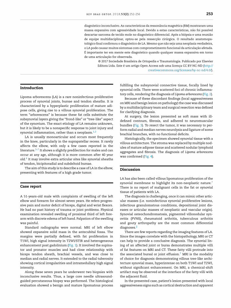
Standard radiographs were normal. MRI of left elbowhowed expansive solid mass in the antecubital fossa. Theargins were partially defined, with fat proliferation in
1WI, high signal intensity in T2WI/STIR and heterogeneousnhancement post gadolinium (Fig. 1). It involved the supina-or and pronator muscles and had close relationship withiceps tendon sheath, brachial vessels, and was close toedian and radial nerves. It extended to the radial tuberosity
howing cortical irregularities and intramedullary high signalntensity.
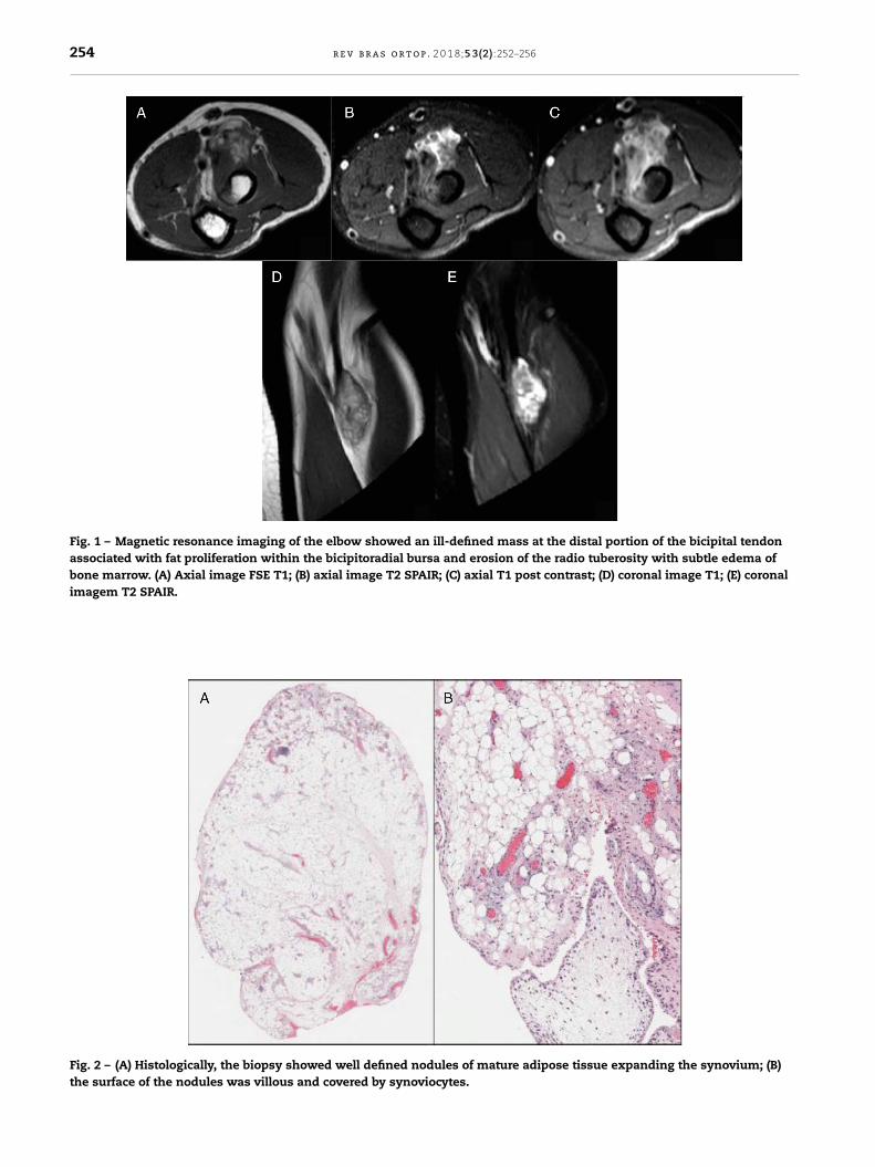
Along these seven years he underwent two biopsies withnconclusive results. Thus, a large core needle ultrasound-uided percutaneous biopsy was performed. The histologicalvaluation showed a benign and mature lipomatous process
creativecommons.org/licenses/by-nc-nd/4.0/).
fulfilling the subsynovial connective tissue, focally lined bysynovial cells. There were scattered foci of chronic inflamma-tory cells, rendering the diagnosis of Lipoma arborescens (Fig. 2).
Because of these discordant findings (local aggressivenesson MRI and benign lesion on pathology) the case was discussedby a multidisciplinary team and surgical resection was definedfor clarifying diagnosis.
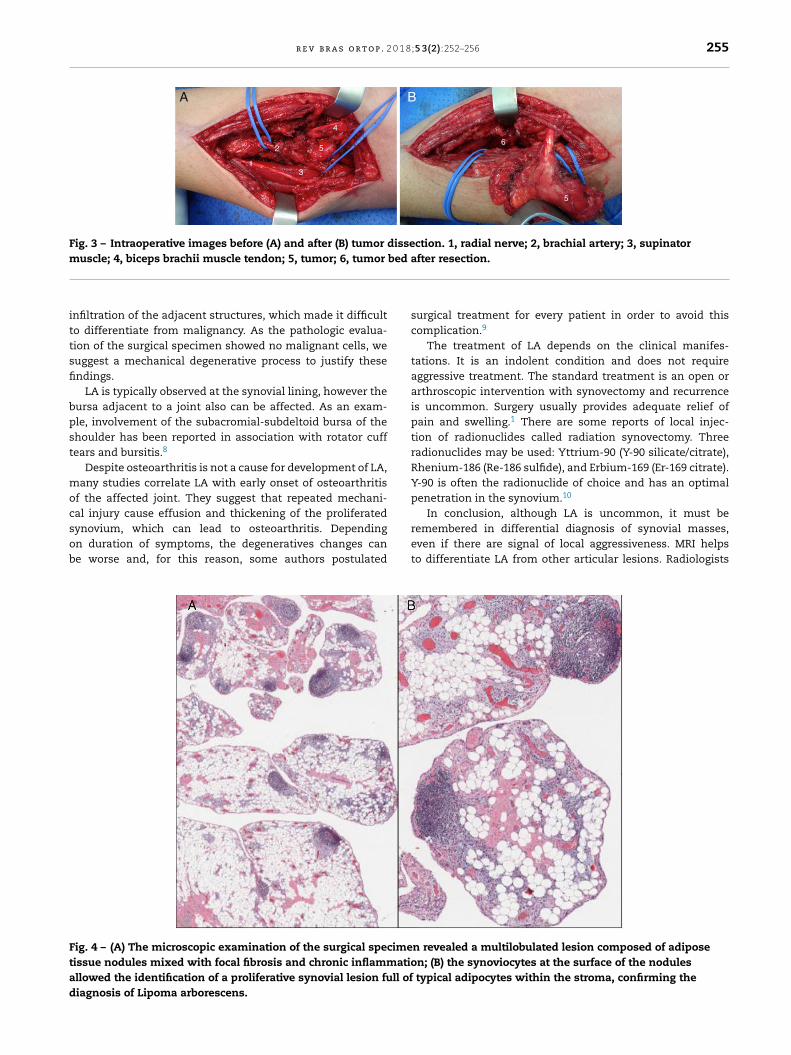
At surgery, the lesion presented as soft mass with ill-defined contours, fibrosis, and adhered to neuromuscularbundles (Fig. 3). To resect the tumor, it was necessary to per-form radial and median nerves neurolysis and ligature of somebrachial branches, with no functional deficits.
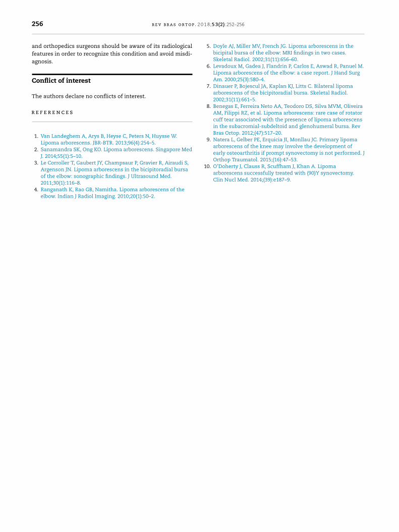
Histologically, the specimen showed synovial tissue with avillous architecture. The stroma was replaced by multiple nod-ules of mature adipose tissue and scattered nodular lymphoidaggregates and fibrosis. The diagnosis of Lipoma arborescenswas confirmed (Fig. 4).
Discussion
LA has also been called villous lipomatous proliferation of thesynovial membrane to highlight its non-neoplastic nature.7
There is no report of malignant cells in the fat or synovialtissue of patients with LA.
The diagnosis is challenging, once it can mimic other artic-ular masses (i.e. noninfectious synovial proliferative lesions,infectious granulomatous conditions, depositional joint dis-eases or articular masses of neoplastic and vascular origin).Synovial osteochondromatosis, pigmented villonodular syn-ovitis (PVNS), rheumatoid arthritis, tuberculous arthritisand gouty arthropathy are the most common differentialdiagnoses.2
There are few reports regarding the imaging features of LA.Since the images correlate with the histopathology, MRI or CTcan help to provide a conclusive diagnosis. The synovial lin-ing of an affected joint or bursa demonstrates multiple villiof fat features on MRI and CT. These fatty villi protrude intothe associated bursal or joint effusion.7 MRI is the modalityof choice for diagnosis demonstrating villous tree-like archi-tecture synovial mass, hyperintense on both T1WI and T2WI,without significant enhancement. On MRI, a chemical-shift
artifact may be observed at the interface of the fatty villi withthe adjacent fluid.In the presented case, patient’s lesion presented with localaggressiveness signs such as cortical destruction and apparent
254 r e v b r a s o r t o p . 2 0 1 8;5 3(2):252–256
Fig. 1 – Magnetic resonance imaging of the elbow showed an ill-defined mass at the distal portion of the bicipital tendonassociated with fat proliferation within the bicipitoradial bursa and erosion of the radio tuberosity with subtle edema ofbone marrow. (A) Axial image FSE T1; (B) axial image T2 SPAIR; (C) axial T1 post contrast; (D) coronal image T1; (E) coronalimagem T2 SPAIR.
Fig. 2 – (A) Histologically, the biopsy showed well defined nodules of mature adipose tissue expanding the synovium; (B)the surface of the nodules was villous and covered by synoviocytes.
r e v b r a s o r t o p . 2 0 1 8;5 3(2):252–256 255
A
1
2
3
5
4
6
5
4
B
Fig. 3 – Intraoperative images before (A) and after (B) tumor dissection. 1, radial nerve; 2, brachial artery; 3, supinatorm bed a
ittsfi
bpst
mocsob
Ftad
uscle; 4, biceps brachii muscle tendon; 5, tumor; 6, tumor
nfiltration of the adjacent structures, which made it difficulto differentiate from malignancy. As the pathologic evalua-ion of the surgical specimen showed no malignant cells, weuggest a mechanical degenerative process to justify thesendings.
LA is typically observed at the synovial lining, however theursa adjacent to a joint also can be affected. As an exam-le, involvement of the subacromial-subdeltoid bursa of thehoulder has been reported in association with rotator cuffears and bursitis.8
Despite osteoarthritis is not a cause for development of LA,any studies correlate LA with early onset of osteoarthritis
f the affected joint. They suggest that repeated mechani-al injury cause effusion and thickening of the proliferated
ynovium, which can lead to osteoarthritis. Dependingn duration of symptoms, the degeneratives changes cane worse and, for this reason, some authors postulatedig. 4 – (A) The microscopic examination of the surgical specimeissue nodules mixed with focal fibrosis and chronic inflammatiollowed the identification of a proliferative synovial lesion full ofiagnosis of Lipoma arborescens.
fter resection.
surgical treatment for every patient in order to avoid thiscomplication.9
The treatment of LA depends on the clinical manifes-tations. It is an indolent condition and does not requireaggressive treatment. The standard treatment is an open orarthroscopic intervention with synovectomy and recurrenceis uncommon. Surgery usually provides adequate relief ofpain and swelling.1 There are some reports of local injec-tion of radionuclides called radiation synovectomy. Threeradionuclides may be used: Yttrium-90 (Y-90 silicate/citrate),Rhenium-186 (Re-186 sulfide), and Erbium-169 (Er-169 citrate).Y-90 is often the radionuclide of choice and has an optimalpenetration in the synovium.10
In conclusion, although LA is uncommon, it must be
remembered in differential diagnosis of synovial masses,even if there are signal of local aggressiveness. MRI helpsto differentiate LA from other articular lesions. Radiologistsn revealed a multilobulated lesion composed of adiposen; (B) the synoviocytes at the surface of the nodules
typical adipocytes within the stroma, confirming the
p . 2 0
r
Orthop Traumatol. 2015;(16):47–53.10. O’Doherty J, Clauss R, Scuffham J, Khan A. Lipoma
256 r e v b r a s o r t o
and orthopedics surgeons should be aware of its radiologicalfeatures in order to recognize this condition and avoid misdi-agnosis.
Conflict of interest
The authors declare no conflicts of interest.
e f e r e n c e s
1. Van Landeghem A, Arys B, Heyse C, Peters N, Huysse W.Lipoma arborescens. JBR-BTR. 2013;96(4):254–5.
2. Sanamandra SK, Ong KO. Lipoma arborescens. Singapore MedJ. 2014;55(1):5–10.
3. Le Corroller T, Gaubert JY, Champsaur P, Gravier R, Airaudi S,Argenson JN. Lipoma arborescens in the bicipitoradial bursa
of the elbow: sonographic findings. J Ultrasound Med.2011;30(1):116–8.4. Ranganath K, Rao GB, Namitha. Lipoma arborescens of theelbow. Indian J Radiol Imaging. 2010;20(1):50–2.
1 8;5 3(2):252–256
5. Doyle AJ, Miller MV, French JG. Lipoma arborescens in thebicipital bursa of the elbow: MRI findings in two cases.Skeletal Radiol. 2002;31(11):656–60.
6. Levadoux M, Gadea J, Flandrin P, Carlos E, Aswad R, Panuel M.Lipoma arborescens of the elbow: a case report. J Hand SurgAm. 2000;25(3):580–4.
7. Dinauer P, Bojescul JA, Kaplan KJ, Litts C. Bilateral lipomaarborescens of the bicipitoradial bursa. Skeletal Radiol.2002;31(11):661–5.
8. Benegas E, Ferreira Neto AA, Teodoro DS, Silva MVM, OliveiraAM, Filippi RZ, et al. Lipoma arborescens: rare case of rotatorcuff tear associated with the presence of lipoma arborescensin the subacromial-subdeltoid and glenohumeral bursa. RevBras Ortop. 2012;(47):517–20.
9. Natera L, Gelber PE, Erquicia JI, Monllau JC. Primary lipomaarborescens of the knee may involve the development ofearly osteoarthritis if prompt synovectomy is not performed. J
arborescens successfully treated with (90)Y synovectomy.Clin Nucl Med. 2014;(39):e187–9.