Embed Size (px)
Citation preview
Eur. J. Biochem. 207,487-497 (1992) 0 FEBS 1992
Lipoamide dehydrogenase from A zotobacter vinelandii: site-directed mutagenesis of the His450-Glu455 diad Kinetics of wild-type and mutated enzymes
Jacques BENEN ', Willem van BERKEL ', Nicole DIETEREN ', David ARSCOTT2, Charles WILLIAMS Jr', Cees VEEGER' and Arie de KOK '
Department of Biochemistry, Agricultural University, Wageningen, The Netherlands Department of Veterans Affairs Medical Centre and Department of Biological Chemistry, University of Michigan, Ann Arbor, Michigan, USA
(Received January 24/April9, 1992) - EJB 92 0095
Three amino acid residues in the active site of lipoamide dehydrogenase from Azotobacter vinelandii were replaced with other residues. His450, the active-site base, was replaced with Ser, Tyr or Phe. Pro451, from X-ray analysis found to be in cis conformation positioning the backbone carbonyl of His450 close to N3 of the flavin, was changed to Ala. Glu455, from X-ray analysis expected to be involved in modulating the pK, of the base (His450), was replaced with Asp and Gln. The general conclusion is that mutation of the His-Glu diad impairs intramolecular electron transfer between the disulfide/dithiol and the FADH-/FAD.
The wild-type enzyme functions according to a ping-pong mechanism in the physiological reaction in which the formation of NADH is rate-limiting. Above pH 8.0 the enzyme is strongly inhibited by the product NADH. The pH dependence of the steady-state kinetics using the NAD' analog 3- acetylpyridine adenine dinucleotide (AcPyAde') reveals a pK, of 8.1 in the pK, AcPyAde+ plot indicating that this pK, is related to the deprotonation of His450 [Benen, J., Berkel van, W., Zak, Z., Visser, T., Veeger, C. & Kok de, A. (1991) Eur. J . Biochem. 202, 863-8721 and to the inhibition by NADH.
The mutations considerably affect turnover. Enzymes with the mutations Pro451 + Ala, His450 + Phe and His450 + Tyr appear to be almost inactive in both directions. Enzyme His450 + Ser is minimally active, V at the pH optimum being 0.5% of wild-type activity in the physiological reaction. Rapid reaction kinetics show that for the His450-mutated enzymes the reductive half reaction using reduced 6,8-thioctic acid amide [Lip(SH),] is rate-limiting and extremely slow when compared to the wild-type enzyme. For enzyme Pro451 + Ala it is concluded that the loss of activity is due to over-reduction by Lip(SH)2 and NADH. The Glu455-mutated enzymes are catalytically competent but show strong inhibition by the product NADH (enzyme Glu455 + Asp more than Glu455 --t Gln). The inhibition can largely be overcome by using AcPyAde' instead of NAD' in the physiological reaction.
The rapid reaction kinetics obtained for enzymes Glu455 +Asp and Glu455hGln deviate from the wild-type enzyme. It is concluded that this difference is due to cooperativity between the active sites in this dimeric enzyme. Rapid reaction kinetics of enzymes His450 + Ser and Glu455 + Gln show the existence of two intermediates at the two-electron reduced level: a species with the NAD' bound, the flavin reduced and the disulfide intact (oxidized) and a species with NAD' bound, the disulfide reduced and the flavin oxidized. No spectral evidence is obtained for the participation of a proposed flavin C4a adduct intermediate [Thorpe, C. & Williams, C. H. Jr (1981) Biochemistry 20, 1507- 15131 in the reaction mechanism of enzymes His450 + Ser and Glu455 + Gln.
Correspondence to A. de Kok, Department of Biochemistry, Agri- cultural University, Dreyenlaan 3, NL-6703 HA Wageningen, The Netherlands
Abbreviations. E,,, oxidized (mutated) lipoamide dehydrogenase; EH,. two-electron-reduced (mutated) lipoamide dehydrogenase with disulfide reduced (530-nm charge-transfer species and tautomeric species); EH-, EH2 one-step deprotonated; E2-, EH2 two-steps deprotonated; EH4, general descriptive term for four-electron-re- duced (mutated) lipoamide dehydrogenase; FADH -, reduced flavin; FIH-. two-electron-reduced species with electrons on the flavin;
Lips,, ~~-6,8-thioctic acid amide, lipoamide; Lip(SH),, reduced lipoamide; Nbs,, 5,5'-dithiobis(2-nitrobenzoate); AcPyAde', 3- acetylpyridine -adenine dinucleotide; AmPyAde', 3-aminopyri- dine-adenine dinucleotide; C121nd, 2,6-dichloroindophenol; PP,/ EDTA buffer, 50 mM sodium pyrophosphate, 0.5 mM EDTA, pH 8.0.
Enzymes. Lipoamide dehydrogenase, NADH:lipoamide oxidoreductase (EC 1.8.1.4); glucose-6-phosphate dehydrogenase, D- glucose-6-phosphate: NADP I-oxidoreductase (EC 1.1.1.49); xan- thine oxidase, xanthine: oxygen oxidoreductase (EC 1 .I .3.22)

488
"S& R
4 INAD' 1
"^""llNADH His{
5
Scheme 1. Proposed reaction intermediates for lipoamide dehydrogenase. The arrows indicate charge-transfer interaction and are drawn from donor to acceptor
Lipoamide dehydrogenase is the flavoprotein component of the pyruvate, oxoglutarate and branched-chain-oxoacid dehydrogenase complexes [I, 21. In the physiological direction, i t catalyses the oxidation of a reduced lipoyl group that is covalently attached to the core protein of these complexes by NAD' . This lipoyl group can be replaced with free lipoamide. Lipoamide dehydrogenase belongs to the family of dimeric flavoenzymes that contain a redox-active disulfide bridge participating in catalysis [l]. The enzyme is composed of two identical subunits with the two active sites built by contacts between the subunits. Other members of this family are glutathione reductase [I 1, mercuric ion reductase [3], trypano- thione reductase [4] and thioredoxin reductase [5] .
Extensive studies on lipoamide dehydrogenase have been performed on the enzymes as isolated from pig heart, rat liver and Escheuicliia coli [6 - 141. All three lipoamide dehydroge- nases were shown to act according to a ping-pong mechanism in the physiological reaction [6, 8, 141. During catalysis, the enzyme shuttles between the oxidized (E0J and the two-elec- tron-reduced (EH,) state [15]. Inhibition due to over-re- duction to the four-electron reduced state (EH,) is most severe in E. coli enzyme and only slight in pig heart enzyme [7, 12, 14. 151. The naturc of the dimeric structure indicates that communication between the active sites seems possible. Evi- dence for positive and negative cooperativity in this family of diineric enzymes has been reported [8, 14, 16- 391.
Detailed studies on pig heart enzyme comprising both steady-state and rapid-reaction kinetics have led to the pro- posal of a reaction mechanism involving a base, histidine, in the active site that becomes protonated during catalysis [9,20, 211. Reccntly, conclusive evidence for the role of the histidine was obtained by site directed mutagenesis studies on gluta- thione reductase [22,23], E. coli [24] and Azotobacter vinelandii
lipoamide dehydrogenase [25]. Scheme 1 depicts proposed and identified reaction intermediates [7, 9 - 11, 15, 17, 26 - 301. Catalysis proceeds around the upper cycle, species 1 - 7. The lower cycle shows species involved in four-electron reduction, species 8 - 11.
In a previous paper the rationale for the selection of amino acids to be mutated was outlined and the spectral properties of the wild-type and mutated enzymes were reported [26]. This paper deals with the kinetic properties and identification of several of the intermediate species as shown in Scheme 1 of A. vinelandii lipoamide dehydrogenase and mutated enzymes.
MATERIALS AND METHODS General
Construction of the mutated enzymes, isolation and treat- ment of the enzymes were performed as described [26]. NAD' (grade I) NADH (grade I), NADP+, xanthine, xanthine oxi- dase (from cow milk), glucose 6-phosphate and glucose-6- phosphate dehydrogenase (from yeast, analytical grade) were from Boehringer. NAD + analogs, 6,g-thioctic acid amide (Lips,), dichloroindophenol (Cl,Ind), 5,5'-dithiobis(2-nitro- benzoate) (Nbs,) and biological buffers were obtained from Sigma Inc. All other chemicals used were of the highest purity available. NAD + , acetylpyridine - adenine dinucleotide (AcPyAde'), NADH, reduced Lipsz [Lip(SH),] and enzyme concentrations were determined as described previously 1261. The thio-NAD + concentration was determined as described for AcPyAde+ .
Steady-state kinetics All activity measurements were performed on a Zeiss M4
QIII spectrophotometer at 25 "C. The forward reaction
489
Redox potential determinations
Visible absorption spectra were recorded on a tempera- ture-controlled Aminco DW2000 spectrophotometer at 25°C. Reductions were carried out in anaerobic cuvettes in 50 mM potassium phosphate pH 7.0, 0.5 mM EDTA. Redox poten- tials were determined in three different ways. Method one was essentially as described for pig heart enzyme [20]. Method two, the xanthine/xanthine oxidase method, was recently de- scribed by Massey [34] and was applied without modification. The co-titrants used, 30 pM in concentration, were safranine T, phenosafranine and benzylviologen. Method three was a modification of method two. Instead of using xanthinelxan- thine oxidase as an electron generating system, NADPH was generated from NADP + (75 pM) using glucose-6-phos- phate dehydrogenase (0.2 -04 m u ) and glucose 6-phospate (400 pM). NADPH proved useful as a reductant for lipo- amide dehydrogenase since quantitative reduction could be achieved yielding spectra identical to reduction by Lip(SH),. Furthermore NADP' did not give rise to the typical long- wavelength absorbance characteristic for FADH-/NAD+ charge-transfer interaction, indicating that no efficient bind- ing occurs. For method one, the titrant was added using a gas- tight Hamilton syringe equipped with a dispenser. Spectra were recorded until changes were complete. For methods two and three the reaction was started by the addition of enzyme (xanthine oxidase or glucose-6-phosphate dehydrogenase) after anaerobiosis was established. In order to achieve equilib- rium the amount of enzyme was chosen such as to complete reduction in a time span of 6-9 h. The amount of enzyme was halved to check whether equilibrium was established [34].
Lip(SH)2/NAD' was determined routinely in 50 mM sodium pyrophospate, 0.5 mM EDTA pH 8.0 (PPJEDTA buffer). In a standard assay, 1.0 mM NAD' and 1 .O mM Lip(SH)2 were used and the formation of NADH was monitored at 340 nm. Steady-state kinetics of the forward reaction of the wild-type enzyme were studied as follows. To a temperature equilibrated cuvette containing 950 p1 PPi buffer, 20 pl of each of the substrates was added, followed by the addition of 10 pl en- zyme at the proper dilution. Substrate concentration combi- nations were chosen such as to avoid errors due to inactivation of enzyme in the time course of the experiment. Therefore the experiment was started with the lowest concentration of substrate A, where substrate B was varied, followed by the highest concentration of substrate A with B varied. Next the second lowest concentration of A was used followed by the second highest concentration of A and so on. Triplicate series were measured.
pH-dependent studies were performed in buffers contain- ing 0.5 mM EDTA and adjusted to ionic strength 150 mM with KCI. Buffers used: pH 5.5-6.0-6.5, 100 mM Mes; pH 7.0-7.5-7.8, 100 mM Hepes; pH 8.0-8.6, 100 mM Hepps; pH 9.0-9.3 100 mM Ches. Data were analyzed ac- cording to a ping-pong mechanism.
The reverse reaction, NADH/LipS,, was determined as described [31]. The NADH-dependent reduction of C1,Ind (diaphorase activity) was measured in PPJEDTA buffer ( E ~ ~ ~ nm for C1,Ind = 22900 M-' cm-' at pH 8.0) by follow- ing the decrease of C1,Ind at 600 nm. The transhydrogenase reaction NADH/thio-NAD + was performed according to [321.
Rapid-reaction kinetics RESULTS
Rapid-reaction kinetics were carried out using a tempera- ture-controlled single-wavelength stopped-flow spectrophoto- meter, type SF-51, from High Tech Scientific Inc. with 1.3 ms dead time. The instrument was interfaced to an IBM computer for data acquisition and analysis. Data were analyzed with a program from High Tech Scientific Inc. Rapid-scan exper- iments were performed with a stopped-flow instrument having a 2-cm light-path cell and a dead time of 3 ms, and using a Tracor Northern diode array spectrophotometer as the detec- tor (scan time 5.42 ms). The detector was interfaced to a Tracor Northern computer for data acquisition and analysis. All experiments were performed anaerobically at 21 .O"C in 100 mM Good buffers containing 0.5 mM EDTA adjusted to ionic strength 150 mM with KCl. Enzyme concentrations after mixing were 26.7 pM. Generation of two-electron-reduced enzyme (EH,) was accomplished by reduction with a small excess of sodium borohydride.
Rate constants at infinite substrate concentration and Kd values were determined from non-linear fitting of apparent rate constants obtained at, at least, five different substrate concentrations. Apparent rate constants represent the average of minimally four shots. In order to achieve pseudo-first-order conditions the lowest substrate concentrations used were five times as high as the enzyme concentration. Since only the L- enantiomer of Lip(SH)2 reacts rapidly [6,9], substrate concen- trations were adjusted to those of the L-enantiomer. Lip(SH)2 and Lips2 were dissolved in bufferlethano1 to yield a final ethanol content of 5% in the mixing chamber.
Simulation of rapid-reaction kinetics was performed using the program KINSIM [33] run on a VAXjVMS minicomputer.
Steady-state kinetics
Wild- type lipoum ide deh y drogenase
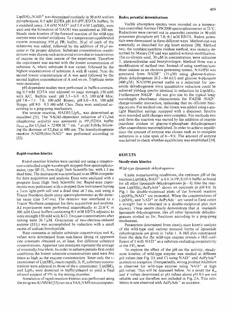
Under nonsaturating conditions, the optimum pH of the reaction Lip(SH),/NAD' is 8.0, in PPJEDTA buffer as found for all other lipoamide dehydrogenases [6 - 81 Also the reac- tion Lip(SH),/AcPyAde+ shows an optimum at pH 8.0. In Fig. 1 the double-reciprocal plots of the forward reaction Lip(SH),/NAD' are presented. When the concentrations of Lip(SH)2 and NAD' or AcPyAde' are varied in fixed ratios a straight line is obtained in a double-reciprocal plot (not shown). These results clearly demonstrate that A . vinelundii lipoamide dehydrogenase, like all other lipoamide dehydro- genases studied so far, functions according to a ping-pong mechanism.
Parameters determined from steady-state kinetic analysis of the wild-type and various mutated forms of lipoamide dehydrogenase are given in Table 1. A Hill plot constructed from the data for the wild-type enzyme reveals a Hill coef- ficient of 1 with NAD' as a substrate excluding cooperativity at the EH2 level.
To explore the effect of the pH on the activity, steady- state kinetics of wild-type enzyme was studied at different pH values (see Fig. 2A and C) using NAD' and AcPyAde' as electron acceptors. Unexpectedly, strong product inhibition is observed for wild-type enzyme using NAD' at high pH values. This will be discussed below. As a result the K,,, and V values determined at pH values above pH 8.0 are not reliable and are therefore not included in Fig. 2A. This inhi- bition is not observed with AcPyAde' as acceptor.
490
25
F- 20 0 7
X 15 ? .g. 10 P
cn
7
5
0
g j \ - a I
E 4 Y a 3
I 1 I I
e'j , , , 0 , , E 4
a3
0 5 10 15 20 25 l/[NAD+] (mM-')
I I 1 I I 0 2 4 6 8 10
l/[lip(SH), ] (mM'')
Fig. 1. Steady-state kinetics of the physiological reaction of A . vinelandii wild-type lipoamide dehydrogenase at the optimum pH 8.0. The tem- perature was 25°C; for other details see Materials and Methods. (A) Lineweaver-Burk plot of the reaction L~P(SH)~/NAD' with "AD+] varied and [Lip(SH),] fixed at: (H) 104pM, (0) 200 pM, (0) 295 pM, ( 0 ) 397 pM, ( A ) 518 pM; (A) extrapolated velocities at infinite [Lip(SH),]. (B) As (A) with [Lip(SH),] varied and "AD'] fixed at : (+) 48wM. ( x ) 72pM, (*) 92pM, ( A ) 147pM, ( 0 ) 194 pM. (0) 51 1 pM, (H) 752 pM, (0) 1044 pM; (A) extrapolated velocities at infinite "AD'].
$1 / , , 2.
5 6 7 8 9 I PH
':g ;7\,,, E 2. ?L
2. a 5 6 7 8 9
PH PH I
E 3.
3 $:'L Y, 5 6 7 8 9
I
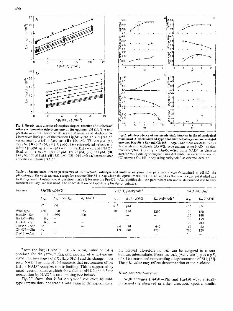
Fig. 2. pH dependence of the steady-state kinetics in the physiological reaction of A . vinelandii wild-type lipoamide dehydrogenase and mutated enzymes His450 + Ser and Glu455 --* Asp. Conditions are described in Materials and Methods. (A) Wild-type enzyme using NAD' as elec- tron acceptor; (B) enzyme His450 + Ser using NAD' as electron acceptor; (C) wild-type enzyme using AcPyAde' as electron acceptor; (D) enzyme Glu455 +Asp using AcPyAde' as electron acceptor.
Table 1. Steady-state kinetic parameters of A . vinelandii wild-type and mutated enzymes. The parameters were determined at pH 8.0, the pH optimum for each enzyme, except for enzyme Glu455 --f Asp where the optimum was pH 7.0. nd signifies that kinetics are not studied due to strong product inhibition. A question mark (?) for enzyme Pro451 +Ala signifies that the parameters can not be determined due to very transient activity (see text also). The concentration of Lip(SH)2 is for the DL-miXtUre.
Enzyme Lip(SH),/NAD+ Lip(SH),/AcPy Ade + NADH/C121nd
k,,, K,,, Lip(SH)Z K,,, NAD' k,,, K, L ~ P ( S H ) ~ K, AcPyAde+ k,,, K,,,NADH
s- ' pM s - l pM s - ' pM Wild-type 420 390 325 His450 + Ser 1.6 1950 300 His450iPhe 0.0 -
His450jTyr 0.0 -
Glu455-Asp nd - -
Glu455-tGIn nd -
Pro45 1 +Ala ? -
-
-
-
-
100 140 1200 - - -
- - -
- - - 2.4 30 640 1.9 240 1750
- - -
170 180 135 140 170 150 170 200 160 50 200 120 ? -
From the log( v> plot in Fig. 2A, a pK, value of 6.4 is obtained for the rate-limiting intermediate of wild-type en- zyme. The invariance of pK, [Lip(SH),] and the change in the pK, [NAD'] around pH 6.4 suggests that protonation of the EH2 . NAD' complex is rate-limiting. This is supported by rapid-reaction kinetics which show that at pH 8.0 and 6.8 the reoxidation by NAD' is rate-limiting (see below).
Fig. 2C shows that V for AcPyAde ' reduction by wild- type enzyme does not reach a maximum in the experimental
pH interval. Therefore no pK, can be assigned to a rate- limiting intermediate. From the pK, [AcPyAde'] plot a pKa of 8.1 is determined representing a deprotonation of EH2 [35]. This pK, value may reflect deprotonation of the histidine.
His45O-mutated enzymes
With enzymes His450 + Phe and His450 --f Tyr virtually no activity is observed in either direction. Spectral studies
49 1
showed that these enzymes still can be reduced by L ~ P ( S H ) ~ though very slowly. It is therefore concluded that the rather bulky aromatic residues impose structural constraints on the enzyme that make it very difficult for L ~ P ( S H ) ~ to interact effectively with the disulfide to exchange reducing equivalents. With enzyme His450 + Ser almost no activity was observed in the reverse reaction, reduction of Lips2, monitored by the oxidation of NADH in the presence of NAD'. About 0.5% of the wild-type activity is found in the forward reaction, L~P(SH)~/NAD+, at the optimum pH 8.0 A kinetic analysis as described for wild-type enzyme demonstrates that enzyme His450 + Ser also functions according to a ping-pong mecha- nism.
Assuming His450 to abstract a proton from the substrate Lip(SH)2 as proposed [9], one might expect the activity of enzyme His450 + Ser would increase at elevated pH values, where L ~ P ( S H ) ~ (pKal = 9.35, free in solution [9]) becomes deprotonated. However V remains constant between pH 8.0 and pH 9.0. In the pH-dependent studies no product inhi- bition by NADH is found in the experimental pH interval. From the log( V) plot in Fig. 2B, a pK, value of 6.3 is obtained for the deprotonation of the rate-limiting intermediate in en- zyme His450 + Ser.
GIu45.5-mutated enzymes
With enzyme Glu455 --t Asp very strong product (NADH) inhibition is observed in the reaction Lip(SH)2/NAD+. The results indicate that this enzyme is inhibited by NADH in a 'dead-end' manner. Addition of excess NAD' after inhibition is complete does not restore activity. Revived NADH pro- duction is only observed after the addition of an extra aliquot of enzyme. This extra enzyme portion becomes inhibited ap- proximately as fast as the initial enzyme portion. No detailed studies were carried out to investigate this phenomenon. How- ever the results indicate that the inhibition is more dependent on time than on the NADH concentration. In view of an increase of the dissociation constant for the monomer dimer equilibrium of the wild-type enzyme upon reduction to the EH4 level [31], it is tempting to speculate that the irreversibility of the inhibition is caused by monomerization.
The reverse reaction as well as the transhydrogenase reac- tion NADH/thio-NAD' are not detectable. When an electron acceptor with higher midpoint potential than NAD' or thio- NAD+ is used, in casu of AcPyAde+ in the forward reaction, and C121nd in the diaphorase reaction, quantitative reduction of the acceptors is observed in the pH interval studied (pH 5.5-9.0) (E; NAD+/NADH = -320 mV [36], EL thioNAD+/thioNADH = - 283 mV [36], E,!,, AcPyAde+/ AcPyAdeH = - 258 mV [36] and E,!,, C121nd/C121ndH2 = + 217 mV [37], pH 7.0). With AcPyAde+ 'dead-end' inhi- bition is observed above pH 7.5.
Enzyme Glu455 + Gln differs in catalytic behavior from enzyme Glu455 +Asp. Here a somewhat less strong inhibitory effect of NADH is found. With AcPyAde+ as acceptor no inhibition is observed.
Under non-saturating fixed substrate concentrations the optimum for the AcPyAde' reduction is at pH 8.0 for enzyme Glu455 +Gln and is lowered to pH 7.0-7.5 for enzyme Glu455 +Asp. Both Glu455-mutated enzymes function ac- cording to a ping-pong mechanism.
For both Glu455-mutated enzymes the steady-state ki- netics of the reaction Lip(SH),/AcPyAde+ were studied as a function of pH. Enzyme Glu455 + Asp shows, as mentioned above, 'dead-end' inhibition with AcPyAde'. This hampers
the determination of the kinetic constants above pH 7.5. Ex- cept for a change in V, the kinetic parameters of enzyme GIu455 +Asp d o not change significantly in the experimental pH range (pH 5 7.5, Fig. 2 D). The pH dependence of steady- state kinetics of enzyme Glu455 + Gln is quite different from enzyme Glu455 +Asp (results not shown). V changes only slightly over the experimental pH range (pH 5.5-9.0) show- ing an optimum at pH 8.0. The K, for L ~ P ( S H ) ~ increases at lower pH but K,,, AcPyAde' remains almost constant as in enzyme Glu455 +Asp. No pKa values are determinable for this enzyme.
Rapid-reaction kinetics
In order to obtain a more detailed insight into the separate half reactions, the reductive reactions with L ~ P ( S H ) ~ and NADH and the oxidative reactions with NAD' and Lipsz were studied in the stopped-flow spectrophotometer.
Wild-type lipoamide dehydrogenase
At pH 7.0 and 8.0 the reduction of E,, by Lip(SH)* and reoxidation of EH2 by NAD' and Lips2 as monitored at 450 nm and 530 nm can essentially be described by a single- exponential function. In Table 2 the extrapolated rate con- stants at infinite substrate concentration and corresponding Kd values are presented. Ping-pong enzymes obey the follow- ing equations:
k i k 3
k 2 k4
k5 k?
kg kn
E + A + E A $ E ' + P (1 1
E ' + B e E ' B = E + Q (2)
for which the steady-state relation
(3)
was derived [38]. Here k3 and k, are the first-order rate con- stants of the reaction of the first and second substrate respec- tively (k, of the appropriate substrate in Table 2). A k,,, was calculated as 670 sK1 at pH 8.0 for the reaction Lip(SH),/ NAD' with data from Table 2 and Eqn (3). This value agrees reasonably well with k,,, calculated from steady-state kinetics : 420 s- ' . Comparison of k3 and k7 demonstrates that, at pH 8.0, the reductive reaction by L ~ P ( S H ) ~ is not rate-limiting as found for the pig heart enzyme [6, 101.
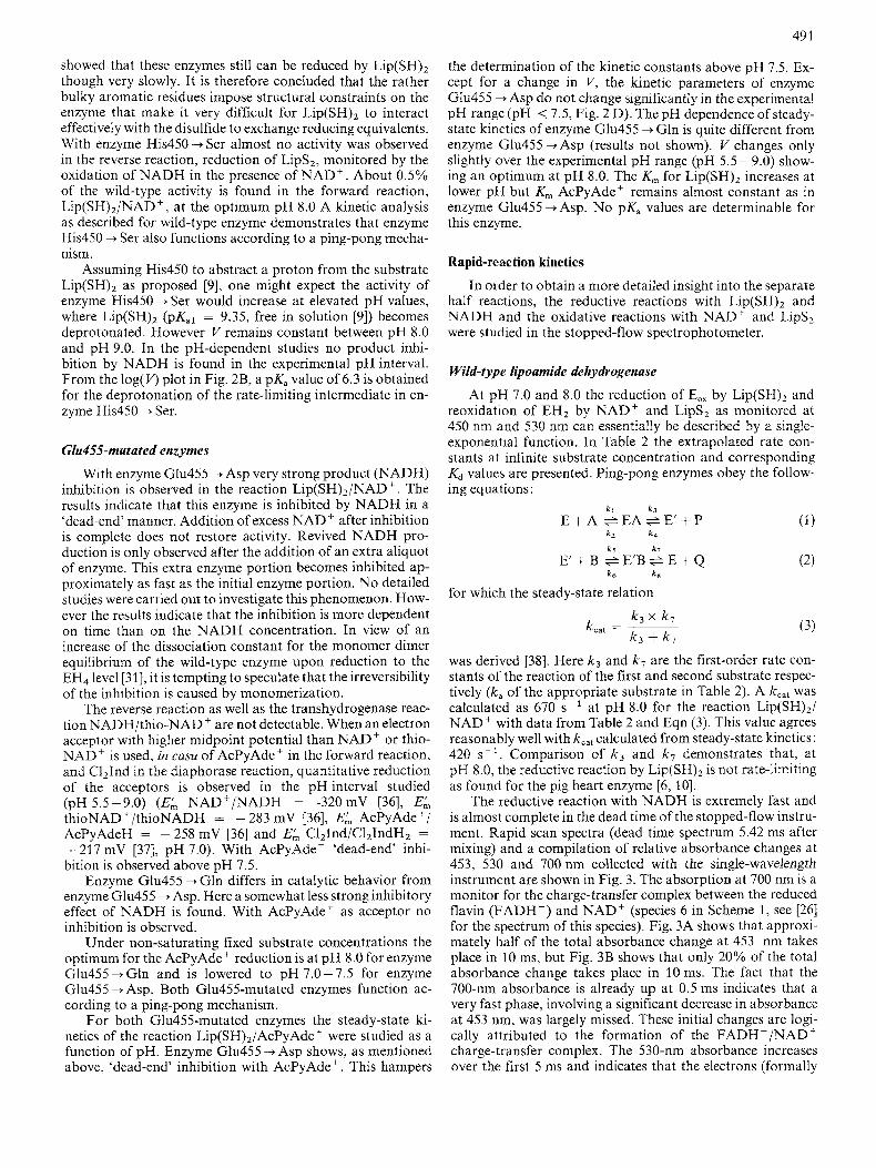
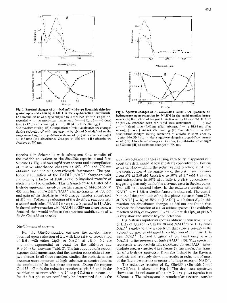
The reductive reaction with NADH is extremely fast and is almost complete in the dead time of the stopped-flow instru- ment. Rapid scan spectra (dead time spectrum 5.42 ms after mixing) and a compilation of relative absorbance changes at 453, 530 and 700 nm collected with the single-wavelength instrument are shown in Fig. 3. The absorption at 700 nm is a monitor for the charge-transfer complex between the reduced flavin (FADH-) and NAD' (species 6 in Scheme 1, see [26] for the spectrum of this species). Fig. 3A shows that approxi- mately half of the total absorbance change at 453 nm takes place in 10 ms, but Fig. 3B shows that only 20% of the total absorbance change takes place in 10ms. The fact that the 700-nm absorbance is already up at 0.5 ms indicates that a very fast phase, involving a significant decrease in absorbance at 453 nm, was largely missed. These initial changes are logi- cally attributed to the formation of the FADH-/NAD' charge-transfer complex. The 530-nm absorbance increases over the first 5 ms and indicates that the electrons (formally
492
Table 2. Rate constants and dissociation constants of A. vinelandii wild-type and mutated enzymes. For NADH reduction the rate constant of reduction of the F A D is shown. A question mark (?) signifies that the constants could not be determined due to the small absorbance changes associatcd at low substratc concentration (see text). Kd values for Lip(SH)Z are expressed for the L-enantiomer. Rate constants k , and kb are extrapolated to infinite [substrate]; the faster rate constant is k , while the slower rate constant is kb. The wavelength at which data were obtaincd is indicated in the last column.
~
Enzyme Reduction Substrate PH k, Kd(.4 k b KdW Wavelength level
S - ' mM S-1 mM nm
Wild-type E O X Lip(SW2 8.0 2000 0.9 E o * LiP(SH)Z 7.0 1000 0.7 E"X NADH 8.0 > 3000 EHz Lipsz 8.0 400 0.5 EH z NAD' 8.0 1000 0.37 EH 2 NAD' 7.0 760 0.47
9.3 8.6 7.8 7.3 6.6 8.0 8.0 8 .O 6.8
8.0 7.0 5.9 8.0 8.0
8.6 7.9 7.3 5.9 8.0 8.0 8.0
two electrons and a proton; a hydride equivalent) are passed over from the flavin to the disulfide (species 6 to 4 or 3 in Scheme l ) , an effect that is more pronounced in the His450-Ser enzyme (see below). Changes occurring after 5 ms are most likely due to further reduction by the excess of NADH.
His450-mutated enzymes
The reductive reaction of E,, by Lip(SH)2 is slow (His450 + Ser) to extremely slow (His450 + Phe and His450 -+ Tyr) upon mutation of the active-site base. No rate constants were determined for enzymes His450 --f Phe and His450 ---* Tyr. Even at the highest possible substrate concen- tration full reduction to EH2 lasted 250 s, yielding an apparent first order rate constant of ~ ~ x I O - ~ s-'. For enzyme His450 - Ser the reduction by Lip(SH), is monophasic pre- ceded by a lag phase (< 300 ms). The reoxidation of EH2 by NAD' is also monophasic but the lag phase is not observed in these traces. The rate constants and Kd values as determined for enzyme His450 + Ser are presented in Table 2. The rate constants and Kd values for the substrates L-Lip(SH), and L- Lips2 should be regarded as approximations since the deter- mined Kd values are approximately 3-4 times higher than the maximal experimental concentration of L-Lip(SH), or L- Lipsz due to solubility limitations.
For the forward overall reaction of enzyme His450 + Ser from Eqn. (3) a k,,, of 1.0 s- ' is calculated that agrees reason-
1.9 5.0 1.5 4.3 1.3 4.7 1 .o 3.7 0.8 3.5 > 3000 c0.005 4.0 0.33 4.4 0.27
450, 530 530 450, 530,700 450, 530 450,530 450, 530
450, 530 530 530 530 530 450, 530,700 530 530 530
? ? 1000 4.7 450, 530 ? ? 600 4.0 530 230 5.4 530 > 3000 ? ? 12 0.55 450, 530 250 2.6 60 2.5 530 <? ? 60 4.2 530 ? ? 35 3.6 530 44 4.8 530 > 3000 450, 530,700 < 0.05 530 58 0.17 14 0.30 530
ably well with k,,, = 1.6 s - ' as obtained from steady-state kinetics. Contrary to the wild-type enzyme however, for en- zyme His450 + Ser the reductive reaction is rate-limiting at both pH 7.0 and 8.0.
The rate constant for the reductive reaction by L ~ P ( S H ) ~ increases only slightly with pH and the traces remain mono- exponential up to pH 9.3. The Kd for L ~ P ( S H ) ~ increases with pH suggesting that the substrate binds to the mutated en- zyme in its protonated state. The reoxidation reaction of His450jSer EH2 by Lips2 is barely detectable at pH 8.0, indicating that the reactivity of the substrate is very poor or the affinity of the enzyme for this substrate is very low.
The reoxidation of EH2 by NAD' proceeds faster in this enzyme than the reduction of E,, by L ~ P ( S H ) ~ (Table 2) although the reaction is also extremely slow when compared to wild-type enzyme.
A possible mechanism of electron transfer from the nascent thiols to the flavin involves the transient formation of a C4a adduct between the charge-transfer thiolate and the flavin [17, 27, 281, species 5 in Scheme 1. This flavin C4a adduct species exhibits characteristic absorbance at 380 nm. Therefore the reoxidation of His450 + Ser EH2 with NAD' was monitored at this wavelength. No absorbance changes compatible with the formation of a flavin C4a adduct species were however detected.
The kinetics of the reduction of enzyme His450 --f Ser by NADH are compatible with a very fast reduction of the flavin
493
Wavelength (nm)
Time (s)
Fig. 3. Spectral changes of A . vinelandii wild-type lipoamide dehydro- genase upon reduction by NADH in the rapid-reaction instruments. (A) Reduction of wild-type enzyme by 5 mol NADH/mol at pH 7.8, recorded with the rapid-scan instrument. (-) EOx; ( ~ . -.) dead time (5.42 ms after mixing); (......) 10.84 ms after mixing; (- - -) 542 ms after mixing. (B) Compilation of relative absorbance changes during reduction of wild-type enzyme by 10 mol NADH/mol in the single-wavelength stopped-flow instrument. (0) Absorbance changes at 453 nm; ( x ) absorbance changes at 530 nm; (m) absorbance changes at 700 nm.
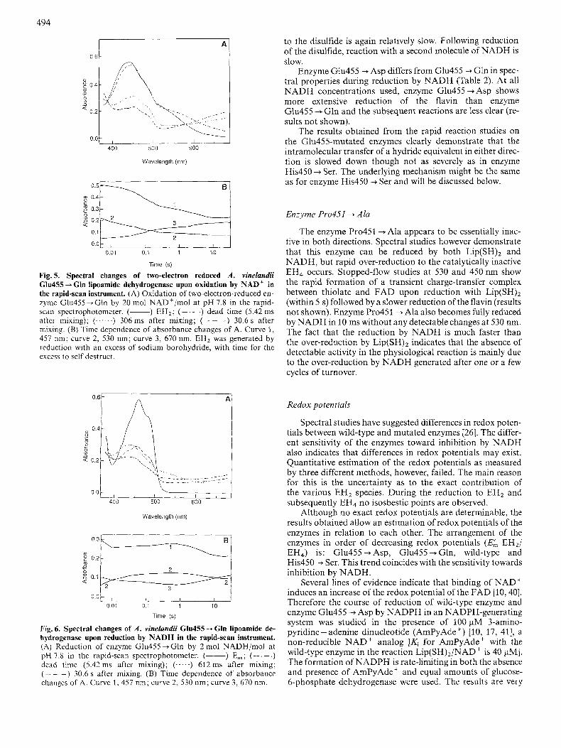
(species 6 in Scheme 1) with subsequent slow transfer of the hydride equivalent to the disulfide (species 4 and 3 in Scheme 1). Fig. 4 shows rapid-scan spectra and a compilation of relative absorbance changes at 453, 530 and 700nm obtained with the single-wavelength instrument. The pro- found stabilization of the FADH -/NAD' charge-transfer compiex by a factor of 1000 is due to impaired transfer of electrons to the disulfide. This intramolecular transfer of a hydride equivalent involves partial regain of absorbance at 453 nm, loss of FADH-/NAD+ charge-transfer at 700 nm and gain of the thiolate to FAD charge-transfer absorbance at 530 nm. FoIlowing reduction of the disulfide, reaction with a second molecule of NADH is very slow (species 3 to 11). Also in the reductive reaction with NADH no 380-nm absorbance is detected that would indicate the transient stabilization of a flavin C4a adduct species.
Glu4.55-mutated enzymes
For the Glu455-mutated enzymes the kinetic traces obtained upon reduction of E,, with L ~ P ( S H ) ~ or reoxidation of EH2 with either Lips2 or NAD' at pH > 6.0 are not mono-exponential as found for the wild-type and His450 + Ser enzymes (Table 2). The introduction of a second exponential demonstrates that the reaction proceeds in at least two phases. In all three reactions studied the biphasic nature becomes more apparent at high substrate concentrations as the amplitude of the fast phase increases. Except for enzyme Glu455 --f Gln in the reductive reaction at pH 8.6 and in the reoxidation reaction with NAD' at pH 8.0 no rate constant for the fast phase can confidently be determined due to the
Wavelength (nrn)
Time (s)
Fig. 4. Spectral changes of A . vinelandii His450 + Ser lipoamide de- hydrogenase upon reduction by NADH in the rapid-reaction instru- ments. (A) Reduction of enzyme His450 + Ser by 10 mot NADH/mol at pH 7.8, recorded with the rapid scan instrument. (-) Eox; (- . - .) dead time (5.42 ms after mixing); (. . . . .) 10.84 ms after mixing; ( - - -) 542 ms after mixing. (B) Compilation of rclative absorbance changes during reduction of enzyme His450 + Ser by 10 mol NADH/mol in the single-wavelength stopped-flow instru- ment. (0) Absorbance changes at 453 nm; ( x ) absorbance changes at 530 nm; (H) absorbance changes at 700 nm.
small absorbance changes causing variability in apparent rate constants determined at low substrate concentration. For en- zyme Glu455 + Gln in the reductive half reaction at pH 8.6, the contribution of the amplitude of the fast phase increases from 5% at 250 pM L ~ P ( S H ) ~ to 30% at 1.7 mM Lip(SH)2 and extrapolates to 50% at infinite Lip(SH)2 concentration suggesting that only half of the enzyme reacts in the fast phase. This will be discussed below. In the oxidative reaction with NAD' at pH 8.0, a similar feature is observed. The contri- bution of the amplitude of the fast phase increases from 25% at [NAD'] = Kd to 58% at [NAD'] = 10 times K d . In this reaction no absorbance changes at 380nm are found that indicate the formation of a C4a adduct species. The oxidative reaction of EH2 ofenzyme Glu455 + Gln with Lips2 at pH 8.0 is very slow and almost beyond detection.
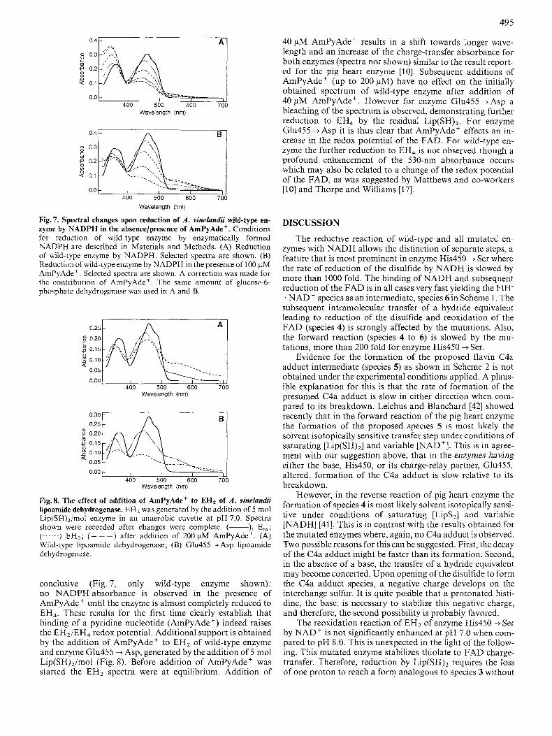
Fig. 5 shows rapid-scan spectra obtained from reoxidation of EH2 of Glu455 + Gln by 20 mol NAD+/mol. EH2 binds NAD' rapidly to give a spectrum that closely resembles the absorption spectra obtained from titration of pig heart EH2 with NAD' [lo] and titration of pig heart enzyme with NADH in the presence of high "AD+] [39]. This spectrum represents a reduced-disulfide/oxidized flavin/NAD' inter- mediate species (species 4 in Scheme 1). Intramolecular trans- fer of a hydride equivalent from the dithiol to the flavin is biphasic and relatively slow, and results in reduction of most of the flavin despite the presence of a large excess of NAD'.
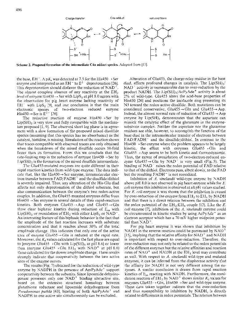
The reductive reaction of E,, Glu455 + Gln with 2 mol NADH/mol is shown in Fig.6. The dead-time spectrum shows that the reduction of the FAD is very fast (species 6 in Scheme 1). The subsequent intramolecular electron transfer
494
A
0.61 I
Wavelength (nm) $:;p 2 0.2 4
0.1
0.0 2
0.01 0.1 1 10
Time (s)
Fig. 5. Spectral changes of two-electron reduced A . vinelundii Glu455 --f Gln lipoamide dehydrogenase upon oxidation by NAD+ in the rapid-scan instrument. (A) Oxidation of two-electron-reduced en- zyme Glu455-tGln by 20 mol NAD+/mol a t pH 7.8 in the rapid- scan spectrophotometer. (-) EH2; (-. -.) dead time (5.42 ms after mixing); (...-..) 306ms after mixing; (- - - ) 30.6s after mixing. (B) Timc dependence of absorbance changes of A. Curve 1, 457 nm; curve 2, 530 nm; curve 3, 670 nm. EH2 was generated by reduction with an excess of sodium borohydride, with time for the excess to self destruct.
0.C
O.r
0 c m D
Q 0.2 0) 0
0.0 I
400 500 600
Wavelength (nrn)
0.01 I , , , I 0 01 0 1 1 10
Time (s)
Fig. 6. Spectral changes of A . vinelundii Glu455 --+ Gln lipoamide de- hydrogenase upon reduction by NADH in the rapid-scan instrument. (A) Reduction of enzyme Glu455+Gln by 2 mol NADH/mol at pH 7.8 in the rapid-scan spectrophotometer. (-) Eox; (- . - .) dead timc (5.42 ms after mixing); (.-...) 612ms after mixing; ( - - -) 30.6 s after mixing. (B) Time dependence of absorbance changes of A. Curve 1,457 nm; curve 2, 530 nm; curve 3,670 nm.
to the disulfide is again relatively slow. Following reduction of the disulfide, reaction with a second molecule of NADH is slow.
Enzyme Glu455 -+ Asp differs from Glu455 + Gln in spec- tral properties during reduction by NADH (Table 2). At all NADH concentrations used, enzyme Glu455 ---f Asp shows more extensive reduction of the flavin than enzyme Glu455 + Gln and the subsequent reactions are less clear (re- sults not shown).
The results obtained from the rapid reaction studies on the Glu455-mutated enzymes clearly demonstrate that the intramolecular transfer of a hydride equivalent in either direc- tion is slowed down though not as severely as in enzyme His450 -+ Ser. The underlying mechanism might be the same as for enzyme His450 + Ser and will be discussed below.
Enzyme Pro451 -+ A h
The enzyme Pro451 -+ Ala appears to be essentially inac- tive in both directions. Spectral studies however demonstrate that this enzyme can be reduced by both L ~ P ( S H ) ~ and NADH, but rapid over-reduction to the catalytically inactive EH4 occurs. Stopped-flow studies at 530 and 450 nm show the rapid formation of a transient charge-transfer complex between thiolate and FAD upon reduction with L ~ P ( S H ) ~ (within 5 s) followed by a slower reduction of the flavin (results not shown). Enzyme Pro451 + Ala also becomes fully reduced by NADH in 10 ms without any detectable changes at 530 nm. The fact that the reduction by NADH is much faster than the over-reduction by Lip(SH)2 indicates that the absence of detectable activity in the physiological reaction is mainly due to the over-reduction by NADH generated after one or a few cycles of turnover.
Redox potentials
Spectral studies have suggested differences in redox poten- tials between wild-type and mutated enzymes [26]. The differ- ent sensitivity of the enzymes toward inhibition by NADH also indicates that differences in redox potentials may exist. Quantitative estimation of the redox potentials as measured by three different methods, however, failed. The main reason for this is the uncertainty as to the exact contribution of the various EH2 species. During the reduction to EH2 and subsequently EH4 no isosbestic points are observed.
Although no exact redox potentials are determinable, the results obtained allow an estimation of redox potentials of the enzymes in relation to each other. The arrangement of the enzymes in order of decreasing redox potentials (I$,, EH2/ EH4) is: Glu455 -+ Asp, Glu455 + Gln, wild-type and His450 --f Ser. This trend coincides with the sensitivity towards inhibition by NADH.
Several lines of evidence indicate that binding of NAD' induces an increase of the redox potential of the FAD [lo, 401. Therefore the course of reduction of wild-type enzyme and enzyme Glu455 -+Asp by NADPH in an NADPH-generating system was studied in the presence of 100pM 3-amino- pyridine - adenine dinucleotide (AmPyAde') [lo, 17, 411, a non-reducible NAD' analog [Ki for AmPyAde' with the wild-type enzyme in the reaction L~P(SH)~/NAD ' is 40 pM]. The formation of NADPH is rate-limiting in both the absence and presence of AmPyAde' and equal amounts of glucose- 6-phosphate dehydrogenase were used. The results are very
49 5
Wavelength (nrn)
Wavelength (nm)
Fig.7. Spectral changes upon reduction of A . vinelundii wild-type en- zyme by NADPH in the absencelpresence of AmPyAde+. Conditions for reduction of wild-type enzyme by enzymatically formed NADPH are described in Materials and Methods. (A) Reduction of wild-type enzyme by NADPH. Selected spectra are shown. (B) Reduction of wild-type enzyme by NADPH in the presence of 100 pM AmPyAde'. Selected spectra are shown. A correction was made for the contribution of AmPyAde+. The same amount of glucose-6- phosphate dehydrogenase was used in A and B.
B n 0.30 0.25
. .~__ --..:.- - - 0.00 I I I I 400 500 600 700
Wavelength (nrn)
Fig.8. The effect of addition of AmPyAde+ to EH2 of A . vinelundii lipoamide dehydrogenase. EH, was generated by the addition of 5 mol Lip(SH),/mol enzyme in an anaerobic cuvette at pH 7.0. Spectra shown were recorded after changes were complete. (-), EOx; (.-....) EH 2 , . ( - - - ) after addition of 200pM AmPyAde'. (A) Wild-type lipoamide dehydrogenase; (B) Glu455 4 Asp lipoamide dehydrogenase.
conclusive (Fig. 7, only wild-type enzyme shown): no NADPH absorbance is observed in the presence of AmPyAde' until the enzyme is almost completely reduced to EH4. These results for the first time clearly establish that binding of a pyridine nucleotide (AmPyAde +) indeed raises the EH2/EH4 redox potential. Additional support is obtained by the addition of AmPyAde' to EH2 of wild-type enzyme and enzyme Glu455 + Asp, generated by the addition of 5 mol Lip(SH),/moI (Fig. 8). Before addition of AmPyAde' was started the EH2 spectra were at equilibrium. Addition of
40 pM AmPyAde' results in a shift towards longer wave- length and an increase of the charge-transfer absorbance for both enzymes (spectra not shown) similar to the result report- ed for the pig heart enzyme [lo]. Subsequent additions of AmPyAde' (up to 200 pM) have no effect on the initially obtained spectrum of wild-type enzyme after addition of 40 pM AmPyAde'. However for enzyme Glu455 +Asp a bleaching of the spectrum is observed, demonstrating further reduction to EH4 by the residual L~P(SH)~. For enzyme Glu455 +Asp it is thus clear that AmPyAde' effects an in- crease in the redox potential of the FAD. For wild-type en- zyme the further reduction to EH4 is not observed though a profound enhancement of the 530-nm absorbance occurs which may also be related to a change of the redox potential of the FAD, as was suggested by Matthews and co-workers [lo] and Thorpe and Williams [17].
DISCUSSION
The reductive reaction of wild-type and all mutated en- zymes with NADH allows the distinction of separate steps, a feature that is most prominent in enzyme His450 4 Ser where the rate of reduction of the disulfide by NADH is slowed by more than 1000 fold. The binding of NADH and subsequent reduction of the FAD is in all cases very fast yielding the FIH- . NAD' species as an intermediate, species 6 in Scheme 1. The subsequent intramolecular transfer of a hydride equivalent leading to reduction of the disulfide and reoxidation of the FAD (species 4) is strongly affected by the mutations. Also, the forward reaction (species 4 to 6) is slowed by the mu- tations, more than 200 fold for enzyme His450 - Ser.
Evidence for the formation of the proposed flavin C4a adduct intermediate (species 5) as shown in Scheme 2 is not obtained under the experimental conditions applied. A plaus- ible explanation for this is that the rate of formation of the presumed C4a adduct is slow in either direction when com- pared to its breakdown. Leichus and Blanchard [42] showed recently that in the forward reaction of the pig heart enzyme the formation of the proposed species 5 is most likely the solvent isotopically sensitive transfer step under conditions of saturating [Lip(SH),] and variable "AD+]. This is in agree- ment with our suggestion above, that in the enzymes having either the base, His450, or its charge-relay partner, Glu455, altered, formation of the C4a adduct is slow relative to its breakdown.
However, in the reverse reaction of pig heart enzyme the formation of species 4 is most likely solvent isotopically sensi- tive under conditions of saturating [Lips2] and variable [NADH] [41]. This is in contrast with the results obtained for the mutated enzymes where, again, no C4a adduct is observed. Two possible reasons for this can be suggested. First, the decay of the C4a adduct might be faster than its formation. Second, in the absence of a base, the transfer of a hydride equivalent may become concerted. Upon opening of the disulfide to form the C4a adduct species, a negative charge develops on the interchange sulfur. It is quite posible that a protonated histi- dine, the base, is necessary to stabilize this negative charge, and therefore, the second possibility is probably favored.
The reoxidation reaction of EH2 of enzyme His450 + Ser by NAD' is not significantly enhanced at pH 7.0 when com- pared to pH 8.0. This is unexpected in the light of the follow- ing. This mutated enzyme stabilizes thiolate to FAD charge- transfer. Therefore, reduction by Lip(SHI2 requires the loss of one proton to reach a form analogous to species 3 without
496
Charge-transfer C4a-adduct reduced flavin
Scheme 2. Proposed formation of the intermediate C4a adduct species. Adapted from [17].
the base, EH-. A pK, was detected at 7.5 for the His450 + Ser enzyme and interpreted as an EH- to E2- deprotonation [26]. This deprotonation should disfavor the reduction of NAD+. The almost complete absence of any reactivity at the EH2 level of enzyme His450 + Ser with Lips2 at pH 8.0 agrees with the observation for pig heart enzyme lacking reactivity of EH- with Lips2 [9], and our conclusion is that the main electronic species of two-electron reduced enzyme His450 + Ser is E2- [26].
The reductive reaction of enzyme His450 + Ser by Lip(SH)z is very slow and fully compatible with the mechan- ism proposed [ l , 91. The observed short lag phase is in agree- ment with a slow formation of the proposed mixed disulfide species (assuming that this species has no absorbance) as the catalyst, histidine, is missing. Simulation of the reaction shows that traces compatible with observed traces are only obtained when the breakdown of the mixed disulfide occurs 10-fold faster than its formation. From this we conclude that the rate-limiting step in the reduction of enzyme His450 + Ser by Lip(SH)2 is the formation of the mixed disulfide intermediate.
The Glu455-mutated enzymes are quite different in their rapid reaction kinetics from wild-type enzyme. The data indi- cate that, like the His450 + Ser enzyme, intramolecular elec- tron transfer between FADiFADH - and the disulfide/dithiol is severely impaired. Thus, any alteration of the His-Glu diad affects not only deprotonation of the dithiol substrate, but also communication between the enzyme's two redox-active couples. In addition, the Glu455-mutated enzymes differ from His450 --f Ser enzyme in several details of their rapid-reaction kinetics. Both enzymes Glu455 + Asp and Glu455 --f Gln show clear biphasic kinetics during reduction of E,, with Lip(SH), or reoxidation of EH2 with either Lips2 or NAD'. An interesting feature of this biphasic behavior is the fact that the amplitude of the fast reaction increases with substrate concentration and that it reaches about 50% of the total amplitude change. This indicates that only one of the active sites of enzyme Glu455 + Gln is reduced at the rapid rate. Moreover, the Kd values calculated for the fast phase are equal to [enzyme Glu455 + Gln with Lip(SH)2 at pH 8.61 or lower than (enzyme Glu455+Cln EH2 with NAD' at pH 8.0) those calculated for the slower amplitude change. These results strongly indicate that cooperativity between the two active sites of the enzyme occurs.
The results (Fig. 7) obtained for the reduction of wild-type enzyme by NADPH in the presence of AmPyAde' support cooperativity between the subunits. Since lipoamide dehydro- genase possesses only one NAD' binding site/active site, based on the extensive structural homology between glutathione reductase and lipoamide dehydrogenase from Pseudornonus putida [43], binding of both AmPyAde+ and NADPH to one active site simultaneously can be excluded.
Alteration of Glu455, the charge-relay residue in the base diad, effects profound changes in catalysis. The Lip(SH),/ NAD ' activity is unmeasurable due to over-reduction by the product NADH. The Li~(sH)~/AcPyAde+ activity is about 2% of wild-type. Glu455 alters the acid-base properties of His450 [26] and positions the imidazole ring presenting its N3 toward the redox-active disulfide. Both mutations can be considered conservative, Glu455 + Gln and Glu455 + Asp. Indeed, the almost normal rate of reduction of Glu455 + Asp enzyme by Lip(SH)2 demonstrates that the aspartate can mimick the catalytic effect of the glutamate in the enzyme- substrate complex. Neither the aspartate nor the glutamine residues are able, however, to accomplish the function of the base diad in the intramolecular transfer of electrons between FADiFADH- and the disulfide/dithiol. In contrast to the His450 + Ser enzyme where the problem appears to be largely kinetic, the effect with enzymes Glu455 + Gln and (3111455 --f Asp seems to be both kinetic and thermodynamic. Thus, the extent of reoxidation of two-electron-reduced en- zyme Glu455 +Gln by NAD' is very small (Fig. 5). The binding of NAD' raises the redox potential of FAD relative to that of the dithiol. Electrons pass, albeit slowly, to the FAD but the resulting FADH- is not reoxidized.
Inhibition of A . vinelandii wild-type enzyme by NADH above pH 8.0 is not observed in pig heart enzyme while for E. coli enzyme this inhibition is observed at all pH values studied. For E. coli enzyme it was shown that the inhibition is caused by over-reduction of the enzyme from EH2 to EH4 by NADH and that there is a direct relation between the inhibition and the redox potential of the EH2/EH, couple [12]. Like the E. coli enzyme [7], inhibition of the A . vinelandii enzyme could be circumvented in kinetic studies by using AcPyAde' as an electron acceptor which has a 70 mV higher midpoint poten- tial than NAD'.
For pig heart enzyme it was shown that inhibition by NADH in the reverse reaction could be prevented by NAD' [15], implying that the relative affinity for NAD' and NADH is important with respect to over-reduction. Therefore, the over-reduction may not only be related to the redox potentials of the different enzymes but the relative affinities and reaction rates of NAD' and NADH at the EH2 level may contribute as well. With respect to A . vinelandii wild-type and mutated enzymes, it can be inferred from the diaphorase activity that the affinity for NADH is not very different among the en- zymes. A similar conclusion is drawn from rapid reaction kinetics of E,, reacting with NADH. Furthermore, the reoxi- dation reaction of EH2 by NAD' shows similar Kd values for enzymes Glu455 + Gln, His450 --t Ser and wild-type enzyme. These data taken together indicate that the over-reduction and thus susceptibility to inhibition by NADH, is directly related to differences in redox potentials. The relation between
497
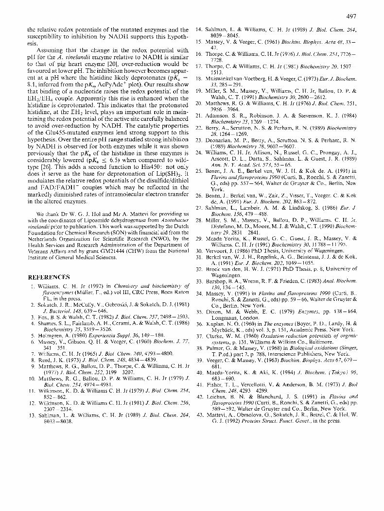
the relative redox potentials of the mutated enzymes and the susceptibility to inhibition by NADH supports this hypoth- esis.
Assuming that the change in the redox potential with pH for the A . vinelundii enzyme relative to NADH is similar to that of pig heart enzyme [20], over-reduction would be favoured at lower pH. The inhibition however becomes appar- ent at a pH where the histidine likely deprotonates (pK, = 8.1, inferred from the pK, AcPyAde' plot). Our results show that binding of a nucleotide raises the redox potential of the EH2/EH4 couple. Apparently this rise is enhanced when the histidine is deprotonated. This indicates that the protonated histidine, at the EH2 level, plays an important role in main- taining the redox potential of the active site carefully balanced to avoid over-reduction by NADH. The catalytic properties of the Glu455-mutated enzymes lend strong support to this hypothesis. Over the entire pH range studied strong inhibition by NADH is observed for both enzymes while it was shown previously that the pK, of the histidine in these enzymes is considerably lowered (pK, 5 6.5) when compared to wild- type [26]. This adds a second function to His450: not only does it serve as the base for deprotonation of L ~ P ( S H ) ~ , it modulates the relative redox potentials of the disulfide/dithiol and FADiFADH - couples which may be reflected in the markedly diminished rates of intramolecular electron transfer in the altered enzymes.
We thank Dr W. G. J. Hol and M r A. Mattevi for providing us with the coordinates of Lipoamide dehydrogenase from Azotobacter vinelandii prior to publication. This work was supported by the Dutch Foundation for Chemical Research (SON) with financial aid from the Netherlands Organization for Scientific Research (NWO), by the Health Services and Research Administration of the Department of Veterans Affairs and by grant GM21444 (CHW) from the National Institute of General Medical Sciences.
REFERENCES 1.
2.
3. 4.
5. 6.
7. 8. 9.
10.
11.
12.
13.
Williams, C . H. Jr (1992) in Chemistry and biochemistry of ,flavoenzymes (Muller, F., ed.) vol 111, CRC Press, Boca Raton FL, in the press.
Sokatch, J. R., McCully, V., Gebroski, J. &Sokatch, D. J. (1981) J . Bacteriol. 148, 639-646.
Fox, B. S. & Walsh, C. T. (1982) J . Biol. Chem. 257,2498-2503. Shames, S . L., Fairlamb, A. H., Cerami, A. & Walsh, C. T. (1986)
Holmgren, A. (1980) Experientia Suppl. 36, 149- 180. Massey, V., Gibson, Q. H. & Veeger, C. (1960) Biochem. J . 77,
Williams, C. H. Jr (1965) J . Biol. Chem. 240,4793-4800. Reed, J. K. (1973) J . Biol. Chem. 248, 4834-4839. Matthews, R. G., Ballou, D. P., Thorpe, C. & Williams, C. H. Jr
Matthews, R. G., Ballou, D. P. & Williams, C. H. Jr (1979) J .
Wilkinson, K. D. & Williams C. H. Jr (1979) J . Bid. Chem. 254,
Wilkinson, K. D. & Williams C. H. Jr (1981) J . Biol. Chem. 256,
Sahlman. L. & Williams. C. H. Jr (1989) J . Biol. Chem. 264,
Biochemistry 25, 3519 - 3526.
341 - 351.
(1977) J . Biol. Chem. 252, 3199-3207.
Biol. Chem. 254,4974-4981.
852-862.
2307 -2314.
14.
15.
16.
17.
18.
19.
20.
21.
22.
23.
24.
25.
26.
27.
28.
29.
30. 31.
32.
33.
34.
35.
36.
37
38
39
40
41
Sahlman, L. & Williams, C. H. Jr (1989) J . Biol. Chem. 264,
Massey, V. & Veeger, C. (1961) Biochim. Biophys. Acta 48, 33-
Thorpe, C. & Williams, C. H. J r (1976)J. Bid. Chem. 251,7726-
Thorpe, C. & Williams, C. H. J r (1981) Biochemistry 20, 1507-
Muiswinkel van-Voetberg, H. &Veeger, C. (1973) Eur. J . Biochem.
Miller, S. M., Massey, V., Williams, C. H. Jr, Ballou, D. P. &
Matthews, R. G . & Williams, C. H. Jr (1976) J . Bid. Chem. 251,
Adamson, S. R., Robinson, J . A. & Stevenson, K. J . (1984)
Berry, A., Scrutton, N. S. & Perham, R. N. (1989) Biochemistry
Deonarian, M. P., Berry, A., Scrutton, N. S. & Perham, R. N. (1989) Biochemistry 28, 9602 -9607.
Williams, C. H. Jr, Allison, N., Russel, G. C., Prongay, A. J., Arscott, D. L., Datta, S., Sahlman, L. & Guest, J. R. (1989) Ann. N . Y. Acad. Sci. 573, 55-65.
Benen, J. A. E., Berkel van, W. J. H. & Kok de, A. (1991) in Flavins andflavoproteins 1990 (Curti, B., Ronchi, S. & Zanetti, G., eds) pp. 557- 564, Walter de Gruyter & Co., Berlin, New York.
Benen, J. , Berkel van, W., Zdk, Z., Visser, T., Veeger, C. & Kok de, A. (1991) Eur. J . Biochem. 202, 863-872.
Sahlman, L., Lambeir, A. M. & Lindskog, S. (1986) Eur. J . Biochem. 156,479-488.
Miller, S. M., Massey, V., Ballou, D. P., Williams, C. H. Jr, Distefano, M. D., Moore, M. J. & Walsh, C. T. (1990) Biochem- istry 29, 2831 - 2841.
Meada-Yorita, K., Russel, G. C., Guest, J. R., Massey, V. & Williams, C. H. Jr (1991) Biochemistry 30, 1 1788 - 11 795.
Vervoort, J . (1986) PhD Thesis, University of Wageningen. Berkel van, W. J. H., Regelink, A. G., Beintema, J . J. & de Kok,
Broek van den, H. W. J. (1971) PhD Thesis, p. 8, University of
Barshop, B. A., Wrenn, R. F. & Frieden, C. (1983) Anal. Biochem.
Massey, V. (1991) in Flavins andflavoproteins 1990 (Curti, B., Ronchi, S. & Zanetti, G., eds) pp. 59-66, Walter de Gruyter & Co., Berlin, New York.
Dixon, M. & Webb, E. C. (1979) Enzymes, pp. 138-164, Longmans, London.
Kaplan, N. 0. (1960) in The enzymes (Boyer, P. D., Lardy, H. & Myrbiick, K., eds) vol. 3, p. 151, Academic Press, New York.
Clarke, W. M. (1960) Oxidation-reduction potentials of organic systems, p. 131, Williams & Wilkins Co., Baltimore.
Palmer, G. & Massey, V. (1968) in Biological oxidations (Singer, T. P.ed.) part 2, p. 288, Interscience Publishers, New York.
Veeger, C. & Massey, V. (1963) Biochim. Biophys. Acta 67, 679- 681.
Maeda-Yorita, K. & Aki, K. (1984) J . Biochem. (Tokyo) 96,
Fisher, T. L., Vercellotti, V. & Anderson, B. M. (1973) J . Biol
8039 - 8045.
47.
7728.
1513.
33, 285 - 291.
Walsh, C. T. (1991) Biochemistry 30, 2600-2612.
3956 - 3964.
Biochemistry 23, 1269 - 1274.
28,1264 - 1269.
A. (1991) Eur. J . Biochem. 202, 1049-1055.
Wageningen.
130, 134-145.
683 - 690.
Chem. 248,4293 -4299. 42. Leichus, B. N . & Blanchard, J. S. (1991) in Flavins and
j7avoproteins 1YY0 (Curti, B., Ronchi, S. & Zanetti, G., eds) pp. 589- 592, Walter de Gruyter and Co., Berlin, New York.
43. Mattevi, A., Obmolova, G., Sokatch, J. R., Betzel, C. & Hol, W. 8033 - 8038. G . J . (1992) Proteins Struct. Funct. Genet., in the press.


![media. · XLS file · Web viewSheet1 gi|226946096|ref|YP_002801169.1| transcriptional regulator protein [Azotobacter vinelandii DJ] gi|308273783|emb|CBX30385.1| Uncharacterized protein](https://img.pdfslide.net/doc/110x75/5aa24f7b7f8b9a84398ce377/media-fileweb-viewsheet1-gi226946096refyp0028011691-transcriptional-regulator.jpg)