Embed Size (px)
Citation preview
LIPOPROTEIN LIPASE: MECHANISM FOR ADAPTATION OF
ACTIVITY TO THE NUTRITIONAL STATE
Gengshu Wu 2004
UMEÅ UNIVERSITY MEDICAL DISSERTATIONS
New series No 866 ISSN 0346-6612 ISBN 91-7305-554-9
From the Department of Medical Biosciences, Physiological Chemistry
Umeå University, Umeå, Sweden

Don't fear failure so much that you refuse to try new things. The
saddest summary of a life contains three descriptions: could have,
might have and should have. -- Louis Boone, American Author
Lipoprotein lipase: mechanism for adaptation of activity to
the nutritional state
AKADEMISK AVHANDLING
som med vederbörligt tillstånd av rektorsämbetet vid Umeå Universitet för
avläggande av doktorsexamen i medicinsk vetenskap, offentligen kommer att
försvaras i sal N350, Umeå Universitet, fredagen den 30 januari 2004, kl 1300
av
Gengshu Wu
Fakultetsopponent: Professor Peter Nilsson-Ehle
Institutionen för Laboratoriemedicin, Klinisk Kemi, Lund Universitet

To all the people who have been loving me

If you believe you can, you probably can. If you believe you won't, you
most assuredly won't. Belief is the ignition switch that gets you off the
launching pad.
-- Denis Waitley, American Author and Speaker
CONTENTS ABBREVIATIONS ……………………………………………….…………..1
LIST OF PAPERS ………………………………………………….……………2
INTRODUCTION ……………………………………………………………….3
Historical Source …..………………………………….…………….……......….3
Where is LPL and why is It Important? ………………….…………….………......….3
LPL is Precisely Regulated …………………………….…………….………......…. 4
What Do We Know About the Structure of LPL? ………………………….………5
Functional LPL …………………………………………………………………5
Present Information on Nutritional Regulation of LPL in Adipose Tissue …………………6
SCOPE OF THIS STUDY ……………………………………….…….……7
PRESENT INVESTIGATION ...…………………………………….………8
Nutritional State and Extracellular LPL in Adipose Tissue . ……………………………8
Nutritional State and Turn-Over of LPL in Adipose Tissue ……….. ……………………8
Heparin Releases Only Extracellular LPL from Adipose Tissue …………………………8
The Ratio between Inactive and Active Forms of LPL in Adipose Tissue ………………9
Effect of Transcription Inhibitors on LPL Activity in Adipose Tissue .………………9
Adipose Tissue-Derived TNF-α and LPL activity ………………………………10
GENERAL DISCUSSION .…………………………………………………12
SUMMARY ………………………………………………………….…14
PERSPECTIVES ......………………………………………….………15
ACKNOWLEDGEMENTS …………………………………….……16
REFERENCES ……………………………………………….… 18
1
ABBREVIATIONS CM chylomicrons FFA free fatty acid HSPG heparan sulfate proteoglycans HDL high density lipoprotein LPL lipoprotein lipase LDL low density lipoprotein LDL-R low density lipoprotein receptor LRP low density lipoprotein receptor-related protein 2-MG 2-monoacylglycerol PPAR peroxisome proliferator-activated receptor TG triglyceride TNF-α tumor necrosis factor-α VLDL very low density lipoprotein VLDL-R very low density lipoprotein receptor
2
LIST OF PAPERS
This thesis is based on the four papers listed below. They will be referred to in the text by their roman numerals.
I. The distribution of lipoprotein lipase in rat adipose tissue. Changes
with nutritional state engage the extracellular enzyme Gengshu Wu, Gunilla Olivecrona, and Thomas Olivecrona Journal of Biological Chemistry (2003) 278, 11925-11930
II. Down-regulation of adipose tissue lipoprotein lipase during fasting
requires that a gene, separate from the lipase gene, is switched on Martin Bergö, Gengshu Wu, Toralph Ruge, and Thomas Olivecrona Journal of Biological Chemistry (2002) 277, 11927-11932
III. Rapid down-regulation of adipose tissue lipoprotein lipase activity on food deprivation. Evidence that TNF-α is involved
Gengshu Wu, Peter Brouckaert, and Thomas Olivecrona American Journal of Physiology (2003) (in the press)
IV. Nutritional regulation of lipoprotein lipase in mice.
Toralph Ruge, Gengshu Wu, Thomas Olivecrona, and Gunilla Olivecrona The International Journal of Biochemistry & Cell Biology (2004) 36,320-329
3
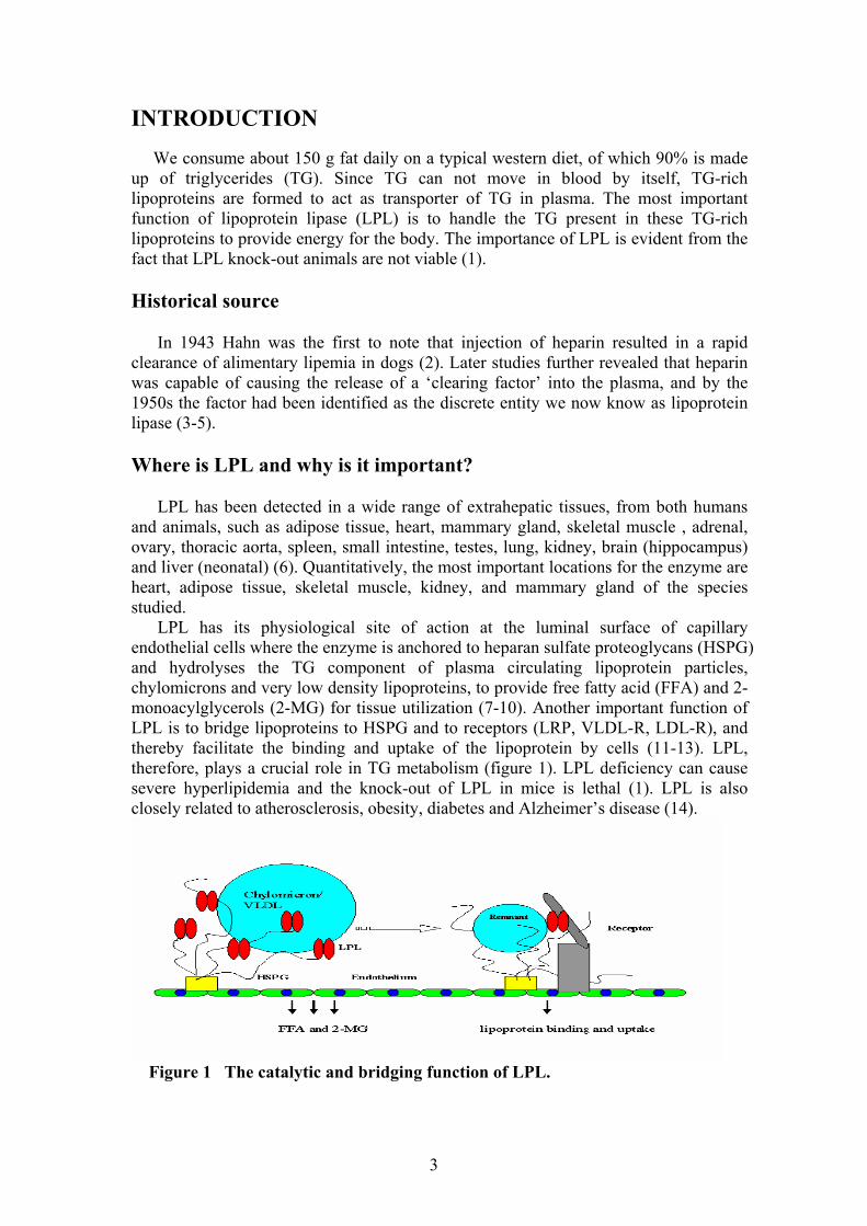
INTRODUCTION We consume about 150 g fat daily on a typical western diet, of which 90% is made up of triglycerides (TG). Since TG can not move in blood by itself, TG-rich lipoproteins are formed to act as transporter of TG in plasma. The most important function of lipoprotein lipase (LPL) is to handle the TG present in these TG-rich lipoproteins to provide energy for the body. The importance of LPL is evident from the fact that LPL knock-out animals are not viable (1). Historical source In 1943 Hahn was the first to note that injection of heparin resulted in a rapid clearance of alimentary lipemia in dogs (2). Later studies further revealed that heparin was capable of causing the release of a ‘clearing factor’ into the plasma, and by the 1950s the factor had been identified as the discrete entity we now know as lipoprotein lipase (3-5). Where is LPL and why is it important? LPL has been detected in a wide range of extrahepatic tissues, from both humans and animals, such as adipose tissue, heart, mammary gland, skeletal muscle , adrenal, ovary, thoracic aorta, spleen, small intestine, testes, lung, kidney, brain (hippocampus) and liver (neonatal) (6). Quantitatively, the most important locations for the enzyme are heart, adipose tissue, skeletal muscle, kidney, and mammary gland of the species studied. LPL has its physiological site of action at the luminal surface of capillary endothelial cells where the enzyme is anchored to heparan sulfate proteoglycans (HSPG) and hydrolyses the TG component of plasma circulating lipoprotein particles, chylomicrons and very low density lipoproteins, to provide free fatty acid (FFA) and 2-monoacylglycerols (2-MG) for tissue utilization (7-10). Another important function of LPL is to bridge lipoproteins to HSPG and to receptors (LRP, VLDL-R, LDL-R), and thereby facilitate the binding and uptake of the lipoprotein by cells (11-13). LPL, therefore, plays a crucial role in TG metabolism (figure 1). LPL deficiency can cause severe hyperlipidemia and the knock-out of LPL in mice is lethal (1). LPL is also closely related to atherosclerosis, obesity, diabetes and Alzheimer’s disease (14).
Figure 1 The catalytic and bridging function of LPL.
4
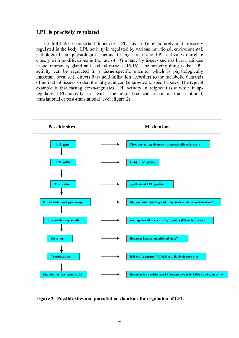
LPL is precisely regulated To fulfil these important functions LPL has to be elaborately and precisely regulated in the body. LPL activity is regulated by various nutritional, environmental, pathological and physiological factors. Changes in tissue LPL activities correlate closely with modifications in the rate of TG uptake by tissues such as heart, adipose tissue, mammary gland and skeletal muscle (15,16). The amazing thing is that LPL activity can be regulated in a tissue-specific manner, which is physiologically important because it directs fatty acid utilization according to the metabolic demands of individual tissues so that the fatty acid can be targeted to specific sites. The typical example is that fasting down-regulates LPL activity in adipose tissue while it up-regulates LPL activity in heart. The regulation can occur at transcriptional, translational or post-translational level (figure 2). Possible sites Mechanisms Figure 2. Possible sites and potential mechanisms for regulation of LPL
LPL gene
LPL mRNA
Translation
Post-translational processing
Intracellular degradation
Secretion
Endothelial (functional) LPL
Cis-trans-acting elements; tissue-specific enhancers
Stability of mRNA
Synthesis of LPL protein
Glycosylation; folding and dimerization; other modification?
Sorting:secretion versus degradation (ER or lysosomal)
Heparin; insulin; nutritional state?
HSPGs fragments, VLDLR and lipolysis products
Heparin; fatty acids; ApoB17;actinomycin D; LPS; nutritional state
Translocation
5
What do we know about the structure of LPL? Consistent with its important multiple functions, LPL also has a rather complicated structure. LPL is a noncovalent homodimer probably arranged in a head to tail conformation (17).It is a glycosylated protein; there are two N-linked oligosaccharides in each subunit. The tertiary structure of LPL has not been directly determined but the structure has been modeled on the coordinates from X-ray crystallography of pancreatic lipase (18). According to this, LPL is organized into two domains, an amino terminal (amino acids 1-312) and a carboxy terminal one (amino acids 313-448) (19,20). N-terminal domain has the catalytic triad which consists of serine-132, aspargine-156 and histidine-241. The entrance to the catalytic triad is covered by a loop, which is an important determinant for the substrate specificity (19). The C-terminal domain contains the receptor-binding and lipoprotein-binding sites (21). Functional LPL LPL with its complicated structure is precisely regulated, and functional LPL is usually the first target for the regulation. What is functional LPL? Attached to the endothelium, LPL hydrolyzes TG-rich lipoproteins and this fraction of LPL is referred to as “functional LPL” and can be released from its binding sites by heparin. This heparin releasable LPL (HR-LPL) is the pool of lipase that has access to its substrate, chylomicron and VLDL triglycerides; therefore, the HR-LPL (functional LPL) may be functionally more relevant than that measured in tissue homogenates. A large body of evidence suggests that the functional site of LPL is at the endothelium of capillary, but not all the LPL in tissues is at an endothelial cell surface. Increases in the endothelial-located enzyme (functional LPL) involve changes in the total amount of enzyme in the tissue (22-24). A decline in endothelial enzyme, when it occurs, is very rapid with a derived turnover time of functional LPL being in the order of 2h for heart and 1h for adipose tissue (25-28). This is in contrast to an intracellular half-life of 4h suggested by other studies (29). But less is known, however, of the mechanisms involved in the removal of enzyme from the endothelial site during periods when the TG uptake capacity of tissues declines. Precise regulation of functional, endothelium-bound LPL activity is required to carefully control TG catabolism and the supply of fatty acids to tissue cells. Nutritional state can change functional LPL, but LPL present in parenchymal cells isolated from lactating mammary gland, or LPL in fat and heart cells is unaltered by the nutritional state of the animals from which they were isolated—except when starvation is particularly severe. Conversely, during nutritional transitions the extracellular LPL and therefore presumed to be functionally active in lipoprotein TG hydrolysis at the endothelium will change dramatically (15,27,30,31). Hence, functional LPL is the window to observe the condition of LPL in intact tissue. The alteration of functional LPL, therefore, should be carefully considered when studying the regulation of LPL in tissues.
6
Present information on nutritional regulation of LPL in adipose tissue LPL activity in adipose tissue is regulated by multiple factors such as nutritional, environmental, physiological and pathological factors, among them nutritional regulation is the most important one. It is well documented that, postprandially, LPL activity is elevated in adipose tissue compared with heart and muscle, resulting in the channeling of circulating TG fatty acids into lipid depots. During fasting the inverse is true: relatively high heart and muscle LPL activities redirect triglyceride fatty acids appropriately into these tissues and away from adipose stores (15).The coordinated regulation of LPL in adipose tissue and muscle during feeding/fasting is critical for maintaining TG homeostasis. The mechanism involved in this tissue-specific regulation of LPL activity, however, is not known. The decrease in adipose tissue LPL activity during starvation in rats has been attributed to both pre- and post-translational mechanism (32-36). Bergo et al found that fasting increases the proportion of inactive LPL in adipose tissue and this effect is blunted in old animals (32,37).In other words, during short-term fasting, LPL specific activity decreases. The specific activity is restored within 4h by refeeding. On longer fasting, LPL mRNA decreases. This becomes significant from 36h.These data show that LPL activity during short-term fasting is regulated through posttranslational mechanism, which allows for quick up-regulation after refeeding. On longer fasting, other mechanisms affecting LPL transcription and synthesis come into play. It should be mentioned that there are discrepant results reported in the literature, this is probably due to the use of animals of different ages or to differences in experimental design and technique (34,35,38,39). LPL activity in adipose tissue is most likely regulated during feeding and fasting by a complex array of factors, such as insulin, glucagon, and glucocorticoid levels as well as sympathetic innervation (9,15). LPL can be regulated by several mechanisms, dependent on the time scale of the required response and the effectors involved (38). The study in this thesis further indicates that adipose tissue-derived TNF-α may be important for nutritional regulation of LPL. The detailed mechanism involved in nutritional regulation of LPL activity in adipose tissue is still an unsolved question. Big effort will be needed due to the complexity of the in vivo experiments and the limited information from the in vitro experiments.
7
SCOPE OF THIS STUDY The general aim of the present study was to investigate the mechanisms involved in nutritional regulation of LPL activity in adipose tissue. Specific aims: 1. To investigate if changes of distribution of LPL in the tissue are involved in the nutritional regulation of LPL in adipose tissue.
2. To study if any gene expression is crucial for the nutritional regulation of LPL activity in adipose tissue.
3. To explore the possible factors involved in nutritional regulation of LPL in adipose tissue.
8
PRESENT INVESTIGATION Nutritional state and extracellular LPL in adipose tissue (I) As reported earlier LPL mass was similar, but LPL activity was higher in adipose tissue from fed compared with fasted rats (32). To study the distribution of LPL within the tissue we used collagenase to digest the extracellular matrix. The adipocytes were then isolated by floatation. Since collagenase can quickly cleave LPL, it is not possible to recover and directly measure extracellular LPL. The pellet of sedimented cells (often called the stromal-vascular cells) contained only 3% of tissue LPL activity and 4% of tissue LPL mass. Hence, virtually all LPL was either in adipocytes or extracellular. The amount of extracellular LPL could therefore be estimated as the difference between tissue total and adipocytes. This activity was 3.5-fold higher in fed compared with fasted rats. In contrast, extracellular LPL mass did not differ significantly between the nutritional states. LPL activity within adipocytes was the same irrespective of the nutritional state so that the difference in tissue LPL activity was always due to a difference in extracellular activity. LPL mass on the other hand did not differ significantly with nutritional state, in tissue total, in adipocytes, or extracellular. Nutritional state and turn-over of LPL in adipose tissue (I and II) Since the size of the pools of LPL are determined in part by the turnover rates, which might change with the nutritional state we studied the effect of nutritional state on the turn-over of LPL in adipose tissue. When protein synthesis was inhibited with cycloheximide, LPL activity and protein mass in adipose tissue from fed rats decreased rapidly and in parallel with half-lives of around 2h (II). The much lower activity in the fasted rats also dropped. To be able to compare the rates between the nutritional states we recalculated the data as percent of initial activity. When expressed in this way, the LPL activities decreased at virtually the same rate in the two nutritional states. The loss of LPL mass and activity during the initial rapid phase (about 2h) and the subsequent slow phase differed between experiments, presumably due to variation in the extent to which protein synthesis was inhibited. One question is obvious here: how about the turn-over rate in active/inactive and intra/extracellular LPL on nutritional change? In a separate experiment LPL mass in adipose tissue decreased by 45-55% in 90 min with cycloheximide treatment. The percentage loss of LPL mass was similar for tissue total, for adipocytes, and for calculated extracellular LPL mass. LPL activity decreased by 53 and 47% in whole tissue and by 52 and 42% in adipocytes from fed and fasted rats, respectively. These differences were not statistically significant. In any case, this experiment shows that all four pools of LPL, active/inactive, intra/extracellular, turn over rapidly and at similar fractional rates (I). Heparin releases only extracellular LPL from adipose tissue (I) Many studies have shown that LPL is released from its endothelial binding sites by heparin (40-42). We questioned how much and what fraction of the total adipose tissue LPL could be released by perfusion with heparin. For this we injected heparin and measured tissue LPL mass and activity at a series of times. There was a gradual
9
decline of LPL activity with time. This was more pronounced in tissues from fed rats, from 1700 to 800 milliunits/g in 60 min compared with 700 to 600 milliunits/g in fasted rats. In contrast, there was no change in the activity within adipocytes. Hence, heparin mobilized a large fraction of the extracellular enzyme, but none of the intracellular. The decline of activity followed the same time course for fed and fasted rats and the percent of the initial extracellular activity that was lost over the 60 min studied was also the same, about 60% in both fed and fasted rats. The changes in LPL mass followed the same trends as for LPL activity. Heparin extracted more LPL mass in the fed compared with the fasted rats, and the changes occurred only in the extracellular portion. Within the adipocytes, LPL mass remained constant. The specific activity of tissue LPL was 0.93 milliunit/ng in the fed rats and 0.39 milliunit/ng in the fasted rats. A calculation of the specific activity for the enzyme that was lost from the tissue in 60 min after heparin injection returns values of 1.8 milliunits/ng in the fed and 1.7 milliunits/ng in the fasted rats. These figures carry a large uncertainty since they were calculated as the ratio of differences between rather large values. Furthermore, they do not take into account the LPL that was produced and released from the adipocytes during the 1-h experiment. Nonetheless, the figures indicate that heparin released only, or at least mainly, the active species of LPL. The ratio between inactive and active forms of LPL in adipose tissue (I and II) Earlier studies have shown that rat tissues contain at least two forms of LPL (32). The catalytically active form has high heparin affinity and elutes at about 1M NaCl from heparin-agarose. Inactive LPL elutes earlier in the gradient. There is more active than inactive LPL in adipose tissue from fed rats, whereas the reverse is true for fasted rats. In the present study, the peak ratio (inactive/active) was 0.49 ± 0.06 and 2.26 ± 0.14 in tissue extracts from fed and fasted rats, respectively. There was no difference in the distribution of LPL between the active and inactive forms within the adipocytes. The peak ratios (inactive/active) were 2.05 ± 0.15 and 2.25 ± 0.04 in fed and fasted rats, respectively. The specific activity for the enzyme that eluted in peak two was similar for all four separations, i.e. for total tissue LPL as well as for adipocyte LPL in fed and in fasted rats (I). Surprisingly when transcription inhibitor actinomycin D was injected into fasted rats, the portion of active forms of LPL increased dramatically. The ratios between the two peaks (inactive/active) were 0.38 in the actinomycin-treated rats compared to 2.27 in the fasted rats that were not given actinomycin. LPL in adipose tissue from fasted rats injected with actinomycin was present mainly in the active form, similar to LPL in adipose tissue from fed rats (II). Effect of transcription inhibitors on LPL activity in adipose tissue (II and IV) Actinomycin D can inhibit DNA-dependent mRNA synthesis. When 24-h fasted rats were injected with actinomycin and then refed, LPL activity increased to the same level as in fed rats. LPL protein mass did not change significantly. Hence, up-regulation of LPL activity during refeeding does not require synthesis of new mRNA. Next, we determined whether down-regulation of LPL activity during fasting requires
10
synthesis of new mRNA. Fed rats were injected with actinomycin or saline, and food was removed from the cages. Eighteen h later, LPL specific activity in adipose tissue from control rats (saline-injected) had decreased, as expected, to 30% of fed control. When transcription was inhibited by actinomycin, LPL mRNA decreased with half-lives of 13.3 and 16.8 h in the fed and fasted states, respectively, demonstrating slow turnover of the LPL transcripts. Surprisingly, in adipose tissue from actinomycin-injected rats, LPL-specific activity was slightly increased (140% of fed, p < 0.05). These findings show that the down-regulation of LPL specific activity during fasting requires synthesis of new mRNA and suggested that, during fasting, a gene is switched on whose product prevents LPL from becoming active even though synthesis of LPL protein continues unabated. We predicted that if we injected actinomycin into 24-h fasted rats with low adipose tissue LPL activity, we would turn off this proposed inhibitory gene, allowing active LPL to be formed again. Our prediction was upheld. 6h after injection of actinomycin into fasted rats, LPL activity in adipose tissue had increased almost 4-fold without refeeding, without any statistically significant change in LPL protein mass. The effect of actinomycin on LPL in adipose tissue could be reproduced with an unrelated transcription inhibitor, α-amanitin (II). Adipose tissue LPL activity in mice is also under nutritional regulation. The activity increases upon feeding and decreases with fasting. And this regulation is mainly post-translational as observed in rats. When transcription was blocked in fasted mice with actinomycin D, adipose tissue LPL activity increased significantly. Actinomycin D could also completely prevent fasting-induced down-regulation of LPL activity (IV). These data indicate that turning off transcription could reproduce the signal that causes up-regulation of LPL activity on refeeding both in rats and in mice. Adipose tissue-derived TNF-α and LPL activity (II and III) Actinomycin D inhibits DNA-dependent mRNA synthesis and prevents fasting-induced down-regulation of LPL activity in adipose tissue (II). The predication is that fasting switches on gene(s) whose product(s) down-regulates LPL activity. Hence, we questioned what this gene is and what the signal(s) is that tells the adipose tissue to rapidly down-regulate LPL activity. From the literature there were two situations when adipose tissue LPL activity is rapidly down-regulated. One is food deprivation (43,44). Another is trauma/sepsis/lipopolysaccharide (45-48). In the latter case TNF-α has been implicated as a major mediator. TNF-α suppresses LPL mRNA in 3T3-L1 adipocytes (49). A single injection of TNF-α decreases LPL activity in adipose tissue (but not other tissues) of mice, rats and guinea pigs (50). It was therefore natural to ask whether TNF-α might also be involved in the response to food deprivation. Since preliminary experiments showed that the rats ate little or nothing after the injection of TNF-α, we compared three group, rats with continued access to food given saline, rats deprived of food given saline, and rats deprived of food given TNF-α. LPL activity decreased by 70% in the rats deprived of food but by 81% in rats given TNF-α. This difference was statistically significant. In both of the groups deprived of food there was a tendency towards a decrease of LPL mass, but this did not reach statistical significance in this experiment. The main change was in the specific activity of LPL which decreased from 0.92 mU/ng in the fed rats to 0.35 and 0.24 mU/ng in the rats deprived of food given saline or TNF-α, respectively. To explore whether TNF-α is involved in the response to food deprivation we measured endogenous adipose tissue
11
TNF-α activity under our experimental conditions. The TNF-α activity was about 40% higher in rats deprived of food for six hour compared to rats with access to food. TNF-α protein level also increased from 1256 in rats with access to food to 2108 pg/pad in rats deprived of food for six hour. If TNF-α is involved in the response of LPL to food deprivation, blocking TNF-α production should impede the response. To test this we used pentoxifylline. This is a non specific inhibitor of phosphodiesterase known to decrease production of TNF-α. The inhibitor had no effect on TNF-α or LPL activity in fed rats but almost abolished the rise of TNF-α and the decrease of LPL activity in rats deprived of food. Similar results were obtained with another inhibitor, theophylline. When the data for TNF-α and LPL activity were plotted against each other there was a rather strong negative correlation. From the literature it seemed possible that the response of LPL to TNF-α might involved nitric oxide or a prostaglandin. Our further results denied this hypothesis. Since the activation of NFκB is one of the most important steps in the signaling pathway of TNF-α, rats were pre-treated with pyrrolidinedithiocarbamate (PDTC), which reversibly suppresses the release of IκB from the latent cytoplasmic form of NFκB in cells treated with TNF-α (51). This caused a small but statistically significant decrease in the response of adipose tissue LPL activity to food deprivation without or with injection of TNF-α. The injection of lipopolysaccharide (LPS) into rats caused an increase of adipose tissue TNF-α and a decrease of LPL activity of similar magnitude to that seen in rats given TNF-α. Both of these effects were strongly impeded by pre-treatment of the rats with pentoxifylline, supporting a role for endogenous TNF-α in the down-regulation of adipose tissue LPL activity by LPS. The results suggested that the down-regulation of adipose tissue LPL activity by LPS is mediated by TNF-α. Taken together, the above studies indicated that TNF-α may be involved in nutritional regulation of adipose tissue LPL activity. Our previous studies had indicated that the signaling during fasting involves activation of a gene, separate from the LPL gene (52). We therefore questioned if this gene is up- or down-stream of TNF-α. To explore this, rats were pre-treated with actinomycin D. As reported before, this virtually abolished the decrease of LPL activity in rats deprived of food. It also abolished the effect of TNF-α. We conclude that the gene is down-stream of TNF-α.
12
GENERAL DISCUSSION The major aim of this project was to investigate the mechanism involved in nutritional regulation of LPL activity in adipose tissue. In white adipose tissue, LPL activity changes during the day according to the nutritional state. This appears to be mediated by posttranscriptional mechanisms. During short term fasting, LPL activity in rat adipose tissue decreases without corresponding changes in the levels of mRNA and protein mass, thereby reducing the specific activity (activity/protein mass ratio) (33,53-55). Bergo et al found that this occurs because the distribution of lipase protein shifts toward an inactive form and that refeeding for 4h can restore the suppressed activity (32,56). The signal and mechanism for these changes in the activity status of the enzyme were unknown. Our study shows that in fasting rats, a gene is switched on in adipose tissue that makes the tissue produce an inactive form of LPL. Inhibition of mRNA synthesis by actinomycin completely blocked the down-regulation of LPL activity in adipose tissue during fasting. Moreover, administration of actinomycin or α-amanitin, another inhibitor of transcription, to fasted rats completely restored LPL activity within 6h. This effect is not due to increased levels of circulating hormone or changed nerve signaling activity to adipose, since it has been shown that actinomycin has the same effect during in vitro incubation (57,58). Although actinomycin could also block the biosynthesis of LPL mRNA, LPL mRNA has a rather long half-life (16.8h in fasted rats). For this reason, mRNA was available for continuous synthesis of LPL protein even after the animals had been given actinomycin. With such stable mRNA, it is not possible for the cell to rapidly regulate LPL activity on a transcriptional level. Instead, the regulation must be mediated on the translational or posttranslational level. If a block in LPL synthesis mediated the decrease in LPL activity on fasting there would have to be an associated block in LPL degradation. This is clearly not the case in our study. LPL protein mass in adipose tissue was not changed by fasting process and the turnover of LPL protein was the same in the fed and in the fasted states. Our findings suggest a perspective on the regulation: the default (fed) state is characterized by a high LPL activity and mass synthesized from a stable mRNA where the newly synthesized LPL is processed mainly into the active form. The enzyme is then secreted and transported to nearby capillaries where it acts on lipoproteins. During periods of caloric restriction (i.e., between meals or during fasting), LPL mRNA and protein mass remain high in adipose tissue. However, LPL activity can be suppressed by the induction of a short-lived gene product that causes newly synthesized LPL to be channeled into an inactive form. Refeeding or administration of actinomycin inhibits the expression of this putative factor, allowing active LPL to be formed. This is a general mechanism operating both in rats and in mice (52,59). We then tried to identify the signaling pathways involved in fasting-induced down-regulation of LPL activity in adipose tissue. Our results indicate that adipose tissue-derived TNF-α may be involved in this physiological short-term regulation of LPL in adipose tissue and NFκB is one of the components in the signaling pathway. Exogenous TNF-α declined LPL activity in adipose tissue, while actinomycin pretreatment could completely block this effect suggesting the gene in question is downstream of TNF-α, and is probably one of the many genes turned on by NFκB (51).
13
When studying the regulation of LPL we considered LPL activity and mass of intact tissue. Actually functional LPL may be more relevant than that measured in tissue homogenates (22,23). In adipose tissue LPL is localized in adipocytes (intracellular) and outside adipocytes (extracellular, including endothelium). We tried to understand if changes in this distribution are involved in the regulation of LPL activity and studies showed that most of the LPL protein in adipose tissue was located extracellularly, irrespective of the nutritional state. The specific activity of intracellular LPL remained the same. It was the specific activity of the extracellular enzyme that changed. This distribution was not due to differences in turnover rate of the extracellular enzymes. Our data thus indicate that the nutritional regulation of adipose LPL is exerted on the activity state of the extracellular enzyme. To gain information on where in the tissue the extracellular LPL is localized we injected heparin. Tissue LPL activity decreased by only 10% during the first 2 min. This presumably represented the LPL molecules that were at the endothelial surface. Over the next 60 min an additional 40% of the LPL activity continued to disappear from the tissue. That tissue LPL activity continued to decrease long after of the endothelial-bound enzyme had been released supports the view that there is a relatively large pool of LPL molecules that recirculate between the luminal surface of the endothelium and extravascular sites in the tissue (43). Heparin releases primarily the active form of LPL. One hour after injection of heparin, tissue LPL activity had decreased by about 50%. This corresponds to more than two-thirds of the extracellular LPL activity. In contrast, LPL activity within the adipocytes did not change. Heparin released more than five times as much LPL activity from the adipose tissue of fed compared with fasted rats. The active, extracellular LPL was equally accessible to release by heparin in both nutritional states. Hence, the larger release in the fed rats was a reflection of the larger amount of active extracellular LPL rather than due to some difference in how the enzyme was located in the tissue. Our studies point out that down-regulation of LPL activity in adipose tissue during fasting requires that a gene, separate from the lipase gene, is switched on. The product of this gene accomplishes the switch from predominantly active to predominantly inactive form(s) of LPL. This switch primarily affects the extracellular LPL and adipose tissue-derived TNF-α may be only one of the factors involved in this process.
14
SUMMARY ● Fasting down-regulates extracellular LPL activity in adipose tissue. There is no difference in intracellular LPL activity or mass in adipose tissue from fed and fasted rats. ● Up-regulation of LPL activity in adipose tissue requires synthesis of new protein but not mRNA. Down-regulation of adipose tissue LPL activity requires synthesis of new mRNA, and this must be the mRNA for some other protein. ● A default state exists in adipose tissue where LPL protein is synthesized on a relatively stable mRNA and is processed into its active form. During fasting, a gene is switched on whose product prevents the enzyme from becoming active even though synthesis of LPL protein continues unabated. ● Adipose tissue-derived TNF-α may be involved in the response of LPL activity to nutritional state. The gene responsible for down-regulation of LPL activity is down-stream of TNF-α.
15
PERSPECTIVES In the present investigation we deal with the mechanism for adaptation of LPL activity in adipose tissue to nutritional state. Fasting switches on gene(s) in adipose tissue whose product down-regulates extracellular LPL activity. Because of the complexity of the systems involved, such as hormones, their receptors, the different transduction mechanisms and the many potential interactions, elucidation of the detailed mechanism for nutritional regulation of LPL activity in adipose tissue is expected to require new experimental approaches and a long-term effort. In vivo experiments are too complex to unravel the mechanism. Studies with adipocytes have shown relatively small or no differences in LPL activity and secretion between cells from fasted compared with fed rats (38,60-62). The next step must therefore be to find a suitable cell system and/or conditions where the putative LPL-regulating gene can be switched on and off and the effects on LPL maturation, trafficking, and stability can be studied in detail. Our present question is: which gene(s) is switched on by fasting and contributes to the down-regulation of extracellular LPL activity in adipose tissue? The response of LPL activity to feeding and fasting appears to occur in a reciprocal manner in adipose tissue, skeletal muscle and heart (63,64). It was suggested that such change may serve as a mechanism to divert circulating TG according to the demands of the body. However, the underlying mechanism is presently not known. What are the specific nutritional signals that are recognized by adipocytes, cardiac myocytes and skeletal myocytes? How are these signals transduced into producing a reciprocal response in functional LPL activity? Do the specific cell types responsible for LPL synthesis and processing in these tissues recognize different signals, or are there selective, tissue-specific biochemical mechanisms that can produce reciprocal effects on enzyme activity in response to the same signal? It is apparently that our study in this thesis did not provide complete information. It is a challenge for all the researchers in this field to give satisfying answers to these questions. Surely, we will better understand the elaborate nutritional regulation of LPL activity in adipose tissue, heart and skeletal muscle if we solve these puzzles. One of the approaches in the near future will be to carry out studies using microarray and proteomics technology to identify the putative gene(s) whose product are involved in nutritional regulation of LPL in adipose tissue, skeletal muscle and heart. On the other hand, to set up a satisfying model system for studying the mechanism involved in regulation is also an urgent task. Our recent study found that endoplasmic reticulum (ER) molecular chaperones such as calreticulin and calnexin promote folding and dimerization of LPL (65). Mikami et al also demonstrated that fasting decreased protein disulfide isomerase in rat (66).It will be interesting to find the possible relation between ER molecular chaperones and the nutritional regulation of LPL activity in adipose tissue.

16
ACKNOWLEDGMENTS I should be proud of myself for never giving up even during the darkest hours, even when I was both mentally and physically exhausted. Though hope is frail, it is hard to kill; there can be miracles when you believe. The day for me to write this acknowledgement finally comes. Excited for completely new life? Or still wandering in the past, going through the unforgettable episodes? It is hard to tell. But one thing is certain that I own a lot to some important persons who accompanied me through these years. Thomas, my main supervisor: thanks for inspiring me so much in science; I believe that the experience spending with you is important for my future academic life. I hope that someday I will finally have your sharp and logical thinking! Gunilla, my co-supervisor: thank you for taking care of me and my family when I firstly came to Umeå. I appreciate all the advice and help from you during these years! Terry, You were the first person to greet me after my long and tired trip from Beijing to Umeå. You are always there for me with the helping hands. What a considerate person you are! The time in Umeå would be really cold if you were not here. I wish you happiness and good luck. Tell Kate that I really enjoyed her beautiful singing, she is indeed talented! Kerstin: a really lovable “old” lady! I will always remember the fishing trip with you on the sea: we did not catch fish, but we really got a lot of joy. We never have a problem in communication although I only know a few words of Swedish and you speak limited English. I know the reason: you are talking with beautiful heart. I wish you good health and happiness in life! Fia: I hope that you could read this from heaven! As my first experimental tutor, thank you for creating a good environment in lab. I believe that God will surely take care of good person like you well! Solvie, Lotta, and Åsa: Thanks for all the help you kindly gave me! You made working in the lab more convenient. Lars: Your ideas for my presentations are always great! And I know that I will be calm and confident in the future when I defense my viewpoints after your efficient training. Aviar, Elena, Valia, and Lucyna: Co-workers in the lab. Let’s remember the time we spent together. Good luck! The “old generation” in lab, Martin, Roger, Anna, Yan, and Toraph: for good memories or co-operation!

17
All the people working in animal house, especially Tord: thanks for taking care of animals and giving me a hand whenever I needed! Urban: thanks for making the teaching much easier! Magnus: thanks for opening the gate to heart perfusion! All my Chinese friends right here or out there in all the world, I thank you all for your understanding and unconditional love, especially Shushun, Fadian, and Xueyu: thanks for making the dullest time enjoyable when I firstly came to Sweden. Enjoy your wonderful life! Guo Yongzi, Wang Shouye, and Li Xiaonan: for nice talking and for sharing the good stories in life! Xiaolei and Wen: It is nice to know you. Thanks for all the happy time we spent together, I will surely treasure it! Good luck and do keep going! All my relatives: I really appreciate all the calls and letters from you, it means a lot to me! But I have to say that it is impossible for me to finish this thesis without support from the most important persons in my life My wife Liyan: I do not need to say one more word here since we went through everything together all these years. I know that you will be always there for me through thick and thin. I love you with all my heart! My son Yang: I am so proud of you! For your excellent job at school and for your persistence and understanding you always show me. I extremely love hearing your singing with guitar during those winter nights, especially “Country roads take me home” and “Streets of London”. Thank you son, for being a “big” friend and making me understand the meaning of life all the time. And finally I want to thank Umeå, this beautiful and peaceful place, for giving me the chance to see the beauty as well as the reality of life. I know that you will always linger in my memory no matter where I am!
18
REFERENCES 1. Weinstock, P. H., Bisgaier, C. L., Aalto-Setala, K., Radner, H., Ramakrishnan, R., Levak-
Frank, S., Essenburg, A. D., Zechner, R., and Breslow, J. L. (1995) J Clin Invest 96, 2555-2568
2. Hahn, P. F. (1943) Science 98, 10-20 3. Anfinsen, C. B., Boyle,E., and Brown, R.K.,. (1952) Science 115, 583 4. Robinson, D. S. a. F., J.E.,. (1953) Q J Exp Physiol 38, 233 5. Korn, E. D. (1955) J.Biol.Chem. 215, 15 6. Braun, J. E., and Severson, D. L. (1992) Biochem J 287 ( Pt 2), 337-347 7. Garfinkel, A. S. a. S., M.C. (1987) Elsevier, Amsterdam, 335-357 8. Eckel, R. H. (1989) N Engl J Med 320, 1060-1068 9. Bensadoun, A. (1991) Annu Rev Nutr 11, 217-237 10. Hamosh, M., and Hamosh, P. (1983) Mol Aspects Med 6, 199-289 11. Beisiegel, U., Weber, W., and Bengtsson-Olivecrona, G. (1991) Proc Natl Acad Sci U S A 88,
8342-8346 12. Eisenberg, S., Sehayek, E., Olivecrona, T., and Vlodavsky, I. (1992) J Clin Invest 90, 2013-
2021 13. Nykjaer, A., Nielsen, M., Lookene, A., Meyer, N., Roigaard, H., Etzerodt, M., Beisiegel, U.,
Olivecrona, G., and Gliemann, J. (1994) J Biol Chem 269, 31747-31755 14. Mead, J. R., Irvine, S. A., and Ramji, D. P. (2002) J Mol Med 80, 753-769 15. Cryer, A. (1981) Int J Biochem 13, 525-541 16. Speake, B. K., Parkin, S. M., and Robinson, D. S. (1985) Biochem Soc Trans 13, 29-31 17. Wong, H., Yang, D., Hill, J. S., Davis, R. C., Nikazy, J., and Schotz, M. C. (1997) Proc Natl
Acad Sci U S A 94, 5594-5598 18. Derewenda, Z. S., and Cambillau, C. (1991) J Biol Chem 266, 23112-23119 19. Wong, H., Davis, R. C., Thuren, T., Goers, J. W., Nikazy, J., Waite, M., and Schotz, M. C.
(1994) J Biol Chem 269, 10319-10323 20. Davis, R. C., Wong, H., Nikazy, J., Wang, K., Han, Q., and Schotz, M. C. (1992) J Biol Chem
267, 21499-21504 21. Nielsen, M. S., Brejning, J., Garcia, R., Zhang, H., Hayden, M. R., Vilaro, S., and Gliemann, J.
(1997) J Biol Chem 272, 5821-5827 22. Jansen, H., Garfinkel, A. S., Twu, J. S., Nikazy, J., and Schotz, M. C. (1978) Biochim Biophys
Acta 531, 109-114 23. Kronquist, K. E., Pedersen, M. E., and Schotz, M. C. (1980) Life Sci 27, 1153-1158 24. Cryer, A. (1985) Biochem Soc Trans 13, 27-28 25. Pedersen, M. E., and Schotz, M. C. (1980) J Nutr 110, 481-487 26. Chajek, T., Stein, O., and Stein, Y. (1975) Biochim Biophys Acta 388, 260-268 27. Cunningham, V. J., and Robinson, D. S. (1969) Biochem J 112, 203-209 28. Davies, P., and Robinson, D. S. (1973) Biochem J 136, 437-439 29. Borensztajn, J., Rone, M. S., and Sandros, T. (1975) Biochim Biophys Acta 398, 394-400 30. Chohan, P., and Cryer, A. (1978) Biochem J 174, 663-666 31. Spencer, I. M., Hutchinson, A., and Robinson, D. S. (1978) Biochim Biophys Acta 530, 375-
384 32. Bergo, M., Olivecrona, G., and Olivecrona, T. (1996) Biochem J 313 ( Pt 3), 893-898 33. Doolittle, M. H., Ben-Zeev, O., Elovson, J., Martin, D., and Kirchgessner, T. G. (1990) J Biol
Chem 265, 4570-4577 34. Ladu, M. J., Kapsas, H., and Palmer, W. K. (1991) Am J Physiol 260, R953-959 35. Oliver, J. D., and Rogers, M. P. (1993) Biochem J 292 ( Pt 2), 525-530 36. Olivecrona, T., Bergo, M., Hultin, M., and Olivecrona, G. (1995) Can J Cardiol 11 Suppl G,
73G-78G 37. Bergo, M., Olivecrona, G., and Olivecrona, T. (1997) Int J Obes Relat Metab Disord 21, 980-
986 38. Lee, J. J., Smith, P. J., and Fried, S. K. (1998) J Nutr 128, 940-946 39. Semb, H., and Olivecrona, T. (1989) Biochem J 262, 505-511 40. Goldberg, I. J. (1996) J Lipid Res 37, 693-707 41. Robinson, D. S., and French, J. E. (1960) Pharmacol Rev 12, 241-263
19
42. Nasstrom, B., Olivecrona, G., Olivecrona, T., and Stegmayr, B. G. (2001) J Lab Clin Med 138, 206-213
43. Bjorntorp, P., Yang, M. U., and Greenwood, M. R. (1983) Am J Clin Nutr 37, 396-402 44. Semb, H., and Olivecrona, T. (1986) Biochim Biophys Acta 876, 249-255 45. Lanza-Jacoby, S., Sedkova, N., Phetteplace, H., and Perrotti, D. (1997) J Lipid Res 38, 701-
710 46. Picard, F., Arsenijevic, D., Richard, D., and Deshaies, Y. (2002) Clin Diagn Lab Immunol 9,
771-776 47. Feingold, K. R., Marshall, M., Gulli, R., Moser, A. H., and Grunfeld, C. (1994) Arterioscler
Thromb 14, 1866-1872 48. Picard, F., Kapur, S., Perreault, M., Marette, A., and Deshaies, Y. (2001) Faseb J 15, 1828-
1830 49. Cornelius, P., Enerback, S., Bjursell, G., Olivecrona, T., and Pekala, P. H. (1988) Biochem J
249, 765-769 50. Semb, H., Peterson, J., Tavernier, J., and Olivecrona, T. (1987) J Biol Chem 262, 8390-8394 51. Wolle, J., Ferguson, E., Keshava, C., Devall, L. J., Boschelli, D. H., Newton, R. S., and
Saxena, U. (1995) Biochem Biophys Res Commun 214, 6-10 52. Bergo, M., Wu, G., Ruge, T., and Olivecrona, T. (2002) J Biol Chem 277, 11927-11932 53. Masuno, H., Blanchette-Mackie, E. J., Schultz, C. J., Spaeth, A. E., Scow, R. O., and Okuda,
H. (1992) J Lipid Res 33, 1343-1349 54. Park, J. W., Oh, M. S., Yang, J. Y., Park, B. H., Rho, H. W., Lim, S. N., Jhee, E. C., and Kim,
H. R. (1995) Biochim Biophys Acta 1254, 45-50 55. Masuno, H., Blanchette-Mackie, E. J., Chernick, S. S., and Scow, R. O. (1990) J Biol Chem
265, 1628-1638 56. Bergo, M., Olivecrona, G., and Olivecrona, T. (1996) Am J Physiol 271, E1092-1097 57. Schotz, M. C., and Garfinkel, A. S. (1965) Biochim Biophys Acta 106, 202-205 58. Wing, D. R., and Robinson, D. S. (1968) Biochem J 106, 667-676 59. Ruge, T., Wu, G., Olivecrona, T., and Olivecrona, G. (2004) The International Journal of
Biochemistry & Cell Biology 36, 320-329 60. Wu, G., Olivecrona, G., and Olivecrona, T. (2003) J Biol Chem 278, 11925-11930 61. Semb, H., and Olivecrona, T. (1987) Biochim Biophys Acta 921, 104-115 62. Cryer, A., Davies, P., Williams, E. R., and Robinson, D. S. (1975) Biochem J 146, 481-488 63. Sugden, M. C., Holness, M. J., and Howard, R. M. (1993) Biochem J 292 ( Pt 1), 113-119 64. Richelsen, B. (1999) J Endocrinol Invest 22, 10-15 65. Zhang, L., Wu, G., Tate, C. G., Lookene, A., and Olivecrona, G. (2003) J Biol Chem 278,
29344-29351 66. Mikami, T., Genma, R., Nishiyama, K., Ando, S., Kitahara, A., Natsume, H., Yoshimi, T.,
Horiuchi, R., and Nakamura, H. (1998) Metabolism 47, 1083-1088