Embed Size (px)
Citation preview
General rights Copyright and moral rights for the publications made accessible in the public portal are retained by the authors and/or other copyright owners and it is a condition of accessing publications that users recognise and abide by the legal requirements associated with these rights.
Users may download and print one copy of any publication from the public portal for the purpose of private study or research.
You may not further distribute the material or use it for any profit-making activity or commercial gain
You may freely distribute the URL identifying the publication in the public portal If you believe that this document breaches copyright please contact us providing details, and we will remove access to the work immediately and investigate your claim.
Downloaded from orbit.dtu.dk on: May 21, 2020
Liposomal Drug Delivery of Anticancer AgentsSynthesis, Biophysical Characterization and Biological Studies of Enzyme SensitivePhospholipid ProdrugsPedersen, Palle Jacob
Publication date:2010
Link back to DTU Orbit
Citation (APA):Pedersen, P. J. (2010). Liposomal Drug Delivery of Anticancer Agents: Synthesis, Biophysical Characterizationand Biological Studies of Enzyme Sensitive Phospholipid Prodrugs. Technical University of Denmark.
Liposomal Drug Delivery
of Anticancer Agents
Synthesis, Biophysical Characterization and Biological
Studies of Enzyme Sensitive Phospholipid Prodrugs
Ph.D. Thesis
Palle Jacob Pedersen
May 2010
Department of Chemistry
Technical University of Denmark

Liposomal Drug Delivery
of Anticancer Agents
Synthesis, Biophysical Characterization
and Biological Studies of Enzyme Sensitive
Phospholipid Prodrugs
Palle Jacob Pedersen
Ph.D. Thesis
May 2010
Department of Chemistry
Technical University of Denmark
Kemitorvet
Building 201
DK-2800 Kgs. Lyngby
Denmark

I
Preface This thesis describes the work carried out during my three years as a Ph.D. student at Department of Chemistry, Technical University of Denmark (DTU) under supervision of Associate Professor Mads H. Clausen, Senior Scientist Thomas L. Andresen and Professor Robert Madsen. Chapter 1–9 covers the work towards a new generation of liposomal drug delivery systems for anticancer agents, including organic synthesis along with biophysical, biological and computational studies. Chapter 10–11 covers the synthesis of small natural-product-like molecules carried out during a 6 month research stay at School of Chemistry, University of Leeds under supervision of Professor Adam Nelson. There are numerous people that have contributed and being involved in the projects I have worked on in the last three years and I would like to thank the following persons for help and collaboration. First of all I would like to thank Associate Professor Mads H. Clausen for your great supervision during the years. You have always motivated and inspired me and during the hard times you remained optimistic and led me in the right directions. Thanks for believing in me. Senior Scientist Thomas L. Andresen and his crew at DTU Nano, Risø are thanked for letting me use their equipment and for sharing their enormous knowledge about phospholipids and liposomes with me. Professor Robert Madsen has been an invaluable resource and a go-to-guy when crucial decision had to be made. I would like to thank all the collaborators on the project of liposomal drug delivery. Dr. Tristan Ruysschaert, Dr. Ahmad Arouri and Professor Ole G. Mouritsen for their involvement in the biophysical characterization, Dr. Fredrik Melander, Dr. Sidsel K. Adolph and Dr. Mogens W. Madsen for doing all the biological studies and Dr. Arun K. Subramanian and Associate Professor Günther H. Peters are thanked for help and guidance with the computational studies in chapter 4. A special thanks goes to Dr. Mikkel S. Christensen for collaboration with the organic synthesis of phospholipids, you have had a significant impact on the work described in this thesis. I thank Dr. Lars Linderoth for introducing me to the field of phospholipid synthesis in the beginning of my Ph.D. The Clausen group; Charlotte, Kasper, Mads, Hélène, Alexandra and Mikkel have along with the bachelor and master students that have been in the group during the years created a positive and inspiring atmosphere which I have appreciated and you are thanked for many great days. The lab technicians; Brian, Anne and Janne are acknowledged for help with apparatus, NMR and chemicals – your service is always with a smile. I would like to thank Professor Adam Nelson for allowing me to come and work in his group at University of Leeds and I am thankful to the entire Nelson group for thanking care
II
of me both in and outside the lab – and no one take away from me that I reach the highest peak in England in the beautiful Lake District. The Danish Council for Strategic Research (NABIIT Program) is acknowledged for financial support of the liposomal drug delivery project, while Augustinus Fonden, Oticon Fonden, Knud Højgaards Fond, Vera og Carl Johan Michaelsens Legat, Civilingeniør Frants Allings Legat and Kemisk Forenings Rejsefond are acknowledged for financial support concerning my stay in Leeds. Finally I would like to thank my family and friends for your support and interest – you are there always for me. I hope you will enjoy the thesis.
_____________________________
Palle Jacob Pedersen Kgs. Lyngby, May 2010
III
Abstract In the first part of the thesis the work towards a new generation of liposomal drug delivery systems for anticancer agents is described. The drug delivery system takes advantage of the elevated level of secretory phospholipase A2 (sPLA2) IIA in many tumors and the enhanced permeability and retention (EPR) effect. The liposomes consists of sPLA2 IIA sensitive phospholipids having anticancer drugs covalently attached to the sn-2 position of the glycerol backbone in the phospholipids, hence drug leakage is avoided from the carrier system. Various known anticancer agents, like chlorambucil, all-trans retinoic acid, α-tocopheryl succinate and calcitriol were examined for their ability to be incorporated into the investigated drug delivery system and syntheses of the phospholipid prodrugs are described. The majority of the phospholipid prodrugs were able to form particles with diameters close to 100 nm upon extrusion at 20 °C indicating that unilamellar vesicles are formed. When subjected to sPLA2 the phospholipid prodrugs were converted into cytotoxic lysolipids and along with the released anticancer drug a chemotherapeutic cocktail is formed. Cytotoxicity studies in several cancer lines revealed that upon sPLA2 triggering the formulated phospholipid prodrugs displayed IC50 values in range from 3–36 µM and complete cell death was observed when higher drug concentrations were applied. Promising for the drug delivery system the majority of the phospholipid prodrugs remain non-toxic in the absence of the enzyme meaning the prodrugs will not damage healthy tissue during the transport in the body. In the second part of the thesis the synthetic studies towards a library of small natural-product-like molecules are described. The collection of molecules was synthesized via a diversity oriented synthesis (DOS) based strategy using a limited number of reaction types. Upon coupling of unsaturated building blocks ring closing metathesis cascades were used to “reprogram” the molecular scaffold and highly diverse structures were obtained. In total 20 novel compounds with a broad structural diversity were prepared in 5 or 6 synthetic steps.
IV
Resumé I den første del af afhandlingen beskrives arbejdet med en ny generation af liposomal drug delivery systemer til brug for cytotoksiske stoffer. Drug delivery systemet udnytter forhøjede koncentrationer af secerneret phospholipase A2 (sPLA2) IIA i kræft svulster og at cancervæv er mere porøst end raskt væv, hvorved nanopartikler som liposomer kan diffundere ind i kræftvæv men ikke ind i raskt væv. Liposomerne består af sPLA2 IIA sensitive phospholipider hvori det cytotoksiske stof kovalent er bundet til sn-2 positionen af glycerol enheden i phospholipiderne således at udsivning fra liposomerne undgås. Adskillige kendte anticancer stoffer, som chlorambucil, retinsyre, α-tocopheryl succinat og calcitriol blev testet for deres evne til at indgå i det undersøgte drug delivery system og syntesen af phospholipid prodrugsene beskrives. Hovedparten af de fremstillede phospholipid prodrugs var i stand til efter ekstrudering ved 20 °C at danne partikler med diametere omkring 100 nm, hvilket indikerer dannelsen af liposomer. I stedeværelse af sPLA2 omdannes phospholipid prodrugsene til cytotoksiske lysolipider og sammen med det frigjorte anticancer stof dannes en cocktail af kemoterapeutiske stoffer. Evnen til at frembringe celledød blev undersøgt i forskellige kræftceller og i stedeværelse af sPLA2 viste phospholipid prodrugsene IC50 værdier mellem 3–36 µM og fuldstændig celledød blev opnået ved anvendelse af højere koncentrationer. Lovende for drug delivery systemet, så var hovedparten af phosphilipid prodrugsene ikke i stand til at frembringe celledød i fravær af enzymet hvorved prodrugsene ikke vil skade raskt væv under transporten i kroppen. I den anden del af afhandlingen beskrives de syntetiske studier mod fremstillingen af et bibliotek af naturstof-lignende molekyler. Samlingen af molekyler blev syntetiseret ved en DOS (Diversity Oriented Synthesis) baseret syntesestrategi ved anvendelse af få reaktionstyper. Simple byggeblokke blev sammensat og ved hjælp af ringluknings metatese kaskader blev det molekylære skellet af de kombinerede byggeblokke omstruktureret og strukturer med en høj diversitet blev opnået. I alt blev 20 nye stoffer med en bred struktural forskellighed fremstillet i 5 eller 6 syntese trin.
V
Publications At the time for submission of this thesis the research had resulted in the following scientific publications. Pedersen, P. J.; Christensen, M. S.; Ruysschaert, T.; Linderoth, L.; Andresen, T. L.; Melander, F.; Mouritsen, O. G.; Madsen, R.; Clausen, M. H. Synthesis and Biophysical Characterization of Chlorambucil Anticancer Ether Lipid Prodrugs. J. Med. Chem. 2009, 52, 3408–3415. Christensen, M. S.; Pedersen, P. J.; Andresen, T. L.; Madsen, R.; Clausen, M. H. Isomerization of all-(E)-Retinoic Acid Mediated by Carbodiimide Activation - Synthesis of ATRA Ether Lipid Conjugates. Eur. J. Org. Chem. 2010, 719-724. Pedersen, P. J.; Adolph, S. K.; Subramanian, A. K.; Arouri, A.; Andresen, T. L.; Mouritsen, O. G.; Madsen, R.; Madsen, M. W.; Peters, G. H.; Clausen, M. H. Liposomal Formulation of Retinoids Designed for Enzyme Triggered Release. J. Med. Chem. 2010, 53, 3782–3792. Pedersen, P. J.; Adolph, S. K.; Andresen, T. L.; Madsen, R.; Madsen, M. W.; Clausen, M. H. Prostaglandin Phospholipid Conjugates with Unusual Biophysical and Cytotoxic Properties. Bioorg. Med. Chem. Lett. Submitted. Kumar, S. A.; Pedersen, P. J.; Andresen, T. L.; Madsen, R.; Clausen, M. H.; Peters, G. H. A Unique Water In-take Mechanism Observed in Secretory Phospholipase A2. J. Am. Chem. Soc. Submitted. O’Leary-Steele, C.; Pedersen, P. J.; James, T.; Lanyon-Hogg, T.; Leach, S.; Hayes, J.; Nelson, A. Synthesis of small molecules with high scaffold diversity: Exploitation of metathesis cascades in combination with inter- and intramolecular DielsAlder reactions. Chem. Eur. J. Accepted.

Contents
1
Contents 1. Introduction to Liposomal Drug Delivery ......................................................................... 5
1.1 Phospholipids and Liposomes ..................................................................................... 6
1.2 Liposomes in Drug Delivery ....................................................................................... 8
1.3 Secretory Phospholipase A2 and its Utilization as a Site Specific Trigger ............... 13
1.4 New Generation of Enzyme Sensitive Phospholipids ............................................... 17
2. Phospholipid Synthesis – An Overview .......................................................................... 21
2.1 Synthesis of 1,2-Diacyl-sn-glycerophospholipids ..................................................... 23
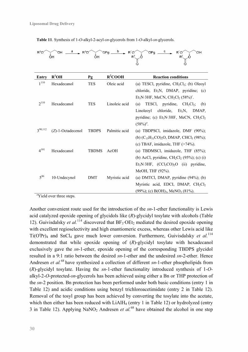
2.2 Synthesis of 1-O-Alkyl-2-acyl-sn-glycerophospholipids .......................................... 29
2.3 Synthesis of Phosphatidylcholine (PC) Headgroup................................................... 33
2.4 Synthesis of Phosphatidylglycerol (PG) Headgroup ................................................. 34
3. Chlorambucil Phospholipid Prodrugs .............................................................................. 35
3.1 Introduction ............................................................................................................... 35
3.2 Synthesis of Chlorambucil Phospholipid Prodrugs ................................................... 35
3.3 Biophysical Characterization and Enzyme Activity .................................................. 37
3.4 Cytotoxicity ............................................................................................................... 40
3.5 Conclusion ................................................................................................................. 41
4. Liposomal Formulation of Retinoids Designed for Enzyme Triggered Release ............. 43
4.1 Introduction ............................................................................................................... 43
4.2 Synthesis of ATRA Phospholipid Prodrugs .............................................................. 43
4.3 Enzymatic Activity .................................................................................................... 46
4.4 Molecular Dynamics Simulations of Enzymatic Activity ......................................... 46
4.5 Revised Prodrug Strategy for Liposomal Delivery of Retinoids ............................... 51
4.6 Synthesis of Retinoid Esters ...................................................................................... 52
4.7 Synthesis of Retinoid Phospholipid Prodrugs ........................................................... 53
4.8 Biophysical Characterization and Enzyme Activity .................................................. 57
4.9 Cytotoxicity ............................................................................................................... 59
4.10 Conclusion ............................................................................................................... 62
5. Enzymatic Studies on Vitamin E Succinate Phospholipid Conjugates ........................... 63
5.1 Introduction ............................................................................................................... 63
5.2 Synthesis of α-Tocopheryl Succinate Phospholipid Prodrugs................................... 64
5.3 Enzyme Activity and Cytotoxicity ............................................................................ 65
Liposomal Drug Delivery
2
5.4 Synthesis and Enzyme Activity of Alkyl and Phenyl Succinate Phospholipids ....... 67
5.5 Conclusion ................................................................................................................. 69
6. Prostaglandin Phospholipid Conjugates with Unusual Properties .................................. 71
6.1 Introduction ............................................................................................................... 71
6.2 Synthesis of Prostaglandin Phospholipid Conjugates ............................................... 72
6.3 Biophysical Characterization and Enzymatic Studies ............................................... 73
6.4 Cytotoxicity ............................................................................................................... 74
7. Towards Liposomal Delivery of Calcitriol ..................................................................... 77
7.1 Introduction ............................................................................................................... 77
7.2 Calcitriol Esters ......................................................................................................... 78
7.3 Synthesis of Phospholipid in Conjugate with α,α-Difluoro Calcitriol Ester ............. 82
7.4 Synthesis of Phospholipid Carbonate in Conjugate with Calcitriol Ester ................. 84
7.5 Biophysical Characterization and Enzyme Activity ................................................. 85
7.6 Biological Studies of the Calcitriol Esters ................................................................ 88
7.7 Conclusion ................................................................................................................. 89
8. Conclusion: Liposomal Drug Delivery ........................................................................... 91
9. Experimental: Liposomal Drug Delivery ........................................................................ 93
9.1 General Experimental ................................................................................................ 93
9.2 Procedures for Biophysical and Biological Characterization ................................... 93
9.3 Experimental Data for Compounds ........................................................................... 96
10. Synthesis of Small Natural-Product-Like Molecules .................................................. 127
10.1 Introduction ........................................................................................................... 127
10.2 Project Outline ....................................................................................................... 129
10.3 Synthesis of Fluorous-Tagged Building Blocks .................................................... 131
10.4 Synthesis of Non-Fluorous-Tagged Building Blocks ........................................... 135
10.5 Coupling of Building Blocks ................................................................................. 137
10.6 Metathesis Cascade Reactions .............................................................................. 139
10.7 Diels Alder Reaction with Cookson’s Reagent ..................................................... 142
10.8 Nosyl Deprotection ............................................................................................... 142
10.9 Synthesis of the Final Products ............................................................................. 144
10.10 Scaffold Analysis ................................................................................................ 148
10.11 Conclusion ........................................................................................................... 149

Contents
3
11. Experimental: Natural-Prodruct-Like Molecules ........................................................ 151
11.1 General Experimental ............................................................................................ 151
11.2 General Procedures ................................................................................................ 151
11.3 Experimental Data for Compounds ....................................................................... 153
Abbreviations .................................................................................................................... 178
References ......................................................................................................................... 181

1. Introduction to Liposomal Drug Delivery
5
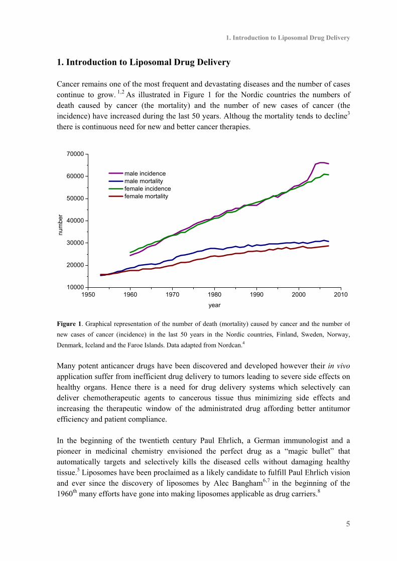
1. Introduction to Liposomal Drug Delivery Cancer remains one of the most frequent and devastating diseases and the number of cases continue to grow. 1,2 As illustrated in Figure 1 for the Nordic countries the numbers of death caused by cancer (the mortality) and the number of new cases of cancer (the incidence) have increased during the last 50 years. Althoug the mortality tends to decline3 there is continuous need for new and better cancer therapies.
1950 1960 1970 1980 1990 2000 201010000
20000
30000
40000
50000
60000
70000
num
ber
year
male incidence male mortality female incidence female mortality
Figure 1. Graphical representation of the number of death (mortality) caused by cancer and the number of
new cases of cancer (incidence) in the last 50 years in the Nordic countries, Finland, Sweden, Norway,
Denmark, Iceland and the Faroe Islands. Data adapted from Nordcan.4
Many potent anticancer drugs have been discovered and developed however their in vivo application suffer from inefficient drug delivery to tumors leading to severe side effects on healthy organs. Hence there is a need for drug delivery systems which selectively can deliver chemotherapeutic agents to cancerous tissue thus minimizing side effects and increasing the therapeutic window of the administrated drug affording better antitumor efficiency and patient compliance. In the beginning of the twentieth century Paul Ehrlich, a German immunologist and a pioneer in medicinal chemistry envisioned the perfect drug as a “magic bullet” that automatically targets and selectively kills the diseased cells without damaging healthy tissue.5 Liposomes have been proclaimed as a likely candidate to fulfill Paul Ehrlich vision and ever since the discovery of liposomes by Alec Bangham6,7 in the beginning of the 1960th many efforts have gone into making liposomes applicable as drug carriers.8
Liposomal Drug Delivery
6
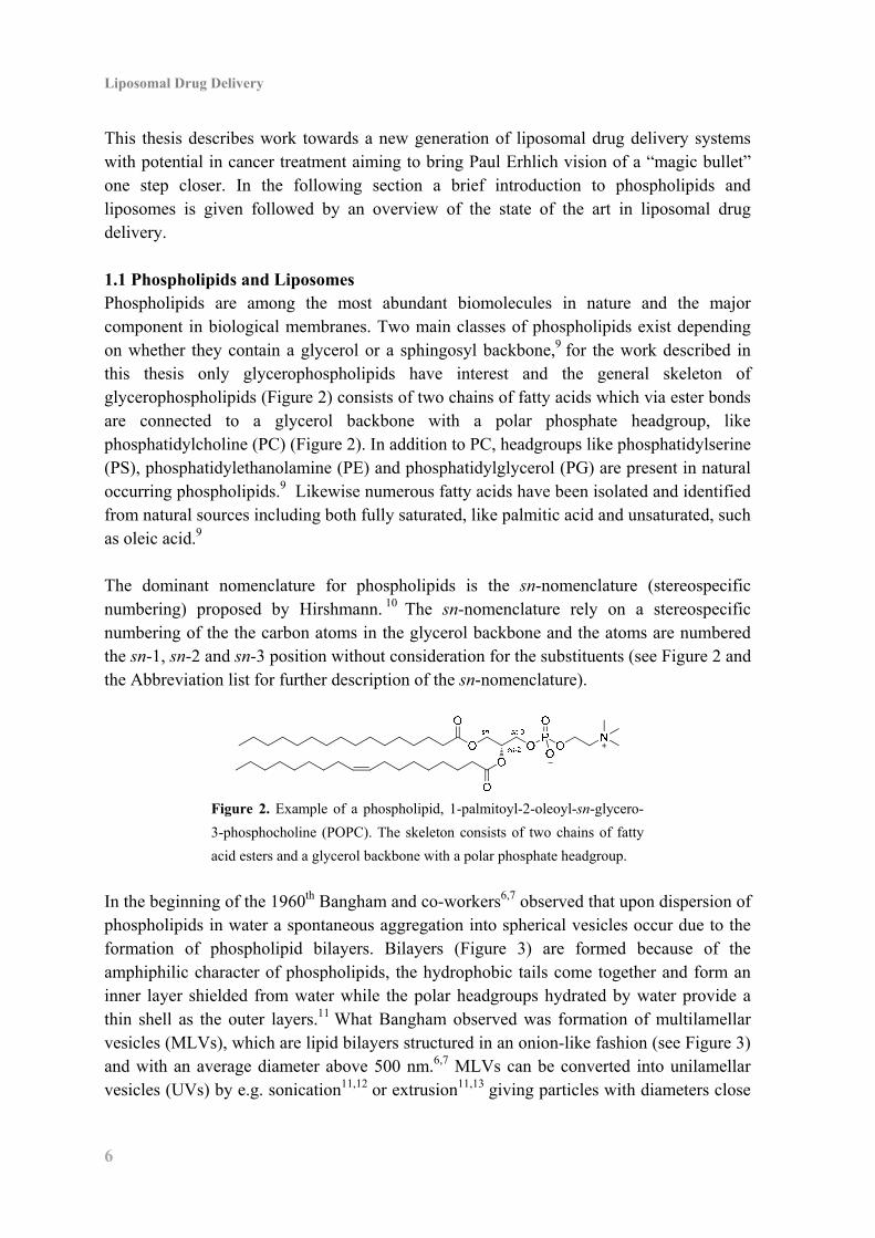
This thesis describes work towards a new generation of liposomal drug delivery systems with potential in cancer treatment aiming to bring Paul Erhlich vision of a “magic bullet” one step closer. In the following section a brief introduction to phospholipids and liposomes is given followed by an overview of the state of the art in liposomal drug delivery. 1.1 Phospholipids and Liposomes Phospholipids are among the most abundant biomolecules in nature and the major component in biological membranes. Two main classes of phospholipids exist depending on whether they contain a glycerol or a sphingosyl backbone,9 for the work described in this thesis only glycerophospholipids have interest and the general skeleton of glycerophospholipids (Figure 2) consists of two chains of fatty acids which via ester bonds are connected to a glycerol backbone with a polar phosphate headgroup, like phosphatidylcholine (PC) (Figure 2). In addition to PC, headgroups like phosphatidylserine (PS), phosphatidylethanolamine (PE) and phosphatidylglycerol (PG) are present in natural occurring phospholipids.9 Likewise numerous fatty acids have been isolated and identified from natural sources including both fully saturated, like palmitic acid and unsaturated, such as oleic acid.9 The dominant nomenclature for phospholipids is the sn-nomenclature (stereospecific numbering) proposed by Hirshmann. 10 The sn-nomenclature rely on a stereospecific numbering of the the carbon atoms in the glycerol backbone and the atoms are numbered the sn-1, sn-2 and sn-3 position without consideration for the substituents (see Figure 2 and the Abbreviation list for further description of the sn-nomenclature).
Figure 2. Example of a phospholipid, 1-palmitoyl-2-oleoyl-sn-glycero-
3-phosphocholine (POPC). The skeleton consists of two chains of fatty
acid esters and a glycerol backbone with a polar phosphate headgroup.
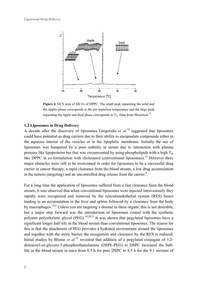
In the beginning of the 1960th Bangham and co-workers6,7 observed that upon dispersion of phospholipids in water a spontaneous aggregation into spherical vesicles occur due to the formation of phospholipid bilayers. Bilayers (Figure 3) are formed because of the amphiphilic character of phospholipids, the hydrophobic tails come together and form an inner layer shielded from water while the polar headgroups hydrated by water provide a thin shell as the outer layers.11 What Bangham observed was formation of multilamellar vesicles (MLVs), which are lipid bilayers structured in an onion-like fashion (see Figure 3) and with an average diameter above 500 nm.6,7 MLVs can be converted into unilamellar vesicles (UVs) by e.g. sonication11,12 or extrusion11,13 giving particles with diameters close

1. Introduction to Liposomal Drug Delivery
7
to 100 nm (see Figure 3). Solutions of MLVs are milky and unclear while UVs provide clear and transparent solutions. By definition liposomes can be seen both as MLVs and UVs but for cancer therapy the smaller UVs are of most interest (vide infra).14 Aqueous phospholipid solutions display a series of temperature dependent reversible phase transitions, which are important for the properties and behavior of liposomes.15 The phase transition temperatures can be demonstrated by obtaining a differential scanning calorimetry (DSC) scan. From the DSC scan of MLVs of 1,2-dipalmitoyl-sn-glycero-3-phosphocholine (DPPC, Figure 4) it is evident that the bilayers undergo a transformation from an ordered solid phase to a more disordered ripple phase (pre-transition temperature) and then into a fluid phase (main phase transition temperature, Tm). The phase transition temperatures depend on the ability of the to phospholipids to create well ordered and dence bilayers and phospholipids having two saturated fatty acids in the sn-1 and sn-2 position have high Tm’s , (Tm of DPPC is 41 °C)16 whereas phospholipids having unsaturated fatty acids have lower Tm’s , (Tm of POPC is –20 °C).17 The physical state of the bilayers affect the permeability, stability and flexibility of the liposomes, hence extrusion of MLVs of 1,2-distearoyl-sn-glycero-3-phosphocholine (DSPC) through a 100 nm filter needs to be performed above the Tm of DSPC at 55 °C,17 as it is impossible below the Tm due to the stiffness of the bilayers.
Figure 3. Illustration of phospholipid bilayers in a multilamellar vesicle (top left) and
phospholipid bilayer in a unilamellar vesicle (top right). Cryo-electro microscopy
pictures of a multilamellar vesicle (bottom left) and unilamellar vesicles (bottom right).
Data from Mouritsen.15
Liposomal Drug Delivery
8
Figure 4. DCS scan of MLVs of DPPC. The small peak separating the solid and
the ripples phase corresponds to the pre-transition temperature and the large peak
separating the ripple and fluid phase corresponds to Tm. Data from Mouritsen.15
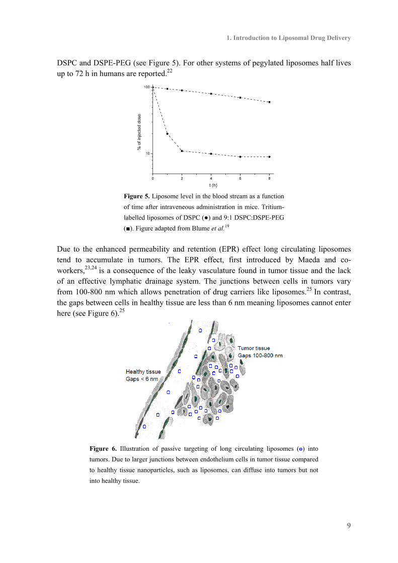
1.2 Liposomes in Drug Delivery A decade after the discovery of liposomes Gregoridis et al.18 suggested that liposomes could have potential as drug carriers due to their ability to encapsulate compounds either in the aqueous interior of the vesicles or in the lipophilic membrane. Initially the use of liposomes was hampered by a poor stability in serum due to interaction with plasma proteins like lipoproteins but that was circumvented by using phospholipids with a high Tm like DPPC in co-formulation with cholesterol (conventional liposomes).14 However three major obstacles were still to be overcomed in order for liposomes to be a successful drug carrier in cancer therapy, a rapid clearance from the blood stream, a low drug accumulation in the tumors (targeting) and an uncontrolled drug release from the carrier.8 For a long time the application of liposomes suffered from a fast clearance from the blood stream, it was observed that when conventional liposomes were injected intravenously they rapidly were recognized and removed by the reticuloendothelial system (RES) hence leading to an accumulation in the liver and spleen followed by a clearance from the body by macrophages.14,8 Unless you are targeting a disease in these organs, this is not desirable, but a major step forward was the introduction of liposomes coated with the synthetic polymer polyethylene glycol (PEG).19,20,21 It was shown that pegylated liposomes have a significant longer half-life in the blood stream than conventional liposomes. The reason for this is that the attachment of PEG provides a hydrated environment around the liposomes and together with the steric barrier the recognition and clearance by the RES is reduced. Initial studies by Blume et al.19 revealed that addition of a pegylated conjugate of 1,2-distearoyl-sn-glycero-3-phosphoethanolamine (DSPE-PEG) to DSPC increased the half-life in the blood stream in mice from 0.5 h for pure DSPC to 8.5 h for the 9:1 mixture of
1. Introduction to Liposomal Drug Delivery
9
DSPC and DSPE-PEG (see Figure 5). For other systems of pegylated liposomes half lives up to 72 h in humans are reported.22
Figure 5. Liposome level in the blood stream as a function
of time after intraveneous administration in mice. Tritium-
labelled liposomes of DSPC (●) and 9:1 DSPC:DSPE-PEG
(■). Figure adapted from Blume et al.19
Due to the enhanced permeability and retention (EPR) effect long circulating liposomes tend to accumulate in tumors. The EPR effect, first introduced by Maeda and co-workers,23,24 is a consequence of the leaky vasculature found in tumor tissue and the lack of an effective lymphatic drainage system. The junctions between cells in tumors vary from 100-800 nm which allows penetration of drug carriers like liposomes.25 In contrast, the gaps between cells in healthy tissue are less than 6 nm meaning liposomes cannot enter here (see Figure 6).25
Figure 6. Illustration of passive targeting of long circulating liposomes (o) into
tumors. Due to larger junctions between endothelium cells in tumor tissue compared
to healthy tissue nanoparticles, such as liposomes, can diffuse into tumors but not
into healthy tissue.
Liposomal Drug Delivery
10
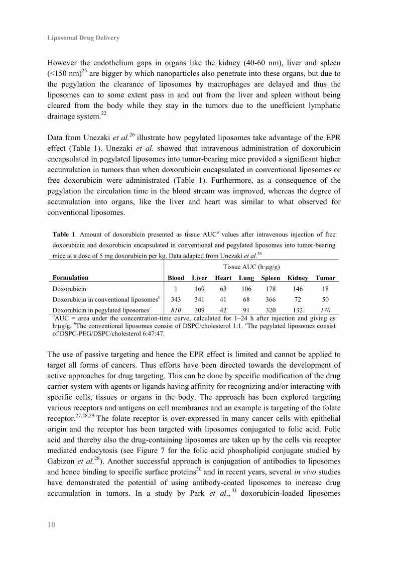
However the endothelium gaps in organs like the kidney (40-60 nm), liver and spleen (<150 nm)25 are bigger by which nanoparticles also penetrate into these organs, but due to the pegylation the clearance of liposomes by macrophages are delayed and thus the liposomes can to some extent pass in and out from the liver and spleen without being cleared from the body while they stay in the tumors due to the unefficient lymphatic drainage system.22 Data from Unezaki et al.26 illustrate how pegylated liposomes take advantage of the EPR effect (Table 1). Unezaki et al. showed that intravenous administration of doxorubicin encapsulated in pegylated liposomes into tumor-bearing mice provided a significant higher accumulation in tumors than when doxorubicin encapsulated in conventional liposomes or free doxorubicin were administrated (Table 1). Furthermore, as a consequence of the pegylation the circulation time in the blood stream was improved, whereas the degree of accumulation into organs, like the liver and heart was similar to what observed for conventional liposomes.
Table 1. Amount of doxorubicin presented as tissue AUCa values after intravenous injection of free
doxorubicin and doxorubicin encapsulated in conventional and pegylated liposomes into tumor-bearing
mice at a dose of 5 mg doxorubicin per kg. Data adapted from Unezaki et al.26
Formulation
Tissue AUC (h·µg/g)
Blood Liver Heart Lung Spleen Kidney Tumor
Doxorubicin 1 169 63 106 178 146 18
Doxorubicin in conventional liposomesb 343 341 41 68 366 72 50
Doxorubicin in pegylated liposomesc 810 309 42 91 320 132 170 aAUC = area under the concentration-time curve, calculated for 1–24 h after injection and giving as h·µg/g. bThe conventional liposomes consist of DSPC/cholesterol 1:1. cThe pegylated liposomes consist of DSPC-PEG/DSPC/cholesterol 6:47:47.
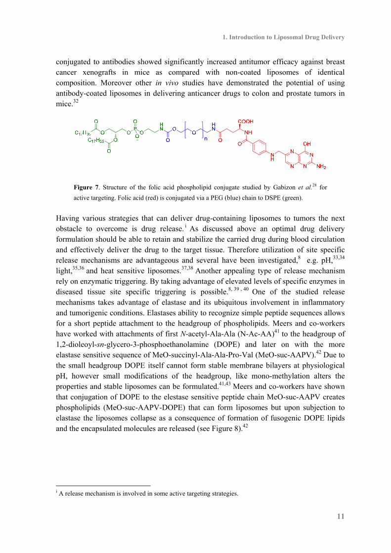
The use of passive targeting and hence the EPR effect is limited and cannot be applied to target all forms of cancers. Thus efforts have been directed towards the development of active approaches for drug targeting. This can be done by specific modification of the drug carrier system with agents or ligands having affinity for recognizing and/or interacting with specific cells, tissues or organs in the body. The approach has been explored targeting various receptors and antigens on cell membranes and an example is targeting of the folate receptor.27,28,29 The folate receptor is over-expressed in many cancer cells with epithelial origin and the receptor has been targeted with liposomes conjugated to folic acid. Folic acid and thereby also the drug-containing liposomes are taken up by the cells via receptor mediated endocytosis (see Figure 7 for the folic acid phospholipid conjugate studied by Gabizon et al.28). Another successful approach is conjugation of antibodies to liposomes and hence binding to specific surface proteins30 and in recent years, several in vivo studies have demonstrated the potential of using antibody-coated liposomes to increase drug accumulation in tumors. In a study by Park et al., 31 doxorubicin-loaded liposomes
1. Introduction to Liposomal Drug Delivery
11
conjugated to antibodies showed significantly increased antitumor efficacy against breast cancer xenografts in mice as compared with non-coated liposomes of identical composition. Moreover other in vivo studies have demonstrated the potential of using antibody-coated liposomes in delivering anticancer drugs to colon and prostate tumors in mice.32
Figure 7. Structure of the folic acid phospholipid conjugate studied by Gabizon et al.28 for
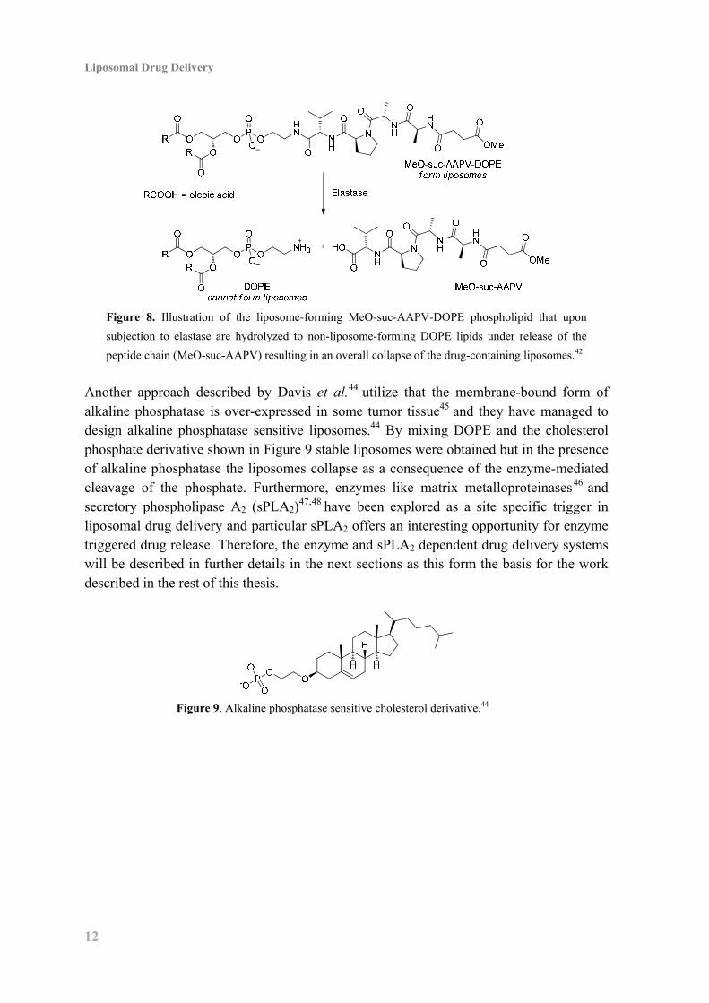
active targeting. Folic acid (red) is conjugated via a PEG (blue) chain to DSPE (green). Having various strategies that can deliver drug-containing liposomes to tumors the next obstacle to overcome is drug release. i As discussed above an optimal drug delivery formulation should be able to retain and stabilize the carried drug during blood circulation and effectively deliver the drug to the target tissue. Therefore utilization of site specific release mechanisms are advantageous and several have been investigated,8 e.g. pH,33,34 light,35,36 and heat sensitive liposomes.37,38 Another appealing type of release mechanism rely on enzymatic triggering. By taking advantage of elevated levels of specific enzymes in diseased tissue site specific triggering is possible.8, 39 , 40 One of the studied release mechanisms takes advantage of elastase and its ubiquitous involvement in inflammatory and tumorigenic conditions. Elastases ability to recognize simple peptide sequences allows for a short peptide attachment to the headgroup of phospholipids. Meers and co-workers have worked with attachments of first N-acetyl-Ala-Ala (N-Ac-AA)41 to the headgroup of 1,2-dioleoyl-sn-glycero-3-phosphoethanolamine (DOPE) and later on with the more elastase sensitive sequence of MeO-succinyl-Ala-Ala-Pro-Val (MeO-suc-AAPV).42 Due to the small headgroup DOPE itself cannot form stable membrane bilayers at physiological pH, however small modifications of the headgroup, like mono-methylation alters the properties and stable liposomes can be formulated.41,43 Meers and co-workers have shown that conjugation of DOPE to the elestase sensitive peptide chain MeO-suc-AAPV creates phospholipids (MeO-suc-AAPV-DOPE) that can form liposomes but upon subjection to elastase the liposomes collapse as a consequence of formation of fusogenic DOPE lipids and the encapsulated molecules are released (see Figure 8).42
i A release mechanism is involved in some active targeting strategies.
Liposomal Drug Delivery
12
Figure 8. Illustration of the liposome-forming MeO-suc-AAPV-DOPE phospholipid that upon
subjection to elastase are hydrolyzed to non-liposome-forming DOPE lipids under release of the
peptide chain (MeO-suc-AAPV) resulting in an overall collapse of the drug-containing liposomes.42
Another approach described by Davis et al.44 utilize that the membrane-bound form of alkaline phosphatase is over-expressed in some tumor tissue45 and they have managed to design alkaline phosphatase sensitive liposomes.44 By mixing DOPE and the cholesterol phosphate derivative shown in Figure 9 stable liposomes were obtained but in the presence of alkaline phosphatase the liposomes collapse as a consequence of the enzyme-mediated cleavage of the phosphate. Furthermore, enzymes like matrix metalloproteinases46 and secretory phospholipase A2 (sPLA2)
47,48 have been explored as a site specific trigger in liposomal drug delivery and particular sPLA2 offers an interesting opportunity for enzyme triggered drug release. Therefore, the enzyme and sPLA2 dependent drug delivery systems will be described in further details in the next sections as this form the basis for the work described in the rest of this thesis.
Figure 9. Alkaline phosphatase sensitive cholesterol derivative.44
1. Introduction to Liposomal Drug Delivery
13
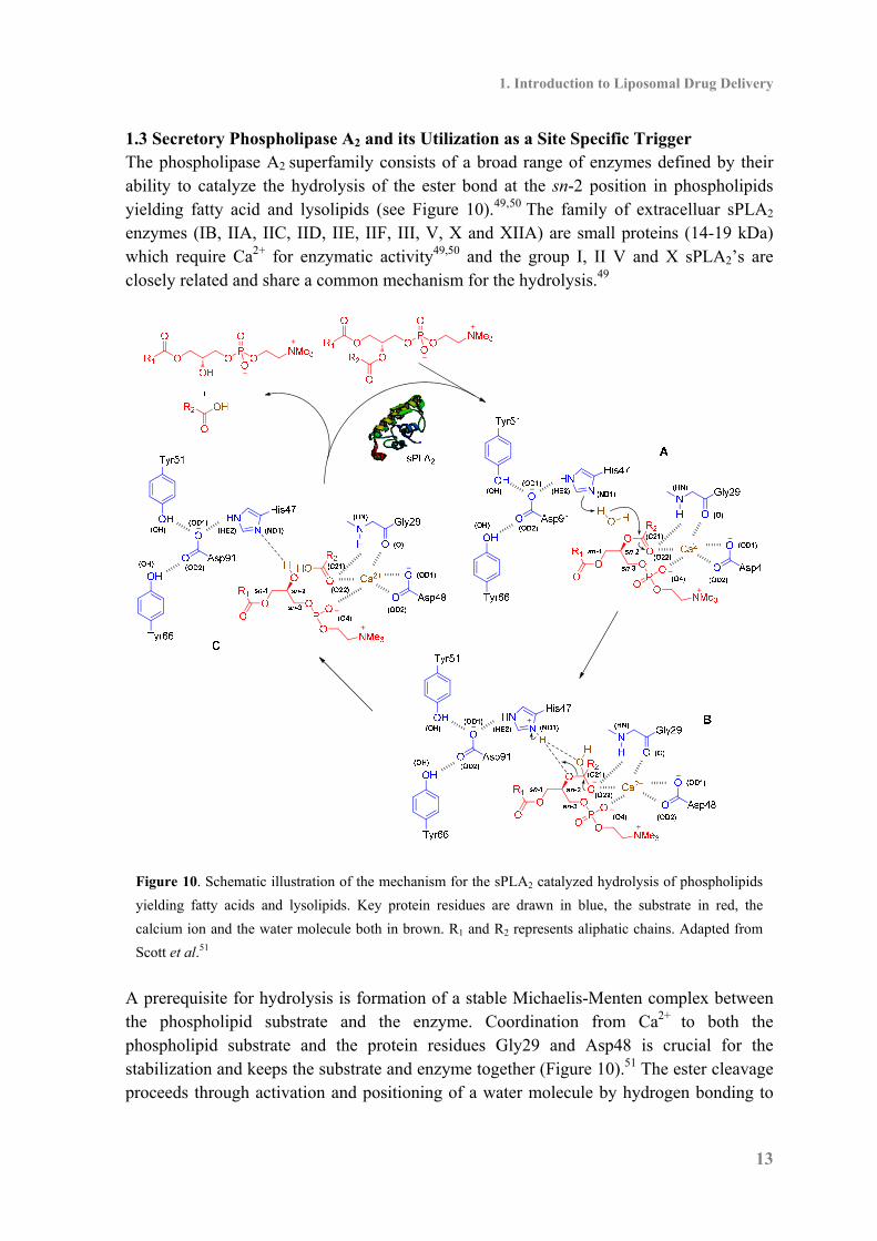
1.3 Secretory Phospholipase A2 and its Utilization as a Site Specific Trigger The phospholipase A2 superfamily consists of a broad range of enzymes defined by their ability to catalyze the hydrolysis of the ester bond at the sn-2 position in phospholipids yielding fatty acid and lysolipids (see Figure 10).49,50 The family of extracelluar sPLA2 enzymes (IB, IIA, IIC, IID, IIE, IIF, III, V, X and XIIA) are small proteins (14-19 kDa) which require Ca2+ for enzymatic activity49,50 and the group I, II V and X sPLA2’s are closely related and share a common mechanism for the hydrolysis.49
Figure 10. Schematic illustration of the mechanism for the sPLA2 catalyzed hydrolysis of phospholipids
yielding fatty acids and lysolipids. Key protein residues are drawn in blue, the substrate in red, the
calcium ion and the water molecule both in brown. R1 and R2 represents aliphatic chains. Adapted from
Scott et al.51 A prerequisite for hydrolysis is formation of a stable Michaelis-Menten complex between the phospholipid substrate and the enzyme. Coordination from Ca2+ to both the phospholipid substrate and the protein residues Gly29 and Asp48 is crucial for the stabilization and keeps the substrate and enzyme together (Figure 10).51 The ester cleavage proceeds through activation and positioning of a water molecule by hydrogen bonding to
Liposomal Drug Delivery
14
His47 assisted by Asp91 (Figure 10A). After abstraction of a hydrogen from the incoming water molecule a nucleophilic attack on the carbonyl in the sn-2 position occurs forming the intermediate (Figure 10B), regeneration of the carbonyl releases the lysolipid and the carboxylic acid by which the catalytic cycle is completed and the enzyme is ready for another substrate (Figure 10).51 The subtype of the enzyme family, sPLA2 IIA has been suggested as a therapeutic target for site specific triggering47,52 due to its over-expression in several human cancer tissues (Table 2)53,54 including breast, stomach, colorectal, pancreatic, prostate and liver cancer.
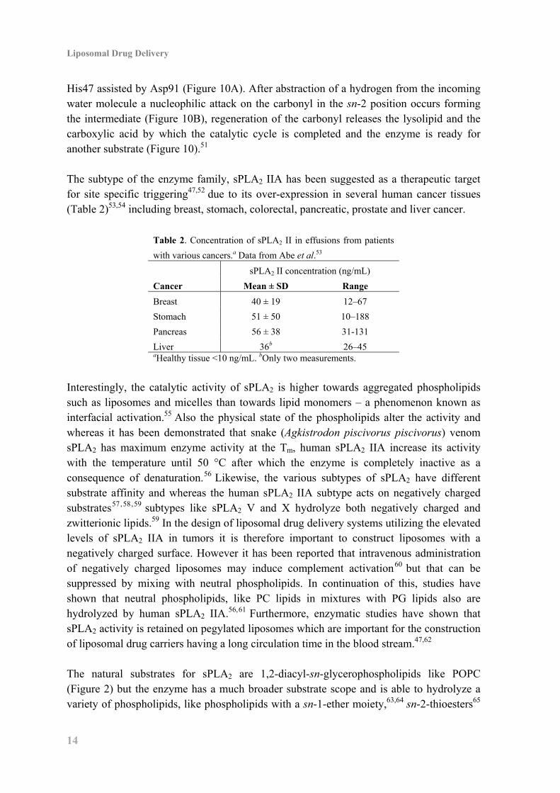
Table 2. Concentration of sPLA2 II in effusions from patients
with various cancers.a Data from Abe et al.53
sPLA2 II concentration (ng/mL)
Cancer Mean ± SD Range
Breast 40 ± 19 12–67
Stomach 51 ± 50 10–188
Pancreas 56 ± 38 31-131
Liver 36b 26–45 aHealthy tissue <10 ng/mL. bOnly two measurements.
Interestingly, the catalytic activity of sPLA2 is higher towards aggregated phospholipids such as liposomes and micelles than towards lipid monomers – a phenomenon known as interfacial activation.55 Also the physical state of the phospholipids alter the activity and whereas it has been demonstrated that snake (Agkistrodon piscivorus piscivorus) venom sPLA2 has maximum enzyme activity at the Tm, human sPLA2 IIA increase its activity with the temperature until 50 °C after which the enzyme is completely inactive as a consequence of denaturation.56 Likewise, the various subtypes of sPLA2 have different substrate affinity and whereas the human sPLA2 IIA subtype acts on negatively charged substrates57,58,59 subtypes like sPLA2 V and X hydrolyze both negatively charged and zwitterionic lipids.59 In the design of liposomal drug delivery systems utilizing the elevated levels of sPLA2 IIA in tumors it is therefore important to construct liposomes with a negatively charged surface. However it has been reported that intravenous administration of negatively charged liposomes may induce complement activation60 but that can be suppressed by mixing with neutral phospholipids. In continuation of this, studies have shown that neutral phospholipids, like PC lipids in mixtures with PG lipids also are hydrolyzed by human sPLA2 IIA.56,61 Furthermore, enzymatic studies have shown that sPLA2 activity is retained on pegylated liposomes which are important for the construction of liposomal drug carriers having a long circulation time in the blood stream.47,62 The natural substrates for sPLA2 are 1,2-diacyl-sn-glycerophospholipids like POPC (Figure 2) but the enzyme has a much broader substrate scope and is able to hydrolyze a variety of phospholipids, like phospholipids with a sn-1-ether moiety,63,64 sn-2-thioesters65
1. Introduction to Liposomal Drug Delivery
15
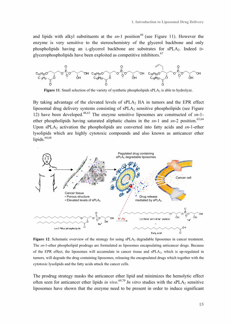
and lipids with alkyl substituents at the sn-1 position66 (see Figure 11). However the enzyme is very sensitive to the stereochemistry of the glycerol backbone and only phospholipids having an L-glycerol backbone are substrates for sPLA2. Indeed D-glycerophospholipids have been exploited as competitive inhibitors.67
Figure 11. Small selection of the variety of synthetic phospholipids sPLA2 is able to hydrolyze.
By taking advantage of the elevated levels of sPLA2 IIA in tumors and the EPR effect liposomal drug delivery systems consisting of sPLA2 sensitive phospholipids (see Figure 12) have been developed.48,63 The enzyme sensitive liposomes are constructed of sn-1-ether phospholipids having saturated aliphatic chains in the sn-1 and sn-2 position.63,64 Upon sPLA2 activation the phospholipids are converted into fatty acids and sn-1-ether lysolipids which are highly cytotoxic compounds and also known as anticancer ether lipids.64,68
Figure 12. Schematic overview of the strategy for using sPLA2 degradable liposomes in cancer treatment.
The sn-1-ether phospholipid prodrugs are formulated as liposomes encapsulating anticancer drugs. Because
of the EPR effect, the liposomes will accumulate in cancer tissue and sPLA2, which is up-regulated in
tumors, will degrade the drug containing liposomes, releasing the encapsulated drugs which together with the
cytotoxic lysolipids and the fatty acids attack the cancer cells.
The prodrug strategy masks the anticancer ether lipid and minimizes the hemolytic effect often seen for anticancer ether lipids in vivo.69,70 In vitro studies with the sPLA2 sensitive liposomes have shown that the enzyme need to be present in order to induce significant
Cancer tissue ▪ Porous structure ▪ Elevated levels of sPLA2
Drug release mediated by sPLA2
Cancer cell
Pegylated drug containing sPLA2 degradable liposomes
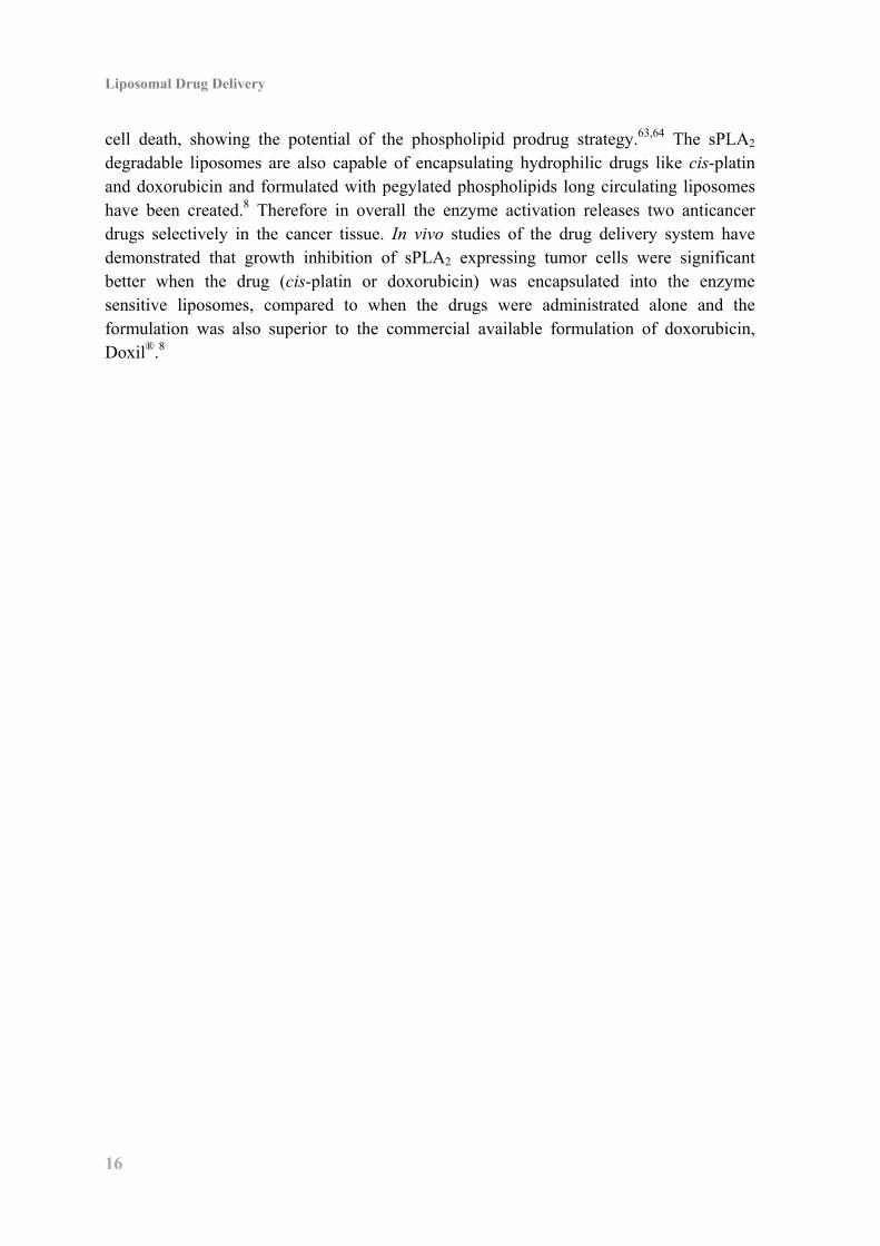
Liposomal Drug Delivery
16
cell death, showing the potential of the phospholipid prodrug strategy.63,64 The sPLA2 degradable liposomes are also capable of encapsulating hydrophilic drugs like cis-platin and doxorubicin and formulated with pegylated phospholipids long circulating liposomes have been created.8 Therefore in overall the enzyme activation releases two anticancer drugs selectively in the cancer tissue. In vivo studies of the drug delivery system have demonstrated that growth inhibition of sPLA2 expressing tumor cells were significant better when the drug (cis-platin or doxorubicin) was encapsulated into the enzyme sensitive liposomes, compared to when the drugs were administrated alone and the formulation was also superior to the commercial available formulation of doxorubicin, Doxil®.8
1. Introduction to Liposomal Drug Delivery
17
1.4 New Generation of Enzyme Sensitive Phospholipids Although liposomes are capable of delivering drugs to tumors a major problem in liposomal drug delivery is leakage of the encapsulated drugs. That can be lowered by addition of cholesterol to the liposomal formulation, but for drug delivery strategies relying on site specific drug release via sPLA2 activation it is desirable to avoid or minimize the addition of cholesterol, as cholesterol harms the enzyme activity and studies have revealed that liposomes having >20% cholesterol are non-degradable by sPLA2.
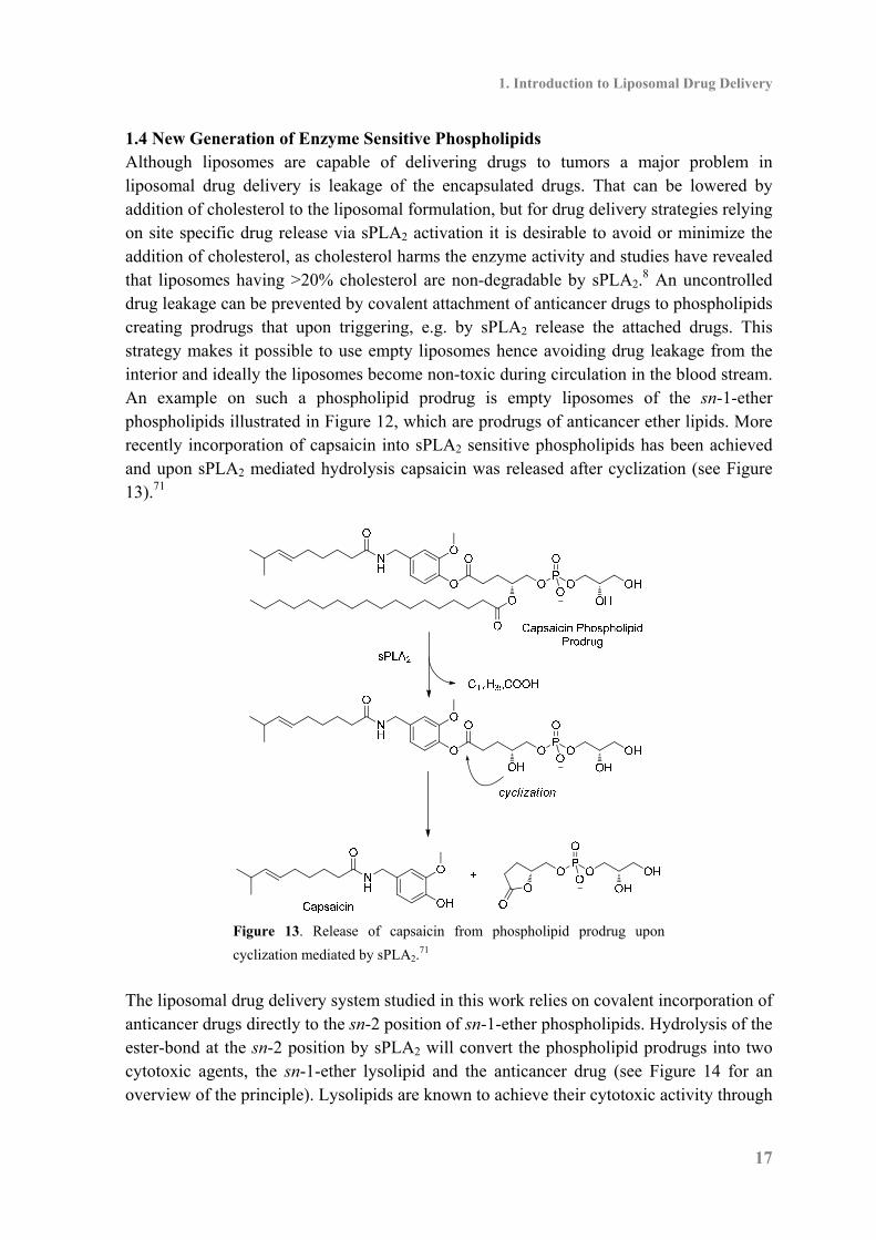
8 An uncontrolled drug leakage can be prevented by covalent attachment of anticancer drugs to phospholipids creating prodrugs that upon triggering, e.g. by sPLA2 release the attached drugs. This strategy makes it possible to use empty liposomes hence avoiding drug leakage from the interior and ideally the liposomes become non-toxic during circulation in the blood stream. An example on such a phospholipid prodrug is empty liposomes of the sn-1-ether phospholipids illustrated in Figure 12, which are prodrugs of anticancer ether lipids. More recently incorporation of capsaicin into sPLA2 sensitive phospholipids has been achieved and upon sPLA2 mediated hydrolysis capsaicin was released after cyclization (see Figure 13).71
Figure 13. Release of capsaicin from phospholipid prodrug upon
cyclization mediated by sPLA2.71
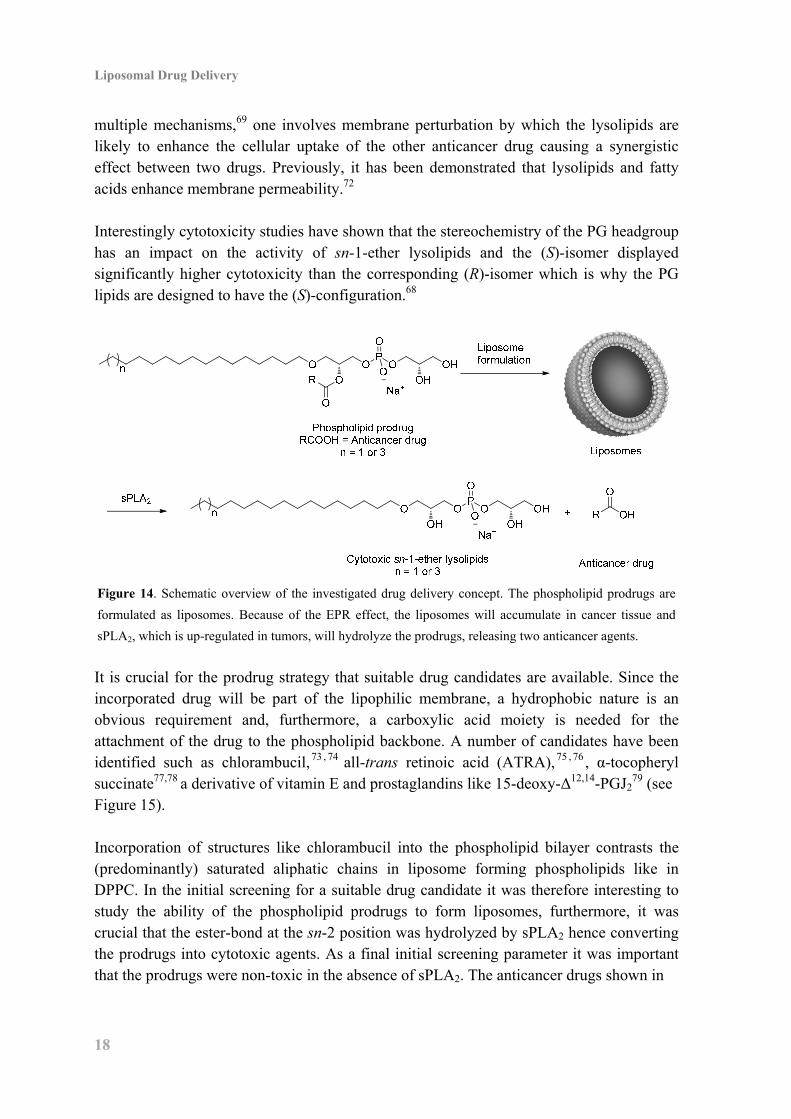
The liposomal drug delivery system studied in this work relies on covalent incorporation of anticancer drugs directly to the sn-2 position of sn-1-ether phospholipids. Hydrolysis of the ester-bond at the sn-2 position by sPLA2 will convert the phospholipid prodrugs into two cytotoxic agents, the sn-1-ether lysolipid and the anticancer drug (see Figure 14 for an overview of the principle). Lysolipids are known to achieve their cytotoxic activity through
Liposomal Drug Delivery
18
multiple mechanisms,69 one involves membrane perturbation by which the lysolipids are likely to enhance the cellular uptake of the other anticancer drug causing a synergistic effect between two drugs. Previously, it has been demonstrated that lysolipids and fatty acids enhance membrane permeability.72 Interestingly cytotoxicity studies have shown that the stereochemistry of the PG headgroup has an impact on the activity of sn-1-ether lysolipids and the (S)-isomer displayed significantly higher cytotoxicity than the corresponding (R)-isomer which is why the PG lipids are designed to have the (S)-configuration.68
Figure 14. Schematic overview of the investigated drug delivery concept. The phospholipid prodrugs are
formulated as liposomes. Because of the EPR effect, the liposomes will accumulate in cancer tissue and
sPLA2, which is up-regulated in tumors, will hydrolyze the prodrugs, releasing two anticancer agents.
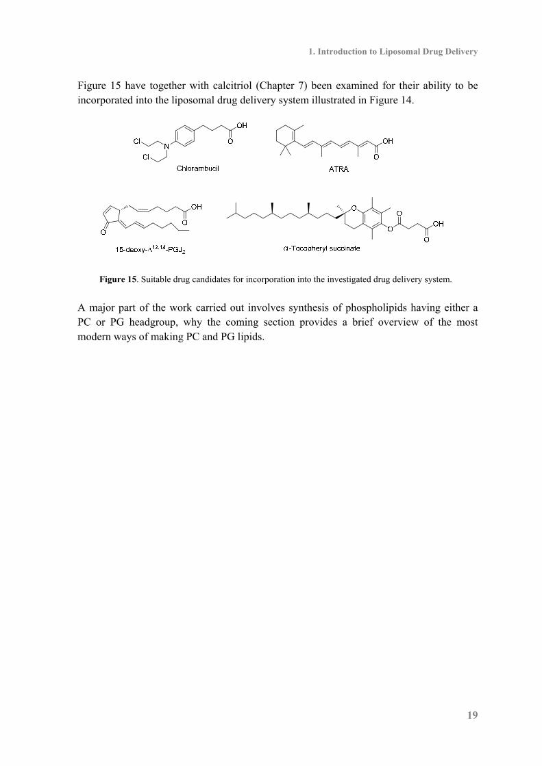
It is crucial for the prodrug strategy that suitable drug candidates are available. Since the incorporated drug will be part of the lipophilic membrane, a hydrophobic nature is an obvious requirement and, furthermore, a carboxylic acid moiety is needed for the attachment of the drug to the phospholipid backbone. A number of candidates have been identified such as chlorambucil,73 , 74 all-trans retinoic acid (ATRA),75 , 76 , α-tocopheryl succinate77,78 a derivative of vitamin E and prostaglandins like 15-deoxy-Δ12,14-PGJ2
79 (see Figure 15). Incorporation of structures like chlorambucil into the phospholipid bilayer contrasts the (predominantly) saturated aliphatic chains in liposome forming phospholipids like in DPPC. In the initial screening for a suitable drug candidate it was therefore interesting to study the ability of the phospholipid prodrugs to form liposomes, furthermore, it was crucial that the ester-bond at the sn-2 position was hydrolyzed by sPLA2 hence converting the prodrugs into cytotoxic agents. As a final initial screening parameter it was important that the prodrugs were non-toxic in the absence of sPLA2. The anticancer drugs shown in
1. Introduction to Liposomal Drug Delivery
19
Figure 15 have together with calcitriol (Chapter 7) been examined for their ability to be incorporated into the liposomal drug delivery system illustrated in Figure 14.
Figure 15. Suitable drug candidates for incorporation into the investigated drug delivery system.
A major part of the work carried out involves synthesis of phospholipids having either a PC or PG headgroup, why the coming section provides a brief overview of the most modern ways of making PC and PG lipids.

2. Phospholipid Synthesis – An Overview
21
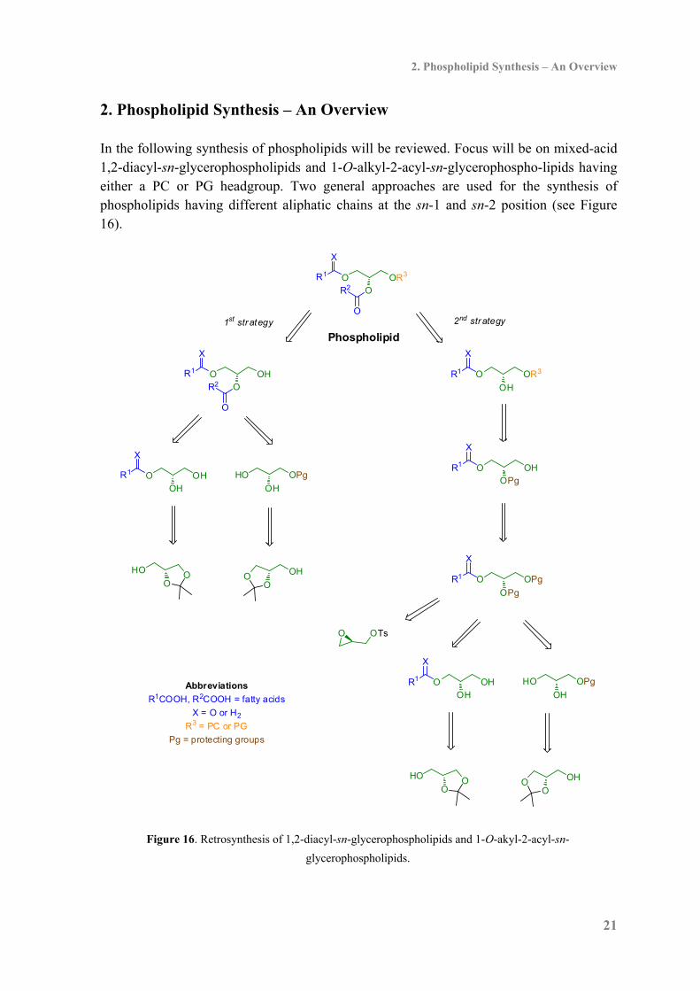
2. Phospholipid Synthesis – An Overview In the following synthesis of phospholipids will be reviewed. Focus will be on mixed-acid 1,2-diacyl-sn-glycerophospholipids and 1-O-alkyl-2-acyl-sn-glycerophospho-lipids having either a PC or PG headgroup. Two general approaches are used for the synthesis of phospholipids having different aliphatic chains at the sn-1 and sn-2 position (see Figure 16).
O
O
R2O OR3R1
X
O
O
R2O OHR1
X
OHO OHR1
X
OH
HO OPg
Abbreviations
R1COOH, R2COOH = fatty acids
X = O or H2
R3 = PC or PG
Pg = protecting groups
OO OH
O
HO O
OH
O OR3R1
X
OPg
O OPgR1
X
OHO OHR1
X
OH
HO OPg
OPgO OHR1
X
OO OH
O
HO O
O OTs
Phospholipid1st strategy 2nd strategy
Figure 16. Retrosynthesis of 1,2-diacyl-sn-glycerophospholipids and 1-O-akyl-2-acyl-sn-
glycerophospholipids.
Liposomal Drug Delivery
22
One strategy relies on installation of the aliphatic chains and then attachment of the phosphate headgroup (1st strategy in Figure 16) whereas in the second strategy the aliphatic chain at the sn-1 position and the phosphate headgroup at the sn-3 position are installed first and then acylation of the hydroxyl at the sn-2 position is achieved at a later stage (2nd strategy in Figure 16). Acyl-migration in unprotected glycerol derivatives is a major challenge in synthesizing phospholipids.80 The migrations are promoted by both base and acid meaning that the synthetic transformations have to be performed under mild conditions. Furthermore, as described in section 1.1 many fatty acids are unsaturated which must be considered when planning the synthesis. The commercially available compounds 1,2-isopropylidene-sn-glycerol and 2,3-isopropylidene-sn-glycerol serve as the most applied starting materials for the synthesis of 1,2-O-diacyl-glycerophospholipids. 1,2-Isopropylidene-sn-glycerol is made from D-mannitol81,82 whereas 2,3-isopropylidene-sn-glycerol can be made from L-erythrulose,83 L -serine82 or L-ascorbic acid.84 In addition to 2,3-isopropylidene-sn-glycerol, glycidols like (R)-glycidyl tosylate are used in the synthesis of sn-1-ether phospholipids. Glycidols can be made in high enantiomeric purity by asymmetric epoxidation of allyl alcohols.85

2. Phospholipid Synthesis – An Overview
23
2.1 Synthesis of 1,2-Diacyl-sn-glycerophospholipids Starting from 2,3-isopropylidene-sn-glycerol acylation of the sn-1 position has been performed using either carbodiimide mediated chemistry or via reaction with acid chlorides (Table 3). The removal of the isopropylidene protecting group without acyl-migration has been achieved under mild acidic conditions using acidic exchange resins, like Amberlyst-H+ (entry 3 in Table 3), however long reaction times (>24 h) have been necessary in order to obtain good yields. Furthermore, hydroxyl coordinating reagents like B(OH)3 in MeNO2 or trifluoroacetic acid (TFA) in B(OEt)3 has been efficient in the desired deprotection as well (entry 1 and 2 in Table 3).
Table 3. Synthesis of 1-acyl-sn-glycerol from 2,3-isopropylidene-sn-glycerol.
Entry R1COOH Reaction conditions
186 6-heptynoic acid (a) Ethyl-(3-dimethylaminopropyl)-carbodiimide
hydro chloride (EDCI), 4-dimethylaminopyridine
(DMAP), CH2Cl2 (97%); (b) B(OH)3, MeNO2 (84%).
287 Stearic acid (a) Stearoyl chloride, Et3N, DMAP, CH2Cl2; (b) TFA,
CF3CH2OH, B(OEt)3 (74%)
388 Stearic acid (a) EDCI, DMAP, CH2Cl2 (84%);
(b) Amberlyst-H+, MeOH (75%).
489 Linoleic acid (a) EDCI, DMAP, THF; (b) Dowex 50W, MeOHa aYield not reported.
Starting from 1,2-isopropylidene-sn-glycerol, protection of the sn-3 position is the first step. Various acid stabile protecting groups have been applied in the synthesis of 3-O-protected-sn-glycerols (Table 4) and among the used protecting groups are p-methoxybenzyl (PMB), tert-butyldiphenylsilyl (TBDPS), benzyl (Bn) and tosyl (Ts). The protecting groups are introduced under basic conditions and the following isopropylidene deprotection is achieved using acid catalysis, and having no ester-functionality acyl-migration is not an issue and strong acids are used, giving short reaction times and excellent yields (Table 4). In addition tetrahydropyranyl (THP, entry 3 in Table 4) is also successfully applied in this type of glycerol functionalization.

Liposomal Drug Delivery
24
Table 4. Synthesis of 3-O-protected-sn-glycerols from 1,2-isopropylidene-sn-glycerol.
Entry Pg Reaction conditions
190 PMB (a) PMBCl, KH, Bu4NI, THF; (b) TsOH, MeOH (98%).
291,92 PMB (a) PMBCl, NaH, DMF (90%); (b) AcOH, MeOH (99%).
393 THP (a) 3,4-dihydropyran (DHP), pyridine p-toluenesulfonate
(PPTS), CH2Cl2 (92%); (b) Bi(OTf)3, THF/H2O 4:1 (76%).
494 TBDPS (a) TBDPSCl, imidazole, THF; (b) HCl, H2O, EtOH (97%).
595 Bn (a) NaH, BnCl, DMSO (55%); (b) AcOH, H2O (95%).
696 Ts (a) TsCl, Pyridine (91%); (b) HCl, H2O, acetone (100%).
The 1-acyl-sn-glycerols (from Table 3) are by using appropriate protecting groups converted into 1,2-diacyl-sn-glycerols (Table 5). It is crucial that both the protecting of the sn-3 position and the final deprotection (step c in Table 5) can be achieved under conditions which avoid racemization. Dimethoxytrityl (DMT) has been used by Neff et al.86 however the group is so acid labile that chromatography through silica must be done with care or with addition of base to the eluent like Neff et al.86 have done. Hence the removal can be done under mild conditions and using B(OH)3 in MeNO2 the desired 1,2-diacyl-sn-glycerol is achieved (entry 1 in Table 5). Gaffney et al.87 have used the 9-(9-phenyl)xanthenyl (pixyl or Px) protecting group, prepared by mixing the glycerol derivative and PxOH in AcOH followed by evaporation in vacuo. Deprotection was done by treatment with a dilute solution of Cl2CHCOOH and pyrrole in CH2Cl2, which was tolerated by the unsaturated arachidonoyl moiety in the sn-2 position (entry 3 in Table 5). Silyl protecting groups like tert-butyldimethylsilyl (TBDMS) was ruled out by Dodd et al.97 as an applicable protecting group for glycerols due to acyl-migration upon treatment with fluorine sources like HF or tetrabutylammonium fluoride (TBAF). However Burgos et al. 98 have shown that applying the rather unusual deprotection conditions, N-bromosuccinimide (NBS) in DMSO and THF99 migration was avoided and the 1,2-diacyl-sn-glycerol was obtained and the enantiomeric purity was verified by Mosher ester analysis100 (entry 2 in Table 5). Recently Stamatov et al.101 have developed a two step procedure for the deprotection of silyl protected glycerols, in which the silyl ether (such as triisopropylsilyl, TIPS) is converted into the trichloroacetate via treatment with Et3N·3HF and trichloroacetic anhydride at 80 °C, then the trichloroacetate is hydrolyzed by pyridine in MeOH and THF affording the 1,2-diacyl-sn-glycerols (entry 5 and 6 in Table 5). A drawback in this method is the reaction temperature at 80 °C required for the trichloroacetate formation which limit the substrate scope, however an oleoyl moiety in both the sn-1 and sn-2 position has resist the conditions. Examination of other unsaturated fatty acids remains.

2. Phospholipid Synthesis – An Overview
25
Table 5. Synthesis of 1,2-diacyl-sn-glycerols from 1-acyl-sn-glycerols.
Entry R1COOH Pg R2COOH Reaction conditions
186 6-Heptynoic acid DMT Myristic acid (a) DMTCl, DMAP, pyridine (94%);
(b) myristic acid, EDCI, DMAP,
CH2Cl2 (92%); (c) B(OH)3, MeNO2
(81%).
298 Stearic acid TBDMS Stearic acid (a) TBDMSCl, imidazole, THF; (b)
C17H35COCl, pyridine (77%); NBS,
DMSO, THF.
387 Stearic acid Px Arachidonic acid (a) PxOH, AcOH (72%); (b)
arachidonic acid, 2,6-Cl2C6H3COCl,
1-methylimidazole, CH2Cl2; (c)
Cl2CHCOOH, pyrrole, CH2Cl2
(90%).
488 Stearic acid DMT Arachidonic acid (a) DMTCl, pyridine (96%); (b)
arachidonic acid, DCC, DMAP,
CH2Cl2 (100%); (c) TFA, pyrrole
(72%).
5101 Oleic acid TBDMS Acetic acid (a) TBDMSCl, imidazole, THF
(85%); (b) AcCl, pyridine (94%); (c)
(i) Et3N·3HF, (CCl3CO)2O; (ii)
pyridine, MeOH, THF (93%).
6101 Oleic acid TIPS Oleic acid (a) TIPSCl, imidazole, THF (80%);
(b) oleoyl chloride, pyridine (93%);
(c) (i) Et3N·3HF, (CCl3CO)2O (ii)
pyridine, MeOH, THF (92%).
Monoacylation of 3-O-protected-glycerols (from Table 4) have been achieved in moderate to good yields using a Steglich esterification102 (Table 6). Applying the coupling reagents in excess double-acylation has also been obtained (entry 2 in Table 6). Likewise attachment of the second fatty acid to the sn-2 position (step b in Table 6) have been achieved using the Steglich coupling or via reaction with acid chlorides. When double bonds are absent the benzyl protected strategy has been used and Martin et al.103 have removed the benzyl ether by hydrogenolysis to obtain pure 1,2-diacyl-sn-glycerols (entry 3 in Table 6). The PMB-group has efficiently been removed by 2,3-dichloro-5,6-dicyano-
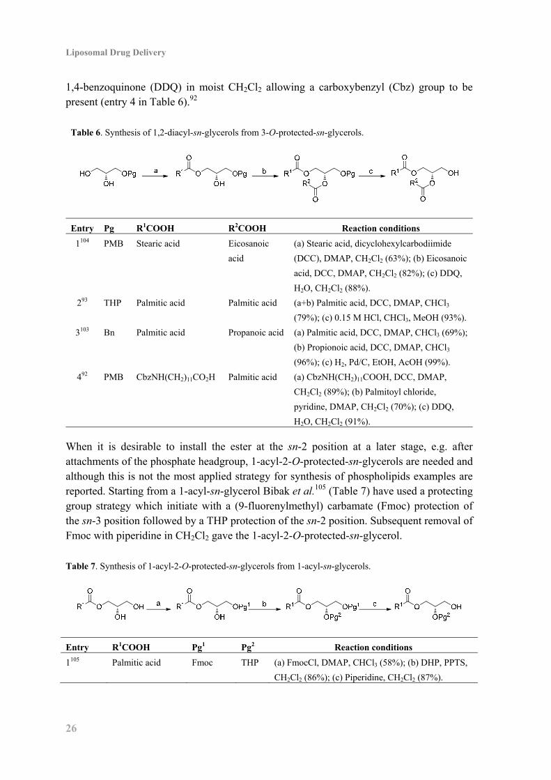
Liposomal Drug Delivery
26
1,4-benzoquinone (DDQ) in moist CH2Cl2 allowing a carboxybenzyl (Cbz) group to be present (entry 4 in Table 6).92
Table 6. Synthesis of 1,2-diacyl-sn-glycerols from 3-O-protected-sn-glycerols.
Entry Pg R1COOH R2COOH Reaction conditions
1104 PMB Stearic acid Eicosanoic
acid
(a) Stearic acid, dicyclohexylcarbodiimide
(DCC), DMAP, CH2Cl2 (63%); (b) Eicosanoic
acid, DCC, DMAP, CH2Cl2 (82%); (c) DDQ,
H2O, CH2Cl2 (88%).
293 THP Palmitic acid Palmitic acid (a+b) Palmitic acid, DCC, DMAP, CHCl3
(79%); (c) 0.15 M HCl, CHCl3, MeOH (93%).
3103 Bn Palmitic acid Propanoic acid (a) Palmitic acid, DCC, DMAP, CHCl3 (69%);
(b) Propionoic acid, DCC, DMAP, CHCl3
(96%); (c) H2, Pd/C, EtOH, AcOH (99%).
492 PMB CbzNH(CH2)11CO2H Palmitic acid (a) CbzNH(CH2)11COOH, DCC, DMAP,
CH2Cl2 (89%); (b) Palmitoyl chloride,
pyridine, DMAP, CH2Cl2 (70%); (c) DDQ,
H2O, CH2Cl2 (91%).
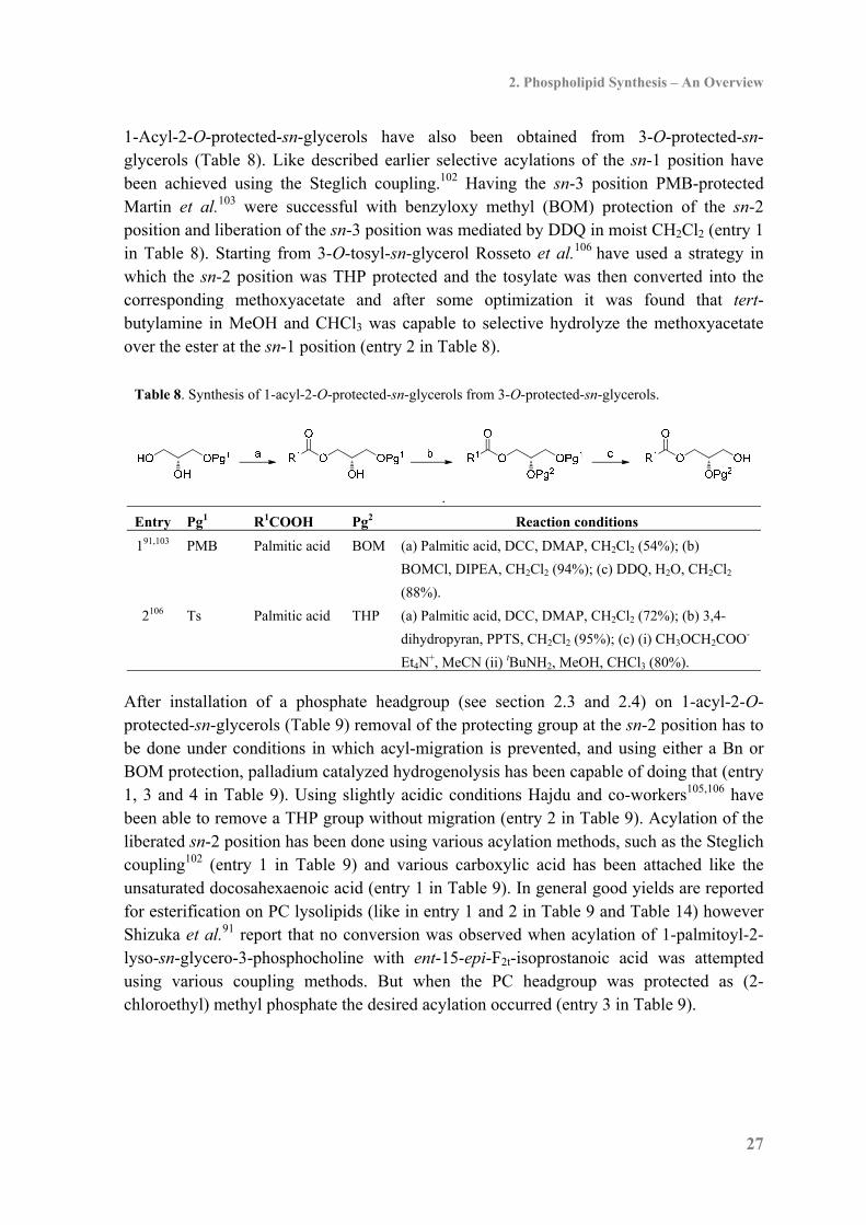
When it is desirable to install the ester at the sn-2 position at a later stage, e.g. after attachments of the phosphate headgroup, 1-acyl-2-O-protected-sn-glycerols are needed and although this is not the most applied strategy for synthesis of phospholipids examples are reported. Starting from a 1-acyl-sn-glycerol Bibak et al.105 (Table 7) have used a protecting group strategy which initiate with a (9-fluorenylmethyl) carbamate (Fmoc) protection of the sn-3 position followed by a THP protection of the sn-2 position. Subsequent removal of Fmoc with piperidine in CH2Cl2 gave the 1-acyl-2-O-protected-sn-glycerol. Table 7. Synthesis of 1-acyl-2-O-protected-sn-glycerols from 1-acyl-sn-glycerols.
Entry R1COOH Pg1 Pg2 Reaction conditions
1105 Palmitic acid Fmoc THP (a) FmocCl, DMAP, CHCl3 (58%); (b) DHP, PPTS,
CH2Cl2 (86%); (c) Piperidine, CH2Cl2 (87%).
2. Phospholipid Synthesis – An Overview
27
1-Acyl-2-O-protected-sn-glycerols have also been obtained from 3-O-protected-sn-glycerols (Table 8). Like described earlier selective acylations of the sn-1 position have been achieved using the Steglich coupling.102 Having the sn-3 position PMB-protected Martin et al.103 were successful with benzyloxy methyl (BOM) protection of the sn-2 position and liberation of the sn-3 position was mediated by DDQ in moist CH2Cl2 (entry 1 in Table 8). Starting from 3-O-tosyl-sn-glycerol Rosseto et al.106 have used a strategy in which the sn-2 position was THP protected and the tosylate was then converted into the corresponding methoxyacetate and after some optimization it was found that tert-butylamine in MeOH and CHCl3 was capable to selective hydrolyze the methoxyacetate over the ester at the sn-1 position (entry 2 in Table 8).
Table 8. Synthesis of 1-acyl-2-O-protected-sn-glycerols from 3-O-protected-sn-glycerols.
.
Entry Pg1 R1COOH Pg2 Reaction conditions
191,103 PMB Palmitic acid BOM (a) Palmitic acid, DCC, DMAP, CH2Cl2 (54%); (b)
BOMCl, DIPEA, CH2Cl2 (94%); (c) DDQ, H2O, CH2Cl2
(88%).
2106 Ts Palmitic acid THP (a) Palmitic acid, DCC, DMAP, CH2Cl2 (72%); (b) 3,4-
dihydropyran, PPTS, CH2Cl2 (95%); (c) (i) CH3OCH2COO-
Et4N+, MeCN (ii) tBuNH2, MeOH, CHCl3 (80%).
After installation of a phosphate headgroup (see section 2.3 and 2.4) on 1-acyl-2-O-protected-sn-glycerols (Table 9) removal of the protecting group at the sn-2 position has to be done under conditions in which acyl-migration is prevented, and using either a Bn or BOM protection, palladium catalyzed hydrogenolysis has been capable of doing that (entry 1, 3 and 4 in Table 9). Using slightly acidic conditions Hajdu and co-workers105,106 have been able to remove a THP group without migration (entry 2 in Table 9). Acylation of the liberated sn-2 position has been done using various acylation methods, such as the Steglich coupling102 (entry 1 in Table 9) and various carboxylic acid has been attached like the unsaturated docosahexaenoic acid (entry 1 in Table 9). In general good yields are reported for esterification on PC lysolipids (like in entry 1 and 2 in Table 9 and Table 14) however Shizuka et al.91 report that no conversion was observed when acylation of 1-palmitoyl-2-lyso-sn-glycero-3-phosphocholine with ent-15-epi-F2t-isoprostanoic acid was attempted using various coupling methods. But when the PC headgroup was protected as (2-chloroethyl) methyl phosphate the desired acylation occurred (entry 3 in Table 9).
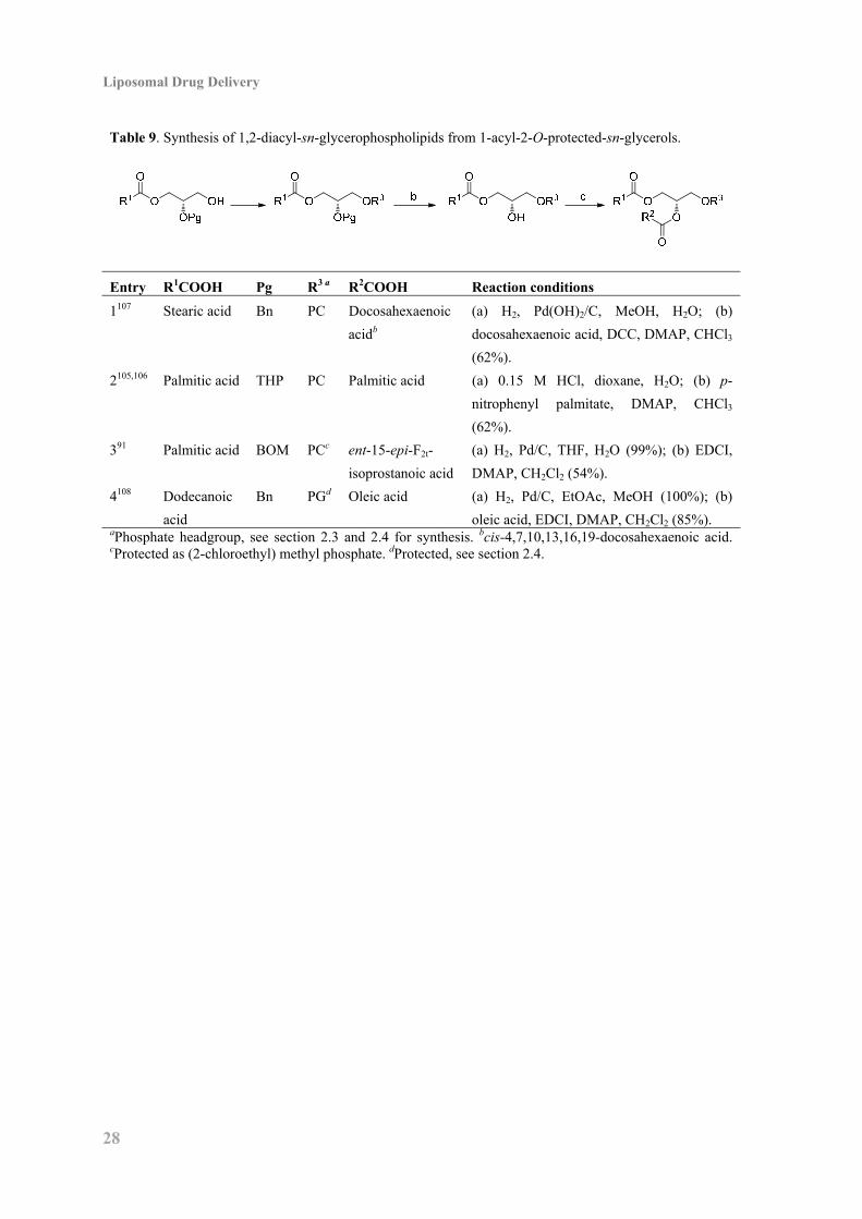
Liposomal Drug Delivery
28
Table 9. Synthesis of 1,2-diacyl-sn-glycerophospholipids from 1-acyl-2-O-protected-sn-glycerols.
Entry R1COOH Pg R3 a R2COOH Reaction conditions
1107 Stearic acid Bn PC Docosahexaenoic
acidb
(a) H2, Pd(OH)2/C, MeOH, H2O; (b)
docosahexaenoic acid, DCC, DMAP, CHCl3
(62%).
2105,106 Palmitic acid THP PC Palmitic acid (a) 0.15 M HCl, dioxane, H2O; (b) p-
nitrophenyl palmitate, DMAP, CHCl3
(62%).
391 Palmitic acid BOM PCc ent-15-epi-F2t-
isoprostanoic acid
(a) H2, Pd/C, THF, H2O (99%); (b) EDCI,
DMAP, CH2Cl2 (54%).
4108 Dodecanoic
acid
Bn PGd Oleic acid (a) H2, Pd/C, EtOAc, MeOH (100%); (b)
oleic acid, EDCI, DMAP, CH2Cl2 (85%). aPhosphate headgroup, see section 2.3 and 2.4 for synthesis. bcis-4,7,10,13,16,19-docosahexaenoic acid. cProtected as (2-chloroethyl) methyl phosphate. dProtected, see section 2.4.
2. Phospholipid Synthesis – An Overview
29
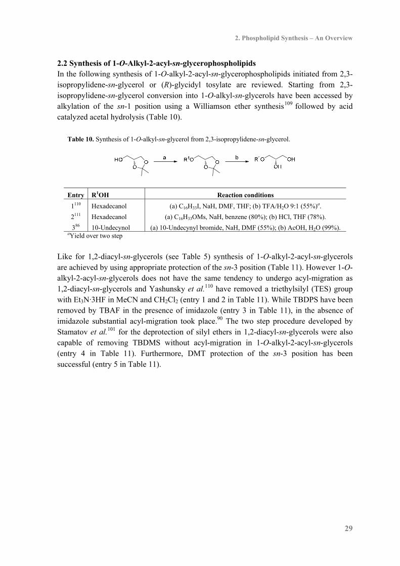
2.2 Synthesis of 1-O-Alkyl-2-acyl-sn-glycerophospholipids In the following synthesis of 1-O-alkyl-2-acyl-sn-glycerophospholipids initiated from 2,3-isopropylidene-sn-glycerol or (R)-glycidyl tosylate are reviewed. Starting from 2,3-isopropylidene-sn-glycerol conversion into 1-O-alkyl-sn-glycerols have been accessed by alkylation of the sn-1 position using a Williamson ether synthesis109 followed by acid catalyzed acetal hydrolysis (Table 10).
Table 10. Synthesis of 1-O-alkyl-sn-glycerol from 2,3-isopropylidene-sn-glycerol.
Entry R1OH Reaction conditions
1110 Hexadecanol (a) C16H33I, NaH, DMF, THF; (b) TFA/H2O 9:1 (55%)a.
2111 Hexadecanol (a) C16H33OMs, NaH, benzene (80%); (b) HCl, THF (78%).
386 10-Undecynol (a) 10-Undecynyl bromide, NaH, DMF (55%); (b) AcOH, H2O (99%). aYield over two step
Like for 1,2-diacyl-sn-glycerols (see Table 5) synthesis of 1-O-alkyl-2-acyl-sn-glycerols are achieved by using appropriate protection of the sn-3 position (Table 11). However 1-O-alkyl-2-acyl-sn-glycerols does not have the same tendency to undergo acyl-migration as 1,2-diacyl-sn-glycerols and Yashunsky et al.110 have removed a triethylsilyl (TES) group with Et3N·3HF in MeCN and CH2Cl2 (entry 1 and 2 in Table 11). While TBDPS have been removed by TBAF in the presence of imidazole (entry 3 in Table 11), in the absence of imidazole substantial acyl-migration took place.90 The two step procedure developed by Stamatov et al.101 for the deprotection of silyl ethers in 1,2-diacyl-sn-glycerols were also capable of removing TBDMS without acyl-migration in 1-O-alkyl-2-acyl-sn-glycerols (entry 4 in Table 11). Furthermore, DMT protection of the sn-3 position has been successful (entry 5 in Table 11).
Liposomal Drug Delivery
30
Table 11. Synthesis of 1-O-alkyl-2-acyl-sn-glycerols from 1-O-alkyl-sn-glycerols.
Entry R1OH Pg R2COOH Reaction conditions
1110 Hexadecanol TES Oleic acid (a) TESCl, pyridine, CH2Cl2; (b) Oleoyl
chloride, Et3N, DMAP, pyridine; (c)
Et3N·3HF, MeCN, CH2Cl2 (58%)c.
2110 Hexadecanol TES Linoleic acid (a) TESCl, pyridine, CH2Cl2; (b)
Linoleoyl chloride, Et3N, DMAP,
pyridine; (c) Et3N·3HF, MeCN, CH2Cl2
(58%)a.
390,112 (Z)-1-Octadecenol TBDPS Palmitic acid (a) TBDPSCl, imidazole, DMF (90%);
(b) (C15H31CO)2O, DMAP, CHCl3 (98%);
(c) TBAF, imidazole, THF (>74%).
4101 Hexadecanol TBDMS AcOH (a) TBDMSCl, imidazole, THF (85%);
(b) AcCl, pyridine, CH2Cl2 (95%); (c) (i)
Et3N·3HF, (CCl3CO)2O (ii) pyridine,
MeOH, THF (92%).
586 10-Undecynol DMT Myristic acid (a) DMTCl, DMAP, pyridine (94%); (b)
Myristic acid, EDCI, DMAP, CH2Cl2
(99%); (c) B(OH)3, MeNO2 (81%). aYield over three steps.
Another convenient route used for the introduction of the sn-1-ether functionality is Lewis acid catalyzed epoxide opening of glycidols like (R)-glycidyl tosylate with alcohols (Table 12). Guivisdalsky et al.114 discovered that BF3·OEt2 mediated the desired epoxide opening with excellent regioselectivity and high enantiomeric excess, whereas other Lewis acid like Ti(OiPr)4 and SnCl4 gave much lower conversion. Furthermore, Guivisdalsky et al.114 demonstrated that while epoxide opening of (R)-glycidyl tosylate with hexadecanol exclusively gave the sn-1-ether, epoxide opening of the corresponding TBDPS glycidol resulted in a 9:1 ratio between the desired sn-1-ether and the undesired sn-2-ether. Hence Andresen et al.68 have synthesized a collection of different sn-1-ether phsopholipids from (R)-glycidyl tosylate. Having the sn-1-ether functionality introduced synthesis of 1-O-alkyl-2-O-protected-sn-glycerols has been achieved using either a Bn or THP protection of the sn-2 position. Bn protection has been performed under both basic conditions (entry 1 in Table 12) and acidic conditions using benzyl trichloroacetimidate (entry 2 in Table 12). Removal of the tosyl group has been achieved by converting the tosylate into the acetate, which then either has been reduced with LiAlH4 (entry 1 in Table 12) or hydrolyzed (entry 3 in Table 12). Applying NaNO2 Andresen et al.68 have obtained the alcohol in one step
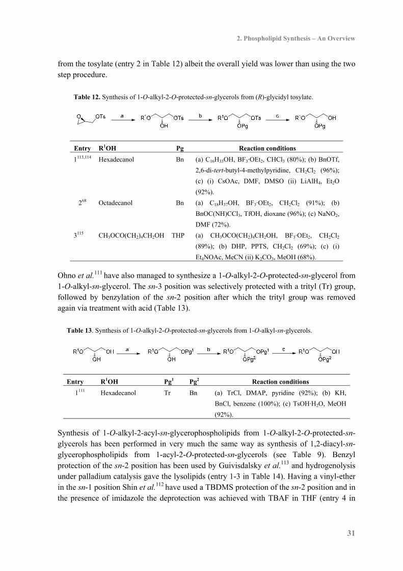
2. Phospholipid Synthesis – An Overview
31
from the tosylate (entry 2 in Table 12) albeit the overall yield was lower than using the two step procedure.
Table 12. Synthesis of 1-O-alkyl-2-O-protected-sn-glycerols from (R)-glycidyl tosylate.
Entry R1OH Pg Reaction conditions
1113,114 Hexadecanol Bn (a) C16H33OH, BF3·OEt2, CHCl3 (80%); (b) BnOTf,
2,6-di-tert-butyl-4-methylpyridine, CH2Cl2 (96%);
(c) (i) CsOAc, DMF, DMSO (ii) LiAlH4, Et2O
(92%).
268 Octadecanol Bn (a) C18H37OH, BF3·OEt2, CH2Cl2 (91%); (b)
BnOC(NH)CCl3, TfOH, dioxane (96%); (c) NaNO2,
DMF (72%).
3115 CH3OCO(CH2)4CH2OH THP (a) CH3OCO(CH2)4CH2OH, BF3·OEt2, CH2Cl2
(89%); (b) DHP, PPTS, CH2Cl2 (69%); (c) (i)
Et4NOAc, MeCN (ii) K2CO3, MeOH (68%).
Ohno et al.111 have also managed to synthesize a 1-O-alkyl-2-O-protected-sn-glycerol from 1-O-alkyl-sn-glycerol. The sn-3 position was selectively protected with a trityl (Tr) group, followed by benzylation of the sn-2 position after which the trityl group was removed again via treatment with acid (Table 13).
Table 13. Synthesis of 1-O-alkyl-2-O-protected-sn-glycerols from 1-O-alkyl-sn-glycerols.
Entry R1OH Pg1 Pg2 Reaction conditions
1111 Hexadecanol Tr Bn (a) TrCl, DMAP, pyridine (92%); (b) KH,
BnCl, benzene (100%); (c) TsOH·H2O, MeOH
(92%).
Synthesis of 1-O-alkyl-2-acyl-sn-glycerophospholipids from 1-O-alkyl-2-O-protected-sn-glycerols has been performed in very much the same way as synthesis of 1,2-diacyl-sn-glycerophospholipids from 1-acyl-2-O-protected-sn-glycerols (see Table 9). Benzyl protection of the sn-2 position has been used by Guivisdalsky et al.113 and hydrogenolysis under palladium catalysis gave the lysolipids (entry 1-3 in Table 14). Having a vinyl-ether in the sn-1 position Shin et al.112 have used a TBDMS protection of the sn-2 position and in the presence of imidazole the deprotection was achieved with TBAF in THF (entry 4 in

Liposomal Drug Delivery
32
Table 14). Interestingly, the following acylation of the sn-2 position has only been reported via reaction with anhydrides (entry 1,2 and 4 in Table 14) albeit in good yields. Table 14. Synthesis of 1-O-alkyl-2-acyl-sn-glycerophospholipids from 1-O-alkyl-2-O-protected-sn-
glycerols.
Entry R1OH Pg R3 a R2COOH Reaction conditions
1113 Hexadecanol Bn PC Palmitic acid (a) H2, Pd(OH)2/C, MeOH, H2O (100%);
(b) (C15H31CO)2O, DMAP, CHCl3 (98%)
268,111 Octadecanol Bn PC AcOH (a) H2, Pd/C, MeOH (>77%); (b) Ac2O,
pyridine (93%).
368 Octadecanol Bn PGb (a) H2, Pd/C, MeOH (>67%).
4112 (Z)-1-Hexadecenol TBDMS PC Palmitic acid (a) TBAF, imidazole, THF (98%); (b)
(C15 H31CO)2O, DMAP, CH2Cl2 (53%). aPhosphate headgroup. bProtected, see section 2.4.
2. Phospholipid Synthesis – An Overview
33
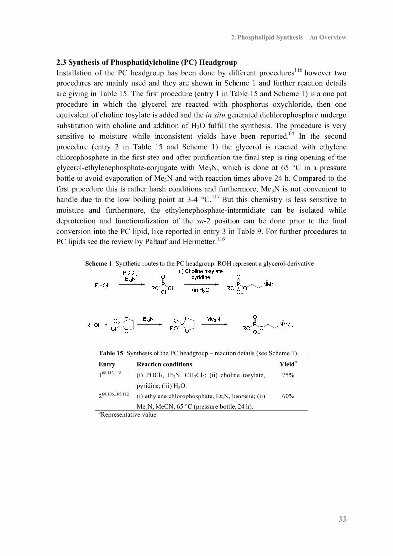
2.3 Synthesis of Phosphatidylcholine (PC) Headgroup Installation of the PC headgroup has been done by different procedures116 however two procedures are mainly used and they are shown in Scheme 1 and further reaction details are giving in Table 15. The first procedure (entry 1 in Table 15 and Scheme 1) is a one pot procedure in which the glycerol are reacted with phosphorus oxychloride, then one equivalent of choline tosylate is added and the in situ generated dichlorophosphate undergo substitution with choline and addition of H2O fulfill the synthesis. The procedure is very sensitive to moisture while inconsistent yields have been reported.64 In the second procedure (entry 2 in Table 15 and Scheme 1) the glycerol is reacted with ethylene chlorophosphate in the first step and after purification the final step is ring opening of the glycerol-ethylenephosphate-conjugate with Me3N, which is done at 65 °C in a pressure bottle to avoid evaporation of Me3N and with reaction times above 24 h. Compared to the first procedure this is rather harsh conditions and furthermore, Me3N is not convenient to handle due to the low boiling point at 3-4 °C.117 But this chemistry is less sensitive to moisture and furthermore, the ethylenephosphate-intermidiate can be isolated while deprotection and functionalization of the sn-2 position can be done prior to the final conversion into the PC lipid, like reported in entry 3 in Table 9. For further procedures to PC lipids see the review by Paltauf and Hermetter.116
Scheme 1. Synthetic routes to the PC headgroup. ROH represent a glycerol-derivative
Table 15. Synthesis of the PC headgroup – reaction details (see Scheme 1).
Entry Reaction conditions Yielda
168,113,118 (i) POCl3, Et3N, CH2Cl2; (ii) choline tosylate,
pyridine; (iii) H2O.
75%
268,106,105,112 (i) ethylene chlorophosphate, Et3N, benzene; (ii)
Me3N, MeCN, 65 °C (pressure bottle, 24 h).
60%
aRepresentative value
Liposomal Drug Delivery
34
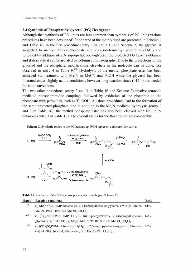
2.4 Synthesis of Phosphatidylglycerol (PG) Headgroup Although that synthesis of PG lipids are less common than synthesis of PC lipids various procedures have been developed116 and three of the mainly used are presented in Scheme 2 and Table 16. In the first procedure (entry 1 in Table 16 and Scheme 2) the glycerol is subjected to methyl dichlorophosphate and 2,2,6,6-tetramethyl piperidine (TMP) and followed by addition of 2,3-isopropylidene-sn-glycerol the protected PG lipid is obtained and if desirable it can be isolated by column chromatography. Due to the protections of the glycerol and the phosphate, modifications elsewhere in the molecule can be done, like observed in entry 4 in Table 9.108 Hydrolysis of the methyl phosphate ester has been achieved via treatment with Me3N in MeCN and iPrOH while the glycerol has been liberated under slightly acidic conditions, however long reaction times (>14 h) are needed for both conversions. The two other procedures (entry 2 and 3 in Table 16 and Scheme 2) involve tetrazole mediated phosphoramidite couplings followed by oxidation of the phosphite to the phosphate with peroxides, such as tBuOOH. All three procedures lead to the formation of the same protected phosphate, and in addition to the Me3N mediated hydrolysis (entry 2 and 3 in Table 16), the methyl phosphate ester has also been cleaved with NaI in 2-butanone (entry 3 in Table 16). The overall yields for the three routes are comparable.
Scheme 2. Synthetic routes to the PG headgroup. ROH represent a glycerol-derivative.
Table 16. Synthesis of the PG headgroup – reaction details (see Scheme 2).
Entry Reaction conditions Yield
168 (i) MeOPOCl2, TMP, toluene; (ii) 2,3-isopropylidene-sn-glycerol, TMP; (iii) Me3N,
MeCN, iPrOH; (iv) HCl, MeOH, CH2Cl2.
61%
268 (i) (iPr)2NPClOMe, TMP, CH2Cl2; (ii) 5-phenyltetrazole, 2,3-isopropylidene-sn-
glycerol; (iii) tBuOOH; (iv) Me3N, MeCN, iPrOH; (v) HCl, MeOH, CH2Cl2.
67%
3108 (i) ((iPr)2N)2POMe, tetrazole, CH2Cl2; (ii) 2,3-isopropylidene-sn-glycerol, tetrazole;
(iii) mCPBA; (iv) NaI, 2-butanone; (v) TFA, MeOH, CH2Cl2.
55%
3. Chlorambucil Phospholipid Prodrugs
35
3. Chlorambucil Phospholipid Prodrugs 3.1 Introduction To demonstrate proof-of-principle chlorambucil was selected for incorporation into the investigated liposomal drug delivery system. Chlorambucil is a chemotherapeutic agent of the mustard gas type119 and it was originally synthesized by Everett et al. in 1953.73 It is used clinically for the treatment of lymphocytic leukemia120 typically in combination with other drugs. Chlorambucil is orally administrated (Leukeran®), but undergoes rapid metabolism, and as a result the stability in aqueous environments is low and chlorambucil has an elimination half-life of 1.5 h.121,122,123 The prodrug formulation could remedy this, since this system will shield chlorambucil from degradation through the incorporation in the lipophilic part of the liposomal membrane and deliver it directly to the tumor, decreasing metabolism compared to the oral administration route. To investigate the effect of sn-1 ether chain length and headgroup charge on enzymatic activity, prodrugs 1 and 2 (Figure 17) were prepared with both C16 and C18 ether chains and a PC and PG headgroup, respectively.
Figure 17. The four target chlorambucil sn-1-ether phospholipid prodrugs. Prodrugs
1a and 1b have a PC headgroup with a C16 and a C18 ether chain respectively. Target
compounds 2a and 2b have the negatively charged PG headgroup.
3.2 Synthesis of Chlorambucil Phospholipid Prodrugs The synthesis of sn-1-ether phospholipids have, as described in section 2.2, been accessed via different routes, starting from 2,3-isopropylidene-sn-glycerol (see Table 10) or (R)-glycidyl tosylate (see Table 12). (R)-Glycidyl tosylate ((–)-3) served as the starting material in this work and the aliphatic ether chain was introduced by ring-opening of the epoxide under Lewis acid catalysis,114 resulting in yields of 89% and 97% for 4a and 4b, respectively (Scheme 3). The PMB group was chosen for protection of the secondary alcohol and introduced by using para-methoxybenzyl trichloroacetimidate (PMBTCA) with La(OTf)3 catalysis.124,125 The resulting tosylate was converted into the acetate with CsOAc in a 9:1 mixture of DMSO and DMF and the ester was hydrolyzed with NaOMe in

Liposomal Drug Delivery
36
MeOH at 40 °C yielding the primary alcohols 5a and 5b in overall yields of 70% and 78% respectively over 3 steps (Scheme 3). It was essential to carry out the hydrolysis at elevated temperature in order to obtain homogeneous reaction mixtures and achieve full conversion in the transformations. Inspired by Andresen et al.68 a direct conversion of the tosylates to the alcohols 5a and 5b using NaNO2 in DMSO was attempted and yields up to 94% over two steps (including the PMB-protection) were achieved, but the result was not reproducible and inconsistent yields from 60% to 94% were obtained. The corresponding carboxylic acid was observed as the major by-product in the experiments giving low yields and since there is precedence for oxidation of alcohols in aqueous environments in the presence of HNO2 and oxygen126,127 it is likely that the oxidation occurred during the aqueous workup. It was attempted to circumvent the undesired oxidation by applying a basic workup, but that did not improve the yield of the desired alcohol.
Scheme 3. Synthesis of phospholipid precursor 5a and 5b.a
aReagents: (a) C16H33OH or C18H37OH, BF3·OEt2, CH2Cl2; (b) (i) PMBTCA, La(OTf)3, toluene; (ii)
CsOAc, DMSO, DMF; (iii) NaOMe, MeOH; (c) (i) PMBTCA, La(OTf)3, toluene; (ii) NaNO2, DMSO.
The PC headgroup was attached to the primary alcohols 5a and 5b by reaction with phosphorus oxychloride and Et3N, followed by addition of choline tosylate, pyridine and finally H2O (Scheme 4 and compare with Table 15). Excess choline tosylate was removed on an MB-3 ion-exchange column and after purification by column chromatography the PMB-group was removed by hydrogenolysis mediated by Pd/C in a quantitative yield, but disappointingly upon scaleup a slow conversion was observed. Instead DDQ in an 18:1 mixture of CH2Cl2 and H2O, 128 was attempted and these conditions resulted in full conversion within 3 h and with satisfactory isolated yields of 6 (Scheme 4). The final attachment of chlorambucil to the phospholipid backbone was achieved via a Steglich esterification102 with DCC and a catalytic amount of DMAP. When the acylation of 6a was performed in ethanol-free chloroform at 20 °C or CH2Cl2 in the temperature range from 0 °C to reflux no incorporation of chlorambucil was observed, but when the conditions were changed to reflux in 1,2-dichloroethane the acylation occurred in a 75% yield, albeit only after adding 5 equivalents of chlorambucil and DCC in portions over 31 h. The acylation of 6b led to an isolated yield of 76% in refluxing chloroform and that was not improved by using 1,2-dichlorethane as the solvent. Changing the coupling reagents to EDCI and DMAP129 did not improve the conversion of the 6. In continuation of this
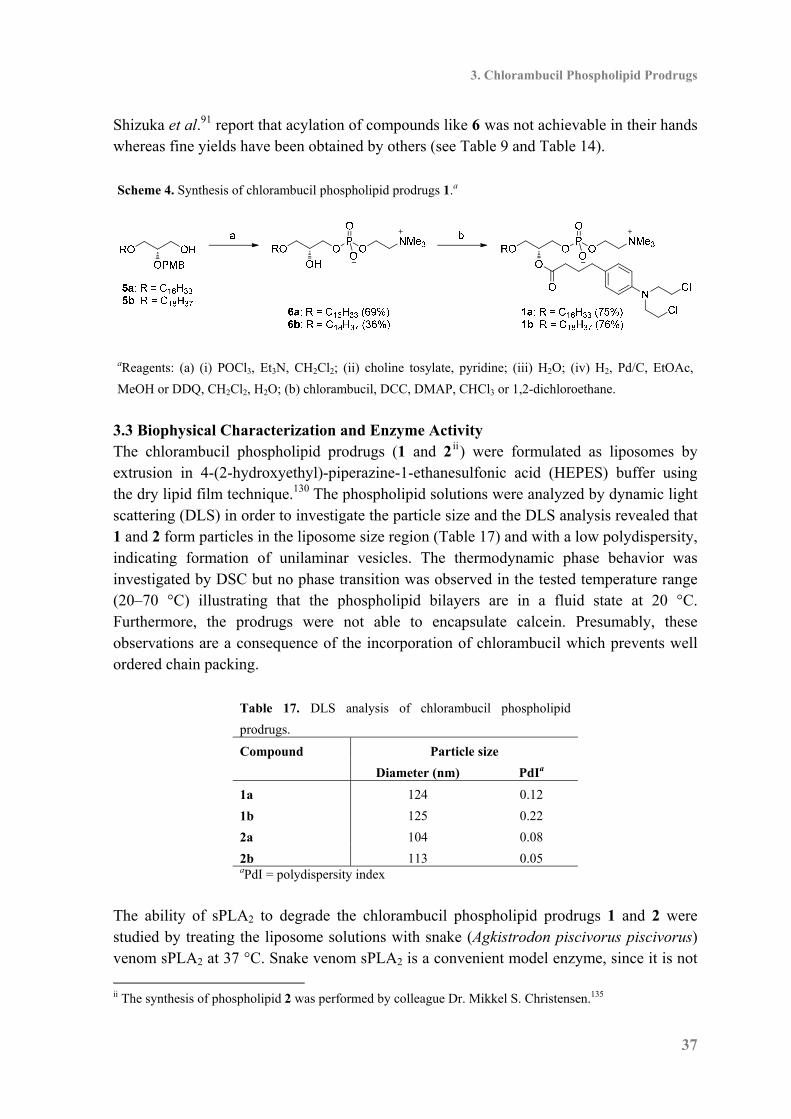
3. Chlorambucil Phospholipid Prodrugs
37
Shizuka et al.91 report that acylation of compounds like 6 was not achievable in their hands whereas fine yields have been obtained by others (see Table 9 and Table 14). Scheme 4. Synthesis of chlorambucil phospholipid prodrugs 1.a
aReagents: (a) (i) POCl3, Et3N, CH2Cl2; (ii) choline tosylate, pyridine; (iii) H2O; (iv) H2, Pd/C, EtOAc,
MeOH or DDQ, CH2Cl2, H2O; (b) chlorambucil, DCC, DMAP, CHCl3 or 1,2-dichloroethane.
3.3 Biophysical Characterization and Enzyme Activity The chlorambucil phospholipid prodrugs (1 and 2 ii) were formulated as liposomes by extrusion in 4-(2-hydroxyethyl)-piperazine-1-ethanesulfonic acid (HEPES) buffer using the dry lipid film technique.130 The phospholipid solutions were analyzed by dynamic light scattering (DLS) in order to investigate the particle size and the DLS analysis revealed that 1 and 2 form particles in the liposome size region (Table 17) and with a low polydispersity, indicating formation of unilaminar vesicles. The thermodynamic phase behavior was investigated by DSC but no phase transition was observed in the tested temperature range (20–70 °C) illustrating that the phospholipid bilayers are in a fluid state at 20 °C. Furthermore, the prodrugs were not able to encapsulate calcein. Presumably, these observations are a consequence of the incorporation of chlorambucil which prevents well ordered chain packing.
Table 17. DLS analysis of chlorambucil phospholipid
prodrugs.
Compound Particle size
Diameter (nm) PdIa
1a 124 0.12
1b 125 0.22
2a 104 0.08
2b 113 0.05 aPdI = polydispersity index
The ability of sPLA2 to degrade the chlorambucil phospholipid prodrugs 1 and 2 were studied by treating the liposome solutions with snake (Agkistrodon piscivorus piscivorus) venom sPLA2 at 37 °C. Snake venom sPLA2 is a convenient model enzyme, since it is not
ii The synthesis of phospholipid 2 was performed by colleague Dr. Mikkel S. Christensen.135
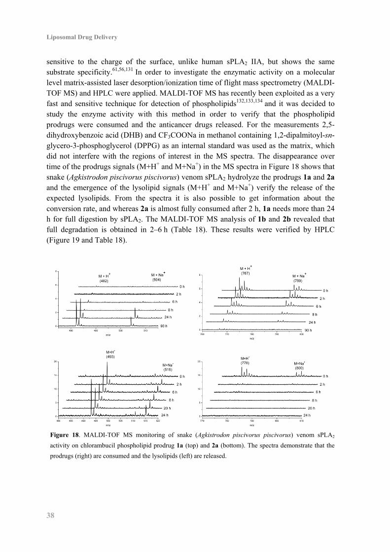
Liposomal Drug Delivery
38
sensitive to the charge of the surface, unlike human sPLA2 IIA, but shows the same substrate specificity.61,56,131 In order to investigate the enzymatic activity on a molecular level matrix-assisted laser desorption/ionization time of flight mass spectrometry (MALDI-TOF MS) and HPLC were applied. MALDI-TOF MS has recently been exploited as a very fast and sensitive technique for detection of phospholipids132,133,134 and it was decided to study the enzyme activity with this method in order to verify that the phospholipid prodrugs were consumed and the anticancer drugs released. For the measurements 2,5-dihydroxybenzoic acid (DHB) and CF3COONa in methanol containing 1,2-dipalmitoyl-sn-glycero-3-phosphoglycerol (DPPG) as an internal standard was used as the matrix, which did not interfere with the regions of interest in the MS spectra. The disappearance over time of the prodrugs signals (M+H+ and M+Na+) in the MS spectra in Figure 18 shows that snake (Agkistrodon piscivorus piscivorus) venom sPLA2 hydrolyze the prodrugs 1a and 2a and the emergence of the lysolipid signals (M+H+ and M+Na+) verify the release of the expected lysolipids. From the spectra it is also possible to get information about the conversion rate, and whereas 2a is almost fully consumed after 2 h, 1a needs more than 24 h for full digestion by sPLA2. The MALDI-TOF MS analysis of 1b and 2b revealed that full degradation is obtained in 2–6 h (Table 18). These results were verified by HPLC (Figure 19 and Table 18).
Figure 18. MALDI-TOF MS monitoring of snake (Agkistrodon piscivorus piscivorus) venom sPLA2
activity on chlorambucil phospholipid prodrug 1a (top) and 2a (bottom). The spectra demonstrate that the
prodrugs (right) are consumed and the lysolipids (left) are released.
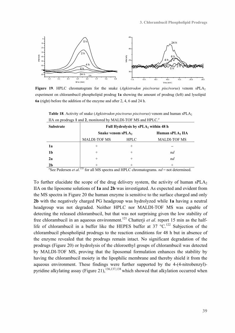
3. Chlorambucil Phospholipid Prodrugs
39
Figure 19. HPLC chromatogram for the snake (Agkistrodon piscivorus piscivorus) venom sPLA2
experiment on chlorambucil phospholipid prodrug 1a showing the amount of prodrug (left) and lysolipid
6a (right) before the addition of the enzyme and after 2, 4, 6 and 24 h.
Table 18. Activity of snake (Agkistrodon piscivorus piscivorus) venom and human sPLA2
IIA on prodrugs 1 and 2, monitored by MALDI-TOF MS and HPLC.a
Substrate Full Hydrolysis by sPLA2 within 48 h
Snake venom sPLA2 Human sPLA2 IIA
MALDI-TOF MS HPLC MALDI-TOF MS
1a + + –
1b + + nd
2a + + nd
2b + + + aSee Pedersen et al.135 for all MS spectra and HPLC chromatograms. nd = not determined.
To further elucidate the scope of the drug delivery system, the activity of human sPLA2 IIA on the liposome solutions of 1a and 2b was investigated. As expected and evident from the MS spectra in Figure 20 the human enzyme is sensitive to the surface charged and only 2b with the negatively charged PG headgroup was hydrolyzed while 1a having a neutral headgroup was not degraded. Neither HPLC nor MALDI-TOF MS was capable of detecting the released chlorambucil, but that was not surprising given the low stability of free chlorambucil in an aqueous environment.121 Chatterji et al. report 15 min as the half-life of chlorambucil in a buffer like the HEPES buffer at 37 °C.122 Subjection of the chlorambucil phospholipid prodrugs to the reaction conditions for 48 h but in absence of the enzyme revealed that the prodrugs remain intact. No significant degradation of the prodrugs (Figure 20) or hydrolysis of the chloroethyl groups of chlorambucil was detected by MALDI-TOF MS, proving that the liposomal formulation enhances the stability by having the chlorambucil moiety in the lipophilic membrane and thereby shield it from the aqueous environment. These findings were further supported by the 4-(4-nitrobenzyl)-pyridine alkylating assay (Figure 21),136,137,138 which showed that alkylation occurred when
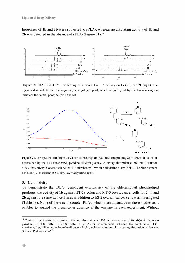
Liposomal Drug Delivery
40
liposomes of 1b and 2b were subjected to sPLA2, whereas no alkylating activity of 1b and 2b was detected in the absence of sPLA2 (Figure 21).iii
Figure 20. MALDI-TOF MS monitoring of human sPLA2 IIA activity on 1a (left) and 2b (right). The
spectra demonstrate that the negatively charged phospholipid 2b is hydrolyzed by the humane enzyme
whereas the neutral phospholipid 1a is not.
Figure 21. UV spectra (left) from alkylation of prodrug 2b (red linie) and prodrug 2b + sPLA2 (blue linie)
determined by the 4-(4-nitrobenzyl)-pyridine alkylating assay. A strong absorption at 560 nm illustrates
alkylating activity. Concept behind the 4-(4-nitrobenzyl)-pyridine alkylating assay (right). The blue pigment
has high UV absorbans at 560 nm. RX = alkylating agent
3.4 Cytotoxicity To demonstrate the sPLA2 dependent cytotoxicity of the chlorambucil phospholipid prodrugs, the activity of 1b against HT-29 colon and MT-3 breast cancer cells for 24 h and 2b against the same two cell lines in addition to ES-2 ovarian cancer cells was investigated (Table 19). None of these cells secrete sPLA2, which is an advantage in these studies as it enables to control the presence or absence of the enzyme in each experiment. Without
iii Control experiments demonstrated that no absorption at 560 nm was observed for 4-(4-nitrobenzyl)-pyridine, HEPES buffer, HEPES buffer + sPLA2 or chlorambucil, whereas the combination 4-(4-nitrobenzyl)-pyridine and chlorambucil gave a highly colored solution with a strong absorption at 560 nm. See also Pedersen et al.135
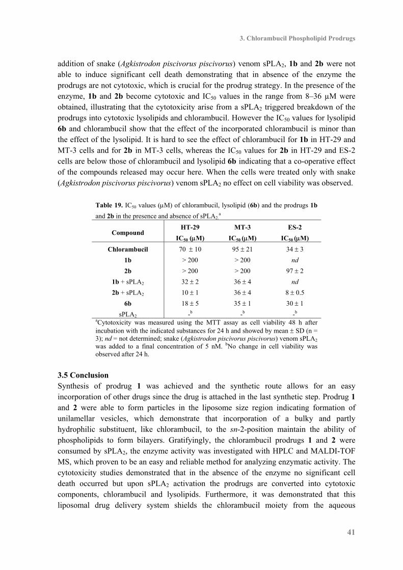
3. Chlorambucil Phospholipid Prodrugs
41
addition of snake (Agkistrodon piscivorus piscivorus) venom sPLA2, 1b and 2b were not able to induce significant cell death demonstrating that in absence of the enzyme the prodrugs are not cytotoxic, which is crucial for the prodrug strategy. In the presence of the enzyme, 1b and 2b become cytotoxic and IC50 values in the range from 8–36 µM were obtained, illustrating that the cytotoxicity arise from a sPLA2 triggered breakdown of the prodrugs into cytotoxic lysolipids and chlorambucil. However the IC50 values for lysolipid 6b and chlorambucil show that the effect of the incorporated chlorambucil is minor than the effect of the lysolipid. It is hard to see the effect of chlorambucil for 1b in HT-29 and MT-3 cells and for 2b in MT-3 cells, whereas the IC50 values for 2b in HT-29 and ES-2 cells are below those of chlorambucil and lysolipid 6b indicating that a co-operative effect of the compounds released may occur here. When the cells were treated only with snake (Agkistrodon piscivorus piscivorus) venom sPLA2 no effect on cell viability was observed.
Table 19. IC50 values (M) of chlorambucil, lysolipid (6b) and the prodrugs 1b
and 2b in the presence and absence of sPLA2.a
Compound HT-29
IC50 (M)
MT-3
IC50 (M)
ES-2
IC50 (M)
Chlorambucil 70 10 95 21 34 3
1b > 200 > 200 nd
2b > 200 > 200 97 2
1b + sPLA2 32 2 36 4 nd
2b + sPLA2 10 1 36 4 8 0.5
6b 18 5 35 1 30 1
sPLA2 -b -b -b
aCytotoxicity was measured using the MTT assay as cell viability 48 h after incubation with the indicated substances for 24 h and showed by mean SD (n = 3); nd = not determined; snake (Agkistrodon piscivorus piscivorus) venom sPLA2
was added to a final concentration of 5 nM. bNo change in cell viability was observed after 24 h.
3.5 Conclusion Synthesis of prodrug 1 was achieved and the synthetic route allows for an easy incorporation of other drugs since the drug is attached in the last synthetic step. Prodrug 1 and 2 were able to form particles in the liposome size region indicating formation of unilamellar vesicles, which demonstrate that incorporation of a bulky and partly hydrophilic substituent, like chlorambucil, to the sn-2-position maintain the ability of phospholipids to form bilayers. Gratifyingly, the chlorambucil prodrugs 1 and 2 were consumed by sPLA2, the enzyme activity was investigated with HPLC and MALDI-TOF MS, which proven to be an easy and reliable method for analyzing enzymatic activity. The cytotoxicity studies demonstrated that in the absence of the enzyme no significant cell death occurred but upon sPLA2 activation the prodrugs are converted into cytotoxic components, chlorambucil and lysolipids. Furthermore, it was demonstrated that this liposomal drug delivery system shields the chlorambucil moiety from the aqueous

Liposomal Drug Delivery
42
environment and enhance the stability. Taken together, the results obtained from this work was very promising for the overall scope of the investigated drug delivery system, although the effect from chlorambucil in the tested cell line was not significant compared to the cytotoxicity of the lysolipids.
4. Liposomal Formulation of Retinoids Designed for Enzyme Triggered Release
43
4. Liposomal Formulation of Retinoids Designed for Enzyme Triggered Release 4.1 Introduction Retinoids,139 such as ATRA (Figure 22), are known for their broad and diverse biological functions and various strategies have been explored to make retinoids applicable as drugs.140 One of the biological functions of ATRA is its anticancer activity towards a broad range of cancer types, like breast, prostate and colon cancer.75,76 For example, orally administrated ATRA is used clinically in the treatment of leukaemia.141,142 However, the oral administration route is restricted by a relative low bioavailability 143 and a fast clearance from the blood stream,144 and thus alternative ways of administrating ATRA would be beneficial. Intravenous administration is hampered by the low water solubility of ATRA but this can be circumvented by formulating ATRA in liposome based drug delivery systems.145 , 146 , 147 , 148 Unfortunately, the liposomal formulation strategies have often been plagued by problems with leakage, which have led to an uncontrolled release of ATRA from the carrier system. Furthermore, ATRA is sensitive to acid and light139 thus a liposomal formulation could be beneficial and the presence of a carboxylic acid moiety allows incorporation into the investigated drug delivery system. The target ATRA phospholipid prodrugs are illustrated in Figure 22.
Figure 22. Structure of ATRA and the target ATRA phospholipids prodrug 7 and 8.
4.2 Synthesis of ATRA Phospholipid Prodrugs With lysolipid 6a in hand (from Scheme 4) the synthesis of phospholipid 7 was attempted by acylation of 6a with ATRA. The reaction was investigated applying the same conditions as used in the synthesis of 1, and as observed under the incorporation of chlorambucil it was necessary to use an excess of ATRA and the coupling reagents in order to get a decent conversion of 6a (Scheme 5). However, 1H NMR analysis of the isolated material revealed that two isomers was formed, corresponding to esters of ATRA ((13E)-retinoic acid) and (13Z)-retinoic acid. The isomers was distinguished in 1H NMR by the signal for the proton alpha to the carbonyl in the retinoids which resonate at 5.8 ppm for the (13E)-isomer and at
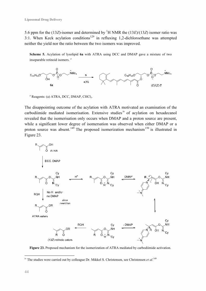
Liposomal Drug Delivery
44
5.6 ppm for the (13Z)-isomer and determined by 1H NMR the (13E)/(13Z) isomer ratio was 3:1. When Keck acylation conditions129 in refluxing 1,2-dichloroethane was attempted neither the yield nor the ratio between the two isomers was improved.
Scheme 5. Acylation of lysolipid 6a with ATRA using DCC and DMAP gave a mixture of two
inseparable retinoid isomers. a
a Reagents: (a) ATRA, DCC, DMAP, CHCl3.
The disappointing outcome of the acylation with ATRA motivated an examination of the carbodiimide mediated isomerisation. Extensive studies iv of acylation on hexadecanol revealed that the isomerisation only occurs when DMAP and a proton source are present, while a significant lower degree of isomersation was observed when either DMAP or a proton source was absent.149 The proposed isomerization mechanism150 is illustrated in Figure 23.
Figure 23. Proposed mechanism for the isomerization of ATRA mediated by carbodiimide activation.
iv The studies were carried out by colleague Dr. Mikkel S. Christensen, see Christensen et al.149
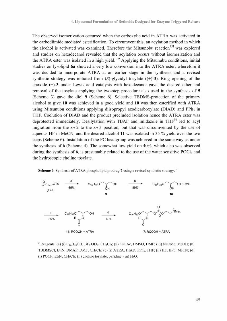
4. Liposomal Formulation of Retinoids Designed for Enzyme Triggered Release
45
The observed isomerization occurred when the carboxylic acid in ATRA was activated in the carbodiimide mediated esterification. To circumvent this, an acylation method in which the alcohol is activated was examined. Therefore the Mitsunobu reaction151 was explored and studies on hexadecanol revealed that the acylation occurs without isomerization and the ATRA ester was isolated in a high yield.149 Applying the Mitsunubu conditions, initial studies on lysolipid 6a showed a very low conversion into the ATRA ester, wherefore it was decided to incorporate ATRA at an earlier stage in the synthesis and a revised synthetic strategy was initiated from (S)-glycidyl tosylate ((+)-3). Ring opening of the epoxide (+)-3 under Lewis acid catalysis with hexadecanol gave the desired ether and removal of the tosylate applying the two-step procedure also used in the synthesis of 5 (Scheme 3) gave the diol 9 (Scheme 6). Selective TBDMS-protection of the primary alcohol to give 10 was achieved in a good yield and 10 was then esterified with ATRA using Mitsunubu conditions applying diisopropyl azodicarboxylate (DIAD) and PPh3 in THF. Coelution of DIAD and the product precluded isolation hence the ATRA ester was deprotected immediately. Desilylation with TBAF and imidazole in THF90 led to acyl migration from the sn-2 to the sn-3 position, but that was circumvented by the use of aqueous HF in MeCN, and the desired alcohol 11 was isolated in 35 % yield over the two steps (Scheme 6). Installation of the PC headgroup was achieved in the same way as under the synthesis of 6 (Scheme 4). The somewhat low yield on 40%, which also was observed during the synthesis of 6, is presumably related to the use of the water sensitive POCl3 and the hydroscopic choline tosylate.
Scheme 6. Synthesis of ATRA phospholipid prodrug 7 using a revised synthetic strategy. a
(+)-3
OTsO a
65%C16H33O OH
OH
9
b
89%C16H33O OTBDMS
OH
10
c
35%
11: RCOOH = ATRA
C16H33O OH
O
O
R
d
40%
C16H33O O
O
P
O
ONMe3
O
O
R
7: RCOOH = ATRA
a Reagents: (a) (i) C16H33OH, BF3·OEt2, CH2Cl2; (ii) CsOAc, DMSO, DMF; (iii) NaOMe, MeOH; (b)
TBDMSCl, Et3N, DMAP, DMF, CH2Cl2; (c) (i) ATRA, DIAD, PPh3, THF; (ii) HF, H2O, MeCN; (d)
(i) POCl3, Et3N, CH2Cl2; (ii) choline tosylate, pyridine; (iii) H2O.

Liposomal Drug Delivery
46
4.3 Enzymatic Activity In order to study sPLA2 activity the phospholipid prodrugs 7 and 8v were formulated as liposomes in HEPES buffer and particles with an average diameter close to 100 nm was formed. The liposome solutions were incubated with snake (Agkistrodon piscivorus piscivorus or Naja mossambica mossambica) venom sPLA2 at 37 °C for 48 h however MALDI-TOF MS analysis of the liposome solutions revealed that no hydrolysis had occurred (Figure 24). Fawzy et al.152 and Hope et al.153 have demonstrated two decades ago that ATRA is an inhibitor of sPLA2 and therefore the hydrolysis of DPPG in the presence of ATRA was investigated. Snake (Naja mossambica mossambica) venom sPLA2 was subjected to solutions of DPPG and 0, 0.1 or 1 eq. ATRA. Complete hydrolysis of DPPG was observed within 24 h in all three experiments,vi by which it can be ruled out that the lack of hydrolysis is a consequence of sPLA2 inhibition. The lack of hydrolysis must therefore be related to the structure of ATRA, the rigidity of the molecule and the presence of a methyl substituent in close vicinity to the carboxylic acid contrast with naturally occurring fatty acids, that are predominantly saturated and flexible molecules without branching. Furthermore, Bonsen et al. have demonstrated that compounds like 2-methylhexanoic acid and 3,3-dimethylbutanoic acid are not released from the sn-2 position by sPLA2,
67 illustrating that substituents in close proximity to the sn-2 position hamper the enzyme activity.
Figure 24. MALDI-TOF MS monitoring of snake (Naja mossambica
mossambica) venom activity on ATRA phospholipid prodrug 8.
4.4 Molecular Dynamics Simulations of Enzymatic Activity To get a better understanding of why substrates like 8 is not hydrolyzed by sPLA2, molecular dynamics (MD) simulations were performed of the Michaelis-Menten complex of 8 in the active site of sPLA2 (denoted as sPLA2–8). The enzymatic activity of sPLA2 on phospholipids depends on several factors, including binding of the enzyme to the membrane surface, membrane properties, formation of the Michaelis-Menten complex, etc.154 In this MD study only the stability of the Michaelis-Menten complex is investigated and it is then assumped that if a stable Michaelis-Menten complex is observed during the
v The synthesis of ATRA phospholipid prodrug 8 was performed by colleague Dr. Mikkel S. Christensen.149 vi See Christensen et al.149 for further details.
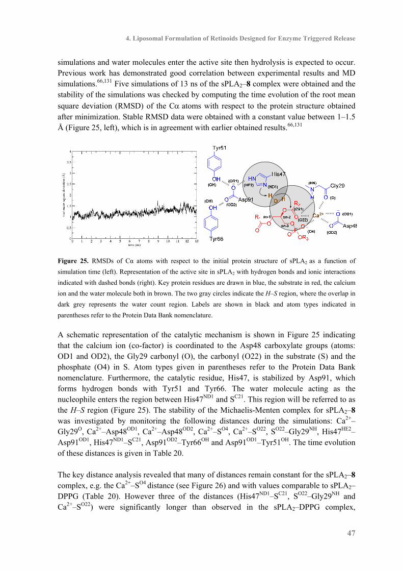
4. Liposomal Formulation of Retinoids Designed for Enzyme Triggered Release
47
simulations and water molecules enter the active site then hydrolysis is expected to occur. Previous work has demonstrated good correlation between experimental results and MD simulations.66,131 Five simulations of 13 ns of the sPLA2–8 complex were obtained and the stability of the simulations was checked by computing the time evolution of the root mean
square deviation (RMSD) of the C atoms with respect to the protein structure obtained after minimization. Stable RMSD data were obtained with a constant value between 1–1.5 Å (Figure 25, left), which is in agreement with earlier obtained results.66,131
Figure 25. RMSDs of Cα atoms with respect to the initial protein structure of sPLA2 as a function of
simulation time (left). Representation of the active site in sPLA2 with hydrogen bonds and ionic interactions
indicated with dashed bonds (right). Key protein residues are drawn in blue, the substrate in red, the calcium
ion and the water molecule both in brown. The two gray circles indicate the H–S region, where the overlap in
dark grey represents the water count region. Labels are shown in black and atom types indicated in
parentheses refer to the Protein Data Bank nomenclature.
A schematic representation of the catalytic mechanism is shown in Figure 25 indicating that the calcium ion (co-factor) is coordinated to the Asp48 carboxylate groups (atoms: OD1 and OD2), the Gly29 carbonyl (O), the carbonyl (O22) in the substrate (S) and the phosphate (O4) in S. Atom types given in parentheses refer to the Protein Data Bank nomenclature. Furthermore, the catalytic residue, His47, is stabilized by Asp91, which forms hydrogen bonds with Tyr51 and Tyr66. The water molecule acting as the nucleophile enters the region between His47ND1 and SC21. This region will be referred to as the H–S region (Figure 25). The stability of the Michaelis-Menten complex for sPLA2–8 was investigated by monitoring the following distances during the simulations: Ca2+–Gly29O, Ca2+–Asp48OD1, Ca2+–Asp48OD2, Ca2+–SO4, Ca2+–SO22, SO22–Gly29NH, His47HE2–Asp91OD1, His47ND1–SC21, Asp91OD2–Tyr66OH and Asp91OD1–Tyr51OH. The time evolution of these distances is given in Table 20. The key distance analysis revealed that many of distances remain constant for the sPLA2–8 complex, e.g. the Ca2+–SO4 distance (see Figure 26) and with values comparable to sPLA2–DPPG (Table 20). However three of the distances (His47ND1–SC21, SO22–Gly29NH and Ca2+–SO22) were significantly longer than observed in the sPLA2–DPPG complex,
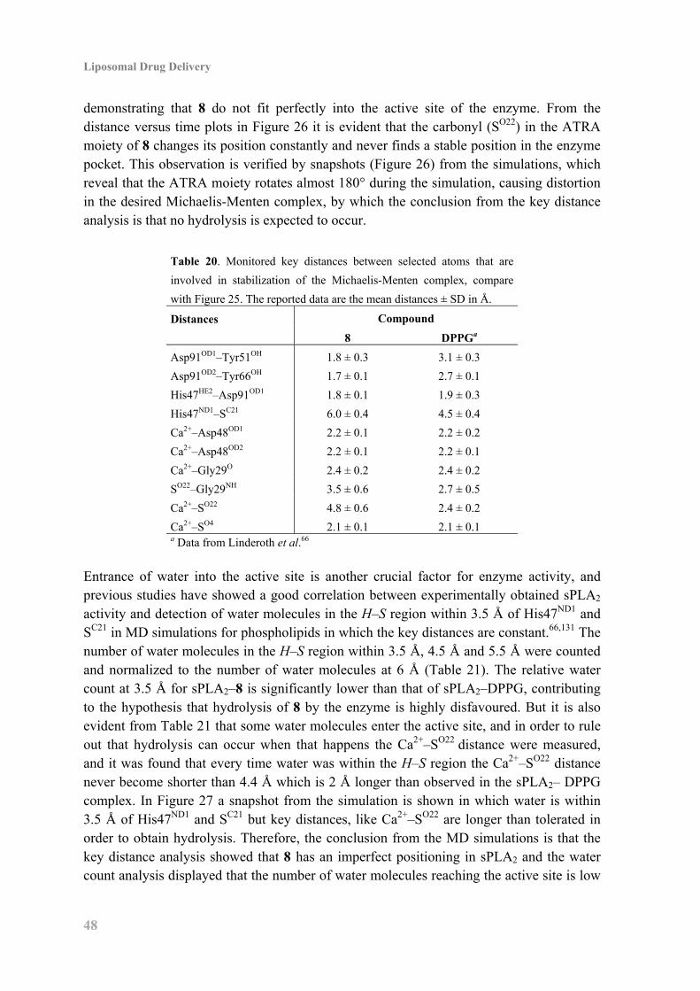
Liposomal Drug Delivery
48
demonstrating that 8 do not fit perfectly into the active site of the enzyme. From the distance versus time plots in Figure 26 it is evident that the carbonyl (SO22) in the ATRA moiety of 8 changes its position constantly and never finds a stable position in the enzyme pocket. This observation is verified by snapshots (Figure 26) from the simulations, which reveal that the ATRA moiety rotates almost 180° during the simulation, causing distortion in the desired Michaelis-Menten complex, by which the conclusion from the key distance analysis is that no hydrolysis is expected to occur.
Table 20. Monitored key distances between selected atoms that are
involved in stabilization of the Michaelis-Menten complex, compare
with Figure 25. The reported data are the mean distances ± SD in Å.
Distances
Compound
8 DPPGa
Asp91OD1–Tyr51OH 1.8 ± 0.3 3.1 ± 0.3
Asp91OD2–Tyr66OH 1.7 ± 0.1 2.7 ± 0.1
His47HE2–Asp91OD1 1.8 ± 0.1 1.9 ± 0.3
His47ND1–SC21 6.0 ± 0.4 4.5 ± 0.4
Ca2+–Asp48OD1 2.2 ± 0.1 2.2 ± 0.2
Ca2+–Asp48OD2 2.2 ± 0.1 2.2 ± 0.1
Ca2+–Gly29O 2.4 ± 0.2 2.4 ± 0.2
SO22–Gly29NH 3.5 ± 0.6 2.7 ± 0.5
Ca2+–SO22 4.8 ± 0.6 2.4 ± 0.2
Ca2+–SO4 2.1 ± 0.1 2.1 ± 0.1 a Data from Linderoth et al.66
Entrance of water into the active site is another crucial factor for enzyme activity, and previous studies have showed a good correlation between experimentally obtained sPLA2 activity and detection of water molecules in the H–S region within 3.5 Å of His47ND1 and SC21 in MD simulations for phospholipids in which the key distances are constant.66,131 The number of water molecules in the H–S region within 3.5 Å, 4.5 Å and 5.5 Å were counted and normalized to the number of water molecules at 6 Å (Table 21). The relative water count at 3.5 Å for sPLA2–8 is significantly lower than that of sPLA2–DPPG, contributing to the hypothesis that hydrolysis of 8 by the enzyme is highly disfavoured. But it is also evident from Table 21 that some water molecules enter the active site, and in order to rule out that hydrolysis can occur when that happens the Ca2+–SO22 distance were measured, and it was found that every time water was within the H–S region the Ca2+–SO22 distance never become shorter than 4.4 Å which is 2 Å longer than observed in the sPLA2– DPPG complex. In Figure 27 a snapshot from the simulation is shown in which water is within 3.5 Å of His47ND1 and SC21 but key distances, like Ca2+–SO22 are longer than tolerated in order to obtain hydrolysis. Therefore, the conclusion from the MD simulations is that the key distance analysis showed that 8 has an imperfect positioning in sPLA2 and the water count analysis displayed that the number of water molecules reaching the active site is low
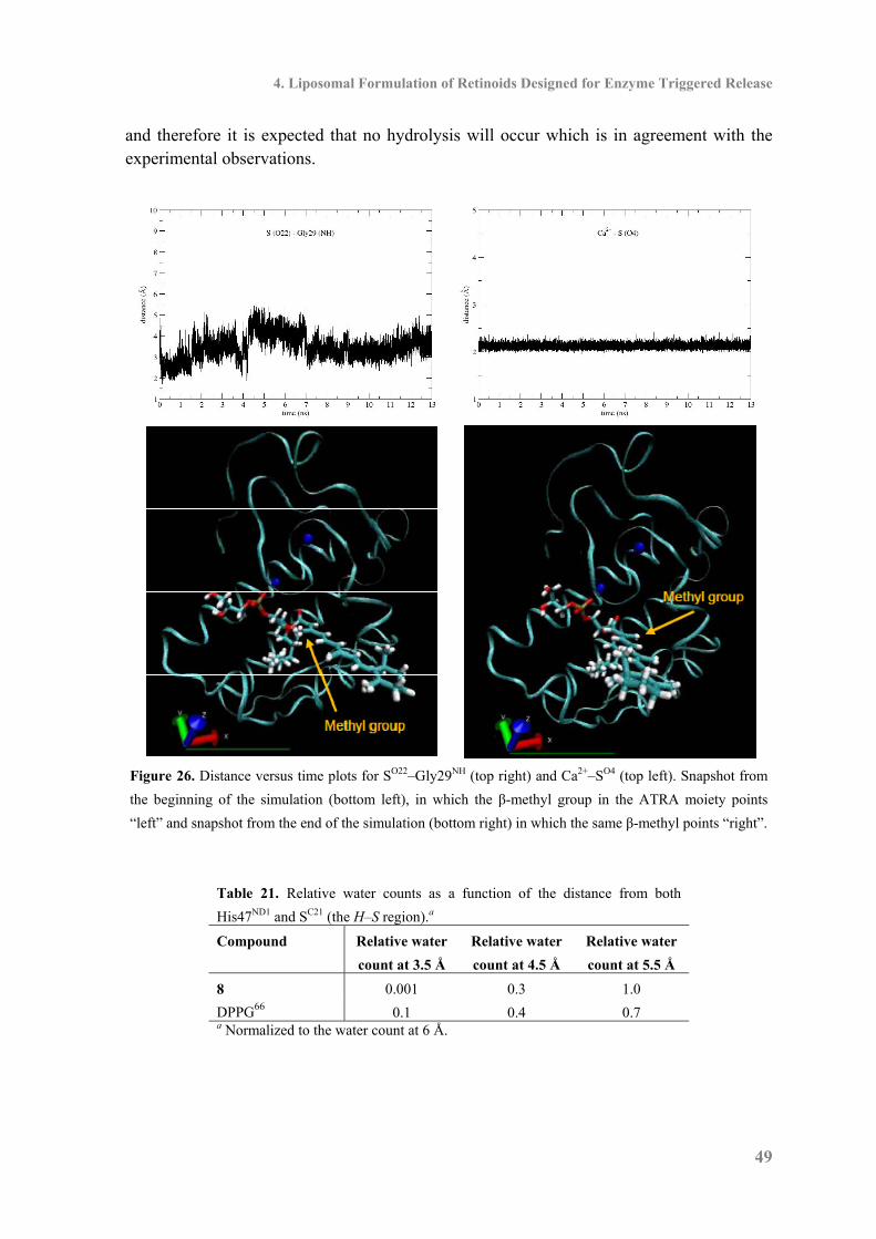
4. Liposomal Formulation of Retinoids Designed for Enzyme Triggered Release
49
and therefore it is expected that no hydrolysis will occur which is in agreement with the experimental observations.
Figure 26. Distance versus time plots for SO22–Gly29NH (top right) and Ca2+–SO4 (top left). Snapshot from
the beginning of the simulation (bottom left), in which the β-methyl group in the ATRA moiety points
“left” and snapshot from the end of the simulation (bottom right) in which the same β-methyl points “right”.
Table 21. Relative water counts as a function of the distance from both
His47ND1 and SC21 (the H–S region).a
Compound Relative water
count at 3.5 Å
Relative water
count at 4.5 Å
Relative water
count at 5.5 Å
8 0.001 0.3 1.0
DPPG66 0.1 0.4 0.7 a Normalized to the water count at 6 Å.
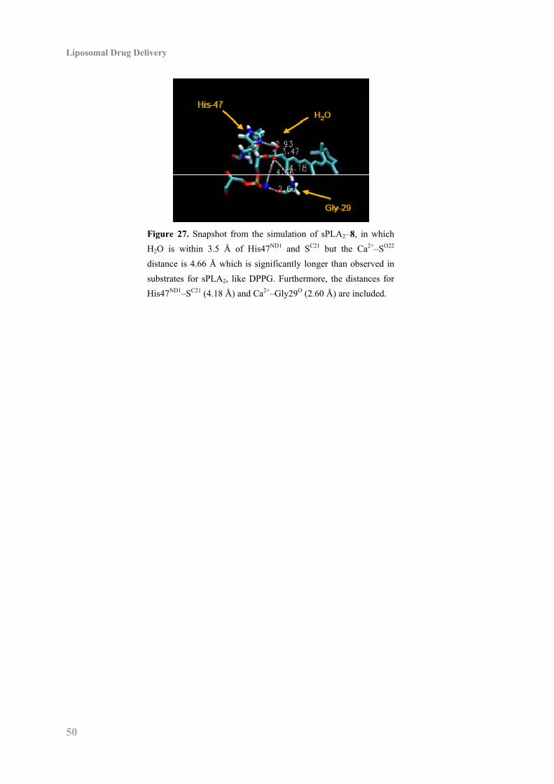
Liposomal Drug Delivery
50
Figure 27. Snapshot from the simulation of sPLA2–8, in which
H2O is within 3.5 Å of His47ND1 and SC21 but the Ca2+–SO22
distance is 4.66 Å which is significantly longer than observed in
substrates for sPLA2, like DPPG. Furthermore, the distances for
His47ND1–SC21 (4.18 Å) and Ca2+–Gly29O (2.60 Å) are included.
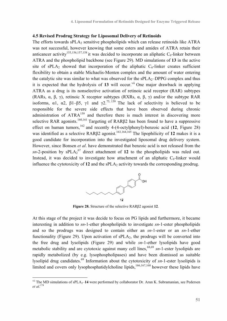
4. Liposomal Formulation of Retinoids Designed for Enzyme Triggered Release
51
4.5 Revised Prodrug Strategy for Liposomal Delivery of Retinoids The efforts towards sPLA2 sensitive phospholipids which can release retinoids like ATRA was not successful, however knowing that some esters and amides of ATRA retain their anticancer activity155,156,157,158 it was decided to incorporate an aliphatic C6-linker between ATRA and the phospholipid backbone (see Figure 29). MD simulations of 13 in the active site of sPLA2 showed that incorporation of the aliphatic C6-linker creates sufficient flexibility to obtain a stable Michaelis-Menten complex and the amount of water entering the catalytic site was similar to what was observed for the sPLA2–DPPG complex and thus it is expected that the hydrolysis of 13 will occur.vii One major drawback in applying ATRA as a drug is its nonselective activation of retinoic acid receptor (RAR) subtypes (RARs, α, β, γ), retinoic X receptor subtypes (RXRs, α, β, γ) and/or the subtype RAR isoforms, α1, α2, β1–β5, γ1 and γ2.75, 159 The lack of selectivity is believed to be responsible for the severe side effects that have been observed during chronic administration of ATRA159 and therefore there is much interest in discovering more selective RAR agonists.160,161 Targeting of RARβ2 has been found to have a suppressive effect on human tumors,162 and recently 4-(4-octylphenyl)-benzoic acid (12, Figure 28) was identified as a selective RARβ2 agonist.163,164,165 The lipophilicity of 12 makes it is a good candidate for incorporation into the investigated liposomal drug delivery system. However, since Bonsen et al. have demonstrated that benzoic acid is not released from the sn-2-position by sPLA2
67 direct attachment of 12 to the phospholipids was ruled out. Instead, it was decided to investigate how attachment of an aliphatic C6-linker would influence the cytotoxicity of 12 and the sPLA2 activity towards the corresponding prodrug.
Figure 28. Structure of the selective RARβ2 agonist 12.
At this stage of the project it was decide to focus on PG lipids and furthermore, it became interesting in addition to sn-1-ether phospholipids to investigate sn-1-ester phospholipids and so the prodrugs was designed to contain either an sn-1-ester or an sn-1-ether functionality (Figure 29). Upon activation of sPLA2, the prodrugs will be converted into the free drug and lysolipids (Figure 29) and while sn-1-ether lysolipids have good metabolic stability and are cytotoxic against many cell lines,68,69 sn-1-ester lysolipids are rapidly metabolized (by e.g. lysophospholipases) and have been dismissed as suitable lysolipid drug candidates.69 Information about the cytotoxicity of sn-1-ester lysolipids is limited and covers only lysophosphatidylcholine lipids,166,167,168 however these lipids have
vii The MD simulations of sPLA2–14 were performed by collaborator Dr. Arun K. Subramanian, see Pedersen et al.174
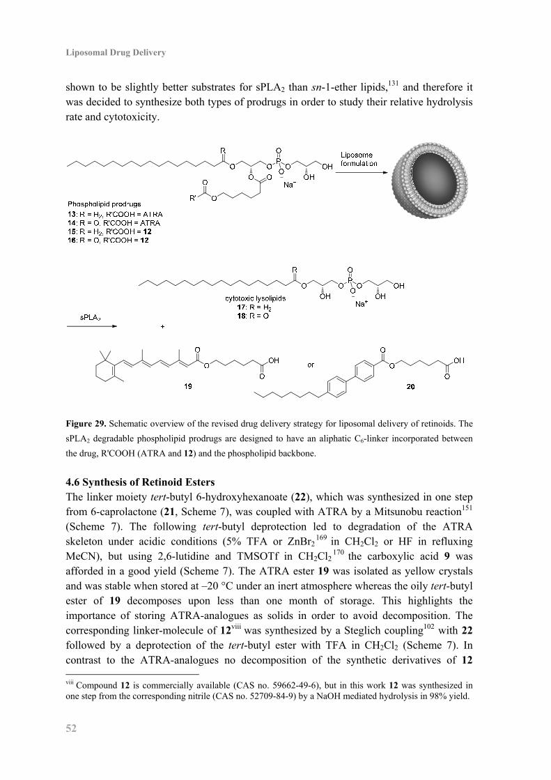
Liposomal Drug Delivery
52
shown to be slightly better substrates for sPLA2 than sn-1-ether lipids,131 and therefore it was decided to synthesize both types of prodrugs in order to study their relative hydrolysis rate and cytotoxicity.
Figure 29. Schematic overview of the revised drug delivery strategy for liposomal delivery of retinoids. The
sPLA2 degradable phospholipid prodrugs are designed to have an aliphatic C6-linker incorporated between
the drug, R'COOH (ATRA and 12) and the phospholipid backbone.
4.6 Synthesis of Retinoid Esters The linker moiety tert-butyl 6-hydroxyhexanoate (22), which was synthesized in one step from 6-caprolactone (21, Scheme 7), was coupled with ATRA by a Mitsunobu reaction151 (Scheme 7). The following tert-butyl deprotection led to degradation of the ATRA skeleton under acidic conditions (5% TFA or ZnBr2
169 in CH2Cl2 or HF in refluxing MeCN), but using 2,6-lutidine and TMSOTf in CH2Cl2
170 the carboxylic acid 9 was afforded in a good yield (Scheme 7). The ATRA ester 19 was isolated as yellow crystals and was stable when stored at –20 °C under an inert atmosphere whereas the oily tert-butyl ester of 19 decomposes upon less than one month of storage. This highlights the importance of storing ATRA-analogues as solids in order to avoid decomposition. The corresponding linker-molecule of 12viii was synthesized by a Steglich coupling102 with 22 followed by a deprotection of the tert-butyl ester with TFA in CH2Cl2 (Scheme 7). In contrast to the ATRA-analogues no decomposition of the synthetic derivatives of 12 viii Compound 12 is commercially available (CAS no. 59662-49-6), but in this work 12 was synthesized in one step from the corresponding nitrile (CAS no. 52709-84-9) by a NaOH mediated hydrolysis in 98% yield.
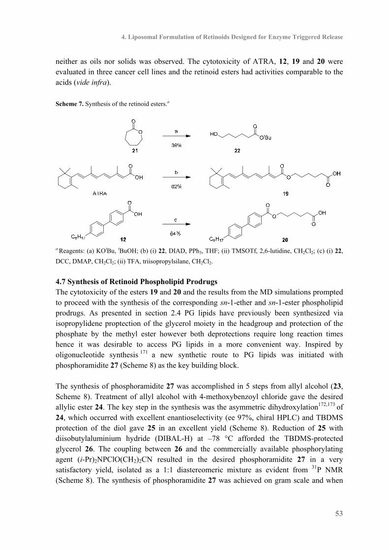
4. Liposomal Formulation of Retinoids Designed for Enzyme Triggered Release
53
neither as oils nor solids was observed. The cytotoxicity of ATRA, 12, 19 and 20 were evaluated in three cancer cell lines and the retinoid esters had activities comparable to the acids (vide infra). Scheme 7. Synthesis of the retinoid esters.a
a Reagents: (a) KOtBu, tBuOH; (b) (i) 22, DIAD, PPh3, THF; (ii) TMSOTf, 2,6-lutidine, CH2Cl2; (c) (i) 22,
DCC, DMAP, CH2Cl2; (ii) TFA, triisopropylsilane, CH2Cl2.
4.7 Synthesis of Retinoid Phospholipid Prodrugs The cytotoxicity of the esters 19 and 20 and the results from the MD simulations prompted to proceed with the synthesis of the corresponding sn-1-ether and sn-1-ester phospholipid prodrugs. As presented in section 2.4 PG lipids have previously been synthesized via isopropylidene proptection of the glycerol moiety in the headgroup and protection of the phosphate by the methyl ester however both deprotections require long reaction times hence it was desirable to access PG lipids in a more convenient way. Inspired by oligonucleotide synthesis 171 a new synthetic route to PG lipids was initiated with phosphoramidite 27 (Scheme 8) as the key building block. The synthesis of phosphoramidite 27 was accomplished in 5 steps from allyl alcohol (23, Scheme 8). Treatment of allyl alcohol with 4-methoxybenzoyl chloride gave the desired allylic ester 24. The key step in the synthesis was the asymmetric dihydroxylation172,173 of 24, which occurred with excellent enantioselectivity (ee 97%, chiral HPLC) and TBDMS protection of the diol gave 25 in an excellent yield (Scheme 8). Reduction of 25 with diisobutylaluminium hydride (DIBAL-H) at –78 °C afforded the TBDMS-protected glycerol 26. The coupling between 26 and the commercially available phosphorylating agent (i-Pr)2NPClO(CH2)2CN resulted in the desired phosphoramidite 27 in a very satisfactory yield, isolated as a 1:1 diastereomeric mixture as evident from 31P NMR (Scheme 8). The synthesis of phosphoramidite 27 was achieved on gram scale and when
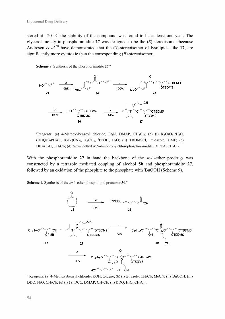
Liposomal Drug Delivery
54
stored at –20 °C the stability of the compound was found to be at least one year. The glycerol moiety in phosphoramidite 27 was designed to be the (S)-stereoisomer because Andresen et al.68 have demonstrated that the (S)-stereoisomer of lysolipids, like 17, are significantly more cytotoxic than the corresponding (R)-stereoisomer.
Scheme 8. Synthesis of the phosphoramidite 27.a
aReagents: (a) 4-Methoxybenzoyl chloride, Et3N, DMAP, CH2Cl2; (b) (i) K2OsO4·2H2O,
(DHQD)2PHAL, K3Fe(CN)6, K2CO3, tBuOH, H2O; (ii) TBDMSCl, imidazole, DMF; (c)
DIBAL-H, CH2Cl2; (d) 2-cyanoethyl N,N-diisopropylchlorophosphoramidite, DIPEA, CH2Cl2.
With the phosphoramidite 27 in hand the backbone of the sn-1-ether prodrugs was constructed by a tetrazole mediated coupling of alcohol 5b and phosphoramidite 27, followed by an oxidation of the phosphite to the phosphate with tBuOOH (Scheme 9). Scheme 9. Synthesis of the sn-1-ether phospholipid precursor 30.a
a Reagents: (a) 4-Methoxybenzyl chloride, KOH, toluene; (b) (i) tetrazole, CH2Cl2, MeCN; (ii) tBuOOH; (iii)
DDQ, H2O, CH2Cl2; (c) (i) 28, DCC, DMAP, CH2Cl2; (ii) DDQ, H2O, CH2Cl2.
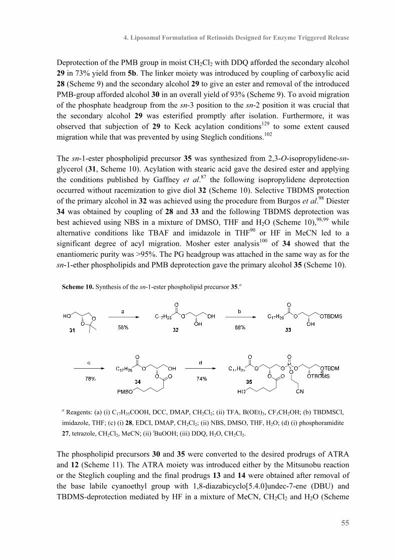
4. Liposomal Formulation of Retinoids Designed for Enzyme Triggered Release
55
Deprotection of the PMB group in moist CH2Cl2 with DDQ afforded the secondary alcohol 29 in 73% yield from 5b. The linker moiety was introduced by coupling of carboxylic acid 28 (Scheme 9) and the secondary alcohol 29 to give an ester and removal of the introduced PMB-group afforded alcohol 30 in an overall yield of 93% (Scheme 9). To avoid migration of the phosphate headgroup from the sn-3 position to the sn-2 position it was crucial that the secondary alcohol 29 was esterified promptly after isolation. Furthermore, it was observed that subjection of 29 to Keck acylation conditions129 to some extent caused migration while that was prevented by using Steglich conditions.102 The sn-1-ester phospholipid precursor 35 was synthesized from 2,3-O-isopropylidene-sn-glycerol (31, Scheme 10). Acylation with stearic acid gave the desired ester and applying the conditions published by Gaffney et al.87 the following isopropylidene deprotection occurred without racemization to give diol 32 (Scheme 10). Selective TBDMS protection of the primary alcohol in 32 was achieved using the procedure from Burgos et al.98 Diester 34 was obtained by coupling of 28 and 33 and the following TBDMS deprotection was best achieved using NBS in a mixture of DMSO, THF and H2O (Scheme 10),98,99 while alternative conditions like TBAF and imidazole in THF90 or HF in MeCN led to a significant degree of acyl migration. Mosher ester analysis100 of 34 showed that the enantiomeric purity was >95%. The PG headgroup was attached in the same way as for the sn-1-ether phospholipids and PMB deprotection gave the primary alcohol 35 (Scheme 10).
Scheme 10. Synthesis of the sn-1-ester phospholipid precursor 35.a
a Reagents: (a) (i) C17H35COOH, DCC, DMAP, CH2Cl2; (ii) TFA, B(OEt)3, CF3CH2OH; (b) TBDMSCl,
imidazole, THF; (c) (i) 28, EDCI, DMAP, CH2Cl2; (ii) NBS, DMSO, THF, H2O; (d) (i) phosphoramidite
27, tetrazole, CH2Cl2, MeCN; (ii) tBuOOH; (iii) DDQ, H2O, CH2Cl2.
The phospholipid precursors 30 and 35 were converted to the desired prodrugs of ATRA and 12 (Scheme 11). The ATRA moiety was introduced either by the Mitsunobu reaction or the Steglich coupling and the final prodrugs 13 and 14 were obtained after removal of the base labile cyanoethyl group with 1,8-diazabicyclo[5.4.0]undec-7-ene (DBU) and TBDMS-deprotection mediated by HF in a mixture of MeCN, CH2Cl2 and H2O (Scheme

Liposomal Drug Delivery
56
11). Thereby the novel route to PG lipids was completed and compared to previous routes (see Table 16) shorter reaction timesix are a major improvement making the chemistry convenient and applicable. Prodrugs 15 and 16 were accessed by coupling between 12 and the primary alcohols 30 and 35, after which the remaining chemistry was similar to the synthesis of 13 and 14 (Scheme 11). 31P NMR of the synthesized prodrugs showed one signal resonating between –1 ppm and 2 ppm, demonstrating high diastereomeric and regioisomeric purity (>95%). Scheme 11. Synthesis of the sn-1-ether and sn-1-ester prodrugs 13, 14, 15 and 16.a
a Reagents: (a) (i) 30, ATRA, DIAD, PPh3, THF or 35, ATRA, DCC, DMAP, Et3N, Et2O; (ii) DBU, CH2Cl2;
(iii) HF, H2O, CH2Cl2, MeCN; (b) (i) 12, DCC, DMAP, CH2Cl2; (ii) DBU, CH2Cl2; (iii) HF, H2O, CH2Cl2,
MeCN.
For the cytotoxicity studies access to lysolipids 17 and 18 are needed as standards for comparison with the tested phospholipid prodrugs and while sn-1-ester lysolipid 18 is commercially available lysolipid 17 was synthesized in two steps from alcohol 29 (Scheme 12). Likewise access to phospholipids with stearic acid in the sn-2 position instead of the retinoid esters are needed and while 1,2-distearoyl-sn-glycero-3-phosphoglycerol (DSPG) is commercially available 1-O-stearyl-2-stearoyl-sn-glycero-3-phosphoglycerol (1-O-DSPG) was synthesized in three steps from alcohol 29 (Scheme 12).
ix The last two deprotections in Scheme 11 were achieved within reaction times of 1 h and 3.5 h, respectively.
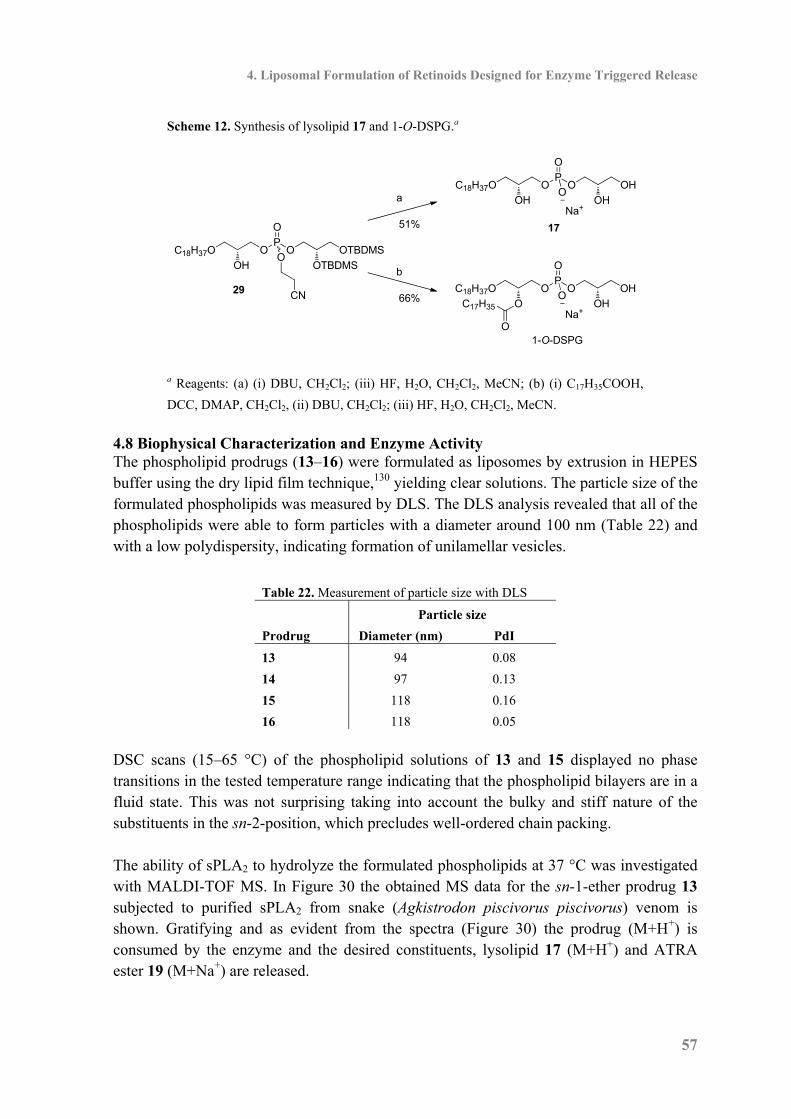
4. Liposomal Formulation of Retinoids Designed for Enzyme Triggered Release
57
Scheme 12. Synthesis of lysolipid 17 and 1-O-DSPG.a
a
29
b
C18H37O O
OH
P
O
O OH
OH
17
O
Na+
C18H37O O
O
P
O
O OH
OH
1-O-DSPG
O
Na+
O
C17H35
51%
66%
O
OH
C18H37OP
O
O
OOTBDMS
CN
OTBDMS
a Reagents: (a) (i) DBU, CH2Cl2; (iii) HF, H2O, CH2Cl2, MeCN; (b) (i) C17H35COOH,
DCC, DMAP, CH2Cl2, (ii) DBU, CH2Cl2; (iii) HF, H2O, CH2Cl2, MeCN. 4.8 Biophysical Characterization and Enzyme Activity The phospholipid prodrugs (13–16) were formulated as liposomes by extrusion in HEPES buffer using the dry lipid film technique,130 yielding clear solutions. The particle size of the formulated phospholipids was measured by DLS. The DLS analysis revealed that all of the phospholipids were able to form particles with a diameter around 100 nm (Table 22) and with a low polydispersity, indicating formation of unilamellar vesicles.
Table 22. Measurement of particle size with DLS
Particle size
Prodrug Diameter (nm) PdI
13 94 0.08
14 97 0.13
15 118 0.16
16 118 0.05 DSC scans (15–65 °C) of the phospholipid solutions of 13 and 15 displayed no phase transitions in the tested temperature range indicating that the phospholipid bilayers are in a fluid state. This was not surprising taking into account the bulky and stiff nature of the substituents in the sn-2-position, which precludes well-ordered chain packing. The ability of sPLA2 to hydrolyze the formulated phospholipids at 37 °C was investigated with MALDI-TOF MS. In Figure 30 the obtained MS data for the sn-1-ether prodrug 13 subjected to purified sPLA2 from snake (Agkistrodon piscivorus piscivorus) venom is shown. Gratifying and as evident from the spectra (Figure 30) the prodrug (M+H+) is consumed by the enzyme and the desired constituents, lysolipid 17 (M+H+) and ATRA ester 19 (M+Na+) are released.

Liposomal Drug Delivery
58
Figure 30. MALDI-TOF MS monitoring of snake (Agkistrodon piscivorus piscivorus) venom sPLA2 activity
on the sn-1-ether prodrug 13. The spectra demonstrate that the prodrug 13 (top right) is consumed and that
the lysolipid 17 (top left) and the ATRA ester 19 (bottom) are released. The MALDI-TOF MS analysis of the sn-1-ether prodrugs 13 and 15 and the sn-1-ester prodrugs 14 and 16 revealed that all of the prodrugs were degraded by the enzyme (Table 23) and the desired molecules were released. The observation that the sn-1-ether prodrugs were consumed by sPLA2 to the same extent as the sn-1-ester prodrugs illustrate and confirm that sn-1-ether phospholipids are excellent substrates for sPLA2. Based on the MALDI-TOF MS analysis of the sPLA2 activity for the various prodrugs it can be concluded that the incorporation of an aliphatic C6-linker between ATRA and the phospholipid backbone resolved the issue of sPLA2 activity on ATRA-phospholipid prodrugs. The experimental observations are in agreement with the outcome from the MD simulations, illustrating the power and potential of using MD simulations in the design of sPLA2 degradable phospholipid prodrugs. In order to rule out that the degradation of the prodrugs was caused by non-enzymatic hydrolysis, a sample of each prodrug was subjected to the reaction conditions in the absence of sPLA2. As evident from the spectra in Figure 30 no degradation of the prodrugs was observed within 48 h and none of the release products were detectable. Finally, the enzyme activity of the purified sPLA2 from snake (Agkistrodon piscivorus piscivorus) venom and commercially available sPLA2 from snake (Naja mossambica mossambica) venom was compared and as can be seen in Table 23, both enzymes were able to hydrolyze the substrates and no significant differences in hydrolysis rate or extent were observed.
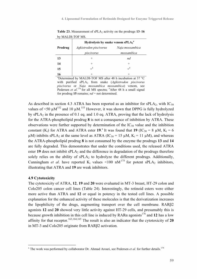
4. Liposomal Formulation of Retinoids Designed for Enzyme Triggered Release
59
Table 23. Measurement of sPLA2 activity on the prodrugs 13–16
by MALDI-TOF MS.
Hydrolysis by snake venom sPLA2a
Prodrug Agkistrodon piscivorus
piscivorus
Naja mossambica
mossambica
13 + nd
14 + +
15 +b +b
16 + + aDetermined by MALDI-TOF MS after 48 h incubation at 37 °C with purified sPLA2 from snake (Agkistrodon piscivorus piscivorus or Naja mossambica mossambica) venom, see Pedersen et al.174 for all MS spectra; bAfter 48 h a small signal for prodrug 15 remains; nd = not determined.
As described in section 4.3 ATRA has been reported as an inhibitor for sPLA2, with IC50 values of <50 µM152 and 10 µM.153 However, it was shown that DPPG is fully hydrolyzed by sPLA2 in the presence of 0.1 eq. and 1.0 eq. ATRA, proving that the lack of hydrolysis for the ATRA-phospholipid prodrug 8 is not a consequence of inhibition by ATRA. These observations were further supported by determination of the IC50 value and the inhibition constant (Ki) for ATRA and ATRA ester 19.x It was found that 19 (IC50 = 8 µM, Ki = 6 µM) inhibits sPLA2 at the same level as ATRA (IC50 = 15 µM, Ki = 11 µM), and whereas the ATRA-phospholipid prodrug 8 is not consumed by the enzyme the prodrugs 13 and 14 are fully degraded. This demonstrates that under the conditions used, the released ATRA ester 19 does not inhibit sPLA2 and the difference in degradation of the prodrugs therefore solely relies on the ability of sPLA2 to hydrolyze the different prodrugs. Additionally, Cunningham et al. have reported Ki values <100 nM 175 for potent sPLA2 inhibitors, illustrating that ATRA and 19 are weak inhibitors. 4.9 Cytotoxicity The cytotoxicity of ATRA, 12, 19 and 20 were evaluated in MT-3 breast, HT-29 colon and Colo205 colon cancer cell lines (Table 24). Interestingly, the retinoid esters were either more active than ATRA and 12 or equal in potency in the tested cell lines. A possible explanation for the enhanced activity of these molecules is that the derivatization increases the lipophilicity of the drugs, augmenting transport over the cell membrane. RARβ2 agonists 12 and 20 showed very little activity against HT-29 cells, and presumably this is because growth inhibition in this cell line is induced by RARα agonists176 and 12 has a low affinity for that receptor.163,164,165 The result is also an indicator that the cytotoxicity of 20 in MT-3 and Colo205 originate from RARβ2 activation.
x The work was performed by collaborator Dr. Ahmad Arouri, see Pedersen et al. for further details.174
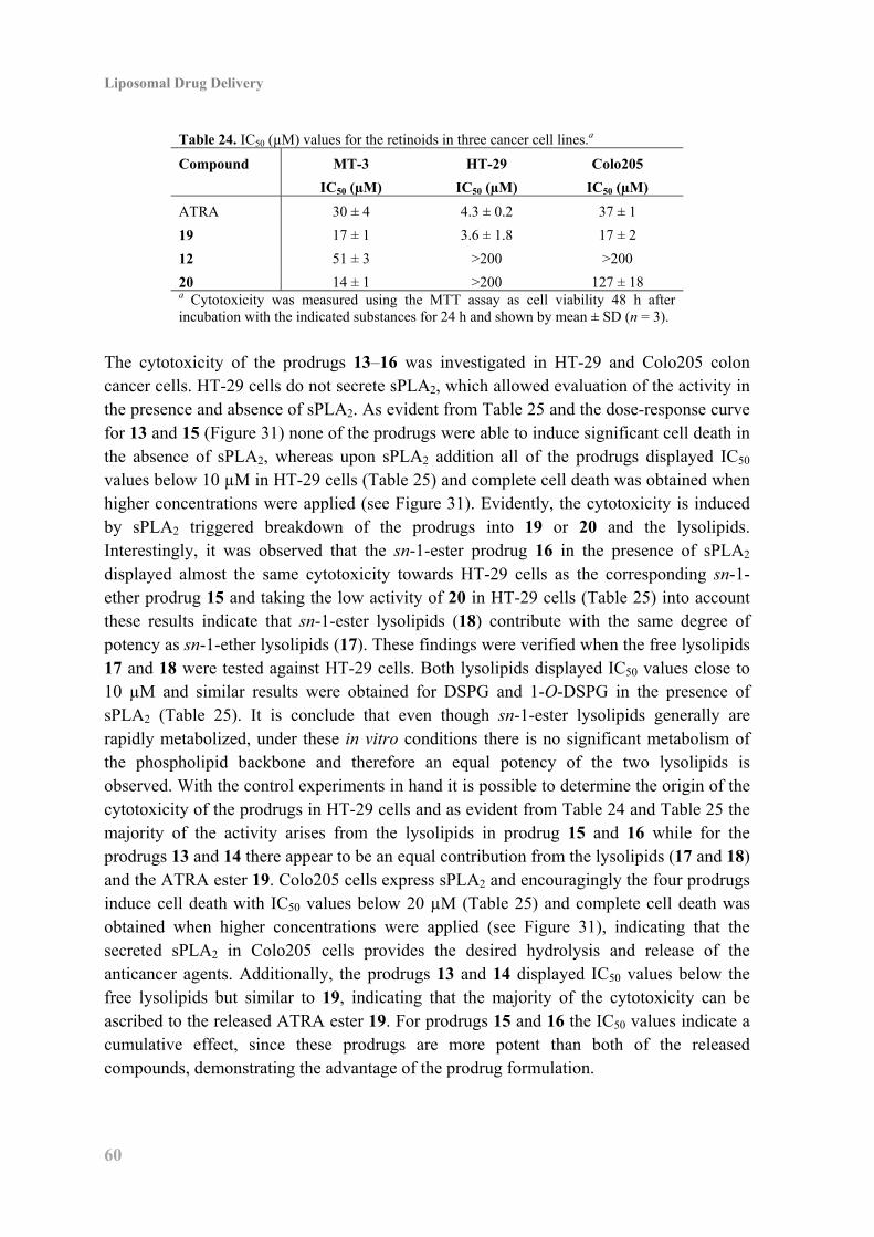
Liposomal Drug Delivery
60
Table 24. IC50 (µM) values for the retinoids in three cancer cell lines.a
Compound MT-3
IC50 (µM)
HT-29
IC50 (µM)
Colo205
IC50 (µM)
ATRA 30 ± 4 4.3 ± 0.2 37 ± 1
19 17 ± 1 3.6 ± 1.8 17 ± 2
12 51 ± 3 >200 >200
20 14 ± 1 >200 127 ± 18 a Cytotoxicity was measured using the MTT assay as cell viability 48 h after incubation with the indicated substances for 24 h and shown by mean ± SD (n = 3).
The cytotoxicity of the prodrugs 13–16 was investigated in HT-29 and Colo205 colon cancer cells. HT-29 cells do not secrete sPLA2, which allowed evaluation of the activity in the presence and absence of sPLA2. As evident from Table 25 and the dose-response curve for 13 and 15 (Figure 31) none of the prodrugs were able to induce significant cell death in the absence of sPLA2, whereas upon sPLA2 addition all of the prodrugs displayed IC50 values below 10 µM in HT-29 cells (Table 25) and complete cell death was obtained when higher concentrations were applied (see Figure 31). Evidently, the cytotoxicity is induced by sPLA2 triggered breakdown of the prodrugs into 19 or 20 and the lysolipids. Interestingly, it was observed that the sn-1-ester prodrug 16 in the presence of sPLA2 displayed almost the same cytotoxicity towards HT-29 cells as the corresponding sn-1-ether prodrug 15 and taking the low activity of 20 in HT-29 cells (Table 25) into account these results indicate that sn-1-ester lysolipids (18) contribute with the same degree of potency as sn-1-ether lysolipids (17). These findings were verified when the free lysolipids 17 and 18 were tested against HT-29 cells. Both lysolipids displayed IC50 values close to 10 µM and similar results were obtained for DSPG and 1-O-DSPG in the presence of sPLA2 (Table 25). It is conclude that even though sn-1-ester lysolipids generally are rapidly metabolized, under these in vitro conditions there is no significant metabolism of the phospholipid backbone and therefore an equal potency of the two lysolipids is observed. With the control experiments in hand it is possible to determine the origin of the cytotoxicity of the prodrugs in HT-29 cells and as evident from Table 24 and Table 25 the majority of the activity arises from the lysolipids in prodrug 15 and 16 while for the prodrugs 13 and 14 there appear to be an equal contribution from the lysolipids (17 and 18) and the ATRA ester 19. Colo205 cells express sPLA2 and encouragingly the four prodrugs induce cell death with IC50 values below 20 µM (Table 25) and complete cell death was obtained when higher concentrations were applied (see Figure 31), indicating that the secreted sPLA2 in Colo205 cells provides the desired hydrolysis and release of the anticancer agents. Additionally, the prodrugs 13 and 14 displayed IC50 values below the free lysolipids but similar to 19, indicating that the majority of the cytotoxicity can be ascribed to the released ATRA ester 19. For prodrugs 15 and 16 the IC50 values indicate a cumulative effect, since these prodrugs are more potent than both of the released compounds, demonstrating the advantage of the prodrug formulation.
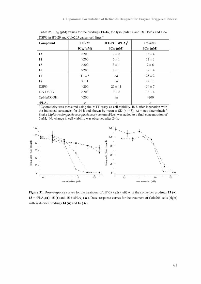
4. Liposomal Formulation of Retinoids Designed for Enzyme Triggered Release
61
Table 25. IC50 (µM) values for the prodrugs 13–16, the lysolipids 17 and 18, DSPG and 1-O-
DSPG in HT-29 and Colo205 cancer cell lines.a
Compound HT-29
IC50 (µM)
HT-29 + sPLA2b
IC50 (µM)
Colo205
IC50 (µM)
13 >200 7 ± 2 16 ± 4
14 >200 6 ± 1 12 ± 3
15 >200 3 ± 1 7 ± 6
16 >200 8 ± 1 19 ± 4
17 11 ± 6 nd 25 ± 2
18 7 ± 1 nd 22 ± 3
DSPG >200 25 ± 11 54 ± 7
1-O-DSPG >200 9 ± 2 33 ± 4
C17H35COOH >200 nd >200
sPLA2 c c c a Cytotoxicity was measured using the MTT assay as cell viability 48 h after incubation with the indicated substances for 24 h and shown by mean ± SD (n ≥ 3); nd = not determined; b Snake (Agkistrodon piscivorus piscivorus) venom sPLA2 was added to a final concentration of 5 nM; c No change in cell viability was observed after 24 h.
0,1 1 10 100
0
20
40
60
80
100
120
livin
g ce
lls (
% o
f co
ntro
l)
concentration (µM)
0,1 1 10 100
0
25
50
75
100
125
livin
g ce
lls (
% o
f co
ntro
l)
concentration (µM)
Figure 31. Dose–response curves for the treatment of HT-29 cells (left) with the sn-1-ether prodrugs 13 (●),
13 + sPLA2 (■), 15 (♦) and 15 + sPLA2 (▲). Dose–response curves for the treatment of Colo205 cells (right)
with sn-1-ester prodrugs 14 (■) and 16 (▲).
Liposomal Drug Delivery
62
4.10 Conclusion The ATRA phospholipid prodrug was synthesized but sPLA2 was not able to hydrolyze the desired ester bond and MD simulations confirmed that 8 has an imperfect positioning in sPLA2. Based on MD simulations, a revised prodrug strategy for enzyme triggered delivery of retinoids was developed in which an aliphatic C6-linker was incorporated between the glycerol backbone of the phospholipids and the retinoids. The phospholipid prodrugs of retinoids (13–16) were synthesized and gratifyingly, it was demonstrated that they can be formulated as liposomes that are degraded by sPLA2. The cytotoxicity studies showed that prodrug 13–16 require sPLA2 in order to induce significant cell death. It is therefore evident that the revised prodrug strategy presents a solution to liposomal formulation of retinoids.
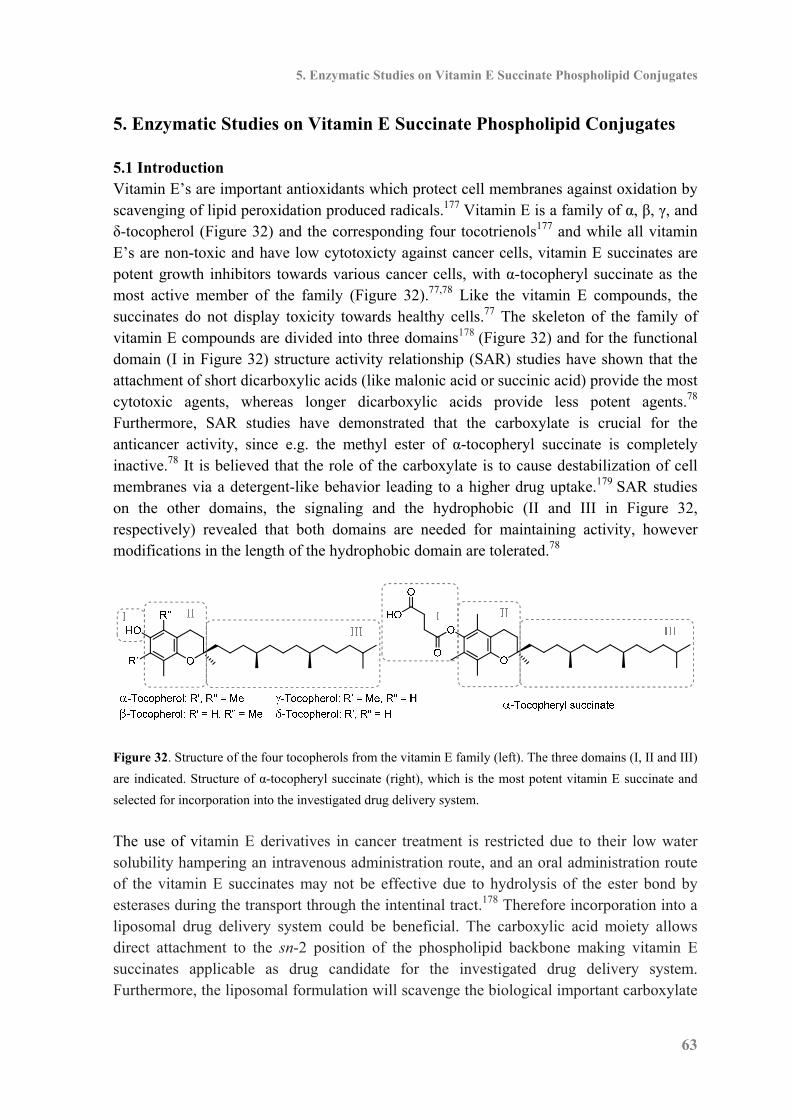
5. Enzymatic Studies on Vitamin E Succinate Phospholipid Conjugates
63
5. Enzymatic Studies on Vitamin E Succinate Phospholipid Conjugates 5.1 Introduction Vitamin E’s are important antioxidants which protect cell membranes against oxidation by scavenging of lipid peroxidation produced radicals.177 Vitamin E is a family of α, β, γ, and δ-tocopherol (Figure 32) and the corresponding four tocotrienols177 and while all vitamin E’s are non-toxic and have low cytotoxicty against cancer cells, vitamin E succinates are potent growth inhibitors towards various cancer cells, with α-tocopheryl succinate as the most active member of the family (Figure 32).77,78 Like the vitamin E compounds, the succinates do not display toxicity towards healthy cells.77 The skeleton of the family of vitamin E compounds are divided into three domains178 (Figure 32) and for the functional domain (I in Figure 32) structure activity relationship (SAR) studies have shown that the attachment of short dicarboxylic acids (like malonic acid or succinic acid) provide the most cytotoxic agents, whereas longer dicarboxylic acids provide less potent agents.78 Furthermore, SAR studies have demonstrated that the carboxylate is crucial for the anticancer activity, since e.g. the methyl ester of α-tocopheryl succinate is completely inactive.78 It is believed that the role of the carboxylate is to cause destabilization of cell membranes via a detergent-like behavior leading to a higher drug uptake.179 SAR studies on the other domains, the signaling and the hydrophobic (II and III in Figure 32, respectively) revealed that both domains are needed for maintaining activity, however modifications in the length of the hydrophobic domain are tolerated.78
Figure 32. Structure of the four tocopherols from the vitamin E family (left). The three domains (I, II and III)
are indicated. Structure of α-tocopheryl succinate (right), which is the most potent vitamin E succinate and
selected for incorporation into the investigated drug delivery system.
The use of vitamin E derivatives in cancer treatment is restricted due to their low water solubility hampering an intravenous administration route, and an oral administration route of the vitamin E succinates may not be effective due to hydrolysis of the ester bond by esterases during the transport through the intentinal tract.178 Therefore incorporation into a liposomal drug delivery system could be beneficial. The carboxylic acid moiety allows direct attachment to the sn-2 position of the phospholipid backbone making vitamin E succinates applicable as drug candidate for the investigated drug delivery system. Furthermore, the liposomal formulation will scavenge the biological important carboxylate

Liposomal Drug Delivery
64
moiety by formation of an ester bond hence only tissue with an over expression of sPLA2 will be affected by the injected liposomes. α-Tocopheryl succinate was selected as the vitamin E succinate to study, since this compound is commercially available, cheap and potent. The target phospholipid prodrugs are shown in Figure 33 and in order to examine differences in biophysical and biological properties along with enzyme activity both the sn-1-ester and the sn-1-ether phospholipid prodrug were synthesized.
Figure 33. Structure of the target α-tocopheryl succinate phospholipid prodrugs.
5.2 Synthesis of α-Tocopheryl Succinate Phospholipid Prodrugs The sn-1-ether phospholipid prodrug 36 was accessible in three synthetic steps from the previously prepared secondary alcohol 29 (Scheme 13). Acylation of 29 with α-tocopheryl succinate gave the desired ester bond and the following deprotections of the cyanoethyl and the TBDMS groups were achieved using the same conditions as in synthesis of 13–16 (Scheme 11), giving the sn-1-ether phospholipid prodrug 36 in 61% yield over the three steps (Scheme 13).
Scheme 13. Synthesis of the sn-1-ether phospholipid prodrug 36.a
aReagents: (a) (i) α-Tocopheryl succinate, EDCI, DMAP, EtOAc, CH2Cl2; (ii) DBU,
CH2Cl2; (iii) HF, H2O, CH2Cl, MeCN.
The synthesis of the sn-1-ester phospholipid prodrug 37 (Scheme 14) was initiated from the previously prepared secondary alcohol 33 (Scheme 10). Alcohol 33 was reacted with α-tocopheryl succinate creating the ester bond and the TBDMS group was removed by the NBS mediated desilylation method, also applied in the synthesis of 34 (Scheme 10), and no migration or degradation of the α-tocopheryl succinyl moiety was observed. The phosphate headgroup was inserted via reaction with the phosphoramidite 27 applying the established

5. Enzymatic Studies on Vitamin E Succinate Phospholipid Conjugates
65
coupling procedure (see e.g. Scheme 9) and after the final deprotections the sn-1-ester phospholipid prodrug 37 was accessed in 44% yield from 38 (Scheme 14). Scheme 14. Synthesis of the sn-1-ester phospholipid prodrug 37.a
aReagents: (a) (i) α-tocopheryl succinate, EDCI, DMAP, CH2Cl2; (ii) NBS, DMSO, THF, H2O; (b) (i)phosphoramidite 27, tetrazole, CH2Cl2, MeCN; (ii) tBuOOH; (iii) DBU, CH2Cl2; (iv) HF, H2O, CH2Cl,MeCN.
5.3 Enzyme Activity and Cytotoxicity The α-tocopheryl succinate phospholipid prodrugs 36 and 37 were hydrated in HEPES buffer yielding milky solutions and a DSC scan (20–65 °C) was obtained. But no Tm was observed in the tested range, indicating that 36 and 37 are in a fluid state at 20 °C. These findings were further supported during the formulation where it was possible to extrude the solutions of 36 and 37 through a 100 nm filter at 20 °C, yielding clear and transparent solutions. DLS analysis of the formulated phospholipids revealed that particles with an average diameter of 115 nm and a low polydispersity was formed (Table 26), indicating formation of unilamellar vesicles.
Table 26. DLS analysis of phospholipid solutions
hydrated in HEPES buffer and extruded through a
100 nm filter.
Compound Particle size
Diameter (nm) PdI
36 115 0.09
37 115 0.06
40 80 0.14
41a 191 0.23
41 115 0.49
42a 168 0.33
42 100 0.35 aNot extruded
In order to study the enzyme activity snake (Naja mossambica mossambica) venom sPLA2 was added to the phospholipid solutions and the mixtures were incubated at 37 °C for 48 h. The activity was monitored by MALDI-TOF MS but disappointingly and as evident from the MS spectra in Figure 34 both phospholipids are poor substrates for the enzyme and
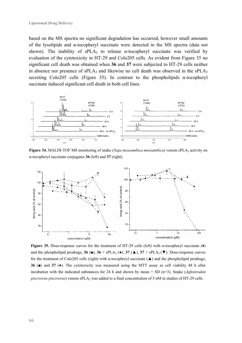
Liposomal Drug Delivery
66
based on the MS spectra no significant degradation has occurred, however small amounts of the lysolipids and α-tocopheryl succinate were detected in the MS spectra (data not shown). The inability of sPLA2 to release α-tocopheryl succinate was verified by evaluation of the cytotoxicity in HT-29 and Colo205 cells. As evident from Figure 35 no significant cell death was obtained when 36 and 37 were subjected to HT-29 cells neither in absence nor presence of sPLA2 and likewise no cell death was observed in the sPLA2 secreting Colo205 cells (Figure 35). In contrast to the phospholipids α-tocopheryl succinate induced significant cell death in both cell lines.
Figure 34. MALDI-TOF MS monitoring of snake (Naja mossambica mossambica) venom sPLA2 activity on
α-tocopheryl succinate conjugates 36 (left) and 37 (right).
Figure 35. Dose-response curves for the treatment of HT-29 cells (left) with α-tocopheryl succinate (♦)
and the phospholipid prodrugs, 36 (■), 36 + sPLA2 (●), 37 (▲), 37 + sPLA2 (▼). Dose-response curves
for the treatment of Colo205 cells (right) with α-tocopheryl succinate (▲) and the phospholipid prodrugs,
36 (■) and 37 (●). The cytotoxicity was measured using the MTT assay as cell viability 48 h after
incubation with the indicated substances for 24 h and shown by mean + SD (n=3). Snake (Agkistrodon
piscivorus piscivorus) venom sPLA2 was added to a final concentration of 5 nM in studies of HT-29 cells.
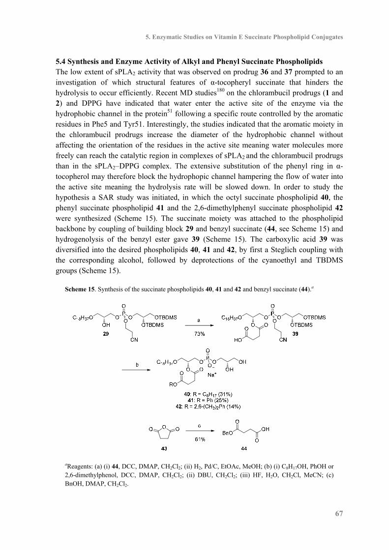
5. Enzymatic Studies on Vitamin E Succinate Phospholipid Conjugates
67
5.4 Synthesis and Enzyme Activity of Alkyl and Phenyl Succinate Phospholipids The low extent of sPLA2 activity that was observed on prodrug 36 and 37 prompted to an investigation of which structural features of α-tocopheryl succinate that hinders the hydrolysis to occur efficiently. Recent MD studies180 on the chlorambucil prodrugs (1 and 2) and DPPG have indicated that water enter the active site of the enzyme via the hydrophobic channel in the protein51 following a specific route controlled by the aromatic residues in Phe5 and Tyr51. Interestingly, the studies indicated that the aromatic moiety in the chlorambucil prodrugs increase the diameter of the hydrophobic channel without affecting the orientation of the residues in the active site meaning water molecules more freely can reach the catalytic region in complexes of sPLA2 and the chlorambucil prodrugs than in the sPLA2–DPPG complex. The extensive substitution of the phenyl ring in α-tocopherol may therefore block the hydrophopic channel hampering the flow of water into the active site meaning the hydrolysis rate will be slowed down. In order to study the hypothesis a SAR study was initiated, in which the octyl succinate phospholipid 40, the phenyl succinate phospholipid 41 and the 2,6-dimethylphenyl succinate phospholipid 42 were synthesized (Scheme 15). The succinate moiety was attached to the phospholipid backbone by coupling of building block 29 and benzyl succinate (44, see Scheme 15) and hydrogenolysis of the benzyl ester gave 39 (Scheme 15). The carboxylic acid 39 was diversified into the desired phospholipids 40, 41 and 42, by first a Steglich coupling with the corresponding alcohol, followed by deprotections of the cyanoethyl and TBDMS groups (Scheme 15).
Scheme 15. Synthesis of the succinate phospholipids 40, 41 and 42 and benzyl succinate (44).a
aReagents: (a) (i) 44, DCC, DMAP, CH2Cl2; (ii) H2, Pd/C, EtOAc, MeOH; (b) (i) C8H17OH, PhOH or 2,6-dimethylphenol, DCC, DMAP, CH2Cl2; (ii) DBU, CH2Cl2; (iii) HF, H2O, CH2Cl, MeCN; (c) BnOH, DMAP, CH2Cl2.

Liposomal Drug Delivery
68
Hydration of 41 and 42 in HEPES buffer at 20 °C gave clear solutions and DLS analysis revealed that particles with an average diameter of 190 nm for 41 and 168 nm for 42 (Table 26) were present, indicating that some kind of non-multilamellar vesicles were formed. Surprisingly, it was not possible to extrude the clear phospholipid solutions of 41 and 42 through a 100 nm filter at 20 °C and the extrusion was therefore performed at 65 °C. DLS analysis of the extrude solutions showed that the average diameter was decreased to 115 and 100 nm for the phospholipid solutions of 41 and 42, respectively whereas the polydispersity was increased (Table 26). The results from the DLS analysis and the extrusions indicate that some form of robust particles with a diameter >100 nm is formed spontaneously upon subjection to HEPES buffer and it is likely this can be associated with the molecular structure of 41 and 42. The significant shorter chain in the sn-2 position than in the sn-1 position gives phospholipids with a different curvature and hence packing properties than phospholipids having chains of (almost) the same length, like in DPPG. But as these results were obtained in last part of the Ph.D. study there was not time for further investigations. Hydration of 40 in HEPES buffer at 20 °C gave the same milky solution as observed for the α-tocopheryl succinate phospholipid prodrugs 36 and 37 and the following extrusion through a 100 nm filter at 20 °C gave a clear solution with particles having an average diameter of 80 nm as measured by DLS (Table 26). The extruded solutions of 40, 41 and 42 were subjected to snake (Naja mossambica mossambica) venom sPLA2 and the enzyme activity was monitored by MALDI-TOF MS. As evident from Figure 36 the octyl succinate phospholipid 40 was completely degraded by the enzyme within 24 h, demonstrating that it is not the succinate moiety that hinders sPLA2 in releasing α-tocopheryl succinate. The enzyme activity on the phenyl succinate phospholipids 41 and 42 was almost as good as on 40, although small peaks for 41 and 42 remain in the MS spectra (Figure 36). Beyond the information that 40, 41 and 42 were consumed by sPLA2 the MS spectra also revealed that the desired lysolipid 17 was released in the three experiments (Figure 36). The enzymatic studies could indicate that the attachment of a phenyl moiety to the succinate to some extent reduce the enzyme activity, but compared to the α-tocopheryl succinate phospholipids the degree of hydrolysis is significantly higher for 41 and 42 by which the lack of hydrolysis on 36 and 37 cannot directly be associated with either the phenyl ring or the two methyl groups ortho to the hydroxyl group in α-tocopherol. It is therefore necessary to extend the SAR study with attachment of further phenols to the succinate moiety and likewise it will be interesting to examine other tocopherols like δ-tocopherol (see Figure 32) in order to get valuable information about the consequence of the methyl groups on the phenyl ring. Furthermore, MD simulations of vitamin E succinate phospholipid conjugates in the active site of sPLA2 would be interesting to obtain in order to study which structural features that hamper the enzyme activity. Potentially, the low degree of hydrolysis on 36 and 37 could also be related to an inhibition of sPLA2 by α-tocopheryl succinate. α-Tocopherol are reported as an inhibitor of sPLA2 by

5. Enzymatic Studies on Vitamin E Succinate Phospholipid Conjugates
69
Chandra et al.,181 whereas no information is available on esters of α-tocopherol. The ability of α-tocopheryl succinate to inhibit sPLA2 was therefore investigated by the same assay as applied during the inhibitions studies of ATRA and the ATRA ester 19. Gratifyingly, the studies demonstrated that α-tocopheryl succinate is a weaker inhibitor than ATRA ester 19 meaning that the low enzyme activity is not due to inhibition.
Figure 36. MALDI-TOF MS monitoring of snake (Naja mossambica mossambica) venom sPLA2 activity
on the succinate phospholipid 40 (top left), 41 (top right) and 42 (bottom left). The lysolipid 17 (bottom
right) was detected in all three experiments and shown from the enzyme study on 41.
5.5 Conclusion The α-tocopheryl succinate phospholipid prodrugs 36 and 37 were prepared in good yields utilizing previously developed chemistry, however the extent of hydrolysis by sPLA2 was low. Thus an investigation of the structural features preventing hydrolysis from occurring efficiently was initiated and the succinate phospholipids 40, 41 and 42 were synthesized. The enzymatic studies demonstrated that the succinate moiety is tolerated by the enzyme, whereas the studies indicated that the phenyl ring to some extent hampers the activity, but more phenyl succinate phospholipid analogues are needed in order to localize which structural motifs in α-tocopheryl succinate that reduce the enzyme activity.

6. Prostaglandin Phospholipid Conjugates with Unusual Properties
71
6. Prostaglandin Phospholipid Conjugates with Unusual Properties 6.1 Introduction Since von Euler182 and Goldblatt183 independently isolated and studied prostaglandins for the first time these fatty acids have attracted attention due to their involvement in many important biological functions.184,185,186 The biosynthetic precursor for protaglandins is arachidonic acid, which by a number of enzyme catalyzed reactions is converted into the diverse selections of prostaglandins known today.184,185,186
Figure 37. Potent antiproliferative prostaglandins.
Many prostaglandins have shown antiproliferative activity in tumor cells, but among the most studied and active prostaglandins are the dienone prostaglandins like Δ12-PGJ2 and Δ7-PGA1 (see Figure 37).187 Likewise, 15-deoxy-Δ12,14-PGJ2, a metabolic derivative of Δ12-PGJ2 (Figure 37) has demonstrated high antitumor activity against L1210 leukemia cells79 and for incorporation into the investigated drug delivery system that compound is more suitable than the former prostaglandins since 15-deoxy-Δ12,14-PGJ2 has a higher lipophilicity. The 15-deoxy-Δ12,14-PGJ2 phospholipid conjugates was designed to have the PG headgroup and C18 chains in the sn-1-position linked through either an ether-bond or ester-bond (see Figure 38).
Figure 38. Targeted 15-deoxy-Δ12,14-PGJ2 phospholipid conjugates.
Liposomal Drug Delivery
72
6.2 Synthesis of Prostaglandin Phospholipid Conjugates The sn-1-ether conjugate 45 (Scheme 16) was prepared by Steglich acylation102 of the phospholipid precurcor 29 with 15-deoxy-Δ12,14-PGJ2 after which the PG headgroup was deprotected under the same conditions as in the synthesis of 13–16 (Scheme 11). Access to the corresponding sn-1-ester conjugate 46 was attempted via the same route as applied in the synthesis of α-tocopheryl succinate prodrug 37 (Scheme 14), but the NBS mediated deprotection of the TBDMS group caused decomposition of the prostaglandin moiety. Therefore an approach similar to the synthesis of the sn-1-ether phospholipid prodrugs was initiated from alcohol 32 (Scheme 17). The secondary alcohol was PMB protected via subjection to PMBTCA in an acidic solution of toluene and the TBDMS group was removed with NBS in a moist mixture of DMSO and THF to afford 47 (Scheme 17). Conversion to phospholipid precursor 48 (Scheme 17) was achieved in the same way as 29 was obtained from 5b (Scheme 9). Then, freshly prepared 48 was acylated with 15-deoxy-Δ12,14-PGJ2 and deprotection of the PG headgroup led to the desired conjugate 46 (Scheme 16). Urea formation was observed during the carbodiimide mediated acylations with 15-deoxy-Δ12,14-PGJ2 which resulted in low yields of 45 and 46 (Scheme 16) but since 15-deoxy-Δ12,14-PGJ2 is expensive the compound was not used in excess and alternative acylation methods was not examined.
Scheme 16 Synthesis of the 15-deoxy-Δ12,14-PGJ2 phospholipid conjugates 45 and 46.a
aReagents: (a) (i) 15-deoxy-Δ12,14-PGJ2, DCC, DMAP, CH2Cl2; (ii) DBU, CH2Cl2; (iii) HF, H2O, CH2Cl2,
MeCN.
Scheme 17. Synthesis of sn-1-ester phospholipid precursors.a
aReagents: (a) (i) PMBTCA, La(OTf)3, toluene; (ii) NBS, DMSO, THF, H2O; (b) (i)
phosphoramidite 27, tetrazole, CH2Cl2, MeCN; (ii) tBuOOH; (iii) DDQ, H2O, CH2Cl2.
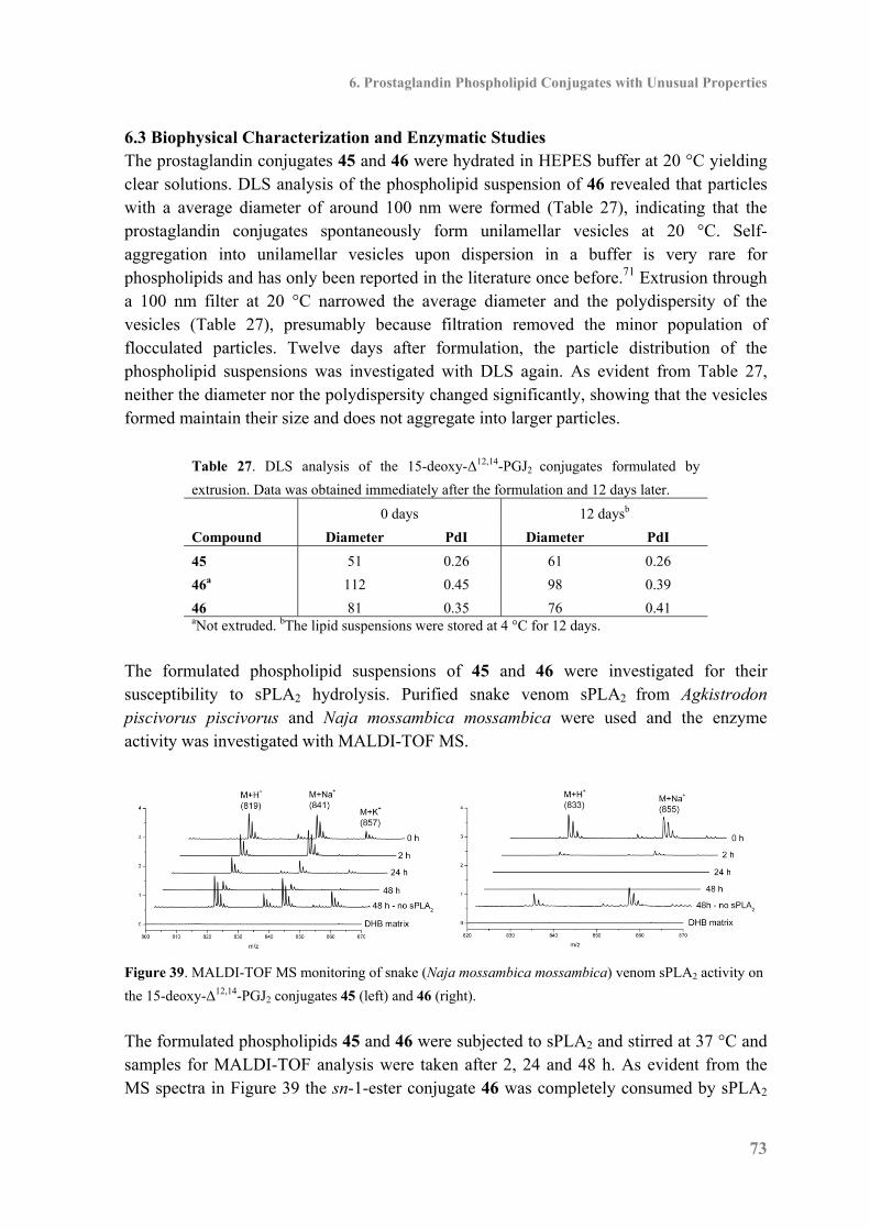
6. Prostaglandin Phospholipid Conjugates with Unusual Properties
73
6.3 Biophysical Characterization and Enzymatic Studies The prostaglandin conjugates 45 and 46 were hydrated in HEPES buffer at 20 °C yielding clear solutions. DLS analysis of the phospholipid suspension of 46 revealed that particles with a average diameter of around 100 nm were formed (Table 27), indicating that the prostaglandin conjugates spontaneously form unilamellar vesicles at 20 °C. Self-aggregation into unilamellar vesicles upon dispersion in a buffer is very rare for phospholipids and has only been reported in the literature once before.71 Extrusion through a 100 nm filter at 20 °C narrowed the average diameter and the polydispersity of the vesicles (Table 27), presumably because filtration removed the minor population of flocculated particles. Twelve days after formulation, the particle distribution of the phospholipid suspensions was investigated with DLS again. As evident from Table 27, neither the diameter nor the polydispersity changed significantly, showing that the vesicles formed maintain their size and does not aggregate into larger particles.
Table 27. DLS analysis of the 15-deoxy-Δ12,14-PGJ2 conjugates formulated by
extrusion. Data was obtained immediately after the formulation and 12 days later.
0 days 12 daysb
Compound Diameter PdI Diameter PdI
45 51 0.26 61 0.26
46a 112 0.45 98 0.39
46 81 0.35 76 0.41 aNot extruded. bThe lipid suspensions were stored at 4 °C for 12 days.
The formulated phospholipid suspensions of 45 and 46 were investigated for their susceptibility to sPLA2 hydrolysis. Purified snake venom sPLA2 from Agkistrodon piscivorus piscivorus and Naja mossambica mossambica were used and the enzyme activity was investigated with MALDI-TOF MS.
Figure 39. MALDI-TOF MS monitoring of snake (Naja mossambica mossambica) venom sPLA2 activity on
the 15-deoxy-Δ12,14-PGJ2 conjugates 45 (left) and 46 (right).
The formulated phospholipids 45 and 46 were subjected to sPLA2 and stirred at 37 °C and samples for MALDI-TOF analysis were taken after 2, 24 and 48 h. As evident from the MS spectra in Figure 39 the sn-1-ester conjugate 46 was completely consumed by sPLA2
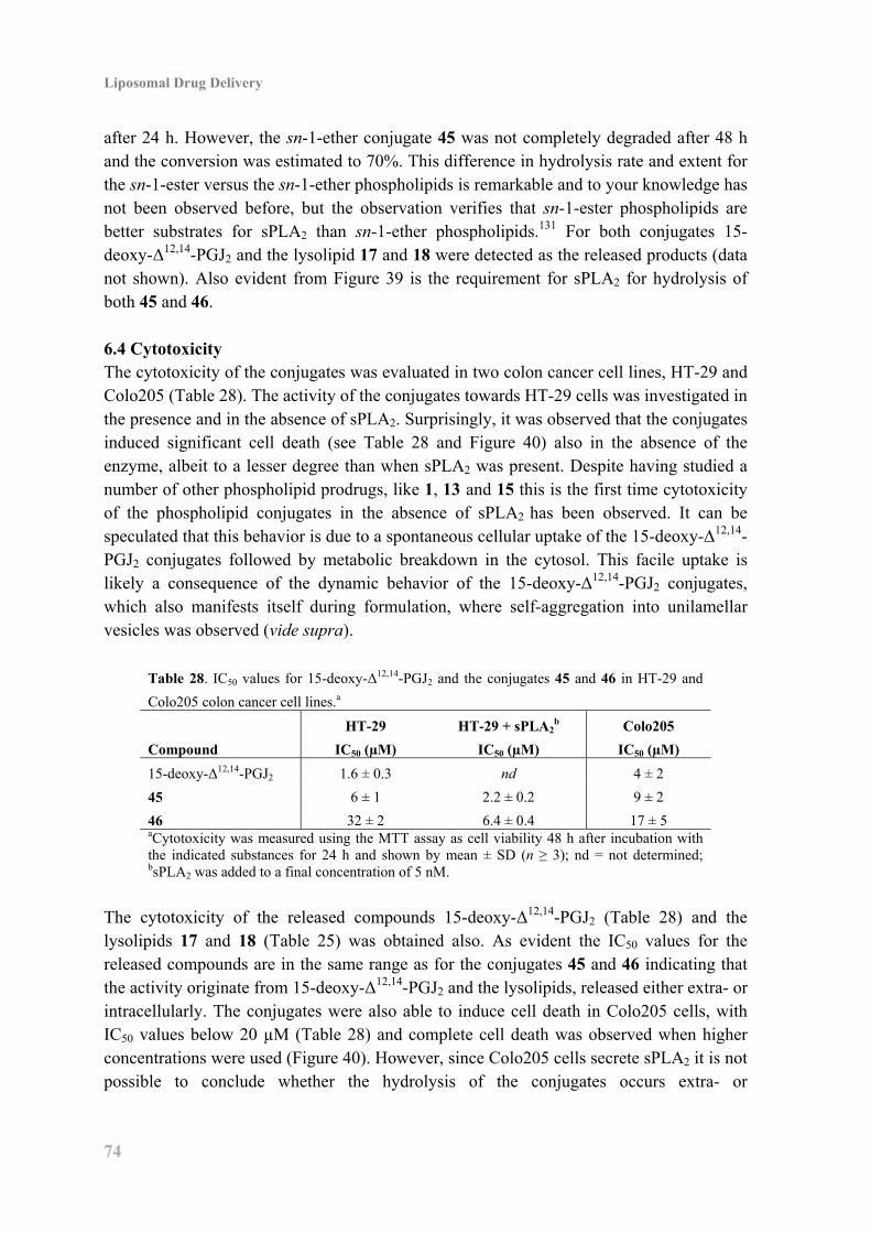
Liposomal Drug Delivery
74
after 24 h. However, the sn-1-ether conjugate 45 was not completely degraded after 48 h and the conversion was estimated to 70%. This difference in hydrolysis rate and extent for the sn-1-ester versus the sn-1-ether phospholipids is remarkable and to your knowledge has not been observed before, but the observation verifies that sn-1-ester phospholipids are better substrates for sPLA2 than sn-1-ether phospholipids.131 For both conjugates 15-deoxy-Δ12,14-PGJ2 and the lysolipid 17 and 18 were detected as the released products (data not shown). Also evident from Figure 39 is the requirement for sPLA2 for hydrolysis of both 45 and 46. 6.4 Cytotoxicity The cytotoxicity of the conjugates was evaluated in two colon cancer cell lines, HT-29 and Colo205 (Table 28). The activity of the conjugates towards HT-29 cells was investigated in the presence and in the absence of sPLA2. Surprisingly, it was observed that the conjugates induced significant cell death (see Table 28 and Figure 40) also in the absence of the enzyme, albeit to a lesser degree than when sPLA2 was present. Despite having studied a number of other phospholipid prodrugs, like 1, 13 and 15 this is the first time cytotoxicity of the phospholipid conjugates in the absence of sPLA2 has been observed. It can be speculated that this behavior is due to a spontaneous cellular uptake of the 15-deoxy-Δ12,14-PGJ2 conjugates followed by metabolic breakdown in the cytosol. This facile uptake is likely a consequence of the dynamic behavior of the 15-deoxy-Δ12,14-PGJ2 conjugates, which also manifests itself during formulation, where self-aggregation into unilamellar vesicles was observed (vide supra).
Table 28. IC50 values for 15-deoxy-Δ12,14-PGJ2 and the conjugates 45 and 46 in HT-29 and
Colo205 colon cancer cell lines.a
HT-29 HT-29 + sPLA2b Colo205
Compound IC50 (µM) IC50 (µM) IC50 (µM)
15-deoxy-Δ12,14-PGJ2 1.6 ± 0.3 nd 4 ± 2
45 6 ± 1 2.2 ± 0.2 9 ± 2
46 32 ± 2 6.4 ± 0.4 17 ± 5 aCytotoxicity was measured using the MTT assay as cell viability 48 h after incubation with the indicated substances for 24 h and shown by mean ± SD (n ≥ 3); nd = not determined; bsPLA2 was added to a final concentration of 5 nM.
The cytotoxicity of the released compounds 15-deoxy-Δ12,14-PGJ2 (Table 28) and the lysolipids 17 and 18 (Table 25) was obtained also. As evident the IC50 values for the released compounds are in the same range as for the conjugates 45 and 46 indicating that the activity originate from 15-deoxy-Δ12,14-PGJ2 and the lysolipids, released either extra- or intracellularly. The conjugates were also able to induce cell death in Colo205 cells, with IC50 values below 20 µM (Table 28) and complete cell death was observed when higher concentrations were used (Figure 40). However, since Colo205 cells secrete sPLA2 it is not possible to conclude whether the hydrolysis of the conjugates occurs extra- or
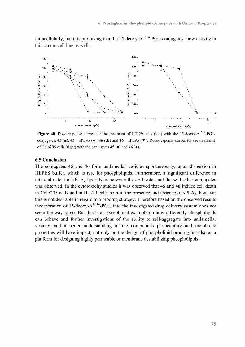
6. Prostaglandin Phospholipid Conjugates with Unusual Properties
75
intracellularly, but it is promising that the 15-deoxy-Δ12,14-PGJ2 conjugates show activity in this cancer cell line as well.
Figure 40. Dose-response curves for the treatment of HT-29 cells (left) with the 15-deoxy-Δ12,14-PGJ2
conjugates; 45 (■), 45 + sPLA2 (●), 46 (▲) and 46 + sPLA2 (▼). Dose-response curves for the treatment
of Colo205 cells (right) with the conjugates 45 (■) and 46 (●).
6.5 Conclusion The conjugates 45 and 46 form unilamellar vesicles spontaneously, upon dispersion in HEPES buffer, which is rare for phospholipids. Furthermore, a significant difference in rate and extent of sPLA2 hydrolysis between the sn-1-ester and the sn-1-ether conjugates was observed. In the cytotoxicity studies it was observed that 45 and 46 induce cell death in Colo205 cells and in HT-29 cells both in the presence and absence of sPLA2, however this is not desirable in regard to a prodrug strategy. Therefore based on the observed results incorporation of 15-deoxy-Δ12,14-PGJ2 into the investigated drug delivery system does not seem the way to go. But this is an exceptional example on how differently phospholipids can behave and further investigations of the ability to self-aggregate into unilamellar vesicles and a better understanding of the compounds permeability and membrane properties will have impact, not only on the design of phospholipid prodrug but also as a platform for designing highly permeable or membrane destabilizing phospholipids.

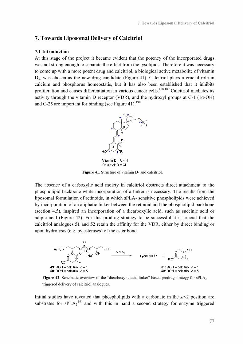
7. Towards Liposomal Delivery of Calcitriol
77
7. Towards Liposomal Delivery of Calcitriol 7.1 Introduction At this stage of the project it became evident that the potency of the incorporated drugs was not strong enough to separate the effect from the lysolipids. Therefore it was necessary to come up with a more potent drug and calcitriol, a biological active metabolite of vitamin D3, was chosen as the new drug candidate (Figure 41). Calcitriol plays a crucial role in calcium and phosphorus homeostatis, but it has also been established that it inhibits proliferation and causes differentiation in various cancer cells.188,189 Calcitriol mediates its activity through the vitamin D receptor (VDR), and the hydroxyl groups at C-1 (1α-OH) and C-25 are important for binding (see Figure 41).190
Figure 41. Structure of vitamin D3 and calcitriol.
The absence of a carboxylic acid moiety in calcitriol obstructs direct attachment to the phospholipid backbone while incorporation of a linker is necessary. The results from the liposomal formulation of retinoids, in which sPLA2 sensitive phospholipids were achieved by incorporation of an aliphatic linker between the retinoid and the phospholipid backbone (section 4.5), inspired an incorporation of a dicarboxylic acid, such as succinic acid or adipic acid (Figure 42). For this prodrug strategy to be successful it is crucial that the calcitriol analogues 51 and 52 retain the affinity for the VDR, either by direct binding or upon hydrolysis (e.g. by esterases) of the ester bond.
Figure 42. Schematic overview of the “dicarboxylic acid linker” based prodrug strategy for sPLA2
triggered delivery of calcitriol analogues.
Initial studies have revealed that phospholipids with a carbonate in the sn-2 position are substrates for sPLA2
191 and with this in hand a second strategy for enzyme triggered
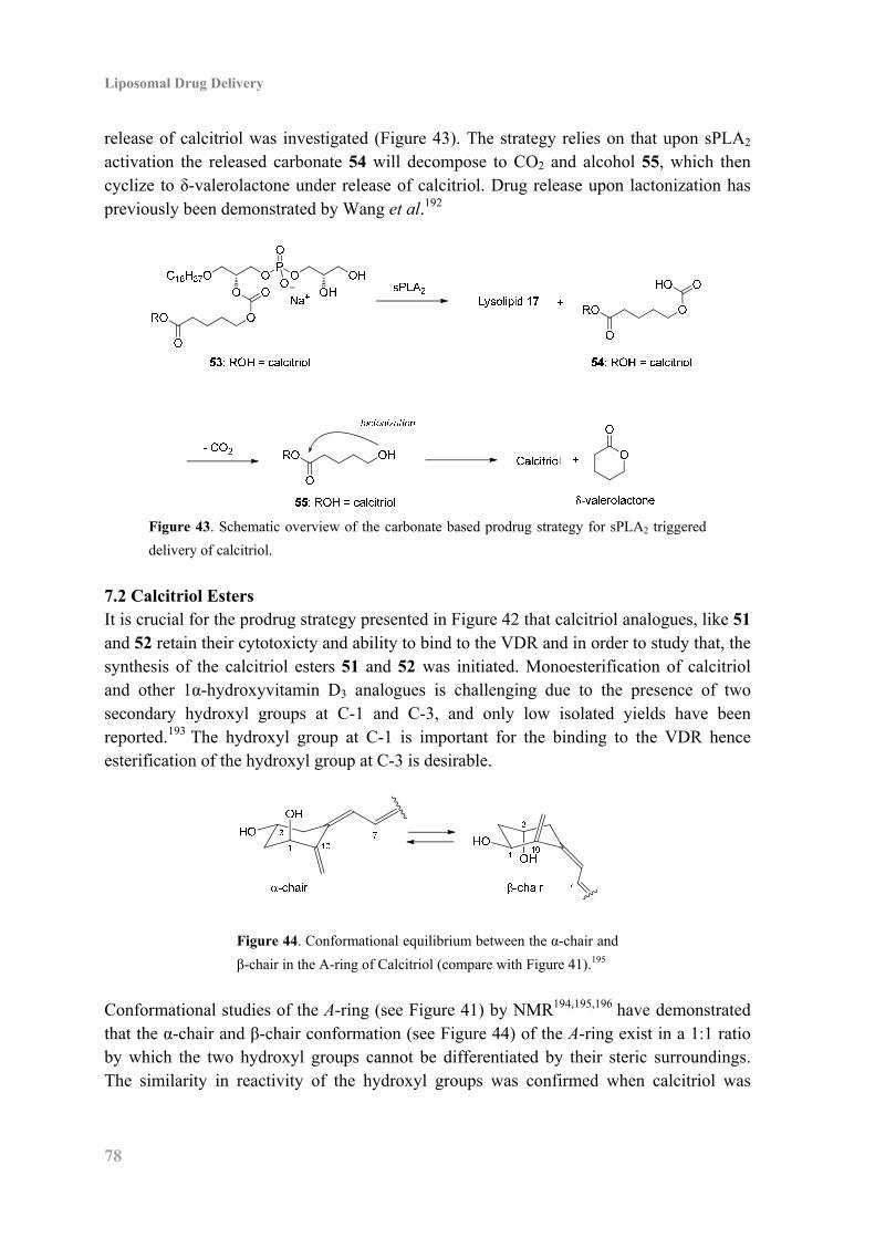
Liposomal Drug Delivery
78
release of calcitriol was investigated (Figure 43). The strategy relies on that upon sPLA2 activation the released carbonate 54 will decompose to CO2 and alcohol 55, which then cyclize to δ-valerolactone under release of calcitriol. Drug release upon lactonization has previously been demonstrated by Wang et al.192
Figure 43. Schematic overview of the carbonate based prodrug strategy for sPLA2 triggered
delivery of calcitriol.
7.2 Calcitriol Esters It is crucial for the prodrug strategy presented in Figure 42 that calcitriol analogues, like 51 and 52 retain their cytotoxicty and ability to bind to the VDR and in order to study that, the synthesis of the calcitriol esters 51 and 52 was initiated. Monoesterification of calcitriol and other 1α-hydroxyvitamin D3 analogues is challenging due to the presence of two secondary hydroxyl groups at C-1 and C-3, and only low isolated yields have been reported.193 The hydroxyl group at C-1 is important for the binding to the VDR hence esterification of the hydroxyl group at C-3 is desirable.
Figure 44. Conformational equilibrium between the α-chair and
β-chair in the A-ring of Calcitriol (compare with Figure 41).195
Conformational studies of the A-ring (see Figure 41) by NMR194,195,196 have demonstrated that the α-chair and β-chair conformation (see Figure 44) of the A-ring exist in a 1:1 ratio by which the two hydroxyl groups cannot be differentiated by their steric surroundings. The similarity in reactivity of the hydroxyl groups was confirmed when calcitriol was
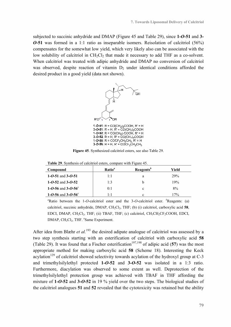
7. Towards Liposomal Delivery of Calcitriol
79
subjected to succinic anhydride and DMAP (Figure 45 and Table 29), since 1-O-51 and 3-O-51 was formed in a 1:1 ratio as inseparable isomers. Reisolation of calcitriol (56%) compensates for the somewhat low yield, which very likely also can be associated with the low solubility of calcitriol in CH2Cl2 that made it necessary to add THF as a co-solvent. When calcitriol was treated with adipic anhydride and DMAP no conversion of calcitriol was observed, despite reaction of vitamin D2 under identical conditions afforded the desired product in a good yield (data not shown).
Figure 45. Synthesized calcitriol esters, see also Table 29.
Table 29. Synthesis of calcitriol esters, compare with Figure 45.
Compound Ratioa Reagentsb Yield
1-O-51 and 3-O-51 1:1 a 29%
1-O-52 and 3-O-52 1:3 b 19%
1-O-56 and 3-O-56c 0:1 c 8%
1-O-56 and 3-O-56c 1:1 c 17% aRatio between the 1-O-calcitriol ester and the 3-O-calcitriol ester. bReagents: (a)
calcitriol, succinic anhydride, DMAP, CH2Cl2, THF; (b) (i) calcitriol, carboxylic acid 58,
EDCI, DMAP, CH2Cl2, THF; (ii) TBAF, THF; (c) calcitriol, CH3CH2CF2COOH, EDCI,
DMAP, CH2Cl2, THF. cSame Experiment.
After idea from Blæhr et al.193 the desired adipate analogue of calcitriol was assessed by a two step synthesis starting with an esterification of calcitriol with carboxylic acid 58 (Table 29). It was found that a Fischer esterification197,198 of adipic acid (57) was the most appropriate method for making carboxylic acid 58 (Scheme 18). Interesting the Keck acylation129 of calcitriol showed selectivity towards acylation of the hydroxyl group at C-3 and trimethylsilylethyl protected 1-O-52 and 3-O-52 was isolated in a 1:3 ratio. Furthermore, diacylation was observed to some extent as well. Deprotection of the trimethylsilylethyl protection group was achieved with TBAF in THF affording the mixture of 1-O-52 and 3-O-52 in 19 % yield over the two steps. The biological studies of the calcitriol analogues 51 and 52 revealed that the cytotoxicity was retained but the ability
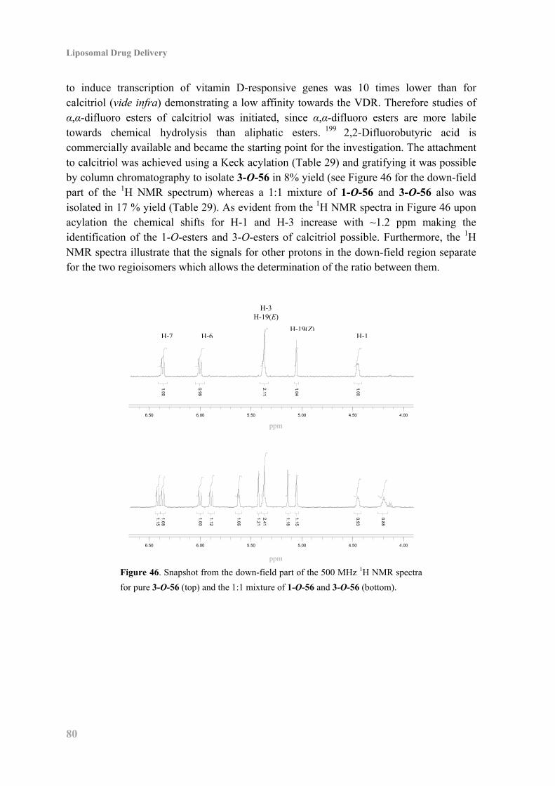
Liposomal Drug Delivery
80
to induce transcription of vitamin D-responsive genes was 10 times lower than for calcitriol (vide infra) demonstrating a low affinity towards the VDR. Therefore studies of α,α-difluoro esters of calcitriol was initiated, since α,α-difluoro esters are more labile towards chemical hydrolysis than aliphatic esters. 199 2,2-Difluorobutyric acid is commercially available and became the starting point for the investigation. The attachment to calcitriol was achieved using a Keck acylation (Table 29) and gratifying it was possible by column chromatography to isolate 3-O-56 in 8% yield (see Figure 46 for the down-field part of the 1H NMR spectrum) whereas a 1:1 mixture of 1-O-56 and 3-O-56 also was isolated in 17 % yield (Table 29). As evident from the 1H NMR spectra in Figure 46 upon acylation the chemical shifts for H-1 and H-3 increase with ~1.2 ppm making the identification of the 1-O-esters and 3-O-esters of calcitriol possible. Furthermore, the 1H NMR spectra illustrate that the signals for other protons in the down-field region separate for the two regioisomers which allows the determination of the ratio between them.
Figure 46. Snapshot from the down-field part of the 500 MHz 1H NMR spectra
for pure 3-O-56 (top) and the 1:1 mixture of 1-O-56 and 3-O-56 (bottom).
4.004.505.005.506.006.50
1.00
0.99
2.11
1.04
1.00
4.004.505.005.506.006.50
1.00
1.12
1.06
1.18
1.15
1.212.41
1.151.08
0.93
0.88
ppm
ppm
H-7 H-6
H-3 H-19(E)
H-19(Z)H-1

7. Towards Liposomal Delivery of Calcitriol
81
Scheme 18. Synthesis of carboxylic acid 58.a
a Reagents: (a) Me3SiCH2CH2OH, TsOH·H2O, toluene.
Encouraging the biological studies demonstrated that 1-O-56 and 3-O-56 binds to the VDR to the same extent as calcitriol (vide infra) hence attachment of a α,α-difluoro dicarboxylic acid to calcitriol was initiated. In order to get the same linker length as in the carbonate based prodrug strategy (Figure 43) it was decide to attach 2,2-difluoroheptanedioic acid. Applying chemistry developed by Burton and co-workers200 coupling of the commercially available α,α-difluoro ester 61 and alkene 60 was successfully achieved and hydrogenolysis of the benzyl ester gave the desired carboxylic acid 62 in 46% yield (Scheme 19). Access to 62 was also achieved in 55% from 61 (Scheme 19) using a three steps procedure involving a dithionite mediated alkylation.201,202 Elimination of iodide by DBU gave the corresponding alkene which was reduced during hydrogenolysis of the benzyl ester.
Scheme 19. Synthesis of α,α-difluoro esters.a
a Reagents: (a) BnOH, EDCI, DMAP, CH2Cl2; (b) (i) 60, NiCl2·6H2O, Zn, THF; (ii) H2, Pd/C,
EtOAc; (c) (i) 60, Na2S2O4, NaHCO3, MeCN, H2O; (ii) DBU, Et2O; (iii) H2, Pd/C, EtOAc; (d)
(i) Me3SiCH2CH2OH or tBuOH, 2,4,6-Cl3C6H2COCl, DMAP, THF; (ii) LiOH, H2O, THF.
Trimethylsilylethyl protection of the carboxylic acid was efficient in the synthesis of 52 (Table 29), and building block 63 (Scheme 19) was prepared via a Yamaguchi esterification203 followed by selective hydrolysis of the α,α-difluoro ethyl ester with LiOH in a cold mixture of H2O and THF (Scheme 19). At this stage of the project the Yamaguchi esterification proved to be advantageous for acylations of calcitriol since better conversion and selectivity towards the 3-O-ester was observed compared to the previously applied carbodiimide chemistry. Exemplified by the coupling of 63 and calcitriol (Scheme 20)
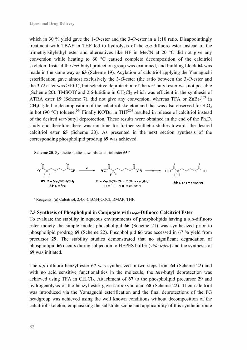
Liposomal Drug Delivery
82
which in 30 % yield gave the 1-O-ester and the 3-O-ester in a 1:10 ratio. Disappointingly treatment with TBAF in THF led to hydrolysis of the α,α-difluoro ester instead of the trimethylsilylethyl ester and alternatives like HF in MeCN at 20 °C did not give any conversion while heating to 60 °C caused complete decomposition of the calcitriol skeleton. Instead the tert-butyl protection group was examined, and building block 64 was made in the same way as 63 (Scheme 19). Acylation of calcitriol applying the Yamaguchi esterification gave almost exclusively the 3-O-ester (the ratio between the 3-O-ester and the 3-O-ester was >10:1), but selective deprotection of the tert-butyl ester was not possible (Scheme 20). TMSOTf and 2,6-lutidine in CH2Cl2 which was efficient in the synthesis of ATRA ester 19 (Scheme 7), did not give any conversion, whereas TFA or ZnBr2
169 in CH2Cl2 led to decomposition of the calcitriol skeleton and that was also observed for SiO2 in hot (90 °C) toluene.204 Finally KOtBu in THF205 resulted in release of calcitriol instead of the desired tert-butyl deprotection. These results were obtained in the end of the Ph.D. study and therefore there was not time for further synthetic studies towards the desired calcitriol ester 65 (Scheme 20). As presented in the next section synthesis of the corresponding phospholipid prodrug 69 was achieved.
Scheme 20. Synthetic studies towards calcitriol ester 65.a
a Reagents: (a) Calcitriol, 2,4,6-Cl3C6H2COCl, DMAP, THF.
7.3 Synthesis of Phospholipid in Conjugate with α,α-Difluoro Calcitriol Ester To evaluate the stability in aqueous environments of phospholipids having a α,α-difluoro ester moiety the simple model phospholipid 66 (Scheme 21) was synthesized prior to phospholipid prodrug 69 (Scheme 22). Phsopholipid 66 was accessed in 67 % yield from precursor 29. The stability studies demonstrated that no significant degradation of phospholipid 66 occurs during subjection to HEPES buffer (vide infra) and the synthesis of 69 was initiated. The α,α-difluoro benzyl ester 67 was synthesized in two steps from 64 (Scheme 22) and with no acid sensitive functionalities in the molecule, the tert-butyl deprotection was achieved using TFA in CH2Cl2. Attachment of 67 to the phospholipid precursor 29 and hydrogenolysis of the benzyl ester gave carboxylic acid 68 (Scheme 22). Then calcitriol was introduced via the Yamaguchi esterification and the final deprotections of the PG headgroup was achieved using the well known conditions without decomposition of the calcitriol skeleton, emphasizing the substrate scope and applicability of this synthetic route
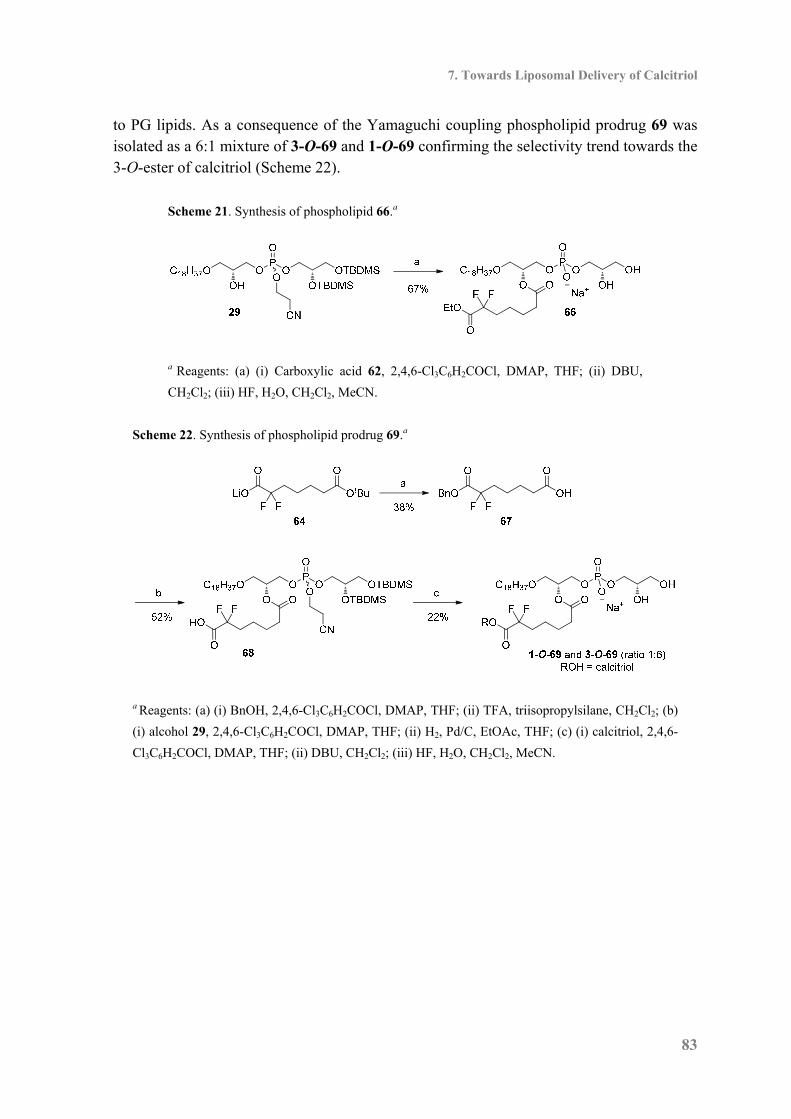
7. Towards Liposomal Delivery of Calcitriol
83
to PG lipids. As a consequence of the Yamaguchi coupling phospholipid prodrug 69 was isolated as a 6:1 mixture of 3-O-69 and 1-O-69 confirming the selectivity trend towards the 3-O-ester of calcitriol (Scheme 22).
Scheme 21. Synthesis of phospholipid 66.a
a Reagents: (a) (i) Carboxylic acid 62, 2,4,6-Cl3C6H2COCl, DMAP, THF; (ii) DBU,
CH2Cl2; (iii) HF, H2O, CH2Cl2, MeCN.
Scheme 22. Synthesis of phospholipid prodrug 69.a
a Reagents: (a) (i) BnOH, 2,4,6-Cl3C6H2COCl, DMAP, THF; (ii) TFA, triisopropylsilane, CH2Cl2; (b)
(i) alcohol 29, 2,4,6-Cl3C6H2COCl, DMAP, THF; (ii) H2, Pd/C, EtOAc, THF; (c) (i) calcitriol, 2,4,6-
Cl3C6H2COCl, DMAP, THF; (ii) DBU, CH2Cl2; (iii) HF, H2O, CH2Cl2, MeCN.
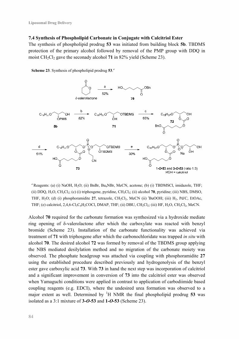
Liposomal Drug Delivery
84
7.4 Synthesis of Phospholipid Carbonate in Conjugate with Calcitriol Ester The synthesis of phospholipid prodrug 53 was initiated from building block 5b. TBDMS protection of the primary alcohol followed by removal of the PMP group with DDQ in moist CH2Cl2 gave the seconady alcohol 71 in 82% yield (Scheme 23).
Scheme 23. Synthesis of phospholipid prodrug 53.a
a Reagents: (a) (i) NaOH, H2O; (ii) BnBr, Bu4NBr, MeCN, acetone; (b) (i) TBDMSCl, imidazole, THF;
(ii) DDQ, H2O, CH2Cl2; (c) (i) triphosgene, pyridine, CH2Cl2; (ii) alcohol 70, pyridine; (iii) NBS, DMSO,
THF, H2O; (d) (i) phosphoramidite 27, tetrazole, CH2Cl2, MeCN (ii) tBuOOH; (iii) H2, Pd/C, EtOAc,
THF; (e) calcitriol, 2,4,6-Cl3C6H2COCl, DMAP, THF; (ii) DBU, CH2Cl2; (iii) HF, H2O, CH2Cl2, MeCN.
Alcohol 70 required for the carbonate formation was synthesized via a hydroxide mediate ring opening of δ-valerolactone after which the carboxylate was reacted with benzyl bromide (Scheme 23). Installation of the carbonate functionality was achieved via treatment of 71 with triphosgene after which the carbonochloridate was trapped in situ with alcohol 70. The desired alcohol 72 was formed by removal of the TBDMS group applying the NBS mediated desilylation method and no migration of the carbonate moiety was observed. The phosphate headgroup was attached via coupling with phosphoramidite 27 using the established procedure described previously and hydrogenolysis of the benzyl ester gave carboxylic acid 73. With 73 in hand the next step was incorporation of calcitriol and a significant improvement in conversion of 73 into the calcitriol ester was observed when Yamaguchi conditions were applied in contrast to application of carbodiimide based coupling reagents (e.g. EDCI), where the undesired urea formation was observed to a major extent as well. Determined by 1H NMR the final phospholipid prodrug 53 was isolated as a 3:1 mixture of 3-O-53 and 1-O-53 (Scheme 23).
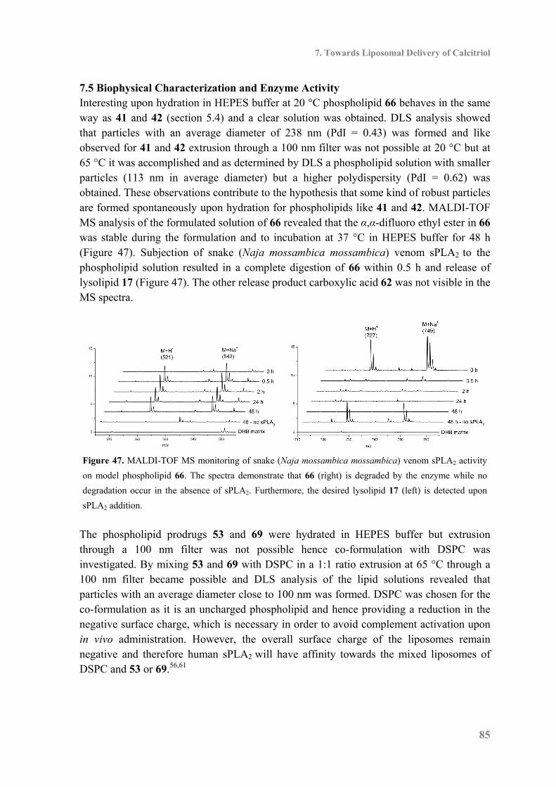
7. Towards Liposomal Delivery of Calcitriol
85
7.5 Biophysical Characterization and Enzyme Activity Interesting upon hydration in HEPES buffer at 20 °C phospholipid 66 behaves in the same way as 41 and 42 (section 5.4) and a clear solution was obtained. DLS analysis showed that particles with an average diameter of 238 nm (PdI = 0.43) was formed and like observed for 41 and 42 extrusion through a 100 nm filter was not possible at 20 °C but at 65 °C it was accomplished and as determined by DLS a phospholipid solution with smaller particles (113 nm in average diameter) but a higher polydispersity (PdI = 0.62) was obtained. These observations contribute to the hypothesis that some kind of robust particles are formed spontaneously upon hydration for phospholipids like 41 and 42. MALDI-TOF MS analysis of the formulated solution of 66 revealed that the α,α-difluoro ethyl ester in 66 was stable during the formulation and to incubation at 37 °C in HEPES buffer for 48 h (Figure 47). Subjection of snake (Naja mossambica mossambica) venom sPLA2 to the phospholipid solution resulted in a complete digestion of 66 within 0.5 h and release of lysolipid 17 (Figure 47). The other release product carboxylic acid 62 was not visible in the MS spectra.
Figure 47. MALDI-TOF MS monitoring of snake (Naja mossambica mossambica) venom sPLA2 activity
on model phospholipid 66. The spectra demonstrate that 66 (right) is degraded by the enzyme while no
degradation occur in the absence of sPLA2. Furthermore, the desired lysolipid 17 (left) is detected upon
sPLA2 addition.
The phospholipid prodrugs 53 and 69 were hydrated in HEPES buffer but extrusion through a 100 nm filter was not possible hence co-formulation with DSPC was investigated. By mixing 53 and 69 with DSPC in a 1:1 ratio extrusion at 65 °C through a 100 nm filter became possible and DLS analysis of the lipid solutions revealed that particles with an average diameter close to 100 nm was formed. DSPC was chosen for the co-formulation as it is an uncharged phospholipid and hence providing a reduction in the negative surface charge, which is necessary in order to avoid complement activation upon in vivo administration. However, the overall surface charge of the liposomes remain negative and therefore human sPLA2 will have affinity towards the mixed liposomes of DSPC and 53 or 69.56,61
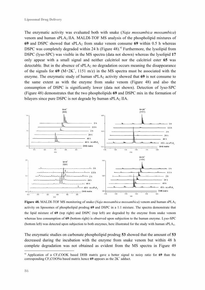
Liposomal Drug Delivery
86
The enzymatic activity was evaluated both with snake (Naja mossambica mossambica) venom and human sPLA2 IIA. MALDI-TOF MS analysis of the phospholipid mixtures of 69 and DSPC showed that sPLA2 from snake venom consume 69 within 0.5 h whereas DSPC was completely degraded within 24 h (Figure 48).xi Furthermore, the lysolipid from DSPC (lyso-SPC) was visible in the MS spectra (data not shown) whereas the lysolipid 17 only appear with a small signal and neither calcitriol nor the calcitriol ester 65 was detectable. But in the absence of sPLA2 no degradation occurs meaning the disappearance of the signals for 69 (M+2K+, 1151 m/z) in the MS spectra must be associated with the enzyme. The enzymatic study of human sPLA2 activity showed that 69 is not consume to the same extent as with the enzyme from snake venom (Figure 48) and also the consumption of DSPC is significantly lower (data not shown). Detection of lyso-SPC (Figure 48) demonstrates that the two phospholipids 69 and DSPC mix in the formation of bilayers since pure DSPC is not degrade by human sPLA2 IIA.
Figure 48. MALDI-TOF MS monitoring of snake (Naja mossambica mossambica) venom and human sPLA2
activity on liposomes of phospholipid prodrug 69 and DSPC in a 1:1 mixture. The spectra demonstrate that
the lipid mixture of 69 (top right) and DSPC (top left) are degraded by the enzyme from snake venom
whereas less consumption of 69 (bottom right) is observed upon subjection to the human enzyme. Lyso-SPC
(bottom left) was detected upon subjection to both enzymes, here illustrated for the study with human sPLA2.
The enzymatic studies on carbonate phospholipid prodrug 53 showed that the amount of 53 decreased during the incubation with the enzyme from snake venom but within 48 h complete degradation was not obtained as evident from the MS spectra in Figure 49 xi Application of a CF3COOK based DHB matrix gave a better signal to noisy ratio for 69 than the corresponding CF3COONa based matrix hence 69 appears as the 2K+ adduct.
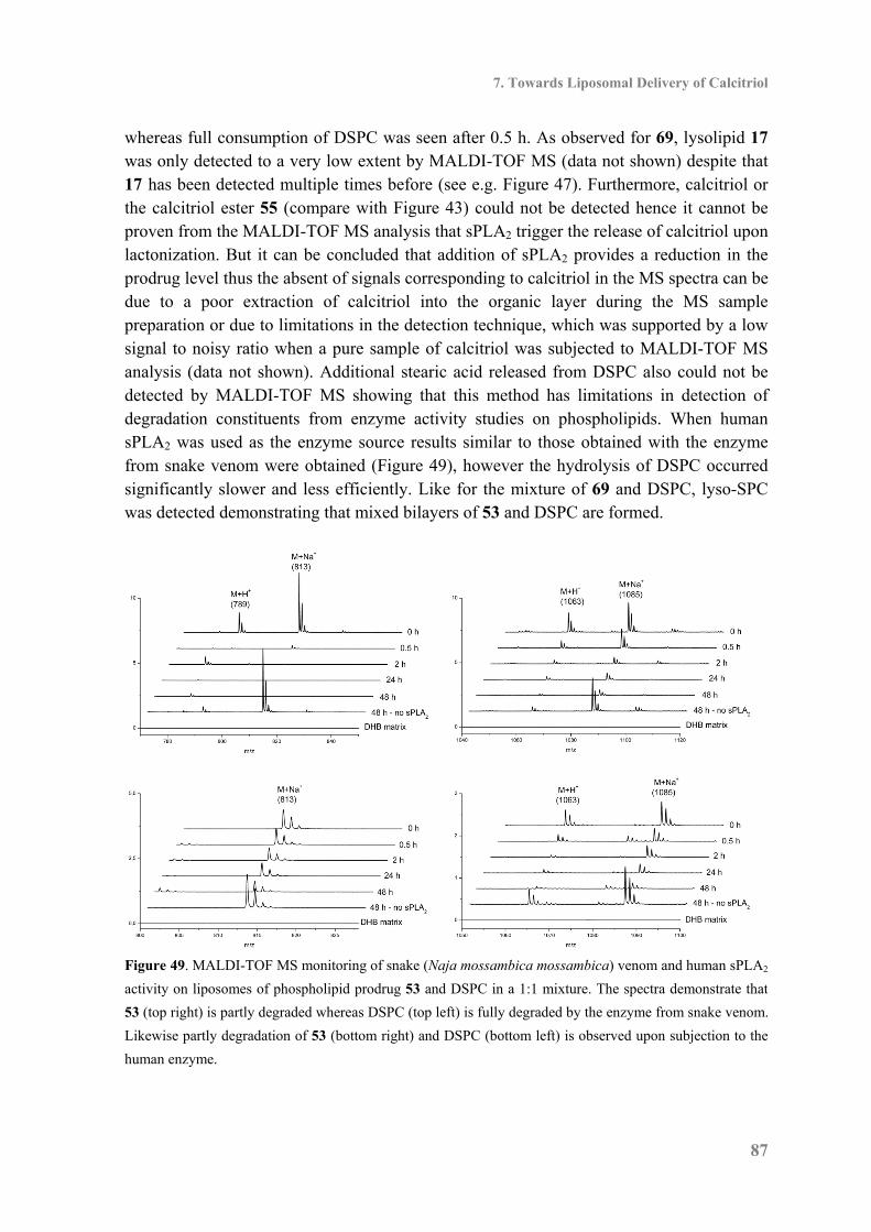
7. Towards Liposomal Delivery of Calcitriol
87
whereas full consumption of DSPC was seen after 0.5 h. As observed for 69, lysolipid 17 was only detected to a very low extent by MALDI-TOF MS (data not shown) despite that 17 has been detected multiple times before (see e.g. Figure 47). Furthermore, calcitriol or the calcitriol ester 55 (compare with Figure 43) could not be detected hence it cannot be proven from the MALDI-TOF MS analysis that sPLA2 trigger the release of calcitriol upon lactonization. But it can be concluded that addition of sPLA2 provides a reduction in the prodrug level thus the absent of signals corresponding to calcitriol in the MS spectra can be due to a poor extraction of calcitriol into the organic layer during the MS sample preparation or due to limitations in the detection technique, which was supported by a low signal to noisy ratio when a pure sample of calcitriol was subjected to MALDI-TOF MS analysis (data not shown). Additional stearic acid released from DSPC also could not be detected by MALDI-TOF MS showing that this method has limitations in detection of degradation constituents from enzyme activity studies on phospholipids. When human sPLA2 was used as the enzyme source results similar to those obtained with the enzyme from snake venom were obtained (Figure 49), however the hydrolysis of DSPC occurred significantly slower and less efficiently. Like for the mixture of 69 and DSPC, lyso-SPC was detected demonstrating that mixed bilayers of 53 and DSPC are formed.
Figure 49. MALDI-TOF MS monitoring of snake (Naja mossambica mossambica) venom and human sPLA2
activity on liposomes of phospholipid prodrug 53 and DSPC in a 1:1 mixture. The spectra demonstrate that
53 (top right) is partly degraded whereas DSPC (top left) is fully degraded by the enzyme from snake venom.
Likewise partly degradation of 53 (bottom right) and DSPC (bottom left) is observed upon subjection to the
human enzyme.

Liposomal Drug Delivery
88
7.6 Biological Studies of the Calcitriol Esters The cytotoxicity of calcitriol and the calcitriol esters 51 and 52 were evaluated in MCF-7 cancer cells and as evident from the dose-response curves in Figure 50 the esters 51 and 52 displayed potency similar to calcitriol itself. The IC50 values <100 nM emphasize the superior potency of calcitriol in contrast to the previously applied drugs, like ATRA. To investigate that the cytotoxicity origins from an activation of the VDR the ability to induce transcription of vitamin D-responsive genes was examined using the 24-hydroxylase luciferase reporter gene assay206 in MCF-7 cells. It was established that the mixture of 1-O-51 and 3-O-51 along with the mixture of 1-O-52 and 3-O-52 had 10 times lower activity with respect to calcitriol (Figure 51) demonstrating that the affinity towards the VDR for 51 and 52 is low and the displayed cytotoxicity must therefore be induced via some unspecific pathways. Gratifying 3-O-56 and the mixture of 1-O-56 and 3-O-56 induce transcription to same extent as calcitriol (Figure 51), and taking into account that the hydroxyl group at C-1 is essential for binding to the VDR the results indicate that hydrolysis of the α,α-difluoroesters occur prior to VDR binding.
Figure 50. Dose-response curves for the treatment of MCF-7 cells
with calcitriol (■), the 1:1 mixture of 1-O-51 and 3-O-51 (▲) and
the 1:3 mixture of 1-O-52 and 3-O-52 (●). The cytotoxicity was
measured using the MTT assay as cell viability 48 h after
incubation with the indicated substances for 24 h and shown by
mean + SD (n=3).
The biological evaluation of the calcitriol phospholipid prodrugs 53 and 69 was not completed at the end of the Ph.D. study hence these results cannot be presented. The biological evaluation will be important in order to clarify the somewhat inconclusive results obtained in the enzymatic studies, particular the 24-hydroxylase luciferase reporter gene assay will provide some valuable information concerning the degree of calcitriol release from the enzyme triggered breakdown of the calcitriol phospholipid prodrugs.

7. Towards Liposomal Delivery of Calcitriol
89
Figure 51: Relative affinity towards the VDR with respect to calcitriol
measured by the 24-hydroxylase luciferase reporter gene assay in
MCF-7 cells after incubation for 48 h with 100 nM of the investigated
compound. Reported as mean ± SD (n = 3).
7.7 Conclusion As calcitriol does not have a carboxylic acid moiety new ways for incorporation and drug release was necessary and two drug delivery strategies utilizing sPLA2 triggering was investigated. Attachment of a dicarboxylic acid linker was studied and it was found that α,α-difluoroesters induce transcription of vitamin D-responsive genes to the same extent as calcitriol and hence synthesis of calcitriol ester 65 was initiated but not accomplished and further synthetic studies are necessary. However the corresponding phospholipid prodrug 69 was synthesized and when co-formulated with DSPC, particles with a diameter close to 100 nm were obtained after extrusion at 65 °C. Likewise a strategy relying on sPLA2 mediated hydrolysis of a phospholipid carbonate conjugate was investigated and the desired phsopholipid prodrug 53 was synthesized and co-formulated with DSPC. The enzymatic studies with snake venom and human sPLA2 showed that the signals in the MS spectra for the phospholipid prodrug decrease but the released products were not detectable in the MS spectra hence no clear conclusions can be made. The results from the biological studies will be highly interesting in order to evaluate the potential of the investigated strategies for liposomal delivery of calcitriol to tumors. In addition, it was discovered that acylation of calcitriol using the Yamaguchi coupling displayed selectivity towards the 3-O-calcitriol esters and gave better conversion why this coupling procedure will be preferable to use in the future.


8. Conclusion: Liposomal Drug Delivery
91
8. Conclusion: Liposomal Drug Delivery During the work towards a new generation of liposomal drug delivery systems for anticancer agents several phospholipid prodrugs have been synthesized. The synthesized phospholipids had either a PC or PG headgroup and they were accessed via short synthetic routes in which derivatization into the various prodrugs was accomplished in the later part of the synthesis. Various know anticancer drugs, like chlorambucil, ATRA and α-tocopheryl succinate have been examined for their ability to be incorporated into the investigated drug delivery system. Initial screening parameters were investigated including the ability of the phospholipid prodrugs to form liposomes, the ability of sPLA2 to liberate the cytotoxic agents and finally the ability of the prodrugs to remain non-toxic in absence of the enzyme. Gratisfyingly, the majority of the phospholipid prodrugs were able to form particles with a diameter close to 100 nm upon extrusion at 20 °C indicating formation of unilamellar vesicles. This demonstrate that incorporation of structures like chlorambucil or ATRA which contrast naturally occurring fatty acids do not hamper the ability of the synthetic phospholipids to form bilayers. However it was necessary to co-formulate the calcitriol prodrugs 53 and 69 with DSPC in order to achieve phospholipid suspensions suitable for extrusion. The ability of sPLA2 to mediate hydrolysis of the ester bond at the sn-2 position and release the incorporated anticancer drug was studied and in general it was observed that the enzyme is sensitive to substituents in close vicinity of the sn-2 position exemplified by ATRA prodrug 8, which was not degraded. But for substrates with no branching in proximity to the sn-2 position, like the chlorambucil prodrugs, full hydrolysis was observed and when the enzyme activity was analyzed with MALDI-TOF MS, that was reliable and convenient to use, it was possible to verify that the desired constituents, the lysolipids and the anticancer drug were released. The cytotoxicity studies revealed that the phospholipid prodrugs in the presence of sPLA2 were able to induce cell death towards various cancer cell lines and IC50 values below 10 µM was obtained. In the absence of the enzyme the phospholipid prodrugs remain non-toxic and no significant cell death was observed, verifying that the displayed cytotoxicity arise from a sPLA2 triggered breakdown of the prodrugs. The non-toxic behavior of the unactivated prodrugs is crucial for the drug delivery concept meaning that the prodrugs during the transport in the body will not harm healthy tissue. The prostaglandin prodrugs 45 and 46 was in this regard an outlier as significant cell death was displayed also in the absence of the enzyme in HT-29 cells.
Liposomal Drug Delivery
92
The overall conclusion from the studies of the phospholipid prodrugs of chlorambucil, retinoids, prostaglandin and α-tocopheryl succintate (chapter 3–6) is that the cytotoxic effect from the incorporated anticancer drug is hard to separate from the cytotoxicity caused by the lysolipids. Hence the next obstacle to overcome in order to bring this drug delivery concept one step further is to indentify or develop a more potent lipophilic drug having a carboxylic acid moiety but no substituents α, β or γ to the carbonyl, because the results described in thesis clearly demonstrate that the concept has potential in future cancer therapy and many valuable experiences and results have been obtained throughout the described work. Work towards incorporation of the more potent calcitriol was initiated however the biological studies remains to be done so the potential of this drug in the investigated drug delivery system cannot be evaluated at this stage.

9. Experimental: Liposomal Drug Delivery
93
9. Experimental: Liposomal Drug Delivery 9.1 General Experimental Starting materials, reagents, and solvents were purchased from Sigma-Aldrich Chemical Co. and used without further purification. 15-deoxy-Δ12,14-Prostaglandin J2 and calcitriol were purchased from Cayman Chemical, USA. The purified snake venom sPLA2 from Agkistrodon piscivorus piscivorus was donated by Dr. R. L. Biltonen (University of Virginia) and sPLA2 from Naja mossambica mossambica was purchased from Sigma-Aldrich Chemical Co. CH2Cl2 was dried over 4 Å molecular sieves and THF was dried over sodium/benzophenone and distilled before use. Evaporation of solvents was done under reduced pressure (in vacuo). Thin layer chromatography (TLC) was performed on Merck aluminum sheets precoated with silica gel 60 F254. Compounds were visualized by charring after dipping in a solution of 6.25 g of (NH4)6Mo7O24 and 1.5 g of Ce(SO4)2 in 250 mL of 10% aqueous H2SO4, in an ethanolic solution of phosphomolybdenic acid (48 g/L) or in a solution of 10 mL of p-anialdehyde, 10 ml of a concentrated aqueous solution of H2SO4 and 250 mL EtOH. Column chromatography was performed using Matrex 60 Å silica gel. The purity of the tested compounds was found to be >95% by HPLC. HPLC was performed on a Waters Alliance HPLC equipped with a diode array detector, using a LiChrospher Si 60 column and eluting with water/isopropanol/hexane mixtures. 207 , 208 NMR spectra were recorded using a Bruker AC 200 MHz spectrometer, a Varian Mercury 300 MHz spectrometer or a Varian Unity Inova 500 MHz spectrometer. Chemical shifts were measured in ppm and coupling constants in Hz, and the field is indicated in each case. IR analysis was carried out on a Bruker Alpha FT-IR spectrometer and optical rotations were measured with a Perkin-Elmer 341 polarimeter, units for 20
589][ are 10-1 deg cm2 g-1.
HRMS was recorded on an Ionspec Ultima Fourier transform mass spectrometer. Melting points were measured by a Buch & Holm melting point apparatus and given in degrees Celsius (°C) uncorrected. Elemental analyses were obtained from H. Kolbe, Mikroanalytisches Laboratorium, Mülheim/Ruhr, Germany. 9.2 Procedures for Biophysical and Biological Characterization Liposome Preparation and Particle Size Determination The phospholipid prodrugs were dissolved in CHCl3 in a test tube and dried under vacuum for 15 h to form a thin film. The phospholipid prodrugs (2 mM) were solubilized by addition of aqueous buffer (0.15 M KCl, 30 µM CaCl2, 10 µM EDTA, 10 mM HEPES, pH 7.5) and vortexed periodically over 1 h at 20 C. Subsequently, the solutions were extruded through a 100 nm polycarbonate cutoff membrane (20-30 repetitions) using a Hamilton syringe extruder (Avanti Polar Lipids, Birmingham, AL). The particle size distribution of the formulated lipids was measured by DLS. The DLS measurements were obtained using a Zetasizer Nano Particle Analyzer (ZS ZEN3600, Malvern Instrument, Westborough, MA).

Liposomal Drug Delivery
94
sPLA2 Activity Measurements Monitored by MALDI-TOF MS and HPLC The formulated phospholipid prodrugs (0.40 mL, 2 mM) were diluted in an aqueous buffer (2.1 mL, 0.15 M KCl, 30 µM CaCl2, 10 µM EDTA, 10 mM HEPES, pH 7.5) and the mixture was stirred at 37 °C in a test tube protected from light. The catalytic reaction was initiated by addition of snake (Agkistrodon piscivorus piscivorus (20 µL, 42 µM) or Naja mossambica mossambica (12 µL, 71 µM)) venom sPLA2 or human sPLA2 IIA (40 µL tear fluid). Sampling was done after 0, 2, 24 and 48 h by collecting 100 µL of the reaction mixture and rapidly mixing it with a solution of CHCl3/MeOH/H2O/AcOH 4:8:1:1 (0.5 mL) in order to stop the reaction. The mixture was washed with water (0.5 mL) and the organic phase (80 µL) was isolated by extraction and then concentrated in vacuo. For the MALDI-TOF MS analysis the extract was mixed with 9 µL of DHB matrix (0.5 M DHB, 2 mM CF3COONa, 1 mg/mL DPPG in MeOH), and 0.5 µL of this mixture was used for the MS analysis. For HPLC 30-75 μL of the organic phase was injected on a 5 μm diol column and eluted with an isocratic eluent (CHCl3/MeOH/H2O 730:230:30 for 1a and 1b; CHCl3/MeOH/25% aqueous NH3 800:195:5 for 2a and 2b). An evaporative light scattering detector was used for detection. Tear fluid was used as the source for human sPLA2 IIA. Tear fluid was collected from healthy volunteers exposed to tiger balm fumes. Tear fluid has a high concentration of sPLA2 IIA and is the only prevalent sPLA2 species found in tears. The sPLA2 IIA content in tears in healthy subjects (54.5 µg/ml) is one of the highest amounts of sPLA2 IIA content reported in human secretions.209,210,211 Alkylating Assay136,137,138 The formulated chlorambucil phospholipid prodrugs (0.40 mL, 2 mM) were diluted in an aqueous buffer (2.1 mL, 0.15 M KCl, 30 μM CaCl2, 10 μM EDTA, 10 μM HEPES, pH = 7.5) and 4-nitrobenzyl pyridine (63 mg, 0.29 mmol) was added and the mixture was stirred at 37 °C. When sPLA2 was used to degrade the liposomes, the catalytic reaction was initiated by addition of snake (Agkistrodon piscivorus piscivorus) venom sPLA2 (20 μL, 42 μM). After 1.5 h incubation 2-amino-1-propanol in tert-butyl alcohol (25% v/v, 3.0 mL) was added and the colaration of the reaction mixture was measured using a UV-visible spectrophometer (PerkinElmer, Lambda 25 UV/Vis Spectrometer) at 450–700 nm with a maximum absorption at 560 nm for the colored pigment. Measurements of pure chlorambucil samples in HEPES buffer with and without 4-nitrobenzyl pyridine, of HEPES buffer with and without sPLA2 and of pure 4-nitrobenzyl pyridine in HEPES buffer were obtained as controls (see Pedersen et al.135). Cytotoxicity Colon cancer HT-29 and ovarian cancer ES-2 cells were cultured in McCoy’s 5A medium in the presence of 10% fetal calf serum and 1% Pen-Strep (InVitrogen). Breast cancer MT-3 and colon cancer Colo205 cells were cultured in RPMI 1640 supplemented with 10 % fetal calf serum and 1% Pen-Strep in a humidified atmosphere containing 5 % CO2. Colo205 cells secrete sPLA2-IIA, however as the cytotoxicity assay was made with low
9. Experimental: Liposomal Drug Delivery
95
cell density and for a short incubation period, inadequate concentrations were reached in the medium, and therefore a conditioned medium was used. The conditioned medium was made as follows: Colo205 cells were grown to confluency for 72h, at which point the cells were pelleted and the medium, which contained approximately 100 ng/ml, was collected and used for the assay. Cells were plated in 96-well plates at a density of 1 × 10⁴ cells per well for HT-29 and 2 × 10⁴ cells per well for Colo205 cells, 24 h prior to addition of the tested compound. Chlorambucil, ATRA, ATRA ester 19, 12, 20, 15-deoxy-Δ12,14-PGJ2 and α-tocopheryl succinate were solubilized in DMSO and water (final DMSO concentration ≤ 0.5%). Liposomes were diluted in PBS, and initial phospholipid concentrations in the liposome solutions were determined by phosphorus analysis.212 After 24 h of incubation, the substances were removed and the cells were washed and incubated in complete medium for another 48 h. Cytotoxic activity was assessed using a standard 3-(4,5-dimethylthiazolyl)-2,5-diphenyltetrazolium bromide (MTT) assay (Cell Proliferation Kit I, Roche, Germany).213 Cell viability is expressed as percentage reduction of incorporated MTT. Molecular Dynamics Simulations The crystal structures of bee-venom (Apis Mellifera) phospholipase A2 complexed with the transition-state analog, 1-O-Octyl-2-heptylphosphonyl-sn-glycero-3-phosphoethanolamine (diC8(2Ph)PE),51 resolved to 2.0 Å and human phospholipase A2 IIA complexed with 6-phenyl-4-(R)-(7-phenyl-heptanoylamino)-hexanoic acid214 resolved to 2.1 Å were obtained from the Protein Data Bank215 (entry codes: 1poc and 1kqu, respectively). The initial modeling step involved placing diC8(2Ph)PE into the binding cleft of human phospholipase A2 IIA, as described previously.131 A previously assembled sPLA2-substrate complex131 was used as the template to place phospholipid prodrugs into the active site. The optimized phospholipid prodrugs were manually overlapped with the pre-existing substrate, which was subsequently deleted. NAMD software216 with the Charmm27 all-atom parameter set and TIP3 water model was used for all simulations. 217 The phospholipid prodrugs were first energy-minimized for 1000 steps before using them in simulations. Missing force field parameters for the prodrug molecules were taken from similar atom types in the CHARMM27 force field. The sPLA2–phospholipid complexes were solvated using the program SOLVATE.218 Eighteen water molecules were randomly replaced with chloride ions to neutralize the systems. Ultimately, the system contained ~4900 water molecules in a simulation cell of dimensions 52.7 x 51.7 x 67.3 Å3. An initial energy minimization of the complex was carried out for 1000 steps, and the system was further minimized for 100, 200, 300, 400 and 500 steps to generate different starting structures. Periodic boundary conditions were applied in x,y, and z directions. Initial MD simulations were conducted for ~100 ps in which each system was slowly heated to 300 K. Simulations were carried out for 13 ns in the NPT ensemble; i.e. at constant number of atoms (N), pressure (P) and temperature (T). A time step of 1 fs and an isotropic pressure of 1 atm using the Langevin piston method219 were employed. Electrostatic forces were
Liposomal Drug Delivery
96
calculated using the particle mesh Ewald method with a uniform grid spacing of ~1 Å.220 A 12 Å cut-off was used for terminating the van der Waals interactions in combination with a switching function starting at 10 Å. Analysis of the trajectories were performed using the Visualization Molecular Dynamics (VMD) software suite.221 9.3 Experimental Data for Compounds 4-Methoxybenzyl trichloroacetimidate (PMBTCA)222 NaH (60% in mineral oil, 81 mg, 2.03 mmol) was washed with heptane (2 × 3 mL) and THF (11 mL) was added followed by dropwise addition of 4-methoxybenzyl alcohol (2.52 mL, 20.3 mmol). The reaction mixture was stirred at 20 °C for 30 min and then cooled to 0 °C. Cl3CCN (2.2 mL, 22.3 mmol) was added dropwise and the reaction mixture was stirred at 0 °C for 1 h and then at 20 °C for 45 min. The reaction mixture was concentrated in vacuo yielding an orange oil which was treated with a mixture of pentane and MeOH (12 mL, pentane/MeOH 275:1) and stirred for 30 min. Then the mixture was filtered through a pad of celite and the filtrate was evaporated in vacuo to afford 5.72 g (99%) of PMBTCA as a yellow oil. 1H NMR (300 MHz, CDCl3): δ 8.37 (s, 1H), 7.39 (d, J = 8.8 Hz, 2H), 6.92 (d, J = 8.8 Hz, 2H), 5.29 (s, 2H), 3.83 (s, 3H). 13C NMR (75 MHz, CDCl3): δ 162.7, 159.8, 129.8 (2C), 127.6, 114.0 (2C), 91.6, 70.8, 55.4. 1-O-Hexadecyl-2-(4-(4-(bis-(2-chloroethyl)-amino)-phenyl)-butanoyl)-sn-glycero-3-phosphocholine (1a) Compound 6a (67 mg, 0.14 mmol) was dissolved in 1,2-dichloroethane (3.5 mL) and the mixture was heated to reflux. DMAP (10 mg, 0.082 mmol), chlorambucil (64 mg, 0.21 mmol) and DCC (1 M in CH2Cl2, 0.2 mL, 0.2 mmol) were added and after 5 h and 19 h additional portions of chlorambucil (64 mg, 0.21 mmol) and DCC (1 M in CH2Cl2, 0.2 mL, 0.2 mmol) were added. After 27 h the mixture was concentrated in vacuo and purified by column chromatography (CH2Cl2/MeOH 4:1 then CH2Cl2/MeOH/H2O 30:20:1) to give 80 mg (75%) of 1a as a colorless amorphous solid. Rf = 0.18 (CH2Cl2/MeOH/H2O 30:20:1). 1H NMR (500 MHz, CDCl3/CD3OD 4:1): δ 7.08 (d, J = 8.6 Hz, 2H), 6.65 (d, J = 8.6 Hz, 2H), 5.16 (p, J = 5.9 Hz, 1H), 4.22 (s, 2H), 4.03–3.99 (m, 2H), 3.72 (t, J = 7.1 Hz, 4H), 3.64 (t, J = 7.1 Hz, 4H), 3.63–3.59 (m, 2H), 3.56 (s, 2H), 3.49–3.39 (m, 2H), 3.19 (s, 9H), 2.56 (t, J = 7.6 Hz, 2H), 2.36 (t, J = 7.6 Hz, 2H), 1.90 (p, J = 7.6 Hz, 2H), 1.54 (p, J = 6.8 Hz, 2H), 1.32-1.26 (m, 26H), 0.88 (t, J = 6.8 Hz, 3H). 13C NMR (75 MHz, CDCl3/CD3OD 4:1) δ 174.1, 144.8, 130.8, 130.0 (2C), 112.6 (2C), 72.2 (d, J = 8.2 Hz), 72.1, 69.6, 66.8, 64.4 (d, J = 5.2 Hz), 59.2 (d, J = 5.2 Hz), 54.5, 54.4, 54.4, 53.9 (2C), 40.9 (2C), 34.2, 34.0, 32.3, 30.1 (8C), 29.9, 29.9, 29.7, 27.2, 26.4, 23.0, 14.3. IR (neat) 2923, 2852, 2366, 1734, 1091 cm-1 HRMS (ESI+) C38H69Cl2N2O7P, m/z [M+H+] 767.4298, found 767.4312.
9. Experimental: Liposomal Drug Delivery
97
1-O-Octadecyl-2-(4-(4-(bis-(2-chloroethyl)-amino)-phenyl)-butanoyl)-sn-glycero-3-phosphocholine (1b) Compound 6b (67 mg, 0.13 mmol) was dissolved in CHCl3 (4 mL) and the mixture was heated to reflux. DMAP (10 mg, 0.082 mmol), chlorambucil (40 mg, 0.13 mmol) and DCC (1 M in CH2Cl2, 130 μL, 0.13 mmol) were added and after 3, 6.5, 23.5, 26.5 and 31 h additional portions of chlorambucil (40 mg, 0.13 mmol) and DCC (1 M in CH2Cl2, 130 μL, 0.13 mmol) were added. After 48 h the mixture was concentrated in vacuo and purified by column chromatography (CH2Cl2/MeOH 4:1 then CH2Cl2/MeOH/H2O 30:20:1) to give 79 mg (76%) of 1b as a colorless amorphous solid. Rf = 0.15 (CH2Cl2/MeOH/H2O 30:20:1). 1H NMR (500 MHz, CDCl3/CD3OD 4:1) δ 7.07 (d, J = 8.5 Hz, 2H), 6.65 (d, J = 8.5 Hz, 2H), 5.16 (p, J = 5.8 Hz, 1H), 4.22 (s, 2H), 4.06–3.94 (m, 2H), 3.72 (t, J = 6.8 Hz, 4H), 3.64 (t, J = 6.8 Hz, 4H), 3.63–3.59 (m, 2H), 3.56 (s, 2H), 3.48–3.40 (m, 2H), 3.20 (s, 9H), 2.56 (t, J = 7.5 Hz, 2H), 2.36 (t, J = 7.5 Hz, 2H), 1.90 (p, J = 7.5 Hz, 2H), 1.54 (m, 2H), 1.32–1.26 (m, 30H), 0.88 (t, J = 6.9 Hz, 3H). 13C NMR (75 MHz, CDCl3/CD3OD 4:1) δ 174.1, 144.9, 130.8, 130.1 (2C), 112.6 (2C), 72.3 (d, J = 8.6 Hz), 72.1, 69.6, 66.9, 64.5 (d, J = 5.2 Hz), 59.4 (d, J = 5.2 Hz), 54.5, 54.4, 54.4, 53.9 (2C), 40.9 (2C), 34.3, 34.0, 32.3, 30.1 (10C), 29.9, 29.9, 29.8, 27.3, 26.4, 23.1, 14.3. IR (neat) 2923, 2852, 2366, 1734, 1518, 1247, 1088, 750 cm-1. HRMS (ESI+) C40H73Cl2N2O7P, m/z [M+Na+] 817.4425, found 817.4430. 1-O-Hexadecyl-3-(p-toluenesulfonyl)-sn-glycerol (4a)68
(R)-Glycidyl tosylate (2.0 g, 8.76 mmol) and hexadecanol (3.0 g, 12.37 mmol) were dissolved in CH2Cl2 (40 mL) and BF3·OEt2 (0.5 mL) was added. The solution was stirred for 24 h at 20 °C and concentrated in vacuo. A white solid was formed and recrystallized from hexane (35 mL) to give 2.75 g of 4a. The filtrate, which contain some product, was concentrated in vacuo and purified by column chromatography (CH2Cl2/Et2O 10:1) to give 0.98 g of 4a (89% overall). Rf = 0.58 (CH2Cl2/Et2O 10:1); mp = 53–55 °C (lit.223 53–54 °C). 1H NMR (300 MHz, CDCl3): δ 7.81 (d, J = 8.4 Hz, 2H), 7.36 (d, J = 8.4 Hz, 2H), 4.12–3.93 (m, 3H), 3.48–3.27 (m, 4H), 2.46 (s, 3H), 2.18 (s, 1H), 1.51 (t, J = 6.4 Hz, 2H), 1.32–1.26 (m, 26H), 0.88 (t, J = 6.7 Hz, 3H).
1-O-Octadecyl-3-(p-toluenesulfonyl)-sn-glycerol (4b)68 Performed as for 4a using (R)-Glycidyl tosylate (5.25 g, 23.0 mmol) and octadecanol (8.7 g, 32.2 mmol) to afford 11.2 g (97 %) of 4b as white crystals. Rf = 0.65 (CH2Cl2/Et2O 10:1); mp = 66–68 °C (lit.224 67–68 °C). 1H NMR (300 MHz, CDCl3): δ 7.80 (d, J = 8.1 Hz, 2H), 7.38 (d, J = 8.1 Hz, 2H), 4.05-3.95 (m, 3H), 3.45-3.35 (m, 4H), 2.43 (s, 3H), 2.10 (s, 1H), 1.51 (m, 2H), 1.35-1.23 (m, 30H), 0.90 (t, J = 6.7 Hz, 3H). 1-O-Hexadecyl-2-O-(4-methoxybenzyl)-sn-glycerol (5a) Alcohol 4a (1.19 g, 2.53 mmol) and PMBTCA (1.10 g, 3.89 mmol) were dissolved in toluene (21 mL) and La(OTf)3 (70 mg, 0.12 mmol) was added. The reaction mixture was
Liposomal Drug Delivery
98
stirred at 20 °C for 2.5 h, then concentrated in vacuo to give a white solid that was dissolved in DMSO (20 mL) and DMF (5 mL). CsOAc (1.49 g, 7.38 mmol) was added and the mixture was heated to 60 °C and stirred for 14 h, after which the reaction mixture was washed with H2O (30 mL) and the organic layer was isolated via extraction with Et2O (3x50 mL). The combined organic phases were dried over MgSO4 and filtration followed by concentration in vacuo gave a solid that was dissolved in MeOH (40 mL). NaOMe (140 mg, 2.59 mmol) was added and the mixture was heated to 40 °C and stirred for 16 h. The mixture was neutralized by addition of concentrated aqueous solution of HCl, washed with H2O (20 mL) and the organic layer was isolated via extraction with CH2Cl2 (3x20 mL). The combined organic phases were dried over MgSO4 and after concentration in vacuo the residue was purified by column chromatography (heptane/EtOAc 1:1) yielding 772 mg (70%) of 5a as a greasy solid. Rf = 0.46 (heptane/EtOAc 1:1). 1H NMR (300 MHz, CDCl3): δ 7.29 (d, J = 8.6 Hz, 2H), 6.89 (d, J = 8.6 Hz, 2H), 4.66 (d, J = 11.4 Hz, 1H), 4.56 (d, J = 11.4 Hz, 1H), 3.81 (s, 3H), 3.78–3.50 (m, 5H), 3.44 (t, J = 6.7 Hz, 2H), 1.61–1.53 (m, 2H), 1.26 (br.s. 26H), 0.88 (t, J = 6.7 Hz, 3H). 13C NMR (75 MHz, CDCl3): δ 159.4, 130.5, 129.6 (2C), 114.0 (2C), 77.5, 72.0, 71.9, 71.3, 63.2, 55.4, 32.1, 29.8 (9C), 29.6, 29.5, 26.3, 22.9, 14.3. Anal. Calcd. C27H48O4: C 74.26, H 11.08; Found: C 74.26, H 11.03. IR (neat) 3374, 2923, 1695 1613, 1513, 1466, 1248, 1110, 824 cm-1. 1-O-Hexadecyl-2-O-(4-methoxybenzyl)-sn-glycerol (5a) using NaNO2 Alcohol 4a (842 mg, 1.79 mmol) and PMBTCA (859 mg, 3.04 mmol) were dissolved in toluene (15 mL) and La(OTf)3 (52 mg, 0.09 mmol) was added. The reaction mixture was stirred at 20 °C for 2.5 h and concentration in vacuo gave a white solid, that along with NaNO2 (3.69 g, 53.6 mmol) were dissolved in degassed DMSO (35 mL). The mixture was stirred at 40 °C for 17 h, then a saturated aqueous solution of NaHCO3 (10 mL) was added and the mixture was stirred for 20 min further after which the organic layer was isolated via extraction with Et2O (3×100 mL). The combined organic phases were dried over MgSO4, filtered, concentrated in vacuo and purified by column chromatography (heptane/EtOAc 1:1) to give 731 mg (94%) of 5a as a greasy solid. 1-O-Octadecyl-2-O-(4-methoxybenzyl)-sn-glycerol (5b) Performed as for 5a, starting from alcohol 4b (2.211 g, 4.433 mmol) and affording 1.60 g (78%) of 5b as a greasy solid. 1H NMR (300 MHz, CDCl3): δ 7.29 (d, J = 8.6Hz, 2H), 6.89 (d, J = 8.6Hz, 2H), 4.66 (d, J = 11.4Hz, 1H), 4.56 (d, J = 11.4 Hz, 1H), 3.81 (s, 3H), 3.78–3.50 (m, 5H), 3.44 (t, J = 6.7 Hz, 2H), 1.62–1.53 (m, 2H), 1.26 (br.s. 30H), 0.88 (t, J = 6.7 Hz, 3H). 13C NMR (75 MHz, CDCl3): δ 159.4, 130.5, 129.6 (2C), 114.0 (2C), 77.5, 72.0, 71.9, 71.3, 63.2, 55.4, 32.1, 29.8 (11C), 29.6, 29.5, 26.3, 22.8, 14.3. Anal. Calcd. C29H52O4: C 74.95, H 11.28; Found: C 75.08, H 11.24. IR (neat) 3524, 2916, 1709 1612, 1511, 1472, 1253, 1108, 1032, 824 cm-1.
9. Experimental: Liposomal Drug Delivery
99
1-O-Octadecyl-2-O-(4-methoxybenzyl)-sn-glycerol using NaNO2 (5b) Performed as for 5a, starting from alcohol 4a (440 mg, 0.882 mmol) and affording 291 mg (71%) of 5b as a greasy solid. 1-O-Hexadecyl-2-lyso-sn-glycero-3-phosphocholine (6a) To a solution of POCl3 (28 μL, 0.30 mmol) in CH2Cl2 (1.2 mL) at 0 °C was added a solution of 5a (104 mg, 0.24 mmol) along with Et3N (44 μL, 0.31 mmol) in CH2Cl2 (1.7 mL) dropwise over 20 min. The reaction was stirred 30 min at 20 °C, after which pyridine (0.15 mL, 1.9 mmol) and choline tosylate (132 mg, 0.48 mmol) were added. The reaction was stirred for 19 h at 20 °C, then H2O (0.15 mL) was added and stirring was continued for 40 min. Continuous concentration with EtOH/toluene 1:1 (6 mL) gave the crude product as a white foam. The residue was dissolved in THF/H2O 9:1 and slowly passed through an MB-3 column. The solvent was removed by continuous concentration with EtOH/toluene 1:1 (6 mL). The crude product was purified by column chromatography (CH2Cl2/MeOH then CH2Cl2/MeOH/H2O 65:25:4) giving 100 mg that was dissolved in CH2Cl2/H2O 18:1 (10 mL) along with DDQ (76 mg, 0.34 mmol). The mixture was stirred for 2 h after which it was concentrated in vacuo and the residue was purified by column chromatography (MeOH until the eluent was colorless, then CH2Cl2/MeOH/H2O 65:25:4) to give 80 mg (69%) of 6a as a colorless amorphous solid. Rf = 0.10 (CH2Cl2/MeOH/H2O 65:25:4). 1H NMR (500 MHz, CDCl3/CD3OD 4:1): δ 4.26 (s, 2H), 3.99–3.82 (m, 3H), 3.61–3.60 (m, 2H), 3.48–3.44 (m, 4H), 3.21 (s, 9H), 1.59–1.54 (m, 2H), 1.32–1.26 (m, 26H), 0.88 (t, J = 6.9 Hz, 3H).68
1-O-Octadecyl-2-lyso-sn-glycero-3-phosphocholine (6b) Performed as for 6a starting from 5b (127 mg, 0.27 mmol) affording 70 mg (36%) of 6b as a colorless amorphous solid. Rf = 0.10 (CH2Cl2/MeOH/H2O 30:10:2). 1H NMR (300 MHz, CDCl3/CD3OD 4:1): δ 4.26 (s, 2H), 3.99–3.82 (m, 3H), 3.61–3.60 (m, 2H), 3.48–3.44 (m, 4H), 3.21 (s, 9H), 1.59–1.54 (m, 2H), 1.32–1.26 (m, 30H), 0.88 (t, J = 6.9 Hz, 3H).68 1-O-Hexadecyl-2-all-trans-retinoyl-sn-glycero-3-phosphocholine (7) To a solution of POCl3 (32 μL, 0.35 mmol) in CH2Cl2 (1.5 mL) at 0 °C was added a solution of alcohol 11 (166 mg, 0.28 mmol) along with Et3N (50 μL, 0.36 mmol) in CH2Cl2 (4 mL) dropwise over 15 min. The reaction mixture was stirred at 20 °C for 30 min, after which pyridine (180 μL, 2.23 mmol) and choline tosylate (153 mg, 0.55 mmol) were added. The reaction mixture was stirred at 20 °C for 16.5 h, then H2O (0.2 mL) was added and stirring was continued for 40 min. Continuous concentration with EtOH/toluene 1:1 (20 mL) gave a yellow residue which was redissolved in THF/H2O 9:1 and slowly passed through a MB-3 column. The solvent was removed by continuous concentration with EtOH/toluene 1:1 (20 mL). The crude product was purified by column chromatography (CH2Cl2/MeOH 4:1 then CH2Cl2/MeOH/H2O 15:10:1) giving 84 mg (40%) of 7 as a yellow oil. Rf = 0.30 (CH2Cl2/MeOH/H2O 15:10:1). 1H NMR (500 MHz,

Liposomal Drug Delivery
100
CDCl3/CD3OD 4:1): δ 7.03 (dd, J = 15.0, 11.5 Hz, 1H), 6.30 (d, J = 16.0 Hz, 1H), 6.29 (d, J = 15.0 Hz, 1H), 6.15 (d, J = 16.0 Hz, 1H), 6.15 (d, J = 11.5 Hz, 1H), 5.79 (s, 1H), 5.20–5.16 (m, 1H), 4.23 (br.s, 2H), 4.09–4.02 (m, 2H), 3.67–3.62 (m, 2H), 3.58 (br.s, 2H), 3.49–3.42 (m, 2H), 3.20 (s, 9H), 2.34 (s, 3H), 2.05–2.00 (m, 5H), 1.72 (s, 3H), 1.66–1.60 (m, 2H), 1.57–1.51 (m, 2H), 1.49–1.47 (m, 2H) 1.26 (s, 26H), 1.03 (s, 6H), 0.88 (t, J = 6.9 Hz, 3H). 13C NMR (75 MHz, CDCl3/CD3OD 4:1): δ 167.2, 154.2, 140.5, 138.0, 137.6, 135.1, 132.0, 130.4, 129.7, 129.3, 118.3, 72.1, 71.6 (d, J = 8.6 Hz), 69.6, 66.9, 64.6 (d, J = 5.3 Hz), 59.2 (d, J = 5.0 Hz), 54.5, 54.5, 54.4, 39.9, 34.6, 33.4, 32.3, 30.0 (9C), 29.8, 29.7, 29.2 (2C), 26.3, 23.0, 21.9, 19.5, 14.3, 14.1, 13.1. 20
589][ = –7.7 (c = 0.64, CHCl3). IR
(neat) 2923, 2853, 2490, 1706, 1458, 1238, 1152, 1085, 967 cm-1. HRMS (ESI+) C44H78NO7P, m/z [M+Na+] 786.5408, found 786.5422. 3-O-Hexadecyl-sn-glycerol (9) (S)-Glycidyl tosylate (2.0 g, 8.76 mmol) and hexadecanol (2.97 g, 12.3 mmol) were dissolved in CH2Cl2 (40 mL) and BF3·OEt2 (0.5 mL) was added. The reaction mixture was stirred at 20 °C for 19 h and then concentrated in vacuo yielding a white solid that was dissolved in DMSO (73 mL) and DMF (18 mL). CsOAc (5.89 g, 30.7 mmol) was added and the reaction mixture was heated to 60 °C and stirred for 14 h, after which the mixture was washed with H2O (50 mL) and the organic layer was isolated via extraction with Et2O (3x200 mL). The combined organic phases were dried over Na2SO4 and filtration followed by concentration in vacuo gave a white solid that was dissolved in MeOH (90 mL) along with NaOMe (482 mg, 8.76 mmol). The reaction mixture was heated to 40 °C and stirred for 12 h, then the mixture was neutralized by addition of concentrated aqueous solution of HCl and concentrated in vacuo. The residue was purified by column chromatography (heptane/EtOAc 1:1) yielding 1.80 g (65%) of 9 as white crystals. Rf = 0.18 (heptane/EtOAc 1:1). mp = 61–63°C (hexane) (lit.225 63–64 °C). 1H NMR (300 MHz, CDCl3):
226 δ 3.91–3.90 (m, 1H), 3.80–3.63 (m, 2H), 3.53 (t, J = 4.8 Hz, 2H), 3.50–3.45 (m, 2H), 2.61 (d, J = 5.2 Hz, 1H, OH), 2.17 (dd, J = 7.0, 5.2 Hz, 1H, OH), 1.60–1.56 (m, 2H), 1.26 (s, 26H), 0.89 (t, J = 6.7 Hz, 3H). 13C NMR (75 MHz, CDCl3): δ 72.7, 72.0, 70.5, 64.5, 32.1, 29.8 (9C), 29.6, 29.5, 26.2, 22.9, 14.3. 1-O-tert-Butyldimethylsilyl-3-O-hexadecyl-sn-glycerol (10) Alcohol 9 (187 mg, 0.59 mmol), Et3N (190 μL, 1.36 mmol) and DMAP (58 mg, 0.47 mmol) were dissolved in CH2Cl2 (5 mL) and DMF (0.5 mL). tert-Butyldimethylsilyl chloride (107 mg, 0.71 mmol) was added and the mixture was stirred at 20 °C for 2 days after which concentration in vacuo followed by purification with column chromatography afforded 228 mg (89%) of 10 as clear oil. Rf = 0.48 (heptanes/EtOAc 4:1). 1H NMR (300 MHz, CDCl3):
227 δ 3.84–3.80 (m, 1H), 3.67–3.63 (m, 2H), 3.48–3.40 (m, 4H), 2.49 (d, J = 4.5 Hz, OH), 1.62–1.53 (m, 2H), 1.26 (s, 26H), 0.91–0.90 (m, 12H), 0.08 (s, 6H). 13C NMR (75 MHz, CDCl3): δ 71.8, 71.5, 70.8, 64.2, 32.1, 29.9 (9C), 29.6, 29.5, 26.3, 26.0 (3C), 22.9, 18.5, 14.3, –5.3 (2C). 20
589][ = +1.5 (c = 1.05, CHCl3).

9. Experimental: Liposomal Drug Delivery
101
1-O-Hexadecyl-2-all-trans-retinoyl-sn-glycerol (11) Alcohol 10 (300 mg, 0.70 mmol), ATRA (314 mg, 1.05 mmol) and PPh3 (365 mg, 1.39 mmol) were dissolved in THF (18 mL) and cooled to 0 °C. DIAD (280 μL, 1.42 mmol) was added dropwise and the reaction mixture was allowed to reach 20 °C and stirred for 3 h, then concentrated in vacuo and purified by column chromatography (heptane/EtOAc 9:1) to give 207 mg of a yellow oil, that was dissolved in MeCN (15 mL) and cooled to 0 °C. Aqueous HF (40%, 0.6 mL) was added dropwise and the reaction mixture was allowed to reach 20 °C and after stirring for 8 h the reaction was quenched by addition of a saturated aqueous solution of NaHCO3 (40 mL). The organic layer was isolated via extraction with CH2Cl2 (3x50 mL) and the combined organic phases were concentrated in vacuo and the residue was purified by column chromatography (heptane/EtOAc 1:1) to afford 144 mg (35%) of 11 as a yellow oil. 1H NMR (300 MHz, CDCl3): δ 7.03 (dd, J = 15.0, 11.3 Hz, 1H), 6.31–6.26 (m, 2H), 6.17–6.12 (m, 2H), 5.84 (s, 1H), 5.05 (p, J = 4.7 Hz, 1H), 3.88–3.84 (m, 2H), 3.71–3.61 (m, 2H), 3.51–3.43 (m, 2H), 2.36 (s, 3H), 2.06–2.00 (m, 5H), 1.72 (s, 3H), 1.64–1.45 (m, 4H), 1.26 (s, 26H), 1.03 (s, 6H), 0.88 (t, J = 6.7 Hz, 3H). 13C NMR (75 MHz, CDCl3): δ 166.9, 154.1, 140.1, 137.8, 137.3, 135.0, 131.6, 130.2, 129.6, 129.0, 118.1, 72.4, 72.1, 70.3, 63.4, 39.7, 34.4, 33.3, 32.1, 29.9 (9C), 29.6, 29.5, 29.1 (2C), 26.2, 22.8, 21.9, 19.3, 14.3, 14.1, 13.1. 20
589][ = +17.1 (c = 1.55, CHCl3).
IR (neat) 3447, 2924, 2855, 1708, 1584, 1458, 1358, 1240, 1152 cm-1. HRMS (ESI+) C39H66O4, m/z [M+Na+] 621.4859, found 621.4848. 4’-octyl-4-phenylbenzoic acid (12) 4’-octyl-4-biphenylcarbonitrile (1.38 g, 4.74 mmol) was dissolved in a mixture of DMSO (200 mL) and H2O (100 mL). NaOH (44.1 g, 1.10 mol) was added and the reaction mixture was heated to 180 °C for 16 h after which the solution was cooled to 20 °C and neutralized by addition of aqueous HCl (37%). The organic layer was isolated by extraction with EtOAc (4x200 mL) and the combined organic layers were concentrated in vacuo and purified by column chromatography (heptane/EtOAc 1:1) to give 1.39 g (95 %) of 12 as a white solid. Rf = 0.11 (heptane/EtOAc 1:1). mp = 148–152 °C (CH2Cl2).
1H NMR (300 MHz, CDCl3): δ 8.17 (d, J = 8.2 Hz, 2H), 7.70 (d, J = 8.2 Hz, 2H), 7.58 (d, J = 8.1Hz, 2H), 7.30 (d, J = 8.1 Hz, 2H), 2.67 (t, J = 7.7 Hz, 2H), 1.71–1.61 (m, 2H), 1.40–1.27 (m, 10H), 0.89 (t, J = 6.7 Hz, 3H). 13C NMR (75 MHz, CDCl3): δ 172.0, 146.6, 143.5, 137.2, 130.9 (2C), 129.2 (2C), 127.7, 127.3 (2C), 127.1 (2C), 35.8, 32.0, 31.6, 29.6, 29.5, 29.4, 22.8, 14.3. 1-O-Octadecyl-2-(6-(all-trans-retinoyloxy)-hexanoyl)-sn-glycero-3-phospho-(S)-glycerol (13) Alcohol 30 (13 mg, 0.014 mmol), ATRA (6 mg, 0.019 mmol) and PPh3 (9 mg, 0.043 mmol) were dissolved in THF (1.0 mL). DIAD (11 µL, 0.043 mmol) was added and the reaction mixture was stirred at 20 °C for 1 h, then concentrated in vacuo and purified by column chromatography (heptane/EtOAc 2:1 then heptane/EtOAc 1:2) to give 13 mg that
Liposomal Drug Delivery
102
was dissolved in CH2Cl2 (1.0 mL) and DBU (2 µL, 0.012 mmol) was added. The reaction mixture was stirred for 30 min at 20 °C and then purified directly by column chromatography (heptane/EtOAc 1:1 then CH2Cl2/MeOH 9:1) to afford 8 mg that was dissolved in MeCN (0.9 mL) and CH2Cl2 (0.3 mL) and cooled to 0 °C. Aqueous HF (40%, 30 µL) was added dropwise and the reaction mixture was allowed to reach 20 °C. After 3.5 h the reaction was quenched by dropwise addition of MeOSiMe3 (0.3 mL) and the mixture was stirred for 30 min, after which NaHCO3 (6 mg, 0.07 mmol) was added and the mixture was concentrated in vacuo and purified by column chromatography (CH2Cl2/MeOH 10:1 then CH2Cl2/MeOH 4:1) to afford 4 mg (31%) of 13 as a yellow amorphous solid. Rf = 0.44 (CH2Cl2/MeOH 4:1). 1H NMR (500 MHz, CDCl3/CD3OD 4:1): δ 7.02 (dd, J = 15.0, 11.4 Hz, 1H), 6.32–6.27 (m, 2H), 6.15 (d, J = 11.4 Hz, 1H), 6.14 (d, J = 16.2 Hz, 1H), 5.77 (s, 1H), 5.18–5.13 (m, 1H), 4.11 (t, J = 6.6 Hz, 2H), 4.03–3.94 (m, 2H), 3.94–3.90 (m, 2H), 3.82–3.77 (m, 1H), 3.62 (t, J = 5.2 Hz, 2H), 3.60–3.57 (m, 2H), 3.49–3.39 (m, 2H), 2.39–2.33 (m, 5H), 2.05–2.00 (m, 5H), 1.72 (s, 3H), 1.71–1.60 (m, 6H), 1.57–1.51 (m, 2H), 1.50–1.45 (m, 2H), 1.45–1.40 (m, 2H), 1.31–1.23 (m, 30H), 1.03 (s, 6H), 0.88 (t, J = 6.9 Hz, 3H).
13C NMR (50 MHz, CDCl3/CD3OD 4:1): δ 174.0, 168.0, 153.7, 140.0, 138.0, 137.7, 135.3, 131.7, 130.3, 129.8, 129.2, 118.5, 72.2 (2C), 71.2, 69.4, 66.7, 64.5, 64.1, 62.7, 40.0, 34.5 (2C), 33.4, 32.3, 30.1, 29.7 (12C), 29.2 (2C), 28.7, 26.4, 25.9, 24.9, 23.0, 22.0, 19.6, 14.3, 14.1, 13.3. IR (neat) 3325, 2923, 2853, 1733, 1709, 1609, 1584, 1457, 1259, 1064, 798 cm-1. HRMS (ESI+) C50H86O11PNa, m/z [M+H+] 917.5879, found 917.5834. 1-Stearoyl-2-(6-(all-trans-retinoyloxy)-hexanoyl)-sn-glycero-3-phospho-(S)-glycerol (14) Alcohol 35 (47 mg, 0.052 mmol), ATRA (31 mg, 0.10 mmol), DMAP (13 mg, 0.10 mmol) and Et3N (72 µL, 0.52 mmol) were dissolved in Et2O (1.2 mL). DCC (27 mg, 0.13 mmol) was added and the reaction mixture was stirred at 20 °C for 22 h, then concentrated in vacuo and purified by column chromatography (toluene then toluene/EtOAc 5:1) to give 32 mg that was dissolved in CH2Cl2 (2.2 mL) and DBU (4.2 µL, 0.028 mmol) was added. The reaction mixture was stirred for 30 min at 20 °C and then purified directly by column chromatography (heptane/EtOAc 1:1 then CH2Cl2/MeOH 9:1) to afford 21 mg that was dissolved in MeCN (1.5 mL) and CH2Cl2 (0.5 mL) and cooled to 0 °C. Aqueous HF (40%, 90 µL) was added dropwise and the reaction mixture was allowed to reach 20 °C. After 3.5 h the reaction was quenched by dropwise addition of MeOSiMe3 (0.3 mL) and the mixture was stirred for 30 min, after which NaHCO3 (6 mg, 0.07 mmol) was added and the mixture was concentrated in vacuo and purified by column chromatography (CH2Cl2/MeOH 10:1 then CH2Cl2/MeOH 4:1 then CH2Cl2/MeOH/H2O 65:25:1) to afford 15 mg (31%) of 14 as a yellow amorphous solid. Rf = 0.56 (CH2Cl2/MeOH 5:1). 1H NMR (500 MHz, CDCl3/CD3OD 4:1): δ 7.02 (dd, J = 15.0, 11.4 Hz, 1H), 6.32–6.27 (m, 2H), 6.15 (d, J = 11.4 Hz, 1H), 6.15 (d, J = 16.2 Hz, 1H), 5.77 (s, 1H), 5.25–5.20 (m, 1H), 4.39 (dd, J = 12.0, 3.3 Hz, 1H), 4.16 (dd, J = 12.0, 6.6 Hz, 1H), 4.11 (t, J = 6.6 Hz, 2H), 3.99–3.91 (m,
9. Experimental: Liposomal Drug Delivery
103
4H), 3.83–3.78 (m, 1H), 3.63 (t, J = 5.1 Hz, 2H), 2.36 (t, J = 7.5 Hz, 2H), 2.34 (s, 3H), 2.31 (t, J = 7.6 Hz, 2H), 2.05–2.00 (m, 5H), 1.71 (s, 3H), 1.71–1.65 (m, 4H), 1.65–1.57 (m, 4H), 1.50–1.45 (m, 2H), 1.45–1.41 (m, 2H), 1.34–1.21 (m, 28H), 1.03 (s, 6H), 0.88 (t, J = 7.0 Hz, 3H). 13C NMR (50 MHz, CDCl3/CD3OD 4:1): δ 174.2, 173.5, 168.0, 153.6, 140.1, 138.0, 137.6, 135.3, 131.6, 130.3, 129.7, 129.1, 118.5, 71.2, 70.7, 66.8, 64.0 (2C), 62.6 (2C), 39.9, 34.3 (3C), 33.4, 32.2, 29.9 (12C), 29.2 (2C), 28.7, 25.8, 25.1, 24.8, 23.0, 21.9, 19.5, 14.3, 14.1, 13.1. 31P NMR (202 MHz, CDCl3/CD3OD 4:1): δ –0.08. IR (neat): 3390, 2924, 2853, 1734, 1709, 1661, 1458, 1260, 1237, 1153, 1050, 805 cm-1. HRMS (ESI+) C50H84O12PNa, m/z [M+H+] 931.5676, found 931.5712. 1-O-Octadecyl-2-(6-(4'-octyl-4-phenylbenzoyloxy)-hexanoyl)-sn-glycero-3-phospho-(S)-glycerol (15) Alcohol 30 (62 mg, 0.068 mmol), carboxylic acid 12 (43 mg, 0.14 mmol) and DMAP (25 mg, 0.20 mmol) were dissolved in CH2Cl2 (4.5 mL). DCC (42 mg, 0.20 mmol) was added and the reaction mixture was stirred at 20 °C for 18 h. The mixture was concentrated in vacuo and purified by column chromatography (toluene then toluene/EtOAc 5:1) to give 65 mg that was dissolved in CH2Cl2 (5.0 mL) and DBU (9 µL, 0.06 mmol) was added. The reaction mixture was stirred for 35 min at 20 °C and then purified directly by column chromatography (heptane/EtOAc 1:1 then CH2Cl2/MeOH 10:1) to afford 58 mg that was dissolved in mixture of MeCN (4.2 mL) and CH2Cl2 (1.4 mL) and cooled to 0 °C. Aqueous HF (40%, 250 µL) was added dropwise and the reaction mixture was allowed to reach 20 °C. After 3.5 h, the reaction was quenched by dropwise addition of MeOSiMe3 (0.93 mL) and the mixture was stirred for 30 min, after which NaHCO3 (8 mg, 0.095 mmol) was added and the mixture was concentrated in vacuo and purified by column chromatography (CH2Cl2/MeOH 10:1 then CH2Cl2/MeOH 4:1) to afford 24 mg (38%) of 15 as a colorless amorphous solid. Rf = 0.23 (CH2Cl2/MeOH 4:1). 1H NMR (500 MHz, CDCl3/CD3OD 4:1): δ 8.08 (d, J = 8.3 Hz, 2H), 7.67 (d, J = 8.3 Hz, 2H), 7.55 (d, J = 8.1 Hz, 2H), 7.29 (d, J = 8.1 Hz, 2H), 5.19–5.14 (m, 1H), 4.34 (t, J = 6.6 Hz, 2H), 4.06–3.93 (m, 4H), 3.85–3.80 (m, 1H), 3.66–3.61 (m, 2H), 3.60–3.56 (m, 2H), 3.48–3.38 (m, 2H), 2.66 (t, J = 7.7 Hz, 2H), 2.40 (t, J = 7.5 Hz, 2H), 1.86–1.79 (m, 2H), 1.76–1.70 (m, 2H), 1.69–1.62 (m, 2H), 1.56–1.49 (m, 4H), 1.39–1.19 (m, 40H), 0.89 (t, J = 6.8 Hz, 3H), 0.88 (t, J = 6.9 Hz, 3H). 13C NMR (50 MHz, CDCl3/CD3OD 4:1): δ 173.9, 167.3, 146.1, 143.6, 137.5, 130.3 (2C), 129.3 (2C), 128.9, 127.4 (2C), 127.1 (2C), 72.1 (2C), 71.3, 69.3, 66.7, 65.2, 64.6, 62.6, 35.9, 34.5, 32.1 (2C), 31.8, 30.0, 29.7 (15C), 28.8, 26.3, 25.8, 24.9, 23.0 (2C), 14.3 (2C). 31P NMR (202 MHz, CDCl3/CD3OD 4:1): δ –1.02. IR (neat): 3320, 2920, 2851, 1717, 1276, 1102, 1069 cm-1. HRMS (ESI+) C51H84O11PNa, m/z [M+H+] 927.5727, found 927.5766.

Liposomal Drug Delivery
104
1-Stearoyl-2-(6-(4'-octyl-4-phenylbenzoyloxy)-hexanoyl)-sn-glycero-3-phospho-(S)-glycerol (16) The synthesis was performed as for 15, starting from alcohol 35 (70 mg, 0.077 mmol) and affording 51 mg (70%) of 16 as a colorless amorphous solid. Rf = 0.54 (CH2Cl2/MeOH 5:1). 1H NMR (500 MHz, CDCl3/CD3OD 4:1): δ 8.08 (d, J = 8.3 Hz, 2H), 7.67 (d, J = 8.3 Hz, 2H), 7.56 (d, J = 8.1 Hz, 2H), 7.29 (d, J = 8.1 Hz, 2H), 5.27–5.22 (m, 1H), 4.39 (dd, J = 12.0, 3.2 Hz, 1H), 4.34 (d, J = 6.6 Hz, 2H), 4.17 (dd, J = 12.0, 6.6 Hz, 1H), 4.05–4.00 (m, 2H), 3.98–3.91 (m, 2H), 3.85–3.80 (m, 1H), 3.65–3.61 (m, 2H), 2.66 (t, J = 7.7 Hz, 2H), 2.40 (t, J = 7.5 Hz, 2H), 2.31 (t, J = 7.6 Hz, 2H), 1.86–1.78 (m, 2H), 1.76–1.69 (m, 2H), 1.68–1.62 (m, 2H), 1.62-1.56 (m, 2H), 1.55–1.49 (m, 2H), 1.39–1.20 (m, 38H), 0.89 (t, J = 6.7 Hz, 3H), 0.88 (t, J = 6.8 Hz, 3H). 13C NMR (50 MHz, CDCl3/CD3OD 4:1): δ 174.4, 173.6, 167.4, 146.3, 143.7, 137.6, 130.4 (2C), 129.4 (2C), 129.0, 127.5 (2C), 127.2 (2C), 71.2, 70.8, 67.1, 65.2, 64.4, 62.7 (2C), 36.0, 34.4 (2C), 32.3 (2C), 31.8, 29.9 (15C), 28.8, 25.9, 25.2, 24.9, 23.0 (2C), 14.3 (2C). 31P NMR (202 MHz, CDCl3/CD3OD 4:1): δ –1.30. IR (neat): 3314, 2921, 2851, 1737, 1467, 1277, 1103 cm-1. HRMS (ESI+) C51H82O12PNa, m/z [M+H+] 941.5520, found 941.5549. 1-O-Octadecyl-2-lyso-sn-glycero-3-phospho-(S)-glycerol (17) Alcohol 29 (92 mg, 0.12 mmol) was dissolved in CH2Cl2 (10.0 mL) and DBU (20 µL, 0.14 mmol) was added. The reaction mixture was stirred for 2 h at 20 °C and then purified directly by column chromatography (heptane/EtOAc 1:1 then CH2Cl2/MeOH 9:1) to afford 63 mg, that was dissolved in MeCN (7.0 mL) and CH2Cl2 (2.3 mL) and cooled to 0 °C. Aqueous HF (40%, 0.24 mL) was added dropwise and the reaction mixture was allowed to reach 20 °C. After 3.5 h the reaction was quenched by dropwise addition of MeOSiMe3 (1.6 mL) and the mixture was stirred for 30 min, after which NaHCO3 (13 mg, 0.15 mmol) was added and the mixture was concentrated in vacuo and purified by column chromatography (CH2Cl2/MeOH 20:1 then CH2Cl2/MeOH/H2O 65:25:4) to give 31 mg (69%) of 17 as an amorphous solid. Rf = 0.47 (CH2Cl2/MeOH/H2O 65:25:4). 1H NMR (300 MHz, CDCl3/CD3OD 4:1):68 δ 4.01–3.87 (m, 5H), 3.85–3.78 (m, 1H), 3.65–3.42 (m, 6H), 1.62–1.52 (m, 2H), 1.36–1.24 (m, 30H), 0.88 (t, J = 6.6 Hz, 3H). 1-O-Octadecyl-2-stearoyl-sn-glycero-3-phospho-(S)-glycerol (1-O-DSPG) Alcohol 29 (70 mg, 0.090 mmol), stearic acid (51 mg, 0.18 mmol) and DMAP (11 mg, 0.090 mmol) were dissolved in CH2Cl2 (2.0 mL). DCC (56 mg, 0.27 mmol) was added and the reaction mixture was stirred at 20 °C. After 19 h the mixture was concentrated in vacuo and purified by column chromatography (heptane then heptane/EtOAc 3:1) to give 90 mg, that was dissolved in CH2Cl2 (5.5 mL) along with DBU (14 µL, 0.095 mmol). The reaction mixture was stirred for 1 h at 20 °C and then purified directly by column chromatography (heptane/EtOAc 1:1 then CH2Cl2/MeOH 9:1) to afford 71 mg, that was dissolved in MeCN (5.8 mL) and CH2Cl2 (2.0 mL) and cooled to 0 °C. Aqueous HF (40%, 0.35 mL) was added dropwise and the reaction mixture was allowed to reach 20 °C. After 4 h the excess
9. Experimental: Liposomal Drug Delivery
105
reagent was quenched by dropwise addition of MeOSiMe3 (1.2 mL) and the mixture was stirred for 30 min, after which NaHCO3 (12 mg, 0.14 mmol) was added and the mixture was concentrated in vacuo and purified by column chromatography (CH2Cl2/MeOH 9:1 then CH2Cl2/MeOH 4:1 then CH2Cl2/MeOH/H2O 65:25:4) to give 47 mg (66%) of 1-O-DSPG as an amorphous colorless solid. Rf = 0.54 (CH2Cl2/MeOH 4:1). 1H NMR (300 MHz, CDCl3/CD3OD 4:1): δ 5.20–5.12 (m, 1H) 4.00–3.87 (m, 4H), 3.83–3.76 (m, 1H), 3.66–3.56 (m, 4H), 3.49–3.39 (m, 2H), 2.33 (t, J = 7.7 Hz, 2H), 1.66–1.48 (m, 4H), 1.40–1.18 (m, 58H), 0.91–0.85 (m, 6H). 13C NMR (50 MHz, CDCl3/CD3OD 4:1): δ 174.4, 72.1, 71.3, 69.5, 66.9, 64.4, 62.8, 34.7, 32.3 (2C), 29.7 (26C), 26.4, 25.3, 23.0 (2C), 14.3 (2C). 31P NMR (202 MHz, CDCl3/CD3OD 4:1): δ 2.20. IR (neat): 3382, 2917, 2850, 1721, 1468, 1252, 1062 cm-1. MALDI-TOF (ES+) C42H84O9PNa, m/z [M+H+] 787.58, found 787.33. 6-(all-trans-Retinoyloxy)-hexanoic acid (19) ATRA (67 mg, 0.22 mmol), tert-butyl 6-hydroxyhexanoate (22, 126 mg, 0.67 mmol) and PPh3 (226 mg, 1.12 mmol) were dissolved in THF (3.4 mL) and cooled to 0 °C. DIAD (0.29 mL, 1.12 mmol) was added dropwise. The mixture was stirred for 1 h after which the mixture was concentrated in vacuo and purified by column chromatography (heptane/EtOAc 4:1) to give 78 mg of a yellow oil that was dissolved in CH2Cl2 (12 mL) and cooled to 0 °C. 2,6-Lutidine (58 μL, 0.50 mmol) and TMSOTf (60 μL, 0.33 mmol) were added and the mixture was stirred at 0 °C for 1 h after which an additional portion of 2,6-lutidine (40 μL, 0.34 mmol) and TMSOTf (0.60 μL, 0.33 mmol) were added and 3 h later another portion of 2,6-lutidine (20 μL, 0.17 mmol) and TMSOTf (0.30 μL, 0.16 mmol) were added. After 5.5 h the excess reagent was quenched by addition of a cold saturated aqueous solution of NH4Cl (10 mL) and the organic layer was isolated by extraction with Et2O (1x30 mL). The combined organic phases were dried over Na2SO4, filtered and concentrated in vacuo. The residue was purified by column chromatography (heptane/EtOAc 2:1) to give 58 mg (62%) of 19 as yellow crystals. Rf = 0.27 (heptane/EtOAc 2:1). mp = 97–100 °C (CH2Cl2).
1H NMR (300 MHz, CDCl3): δ 7.00 (dd, J = 15.0 Hz and 11.4 Hz, 1H), 6.31–6.26 (m, 2H), 6.15 (d, J = 11.4 Hz, 1H), 6.14 (d, J = 16.0 Hz, 1H), 5.77 (s, 1H), 4.12 (t, J = 6.5 Hz, 2H), 2.41–2.36 (m, 5H), 2.05–2.00 (m, 5H), 1.71–1.57 (m, 9H), 1.49–1.45 (m, 4H), 1.03 (s, 6H). 13C NMR (75 MHz, CDCl3): δ 178.1, 167.4, 153.0, 139.8, 137.8, 137.4, 135.3, 131.2, 130.2, 129.6, 128.8, 118.6, 63.6, 39.7, 34.4, 33.7, 33.2, 29.1 (2C), 28.6, 25.7, 24.5, 21.9, 19.4, 14.0, 13.1. IR (neat): 2926, 1706, 1578, 1458, 1356, 1238, 1152, 966 cm-1. HRMS (ESI+) C26H38O4, m/z [M+Na+] 437.2663, found 437.2657. 6-(4'-Octyl-4-phenylbenzoyloxy)-hexanoic acid (20) 4’-octyl-4-phenylbenzoic acid (12, 72 mg, 0.23 mmol), tert-butyl 6-hydroxyhexanoate (22, 131 mg, 0.69 mmol) and DMAP (113 mg, 0.93 mmol) were dissolved in CH2Cl2 (7.2 mL). DCC (1M in CH2Cl2, 0.94 mL, 0.94 mmol) was added dropwise. The mixture was stirred for 1.5 h after which the mixture was concentrated in vacuo and purified by column
Liposomal Drug Delivery
106
chromatography (heptane/toluene 1:1 then heptane/EtOAc 20:1) to give 82 mg of the desired ester that was dissolved in CH2Cl2 (5 mL). TFA (0.39 mL, 5.12 mmol) and triisopropylsilane (176 μL, 0.86 mmol) were added and the mixture was stirred at 20 °C. Additional portions of TFA (0.20 mL, 2.61 mmol) were added after 7.5 and 23.5 h. After 27.5 h toluene (10 mL) was added and the mixture was concentrated in vacuo and purified by column chromatography (heptane/EtOAc 1:1) to give 63 mg (64%) of 20 as white crystals. Rf = 0.24 (heptane/EtOAc 1:1). mp = 90–92 °C (CH2Cl2).
1H NMR (300 MHz, CDCl3): δ 8.09 (d, J = 8.4 Hz, 2H), 7.66 (d, J = 8.4 Hz, 2H), 7.55 (d, J = 8.1 Hz, 2H), 7.29 (d, J = 8.1 Hz, 2H), 4.35 (t, J = 6.5 Hz, 2H), 2.66 (t, J = 7.7 Hz, 2H), 2.42 (t, J = 7.4 Hz, 2H), 1.88–1.50 (m, 8H), 1.41–1.27 (m, 10H), 0.89 (t, J = 6.6 Hz, 3H). 13C NMR (75 MHz, CDCl3): δ 179.1, 166.7, 145.7, 143.3, 137.4, 130.2 (2C), 129.1 (2C), 128.9, 127.2 (2C), 126.9 (2C), 64.8, 35.8, 33.9, 32.0, 31.6, 29.6, 29.5, 29.4, 28.6, 25.7, 24.5, 22.8, 14.3. IR (neat): 2921, 1711, 1275, 1123, 766 cm-1. HRMS (ESI+) C27H36O4, m/z [M+Na+] 447.2512, found 447.2501. tert-Butyl 6-hydroxyhexanoate (22)228 6-Caprolactone (21, 2.00 g, 17.5 mmol) and KOtBu (2.16 g, 19.3 mmol) were dissolved in tBuOH (55 mL) and heated to reflux. After 24 h H2O (20 mL) was added and the organic layer was isolated by extraction with Et2O (2x75 mL). The combined organic phases were concentrated in vacuo and purification by column chromatography (heptane/EtOAc 1:1) gave 1.29 g (39%) of 22 as a colorless oil. Rf = 0.38 (heptane/EtOAc 1:1). 1H NMR (300 MHz, CDCl3): δ 3.64 (t, J = 6.5 Hz, 2H), 2.23 (t, J = 7.4 Hz, 2H), 1.68–1.54 (m, 4H), 1.44 (s, 9H), 1.41–1.38 (m, 2H). 13C NMR (75 MHz, CDCl3): δ 173.3, 80.2, 62.8, 35.6, 32.5, 28.3 (3C), 25.3, 24.9. Allyl 4-Methoxybenzoate (24) Allyl alcohol (23, 5.0 mL, 73.5 mmol), Et3N (15.4 mL, 110.3 mmol) and DMAP (0.45 g, 3.7 mmol) were dissolved in CH2Cl2 (250 mL). 4-Methoxybenzoyl chloride (11.9 mL, 88.2 mmol) was added dropwise and the reaction mixture was stirred at 20 °C for 2 h after which H2O (200 mL) was added and the organic layer was isolated via extraction with CH2Cl2 (3x300 mL). The combined organic phases were dried over MgSO4 and then concentrated in vacuo yielding 14.1 g (100%) of 24 as colorless oil. Rf = 0.77 (heptane/EtOAc 1:1). 1H NMR (300 MHz, CDCl3):
173 δ 8.03 (d, J = 9.0 Hz, 2H), 6.93 (d, J = 9.0 Hz, 2H), 6.05 (ddt, J = 17.2, 10.5, 5.6 Hz, 1H), 5.41 (dd, J = 17.2, 1.6 Hz, 1H), 5.31–5.26 (m, 1H), 4.81 (dt, J = 5.6, 1.4 Hz, 2H), 3.78 (s, 3H). 1-(4-Methoxybenzoyl)-2,3-O-di-tert-butyldimethylsilyl-sn-glycerol (25) K2OsO4·2H2O (271 mg, 0.74 mmol), (DHQD)2PHAL (687 mg, 0.88 mmol), K3Fe(CN)6
(72.6 g, 220.6 mmol) and K2CO3 (30.5 g, 220.6 mmol) were dissolved in a mixture of tBuOH (370 mL) and H2O (370 mL) and stirred at 20 °C for 15 min after which 24 (14.1 g, 73.5 mmol) was added. After 2 h excess reagent was quenched by addition of Na2SO3 (111
9. Experimental: Liposomal Drug Delivery
107
g, 0.88 mol) and the organic layer was isolated by extraction with EtOAc (3x500 mL). Toluene (200 mL) was added to the combined organic phases and concentration in vacuo afforded (S)-1-O-(4-methoxybenzoyl)-glycerol as a white solid, 1H NMR was similar to the data reported.173 300 MHz 1H NMR of the crude product indicates that further purification was not necessary and the enantiomeric excess (>97%) was determined from chiral HPLC. HPLC (chiral) Chiralpak AS-H at at 20 °C, λ= 254 nm, hexane/2-propanol 75:25, retention times 17.8 min (S), 23.4 min (R) at 0.4 mL/min flow rate. The crude product of (S)-1-O-(4-methoxybenzoyl)-glycerol and imidazole (16.0 g, 235.3 mmol) were dissolved in DMF (78 mL) and heated to 60 °C after which TBDMSCl (33.2 g, 220.6 mmol) was added. After 4 h an extra portion of TBDMSCl (5.5 g, 36.5 mmol) and imidazole (2.5 g, 36.7 mmol) were added. After 6 h H2O (50 mL) and Et2O (150 mL) were added and the organic layer was isolated by extraction with Et2O (3x150 mL mL). The combined organic phases were washed with a saturated aqueous solution of NaHCO3 (100 mL). Toluene (200 mL) was added to the combined organic phases and concentration in vacuo gave a colorless oil which was purified by column chromatography (heptane/EtOAc 8:1) to give 32.6 g (99%) of 25 as clear oil. Rf = 0.28 (heptane/EtOAc 10:1). 1H NMR (300 MHz, CDCl3): δ 8.01 (d, J = 9.0 Hz, 2H), 6.93 (d, J = 8.6 Hz, 2H), 4.41 (dd, J = 11.3, 4.0 Hz, 1H), 4.25 (dd, J = 11.2, 6.1 Hz), 4.06–4.00 (m, 1H), 3.89 (s, 3H), 3.65–3.63 (m, 2H), 0.90 (s, 9H), 0.88 (s, 9H), 0.10–0.07 (m, 12H). 13C NMR (75 MHz, CDCl3): δ 166.4, 163.4, 131.8 (2C), 122.9, 113.7 (2C), 71.5, 66.5, 65.0, 55.5, 26.1 (3C), 25.9 (3C), 18.5, 18.2, –4.4, –4.6, –5.2, –5.3. Anal. Calcd. C23H42O5Si2: C 60.75, H 9.31; Found C 60.83, H 9.38. IR (neat) 2955, 2857, 1718, 1607, 1512, 1472, 1256, 1102, 840, 774 cm-1. 2,3-O-Di-tert-butyldimethylsilyl-sn-glycerol (26) Ester 25 (9.80 g, 21.6 mmol) was dissolved in CH2Cl2 (75 mL) and cooled to –78 °C under atmosphere of N2. DIBAL-H (1 M in hexane, 41.0 mL) was added dropwise and the reaction mixture was kept at –78 °C. After 2.5 h an additional portion of DIBAL-H (1 M in hexane, 6.5 mL) was added dropwise. After 3 h MeOH (2.0 mL) was added and 1 h later Rochelle’s salt (2.0 mL) was added and the mixture was allowed to reach 20 °C. H2O (100 mL) was added and the organic layer was isolated via extraction with EtOAc (3x200 mL). The combined organic phases were concentrated in vacuo and purified by column chromatography (CH2Cl2) to give 6.05 g (88%) of 26 as a clear oil. Rf = 0.45 (CH2Cl2).
1H NMR (300 MHz, CDCl3): δ 3.81–3.71 (m, 1H), 3.69–3.56 (m, 4H), 2.17 (dd, J = 7.5, 5.1 Hz, 1H, OH), 0.89 (br. s, 18H), 0.09 (s, 6H), 0.06 (s, 6H). 13C NMR (75 MHz, CDCl3): δ 72.7, 65.0, 64.9, 26.0 (3C), 25.9 (3C), 18.4, 18.2, –4.4, –4.7, –5.3, –5.4. 20
589][ = +18.5° (c
0.6, CHCl3). HRMS (ESI+) C15H36O3Si2, m/z [M+Na+] 343.2101, found 343.2110. (2,3-O-Di-tert-butyldimethylsilyl)-sn-glycero-2-cyanoethyl-N,N-diisopropylphosphoramidite (27) Alcohol 26 (904 mg, 2.82 mmol) and diisopropylethylamine (1.0 mL, 5.92 mmol) were dissolved in CH2Cl2 (10 mL). 2-Cyanoethyl-N,N-diisopropylchlorophosphoramidite (1.0 g,
Liposomal Drug Delivery
108
4.22 mmol) was added dropwise and the reaction mixture was stirred at 20 °C for 1.5 h after which EtOAc (20 mL) and a saturated aqueous solution of NaHCO3 (50 mL) were added and the organic layer was isolated by extraction with EtOAc (2x50 mL). The combined organic phases were concentrated in vacuo and the residue was purified by column chromatography (EtOAc) to give 1.35 g (92%) of 27 (two diastereoisomers 1:1) as a clear oil. Rf = 1.0 (EtOAc). 1H NMR (300 MHz, CDCl3, two diastereoisomers): δ 3.87–3.46 (m, 5H), 2.67–2.61 (m, 2H), 1.20–1.17 (m, 12H), 0.90 (s, 9H), 0.89 (s, 9H), 0.09–0.06 (m, 12H). 13C NMR (75 MHz, CDCl3, two diastereoisomers); δ 117.8 (0.5C), 117.7 (0.5C), 73.3 (d, J = 7.3 Hz, 0.5C), 73.2 (d, J = 8.3 Hz, 0.5C), 65.1 (d, J = 15.2 Hz, 0.5C), 65.0 (0.5C), 64.9 (0.5C), 64.8 (d, J = 15.7 Hz, 0.5C), 58.7 (d, J = 11.9 Hz, 0.5C), 58.4 (d, J = 12.5 Hz, 0.5C), 43.2 (d, J = 4.7 Hz), 43.1 (d, J = 4.6 Hz), 26.1 (3C), 26.0 (3C), 24.8, 24.7 (2C), 24.6, 20.6 (0.5C), 20.5 (0.5C), 18.5, 18.3, –4.3 (0.5C), –4.4 (0.5C), –4.5, –5.2, –5.3. 31P NMR (202 MHz, CDCl3): δ 149.0, 148.4. HRMS (ESI+) C24H53N2O4PSi2, m/z [M+Na+] 543.3179, found 543.3186. IR (neat): 2958, 2929, 2883, 2857 cm-1. 6-(4-Methoxybenzyloxy)-hexanoic acid (28) 6-Caprolactone (21, 1.0 g, 8.76 mmol), KOH (2.46 g, 43.8 mmol) and 4-methoxybenzyl chloride (3.6 mL, 26.3 mmol) were dissolved in toluene (15 mL) and heated to reflux. After 18 h H2O (50 mL) was added and the solution was acidified until pH 2 with aqueous HCl (37%). The organic layer was isolated by extraction with Et2O (2×100 mL) and the combined organic phases were concentrated in vacuo and purified by column chromatography (heptane/EtOAc 3:2) to give 1.6 mg (74%) of 28 as a colorless clear oil. Rf = 0.15 (heptane/EtOAc 3:2). 1H NMR (300 MHz, CDCl3):
229 δ 7.27 (d, J = 8.7 Hz, 2H), 6.89 (d, J = 8.7 Hz, 2H), 4.39 (s, 2H), 3.81 (s, 3H), 3.45 (t, J = 6.5 Hz, 2H), 2.37 (t, J = 7.5 Hz, 2H), 1.71–1.59 (m, 4H), 1.48–1.39 (m, 2H). 13C NMR (75 MHz, CDCl3): δ 179.7, 159.2, 130.7, 129.4 (2C), 113.9 (2C), 72.7, 69.9, 55.4, 34.1, 29.5, 25.8, 24.6. 1-O-Octadecyl-sn-glycero-3-(2-cyanoethylphospho)-(S)-2,3-O-di-tert-butyldimethylsilyl-glycerol (29) Alcohol 5b (268 mg, 0.58 mmol) and phosphoramidite 27 (405 mg, 0.77 mmol) were dissolved in CH2Cl2 (4.3 mL), tetrazole (0.45 M in MeCN, 1.7 mL, 0.77 mmol) was added and the mixture was stirred for 30 min at 20 °C after which tBuOOH (5.5 M in nonane, 0.14 mL, 0.77 mmol) was added. After 70 min the mixture was concentrated in vacuo and purified by column chromatography (heptane/EtOAc 3:1) to afford 452 mg that was dissolved in CH2Cl2 (10 mL) and H2O (0.6 mL). DDQ (171 mg, 0.75 mmol) was added and the mixture was stirred at 20 °C for 1.5 h after which Na2SO4 (15 g) was added and the mixture was purified directly by column chromatography (CH2Cl2 then heptane/EtOAc 1:1) to give 328 mg (73%) of 29 (two distereoisomers 1:1) as a colorless oil. Rf = 0.21 (heptane/EtOAc 1:1). 1H NMR (300 MHz, CDCl3, two diastereoisomers): δ 4.32–3.97 (m, 8H), 3.92–3.84 (m, 1H), 3.59–3.43 (m, 6H), 2.78 (t, J = 6.3 Hz, 2H), 1.59–1.52 (m, 2H), 1.33–1.24 (m, 30H), 0.92–0.86 (m, 21H), 0.10 (s, 3H), 0.09 (s, 3H), 0.06 (s, 6H). 13C NMR
9. Experimental: Liposomal Drug Delivery
109
(75 MHz, CDCl3, two diastereoisomers): δ 116.5, 77.4, 72.0, 70.8, 70.8, 69.7 (0.5C), 69.6 (0.5C), 69.5 (0.5C), 69.5 (0.5C), 64.0, 62.1 (0.5C), 62.0 (0.5C), 32.1, 29.9 (12C), 29.6, 29.5, 26.2, 26.0 (3C), 25.9 (3C), 22.9, 19.8 (0.5C), 19.7 (0.5C), 18.4 (0.5C), 18.2 (0.5C), 14.3, –4.5, –4.6, –5.3, –5.3. 31P NMR (202 MHz, CDCl3): δ –0.10 and –0.19. HRMS (ESI+) C39H82NO8PSi2, m/z [M+Na+] 802.5209, found 802.5205. 1-O-Octadecyl-2-(6-hydroxyhexanoyl)-sn-glycero-3-(2-cyanoethylphospho)-(S)-2,3-O-di-tert-butyldimethylsilyl-glycerol (30) Alcohol 29 (65 mg, 0.083 mmol), carboxylic acid 28 (42 mg, 0.17 mmol) and DMAP (0.5 mg, 0.004 mmol) were dissolved in CH2Cl2 (4.0 mL) and EtOAc (1.4 mL). DCC (0.25 mL, 1 M in CH2Cl2, 0.25 mmol) was added and the reaction mixture was stirred at 20 °C. After 70 min 28 (42 mg, 0.17 mmol) and DCC (0.25 mL, 1 M in CH2Cl2, 0.25 mmol) were added and the reaction mixture was stirred for additional 21 h after which the mixture was concentrated in vacuo and purified by column chromatography (toluene/EtOAc 5:1) to give a residue that was dissolved in CH2Cl2 (6 mL) and H2O (0.3 mL) along with DDQ (28 mg, 0.12 mmol). The reaction mixture was stirred at 20 °C for 1 h after which Na2SO4 was added and the mixture was purified directly by column chromatography (heptane/EtOAc 2:1 then EtOAc/heptane 1:2) to give 70 mg (93%) of 30 as a colorless oil. Rf = 0.50 (heptane/EtOAc 1:2). 1H NMR (300 MHz, CDCl3, two diastereoisomers): δ 5.23–5.16 (m, 1H), 4.31–4.14 (m, 5H), 4.04–3.96 (m, 1H), 3.92–3.84 (m, 1H), 3.65 (t, J = 6.3 Hz, 2H), 3.59–3.54 (m, 4H), 3.47–3.40 (m, 2H), 2.77 (td, J = 6.3 and 2.0 Hz, 2H), 2.39 (t, J = 7.3 Hz, 2H), 1.74–1.63 (m, 2H), 1.63–1.51 (m, 4H), 1.49–1.39 (m, 2H), 1.33–1.23 (m, 30H), 0.92–0.86 (m, 21H), 0.11 (s, 3H), 0.10 (s, 3H), 0.07 (s, 6H). 31P NMR (202 MHz, CDCl3): δ –0.87 and –1.08. IR (neat): 3443, 2925, 2854, 1741, 1463, 1255, 1101, 1035, 836 cm-1. HRMS (ESI+) C45H92NO10PSi2, m/z [M+Na+] 893.5890, found 893.5878. 1-Stearoyl-sn-glycerol (32) 2,3-O-Isopropylidene-sn-glycerol (31, 1.12 g, 8.47 mmol), octadecanol (3.13 g, 11.0 mmol) and DMAP (259 mg, 2.12 mmol) were dissolved in CH2Cl2 (75 mL) and DCC (3.49 g, 16.9 mmol) was added. The mixture was stirred at 20 °C for 2 h, then concentrated in vacuo and purified by column chromatography (CH2Cl2) to give a white solid that was dissolved in B(OEt)3 (45 mL) and CF3CH2OH (5.6 mL). CF3COOH (5.6 mL) was added and the reaction mixture was stirred at 20 °C for 5 h, then toluene (2×50 mL) was added and the resulting solution was concentrated in vacuo. The residue was dissolved in MeOH (120 mL) and carefully H2O (120 mL) was added. A colorless precipitate was formed and collected by filtration, washed with H2O, then dissolved in Et2O (200 mL) and dried over Na2SO4. After filtration the mixture was concentrated in vacuo and the residue was crystallized from first hexane and then CH2Cl2 to give 1.77 g (58%) of 32 as white crystals. Rf = 0.24 (heptane/EtOAc 1:1). mp = 78–80 °C (lit.87 69 °C). 1H NMR (300 MHz, CDCl3): δ 4.25–4.13 (m, 2H), 3.99–3.91 (m, 1H), 3.75–3.66 (m, 1H), 3.65–3.56 (m, 1H), 2.51 (d, J = 5.1 Hz, 1H, OH), 2.36 (t, J = 7.5 Hz, 2H), 2.06 (d, J = 6.2 Hz, 1H, OH), 1.69–1.59 (m,
Liposomal Drug Delivery
110
2H), 1.39–1.20 (m, 28H), 0.89 (t, J = 6.2 Hz, 3H). 20589][ = +3.47 (c = 3.0, pyridine) (lit.98
25D][ =+3.55 (c = 5.24, pyridine).
1-Stearoyl-3-O-tert-butyldimethylsilyl-sn-glycerol (33) Alcohol 32 (300 mg, 0.84 mmol) and imidazole (142 mg, 2.09 mmol) were dissolved in THF (8.5 mL) and tert-butyldimethylsilyl chloride (202 mg, 1.34 mmol) was added. The reaction mixture was stirred at 20 °C for 2 h, then concentrated in vacuo and purified by column chromatography (heptane/EtOAc 3:1) to give 350 mg (88%) of 33 as a colorless oil. Rf = 0.75 (heptane/EtOAc 1:1). 1H NMR (300 MHz, CDCl3):
98 δ 4.17 (dd, J = 10.7, 4.2 Hz, 1H), 4.11 (dd, J = 10.7, 5.1 Hz, 1H), 3.93–3.84 (m, 1H), 3.69 (dd, J = 10.1, 4.6 Hz, 1H), 3.61 (dd, J = 10.1, 5.6 Hz, 1H), 2.51 (d, J = 5.4 Hz, 1H, OH), 2.35 (t, J = 7.5 Hz, 2H), 1.69–1.59 (m, 2H), 1.37–1.22 (m, 28H), 0.92–0.86 (m, 12H), 0.09 (s, 6H). 13C NMR (50 MHz, CDCl3): δ 174.1, 70.2, 65.1, 63.9, 34.4, 32.1, 29.8 (8C), 29.6, 29.5, 29.4, 29.3, 26.0 (3C), 25.1, 22.8, 18.4, 14.3, –5.3 (2C). 20
589][ = +1.79 (c = 5.2, CHCl3) (lit.98 20
D][ =+1.70
(c = 6.0, CHCl3). 1-Stearoyl-2-(6-(4-methoxybenzyloxy)hexanoyl)-sn-glycerol (34) Alcohol 33 (89 mg, 0.19 mmol), carboxylic acid 28 (95 mg, 0.38 mmol) and DMAP (53 mg, 0.44 mmol) were dissolved in CH2Cl2 (2.7 mL) and EDCI (79 mg, 0.41 mmol) was added. The reaction mixture was stirred at 20 °C for 20 h, then concentrated in vacuo and purified by column chromatography (heptane/EtOAc 7:1) to give 126 mg of the desired ester, that was dissolved in a mixture of DMSO (3.4 mL), THF (2.8 mL) and H2O (0.34 mL) in a flask protected from light. NBS (149 mg, 0.84 mmol) was added and the reaction mixture was stirred at 20 °C for 21 h, after which excess reagent was quenched by addition of a aqueous solution of Na2S2O3 (1%, 7.7 mL). The mixture was stirred at 20 °C for 10 min, then the organic layer was isolated by extraction with Et2O (3x20 mL). The combined organic phases were dried over Na2SO4, filtrated, concentrated in vacuo and purified by column chromatography (heptane/EtOAc 1:1) to give 87 mg (78%) of 34 as a colorless oil. Rf = 0.44 (heptane/EtOAc 1:1). 1H NMR (300 MHz, CDCl3): δ 7.26 (d, J = 8.3 Hz, 2H), 6.88 (d, J = 8.3 Hz, 2H), 5.12–5.05 (m, 1H), 4.43 (s, 2H), 4.32 (dd, J = 11.9 and 4.6 Hz, 1H), 4.23 (dd, J = 11.9 and 5.7 Hz, 1H), 3.81 (s, 3H), 3.73–3.71 (m, 2H), 3.45 (dt, J = 6.3 and 1.1 Hz, 2H), 2.36 (t, J = 7.4 Hz, 2H), 2.32 (t, J = 7.5 Hz, 2H), 1.71–1.58 (m, 6H), 1.48–1.38 (m, 2H), 1.33-1.24 (m, 28H), 0.88 (t, J = 6.7 Hz, 3H). 13C NMR (75 MHz, CDCl3): δ 174.0, 173.5, 159.4, 130.8, 129.5 (2C), 114.0 (2C), 72.8, 72.4, 70.0, 62.2, 61.7, 55.5, 34.4, 32.2, 29.9 (10C), 29.7, 29.6, 29.5, 29.4, 25.9, 25.1, 25.0, 23.0, 14.4. 20
589][ = –
2.58 (c = 2.4, CHCl3). IR (neat): 3466, 2923, 2853, 1739, 1513, 1247, 1171, 1098 cm-1. HRMS (ESI+) C35H60O7, m/z [M+Na+] 615.4231, found 615.4256.
9. Experimental: Liposomal Drug Delivery
111
Mosher ester analysis of compound 34
1-Stearoyl-2-(6-(4-methoxybenzyloxy)hexanoyl)-3-((R)-methoxytrifluoromethylphenylacetoyl)-sn-glycerol (Mosher (R)-ester) Alcohol 34 (21 mg, 0.035 mmol) was dissolved in pyridine (0.35 mL) and (R)-methoxytrifluoromethylphenylacetyl chloride (27 mg, 0.11 mmol) was added and the mixture was stirred at 20 °C. After 45 min a saturated aqueous solution of NaHCO3 (4 mL) was added and the organic layer was isolated by extraction with CH2Cl2 (3×4 mL). The combined organic layers were dried over Na2SO4, filtered and concentrated in vacuo to give 26 mg of the crude ester which was subjected to NMR analysis. 1H NMR (300 MHz, CDCl3): δ 7.53–7.39 (m, 5H), 7.26 (d, J = 8.3 Hz, 2H), 6.88 (d, J = 8.3 Hz, 2H), 5.34–5.27 (m, 1H), 4.60 (dd, J = 11.9 and 3.9 Hz, 1H), 4.43 (s, 2H), 4.37 (dd, J = 11.9 and 5.6 Hz, 1H), 4.27 (dd, J = 11.9 and 4.8 Hz, 1H), 4.12 (dd, J = 11.9 and 5.7 Hz, 1H), 3.81 (s, 3H), 3.54 (s, 3H), 3.43 (t, J = 6.5 Hz, 2H), 2.33–2.23 (m, 4H), 1.65–1.54 (m, 6H), 1.43–1.34 (m, 2H), 1.33–1.21 (m, 28H), 0.88 (t, J = 6.7 Hz, 3H). 19F NMR (282 MHz, CDCl3): δ –72.05.
1-Stearoyl-2-(6-(4-methoxybenzyloxy)hexanoyl)-3-((S)-methoxytrifluoromethylphenylacetoyl)-sn-glycerol (Mosher (S)-ester) Alcohol 34 (21 mg, 0.035 mmol) was dissolved in pyridine (0.35 mL) and (S)-methoxytrifluoromethylphenylacetyl chloride (27 mg, 0.11 mmol) was added and the mixture was stirred at 20 °C. After 45 min a saturated aqueous solution of NaHCO3 (4 mL) was added and the organic layer was isolated by extraction with CH2Cl2 (3×4 mL). The combined organic layers were dried over Na2SO4, filtered and concentrated in vacuo to give 23 mg of the crude ester which was subjected to NMR analysis. 1H NMR (300 MHz, CDCl3): δ 7.53–7.40 (m, 5H), 7.26 (d, J = 8.3 Hz, 2H), 6.88 (d, J = 8.3 Hz, 2H), 5.33–5.26 (m, 1H), 4.60 (dd, J = 11.9 and 3.9 Hz, 1H), 4.43 (s, 2H), 4.37 (dd, J = 11.9 and 5.8 Hz, 1H), 4.28 (dd, J = 11.9 and 4.7 Hz, 1H), 4.08 (dd, J = 11.9 and 5.6 Hz, 1H), 3.81 (s, 3H), 3.53 (s, 3H), 3.43 (t, J = 6.5 Hz, 2H), 2.33–2.24 (m, 4H), 1.65–1.55 (m, 6H), 1.43–1.34 (m, 2H), 1.33–1.23 (m, 28H), 0.88 (t, J = 6.7 Hz, 3H). 19F NMR (282 MHz, CDCl3): δ –72.06. 1-Stearoyl-2-(6-hydroxyhexanoyl)-sn-glycero-3-(2-cyanoethylphospho)-(S)-2,3-O-di-tert-butyldimethylsilyl-glycerol (35) Alcohol 34 (75 mg, 0.13 mmol) and phosphoramidite 27 (88 mg, 0.17 mmol) were dissolved in CH2Cl2 (0.90 mL) and tetrazole (0.45 M in MeCN, 193 µL, 0.17 mmol) was added and the mixture was stirred 20 °C for 30 min after which tBuOOH (5.5 M in nonane, 31 µL, 0.17 mmol) was added. After 90 min the reaction mixture was concentrated in vacuo and purified by column chromatography (heptane/EtOAc 1:1) to give 118 mg that was dissolved in CH2Cl2 (2.3 mL) and H2O (0.13 mL). DDQ (39 mg, 0.17 mmol) was added and the reaction mixture was stirred for 1.5 h at 20 °C. Na2SO4 was added and the mixture was purified directly by column chromatography (heptane/EtOAc 3:1 then heptane/EtOAc 1:1) to give 85 mg (74%) of 35 (two diastereoisomers 1:1) as a colorless oil. Rf = 0.12 (heptane/EtOAc 1:1). 1H NMR (300 MHz, CDCl3, two diastereoisomers): δ
Liposomal Drug Delivery
112
5.30–5.22 (m, 1H), 4.38–4.11 (m, 7H), 4.05–3.96 (m, 1H), 3.90–3.83 (m, 1H), 3.63 (t, J = 6.3 Hz, 2H), 3.59–3.52 (m, 2H), 2.77 (t, J = 6.3 Hz, 2H), 2.37 (t, J = 7.3 Hz, 2H), 2.32 (t, J = 7.6 Hz, 2H), 1.74–1.53 (m, 6H), 1.48–1.38 (m, 2H), 1.33–1.23 (m, 28H), 0.92–0.86 (m, 21H), 0.10 (s, 3H), 0.09 (s, 3H), 0.06 (s, 6H). IR (neat) 3507, 2925, 2855, 1743, 1463, 1255, 1147, 1102, 1037, 836 cm-1. HRMS (ESI+) C45H90NO11PSi2, m/z [M+Na+] 930.5688, found 930.5649. 1-O-Octadecyl-2-(4-(α-tocopheroxy)-4-oxobutanoyl)-sn-glycero-3-phospho-(S)-glycerol (36) Alcohol 29 (48 mg, 0.062 mmol), α-tocopheryl succinate (0.032 M in EtOAc, 1.5 mL, 0.048 mmol) and DMAP (18 mg, 0.14 mmol) were dissolved in CH2Cl2 (4 mL). EDCI (28 mg, 0.14 mmol) was added and the reaction mixture was stirred for 1 h at 20 °C, then concentrated in vacuo and purified by column chromatography (heptane/EtOAc 2:1 then heptane/EtOAc 1:2) to give 61 mg of the desired ester, that was dissolved in CH2Cl2 (1.3 mL) along with DBU (8 µL, 0.050 mmol). The reaction mixture was stirred at 20 °C for 40 min and then purified directly by column chromatography (CH2Cl2/MeOH 10:1) to afford 41 mg that was dissolved in MeCN (2.3 mL) and CH2Cl2 (0.8 mL) and cooled to 0 °C. Aqueous HF (40%, 0.14 mL) was added dropwise and the reaction mixture was allowed to reach 20 °C. After 3 h the reaction was quenched by dropwise addition of MeOSiMe3 (0.6 mL) and the mixture was stirred for 30 min, after which NaHCO3 (5 mg, 0.059 mmol) was added and the mixture was concentrated in vacuo and purified by column chromatography (CH2Cl2/MeOH 20:1 then CH2Cl2/MeOH 4:1) to give 30 mg (61%) of 36 as a colorless amorphous solid. Rf = 0.09 (CH2Cl2/MeOH 9:1). 1H NMR (300 MHz, CDCl3/CD3OD 4:1): δ 5.22–5.14 (m, 1H), 4.05–3.98 (m, 2H), 3.98–3.90 (m, 2H), 3.86–3.79 (m, 1H), 3.67–3.56 (m, 4H), 3.47–3.40 (m, 2H), 2.98–2.90 (m, 2H), 2.83–2.76 (m, 2H), 2.60 (t, J = 6.4 Hz), 2.09 (s, 3H), 2.01 (s, 3H), 1.97 (s, 3H), 1.84–1.74 (m, 2H), 1.59–1.48 (m, 7H), 1.47–1.01 (m, 49H), 0.91–0.83 (m, 15H). 13C NMR (75 MHz, CDCl3/CD3OD 4:1): δ 172.4, 171.8, 149.7, 140.7, 126.9, 125.2, 123.3, 117.8, 75.4, 72.6, 72.1, 71.1, 69.1, 66.7, 64.4, 62.4, 39.7 (2C), 37.7 (3C), 37.6, 33.1, 33.0, 32.2, 31.4, 30.0 (13C), 29.3, 29.0, 28.2, 26.3, 25.1, 24.7, ~24, 23.0, 22.9, 22.8, 21.3, 20.9, 20.0, 19.9, 14.3, 13.1, 12.2, 12.0. 31P NMR (202 MHz, CDCl3/CD3OD 4:1): δ 3.43. IR (neat) 3423, 2923, 2853, 1722, 1696, 1660, 1397, 1264, 1062, 804 cm-1. HRMS (ESI+) C57H102O12PNa, m/z [M+H+] 1033.7085, found 1033.7094. 1-Stearoyl-2-(4-(α-tocopheroxy)-4-oxobutanoyl)-sn-glycero-3-phospho-(S)-glycerol (37) Alcohol 38 (56 mg, 0.064 mmol) and phosphoramidite 27 (45 mg, 0.087 mmol) were dissolved in CH2Cl2 (0.45 mL) and the mixture was stirred for 15 min at 20 °C before tetrazole (0.45 M in MeCN, 193 µL, 0.087 mmol) was added. 30 min later tBuOOH (5.5 M in nonane, 15 µL, 0.087 mmol) was added and the reaction mixture was stirred for 70 min after which the mixture was concentrated in vacuo and purified by column chromatography

9. Experimental: Liposomal Drug Delivery
113
(heptane/EtOAc 2:1) to give 76 mg that was dissolved in CH2Cl2 (4.5 mL) and DBU (10 µL, 0.064 mmol) was added. The reaction mixture was stirred for 1 h at 20 °C and then purified directly by column chromatography (heptane/EtOAc 1:1 then CH2Cl2/MeOH 9:1) to afford 58 mg that was dissolved in MeCN (3.8 mL) and CH2Cl2 (1.3 mL) and cooled to 0 °C. Aqueous HF (40%, 125 µL) was added dropwise and the reaction mixture was allowed to reach 20 °C. After 4 h the reaction was quenched by dropwise addition of MeOSiMe3 (0.84 mL) and the mixture was stirred for 30 min, after which NaHCO3 (7 mg, 0.083 mmol) was added and the mixture was concentrated in vacuo and purified by column chromatography (CH2Cl2/MeOH 20:1 then CH2Cl2/MeOH 4:1) to give 30 mg (44%) of 37 as a colorless amorphous solid. Rf = 0.26 (CH2Cl2/MeOH 4:1). 1H NMR (500 MHz, CDCl3/CD3OD 4:1): δ 5.29–5.24 (m, 1H), 4.37 (dd, J = 12.0, 3.4 Hz, 1H), 4.20 (dd, J = 12.0, 6.5 Hz, 1H), 4.08–4.04 (m, 2H), 4.00–3.91 (m, 2H), 3.83–3.78 (m, 1H), 3.65–3.58 (m, 2H), 2.97–2.92 (m, 2H), 2.80 (t, J = 6.7 Hz, 2H), 2.60 (t, J = 6.6 Hz, 2H), 2.28 (t, J = 7.6 Hz, 2H), 2.09 (s, 3H), 2.01 (s, 3H), 1.97 (s, 3H), 1.85–1.74 (m, 2H), 1.60–1.03 (m, 54H), 0.90–0.85 (m, 15H).
13C NMR (50 MHz, CDCl3/CD3OD 4:1): δ 174.3, 172.2, 171.6, 149.8, 140.8, 126.9, 125.2, 123.3, 117.8, 75.5, 71.1 (2C), 67.2, 64.3, 62.6 (2C), 40 (br), 39.7, 37.8 (4C), 34.3, 33.1 (2C), 32.2, 31.5, 30.0 (12C), 29.4, 29.0, 28.3, 25.1 (2C), 24.7, ~24, 22.9 (3C), 21.3, 20.9, 20.0 (2C), 14.3, 13.1, 12.2, 12.0. 31P NMR (202 MHz, CDCl3/CD3OD 4:1): δ –0.88. IR (neat) 3406, 2924, 2853, 1728, 1518, 1449, 1247, 1144, 1105, 740 cm-1. HRMS (ESI+) C57H100O13PNa, m/z [M+H+] 1047.6879, found 1047.6858. 1-Stearoyl-2-(4-(α-tocopheroxy)-4-oxobutanoyl)-sn-glycerol (38) Alcohol 33 (63 mg, 0.13 mmol), α-tocopheryl succinate (84 mg, 0.16 mmol) and DMAP (26 mg, 0.21 mmol) were dissolved in CH2Cl2 (1.9 mL). EDCI (38 mg, 0.20 mmol) was added and the reaction mixture was stirred at 20 °C for 2 h, then concentrated in vacuo and purified by column chromatography (heptane/EtOAc 7:1) to give 122 mg of the desired ester, that was dissolved in a mixture of DMSO (2.4 mL), THF (2.0 mL) and H2O (0.24 mL) in a flask protected from light. NBS (104 mg, 0.58 mmol) was added and the reaction mixture was stirred at 20 °C for 70 h, after which the reaction was quench by addition of aqueous Na2S2O3 (1%, 5.4 mL). The mixture was stirred at 20 °C for 10 min, then the organic layer was isolated via extraction with Et2O (3x14 mL). The combined organic phases were dried over Na2SO4, filtrated, concentrated in vacuo and purified by column chromatography (heptane/EtOAc 1:1) to give 56 mg (48%) of 38 as a colorless oil. Rf = 0.45 (heptane/EtOAc 1:1). 1H NMR (300 MHz, CDCl3): δ 5.16–5.09 (m, 1H), 4.33 (dd, J = 12.0, 4.7 Hz, 1H), 4.26 (dd, J = 12.0, 5.6 Hz, 1H), 3.77 (dd, J = 12.3, 4.4 Hz, 1H), 3.71 (dd, J = 12.3, 5.1 Hz, 1H), 3.01–2.94 (m, 2H), 2.83–2.76 (m, 2H), 2.59 (t, J = 6.4 Hz. 2H), 2.32 (t, J = 7.5 Hz, 2H), 2.09 (s, 3H), 2.02 (s, 3H), 1.97 (s, 3H), 1.85–1.72 (m, 2H), 1.66–1.02 (m, 54H), 0.92–0.84 (m, 15H). 13C NMR (50 MHz, CDCl3): δ 173.9, 171.9, 171.2, 149.6, 140.6, 126.7, 125.0, 123.2, 117.6, 75.2, 73.1, 62.0, 61.5, 40.3, 39.5, 37.6 (3C), 37.4, 34.2, 32.9 (2C), 32.1, 31.2, 29.8 (12C), 29.3, 29.1, 28.1, 25.0 (2C), 24.6, 24, 22.8 (3C),
Liposomal Drug Delivery
114
21.2, 20.7, 19.8 (2C), 14.3, 13.1, 12.2, 12.0. IR (neat) 3493, 2923, 2853, 1741, 1462, 1377, 1146 cm-1. HRMS (ESI+) C54H94O8, m/z [M+Na+] 893.6847, found 893.6817. 1-O-Octadecyl-2-(4-hydroxy-4-oxobutanoyl)-sn-glycero-3-(2-cyanoethylphospho)-(S)-2,3-di-O-tert-butyldimethylsilyl-glycerol (39) Alcohol 29 (96 mg, 0.12 mmol) and carboxylic acid 44 (51 mg, 0.25 mmol) were dissolved in CH2Cl2 (2.5 mL). DCC (76 mg, 0.37 mmol) and DMAP (15 mg, 0.12 mmol) were added and the reaction mixture was stirred at 20 °C for 1 h, then concentrated in vacuo and purified by column chromatography (heptane then heptane/EtOAc 2:1) to afford 81 mg of the desired ester, that was dissolved in a mixture of EtOAc (0.7 mL) and MeOH (0.7 mL). Pd/C (10 wt%, 12 mg, 0.012 mmol) was added and the mixture was stirred under an atmosphere of H2 for 2 h at 20 °C, after which filtration through celite gave 74 mg (73%) of 39 (two diastereoisomers 1:1) as a colorless oil. 1H NMR (300 MHz, CDCl3, two diastereoisomers): δ δ 5.23–5.16 (m, 1H), 4.32–4.13 (m, 5H), 4.06–3.97 (m, 1H), 3.92–3.84 (m, 1H), 3.62–3.53 (m, 4H), 3.48–3.39 (m, 2H), 2.77 (t, J = 6.3 Hz, 2H), 2.70–2.64 (m, 4H), 1.59–1.49 (m, 2H), 1.32–1.23 (m, 30H), 0.92–0.86 (m, 21H), 0.10 (s, 3H), 0.09 (s, 3H), 0.07 (s, 6H). 13C NMR (50 MHz, CDCl3, two diastereoisomers): δ 175.1, 171.5, 116.5, 72.1 (2C), 71.2, 69.7, 68.2, 66.7, 64.1, 62.2, 32.1, 29.8 (13C), 29.0 (2C), 26.0 (7C), 22.8, 19.6, 18.4 (2C), 14.2, -4.6 (2C), -5.3 (2C). IR (neat): 2924, 2854, 1737, 1463, 1252, 1152, 1102. HRMS (ESI+) C43H86NO11PSi2, m/z [M+Na+] 902.5370, found 902.5415. 1-O-Octadecyl-2-(4-octanoxy-4-oxobutanoyl)-sn-glycero-3-phospho-(S)-glycerol (40) Carboxylic acid 39 (26 mg, 0.031 mmol) and octanol (7.2 µL, 0.045 mmol) were dissolved in CH2Cl2 (0.5 mL). DCC (8 mg, 0.04 mmol) and DMAP (2 mg, 0.016 mmol) were added and the reaction mixture was stirred at 20 °C for 1 h, after which the mixture was purified directly by column chromatography (heptane/EtOAc 4:1 then heptane/EtOAc 2:1) to give 12 mg of the desired ester, that was dissolved in CH2Cl2 (1.1 mL) along with DBU (1.9 µL, 0.013 mmol). The reaction mixture was stirred for 1 h and then purified directly by column chromatography (heptane/EtOAc 1:1 then CH2Cl2/MeOH 9:1) to give 11 mg of a colorless oil, that was dissolved in a mixture of MeCN (1.0 mL) and CH2Cl2 (0.33 mL) and cooled to 0 °C. Aqueous HF (40%, 60 µL) was added dropwise and the reaction mixture was allowed to reach 20 °C. After 3.5 h excess reagent was quenched by dropwise addition of MeOSiMe3 (225 µL) and the mixture was stirred for 30 min, after which NaHCO3 (3 mg, 0.035 mmol) was added and the mixture was concentrated in vacuo and purified by column chromatography (CH2Cl2/MeOH 10:1 then CH2Cl2/MeOH 4:1) to give 7 mg (31%) of 40 as a colorless amorphous solid. Rf = 0.38 (CH2Cl2/MeOH 4:1). 1H NMR (300 MHz, CDCl3/CD3OD 4:1): δ 5.18–5.11 (m, 1H), 4.08 (t, J = 6.8 Hz, 2H), 3.97–3.90 (m, 4H), 3.85–3.79 (m, 1H), 3.66–3.62 (m, 2H), 3.60–3.55 (m, 2H), 3.47–3.40 (m, 2H), 2.68–2.61 (m, 4H), 1.67–1.58 (m, 2H), 1.58–1.50 (m, 2H), 1.35–1.23 (m, 40H), 0.89 (t, J = 6.0 Hz, 3H), 0.88 (t, J = 5.8 Hz, 3H). 13C NMR (50 MHz, CDCl3/CD3OD 4:1): δ 172.3, 172.0, 71.8 (2C), 70.4, 68.1, 67.3, 65.1 (2C), 62.2, 31.7 (2C), 29.6 (18C), 25.8, 24.0, 22.5 (2C),

9. Experimental: Liposomal Drug Delivery
115
13.8 (2C). IR (neat): 3352, 2922, 2853, 1736, 1220, 1162, 1118, 1053. HRMS (ESI-) C36H70O11P, m/z [M-] 709.4660, found 709.4687. 1-O-Octadecyl-2-(4-phenoxy-4-oxobutanoyl)-sn-glycero-3-phospho-(S)-glycerol (41) The synthesis was performed as for 40, starting from carboxylic acid 39 (30 mg, 0.034 mmol) and phenol (10 mg, 0.10 mmol) and affording 6 mg (25 %) of 41 as a colorless amorphous solid. Rf = 0.14 (CH2Cl2/MeOH 4:1). 1H NMR (500 MHz, CDCl3/CD3OD 4:1): δ 7.38 (t, 2H, J = 7.8 Hz), 7.24 (t, J = 7.8 Hz, 1H), 7.09 (d, J = 7.8 Hz, 2H), 5.22–5.17 (m, 1H), 4.05–3.96 (m, 2H), 3.93–3.89 (m, 2H), 3.79–3.76 (m, 1H), 3.63–3.57 (m, 4H), 3.47–3.38 (m, 2H), 2.90 (t, J = 6.7 Hz, 2H), 2.79 (t, J = 6.7 Hz, 2H), 1.55–1.49 (m, 2H), 1.31–1.22 (m, 30H), 0.88 (t, J = 6.9 Hz, 3H). 13C NMR (50 MHz, CDCl3/CD3OD 4:1): δ 172.5, 171.7, 150.9, 129.8 (2C), 126.3, 121.8 (2C), 72.6, 72.2, 71.2, 69.2, 66.8, 64.5, 62.5, 32.2, 30.0 (15C), 26.3, 23.0, 14.3. 31P NMR (202 MHz, CDCl3/CD3OD 4:1): δ –0.64. IR (neat): 3333, 2922, 2852, 1763, 1737, 1251, 1198, 1131, 1068. HRMS (ESI+) C34H58O11P, m/z [M-] 673.3721, found 673.3702. 1-O-Octadecyl-2-(2,6-dimethylphenoxy-4-oxobutanoyl)-sn-glycero-3-phospho-(S)-glycerol (42) The synthesis was performed as for 40, starting from carboxylic acid 39 (71 mg, 0.086 mmol) and 2,6-dimethylphenol (32 mg, 0.26 mmol) and affording 9 mg (14 %) of 42 as a colorless amorphous solid. Rf = 0.09 (CH2Cl2/MeOH 4:1). 1H NMR (500 MHz, CDCl3/CD3OD 4:1): δ 7.06–7.05 (m, 3H), 5.20–5.15 (m, 1H), 4.05–3.97 (m, 2H), 3.95–3.89 (m, 2H), 3.82–3.77 (m, 1H), 3.63–3.56 (m, 4H), 3.47–3.38 (m, 2H), 2.95 (t, J = 6.7 Hz, 2H), 2.80 (t, J = 6.7 Hz, 2H), 2.14 (s, 6H), 1.55–1.49 (m, 2H), 1.30–1.22 (m, 30H), 0.88 (t, J = 6.9 Hz, 3H). 13C NMR (50 MHz, CDCl3/CD3OD 4:1): δ 172.4, 171.1, 148.3, 130.4 (2C), 129.0 (2C), 126.3, 72.6, 72.2, 71.2, 69.2, 66.8, 64.5, 62.5, 32.2, 30.0, 29.3 (13C), 28.9, 26.3, 23.0, 16.4 (2C), 14.3. 31P NMR (202 MHz, CDCl3/CD3OD 4:1): δ –2.62. IR (neat): 3331, 2922, 2853, 1753, 1738, 1233, 1139, 1058. HRMS (ESI-) C36H62O11P, m/z [M-] 701.7034, found 701.4017. Benzyl succinate (44)230 Succinic anhydride (43, 1.0 g, 9.93 mmol), benzyl alcohol (1.12 mL, 10.9 mmol) and DMAP (1.22 g, 10.0 mmol) were dissolved in CH2Cl2 (10 mL). The reaction mixture was stirred at 20 °C for 20.5 h, after which CH2Cl2 (50 mL) and a saturated aqueous solution of NH4Cl (50 mL) were added and the organic layer was isolated by extraction with CH2Cl2 (2×50 mL). The combined organic phases were dried over Na2SO4 and concentration in vacuo gave 1.27 g (61%) of 44 as a colorless oil. 1H NMR (300 MHz, CDCl3): δ 7.31–7.22 (m, 5H), 5.07 (s, 2H), 2.68–2.57 (m, 4H). 13C NMR (75 MHz, CDCl3): δ 176.9, 173.2, 136.2, 128.5 (2C), 128.0 (3C), 66.1, 30.8, 30.3.
Liposomal Drug Delivery
116
1-O-Octadecyl-2-(11-oxo-(5Z,9Z,12E,14E)-prostatetraenoyl)-sn-glycero-3-phospho-(S)-glycerol (45) Alcohol 29 (16 mg, 0.021 mmol), DMAP (0.4 mg, 0.003 mmol) and 15-deoxy-Δ12,14-prostaglandin J2 (0.5 mL, 0.032 M in MeOAc, 0.016 mmol) were dissolved in CH2Cl2 (1.5 mL) and DCC (40 µL, 1.0 M in CH2Cl2, 0.04 mmol) was added. The reaction mixture was stirred for 4.5 h at 20 °C after which the mixture was concentrated in vacuo and purified by column chromatography (heptane/EtOAc 2:1 then heptane/EtOAc 1:2) to give 11 mg of the desired ester that was dissolved in CH2Cl2 (0.8 mL). DBU (2 µL, 0.011 mmol) was added and the reaction mixture was stirred for 1 h at 20 °C and then purified directly by column chromatography (CH2Cl2/MeOH 10:1) to give 8 mg, that was dissolved in MeCN (0.9 mL) and CH2Cl2 (0.3 mL) and cooled to 0 °C. Aqueous HF (40%, 30 µL) was added dropwise and the reaction mixture was allowed to reach 20 °C. After 2.5 h the reaction was quenched by dropwise addition of MeOSiMe3 (0.2 mL) and the mixture was stirred for 30 min, after which NaHCO3 (5 mg, 0.06 mmol) was added and the mixture was concentrated in vacuo and purified by column chromatography (CH2Cl2/MeOH 10:1 then CH2Cl2/MeOH 4:1 then CH2Cl2/MeOH/H2O 65:25:1) to give 4 mg (22%) of 45 as a colorless amorphous solid. Rf = 0.22 (CH2Cl2/MeOH 4:1). 1H NMR (500 MHz, CDCl3/CD3OD 4:1): δ 7.55 (dd, J = 5.8, 1.8 Hz, 1H), 6.96 (d, J = 11.0 Hz, 1H), 6.39–6.32 (m, 2H), 6.30 (dt, J = 15.0, 6.6 Hz, 1H), 5.47 (dt, J = 10.4, 7.2 Hz, 1H), 5.36 (dt, J = 10.4, 7.2 Hz, 1H), 5.16–5.12 (m, 1H), 3.99–3.91 (m, 4H), 3.81–3.79 (m, 1H), 3.66–3.61 (m, 3H), 3.59–3.56 (m, 2H), 3.49–3.40 (m, 2H), 2.64–2.59 (m, 1H), 2.35–2.30 (m, 3H), 2.28–2.24 (m, 2H), 2.04 (q, J = 7.2 Hz, 2H), 1.68–1.62 (m, 2H), 1.54–1.52 (m, 2H), 1.49–1.45 (m, 2H), 1.34–1.31 (m, 4H), 1.30–1.23 (m, 30H), 0.92–0.87 (m, 6H). 13C NMR (50 MHz, CDCl3/CD3OD 4:1): δ 198.9, 173.9, 162.5, 148.3, 135.3 (2C), 133.1, 131.9, 126.0(2C), 72.1 (2C), 71.3, 69.5, 66.7, 64.5, 62.6, 44.0, 34.1, 33.9, 32.3, 31.8, 30.9, 30.1 (12C), 29.7, 28.2, 27.0, 26.4, 25.1, 23.0, 22.9, 14.3 (2C). 31P NMR (202 MHz, CDCl3/CD3OD 4:1): δ 3.60. IR (neat) 3364, 2923, 2853, 1733, 1695, 1632, 1464, 1259, 1207, 1097, 1058, 800 cm-1. HRMS (ESI+) C44H76O10PNa, m/z [M+H+] 819.5147, found 819.5166. 1-Stearoyl-2-(11-oxo-(5Z,9Z,12E,14E)-prostatetraenoyl)-sn-glycero-3-phospho-(S)-glycerol (46) Alcohol 47 (18 mg, 0.037 mmol) and phosphoamidite 27 (26 mg, 0.050 mmol) were dissolved in CH2Cl2 (260 µL) and the mixture was stirred for 15 min at 20 °C before tetrazole (0.45 M in MeCN, 111 µL, 0.050 mmol) was added. 30 min later tBuOOH (5.5 M in nonane, 9 µL, 0.050 mmol) was added and the reaction mixture was stirred for 30 min after which the mixture was concentrated in vacuo and purified by column chromatography (heptane/EtOAc 3:1) to give 30 mg that was dissolved in CH2Cl2 (0.70 mL) and H2O (40 µL). DDQ (12 mg, 0.052 mmol) was added and the reaction mixture was stirred for 3 h at 20 °C. Na2SO4 was added and the mixture was purified directly by column chromatography (CH2Cl2 then heptane/EtOAc 1:1) to afford 21 mg of 48, which was dissolved in CH2Cl2 (0.40 mL) along with 15-deoxy-Δ12,14-prostaglandin J2 (10 mg, 0.32
9. Experimental: Liposomal Drug Delivery
117
mmol) and DMAP. DCC (13 mg, 0.065 mmol) was added and the reaction mixture was stirred for 2.5 h at 20 °C, then purified directly by column chromatography (toluene then toluene/EtOAc 5:1) to afford 15 mg that was dissolved in CH2Cl2 (1.2 mL) and treated with DBU (2.2 µL, 0.015 mmol). The reaction mixture was stirred for 30 min at 20 °C and then purified directly by column chromatography (heptane/EtOAc 1:1 then CH2Cl2/MeOH 9:1) to afford 7 mg, that was dissolved in MeCN (560 µL), CH2Cl2 (352 µL) and H2O (34 µL) and cooled to 0 °C. Aqueous HF (40%, 34 µL) was added dropwise and the reaction mixture was allowed to reach 20 °C. After 3.5 h the reaction was quenched by dropwise addition of MeOSiMe3 (120 µL) and the mixture was stirred for 30 min, after which NaHCO3 (1 mg, 0.012 mmol) was added and the mixture was concentrated in vacuo and purified by column chromatography (CH2Cl2/MeOH 20:1 then CH2Cl2/MeOH 5:1 then CH2Cl2/MeOH/H2O 65:25:1) to give 4 mg (13%) of 46 as a colorless amorphous solid. Rf = 0.63 (CH2Cl2/MeOH 5:1). 1H NMR (300 MHz, CDCl3/CD3OD 4:1): δ 7.54 (dd, J = 6.5, 2.5 Hz, 1H), 6.96 (d, J = 10.5 Hz, 1H), 6.40–6.28 (m, 3H), 5.51–5.35 (m, 2H), 5.25–5.18 (m, 1H), 4.39 (dd, J = 12.1, 3.4 Hz, 1H), 4.16 (dd, J = 12.1, 6.4 Hz, 1H), 3.99–3.85 (m, 4H), 3.82–3.77 (m, 1H), 3.66–3.60 (m, 3H), 2.67–2.56 (m, 1H), 2.41–2.21 (m, 7H), 2.05 (q, J = 7.0 Hz, 2H), 1.70–1.55 (m, 4H), 1.51–1.40 (m, 2H), 1.38–1.22 (m, 32H), 0.91 (t, J = 7.0 Hz, 3H), 0.88 (t, J = 6.2 Hz, 3H). 13C NMR (50 MHz, CDCl3/CD3OD 4:1): δ 197.2, 174.3, 173.4, 162.3, 148.2, 135.3 (2C), 132.8, 131.7, 126.1, 126.0, 71.2, 70.9, 66.8, 64.0, 62.8, 62.6, 43.9, 34.4, 33.8, 32.4, 31.7, 30.0 (14C), 28.8, 26.9, 25.2, 25.0, 23.0, 22.8, 14.3 (2C). IR (neat) 3391, 2924, 2854, 1737, 1697, 1633, 1459, 1377, 1227, 1103, 1066, 816 cm-1. HRMS (ESI+) C44H74O11PNa, m/z [M+H+] 833.4939, found 833.4958. 1-Stearoyl-2-O-(4-methoxybenzyl)-sn-glycerol (47) Alcohol 32 (189 mg, 0.091 mmol) and PMBTCA (193 mg, 0.70 mmol) were dissolved in toluene (4.0 mL) and La(OTf)3 (12 mg, 0.02 mmol) was added. The reaction mixture was stirred at 20 °C for 3.5 h, then concentrated in vacuo and purified by column chromatography (heptane then heptane/EtOAc 7:1) to give 194 mg that subsequently was dissolved in a mixture of DMSO (6.5 mL), THF (5.4 mL) and H2O (0.65 mL) in a flask protected from light. NBS (274 mg, 1.54 mmol) was added and the reaction mixture was stirred at 20 °C for 17 h, after which the reaction was quenched by addition of an aqueous solution of Na2S2O3 (1%, 15 mL). The mixture was stirred at 20 °C for 10 min, then the organic layer was isolated by extraction with Et2O (3x40 mL). The combined organic phases were dried over Na2SO4, filtered, concentrated in vacuo and purified by column chromatography (heptane/EtOAc 2:1) to give 120 mg (63%) of 47 as a colorless oil. Rf = 0.51 (heptane/EtOAc 1:1). 1H NMR (300 MHz, CDCl3): δ 7.27 (d, J = 8.7 Hz, 2H), 6.89 (d, J = 8.7 Hz, 2H), 4.65 (d, J = 11.3 Hz, 1H), 4.52 (d, J = 11.3 Hz, 1H), 4.23-4.20 (m, 2H), 3.81 (s, 3H), 3.71–3.56 (m, 3H), 2.32 (t, J = 7.5 Hz, 2H), 1.67–1.55 (m, 2H), 1.33–1.21 (m, 28H), 0.88 (t, J = 6.3 Hz, 3H). 13C NMR (50 MHz, CDCl3): δ 174.0, 159.6, 130.1, 129.7 (2C), 114.1 (2C), 77.3, 72.0, 62.9, 62.2, 55.4, 38.2, 34.4, 29.6 (12C), 25.1, 22.8,
Liposomal Drug Delivery
118
14.3. IR (neat) 2926, 2855, 1740, 1514, 1464, 1251, 1106, 1036, 836 cm-1. HRMS (ESI+) C29H50O5, m/z [M+Na+] 501.3550, found 501.3560. 1-O-(4-hydroxy-4-oxobutanoyl) 1α,25-dihydroxyvitamin D3 (1-O-51) and 3-O-(4-hydroxy-4-oxobutanoyl) 1α,25-dihydroxyvitamin D3 (3-O-51) Calcitriol (10 mg, 024 mmol), succinic anhydride (2.4 mg, 0.024 mmol) and DMAP (2.9 mg, 0.024 mmol) were dissolved in a mixture of CH2Cl2 (0.66 mL) and THF (80 µL). The reaction mixture was stirred at 20 °C for 40 h and then purified directly by column chromatography (heptane/EtOAc 1:3 then CH2Cl2/MeOH 4:1) to afford 3.6 mg (29%) as a 1:1 mixture of 1-O-51 and 3-O-51 (deterimed by 300 MHz 1H NMR). Also, 5.6 mg (56%) of Calcitriol was reisolated. 1H NMR (300 MHz, CDCl3, for 1-O-51): δ 6.37 (d, J = 11.2 Hz, 1H), 5.91 (d, J = 11.2 Hz, 1H), 5.53–5.46 (m, 1H), 5.30 (s, 1H), 5.03 (s, 1H), 4.18–4.09 (m, 1H), 2.87–2.24 (m, 7H), 2.20-0.80 (m, 20H), 1.21 (s, 6H), 0.93 (d, J = 5.5 Hz, 3H), 0.51 (s, 3H). 1H NMR (300 MHz, CDCl3, for 3-O-51): δ 6.32 (d, J = 11.2 Hz, 1H), 6.02 (d, J = 11.2 Hz, 1H), 5.36 (s, 1H), 5.27–5.20 (m, 1H), 5.03 (s, 1H), 4.42–4.35 (m, 1H), 2.87–2.24 (m, 7H), 2.20–0.80 (m, 20H), 1.21 (s, 6H), 0.93 (d, J = 5.5 Hz, 3H), 0.54 (s, 3H). MALDI-TOF (ES+) Calcd. for C31H48O6 (M+Na+) 539.33. Found: 539.20. 1-O-(6-hydroxy-6-oxohexanoyl) 1α,25-dihydroxyvitamin D3 (1-O-52) and 3-O-(6-hydroxy-6-oxohexanoyl) 1α,25-dihydroxyvitamin D3 (3-O-52) Calcitriol (10 mg, 0.024 mmol) and carboxylic acid 58 (7 mg, 0.03 mmol) were dissolved in CH2Cl2 (0.66 mL) and THF (80 µL). EDCI (6 mg, 0.03 mmol) and DMAP (4 mg, 0.03 mmol) were added and the reaction mixture was stirred at 20 °C. After 2 h further amounts of the carboxylic acid 58 (2 mg, 0.008 mmol), EDCI (2 mg, 0.01 mmol) and DMAP (2 mg, 0.016 mmol) were added and the mixture was stirred for 3 h more, then concnetrated in vacuo and purified by column chromatography (heptane/EtOAc 3:1 then heptane/EtOAc 1:1) to afford 4.1 mg, that was dissolved in THF (0.5 mL) and TBAF (1.0 M in THF, 20 µL, 0.019 mmol) was added and the reaction mixture was stirred at 20 °C. After 2 h an additional amount of TBAF (1.0 M in THF, 20 µL, 0.019 mmol) was added and the mixture was stirred for 1 h more, then EtOAc (1.5 mL) and heptane (1.5 mL) were added and the mixture was purified directly by column chromatography (heptane/EtOAc 1:1 then CH2Cl2/MeOH 10:1) to afford 2.5 mg (19%) as a 1:3 mixture of 1-O-52 and 3-O-52 (determined by 500 MHz 1H NMR). 1H NMR (500 MHz, CDCl3, for 1-O-52): δ 6.38 (d, J = 11.2 Hz, 1H), 5.89 (d, J = 11.2 Hz, 1H), 5.52–5.50 (m, 1H), 5.34 (s, 1H), 5.05 (s, 1H), 4.18–4.13 (m, 1H), 2.85–2.79 (m, 1H), 2.65 (dd, J = 13.0, 3.7 Hz, 1H), 2.44–0.80 (m, 29H), 1.23 (s, 6H), 0.93 (d, J = 6.5 Hz, 3H), 0.52 (s, 3H). 1H NMR (500 MHz, CDCl3, for 3-O-52): δ 6.33 (d, J = 11.4 Hz, 1H), 6.03 (d, J = 11.4 Hz, 1H), 5.36 (s, 1H), 5.26–5.22 (m, 1H), 5.03 (s, 1H), 4.44–4.40 (m, 1H), 2.85–2.79 (m, 1H), 2.59 (dd, J = 13.4, 2.7 Hz, 1H), 2.44–0.80 (m, 29H), 1.23 (s, 6H), 0.95 (d, J = 6.5 Hz, 3H), 0.55 (s, 3H). IR (neat): 3399, 2930, 2871, 1732, 1457, 1376, 1260, 1064. MALDI-TOF (ES+) Calcd. for C33H52O6 (M+Na+) 567.37. Found: 567.19.
9. Experimental: Liposomal Drug Delivery
119
1-O-Octadecyl-2-((5-((5Z,7E)-(1S,3R)-9,10-seco-5,7,10(19)-cholestatriene-3,25-diol-1-oxy)-5-oxopentyloxy)-carbonyl)-sn-glycero-3-phospho-(S)-glycerol (1-O-53) and 1-O-Octadecyl-2-((5-((5Z,7E)-(1S,3R)-9,10-seco-5,7,10(19)-cholestatriene-1,25-diol-3-oxy)-5-oxopentyloxy)-carbonyl)-sn-glycero-3-phospho-(S)-glycerol (3-O-53) Carboxylic acid 72 (20 mg, 0.022 mmol) and calcitriol (9 mg, 0.022 mmol) were dissolved in THF (1.3 mL). 2,4,6-Trichlorobenzoyl chloride (4.4 µL, 0.029 mmol) and then DMAP (5 mg, 0.044 mmol) were added and the mixture was stirred at 20 °C for 1.5 h, then concentrated in vacuo and purified by column chromatography (CH2Cl2 then CH2Cl2/EtOAc 1:1) to give 15 mg of a colorless oil, that was dissolved in CH2Cl2 (1.0 mL) along with DBU (1.8 µL, 0.012 mmol). The reaction mixture was stirred at 20 °C for 1 h and then purified directly by column chromatography (heptane/EtOAc 1:3 then CH2Cl2/MeOH 9:1) to give 13 mg of a colorless oil, that was dissolved in a mixture of MeCN (1.0 mL), CH2Cl2 (0.33 mL) and H2O (0.16 mL) and cooled to 0 °C. Aqueous HF (40%, 60 µL) was added and the reaction mixture was allowed to reach 20 °C. After 3 h excess reagent was quenched by dropwise addition of MeOSiMe3 (0.20 mL) and the mixture was stirred for 30 min, after which NaHCO3 (3 mg, 0.036 mmol) was added and the mixture was concentrated in vacuo and purified by column chromatography (CH2Cl2/MeOH 9:1 then CH2Cl2/MeOH 4:1) to give 7 mg (30%) as a 1:4 mixture of 1-O-53 and 3-O-53 (determined by 500 MHz 1H NMR) as a colorless amorphous solid. Rf = 0.10 (CH2Cl2/MeOH 4:1). 1H NMR (500 MHz, CDCl3/CD3OD 4:1, for 1-O-53): δ 6.37 (d, J = 11.1 Hz, 1H), 5.90 (d, J = 11.1 Hz, 1H), 5.48–5.45 (m, 1H), 5.33 (s, 1H), 5.05 (s, 1H), 5.00–4.95 (m, 1H), 4.16–4.07 (m, 3H), 4.06–3.99 (m, 2H), 3.96–3.89 (m, 2H), 3.83–3.77 (m, 1H), 3.66–3.59 (m, 4H), 3.49–3.40 (m, 2H), 2.63–2.59 (m, 1H), 2.47–2.40 (m, 1H), 2.40–2.30 (m, 3H), 2.16–0.93 (m, 56H), 1.20 (s, 6H), 0.94 (d, J = 6.4 Hz, 3H), 0.88 (t, J = 6.9 Hz, 3H), 0.51 (s, 3H). 1H NMR (500 MHz, CDCl3/CD3OD 4:1, for 3-O-53): δ 6.29 (d, J = 11.1 Hz, 1H), 6.05 (d, J = 11.1 Hz, 1H), 5.38 (s, 1H), 5.23–5.19 (m, 1H), 5.01 (s, 1H), 5.00–4.95 (m, 1H), 4.38–4.34 (m, 1H), 4.16–4.07 (m, 2H), 4.06–3.99 (m, 2H), 3.96–3.89 (m, 2H), 3.83–3.77 (m, 1H), 3.66–3.59 (m, 4H), 3.49–3.40 (m, 2H), 2.87–2.77 (m, 1H), 2.58–2.53 (m, 1H), 2.40–2.30 (m, 3H), 2.16–0.93 (m, 56H), 1.20 (s, 6H), 0.94 (d, J = 6.4 Hz, 3H), 0.88 (t, J = 6.9 Hz, 3H), 0.55 (s, 3H). 13C NMR (50 MHz, CDCl3/CD3OD 4:1, for 3-O-53): δ 173.5, 155.2, 147.9, 143.2, 133.2, 124.7, 117.5, 111.6, 75.6, 72.3 (2C), 71.2, 71.0, 70.7, 69.8, 68.8, 68.2, 65.4, 62.7, 57.0, 56.7, 46.3, 44.6, 41.6, 40.9, 40.0, 36.8, 36.5, 34.3, 32.3, 30.0 (16C), 28.0, 26.3 (2C), 24.0, 23.0, 22.5, 21.5, 21.2, 19.0, 14.3, 12.2. IR (neat): 3371, 2923, 2853, 1737, 1641, 1464, 1377, 1261, 1104, 1065. HRMS (ESI-) C57H100O14P, m/z [M-] 1039.6856, found 1039.6833. 1-O-(2,2-difluorobutanoyl) 1α,25-dihydroxyvitamin D3 (1-O-56) and 3-O-(2,2-difluorobutanoyl) 1α,25-dihydroxyvitamin D3 (3-O-56) Calcitriol (5.0 mg, 0.012 mmol) and 2,2-difluorobutanoic acid (1.5 µL, 0.014 mmol) were dissolved in CH2Cl2 (0.33 mL) and THF (40 µL). EDCI (3 mg, 0.016 mmol) and DMAP (2 mg, 0.016 mmol) were added and the reaction mixture was stirred at 20 °C. After 2 h

Liposomal Drug Delivery
120
further amounts of 2,2-difluorobutanoic acid (1.5 µL, 0.014 mmol), EDCI (3 mg, 0.016 mmol) and DMAP (2 mg, 0.016 mmol) were added and the mixture was stirred for 18 h more, then concnetrated in vacuo and purified by column chromatography (heptane/EtOAc 3:1 then heptane/EtOAc 1:1) to afford 0.5 mg (8%) of pure 3-O-56 and 1.1 mg (17%) as a 1:1 mixture of 1-O-56 and 3-O-56 (determined by 500 MHz 1H NMR). Also, 2.0 mg (40%) of calcitriol was reisolated. 1H NMR (500 MHz, CDCl3, for 1-O-56): δ 6.42 (d, J = 11.2 Hz, 1H), 5.89 (d, J = 11.2 Hz, 1H), 5.64–5.61 (m, 1H), 5.43 (s, 1H), 5.14 (s, 1H), 4.22–4.17 (m, 1H), 2.83 (dd, J = 10.6, 3.5 Hz, 1H), 2.67 (dd, J = 12.2, 4.2 Hz, 1H), 2.32 (dd, J = 12.2, 9.5 Hz, 1H), 2.24–0.80 (m, 22H), 1.23 (s, 6H), 1.01 (t, J = 7.5 Hz, 3H), 0.94 (d, J = 6.2 Hz, 3H), 0.50 (s, 3H). 1H NMR (500 MHz, CDCl3, for 3-O-56): δ 6.37 (d, J = 11.2 Hz, 1H), 6.00 (d, J = 11.2 Hz, 1H), 5.40–5.35 (m, 2H), 5.05 (s, 1H), 4.47–4.43 (m, 1H), 2.80 (dd, J = 12.1, 4.1 Hz, 1H), 2.68 (dd, J = 13.4, 3.0 Hz, 1H), 2.49 (dd, J = 13.4, 6.8 Hz, 1H), 2.16–0.80 (m, 22H), 1.23 (s, 6H), 1.02 (t, J = 7.5 Hz, 3H), 0.95 (d, J = 6.4 Hz, 3H), 0.55 (s, 3H). MALDI-TOF (ES+) Calcd. for C31H48F2O4 (M+Na+) 545.34. Found: 545.05. 2-Trimethylsilylethyl adipate (58)198 Adipic acid (57, 730 mg, 5.0 mmol), 2-trimethylsilylethanol (1.01 mL, 7.1 mmol) and TsOH·H2O (5 mg, 0.025 mmol) were dissolved in toluene (5.0 mL). A Dean Stark distilling trap was attached to the reaction flask and the reaction mixture was heated to reflux. After 2.5 h the mixture was purified directly by column chromatography (heptane then heptane/EtOAc 3:1) to afford 587 mg (48%) of 58 as a colorless oil. 1H NMR (300 MHz, CDCl3): δ 11.6 (s, 1H, OH), 4.17–4.11 (m, 2H), 2.39–2.26 (m, 4H), 1.69–1.61 (m, 4H), 1.00–0.92 (m, 2H), 0.01 (s, 9H). 13C NMR (75 MHz, CDCl3): δ 179.7, 173.6, 62.7, 34.1, 33.8, 24.4, 24.2, 17.4, –1.4 (3C). Benzyl 4-pentenoate (60) 4-Pentenoic acid (59, 2.0 g, 20.0 mmol), BnOH (3.1 mL, 30.0 mmol) and DMAP (1.2 g, 10.0 mmol) were dissolved in CH2Cl2 (50 mL). EDCI (5.0 g, 26.0 mmol) was added and the reaction mixture was stirred at 20 °C for 1.5 h, then the mixture was concentrated in vacuo and purified by column chromatography (heptane then heptane/EtOAc 5:1) to afford 2.46 g (65%) of 60 as a colorless oil. Rf = 0.77 (heptane/EtOAc 1:1). 1H NMR (300 MHz, CDCl3):
231 δ 7.30–7.24 (m, 5H), 6.05 (tdd, J = 5.8, 10.3, 16.5 Hz, 1H), 5.04 (s, 2H), 5.01–4.89 (m, 2H), 2.41–2.30 (m, 4H). 13C NMR (75 MHz, CDCl3): δ 172.9, 136.7, 136.1, 128.6 (2C), 128.3 (3C), 115.7, 66.3, 33.6, 28.9. 7-Ethoxy-6,6-difluoro-7-oxoheptanoic acid (62) using NiCl2 and Zn NiCl2·6H2O (110 mg, 0.46 mmol) and Zn (312 mg, 4.80 mmol) were dissolved in THF (4.8 mL), one drop of H2O was added and the mixture was stirred at 20 °C for 10 min under N2 atmosphere. Then alkene 60 (913 mg, 4.80 mmol) in THF (0.9 ml) and ethyl 2,2-difluoro-2-iodo-acetate (61, 600 mg, 2.40 mmol) in THF (0.9 mL) were added. The
9. Experimental: Liposomal Drug Delivery
121
reaction mixture was stirred at 20 °C for 16 h and then transferred to a flask with a saturated aqueous solution of NH4Cl (15 mL) and Et2O (30 mL). The black solid was removed by filtration and the organic layer was isolated by extraction with Et2O (2×20 mL), the combined organic layers were dried over Na2SO4 and concentrated in vacuo. The residue was purified by column chromatography (heptane/EtOAc 10:1) to afford 344 mg of a colorless oil, that was dissolved in EtOAc (12 mL) and Pd/C (10 wt%, 51 mg, 0.058 mmol) was added. The mixture was stirred at 20 °C for 1 h under an atmosphere of H2, after which filtration through celite gave 245 mg (46%) of 62 as a colorless oil. 1H NMR (300 MHz, CDCl3): δ 11.4 (s, 1H, OH), 4.33 (q, J = 7.1 Hz, 2H), 2.39 (t, J = 7.3 Hz, 2H), 2.17–2.01 (m, 2H), 1.71 (p, J = 7.3 Hz, 2H), 1.61–1.50 (m, 2H), 1.36 (t, J = 7.1 Hz, 3H). 13C NMR (75 MHz, CDCl3): δ 179.7, 164,4 (t, J = 33 Hz), 116,2 (t, J = 250 Hz), 63.0, 34.2 (t, J = 23.4 Hz), 33.8, 24.1, 21.1 (t, J = 4.4 Hz), 14.1. 19F NMR (282 MHz, CDCl3): δ –106.5 (t, J = 16.8 Hz). IR (neat): 2944, 2878, 1760, 1706, 1181, 1095, 1020. HRMS (ESI+) C9H14O4F2, m/z [M+NH4
+] 224.1204, found 224.1200. 7-Ethoxy-6,6-difluoro-7-oxoheptanoic acid (62) using Na2S2O4 Alkene 60 (456 mg, 2.40 mmol) and ethyl 2,2-difluoro-2-iodo-acetate (61, 600 mg, 2.40 mmol) were dissolved in a mixture of MeCN (7.2 mL) and H2O (2.4 mL) and cooled to 0 °C. A mixture of Na2S2O4 (692 mg, 3.60 mmol) and NaHCO3 (302 mg) was added and the reaction mixture was allowed to reach 20 °C. After 15.5 h H2O (10 mL) was added and the organic layer was isolated by extraction with Et2O (2×40 mL), the combined organic layers were dried over Na2SO4, filtered and concentrated in vacuo. The residue was dissolved in Et2O (4.3 mL) and DBU (0.32 mL, 2.12 mmol) was added and the mixture was stirred at 20 °C. After 3.5 h a saturated aqueous solution of NH4Cl (40 mL) was added and the organic layer was isolated by extraction with Et2O (2×60 mL). The combined organic layers were dried over Na2SO4 and concentrated in vacuo. The residue was purified by column chromatography (heptane/EtOAc 6:1 then heptane/EtOAc 3:1) to afford 415 mg of a colorless oil that was dissolved in EtOAc (18.3 mL) along with Pd/C (10 wt%, 79 mg, 0.065 mmol). The mixture was stirred at 20 °C for 3 h under an atmosphere of H2, after which filtration through celite gave 298 mg (55%) of 62 as a colorless oil. Lithium 2,2-difluoro-7-oxo-7-(2-(trimethylsilyl)-ethoxy)-heptanoate (63) Carboxylic acid 62 (81 mg, 0.36 mmol) and 2-trimethylsilyl ethanol (67 µL, 0.47 mmol) were dissolved in THF (3.6 mL). 2,4,6-Trichlorobenzoyl chloride (61 µL, 0.40 mmol) and then DMAP (88 mg, 0.72 mmol) were added. The mixture was stirred at 20 °C for 50 min, then concentrated in vacuo and purified by column chromatography (heptane/EtOAc 8:1) to give 117 mg of a colorless oil, that was dissolved in THF (5.5 mL) and H2O (1.4 mL) and cooled to 0 °C. To the cold solution LiOH (2.9 mg, 0.12 mmol) was added and after 20 min and 40 min the same amount of LiOH (2.9 mg, 0.12 mmol) was added. After 1 h toluene (40 mL) was added and the mixture was concentrated in vacuo to afford 108 mg (99%) of 63 as a colorless amorphous solid. 1H NMR (300 MHz, CDCl3): δ 4.19–4.12 (m,
Liposomal Drug Delivery
122
2H), 2.26 (t, J = 7.2 Hz, 2H), 2.06–1.85 (m, 2H), 1.65–1.51 (m, 2H), 1.50–1.36 (m, 2H), 1.02–0.92 (m, 2H), 0,03 (s, 9H). 13C NMR (50 MHz, CDCl3): δ 175.0, 171.1 (t, J = 33 Hz), 118.3 (t, J = 244 Hz), 63.1, 34.2, 33.6 (t, J = 23 Hz), 24.4, 21.4, 17.2, –1.4. 19F NMR (282 MHz, CDCl3): δ –106.1. IR (neat): 2954, 2899, 1731, 1639, 1452, 1249, 1099 cm-1. HRMS (ESI+) C12H22O4F2Si, m/z [M+NH4
+] 314.1599, found 314.1598. Lithium 7-(tert-butoxy)-2,2-difluoro-7-oxoheptanoate (64) The synthesis was performed as for 63, starting from carboxylic acid 62 (64 mg, 0.29 mmol) and tert-butanol (53 µL, 0.57 mmol) and affording 74 mg (86 %) of 64 as a colorless amorphous solid. 1H NMR (300 MHz, CDCl3): δ 2.26–2.12 (m, 2H), 2.05–1.83 (m, 2H), 1.63–1.48 (m, 2H), 1.48–1.35 (m, 11H). 13C NMR (75 MHz, CDCl3): δ 174.4, 171.2 (t, J = 29.0 Hz), 118.2 (t, J = 249 Hz), 81.0, 35.4, 33.9 (t, J = 24.7 Hz), 28.1 (3C), 24.6, 21.1. 19F NMR (282 MHz, CDCl3): δ –106.2 (t, J = 17.2 Hz). IR (neat): 3468, 2978, 2936, 1727, 1637, 1451, 1368, 1152, 1100. HRMS (ESI+) C11H18O4F2, m/z [M+NH4
+] 270.1517, found 270.1517. 1-O-Octadecyl-2-(7-ethoxy-6,6-difluoro-7-oxoheptanoyl)-sn-glycero-3-phospho-(S)-glycerol (66) Alcohol 29 (40 mg, 0.051 mmol) and carboxylic acid 62 (17 mg, 0.077 mmol) were dissolved in THF (1.0 mL). 2,4,6-Trichlorobenzoyl chloride (12 µL, 0.077 mmol) and then DMAP (13 mg, 0.10 mmol) were added and the reaction mixture was stirred at 20 °C for 1 h, then concentrated in vacuo and purified by column chromatography (heptane/EtOAc 5:1 then hepatne/EtOAc 3:1) to afford 43 mg of a colorless oil, that was dissolved in CH2Cl2 (4.0 mL) along with DBU (6.0 µL, 0.040 mmol). The reaction mixture was stirred at 20 °C for 1 h and then purified directly by column chromatography (heptane/EtOAc 1:1 then CH2Cl2/MeOH 9:1) to give 38 mg of a colorless oil, that was dissolved in a mixture of MeCN (3.3 mL) and CH2Cl2 (1.1 mL) and cooled to 0 °C. Aqueous HF (40%, 0.20 mL) was added and the reaction mixture was allowed to reach 20 °C. After 3.5 h excess reagent was quenched by dropwise addition of MeOSiMe3 (0.75 mL) and the mixture was stirred for 30 min, after which NaHCO3 (10 mg, 0.12 mmol) was added and the mixture was concentrated in vacuo and purified by column chromatography (CH2Cl2/MeOH 10:1 then CH2Cl2/MeOH 4:1) to give 25 mg (67%) of 66 as a colorless amorphous solid. Rf = 0.11 (CH2Cl2/MeOH 4:1). 1H NMR (500 MHz, CDCl3/CD3OD 4:1): δ 5.19–5.14 (m, 1H), 4.33 (q, J = 7.1 Hz, 2H), 4.12–4.03 (m, 2H), 4.01–3.95 (m, 2H), 3.80–3.85 (m, 1H), 3.64–3.61 (m, 2H), 3.59–3.57 (m, 2H), 3.49–3.39 (m, 2H), 2.39 (t, J = 7.4 Hz, 2H), 2.14–2.04 (m, 2H), 1.73–1.67 (m, 2H), 1.57–1.51 (m, 4H), 1.36 (t, J = 7.1 Hz, 3H), 1.32–1.23 (m, 30H), 0.88 (t, J = 6.9 Hz, 3H). 13C NMR (50 MHz, CDCl3/CD3OD 4:1): δ 173.4, 164.8, 116.5 (t, J = 250 Hz), 72.2, 71.7, 71.0, 69.0, 67.7, 65.4, 63.4, 62.2, 34.1 (t, J = 23 Hz), 33.9, 32.3, 29.8 (13C), 26.3, 24.6, 23.0, 21.3, 14.3, 14.1. 31P NMR (202 MHz, CDCl3/CD3OD 4:1): δ –0.47. IR (neat): 3406, 2924, 2853, 1765, 1738, 1452, 1241, 1098, 1065, 1044 cm-1. HRMS (ESI-) C33H62O11F2P, m/z [M-] 703.4002, found 703.3981.
9. Experimental: Liposomal Drug Delivery
123
7-Benzyloxy- 6,6-difluoro-7-oxoheptanoic acid (67) Lithium salt 64 (31 mg, 0.12 mmol) and benzyl alcohol (19 µL, 0.18 mmol) were dissolved in THF (1.1 mL). 2,4,6-Trichlorobenzoyl chloride (21 µL, 0.14 mmol) and then DMAP (29 mg, 0.24 mmol) were added and the mixture was stirred at 20 °C for 3 h, then concentrated in vacuo and purified by column chromatography (heptane/EtOAc 10:1) to give 27 mg of a colorless oil, that was dissolved in CH2Cl2 (2.0 mL). Triisopropylsilane (79 µL, 0.40 mmol) and TFA (0.36 mL, 4.74 mmol) were added and after stirring for 1 h at 20 °C toluene (5 mL) was added and the mixture was concentrated in vacuo. The residue was purified by column chromatography (heptane/EtOAc 10:1 then heptane/EtOAc 2:1) to give 13 mg (38%) of 67 as a colorless oil. 1H NMR (300 MHz, CDCl3): δ 7.40–7.38 (m, 5H), 5.29 (s, 2H), 2.34 (t, J = 7.3 Hz, 2H), 2.17–2.00 (m, 2H), 1.67 (p, J = 7.3 Hz, 2H), 1.55–1.44 (m, 2H). 13C NMR (75 MHz, CDCl3): δ 179.2, 164.2 (t, J = 33 Hz), 134.4, 129.0, 128.9, 128.6, 116.2 (t, J = 250 Hz), 68.4, 34.3 (s, J = 23 Hz), 33.6, 24.1, 21.1 (t, J = 4.4 Hz). 19F NMR (282 MHz, CDCl3): δ –106.2 (t, J = 16.8 Hz). IR (neat): 3036, 2949, 1763, 1706, 1456, 1293, 1172, 1098. HRMS (ESI+) C14H16O4F2, m/z [M+NH4
+] 304.1361, found 304.1358. 1-O-Octadecyl-2-(7-hydroxy-6,6-difluoro-7-oxoheptanoyl)-sn-glycero-3-(2-cyanoethylphospho)-(S)-2,3-di-O-tert-butyldimethylsilyl-glycerol (68) Alcohol 29 (25 mg, 0.032 mmol) and carboxylic acid 67 (13 mg, 0.045 mmol) were dissolved in THF (1.0 mL). 2,4,6-Trichlorobenzoyl chloride (6.9 µL, 0.045 mmoL) and then DMAP (8 mg, 0.064 mmol) were added and the reaction mixture was stirred at 20 °C for 21 h, then concentrated in vacuo and purified by column chromatography (heptane/EtOAc 10:1 then hepatane/EtOAc 3:1) to afford 20 mg of the desired ester, that was dissolved in a mixture of EtOAc (2.0 mL) and THF (1.0 mL). Pd/C (10 wt%, 10 mg, 0.01 mmol) was added and the mixture was stirred under an atmosphere of H2 for 4 h at 20 °C, after which filtration through celite gave 16 mg (52%) of 68 (two diastereoisomers 1:1) as a colorless oil. 1H NMR (300 MHz, CDCl3, two diastereoisomers): δ 5.21–5.12 (m, 1H), 4.32–4.11 (m, 5H), 4.03–3.95 (m, 1H), 3.90–3.83 (m, 1H), 3.60–3.50 (m, 4H), 3.46–3.38 (m, 2H), 2.78 (t, J = 5.7 Hz, 2H), 2.37 (t, J = 6.8 Hz, 2H), 2.10–1.92 (m, 2H), 1.72–1.60 (m, 2H), 1.60–1.42 (m, 4H), 1.35–1.17 (m, 30H), 0.93–0.81 (m, 21H), 0.10 (s, 3H), 0.09 (s, 3H), 0.07 (s, 6H). 31P NMR (202 MHz, CDCl3/CD3OD 4:1): δ –1.27, –1.47. IR (neat): 2924, 2854, 1737, 1655, 1463, 1251, 1099, 1036. HRMS (ESI+) C46H90O11F2NPSi2, m/z [M+Na+] 980.5651, found 980.5623. 1-O-Octadecyl-2-(7-((5Z,7E)-(1S,3R)-9,10-seco-5,7,10(19)-cholestatriene-1,25-diol-3-oxy)-6,6-difluoro-7-oxoheptanoyl)-sn-glycero-3-phospho-(S)-glycerol (3-O-69) Carboxylic acid 68 (31 mg, 0.033 mmol) and calcitriol (14 mg, 0.033 mmol) were dissolved in THF (2.0 mL). 2,4,6-Trichlorobenzoyl chloride (5.0 µL, 0.033 mmol) and then DMAP (8 mg, 0.066 mmol) were added and the mixture was stirred at 20 °C for 4 h, then concentrated in vacuo and purified by column chromatography (CH2Cl2 then
Liposomal Drug Delivery
124
CH2Cl2/EtOAc 2:1) to give 14 mg of a colorless oil, that was dissolved in CH2Cl2 (2.0 mL) along with DBU (1.6 µL, 0.010 mmol). The reaction mixture was stirred at 20 °C for 1 h and then purified directly by column chromatography (heptane/EtOAc 1:3 then CH2Cl2/MeOH 9:1) to give 9 mg of a colorless oil, that was dissolved in a mixture of MeCN (1.0 mL) and CH2Cl2 (0.33 mL) and cooled to 0 °C. Aqueous HF (40%, 60 µL) was added and the reaction mixture was stirred at 0 °C. After 3 h excess reagent was quenched by dropwise addition of MeOSiMe3 (0.25 mL) and the mixture was stirred for 30 min, after which NaHCO3 (4 mg, 0.048 mmol) was added and the mixture was concentrated in vacuo and purified by column chromatography (CH2Cl2/MeOH 9:1 then CH2Cl2/MeOH 4:1) to give 8 mg (22%) of 1:6 mixture of 1-O-69 and 3-O-69 (determined by 500 MHz 1H NMR) as a colorless amorphous solid. Rf = 0.14 (CH2Cl2/MeOH 4:1). 1H NMR (500 MHz, CDCl3/CD3OD 4:1 for 3-O-69): δ 6.31 (d, J = 11.1 Hz, 1H), 6.05 (dd, J = 11.1 Hz, 1H), 5.40 (s, 1H), 5.37–5.33 (m, 1H), 5.19–5.13 (m, 1H), 5.04 (s, 1H), 4.39–4.34 (m, 1H), 4.18–4.06 (m, 2H), 4.06–3.96 (m, 2H), 3.85–3.81 (m, 1H), 3.65–3.56 (m, 4H), 3.49–3.41 (m, 2H), 2.81 (dd, J = 11.8, 3.4 Hz, 1H), 2.66–2.61 (m, 1H), 2.52 (dd, J = 14.1, 5.8 Hz, 1H), 2.41–2.35 (m, 2H), 2.19–0.98 (m, 58H), 1.20 (s, 6H), 0.94 (d, J = 6.4 Hz, 3H), 0.88 (t, J = 6.9 Hz, 3H), 0.55 (s, 3H). 13C NMR (50 MHz, CDCl3/CD3OD 4:1 for 3-O-69): δ 173.2, 164 (missing), 147.2, 143.7, 131.9, 125.2, 117.3, 116.0, 112.2, 73.6, 72.2 (2C), 71.2, 70.8, 69.7, 68.8, 68.0, 65.7, 62.6, 56.9, 56.6, 46.2, 44.5, 44.0, 41.3, 40.8, 36.7, 36.4, 34.4, 34.0, 32.2, 30.0 (15C), 27.9, 26.3 (2C), 24.5, 23.8, 23.0, 22.5, 21.2 (2C), 19.0, 14.3, 12.1. IR (neat): 3337, 2925, 2854, 1737, 1717, 1658, 1585, 1479, 1259, 1198, 1146, 1073, 1045. HRMS (ESI+) C58H100O13F2PNa, m/z [M+H+] 1097.6840, found 1097.6872. Benzyl 5-hydroxypentanoate (70) δ-Valerolactone (5.90 g, 59.2 mmol) and NaOH (2.37 g, 59.2 mmol) were dissolved in H2O (59 mL) and the mixture was stirred at 70 °C for 5.5 h. Concentration in vacuo gave a white solid, which was concentrated with toluene (2×20 mL) to remove traces of H2O. The white solid was dissolved in a mixture of MeCN (60 mL) and acetone (60 mL) along with benzyl bromide (8.46 mL, 71.1 mmol) and Bu4NBr (955 mg, 2.96 mmol). The reaction mixture was stirred at 55 °C for 22 h, then concentrated in vacuo and purified by column chromatography (heptane/EtOAc 1:1) to afford 6.20 g (54%) of 70 as a colorless oil. 1H NMR (300 MHz, CDCl3):
232 δ 7.33–7.29 (m, 5H), 5.08 (s, 2H), 3.57 (t, J = 6.2 Hz, 2H), 2.69 (s, 1H, OH), 2.36 (t, J = 7.3 Hz, 2H), 1.74–1.64 (m, 2H), 1.59–1.49 (m, 2H). 13C NMR (75 MHz, CDCl3): δ 173.5, 135.9, 128.4 (2C), 128.1, 128.0 (2C), 66.1, 61.8, 33.8, 31.9, 21.1. 1-O-Octadecyl-3-O-(tert-butyldimethylsilyl)-sn-glycerol (71) Alcohol 5b (500 mg, 1.08 mmol) was dissolved in THF (3 mL), TBDMSCl (206 mg, 1.37 mmol) and imidazole (138 mg, 2.03 mmol) were added and the reaction mixture was stirred at 20 °C for 1 h and then concentrated in vacuo. The residue was purified by column chromatography (heptane then heptane/EtOAc 5:1) to afford 642 mg of a white solid, that
9. Experimental: Liposomal Drug Delivery
125
was dissolved in CH2Cl2 (8 mL) followed by addition of DDQ (277 mg, 1.22 mmol) and H2O (0.44 mL). The reaction mixture was stirred at 20 °C for 1 h, then Na2SO4 was added and the mixture was purified directly by column chromatography (heptane then heptane/EtOAc 10:1) to give 407 mg (82%) of 71 as a colorless oil. 1H NMR (300 MHz, CDCl3):
233 δ 3.87–3.85 (m, 1H), 3.70–3.60 (m, 2H), 3.50–3.41 (m, 4H), 2.48 (d, J = 4.5 Hz, OH), 1.67–1.53 (m, 2H), 1.26 (s, 30H), 0.91–0.88 (m, 12H), 0.08 (s, 6H). 13C NMR (75 MHz, CDCl3): δ 71.8, 71.5, 70.8, 64.2, 32.1, 29.9 (11C) 29.6, 29.5, 26.3, 26.0 (3C), 22.9, 18.4, 14.3, –5.3 (2C). 20
589][ = + 0.7 (c = 2.0, CHCl3).
1-O-Octadecyl-2-((5-benzyloxy-5-oxopentyloxy)-carbonyl)-sn-glycerol (72) Alcohol 71 (100 mg, 0.22 mmol) was dissolved in CH2Cl2 (1.1 mL) and cooled to 0 °C. Triphosgene (33 mg, 0.11 mmol) and pyridine (30 µL, 0.35 mmol) were added and the reaction mixture was stirred at 0 °C for 1.5 h after which alcohol 70 (124 mg, 0.65 mmol) and pyridine (30 µL, 0.35 mmol) were added. The reaction mixture was allowed to reach 20 °C and after 19.5 h the mixture was purified directly by column chromatography (toluene then toluene/EtOAc 10:1) to afford 117 mg of a colorless oil that was dissolved in a mixture of DMSO (4.3 mL), THF (3.5 mL) and H2O (0.43 mL) in a flask protected from light. NBS (184 mg, 1.03 mmol) was added and the reaction mixture was stirred at 20 °C for 18 h after which excess reagents were quenched by addition of an aqueous solution of Na2S2O3 (1%, 9.5 mL). The mixture was stirred for 10 min at 20 °C, then the organic layer was isolated by extraction with Et2O (3×25 mL). The combined organic phases were dried over Na2SO4, concentrated in vacuo and purified by column chromatography (heptane/EtOAC 5:1 then heptane/EtOAc 3:1) to give 82 mg (65%) of 72 as a colorless oil. 1H NMR (300 MHz, CDCl3): δ 7.39–7.33 (m, 5H), 5.12 (s, 2H), 4.89–4.82 (m, 1H), 4.19–4.14 (m, 2H), 3.88–3.83 (m, 2H), 3.67 (dd, J = 10.5 and 5.1 Hz, 1H), 3.63 (dd, J = 10.5 and 5.2 Hz, 1H), 3.52–3.40 (m, 2H), 2.41 (t, J = 7.1 Hz, 2H), 2.25 (t, J = 6.4 Hz, 1H), 1.77–1.71 (m, 4H), 1.59–1.52 (m, 2H), 1.33–1.22 (m, 30H), 0.89 (t, J = 6.7 Hz, 3H). 13C NMR (50 MHz, CDCl3): δ 173.2, 155.1, 136.1, 128.7 (2C), 128.4 (3C), 77.0, 72.1, 69.8, 67.8, 66.4, 62.8, 33.8, 32.1, 29.8 (13C), 28.2, 26.2, 22.8, 21.4, 14.3. 20
589][ = – 3.3 (c = 1.4,
CHCl3). IR (neat): 3479, 2917, 2805, 1737, 1466, 1257, 1162, 1030. HRMS (ESI+) C34H58O7, m/z [M+Na+] 601.4075, found 601.4048. 1-O-Octadecyl-2-((5-hydroxy-5-oxopentyloxy)-carbonyl)-sn-glycero-3-(2-cyanoethylphospho)-(S)-2,3-di-O-tert-butyldimethylsilyl-glycerol (73) Alcohol 72 (80 mg, 0.14 mmol) and phosphoramidite 27 (99 mg, 0.19 mmol) were dissolved in CH2Cl2 (1.0 mL) and tetrazole (0.45 M in MeCN, 0.42 mL, 0.19 mmol) was added and the mixture was stirred for 30 min at 20 °C after which tBuOOH (5.5 M in nonane, 35 µL, 0.19 mmol) was added. After 80 min the mixture was concentrated in vacuo and purified by column chromatography (heptane/EtOAc 3:1 then heptane/EtOAc 1:1) to afford 108 mg of colorless oil, that was dissolved in mixture of EtOAc (0.8 mL) and THF (0.8 mL). Pd/C (10 wt%, 5 mg, 0.005 mmol) was added and the mixture was

Liposomal Drug Delivery
126
stirred under an atmosphere of H2 for 5 h at 20 °C, then concentrated in vacuo and purified by column chromatography (heptane/EtOAc 1:1 then CH2Cl2/MeOH 9:1) to afford 77 mg (61%) of 73 (two diastereoisomers 1:1) as a colorless oil. 1H NMR (300 MHz, CDCl3, two diastereoisomers): δ 5.05–4.97 (m, 1H), 4.41–4.12 (m, 7H), 4.07–3.96 (m, 1H), 3.92–3.83 (m, 1H), 3.66–3.52 (m, 4H), 3.49–3.40 (m, 2H), 2.78 (t, J = 6.3 Hz, 2H), 2.43–2.37 (m, 2H), 1.81–1.72 (m, 4H), 1.60–1.50 (m, 2H), 1.38–1.22 (m, 30H), 0.91–0.88 (m, 21H), 0.10 (s, 3H), 0.09 (s, 3H), 0.06 (s, 6H). 13C NMR (50 MHz, CDCl3, two diastereoisomers): δ 177.5, 154.6, 116.5, 74.6, 72.1 (2C), 69.7, 68.1 (2C), 66.6, 64.0, 62.2, 33.8, 32.1, 29.8 (13C), 28.0, 25.9 (7C), 22.8, 21.4, 19.6, 18.2 (2C), 14.2, –4.7 (2C), –5.3 (2C). 31P NMR (202 MHz, CDCl3): δ –1.05, –1.27. HRMS (ESI+) C45H90NO12PSi2, m/z [M+Na+] 946.5641, found 946.5649.

10. Synthesis of Small Natural-Product-Like Molecules
127
10. Synthesis of Small Natural-Product-Like Molecules 10.1 Introduction This part of the thesis describes the synthesis of small natural-product-like molecules using a diversity-oriented synthesis (DOS) based strategy. The work was carried out during a six month research stay at University of Leeds under supervision of Professor Adam Nelson. This chapter constitutes an independent part of the thesis and have no relation with the investigation of liposomal drug delivery presented in the first part. In the aspiration to discover novel small molecules with biological activity the exploration of unknown chemical space has been suggested as a reasonable way to go.234,235 The introduction of combinatorial chemistry and DOS234,236,237,238 have liberated libraries of small molecules with high substitutional, stereochemical and/or scaffold diversity,239,240,241,242,243 but a lot of the chemical space remain unexplored. Recent studies by Lipkus et al. of the scaffold for known cyclic organic molecules244 have revealed that 50% of the compounds are described by 0.25% of the known hetero frameworks (Table 30) and the similarity in molecular skeletons for known organic molecules is even more significant when the scaffold analysis is made on graph frameworks (Table 30). Lipkus et al.244 conclude that the uneven distribution of frameworks amongst known organic compounds can reflects the way in which chemical space is usually explored, whereas Schreiber and co-workers see the lack of structural diversity as a potential bottleneck in the discovery of new drugs.234,244
Table 30. Scaffold analysis for known cyclic organic molecules, registred in the CAS registry244 –
examples of the terms hetero framework and graph framework is illustrated in the bottom of the Table.
Hetero frameworks Graph frameworks
● 2 594 176 frameworks in total. ● 836 708 frameworks in total.
● 50% of the compounds are described by
0.25% of the frameworks.
● 70% of the compounds are described by
0.20% of the frameworks.
● 17% of the compounds have one of the 30
most common frameworks.
● 36% of the compounds have one of the 30
most common frameworks.
● 51% of the frameworks were found in just one
compound.
● 47% of the frameworks were found in just
one compound.
Thus there is a need for new synthetic strategies for making diverse molecules and recently, Nelson and co-workers245 have prepared a DOS library using a “double branching

Natural-Product-Like Molecules
128
then folding”xii strategy by which the synthesis of over 80 distinct scaffolds were achieved in 4 or 5 steps from simple building blocks using not more than six different reaction types (see Figure 53). The powerful strategy involved the attachment of pairs of unsaturated building blocks to a fluorous-tagged linker246 to give compounds like 81 and 82 (Figure 53). The fluorous tag allowed removal of excess reagents at each stage by fluorous-solid phase extraction (F-SPE) only. 247 Metathesis cascades were used, as the “folding” pathway, to “reprogram” the scaffolds of the metathesis substrates and the design of the fluorous-tagged linker246 allowed only cyclised products to be released from the linker. Desilylation of the cyclic silaketals afforded the final products (like 85 and 86), and applying this strategy Nelson and co-workers synthesized more than 90 natural-product-like molecules with an unprecedented structural diversity. 248 , 249 , 250 The synthesized molecules have many of the structural features that are present in natural products, including isolated, fused and spirocyclic ring systems, hydrogen bonding moieties, unsaturated carbon atoms and chiral centers, by which the molecules can be classified as natural-product-like. Known alkaloids like morphine, piperine and anabasine ( Figure 52) are examples on natural products having the mentioned features.
Figure 52. Biological active alkaloids – which have some of the
same structural features as in the natural-product-like molecules
prepared by Nelson and co-workers.245
Many of the natural-product-like molecules prepared by Nelson and co-workers have, due to the 2-nitrophenylsulfonyl (nosyl or Ns) protected amine, a scope for further derivatisation and the aim of this work was to prepare in a similar and elegant way a small library of natural-product-like molecules which after nosyl deprotection can be functionalized with various electrophiles.
xii In DOS terminology a “branching pathway”238,243 is when the same substrate is exposed to different reactions conditions (e.g. attachment of various building blocks using different reaction types) and a “folding pathway”238,242 is when different substrates are exposed to the same reaction conditions (e.g. a metathesis cascade).
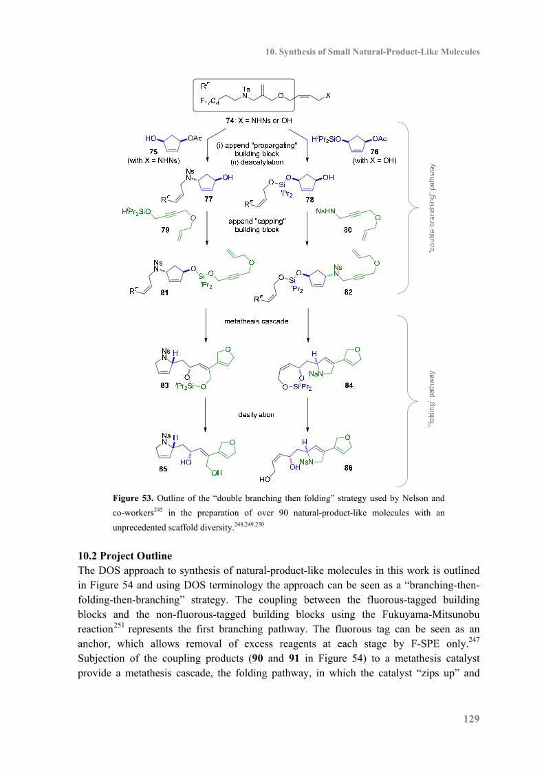
10. Synthesis of Small Natural-Product-Like Molecules
129
Figure 53. Outline of the “double branching then folding” strategy used by Nelson and
co-workers245 in the preparation of over 90 natural-product-like molecules with an
unprecedented scaffold diversity.248,249,250
10.2 Project Outline The DOS approach to synthesis of natural-product-like molecules in this work is outlined in Figure 54 and using DOS terminology the approach can be seen as a “branching-then-folding-then-branching” strategy. The coupling between the fluorous-tagged building blocks and the non-fluorous-tagged building blocks using the Fukuyama-Mitsunobu reaction251 represents the first branching pathway. The fluorous tag can be seen as an anchor, which allows removal of excess reagents at each stage by F-SPE only.247 Subjection of the coupling products (90 and 91 in Figure 54) to a metathesis catalyst provide a metathesis cascade, the folding pathway, in which the catalyst “zips up” and
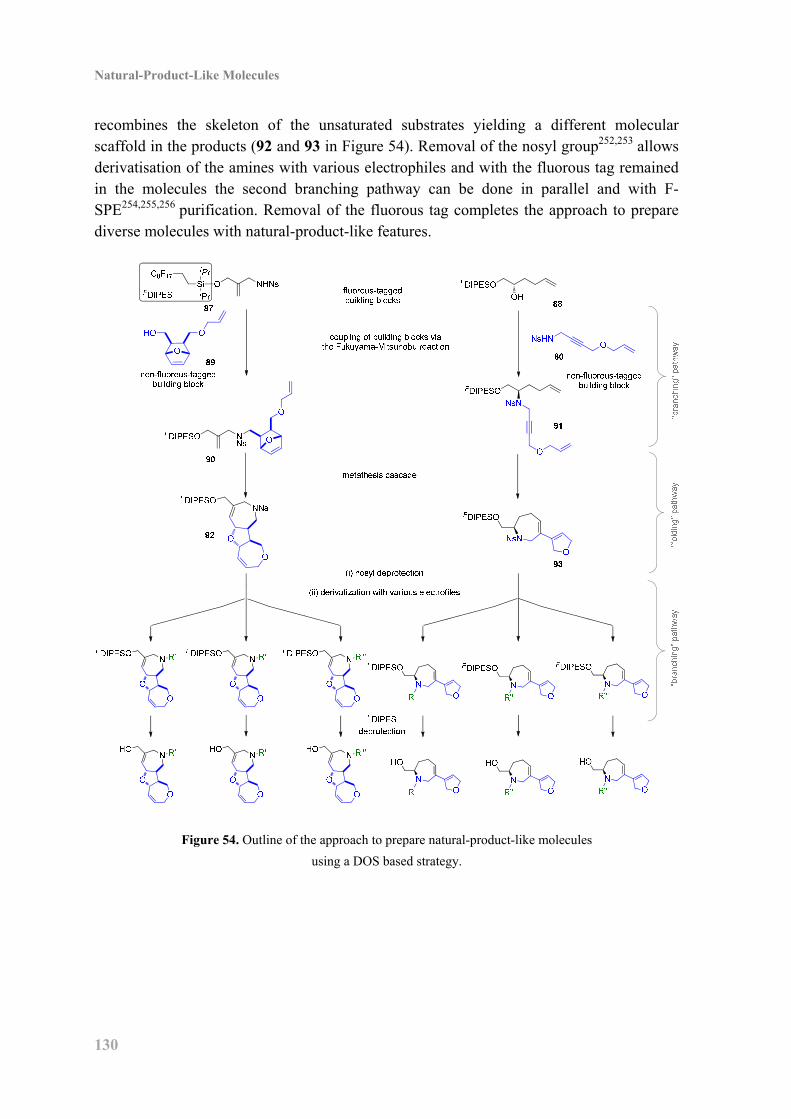
Natural-Product-Like Molecules
130
recombines the skeleton of the unsaturated substrates yielding a different molecular scaffold in the products (92 and 93 in Figure 54). Removal of the nosyl group252,253 allows derivatisation of the amines with various electrophiles and with the fluorous tag remained in the molecules the second branching pathway can be done in parallel and with F-SPE254,255,256 purification. Removal of the fluorous tag completes the approach to prepare diverse molecules with natural-product-like features.
Figure 54. Outline of the approach to prepare natural-product-like molecules
using a DOS based strategy.
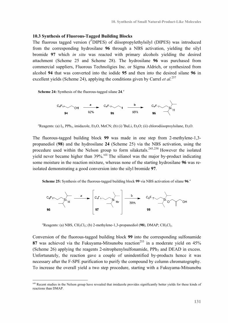
10. Synthesis of Small Natural-Product-Like Molecules
131
10.3 Synthesis of Fluorous-Tagged Building Blocks The fluorous tagged version (FDIPES) of diisopropylethylsilyl (DIPES) was introduced from the corresponding hydrosilane 96 through a NBS activation, yielding the silyl bromide 97 which in situ was reacted with primary alcohols yielding the desired attachment (Scheme 25 and Scheme 28). The hydrosilane 96 was purchased from commercial suppliers, Fluorous Technoligies Inc. or Sigma Aldrich, or synthesized from alcohol 94 that was converted into the iodide 95 and then into the desired silane 96 in excellent yields (Scheme 24), applying the conditions given by Carrel et al.257
Scheme 24: Synthesis of the fluorous-tagged silane 24.a
aReagents: (a) I2, PPh3, imidazole, Et2O, MeCN; (b) (i) tBuLi, Et2O; (ii) chlorodiisoproylsilane, Et2O.
The fluorous-tagged building block 99 was made in one step from 2-methylene-1,3-propanediol (98) and the hydrosilane 24 (Scheme 25) via the NBS activation, using the procedure used within the Nelson group to form silaketals.245,258 However the isolated yield never became higher than 39%.xiii The silianol was the major by-product indicating some moisture in the reaction mixture, whereas none of the starting hydrosilane 96 was re-isolated demonstrating a good conversion into the silyl bromide 97.
Scheme 25: Synthesis of the fluorous-tagged building block 99 via NBS activation of silane 96.a
aReagents: (a) NBS, CH2Cl2; (b) 2-methylene-1,3-propanediol (98), DMAP; CH2Cl2.
Conversion of the fluorous-tagged building block 99 into the corresponding sulfonamide 87 was achieved via the Fukuyama-Mitsunobu reaction253 in a moderate yield on 45% (Scheme 26) applying the reagents 2-nitrophenylsulfonamide, PPh3 and DEAD in excess. Unfortunately, the reaction gave a couple of unindentified by-products hence it was necessary after the F-SPE purification to purify the compound by column chromatography. To increase the overall yield a two step procedure, starting with a Fukuyama-Mitsunobu
xiii Recent studies in the Nelson group have revealed that imidazole provides significantly better yields for these kinds of reactions than DMAP.
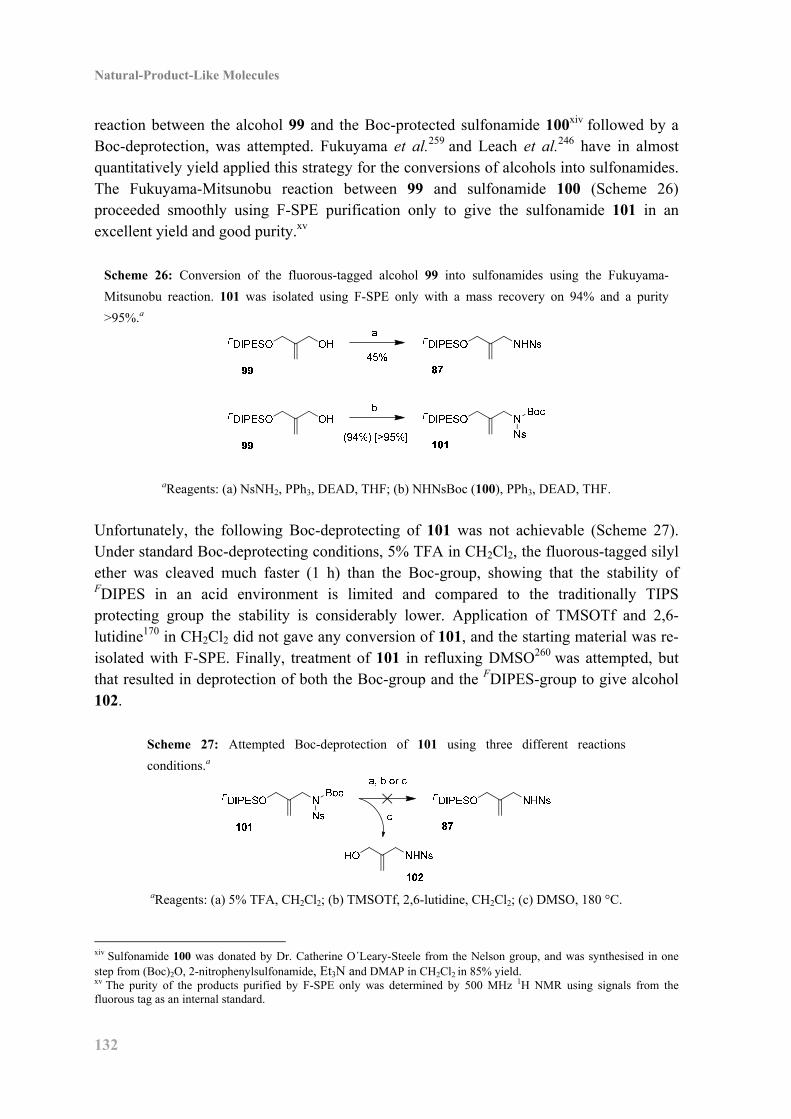
Natural-Product-Like Molecules
132
reaction between the alcohol 99 and the Boc-protected sulfonamide 100xiv followed by a Boc-deprotection, was attempted. Fukuyama et al.259 and Leach et al.246 have in almost quantitatively yield applied this strategy for the conversions of alcohols into sulfonamides. The Fukuyama-Mitsunobu reaction between 99 and sulfonamide 100 (Scheme 26) proceeded smoothly using F-SPE purification only to give the sulfonamide 101 in an excellent yield and good purity.xv
Scheme 26: Conversion of the fluorous-tagged alcohol 99 into sulfonamides using the Fukuyama-
Mitsunobu reaction. 101 was isolated using F-SPE only with a mass recovery on 94% and a purity
>95%.a
aReagents: (a) NsNH2, PPh3, DEAD, THF; (b) NHNsBoc (100), PPh3, DEAD, THF.
Unfortunately, the following Boc-deprotecting of 101 was not achievable (Scheme 27). Under standard Boc-deprotecting conditions, 5% TFA in CH2Cl2, the fluorous-tagged silyl ether was cleaved much faster (1 h) than the Boc-group, showing that the stability of FDIPES in an acid environment is limited and compared to the traditionally TIPS protecting group the stability is considerably lower. Application of TMSOTf and 2,6-lutidine170 in CH2Cl2 did not gave any conversion of 101, and the starting material was re-isolated with F-SPE. Finally, treatment of 101 in refluxing DMSO260 was attempted, but that resulted in deprotection of both the Boc-group and the FDIPES-group to give alcohol 102.
Scheme 27: Attempted Boc-deprotection of 101 using three different reactions
conditions.a
aReagents: (a) 5% TFA, CH2Cl2; (b) TMSOTf, 2,6-lutidine, CH2Cl2; (c) DMSO, 180 °C.
xiv Sulfonamide 100 was donated by Dr. Catherine O´Leary-Steele from the Nelson group, and was synthesised in one step from (Boc)2O, 2-nitrophenylsulfonamide, Et3N and DMAP in CH2Cl2 in 85% yield. xv The purity of the products purified by F-SPE only was determined by 500 MHz 1H NMR using signals from the fluorous tag as an internal standard.
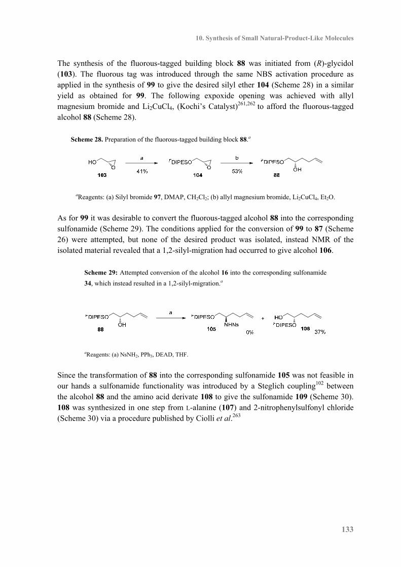
10. Synthesis of Small Natural-Product-Like Molecules
133
The synthesis of the fluorous-tagged building block 88 was initiated from (R)-glycidol (103). The fluorous tag was introduced through the same NBS activation procedure as applied in the synthesis of 99 to give the desired silyl ether 104 (Scheme 28) in a similar yield as obtained for 99. The following expoxide opening was achieved with allyl magnesium bromide and Li2CuCl4, (Kochi’s Catalyst)261,262 to afford the fluorous-tagged alcohol 88 (Scheme 28).
Scheme 28. Preparation of the fluorous-tagged building block 88.a
aReagents: (a) Silyl bromide 97, DMAP, CH2Cl2; (b) allyl magnesium bromide, Li2CuCl4, Et2O.
As for 99 it was desirable to convert the fluorous-tagged alcohol 88 into the corresponding sulfonamide (Scheme 29). The conditions applied for the conversion of 99 to 87 (Scheme 26) were attempted, but none of the desired product was isolated, instead NMR of the isolated material revealed that a 1,2-silyl-migration had occurred to give alcohol 106.
Scheme 29: Attempted conversion of the alcohol 16 into the corresponding sulfonamide
34, which instead resulted in a 1,2-silyl-migration.a
aReagents: (a) NsNH2, PPh3, DEAD, THF.
Since the transformation of 88 into the corresponding sulfonamide 105 was not feasible in our hands a sulfonamide functionality was introduced by a Steglich coupling102 between the alcohol 88 and the amino acid derivate 108 to give the sulfonamide 109 (Scheme 30). 108 was synthesized in one step from L-alanine (107) and 2-nitrophenylsulfonyl chloride (Scheme 30) via a procedure published by Ciolli et al.263
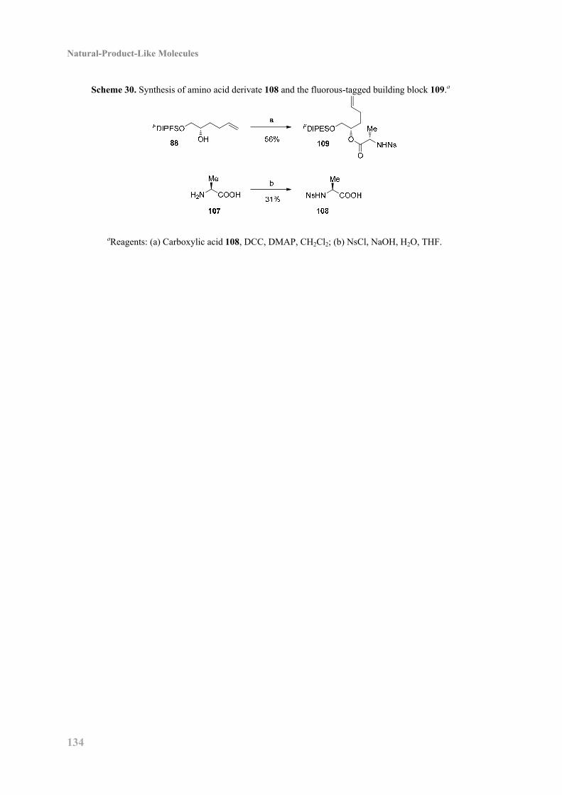
Natural-Product-Like Molecules
134
Scheme 30. Synthesis of amino acid derivate 108 and the fluorous-tagged building block 109.a
aReagents: (a) Carboxylic acid 108, DCC, DMAP, CH2Cl2; (b) NsCl, NaOH, H2O, THF.
10. Synthesis of Small Natural-Product-Like Molecules
135
10.4 Synthesis of Non-Fluorous-Tagged Building Blocks The building blocks applied in this work are shown in Figure 55. Sulfonamide 80xvi and alcohol 111xvii were donated from members of the Nelson group.
Figure 55. The applied non-fluorous-tagged building blocks – two kinds of building blocks
were used; sulfonamides and alcohols.
The sulfonamide 110 was synthesized from the commercial available amine 113 and 2-nitrophenylsulfonyl chloride (Scheme 31) in a 71% yield as white needles. It was crucial that the recrystallisation from 1:1 toluene–hexane removed all traces of 2-nitrophenylsulfonyl derivates; otherwise it was observed that the Fukuyama-Mitsunobu reaction between 110 and various alcohols did not occur. The vinylacetate 112 was synthesized by a Steglich coupling102 between the diol 114 and vinylacetic acid (115, Scheme 31) in a poor yield on 31%. Although the reaction was carried out under dilute conditions (0.01 M) the diester 116 was also isolated in 11% yield.
Scheme 31. Synthesis of the sulfonamide 110 and the ester 112.a
H2Na
71%NsHN
OHHO
O
OHb
31%
OHO
O
OO
O
O114 115 112 116 11%
110113
aReagents: (a) NsCl, K2CO3, CH2Cl2; (b) DCC, DMAP, CH2Cl2.
xvi Alcohol 111 was donated by Dr. Stuart Leach, former member of the Nelson group, and was synthesised from 1,4-butyndiol, allyl bromide and NaH in DMF in 63% yield. xvii Sulfonamide 80 was donated by Dr. Catherine O´Leary-Steele from the Nelson group and was synthesised over two step in 72%. The first step was a Fukuyama-Mitsunobu coupling between the alcohol 111 and the sulfonamide 100 activated by PPh3 and DEAD in THF, followed by a Boc-deprotection with 10% TFA in CH2Cl2.
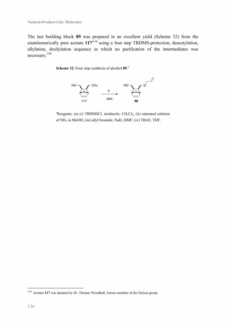
Natural-Product-Like Molecules
136
The last building block 89 was prepared in an excellent yield (Scheme 32) from the enantiomerically pure acetate 117xviii using a four step TBDMS-protection, deacetylation, allylation, desilylation sequence in which no purification of the intermediates was necessary.258
Scheme 32. Four step synthesis of alcohol 89.a
aReagents: (a) (i) TBDMSCl, imidazole, CH2Cl2; (ii) saturated solution
of NH3 in MeOH; (iii) allyl bromide, NaH, DMF; (iv) TBAF, THF.
xviii Acetate 117 was donated by Dr. Thomas Woodhall, former member of the Nelson group.
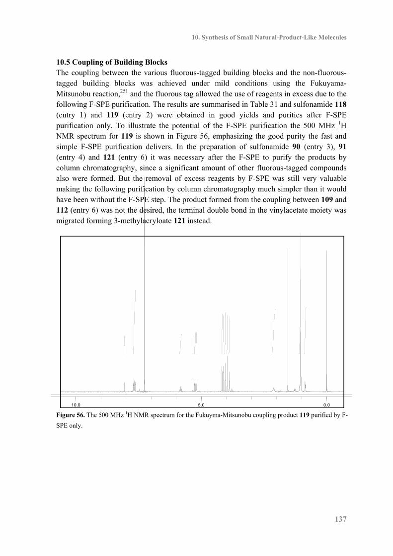
10. Synthesis of Small Natural-Product-Like Molecules
137
10.5 Coupling of Building Blocks The coupling between the various fluorous-tagged building blocks and the non-fluorous-tagged building blocks was achieved under mild conditions using the Fukuyama-Mitsunobu reaction,251 and the fluorous tag allowed the use of reagents in excess due to the following F-SPE purification. The results are summarised in Table 31 and sulfonamide 118 (entry 1) and 119 (entry 2) were obtained in good yields and purities after F-SPE purification only. To illustrate the potential of the F-SPE purification the 500 MHz 1H NMR spectrum for 119 is shown in Figure 56, emphasizing the good purity the fast and simple F-SPE purification delivers. In the preparation of sulfonamide 90 (entry 3), 91 (entry 4) and 121 (entry 6) it was necessary after the F-SPE to purify the products by column chromatography, since a significant amount of other fluorous-tagged compounds also were formed. But the removal of excess reagents by F-SPE was still very valuable making the following purification by column chromatography much simpler than it would have been without the F-SPE step. The product formed from the coupling between 109 and 112 (entry 6) was not the desired, the terminal double bond in the vinylacetate moiety was migrated forming 3-methylacryloate 121 instead.
Figure 56. The 500 MHz 1H NMR spectrum for the Fukuyma-Mitsunobu coupling product 119 purified by F-
SPE only.
0.05.010.0
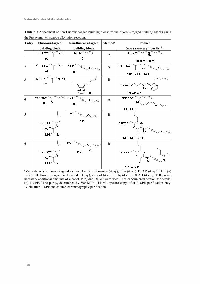
Natural-Product-Like Molecules
138
Table 31: Attachment of non-fluorous-tagged building blocks to the fluorous tagged building blocks using
the Fukuyama-Mitsunobu alkylation reaction.
Entry Fluorous-tagged
building block
Non-fluorous-tagged
building block
Methoda Product
(mass recovery) [purity] b
1
A
2
A
3
B
4
A
5
B
6
B
aMethods: A: (i) fluorous-tagged alcohol (1 eq.), sulfonamide (4 eq.), PPh3 (4 eq.), DEAD (4 eq.), THF. (ii) F–SPE; B: fluorous-tagged sulfonamide (1 eq.), alcohol (4 eq.), PPh3 (4 eq.), DEAD (4 eq.), THF, when necessary additional amounts of alcohol, PPh3 and DEAD were used – see experimental section for details. (ii) F–SPE. bThe purity, determined by 500 MHz 1H-NMR spectroscopy, after F–SPE purification only. cYield after F–SPE and column chromatography purification.
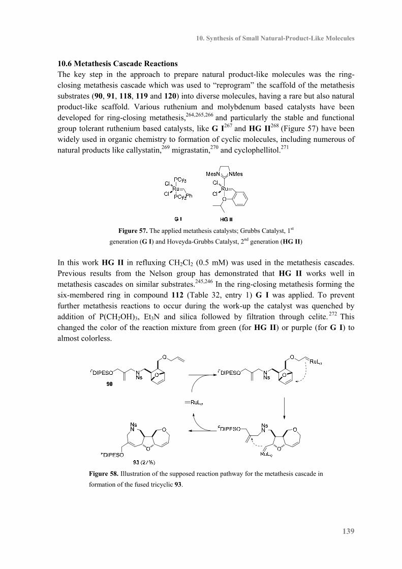
10. Synthesis of Small Natural-Product-Like Molecules
139
10.6 Metathesis Cascade Reactions The key step in the approach to prepare natural product-like molecules was the ring-closing metathesis cascade which was used to “reprogram” the scaffold of the metathesis substrates (90, 91, 118, 119 and 120) into diverse molecules, having a rare but also natural product-like scaffold. Various ruthenium and molybdenum based catalysts have been developed for ring-closing metathesis,264,265,266 and particularly the stable and functional group tolerant ruthenium based catalysts, like G I267 and HG II268 (Figure 57) have been widely used in organic chemistry to formation of cyclic molecules, including numerous of natural products like callystatin,269 migrastatin,270 and cyclophellitol.271
Figure 57. The applied metathesis catalysts; Grubbs Catalyst, 1st
generation (G I) and Hoveyda-Grubbs Catalyst, 2nd generation (HG II)
In this work HG II in refluxing CH2Cl2 (0.5 mM) was used in the metathesis cascades. Previous results from the Nelson group has demonstrated that HG II works well in metathesis cascades on similar substrates.245,246 In the ring-closing metathesis forming the six-membered ring in compound 112 (Table 32, entry 1) G I was applied. To prevent further metathesis reactions to occur during the work-up the catalyst was quenched by addition of P(CH2OH)3, Et3N and silica followed by filtration through celite. 272 This changed the color of the reaction mixture from green (for HG II) or purple (for G I) to almost colorless.
Figure 58. Illustration of the supposed reaction pathway for the metathesis cascade in
formation of the fused tricyclic 93.
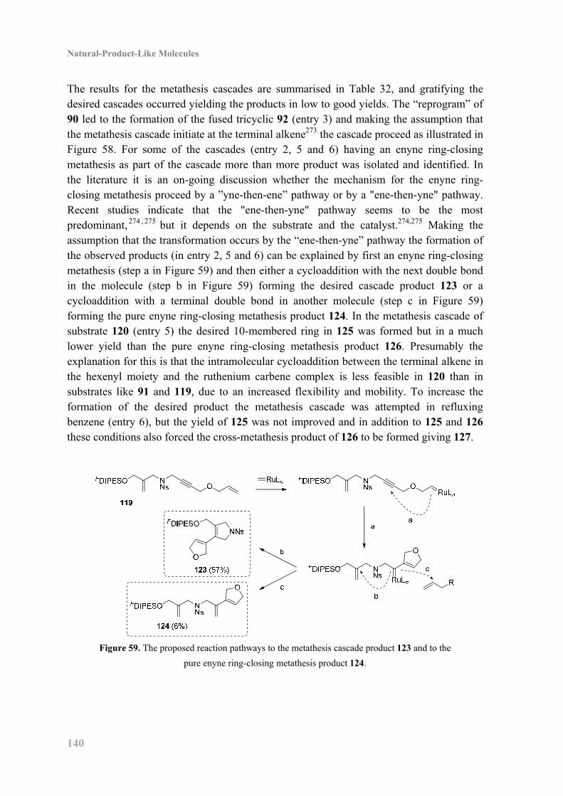
Natural-Product-Like Molecules
140
The results for the metathesis cascades are summarised in Table 32, and gratifying the desired cascades occurred yielding the products in low to good yields. The “reprogram” of 90 led to the formation of the fused tricyclic 92 (entry 3) and making the assumption that the metathesis cascade initiate at the terminal alkene273 the cascade proceed as illustrated in Figure 58. For some of the cascades (entry 2, 5 and 6) having an enyne ring-closing metathesis as part of the cascade more than more product was isolated and identified. In the literature it is an on-going discussion whether the mechanism for the enyne ring-closing metathesis proceed by a ”yne-then-ene” pathway or by a "ene-then-yne" pathway. Recent studies indicate that the "ene-then-yne" pathway seems to be the most predominant, 274 , 275 but it depends on the substrate and the catalyst.274,275 Making the assumption that the transformation occurs by the “ene-then-yne” pathway the formation of the observed products (in entry 2, 5 and 6) can be explained by first an enyne ring-closing metathesis (step a in Figure 59) and then either a cycloaddition with the next double bond in the molecule (step b in Figure 59) forming the desired cascade product 123 or a cycloaddition with a terminal double bond in another molecule (step c in Figure 59) forming the pure enyne ring-closing metathesis product 124. In the metathesis cascade of substrate 120 (entry 5) the desired 10-membered ring in 125 was formed but in a much lower yield than the pure enyne ring-closing metathesis product 126. Presumably the explanation for this is that the intramolecular cycloaddition between the terminal alkene in the hexenyl moiety and the ruthenium carbene complex is less feasible in 120 than in substrates like 91 and 119, due to an increased flexibility and mobility. To increase the formation of the desired product the metathesis cascade was attempted in refluxing benzene (entry 6), but the yield of 125 was not improved and in addition to 125 and 126 these conditions also forced the cross-metathesis product of 126 to be formed giving 127.
Figure 59. The proposed reaction pathways to the metathesis cascade product 123 and to the
pure enyne ring-closing metathesis product 124.
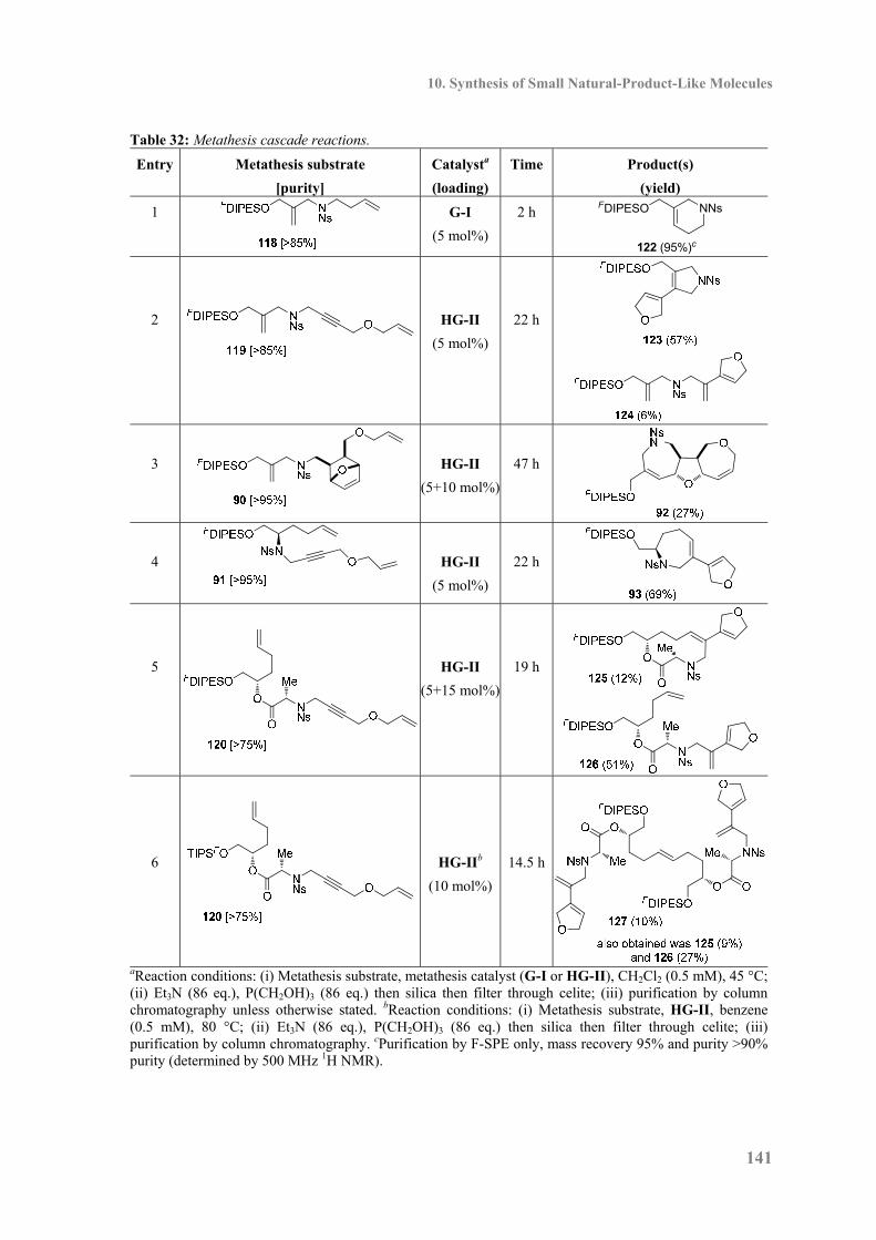
10. Synthesis of Small Natural-Product-Like Molecules
141
Table 32: Metathesis cascade reactions.
Entry Metathesis substrate
[purity]
Catalysta
(loading)
Time Product(s)
(yield)
1
G-I
(5 mol%)
2 h NNsFDIPESO
122 (95%)c
2
HG-II
(5 mol%)
22 h
3
HG-II
(5+10 mol%)
47 h
4
HG-II
(5 mol%)
22 h
5
HG-II
(5+15 mol%)
19 h
6
HG-IIb
(10 mol%)
14.5 h
aReaction conditions: (i) Metathesis substrate, metathesis catalyst (G-I or HG-II), CH2Cl2 (0.5 mM), 45 °C; (ii) Et3N (86 eq.), P(CH2OH)3 (86 eq.) then silica then filter through celite; (iii) purification by columnchromatography unless otherwise stated. bReaction conditions: (i) Metathesis substrate, HG-II, benzene (0.5 mM), 80 °C; (ii) Et3N (86 eq.), P(CH2OH)3 (86 eq.) then silica then filter through celite; (iii) purification by column chromatography. cPurification by F-SPE only, mass recovery 95% and purity >90% purity (determined by 500 MHz 1H NMR).
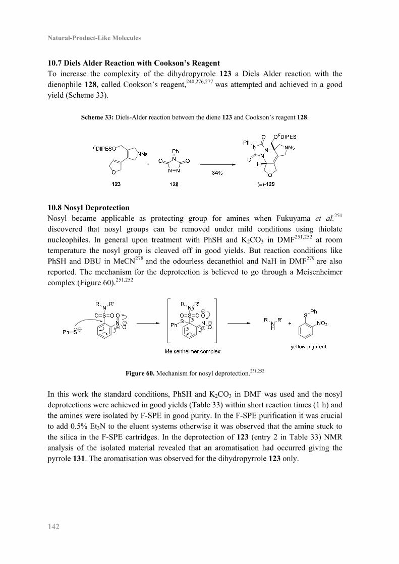
Natural-Product-Like Molecules
142
10.7 Diels Alder Reaction with Cookson’s Reagent To increase the complexity of the dihydropyrrole 123 a Diels Alder reaction with the dienophile 128, called Cookson’s reagent,240,276,277 was attempted and achieved in a good yield (Scheme 33).
Scheme 33: Diels-Alder reaction between the diene 123 and Cookson’s reagent 128.
10.8 Nosyl Deprotection Nosyl became applicable as protecting group for amines when Fukuyama et al.251 discovered that nosyl groups can be removed under mild conditions using thiolate nucleophiles. In general upon treatment with PhSH and K2CO3 in DMF251,252 at room temperature the nosyl group is cleaved off in good yields. But reaction conditions like PhSH and DBU in MeCN278 and the odourless decanethiol and NaH in DMF279 are also reported. The mechanism for the deprotection is believed to go through a Meisenheimer complex (Figure 60).251,252
Figure 60. Mechanism for nosyl deprotection.251,252
In this work the standard conditions, PhSH and K2CO3 in DMF was used and the nosyl deprotections were achieved in good yields (Table 33) within short reaction times (1 h) and the amines were isolated by F-SPE in good purity. In the F-SPE purification it was crucial to add 0.5% Et3N to the eluent systems otherwise it was observed that the amine stuck to the silica in the F-SPE cartridges. In the deprotection of 123 (entry 2 in Table 33) NMR analysis of the isolated material revealed that an aromatisation had occurred giving the pyrrole 131. The aromatisation was observed for the dihydropyrrole 123 only.
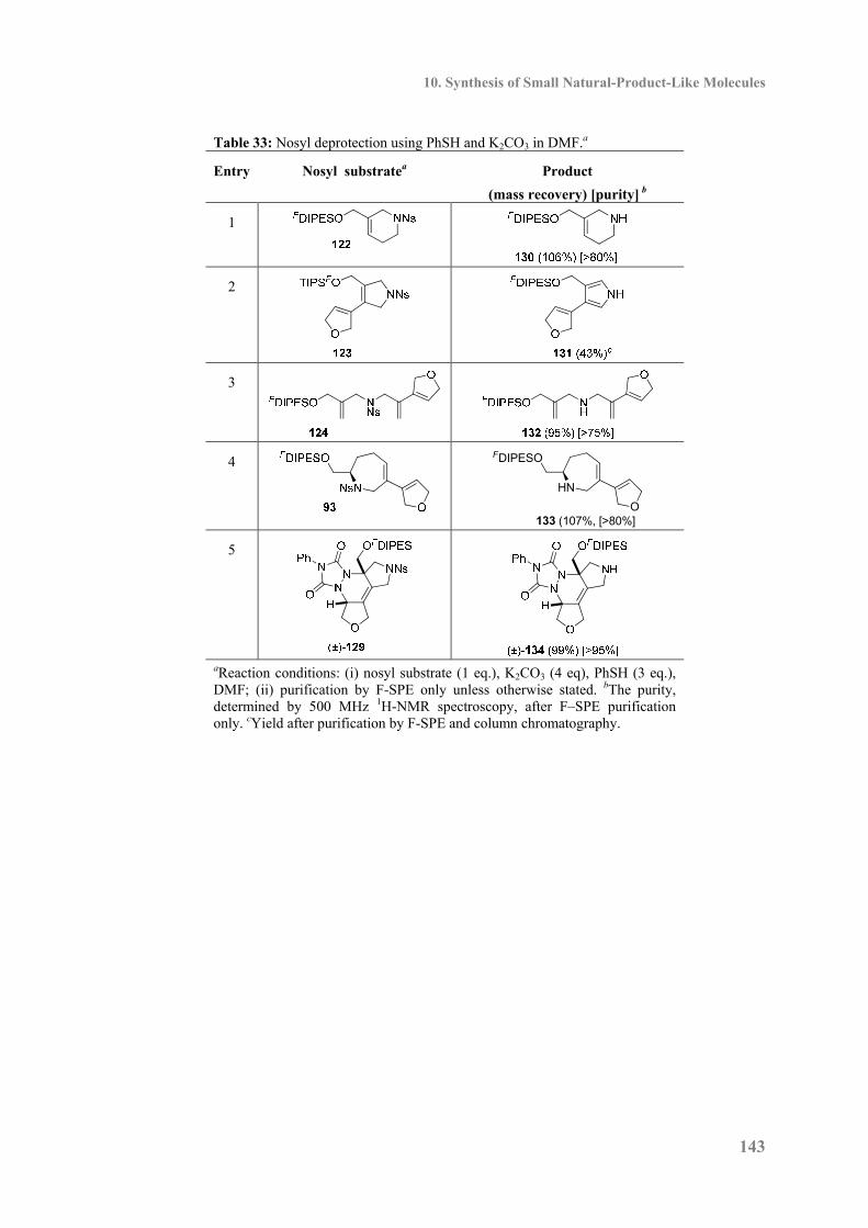
10. Synthesis of Small Natural-Product-Like Molecules
143
Table 33: Nosyl deprotection using PhSH and K2CO3 in DMF.a
Entry Nosyl substratea Product
(mass recovery) [purity] b
1
2
3
4
133 (107%, [>80%]
FDIPESO
HN
O
5
aReaction conditions: (i) nosyl substrate (1 eq.), K2CO3 (4 eq), PhSH (3 eq.), DMF; (ii) purification by F-SPE only unless otherwise stated. bThe purity, determined by 500 MHz 1H-NMR spectroscopy, after F–SPE purification only. cYield after purification by F-SPE and column chromatography.
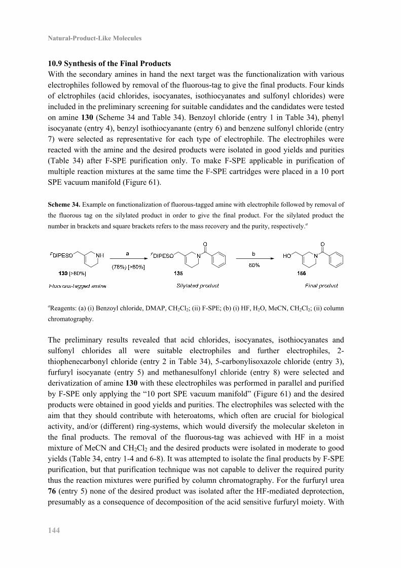
Natural-Product-Like Molecules
144
10.9 Synthesis of the Final Products With the secondary amines in hand the next target was the functionalization with various electrophiles followed by removal of the fluorous-tag to give the final products. Four kinds of elctrophiles (acid chlorides, isocyanates, isothiocyanates and sulfonyl chlorides) were included in the preliminary screening for suitable candidates and the candidates were tested on amine 130 (Scheme 34 and Table 34). Benzoyl chloride (entry 1 in Table 34), phenyl isocyanate (entry 4), benzyl isothiocyanante (entry 6) and benzene sulfonyl chloride (entry 7) were selected as representative for each type of electrophile. The electrophiles were reacted with the amine and the desired products were isolated in good yields and purities (Table 34) after F-SPE purification only. To make F-SPE applicable in purification of multiple reaction mixtures at the same time the F-SPE cartridges were placed in a 10 port SPE vacuum manifold (Figure 61). Scheme 34. Example on functionalization of fluorous-tagged amine with electrophile followed by removal of
the fluorous tag on the silylated product in order to give the final product. For the silylated product the
number in brackets and square brackets refers to the mass recovery and the purity, respectively.a
aReagents: (a) (i) Benzoyl chloride, DMAP, CH2Cl2; (ii) F-SPE; (b) (i) HF, H2O, MeCN, CH2Cl2; (ii) column
chromatography.
The preliminary results revealed that acid chlorides, isocyanates, isothiocyanates and sulfonyl chlorides all were suitable electrophiles and further electrophiles, 2-thiophenecarbonyl chloride (entry 2 in Table 34), 5-carbonylisoxazole chloride (entry 3), furfuryl isocyanate (entry 5) and methanesulfonyl chloride (entry 8) were selected and derivatization of amine 130 with these electrophiles was performed in parallel and purified by F-SPE only applying the “10 port SPE vacuum manifold” (Figure 61) and the desired products were obtained in good yields and purities. The electrophiles was selected with the aim that they should contribute with heteroatoms, which often are crucial for biological activity, and/or (different) ring-systems, which would diversify the molecular skeleton in the final products. The removal of the fluorous-tag was achieved with HF in a moist mixture of MeCN and CH2Cl2 and the desired products were isolated in moderate to good yields (Table 34, entry 1-4 and 6-8). It was attempted to isolate the final products by F-SPE purification, but that purification technique was not capable to deliver the required purity thus the reaction mixtures were purified by column chromatography. For the furfuryl urea 76 (entry 5) none of the desired product was isolated after the HF-mediated deprotection, presumably as a consequence of decomposition of the acid sensitive furfuryl moiety. With
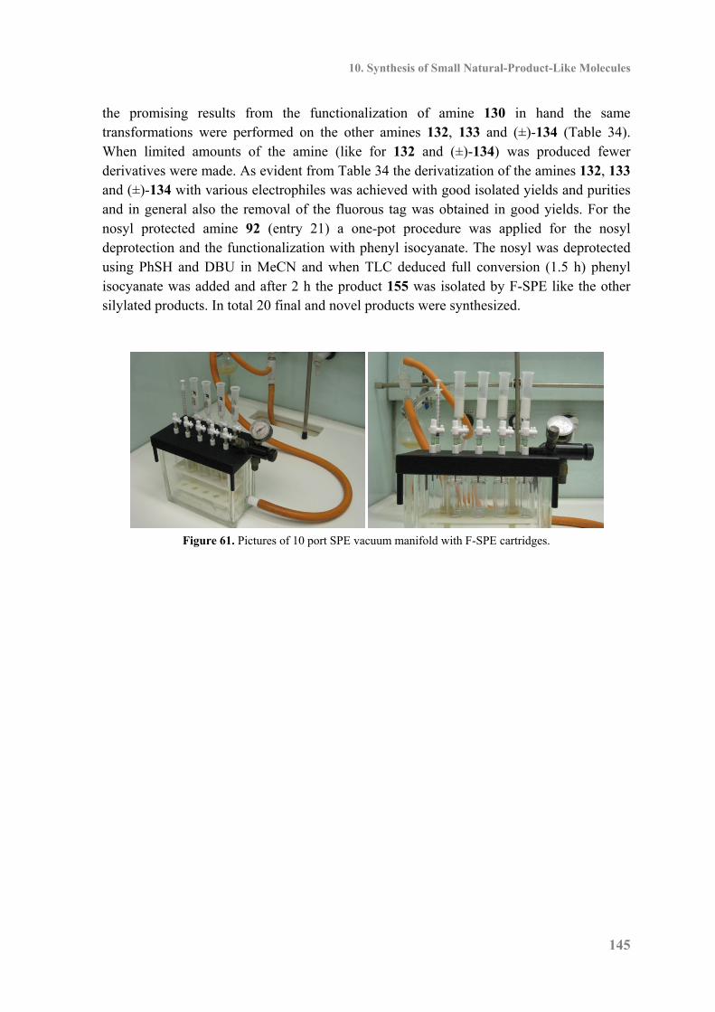
10. Synthesis of Small Natural-Product-Like Molecules
145
the promising results from the functionalization of amine 130 in hand the same transformations were performed on the other amines 132, 133 and (±)-134 (Table 34). When limited amounts of the amine (like for 132 and (±)-134) was produced fewer derivatives were made. As evident from Table 34 the derivatization of the amines 132, 133 and (±)-134 with various electrophiles was achieved with good isolated yields and purities and in general also the removal of the fluorous tag was obtained in good yields. For the nosyl protected amine 92 (entry 21) a one-pot procedure was applied for the nosyl deprotection and the functionalization with phenyl isocyanate. The nosyl was deprotected using PhSH and DBU in MeCN and when TLC deduced full conversion (1.5 h) phenyl isocyanate was added and after 2 h the product 155 was isolated by F-SPE like the other silylated products. In total 20 final and novel products were synthesized.
Figure 61. Pictures of 10 port SPE vacuum manifold with F-SPE cartridges.
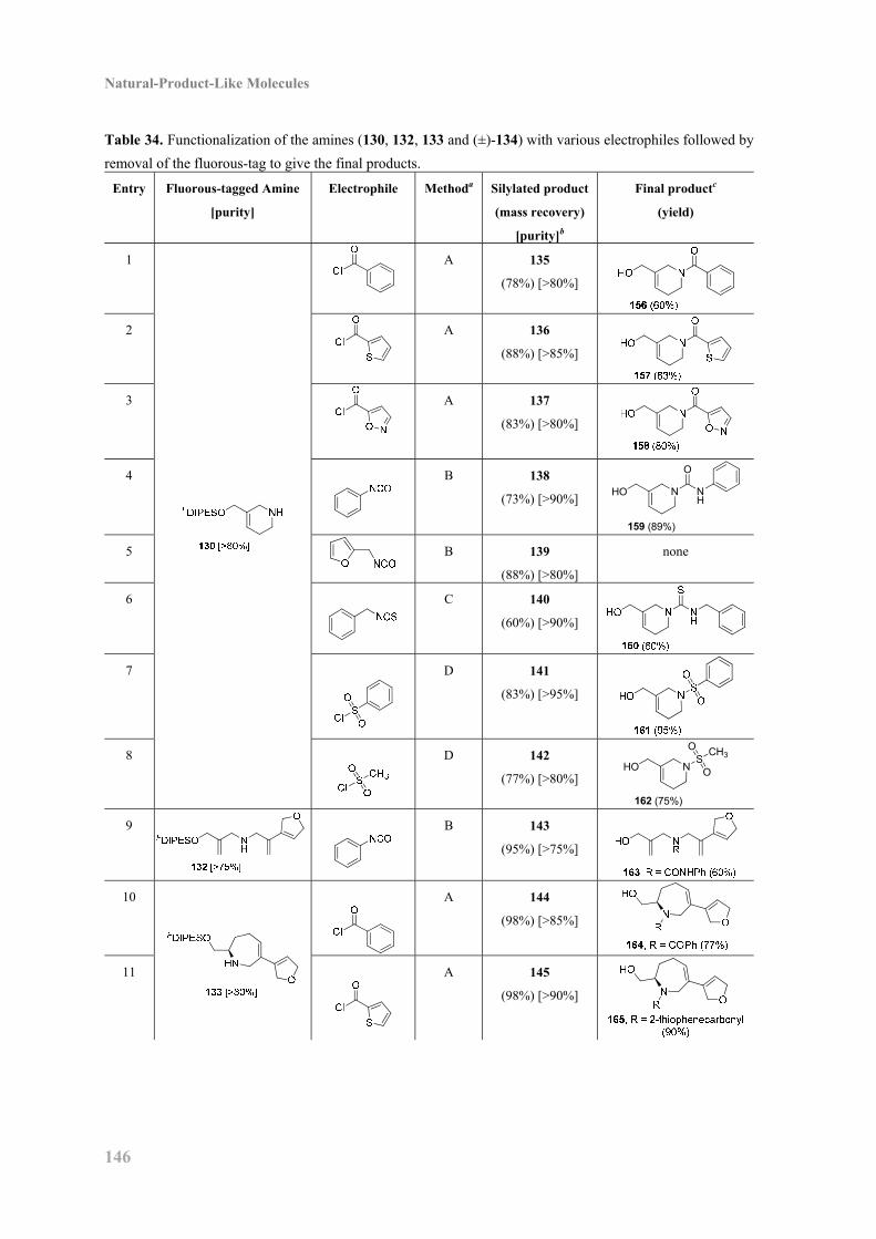
Natural-Product-Like Molecules
146
Table 34. Functionalization of the amines (130, 132, 133 and (±)-134) with various electrophiles followed by
removal of the fluorous-tag to give the final products.
Entry Fluorous-tagged Amine
[purity]
Electrophile Methoda Silylated product
(mass recovery)
[purity]b
Final productc
(yield)
1
A 135
(78%) [>80%]
2
A
136
(88%) [>85%]
3
A
137
(83%) [>80%]
4
B
138
(73%) [>90%] NHO
159 (89%)
O
NH
5
B 139
(88%) [>80%]
none
6
C
140
(60%) [>90%]
7
D 141
(83%) [>95%]
8
D
142
(77%) [>80%] NHO
162 (75%)
SO CH3
O
9
B 143
(95%) [>75%]
10
A 144
(98%) [>85%]
11
A 145
(98%) [>90%]
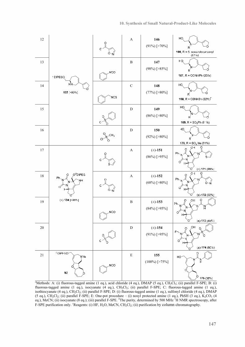
10. Synthesis of Small Natural-Product-Like Molecules
147
12
A 146
(91%) [>70%]
13
B 147
(98%) [>85%]
14
C 148
(77%) [>80%]
15 D 149
(86%) [>80%]
16
D 150
(92%) [>80%]
17
A (±)-151
(86%) [>95%]
18
A (±)-152
(68%) [>80%]
19
B (±)-153
(84%) [>95%]
20
D (±)-154
(91%) [>95%]
21
E 155
(100%) [>75%]
aMethods: A: (i) fluorous-tagged amine (1 eq.), acid chloride (4 eq.), DMAP (5 eq.), CH2Cl2; (ii) parallel F-SPE; B: (i)fluorous-tagged amine (1 eq.), isocyanate (4 eq.), CH2Cl2; (ii) parallel F-SPE; C: fluorous-tagged amine (1 eq.),isothiocyanate (4 eq.), CH2Cl2; (ii) parallel F-SPE; D: (i) fluorous-tagged amine (1 eq.), sulfonyl chloride (4 eq.), DMAP(5 eq.), CH2Cl2; (ii) parallel F-SPE; E: One-pot procedure – (i) nosyl protected amine (1 eq.), PhSH (3 eq.), K2CO3 (4eq.), MeCN; (ii) isocyanate (8 eq.); (iii) parallel F-SPE. bThe purity, determined by 500 MHz 1H NMR spectroscopy, afterF-SPE purification only. cReagents: (i) HF, H2O, MeCN, CH2Cl2; (ii) purification by column chromatography.
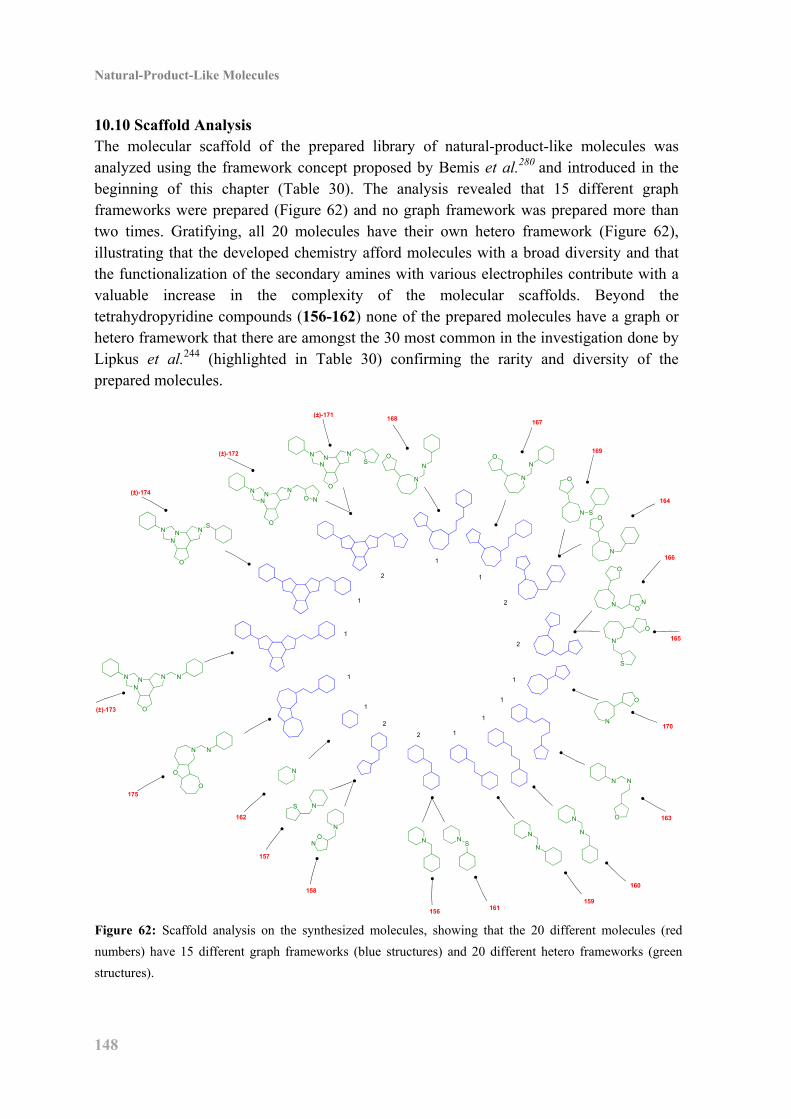
Natural-Product-Like Molecules
148
10.10 Scaffold Analysis The molecular scaffold of the prepared library of natural-product-like molecules was analyzed using the framework concept proposed by Bemis et al.280 and introduced in the beginning of this chapter (Table 30). The analysis revealed that 15 different graph frameworks were prepared (Figure 62) and no graph framework was prepared more than two times. Gratifying, all 20 molecules have their own hetero framework (Figure 62), illustrating that the developed chemistry afford molecules with a broad diversity and that the functionalization of the secondary amines with various electrophiles contribute with a valuable increase in the complexity of the molecular scaffolds. Beyond the tetrahydropyridine compounds (156-162) none of the prepared molecules have a graph or hetero framework that there are amongst the 30 most common in the investigation done by Lipkus et al.244 (highlighted in Table 30) confirming the rarity and diversity of the prepared molecules.
1
1
2
1
1
2
2
1
1
1
12
2
1
1
N
NS
N
ON N N
S
N
N
N
N
N N
O
N
S
O
N
O
NO
N
O
N
N S
O
O
N
NO
N
N
ON
O
NNN
S
N
O
NNN
O N
N
O
NNN
S
N
O
NNN N
N
O
O
N
162
157
158
156161
159
160
163
170
165
166
164
169
167168
(±)-171
(±)-172
(±)-174
(±)-173
175
Figure 62: Scaffold analysis on the synthesized molecules, showing that the 20 different molecules (red
numbers) have 15 different graph frameworks (blue structures) and 20 different hetero frameworks (green
structures).
10. Synthesis of Small Natural-Product-Like Molecules
149
10.11 Conclusion In total 20 novel compounds with a broad structural diversity were prepared using a short 5 or 6 steps “branching-then-folding-then-branching” strategy in overall yields from 75% to 5% (36% in average). A limited number of reaction types were applied, including the Fukuyama-Mitsunobu reaction and the ring closing metathesis cascade as the key reactions. Furthermore, in average the rapid F-SPE purification was sufficient in 3 out of 5 steps limiting the use of column chromatography. The prepared natural-product-like molecules are subjected to biological testing and it will be interesting to see whether some of the molecules will provide a biological response and potentially be a novel lead compound in the development of a new drug.

11. Experimental: Natural-Prodruct-Like Molecules
151
11. Experimental: Natural-Prodruct-Like Molecules 11.1 General Experimental Starting materials, reagents and solvents were purchased from Sigma–Aldrich, Alfa Aesar or Fluorous Technologies and used without further purification unless otherwise stated. DMF, MeCN and CH2Cl2 were dried over 4 Å molecular sieves. THF was freshly distilled from Na, using benzophenone as a self-indicator. Saturated NH3 in MeOH (ca. 0.025 M) was prepared by bubbling NH3 gas through MeOH for a minimum of 30 min. NBS was recrystallised from water and dried under vacuum prior to use. Solvents were removed under reduced pressure using a Büchi rotary evaporator and a Vacuubrand diaphragm pump or a Genevac HT-4 evaporation system. Petrol refers to petroleum spirit (bp 40–60 °C). Column chromatography was carried out using silica (35-70 μm particles). TLC was carried out on commercially available pre-coated plates (Merck silica Kieselgel 60F254). 1H and 13C NMR spectra were recorded on a Bruker Avance 500, Avance DPX300 or DRX500 spectrophotometer with an internal deuterium lock. Chemical shifts are quoted in ppm downfield of tetramethylsilane and values of coupling constants are given in Hz. 13C NMR spectra were recorded with broad band proton decoupling. Melting points were determined on a Reichert hot stage apparatus and are uncorrected. Infrared spectra were recorded on a Perkin Elmer spectrum one FT-IR infrared spectrophotometer. Mass spectra were recorded using a Micromass LCT-KA111 electrospray mass spectrometer (for ES+ mode) or a Quatro II triple quadrupole instrument (for EI mode). Optical activity measurements were recorded at room temperature (20 °C) on an AA-1000
polarimeter; units for 20D are 10–1 deg cm2 g–1.
11.2 General Procedures General Procedure for F-SPE Purification Fluorous–Solid Phase Extraction (F–SPE) was carried out using pre-packed FluoroFlash®
cartridges purchased from Fluorous Technologies Inc. The F–SPE cartridges were washed extensively with acetone and were pre-conditioned with 8:2 MeOH–H2O. Crude reaction mixtures were loaded onto cartridges using CH2Cl2 or 8:2 MeOH–H2O, and eluted using 8:2 MeOH–H2O as the fluorophobic eluent, and then MeOH as the fluorophilic eluent; compressed air was used in a manner similar to that described by Curran et al.247 Procedure for Parallel F-SPE Purification The F–SPE cartridges (2 g) were placed in the ports of a 10 port SPE vacuum manifold and washed extensively with acetone and pre-conditioned with 160:40:1 MeOH–H2O–Et3N. The crude reaction mixtures were loaded onto the F–SPE cartridge by CH2Cl2 (0.2 mL), and the reaction flasks were rinsed with 160:40:1 MeOH–H2O–Et3N (1 mL) which was added to F–SPE cartridge as well. Elution under vacuum (ca. 100 mmHg) with 160:40:1 MeOH–H2O–Et3N (3×3 mL) gave the fluorofrobic fraction which were collected in 17.5
Natural-Product-Like Molecules
152
mL vials after which elution under vacuum (ca. 100 mmHg) with 200:1 MeOH–Et3N (3×3 mL) gave the fluorophilic fraction which were collected in 17.5 mL vials. The vials containing the fluorophilic fractions were concentrated on a Genevac EZ-2 vacuum centrifuge to give the purified products. The purity of the products purified by F-SPE only was determined by 500 MHz 1H NMR using signals from the fluorous tag as an internal standard. General Procedure A: Method for Fukuyama–Mitsunobu Reactions of Fluorous-tagged Alcohols with a non-Fluorous-tagged Sulfonamide. The fluorous-tagged alcohol (1 eq.), the sulfonamide (4 eq.) and PPh3 (4 eq.) were dissolved in THF (0.022 M in alcohol) and cooled to 0 °C. DEAD (4 eq.) was added dropwise and the reaction mixture was allowed to reach room temperature and stirred for 1 h after which the mixture was concentrated under reduced pressure and purified by F–SPE. General Procedure B: Method for Ring-closing Metathesis Reactions of Fluorous-tagged Substrates. The metathesis substrate (1 eq.) was dissolved in CH2Cl2 (0.5 mM) and heated to reflux. Grubbs Catalyst, 1st generation (5 mol%) or Hoveyda-Grubbs Catalyst, 2nd generation (5 mol%) was added and the reaction mixture was stirred until completion was indicated by TLC. The reaction mixture was cooled to room temperature, Et3N (86 eq.) and tris(hydroxymethyl)phosphine (86 eq.) were added and the mixture was stirred for 15 min after which silica (ca. 10 g per mmol of substrate) was added and the mixture was stirred for another 15 min. The suspension was filtrated through a pad of celite, washed with EtOAc, concentrated under reduced pressure and purified by F-SPE or column chromatography. General Procedure C: Method for 2-Nitrophenylsulfonyl Deprotection of Fluorous-tagged Sulfonamides. The fluorous-tagged sulfonamide (1 eq.) and K2CO3 (4 eq.) were dissolved in DMF (0.05 M). PhSH (3 eq.) was added dropwise and the reaction mixture was stirred at room temperature for 1 h. A solution of 160:40:1 MeOH–H2O–Et3N (5 mL per mL DMF) was added and the mixture was purified directly by F–SPE, eluting with 160:40:1 MeOH–H2O–Et3N then 200:1 MeOH–Et3N. General Procedure D: Method for Amide Formation from Fluorous-tagged Amines. The fluorous-tagged amine (0.030 mmol) and DMAP (18 mg, 0.15 mmol) were dissolved in CH2Cl2 (0.5 mL) and the acid chloride (0.12 mmol) was added. The reaction mixture was stirred for 2 h at room temperature, concentrated under a flow of N2 and purified by F–SPE.
11. Experimental: Natural-Prodruct-Like Molecules
153
General Procedure E: Method for Urea Formation from Fluorous-tagged Amines. The fluorous-tagged amine (0.030 mmol) was dissolved in CH2Cl2 (0.5 mL) and the isocyanate (0.12 mmol) was added. The reaction mixture was stirred for 2 h at room temperature, concentrated under a flow of N2 and purified by F–SPE. General Procedure F: Method for Thiourea Formation from Fluorous-tagged Amines. The fluorous-tagged amine (0.030 mmol) was dissolved in CH2Cl2 (0.5 mL) and the isothiocyanate (0.12 mmol) was added. The reaction mixture was stirred for 2 h at room temperature, concentrated under a flow of N2 and purified by F–SPE. General Procedure G: Method for Sulfonamide Formation from Fluorous-tagged Amines. The fluorous-tagged amine (0.030 mmol) and DMAP (18 mg, 0.15 mmol) were dissolved in CH2Cl2 (0.5 mL) and the sulfonyl chloride (0.12 mmol) was added. The reaction mixture was stirred for 2 h at room temperature, concentrated under a flow of N2 and purified by F–SPE. General Procedure H: Method for the FDIPES-deprotection The fluorous-tagged silyl ether (0.030 mmol) was dissolved in MeCN (0.5 mL) and CH2Cl2 (0.4 mL) and HF (50% aq, 0.1 mL) was added. The reaction mixture was stirred for 1.5 h at room temperature after which MeOSiMe3 (0.36 mL) was added and the mixture was stirred for another 1.5 h, concentrated under a flow a of N2 and purified by column chromatography, eluting with either 20:1 CH2Cl2–MeOH or 25:1 CH2Cl2–MeOH. 11.3 Experimental Data for Compounds N-(3-((3',3',4',4',5',5',6',6',7',7',8',8',9',9',10',10',10'-Heptadecafluorodecyl)-diisopropyl-silyloxy)-2-methylene-propyl) 2-nitrobenzenesulfonamide (87) By general procedure A, the alcohol 99 (600 mg, 0.93 mmol), 2-nitrophenylsulfonamide (726 mg, 3.59 mmol), PPh3 (942 mg, 3.59 mmol) and DEAD (0.57 mL, 3.59 mmol) in THF (55 mL) gave a crude product which was purified by column chromatography, eluting with CH2Cl2, to give the sulfonamide 87 (349 mg, 45%) as a colorless oil, Rf 0.10 (10:1 petrol–EtOAc); νmax/cm-1 (neat) 3348, 2948, 2869, 1542, 1359, 1208; δH (500 MHz, CDCl3) 8.12–8.10 (1H, m, nosyl 3-H), 7.87–7.85 (1H, m, nosyl 5-H), 7.74–7.72 (2H, m, nosyl 4-H and 6-H), 5.53 (1H, t, J 5.8, NH), 5.10–5.09 (1H, m, 2-CH2a), 5.05–5.04 (1H, m, 2-CH2b), 4.17–4.16 (2H, m, 3-H2), 3.79–3.77 (2H, m, 1-H2), 2.14–2.02 (2H, m, 2'-H2), 1.08-1.00 (14H, m, iPr), 0.87–0.83 (2H, m, 1'-H2); δC (75 MHz, CDCl3) 143.0, 133.7, 132.8, 131.2, 125.4, 113.3, 64.7, 46.2, 17.6 (2C), 17.5 (2C), 12.5 (2C) (the signals for nosyl 1-C, 2-C, 1'-C, 2'-C and C8F17 are missing); m/z (ES+) (Found: MNa+ 855.1177, C26H29F17N2O5SSi requires MNa 855.1187).

Natural-Product-Like Molecules
154
(S)-(3',3',4',4',5',5',6',6',7',7',8',8',9',9',10',10',10'-Heptadecafluorodecyl)-diisopropylsilyl 2-hydroxy-5-hexenyl ether (88) The epoxide 104 (658 mg, 1.04 mmol) was dissolved in Et2O (15 mL) and the mixture was cooled to –30 °C. Li2CuCl4 (0.1 M in THF, 0.26 mL, 0.025 mmol) and then allyl magnesium bromide (1 M in Et2O, 1.50 mL, 1.50 mmol) were added dropwise and the reaction mixture was stirred for 2 h at -30 °C, after which a saturated aqueous solution of NH4Cl (6 mL) was added and the mixture was allowed to reach room temperature and H2O (60 mL) was added. The organic layer was isolated by extraction with Et2O (3×60 mL) and the combined organic phases were concentrated under reduced pressure and purified by column chromatography, eluting with 97:3 petrol–EtOAc, to give the alcohol 88 (374 mg,
53%) as a clear colorless oil, Rf 0.60 (9:1 petrol–EtOAc); 20D –3.0 (c 0.009 in CHCl3); δH
(500 MHz, CDCl3) 5.83 (1H, ddt, J 17.0, 10.2 and 6.7, 5-H), 5.05 (1H, ddd, J 17.0, 3.4 and 1.2, 6-Ha), 4.98 (1H, ddt, J 10.2, 2.1 and 1.2, 6-Hb), 3.70–3.67 (2H, m, 1-Ha and 2-H), 3.49 (1H, dd, J 10.6 and 8.2, 1-Hb), 2.31 (1H, d, J 3.5, OH), 2.28–2.07 (4H, m, 4-H2 and 2'-H2), 1.58–1.47 (2H, m, 3-H2), 1.08–1.03 (14H, m, iPr), 0.91–0.87 (2H, m, 1'-H2). Allyl (((1S,2S,3R,4R)-3-hydroxymethyl-7-oxa-bicyclo[2.2.1]-5-hepten-2-yl)-methyl) ether (89)258 The alcohol 117 (1.35 g, 7.25 mmol) and imidazole (0.98 g, 14.5 mmol) were dissolved in CH2Cl2 (73 mL) and TBDMSCl (1.64 g, 10.9 mmol) was added. The reaction mixture was stirred at room temperature for 1.5 h after which the reaction mixture was concentrated under reduced pressure to give the crude ((1S,2S,3R,4R)-3-((tert-butyldimethylsilyloxy)-methyl)-7-oxa-bicyclo[2.2.1]-hept-5-en-2-yl)-methyl acetate, Rf 0.82 (1:1 Petrol–EtOAc). The crude product was redissolved in a saturated solution of NH3 in MeOH (150 mL, 0.025 M) and stirred at room temperature for 3 days after which the reaction mixture was concentrated under reduced pressure to give a colorless amorphous solid, which was treated with a saturated aqueous solution of NH4Cl (10 mL), H2O (150 mL) and Et2O (150 mL). The organic layer was isolated by extraction with Et2O (2×150 mL) and the combined organic phases were concentrated under reduced pressure to give the crude ((1S,2S,3R,4R)-3-((tert-butyldimethylsilyloxy)-methyl)-7-oxa-bicyclo[2.2.1]-hept-5-en-2-yl)-methanol, Rf 0.10 (3:1 petrol–EtOAc). The crude product and allyl bromide (2.50 mL, 29.0 mmol) were dissolved in DMF (30 mL) and cooled to 0 °C. NaH (700 mg, 60% suspension) was added and the reaction mixture was stirred at room temperature for 16 h after which the reaction was quenched by addition of a saturated aqueous solution of NH4Cl (10 mL) and H2O (150 ml). The organic layer was isolated by extraction with Et2O (3×150 mL). The combined organic phases were concentrated under reduced pressure to give the crude allyl ((1S,2S,3R,4R)-3-((tert-butyldimethylsilyloxy)-methyl)-7-oxa-bicyclo[2.2.1]-hept-5-en-2-yl)-methyl ether, Rf 0.68 (3:1 petrol–EtOAc). The crude product was dissolved in THF and TBAF (29 mL, 1.0 M in THF) was added. The reaction mixture was stirred at room temperature for 3 h after which the mixture was concentrated under reduced pressure and purified by column chromatography, eluting with 1:1 Petrol–EtOAc,
11. Experimental: Natural-Prodruct-Like Molecules
155
to give the alcohol 89 (1.34 g, 94%) as a yellow oil, Rf 0.19 (1:1 Petrol–EtOAc); δH (500 MHz, CDCl3) 6.41 (1H, dd, J 5.8 and 1.6, 5-H or 6-H), 6.38 (1H, dd, J 5.8 and 1.6, 5-H or 6-H), 5.92 (1H, ddt, J 17.2, 10.4 and 5.8, allyl 2-H), 5.30 (1H, ddd, J 17.2, 2.9 and 1.4, allyl 3-Htrans), 5.22 (1H, ddd, J 10.4, 2.4 and 1.1, allyl 3-Hcis), 4.74–4.72 (1H, m, 1-H or 4-H), 4.71–4.69 (1H, m, 1-H or 4-H), 4.06–3.99 (2H, m, allyl 1-H2), 3.83 (1H, dd, J 11.1 and 8.1, 3-CH2a), 3.71–3.65 (1H, m, 3-CH2b), 3.67 (1H, t, J 9.1, 3-CH2a), 3.60 (1H, dd, J 9.1 and 6.6, 3-CH2b), 3.12–3.07 (1H, m, OH), 2.04 (1H, ddd, J 9.1, 8.1 and 6.6, 2-H), 1.94 (1H, td, J 8.1 and 5.8, 3-H). N-(((1R,2R,3S,4S)-3-(Allyloxymethyl)-7-oxa-bicyclo[2.2.1]-5-hepten-2-yl)-methyl) N-((3-(3',3',4',4',5',5',6',6',7',7',8',8',9',9',10',10',10'-heptadecafluorodecyl)-diisopropyl-silyloxy)-2-methylene-propyl) 2-nitrobenzenesulfonamide (90) The sulfonamide 87 (20 mg, 0.024 mmol), the alcohol 89 (19 mg, 0.096 mmol) and PPh3 (25 mg, 0.096 mmol) were dissolved in THF (1.1 mL) and cooled to 0 °C. DEAD (0.15 µL, 0.096 mmol) was added dropwise and the reaction mixture was allowed to reach room temperature. After 1 h PPh3 (25 mg, 0.096 mmol) and DEAD ((0.15 µL, 0.096 mmol) were added and after 4 h the mixture was concentrated under reduced pressure and purified by F–SPE, eluting with 8:2 MeOH–H2O then MeOH, and by column chromatography, eluting with CH2Cl2, to give the sulfonamide 90 (11 mg, 46%) as a colorless oil, Rf 0.45
(3:1 petrol–EtOAc); 20D –24.4 (c 0.0095 in CHCl3); νmax/cm-1 (neat) 3088, 2948, 2869,
1547, 1371, 1241; δH (500 MHz, CDCl3) 8.07 (1H, dd, J 7.7 and 1.6, nosyl 3-H), 7.73–7.64 (3H, m, nosyl 4-H, 5-H and 6-H), 6.32 (1H, dd, J 5.8 and 1.7, 5-H or 6-H), 6.19 (1H, dd, J 5.8 and 1.7, 5-H or 6-H), 5.89 (1H, ddt, J 17.0, 10.4 and 5.7, allyl 2-H), 5.29–5.23 (2H, m, propyl 2-CH2a and allyl 3-Ha), 5.18 (1H, ddd, J 10.4, 2.7 and 1.3, allyl 3-Hb), 5.12–5.09 (1H, m, propyl 2-CH2b), 4.76–4.74 (1H, m, 1-H or 4-H), 4.62–4.60 (1H, m, 1-H or 4-H), 4.15–3.94 (6H, m, propyl 1-H2, propyl 3-H2 and allyl 1-H2), 3.46–3.33 (4H, m, 2-CH2 and 3-CH2), 2.16–2.05 (2H, m, 2'-H2), 1.96–1.85 (2H, m, 2-H and 3-H), 1.09–1.00 (14H, m, iPr), 0.87–0.82 (2H, m, 1'-H2); δC (75 MHz, CDCl3) 148.1, 142.6, 136.3, 135.1, 134.7, 133.8, 133.4, 131.8, 131.7, 124.4, 117.3, 113.8, 80.9, 80.4, 72.3, 69.7, 64.2, 50.3, 47.2, 40.7, 38.0, 17.7 (2C), 17.6 (2C), 12.5, 12.5 (the signals for 1'-C, 2'-C and C8F17 are missing); m/z (ES+) (Found: MNa+ 1033.2155, C37H43F17N2O7SSi requires MNa 1033.2181). (R)-N-(4-Allyloxy-2-butynyl) N-1-((3',3',4',4',5',5',6',6',7',7',8',8',9',9',10',10',10'-heptadecafluorodecyl)-diisopropyl-silyloxy)-5-hexen-2-yl 2-nitrobenzenesulfonamide (91) By general procedure A, the alcohol 88 (195 mg, 0.29 mmol), the sulfonamide 80 (357 mg, 1.15 mmol), PPh3 (300 mg, 1.15 mmol) and DEAD (0.18 mL, 1.15 mmol) in THF (13 mL) gave a crude product which was purified by column chromatography, eluting with 9:1 petrol–EtOAc, to give the sulfonamide 91 (155 mg, 55%) as a clear colorless oil, Rf 0.38
(9:1 petrol–EtOAc); 20D –6.9 (c 0.011 in CHCl3); νmax/cm-1 (neat) 2951, 2870, 1548,
Natural-Product-Like Molecules
156
1372, 1244, 1209; δH (500 MHz, CDCl3) 8.16 (1H, dd, J 7.4 and 2.0, nosyl 3-H), 7.70–7.61 (3H, m, nosyl 4-H, 5-H and 6-H), 5.85 (1H, ddt, J 17.2, 10.3 and 5.7, allyl 2-H), 5.71 (1H, ddt, J 17.1 10.1 and 6.5, 5-H), 5.27 (1H, ddd, J 17.2, 3.0 and 1.2, allyl 3-Htrans), 5.20 (1H, ddd, J 10.4, 3.0 and 1.2, allyl 3-Hcis), 4.96–4.91 (2H, m, 6-H2), 4.36 (1H, dt, J 18.6 and 1.7, 1-Ha), 4.26 (1H, dt, J 18.6 and 1.9, 1-Hb), 4.00 (2H, t, J 1.6, butynyl 1-H2 or 4-H2), 3.95–3.89 (1H, m, 2-H), 3.94 (2H, dt, J 5.7 and 1.2, allyl 1-H2), 3.88–3.85 (2H, m, butynyl 1-H2 or 4-H2), 2.14–2.03 (2H, m, 2'-H2), 1.99–1.80 (2H, m, 4-H2), 1.06–1.00 (14H, m, iPr), 0.89–0.80 (4H, m, 3-H2 and 1'-H2); δC (75 MHz, CDCl3) 148.2, 137.4, 134.1, 134.0, 133.6, 131.8, 131.6, 124.2, 117.8, 115.2, 82.3, 80.6, 70.7, 65.4, 59.4, 57.3, 34.1, 29.8, 28.6, 17.6 (2C), 17.5 (2C), 12.3 (2C) (the signals for 1'-C, 2'-C and C8F17 are missing); m/z (ES+) (Found: MNa+ 991.2035, C35H41F17N2O6SSi requires MNa 991.2075). (1S,3R,9R,10S)-5-(((3',3',4',4',5',5',6',6',7',7',8',8',9',9',10',10',10'-Heptadecafluorodecyl)-diisopropyl-silyloxy)-methyl) 7-(2-nitrobenzenesulfonyl)-2,12-dioxa-7-azatricyclo[8.5.0.03,9]4,14-pentadecadiene (92) By general procedure B, after 47 h the metathesis substrate 90 (11 mg, 0.011 mmol) and Hoveyda-Grubbs Catalyst, 2nd generation (0.3 mg, 0.5 µmol and 0.6 mg, 0.001 mmol) in CH2Cl2 (22 mL) gave the crude product, which was purified by column chromatography, eluting with a gradient 5:1→1:1 petrol–EtOAc, to give the tricycle 92 (3 mg, 27%) as a
colorless oil, Rf 0.62 (1:1 petrol–EtOAc); 20D –48.0 (c 0.0040 in CHCl3); νmax/cm-1 (neat)
2946, 2868, 1546, 1371, 1240, 1205; δH (500 MHz, CDCl3) 7.99 (1H, dd, J 7.6 and 1.5, nosyl 3-H), 7.71 (1H, td, J 7.6 and 1.5, nosyl 5-H), 7.66 (1H, td, J 7.6 and 1.5, nosyl 4-H), 7.62 (1H, dd, J 7.6 and 1.5, nosyl 6-H), 6.06–6.04 (2H, m, 4-H and 15-H), 5.69 (1H, ddt, J 10.8, 5.2 and 2.8, 14-H), 4.61 (1H, d, J 9.8, 1-H ), 4.39 (1H, d, J 9.8, 3-H), 4.26–4.10 (4H, m, 5-CH2 and 13-H2), 4.02 (1H, d, J 17.3, 6-Ha), 3.88 (1H, dd, J 10.8 and 6.6, 11-Ha), 3.80 (1H, d, J 17.3, 6-Hb), 3.76 (1H, dd, J 12.4 and 10.8, 11-Hb), 3.56 (1H, dd, J 12.4 and 5.3, 8-Ha), 3.50 (1H, t, J 12.4, 8-Hb), 2.67 (1H, tdd, J 12.4, 9.8 and 6.6, 10-H), 2.46 (1H, tdd, J 12.4, 9.8 and 5.3, 9-H), 2.19–2.06 (2H, m, 2’-H2), 1.11–0.99 (14H, m, iPr), 0.90–0.85 (2H, m, 1’-H2); δC (75 MHz, CDCl3) 147.7 (HMBC), 137.6, 133.8, 133.1, 132.4, 131.8, 131.0, 129.5, 128.5, 124.3, 82.8, 82.2, 67.6, 67.4, 65.7, 47.5, 46.7, 46.5, 44.9, 17.7 (2C), 17.6 (2C), 12.5 (2C) (the signals for 1'-C, 2'-C and C8F17 are missing); m/z (ES+) (Found: MNa+ 1005.1826, C35H39F17N2O7SSi requires MNa 1005.1868). (R,Z)-6-(2,5-Dihydrofuran-3-yl)-2-(((3',3',4',4',5',5',6',6',7',7',8',8',9',9',10',10',10'-heptadecafluorodecyl)-diisopropyl-silyloxy)-methyl)-1-(2-nitrophenylsulfonyl)-2,3,4,7-tetrahydro-1H-azepine (93) By general procedure B, after 22 h the metathesis substrate 91 (245 mg, 0.25 mmol) and Hoveyda-Grubbs Catalyst, 2nd generation (8 mg, 0.013 mmol) in CH2Cl2 (500 mL) gave the crude product, which was purified by column chromatography, eluting with CH2Cl2, to give the tetrahydro-1H-azepine 93 (162 mg, 69%) as a colorless oil, Rf 0.44 (CH2Cl2);
20D +22.9 (c 0.003 in CHCl3); νmax/cm-1 (neat) 2928, 2866, 1546, 1371, 1245, 1204; δH

11. Experimental: Natural-Prodruct-Like Molecules
157
(500 MHz, CDCl3) 7.91 (1H, dd, J 7.8 and 1.3, nosyl 3-H), 7.64 (1H, td, J 7.8 and 1.3, nosyl 5-H), 7.55 (1H, td, J 7.8 and 1.3, nosyl 4-H), 7.52 (1H, dd, J 7.8 and 1.3, nosyl 6-H), 5.86–5.84 (1H, m, dihydrofuran 4-H), 5.25-5.22 (1H, m, 5-H), 4.83–4.71 (2H, m, dihydrofuran 2-H2), 4.69 (1H, d, J 18.1, 7-Ha), 4.64–4.58 (1H, m, dihydrofuran 5-Ha), 4.56–4.50 (1H, m, dihydrofuran 5-Hb), 4.33–4.27 (1H, m, 7-Hb), 4.20–4.14 (1H, m, 2-H), 3.76 (1H, dd, J 10.0 and 5.5, 2-CH2a), 3.71 (1H, dd, J 10.0 and 3.8, 2-CH2a), 2.35–2.00 (6H, m, 3-H2, 4-H2 and 2'-H2), 1.05–0.97 (14H, m, iPr), 0.84–0.81 (2H, m, 1'-H2); δC (75 MHz, CDCl3) 138.6, 133.4, 131.2, 130.4, 123.7, 120.0, 76.9, 75.1, 66.7, 59.0, 44.1, 29.8, 26.4, 17.5 (2C), 17.5 (2C), 12.3, 12.3 (the signals for 4 unsaturated C, 1'-C, 2'-C and C8F17 are missing); m/z (ES+) (Found: MNa+ 963.1758, C33H37F17N2O6SSi requires MNa 963.1762). 3,3,4,4,5,5,6,6,7,7,8,8,9,9,10,10,10-Heptadecafluorodecane iodide (95)257 3,3,4,4,5,5,6,6,7,7,8,8,9,9,10,10,10-heptadecafluorodecanol (8.60 g, 18.5 mmol), imidazole (3.80 g, 55.6 mmol) and PPh3 (7.29 g, 27.8 mmol) were dissolved in Et2O (46 mL) and MeCN (15 mL) and the mixture was cooled to 0 °C. I2 (7.06 g, 27.8 mmol) was added over 15 min and the reaction mixture was stirred at 0 °C for 15 min and then at room temperature for 15 h. Et2O (200 mL) was added and the mixture was lead through a pad of celite and the celite was washed with Et2O (4×50 mL). The colorless organic mixture was concentrated under reduced pressure and purified by column chromatography, eluting with hexane, to give the iodide 95 (9.83 g, 92%) as a colorless amorphous solid, mp = 51.5–52.0 °C (hexane), (lit.281 54.0–55.5 °C); Rf 0.76 (hexane); δH (500 MHz, CDCl3) 3.27–3.23 (2H, m, 1-H2), 2.73–2.66 (2H, m, 2-H2). (3,3,4,4,5,5,6,6,7,7,8,8,9,9,10,10,10-Heptadecafluorodecyl)-diisopropylsilane (96)257 To a stirred solution of tBuLi (1.7 M in pentane, 20.1 mL, 34.2 mmol) ín Et2O (58 mL) at –78 °C was added a solution of the iodide 95 (9.80 g, 17.1 mmol) in Et2O (80 mL) over 15 min. The reaction mixture was stirred for 1 h at -78°C after which chlorodiisopropylsilane (2.06 g, 13.7 mmol) was added and the mixture was allowed to reach room temperature. After 2 h the reaction was quenched by carefully addition of a saturated aqueous solution of NH4Cl (50 mL). H2O (ca. 20 mL) was added until the aqueous phase was clear and the organic layer was isolated by extraction. The aqueous phase was washed with Et2O (2×100 mL) and the combined organic phases were concentrated under reduced pressure and purified by column chromatography, eluting with hexane, to the give silane 96 (7.29 g, 95%) as colorless oil, Rf 0.89 (hexane); δH (500 MHz, CDCl3) 3.49 (1H, s, Si-H), 2.18–2.06 (2H, m, 2-H2), 1.08–1.00 (14H, m, iPr), 0.87–0.82 (2H, m, 1-H2). 3-((3',3',4',4',5',5',6',6',7',7',8',8',9',9',10',10',10'-Heptadecafluorodecyl)-diisopropyl-silyloxy) 2-methylene-propanol (99) NBS (1.19 g, 6.68 mmol) was added to a stirred solution of silane 96 (3.76 g 6.68 mmol) along with 4 Å molecular sieves in CH2Cl2 (60 mL) at 0 °C. The reaction mixture was
Natural-Product-Like Molecules
158
allowed to reach room temperature and stirred for 20 min, after which the mixture was added to a solution of 2-methylene-1,3-propanediol (491 mg, 5.57 mmol), DMAP (1.70 g, 13.9 mmol) and 4 Å molecular sieves in CH2Cl2 (500 mL). The reaction mixture was stirred at room temperature for 1.5 h, then the molecular sieves were removed by filtration and the filtrate was concentrated under reduced pressure and purified by column chromatography, eluting with 10:1 petrol–EtOAc, to give the silyl ether 99 (1.42 mg, 39%) as a clear colorless oil, Rf 0.40 (10:1 petrol–EtOAc); νmax/cm-1 (neat) 3338, 2927, 2870, 1708, 1463, 1368, 1243, 1206; δH (500 MHz, CDCl3) 5.14–5.11 (2H, m, 2-CH2), 4.30 (2H, s, 1-H2), 4.18 (2H, d, J 6.0, 3-H2), 2.19–2.08 (2H, m, 2'-H2), 1.07–1.04 (14H, m, iPr) and 0.91–0.83 (2H, m, 1'-H2); δC (75 MHz, CDCl3) 147.4, 111.3, 65.1, 64.5, 17.7, 17.6, 12.6 (the signals for 1'-C, 2'-C and C8F17 are missing); m/z (ES+) (Found: MH+ 649.1398, C20H25F17O2Si requires MH 649.1425). N-tert-Butoxycarbonyl N-(3-((3',3',4',4',5',5',6',6',7',7',8',8',9',9',10',10',10'-heptadecafluorodecyl)-diisopropyl-silyloxy)-2-methylene-propyl) 2-nitrobenzenesulfonamide (101) By general procedure A, the alcohol 99 (300 mg, 0.46 mmol), the sulfonamide 100 (559 mg, 1.85 mmol), PPh3 (485 mg, 1.85 mmol) and DEAD (0.29 mL, 1.85 mmol) in THF (21 mL) gave the sulfonamide 101 (404 mg, 94%, >95% purity by 500 MHz 1H NMR spectroscopy) as a colorless oil, Rf 0.15 (10:1 petrol–EtOAc); νmax/cm-1 (neat) 2946, 2869, 1737, 1547, 1369, 1206; δH (500 MHz, CDCl3) (1H, m, nosyl 3-H), 7.77–7.74 (3H, m, nosyl 4-H, 5-H and 6-H), 5.27–5.25 (1H, m, 2-CH2a), 5.21–5.19 (1H, m, 2-CH2b), 4.39–4.37 (2H, m, 1-H2 or 3-H2), 4.29–4.27 (2H, m, 1-H2 or 3-H2), 2.23–2.11 (2H, m, 2'-H2), 1.35 (9H, s, tBu), 1.09–1.05 (14H, m, iPr), 0.92–0.88 (2H, m, 1'-H2); δC (75 MHz, CDCl3) 143.6, 134.3, 133.9, 131.8, 124.5, 111.0, 85.2, 64.6, 49.6, 27.9, 17.7 (2C), 17.6 (2C), 12.6 (2C) (the signals for nosyl 1-C, 2-C, C=O, 1'-C, 2'-C and C8F17 are missing); m/z (ES+) (Found: MNa+ 955.1724, C31H37F17N2O7SSi requires MNa 955.1711). (S)-(3',3',4',4',5',5',6',6',7',7',8',8',9',9',10',10',10'-Heptadecafluorodecyl)-diisopropylsilyl oxiranylmethyl ether (104) NBS (548 mg, 3.08 mmol) was added to a stirred solution of silane 96 (1.73 g, 3.08 mmol) and 4 Å molecular sieves in CH2Cl2 (27 mL) at 0 °C. The reaction mixture was allowed to reach room temperature and stirred for 25 min, after which the mixture was added to a solution of (R)-glycidol (190 mg, 2.57 mmol), DMAP (786 mg, 6.43 mmol) and 4 Å molecular sieves in CH2Cl2 (225 mL). The reaction mixture was stirred at room temperature for 1.5 h, then the molecular sieves were removed by filtration and the filtrate was concentrated under reduced pressure and purified by column chromatography, eluting with 45:1 petrol–EtOAc, to give the silyl ether 104 (660 mg, 40%) as a clear colorless oil,
Rf 0.28 (45:1 petrol–EtOAc); 20D –8.5 (c 0.016 in CHCl3); δH (300 MHz, CDCl3) 3.95
(1H, dd, J 11.8 and 2.9, 3-Ha), 3.68 (1H, dd, J 11.8 and 5.0, 3-Hb), 3.13–3.07 (1H, dddd, J 5.0, 4.1, 2.9 and 2.7, 2-H), 2.78 (1H, dd, J 5.1 and 4.1, 1-Ha), 2.63 (1H, dd, J 5.1 and 2.7,
11. Experimental: Natural-Prodruct-Like Molecules
159
1-Hb), 2.25–2.06 (2H, m, 2'-H2), 1.08–1.02 (14H, m, iPr), 0.91–0.84 (2H, m, 1'-H2); δC (75 MHz, CDCl3) 64.3, 52.5, 44.5, 24.6 (t, J 24), 17.7, 17.6, 12.6 (the signals for 1'-C and C8F17 are missing). L-N-(2-Nitrophenylsulfonyl) alanine (108) L-Alanine (1.50 g, 16.8 mmol) was dissolved in mixture of THF (18 mL) and aqueous NaOH (1M, 20 mL, 20 mmol) and cooled to 0 °C. A solution of 2-nitrobenzenesulfonyl chloride (4.48 g, 20.2 mmol) in aqueous NaOH (1M, 24 mL, 24 mmol) was added in small portions over 1 h and the reaction mixture was stirred at 0 °C for an additional 30 min after which THF was removed by concentration under reduced pressure. The remaining aqueous solution was washed with Et2O (2×15 mL) then the pH was adjusted to 1 via addition of concentrated HCl yielding a colorless amorphous solid in the aqueous solution. The solid was isolated by filtration and recrystallation from H2O gave the sulfonamide 108 (1.44 g,
31%) as white needles, mp = 166.8–168.1 °C (H2O); 20D –137.6 (c 0.017 in acetone);
νmax/cm-1 (neat) 3299, 3099, 1723, 1543, 1351, 1171; δH (500 MHz, acetone-d6) 8.15-8.13 (1H, m, nosyl 3-H), 7.98–7.96 (1H, m, nosyl 5-H), 7.93–7.86 (2H, m, nosyl 4-H and 6-H), 7.01–6.96 (1H, m, NH), 4.27–4.20 (1H, m, 2-H), 1.48 (3H, d, J 7.2, CH3); δC (75 MHz, acetone-d6) 173.1, 134.9, 133.6, 131.3, 125.9, 52.8, 29.8, 19.6 (the signal for nosyl 2-C is missing); m/z (ES+) (Found: MNa+ 297.0153, C9H10O6S requires MNa 297.0152). (S)-((S)-1-((3',3',4',4',5',5',6',6',7',7',8',8',9',9',10',10',10'-Heptadecafluorodecyl)-diisopropyl-silyloxy)-5-hexen-2-yl) 2-(2-nitrophenylsulfonamido)-propanoate (109) The alcohol 88 (200 mg, 0.30 mmol), the carboxylic acid 108 (325 mg, 1.19 mmol) and DMAP (18 mg, 0.15 mmol) were dissolved in THF (22 mL) and cooled to 0 °C. A solution of DCC (246 mg, 1.19 mmol) in THF (8 mL) was added dropwise over 5 min. The reaction mixture was stirred at 0 °C for 1.5 h after which the mixture was concentrated under reduced pressure and purified by F–SPE, eluting with 8:2 MeOH–H2O then MeOH, and column chromatography, eluting with a gradient 9:1→6:1 Petrol–EtOAc, to give the ester
109 (155 mg, 56%) as a slightly yellow oil, Rf 0.10 (9:1 Petrol–EtOAc); 20D –53.0 (c
0.014 in CHCl3); νmax/cm-1 (neat) 2949, 2871, 1743, 1546, 1360, 1244, 1208; δH (500 MHz, CDCl3) 8.10–8.07 (1H, m, nosyl 3-H), 7.94–7.92 (1H, m, nosyl 6-H), 7.73–7.67 (2H, m, nosyl 4-H and 5-H), 6.16 (1H, d, J 7.3, NH), 5.68 (1H, ddt, J 16.9, 10.4 and 6.6, hexenyl 5-H), 4.96–4.90 (2H, m, hexenyl 6-H2), 4.78 (1H, dt, J 10.1 and 5.1, hexenyl 2-H), 4.25 (1H, p, J 7.3, 2-H), 3.60 (2H, d, J 5.1, hexenyl 1-H2), 2.11–1.99 (2H, m, 2'-H2), 1.95–1.81 (2H, m, hexenyl 4-H2), 1.61–1.49 (2H, m, hexenyl 3-H2), 1.48 (3H, d, J 7.3, 3-H3), 1.03–0.98 (14H, m, iPr), 0.84–0.79 (2H, m, 1'-H2); δC (75 MHz, CDCl3) 171.6, 147.9, 137.2, 134.7, 133.6, 133.0, 130.5, 125.8, 115.6, 75.8, 64.4, 52.7, 29.7, 29.4, 25.5 (t, J 23), 20.0, 17.6, 17.5, 17.5 (2C), 12.4, 12.4 (the signals for 1'-C and C8F17 are missing); m/z (ES+) (Found: MNa+ 955.1691, C31H37N2O7SSi requires MNa 955.1711).
Natural-Product-Like Molecules
160
N-3-Butenyl 2-nitrobenzenesulfonamide (110)245 3-Butenyl amine hydrochloride (790 mg, 7.34 mmol), 2-nitrobenzenesulfonyl chloride (1.63 g, 7.34 mmol) and K2CO3 (2.23 g, 16.1 mmol) were dissolved in CH2Cl2 (13 mL) and the reaction mixture was stirred for 64 h at room temperature. Carefully, H2O (1.3 mL) and an aqueous solution of HCl (10%, 13 mL) were added and the organic layer was isolated by extraction, washed with a saturated aqueous solution of Na2SO4 (12 mL) and dried over MgSO4. Concentration under reduced pressure and recrystallisation (1:1 toluene–hexane) gave the sulfonamide 110 (1.33 mg, 71%) as white needles, mp = 53.0–53.5 °C (1:1 toluene–hexane); Rf 0.55 (3:1 toluene–EtOAc); δH (300 MHz, CDCl3) 8.18–8.13 (1H, m, nosyl 3-H), 7.90–7.86 (1H, m, nosyl 6-H), 7.78–7.73 (2H, m, nosyl 4-H and 5-H), 5.73–5.59 (1H, m, 3-H), 5.33 (1H, br s, NH), 5.10–5.02 (2H, m, 4-H2), 3.19 (2H, dd, J 12.8 and 6.6, 1-H2) and 2.32–2.25 (2H, m, 2-H2). 4-Hydroxy-2-butynyl vinylacetate (112) 4-hydroxy-2-butynol (1.00 g, 11.6 mmol), vinylacetic acid (1.10 g, 12.8 mmol) and DMAP (280 mg, 2.3 mmol) were dissolved in CH2Cl2 (1160 mL). DCC (4.78 g, 23.2 mmol) in CH2Cl2 (20 mL) was added and the reaction mixture was stirred for 3 h, then concentrated under reduced pressure and purified by column chromatography, eluting with 4:1 CH2Cl2–MeOH, to give the monoester 112, contaminated with DCC, hence the mixture was purified by column chromatography, eluting with 1:1 petrol–EtOAc, to give the pure monoester 112 (559 mg, 31%) as a colorless oil, Rf 0.70 (2:1 petrol–EtOAc); νmax/cm-1 (neat) 3434, 1743, 1167, 1144, 1023, 986; δH (500 MHz, CDCl3) 5.92 (1H, ddt, J 16.7, 9.8 and 6.9, 3-H), 5.22–5.17 (2H, m, 4-H2), 4.74 (2H, t, J 1.8, butynyl 1-H2), 4.31 (2H, dt, J 6.1 and 1.8, butynyl 4-H2), 3.15 (2H, dt, J 6.9 and 1.4, 2-H2), 2.23 (1H, t, J 6.1, OH); δC (75 MHz, CDCl3) 171.0, 129.7, 119.1, 85.4, 79.6, 52.6, 51.0, 38.9; m/z (ES+) (Found: MNa+ 177.0516, C8H10O3 requires MNa 177.0522). N-3-Butenyl N-(3-((3',3',4',4',5',5',6',6',7',7',8',8',9',9',10',10',10'-heptadecafluorodecyl)-diisopropyl-silyloxy))-2-methylene-propyl 2-nitrobenzenesulfonamide (118) By general procedure A, the alcohol 99 (260 mg, 0.40 mmol), the sulfonamide 110 (410 mg, 1.60 mg), PPh3 (420 mg, 1.60 mmol) and DEAD (0.25 mL, 1.60 mmol) in THF (18 mL) gave the sulfonamide 118 (338 mg, 95%, >85% purity by 500 MHz 1H NMR spectroscopy) as a colorless oil, Rf 0.37 (10:1 petrol–EtOAc); νmax/cm-1 (neat) 2926, 2869, 1729, 1548, 1463, 1372, 1242, 1206; δH (500 MHz, CDCl3) 8.05 (1H, dd, J 7.4 and 1.8, nosyl 3-H), 7.71–7.62 (3H, m, nosyl 4-H, 5-H, 6-H), 5.66–5.57 (1H, m, butenyl 3-H), 5.27–5.26 (1H, m, 2-CH2a), 5.10 (1H, d, J 1.0, 2-CH2b), 5.00 (1H, ddd, J 17.2, 3.0 and 1.4, butenyl 4-Htrans), 4.98–4.95 (1H, m, butenyl 4-Hcis), 4.08 (2H, s, 3-H2), 4.04 (2H, s, 1-H), 3.36–3.33 (2H, m, butenyl 1-H2), 2.26–2.21 (2H, m, butenyl 2-H2), 2.15–2.05 (2H, m, 2'-H2), 1.09–1.01 (14H, m, iPr), 0.86–0.82 (2H, m, 1'-H2); δC (75 MHz, CDCl3) 148.3, 142.7, 134.5, 133.8, 131.9, 131.5, 124.6, 117.7, 114.0, 64.1, 50.0, 46.6, 32.5, 17.9 (2C), 17.8

11. Experimental: Natural-Prodruct-Like Molecules
161
(2C), 12.7 (2C) (the signals for 1''-C, 2''-C, C8F17 and nosyl 1-C are missing); m/z (ES+) (Found: MNa+ 909.1677, C30H35F17N2O5SSi requires MNa 909.1657). N-(4-Allyloxy-2-butynyl) N-3-((3',3',4',4',5',5',6',6',7',7',8',8',9',9',10',10',10'-heptadecafluorodecyl)-diisopropyl-silyloxy)-2-methylene-propyl 2-nitrobenzenesulfonamide (119) By general procedure A, the alcohol 99 (200 mg, 0.31 mmol), the sulfonamide 80 (382 mg, 1.23 mmol), PPh3 (322 mg, 1.23 mmol) and DEAD (0.19 mL, 1.23 mmol) in THF (14 mL) gave the sulfonamide 119 (280 mg, 96%, >85% purity by 500 MHz 1H NMR spectroscopy) as a colorless oil, Rf 0.10 (10:1 petrol–EtOAc); νmax/cm-1 (neat) 2947, 2868, 1548, 1371, 1205; δH (500 MHz, CDCl3) 8.08–8.06 (1H, m, nosyl 3-H), 7.70–7.62 (3H, m, nosyl 4-H, 5-H and 6-H), 5.83 (1H, ddt, J 17.2, 10.4 and 5.7, allyl 2-H), 5.34–5.32 (1H, d, J 1.4, 2-CH2a), 5.24 (1H, ddd, J 17.2, 3.0 and 1.3, allyl 3-Htrans), 5.20 (1H, ddd, J 10.4, 3.0 and 1.3, alllyl 3-Hcis), 5.18–5.17 (1H, m, 2-CH2b), 4.17 (2H, t, J 1.7, 1-H2 or 3-H2), 4.13 (2H, s, butynyl 1-H2 or 3-H2), 4.05 (2H, s, butynyl 1-H2 or 3-H2), 3.97 (2H, t, J 1.8, 1-H2 or 3-H2), 3.88 (2H, dt, J 5.7 and 1.3, allyl 1-H2), 2.17–2.06 (2H, m, 2'-H2), 1.05–1.02 (14H, m, iPr), 0.87–0.83 (2H, m, 1'-H2); δC (75 MHz, CDCl3) 161.0, 150.5, 141.6, 133.8, 131.7, 131.3, 124-3, 118.0, 114.8, 112.8, 77.4, 70.7, 63.7, 61.1, 57.2, 49.4, 36.3, 17.7 (2C), 17.6 (2C), 12.5 (2C) (the signals for 1'-C, 2'-C and C8F17 are missing); m/z (ES+) (Found: MNa+ 963.1718, C33H37F17N2O6SSi requires MNa 963.1762). (S)-((S)-1-((3',3',4',4',5',5',6',6',7',7',8',8',9',9',10',10',10'-Heptadecafluorodecyl)-diisopropyl-silyloxy)-5-hexen-2-yl) 2-N-(4-allyloxy-2-butynyl) 2-(2-nitrophenylsulfonamido)-propanoate (120) The sulfonamide 109 (155 mg, 0.17 mmol), the alcohol 111 (105 mg, 0.83 mmol) and PPh3 (218 mg, 0.83 mmol) were dissolved in THF (7.5 mL) and cooled to 0 °C. DEAD (0.13 mL, 0.83 mmol) was added dropwise and the reaction mixture was stirred at room temperature. After 1 h PPh3 (109 mg, 0.41 mmol) and DEAD (0.07 mL, 0.41 mmol) were added and the reaction mixture was stirred for another 1.5 h after which the mixture was concentrated under reduced pressure and purified by F–SPE, eluting with 8:2 MeOH–H2O then MeOH, to give the sulfonamide 120 (120 mg, 68%, >75% purity by 500 MHz 1H NMR spectroscopy) as a colorless oil, Rf 0.64 (3:1 petrol–EtOAc); δH (500 MHz, CDCl3) 8.19–8.16 (1H, m, nosyl 3-H), 7.70–7.64 (3H, m, nosyl 4-H, 5-H and 6-H), 5.88–5.79 (1H, m, allyl 2-H), 5.75 (1H, ddd, J 16.9, 10.1 and 6.6, hexenyl 5-H), 5.25 (1H, ddd, J 17.1, 3.0 and 1.4, allyl 3-Htrans), 5.21–5.18 (1H, m, allyl 3-Hcis), 5.02–4.95 (2H, m, hexenyl 6-H2), 4.93–4.88 (1H, m, hexenyl 2-H), 4.84 (1H, q, J 7.4, 2-H), 4.51 (1H, dt, J 18.8 and 1.8, butynyl 1-Ha), 4.11 (1H, dt, J 18.8 and 1.8, butynyl 1-Hb), 3.99–3.97 (2H, m, butynyl 3-H2), 3.91 (2H, dt, J 5.8 and 1.4, allyl 1-H2), 3.67–3.65 (2H, m, hexenyl 1-H2), 2.15–1.99 (4H, m, hexenyl 4-H2 and 2'-H2), 1.68–1.63 (2H, m, hexenyl 3-H2), 1.63 (3H, d, J 7.4, 3-H3), 1.06–0.99 (14H, m, iPr), 0.86–0.81 (2H, m, 1'-H2); m/z (ES+) (Found: MNa+ 1063.2240, C38H45F17N2O8SSi requires MNa 1063.2287).
Natural-Product-Like Molecules
162
3-((3',3',4',4',5',5',6',6',7',7',8',8',9',9',10',10',10'-Heptadecafluorodecyl)-diisopropyl-silyloxy)-methyl-1-(2-nitrophenylsulfonyl)-1,2,5,6-tetrahydropyridine (122) By general procedure B, after 2 h the metathesis substrate 118 (330 mg, 0.37 mmol) and Grubbs Catalyst, 1st generation (15 mg, 0.019 mmol) in CH2Cl2 (370 mL) gave the crude product, which was purified by F–SPE, eluting with 8:2 MeOH–H2O then MeOH, to give the tetrahydropyridine 122 (300 mg, 95%, >90% purity by 500 MHz 1H NMR spectroscopy) as a brown amorphous solid, Rf 0.33 (10:1 petrol–EtOAc); νmax/cm-1 (neat) 2924, 2853, 1548, 1462, 1374, 1242, 1207; δH (500 MHz, CDCl3) 8.00 (1H, dd, J 7.7 and 1.6, nosyl 3-H), 7.71–7.60 (3H, m, nosyl 4-H, 5-H and 6-H), 5.77–5.74 (1H, m, 4-H), 4.13 (2H, s, 3-CH2), 3.81 (2H, d, J 1.8, 2-H2), 3.44 (2H, t, J 5.8, 6-H2), 2.26–2.22 (2H, m, 5-H2), 2.15–2.04 (2H, m, 2'-H2), 1.09–1.00 (14H, m, iPr) and 0.88–0.83 (2H, m, 1'-H2); δC (75 MHz, CDCl3) 148.0, 132.7, 132.5, 131.1, 130.4, 129.8, 123.1, 119.3, 64.2, 43.8, 41.6, 23.7, 16.5 (2C), 16.4 (2C), 11.4 (2C) (the signals for 1'-C’, 2'-C and C8F17 are missing); m/z (ES+) (Found: MNa+ 881.1373, C28H31F17N2O5SSi requires MNa 881.1344). 3-(2,5-Dihydro-3-furanyl)-4-(((3',3',4',4',5',5',6',6',7',7',8',8',9',9',10',10',10'-heptadecafluorodecyl)-diisopropyl-silyloxy)-methyl)-1-(2-nitrophenylsulfonyl)-2,5-dihydro-pyrrole (123) By general procedure B, after 22 h the metathesis substrate 119 (246 mg, 0.26 mmol) and Hoveyda-Grubbs Catalyst, 2nd generation (8 mg, 0.013 mmol) in CH2Cl2 (500 mL) gave the crude product, which was purified by column chromatography, eluting with CH2Cl2, to give the dihydropyrrole 123 (136 mg, 57%) as a colorless amorphous solid, Rf 0.45 (CH2Cl2); νmax/cm-1 (neat) 2949, 2868, 1548, 1372, 1206; δH (500 MHz, CDCl3) 8.01 (1H, dd, J 7.6 and 1.6, nosyl 3-H), 7.72–7.66 (2H, m, nosyl 4-H and 5-H), 7.63 (1H, dd, J 7.3 and 1.8, nosyl 6-H), 5.80–5.78 (1H, m, dihydrofuran 4-H), 4.79–4.76 (2H, m, dihydrofuran 2-H2 or 5-H2), 4.71–4.68 (2H, m, dihydrofuran 2-H2 or 5-H2), 4.50–4.45 (4H, m, 2-H2 and 5-H2), 4.36–4.35 (2H, m, 3-CH2), 2.12–2.01 (2H, m, 2'-H2), 1.07–0.99 (14H, m, iPr), 0.87–0.82 (2H, m, 1'-H2); δC (75 MHz, CDCl3) 133.9, 133.8, 132.1, 132.0, 131.7, 130.7, 126.6, 124.3, 123.9; 75.4, 75.2, 59.1, 57.0, 56.9, 17.6 (2C), 17.5 (2C), 12.4 (2C) (the signals for nosyl 1-C, 1'-C, 2'-C and C8F17 are missing); m/z (ES+) (Found: MNa+ 935.1443, C31H33F17N2O6SSi requires MNa 935.1449). Also obtained was N-(2-(2,5-dihydrofuran-3-yl)-allyl)-N-(3-(((3',3',4',4',5',5',6',6',7',7',8',8',9',9', 10',10',10'-heptadecafluorodecyl)-diisopropyl)-silyloxy)-2-methylene-propyl)-2-nitrobenzenesulfonamide 124 (14 mg, 6 %) as a clear colorless oil; Rf 0.59 (CH2Cl2); δH (500 MHz, CDCl3) 8.05–8.03 (1H, m, nosyl 3-H), 7.70–7.63 (3H, m, nosyl 4-H, 5-H and 6-H), 5.81–5.79 (1H, m, dihydrofuran 4-H), 5.21–5.19 (1H, m, 2-CH2 or allyl 3-H), 5.01–4.99 (2H, m, 2-CH2 or allyl 3-H), 4.84–4.82 (1H, m, 2-CH2 or allyl 3-H), 4.21–4.19 (2H, m, 1-H2, 3-H2 or allyl 1-H2), 4.15–4.12 (4H, m, dihydrofuran 2-H2 and 5-H2), 4.04–4.02 (2H, m, 1-H2, 3-H2 or allyl 1-H2), 4.02–4.00 (2H, m, 1-H2, 3-H2 or allyl 1-H2), 2.14–2.02 (2H, m, 2'-H2), 1.05–0.99 (14H, m, iPr), 0.84–0.81 (2H, m, 1'-H2); δC (75 MHz, CDCl3) 147.9, 142.3, 138.1, 134.1, 133.7, 131.8, 131.5, 128.5,

11. Experimental: Natural-Prodruct-Like Molecules
163
127.2, 124.3, 113.4, 108.2, 69.7, 66.2, 64.0, 50.0, 47.6, 17.6 (2C), 17.5 (2C), 12.5 (2C) (the signals for 1’-C', 2’-C and C8F17 are missing); m/z (ES+) (Found: MNa+ 963.1742, C33H37F17N2O6SSi requires MNa 963.1762). (3S,10S,Z)-6-(2,5-Dihydrofuran-3-yl)-10-(((3',3',4',4',5',5',6',6',7',7',8',8',9',9',10',10',10'-heptadecafluorodecyl)-diisopropyl)-silyloxy)-methyl)-3-methyl-4-(2-nitrophenylsulfonyl)-4,5,9,10-tetrahydro-3H-1,4-oxazecin-2(8H)-one (125) By general procedure B, after 19 h the metathesis substrate 120 (71 mg, 0.068 mmol) and Hoveyda-Grubbs catalyst, 2nd generation (2.3 mg, 0.004 mmol and 6.0 mg, 0.010 mmol) in CH2Cl2 (150 mL) gave the crude product, which was purified by column chromatography, eluting with 3:1 petrol–EtOAc, to give the tetrahydro-3H-1,4-oxazecin-2(8H)-one 125 (8
mg, 12%) as a colorless oil, Rf 0.26 (3:1 petrol–EtOAc); 20D –10.0 (c 0.004 in CHCl3);
νmax/cm-1 (neat) 2947, 2868, 1738, 1546, 1371, 1205; δH (500 MHz, CDCl3) 8.06 (1H, dd, J 7.6 and 1.4, nosyl 3-H), 7.73 (1H, td, J 7.6 and 1.4, nosyl 5-H), 7.69 (1H, td, J 7.6 and 1.4, nosyl 4-H), 7.65 (1H, dd, J 7.6 and 1.4, nosyl 6-H), 5.99–5.94 (1H, m, 7-H), 5.79–5.77 (1H, m, dihydrofuran 4-H), 4.95–4.89 (1H, m, 10-H), 4.85–4.64 (5H, m, 3-H, dihydrofuran 2-H2 and 5-H2), 4.61–4.55 (1H, m, 5-Ha), 3.70–3.65 (2H, m, 10-CH2a and 5-Hb), 3.61 (1H, dd, J 10.4 and 5.6, 10-CH2b), 2.42–2.28 (2H, m, 8-H2), 2.14–2.03 (2H, m, 2'-H2), 1.86–1.74 (2H, m, 9-H2), 1.11 (3H, d, J 7.0, 3-CH3), 1.06–0.98 (14H, m, iPr), 0.86–0.80 (2H, m, 1'-H2); δC (75 MHz, CDCl3) 170.2, 137.2, 134.6, 133.9, 131.8, 131.0, 124.9, 124.4, 124.1, 77.0, 76.1, 75.6, 65.4, 53.7, 47.1, 29.9, 28.0, 17.5, 17.5, 17.5 (2C), 13.3, 12.4, 12.4 (the signals for 5-C, nosyl 2-C, 1'-C, 2'-C and C8F17 are missing); m/z (ES+) (Found: MNa+ 1035.1988, C36H41N2O8SSi requires MNa 1035.1974). Also obtained was (S)-((S)-1-((3',3',4',4',5',5',6',6',7',7',8',8',9',9',10',10',10'-heptadecafluorodecyl)-diisopropyl-silyloxy)-5-hexen-2-yl) 2-N-(2-(2,5-dihydro-3-furanyl)-allyl) 2-(2-nitrophenylsulfonamido)-propanoate 126 (36 mg, 51%) as a colorless oil, Rf
0.52 (3:1 petrol–EtOAc); 20D –14.6 (c 0.015 in CHCl3); νmax/cm-1 (neat) 2947, 2867,
1741, 1546, 1243, 1206, 1152; δH (500 MHz, CDCl3) 8.03 (1H, dd, J 7.6 and 1.6, nosyl 3-H), 7.68 (1H, tt, J 7.6 and 1.6, nosyl 5-H), 7.64 (1H, tt, J 7.6 and 1.6, nosyl 4-H), 7.60 (1H, dd, J 7.6 and 1.6, nosyl 6-H), 5.96–5.94 (1H, m, dihydrofuran 4-H), 5.74 (1H, ddt, J 17.0, 10.3 and 6.6, hexenyl 5-H), 5.27–5.25 (1H, m, allyl 3-Ha), 5.02–4.98 (1H, m, hexenyl 6-Ha), 4.98–4.96 (1H, m, hexenyl 6-Hb), 4.90 (1H, t, J 7.4, 2-H), 4.86–4.81 (2H, hexenyl 2-H and allyl 3-Hb), 4.77–4.72 (2H, hexenyl 1-H2), 4.71–4.64 (3H, dihydrofuran 2-Ha and 5-H2), 4.01 (1H, d, J 17.9, dihydrofuran 2-Hb), 3.64–3.61 (2H, m, allyl 1-H2), 2.13–1.96 (4H, hexenyl 4-H2 and 2'-H2), 1.68–1.57 (2H, hexenyl 3-H2), 1.48 (3H, d, J 7.4, 3-H3), 1.05–0.98 (14H, m, iPr), 0.85–0.80 (2H, m, 1'-H2); δC (75 MHz, CDCl3) 171.1, 148.1, 137.4, 137.1, 136.2, 133.7, 131.6, 131.5, 129.2, 124.1, 122.3, 115.5, 115.2, 76.9, 75.5, 75.3, 64.3, 56.6, 48.2, 29.7, 29.3, 17.5 (2C), 17.5 (2C), 17.0, 12.4 (2C) (the signals for 1'-C, 2'-C and C8F17 are missing); m/z (ES+) (Found: MNa+ 1063.2278, C38H45F17N2O8SSi requires MNa 1063.2287).

Natural-Product-Like Molecules
164
N
O
SiC8F17
O
SO
O
NO2N
NN
O
O
Ph
H13
3a
88a
11
(2S,2'S)-((14S,21S,E)-1,1,1,2,2,3,3,4,4,5,5,6,6,7,7,8,8,27,27,28,28,29,29,30,30,31,31,32,32,33,33,34,34,34-Tetratriacontafluoro-11,11,24,24-tetraisopropyl-12,23-dioxa-11,24-disilatetratriacont-17-ene-14,21-diyl) bis(2-(N-(2-(2,5-dihydrofuran-3-yl)-allyl)-2-nitrophenylsulfonamido)-propanoate) (127) The metathesis substrate 120 (120 mg, 0.12 mmol) was dissolved in benzene (230 mL) and heated to reflux. Hoveyda-Grubbs Catalyst, 2nd generation (7.2 mg, 0.011 mmol) was added and the reaction mixture was stirred for 14.5 h. The reaction mixture was cooled to room temperature, Et3N (1.5 mL, 10.3 mmol) and tris(hydroxymethyl)phosphine (500 mg, 4.03 mmol) were added and the mixture was stirred for 15 min after which silica (1.5 g) was added and the mixture was stirred for another 15 min. The suspension was filtrated through a pad of celite, washed with EtOAc, concentrated under reduced pressure and purified by column chromatography, eluting with 3:1 petrol–EtOAc, to give the 11,24-disilatetratriacont-17-ene 127 (24 mg, 10%) as a colorless oil, Rf 0.08 (3:1 petrol–EtOAc);
20D –16.0 (c 0.012 in CHCl3); νmax/cm-1 (neat) 2947, 2869, 1739, 1547, 1372, 1206, 1152;
δH (500 MHz, CDCl3) 8.02 (2H, dd, J 7.8 and 1.4, nosyl 3-H), 7.71–7.60 (6H, m, nosyl 4-H, 5-H and 6-H), 5.96–5.93 (2H, dihydrofuran 4-H), 5.34–5.31 (2H, 17-H and 18-H), 5.23–5.21 (2H, allyl 3-Ha), 4.90 (2H, q, J 7.4, propanoate 2-H), 4.87–4.82 (2H, 14-H and 21-H), 4.82–4.80 (2H, allyl 3-Hb), 4.76–4.65 (10H, allyl 1-Ha, dihydrofuran 2-H2 and 5-H2), 4.03 (2H, d, J 18.1, allyl 1-Hb), 3.65–3.61 (4H, 13-H2 and 22-H2), 2.14–1.88 (8H, 9-H2, 16-H2, 19-H2 and 26-H2), 1.59–1.53 (4H, 15-H2 and 20-H2), 1.48 (6H, d, J 7.4, propanoate 3-H3), 1.04–0.97 (28H, iPr), 0.85–0.80 (4H, 10-H2 and 25-H2); δC (75 MHz, CDCl3) 171.2 (2C), 148.0 (2C), 137.1 (2C), 136.2 (2C), 133.8 (2C), 133.7 (2C), 131.7 (2C), 131.4 (2C), 129.9 (2C), 124.1 (2C), 122.3 (2C), 115.1 (2C), 76.9 (2C), 75.5 (2C), 75.3 (2C), 64.5 (2C), 56.6 (2C), 48.1 (2C), 30.3 (2C), 28.1 (2C), 17.5 (2C), 17.5 (2C), 17.4 (4C), 16.9 (2C), 12.4 (4C) (the signals for 9-C, 10-C, 24-C, 25-C and C8F17 are missing); m/z (ES+) (Found: MNa+ 2075.4348, C74H86F34N4O16S2Si2 requires MNa 2075.4368). Also obtained was 125 (11 mg, 9%) and 126 (32 mg, 27%). (3aR,8aS)-8a-(((3’,3’,4’,4’,5’,5’,6’,6’,7’,7’,8’,8’,9’,9’,10’,10’-Heptadecafluorodecyl)-diisopropyl-silyloxy)-methyl)-10-(2-nitrophenylsulfonyl)-6-phenyl-3,3a,8a,9,10,11-hexahydrofuro[3,4-e]pyrrolo[3,4-c][1,2,4]triazolo[1,2-a]pyridazine-5,7(1H,6H)-dione ((±)-129) The diene 60 (97 mg, (0.089 mmol) was dissolved in CH2Cl2 (3 mL) and 4-phenyl-[1,2,4]-triazole-3,5-dione (128, 156 mg, 0.89 mmol) was added. The reaction mixture was stirred at room temperature for 1.5 h after which the mixture was concentrated under reduced pressure and purified by F–SPE, eluting with 8:2 MeOH–H2O then MeOH, and column chromatography, eluting with 3:1 Petrol–EtOAc, to give the tetracycle (±)-129 (93 mg, 84%) as a colorless amorphous solid, Rf 0.10 (3:1 Petrol–
11. Experimental: Natural-Prodruct-Like Molecules
165
EtOAc); νmax/cm-1 (neat) 2947, 2869, 1767, 1708, 1547, 1242, 1206; δH (500 MHz, CDCl3) 8.09 (1H, dd, J 7.6 and 1.7, nosyl 3-H), 7.78–7.72 (2H, m, nosyl 4-H and 5-H), 7.67 (1H, dd, J 7.6 and 1.7, nosyl 6-H), 7.48–7.42 (4H, m, phenyl 2-H and 3-H), 7.39–7.34 (1H, m, phenyl 4-H), 4.75 (1H, dd, J 8.7 and 7.3, 9-Ha), 4.53–4.47 (2H, m, 3a-H and 1-Ha), 4.45–4.41 (1H, m, 1-Hb), 4.33 (1H, d, J 10.4, 8a-CH2a), 4.27–4.24 (2H, m, 11-H2), 4.22 (1H, d, J 10.7, 9-Ha), 3.76 (1H, d, J 10.4, 8a-CH2b), 3.62 (1H, dd, J 9.4 and 8.7, 3-Hb), 3.56 (1H, d, J 10.7, 9-Hb), 2.08–1.95 (2H, m, 1'-H2), 1.02–0.94 (14H, m, iPr), 0.83–0.78 (2H, m, 2'-H2); δC (75 MHz, CDCl3) 150.1, 149.6, 148.2, 134.4, 133.0, 132.1, 131.6, 131.5, 131.2, 129.3 (2C), 128.4, 125.2 (2C), 125.0, 124.5, 71.3, 67.3, 64.9, 63.7, 54.4, 53.9, 47.8, 17.4 (2C), 17.4 (2C), 12.3, 12.2 (the signals for 1'-C, 2'-C and C8F17 are missing); m/z (ES+) (Found: MNa+ 1110.1800, C39H38F17N5O8SSi requires MNa 1110.1831). 3-((3',3',4',4',5',5',6',6',7',7',8',8',9',9',10',10',10'-Heptadecafluorodecyl)-diisopropyl-silyloxy)-methyl-1,2,5,6-tetrahydropyridine (130) By general procedure C, the sulfonamide 122 (100 mg, 0.12 mmol), K2CO3 (64 mg, 0.46 mmol) and PhSH (35 µL, 0.35 mmol) in DMF (2 mL) gave the tetrahydropyridine 130 (83 mg, 106%, >80% purity by 500 MHz 1H NMR spectroscopy) as a colorless oil, νmax/cm-1 (neat) 2926, 2868, 1463, 1368, 1241, 1206; δH (500 MHz, CDCl3) 5.75–5.72 (1H, m, 4-H), 4.07 (2H, d, J 0.5, 3-CH2), 3.30 (2H, d, J 1.7, 2-H2), 2.92 (2H, t, J 5.7, 6-H2), 2.18–2.05 (4H, m, 5-H2 and 2'-H2), 1.07–1.03 (14H, m, iPr) and 0.89–0.82 (2H, m, 1'-H2); δC (75 MHz, CDCl3) 137.3, 120.8, 66.1, 45.6, 43.2, 29.9, 25.6, 17.7 (2C), 17.6 (2C) 12.6 (2C) (the signals for 1'-C and C8F17 are missing); m/z (EI+) (Found: M+ 673.1664, C22H28F17NOSi requires M 673.1669). (R,Z)-6-(2,5-Dihydrofuran-3-yl)-2-(((3',3',4',4',5',5',6',6',7',7',8',8',9',9',10',10',10'-heptadecafluorodecyl)-diisopropyl-silyloxy)-methyl)-2,3,4,7-tetrahydro-1H-azepine (133) By general procedure C, the sulfonamide 93 (155mg, 0.16 mmol), K2CO3 (88 mg, 0.64 mmol) and PhSH (50 µL, 0.49 mmol) in DMF (3 mL) gave the tetrahydro-1H-azepine 133 (129 mg, 107%, >80% purity by 500 MHz 1H NMR spectroscopy) as a colorless oil, Rf 0.12 (2:1 petrol–EtOAc); νmax/cm-1 (neat) 2922, 2852, 1461, 1375, 1240, 1205; δH (500 MHz, CDCl3) 5.73–5.71 (1H, m, dihydrofuran 4-H), 5.66–5.62 (1H, m, 5-H), 4.76–4.69 (4H, m, dihydrofuran 2-H2 and 5-H2), 3.79 (1H, d, J 15.4, 7-Ha), 3.59–3.55 (2H, m, 2-CH2), 3.51–3.47 (1H, m, 7-Hb), 2.99–2.94 (1H, m, 2-H), 2.44–2.37 (1H, m, 3-Ha), 2.33–2.25 (1H, m, 3-Hb), 2.18–2.06 (2H, m, 2'-H2), 1.79–1.66 (1H, m, 4-Ha), 1.42–1.23 (1H, m, 4-Hb), 1.10–0.98 (14H, m, iPr), 0.90–0.82 (2H, m, 1'-H2); m/z (ES+) (Found: MH+ 756.2128, C27H35F17NO2Si requires MH 756.2160).
Natural-Product-Like Molecules
166
(3aR,8aS)-8a-(((3’,3’,4’,4’,5’,5’,6’,6’,7’,7’,8’,8’,9’,9’,10’,10’-Heptadecafluorodecyl)-diisopropyl-silyloxy)-methyl)-6-phenyl-3,3a,8a,9,10,11-hexahydrofuro[3,4-e]pyrrolo[3,4-c][1,2,4]triazolo[1,2-a]pyridazine-5,7(1H,6H)-dione ((±)-134) By general procedure C, the sulfonamide (±)-129 (113 mg, 0.10 mmol), K2CO3 (55 mg, 0.40 mmol) and PhSH (31 µL, 0.30 mmol) in DMF (2.5 mL) gave the amine (±)-134 (89 mg, 99%, >95% purity by 500 MHz 1H NMR spectroscopy) as a colorless amorphous solid, νmax/cm-1 (neat) 3335, 2949, 2870, 1755, 1693, 1426, 1203; δH (500 MHz, CDCl3) 7.52–7.49 (2H, m, phenyl 2-H), 7.47–7.43 (2H, m, phenyl 3-H), 7.37–7.33 (1H, m, phenyl 4-H), 4.76 (1H, dd, J 8.4 and 7.2, 3-Ha), 4.59–4.53 (1H, m, 3a-H), 4.47 (1H, ddd, J 13.6, 3.5 and 2.0, 1-Ha), 4.39 (1H, ddd, J 13.6, 3.0 and 1.7, 1-Hb), 4.27 (1H, d, J 10.1, 8a-CH2a), 3.81 (1H, d, J 10.1, 8a-CH2b), 3.74 (1H, d, J 11.8, 9-Ha), 3.69–3.66 (2H, m, 11-H2), 3.59 (1H, dd, J 9.1 and 8.4, 3-Hb), 3.25 (1H, d, J 11.8, 9-Hb), 2.10–1.98 (2H, m, 2'-H2), 1.05–0.98 (14H, iPr), 0.89–0.82 (2H, m, 1'-H2); δC (75 MHz, CDCl3) 149.8, 149.7, 131.6, 131.4, 129.7 (2C), 128.1, 125.3 (2C), 71.6, 67.4, 66.1, 64.7, 54.9, 53.9, 47.4, 17.5 (2C), 17.5 (2C), 12.4, 12.4, (the signals for 11a-C, 1'-C, 2'-C and C8F17 are missing); m/z (ES+) (Found: MH+ 903.2208, C33H35F17N4O4Si requires MH 903.2234). 3-((3',3',4',4',5',5',6',6',7',7',8',8',9',9',10',10',10'-Heptadecafluorodecyl)-diisopropyl-silyloxy)-methyl-1-(furfuryl aminecarbonyl)-1,2,5,6-tetrahydropyridine (139) By general procedure E, the amine 130 (20 mg, 0.030 mmol) and furfuryl isocyanate (13 µL, 0.12 mmol) gave 3-((3',3',4',4',5',5',6',6',7',7',8',8',9',9',10',10',10'-heptadecafluorodecyl)-diisopropyl-silyloxy)-methyl-1-(furfuryl aminecarbonyl)-1,2,5,6-tetrahydropyridine 139 (21 mg, 88%, >80% purity by 500 MHz 1H NMR spectroscopy) as a colorless oil, δH (500 MHz, CDCl3) 7.34 (1H, dd, J 1.9 and 0.8, furfuryl 5-H), 6.31 (1H, dd, J 3.1 and 1.9, furfuryl 4-H), 6.23 (1H, dd, J 3.1 and 0.8, furfuryl 3-H), 5.83–5.80 (1H, m, 4-H), 4.71 (1H, t, J 5.4, NH), 4.44 (2H, d, J 5.4, furfuryl 3-CH2), 4.14–4.12 (2H, m, 3-CH2), 3.82–3.80 (2H, m, 2-H2), 3.47 (2H, t, J 5.7, 6-H2), 2.19–2.05 (4H, m, 5-H2 and 2'-H2), 1.09–1.00 (14H, m, iPr), 0.89–0.85 (2H, m, 1'-H2). 3-hydroxymethyl-1-benzenecarbonyl-1,2,5,6-tetrahydropyridine (156) By general procedure D, the amine 130 (20 mg, 0.030 mmol), DMAP (18 mg, 0.12 mmol) and benzyl chloride (14 µL, 0.12 mmol) gave 3-((3',3',4',4',5',5',6',6',7',7',8',8',9',9',10',10',10'-heptadecafluorodecyl)-diisopropyl-silyloxy)-methyl-1-benzenecarbonyl-1,2,5,6-tetrahydropyridine 135 (18 mg, 78%, 55:45 mixture of rotamers, >80% purity by 500 MHz 1H NMR spectroscopy) as a colorless oil, δH (500 MHz, CDCl3) 7.43–7.37 (5H, m, phenyl 2-H, 3-H and 4-H), 5.85–5.78 (1H, m, 4-H), 4.22–4.15 (2H, 3-CH2), 4.04–3.99 (2H, 2-H2
maj), 3.93–3.89 (2H, m, 2-H2min), 3.86–3.80 (2H, m,
6-H2min), 3.49–3.42 (2H, m, 6-H2
maj), 2.31–2.07 (4H, m, 5-H2 and 2'-H2), 1.13–0.73 (16H, m, 1'-H2 and iPr). By general procedure H, the silyl ether 135 (18 mg, 0.023 mmol) gave the amide 156 (3 mg, 60%, 10:7 mixture of rotamers) as a colorless oil, Rf 0.46 (20:1 CH2Cl2–MeOH);
11. Experimental: Natural-Prodruct-Like Molecules
167
νmax/cm-1 (neat) 3396, 2926, 2854, 1614, 1447, 1255, 1137; δH (500 MHz, CDCl3) 7.45–7.38 (5H, m, phenyl 2-H, 3-H and 4-H), 5.90–5.86 (1H, m, 4-H), 4.28–4.22 (2H, m, 2-H2
maj), 4.17–4.12 (4H, m, 2-H2min and 3-CH2
min), 4.00–3.93 (2H, m, 3-CH2maj), 3.86–3.80
(2H, m, 6-H2min), 3.49–3.43 (2H, m, 6-H2
maj), 2.33–2.25 (2H, m, 5-H2min), 2.23–2.15 (2H,
m, 5-H2maj); m/z (EI+) (Found: M+ 217.1107, C13H15NO2 requires M 217.1103).
3-Hydroxymethyl-1-(2-thiophenecarbonyl)-1,2,5,6-tetrahydropyridine (157) By general procedure D, the amine 130 (20 mg, 0.030 mmol), DMAP (18 mg, 0.12 mmol) and 2-thiophenecarbonyl chloride (13 µL, 0.12 mmol) gave 3-((3',3',4',4',5',5',6',6',7',7',8',8',9',9',10',10',10'-heptadecafluorodecyl)-diisopropyl-silyloxy)-methyl-1-(2-thiophenecarbonyl)-1,2,5,6-tetrahydropyridine 136 (21 mg, 88%, >85% purity by 500 MHz 1H NMR spectroscopy) as a colorless oil, δH (500 MHz, CDCl3) 7.44 (1H, dd, J 5.0 and 1.1, thiophene 5-H), 7.32 (1H, dd, J 3.6 and 1.1, thiophene 3-H), 7.04 (1H, dd, J 5.0 and 3.6, thiophene 4-H), 5.84–5.82 (1H, m, 4-H), 4.22–4.12 (4H, m, 2-H2 and 3-CH2), 3.77 (2H, t, J 5.6, 6-H2), 2.29–2.25 (2H, m, 5-H2), 2.16–2.03 (2H, m, 2'-H2), 1.08–0.95 (14H, m, iPr), 0.90–0.83 (2H, m, 1'-H2). By general procedure H, the silyl ether 136 (21 mg, 0.027 mmol) gave the amide 157 (5 mg, 83%) as a colorless oil, Rf 0.27 (20:1 CH2Cl2–MeOH); νmax/cm-1 (neat) 3400, 2528, 1601, 1443, 1084; δH (500 MHz, CD3OD) 7.65 (1H, dd, J 5.0 and 1.1, thiophene 5-H), 7.46–7.44 (1H, m, thiophene 3-H), 7.13 (1H, dd, J 5.0 and 3.7, thiophene 4-H), 5.90–5.87 (1H, m, 4-H), 4.31–4.22 (2H, m, 2-H2), 4.03–3.97 (2H, m, 3-CH2), 3.79 (2H, t, J 5.8, 6-H2), 2.31–2.26 (2H, m, 5-H2); m/z (ES+) (Found: MNa+ 246.0553, C11H13NO2S requires MNa 246.0559). 3-Hydroxymethyl-1-(5-isoxazolecarbonyl)-1,2,5,6-tetrahydropyridine (158) By general procedure D, the amine 130 (20 mg, 0.030 mmol), DMAP (18 mg, 0.12 mmol) and 5-isoxazolecarbonyl chloride (15 µL, 0.12 mmol) gave 3-((3',3',4',4',5',5',6',6',7',7',8',8',9',9',10',10',10'-heptadecafluorodecyl)-diisopropyl-silyloxy)-methyl-1-(5-isoxazolecarbonyl)-1,2,5,6-tetrahydropyridine 137 (19 mg, 83%, 55:45 mixture of rotamers, >80% purity by 500 MHz 1H NMR spectroscopy) as a colorless oil, δH (500 MHz, CDCl3) 8.32 (1H, d, J 1.7, isoxazole 3-Hmaj) and 8.29 (1H, d, J 1.7, isoxazole 3-Hmin), 6.78–6.77 (1H, m, isoxazole 4-H), 5.86–5.82 (1H, m, 4-H2), 4.22–4.10 (4H, m, 2-H2 and 3-CH2), 3.82 (2H, t, J 5.7, 6-Hmin) and 3.73 (2H, t, J 5.7, 6-Hmaj), 2.34–2.26 (2H, m, 5-H2), 2.18–2.02 (2H, m, 2'-H2), 1.09–0.99 (14H, m, iPr), 0.91–0.83 (2H, m, 1'-H2). By general procedure H, the silyl ether 137 (19 mg, 0.025 mmol) gave the amide 158 (4 mg, 80%, 60:40 mixture of rotamers) as a colorless oil, Rf 0.21 (20:1 CH2Cl2–MeOH); νmax/cm-1 (neat) 3407, 2923, 2853, 1633, 1428; δH (500 MHz, CD3OD) 8.52 (1H, d, J 1.8, isoxazole 3-H), 6.86 (1H, d, J 1.8, isoxazole 4-Hmin) and 6.83 (1H, d, J 1.8, isoxazole 4-Hmaj), 5.90–5.87 (1H, m, 4-H), 4.23–4.20 (2H, m, 3-CH2), 4.06–3.96 (2H, m, 2-H2), 3.82
Natural-Product-Like Molecules
168
(2H, t, J 5.8, 6-H2min) and 3.67 (2H, t, J 5.8, 6-H2
maj), 2.34–2.26 (2H, m, 5-H2); m/z (ES+) (Found: MNa+ 231.0740, C10H12N2O3 requires MNa 231.0740). 3-Hydroxymethyl-1-(phenyl-aminecarbonyl)-1,2,5,6-tetrahydropyridine (159) By general procedure E, the amine 130 (35 mg, 0.052 mmol) and phenyl isocyanate (23 µL, 0.21 mmol) gave 3-((3',3',4',4',5',5',6',6',7',7',8',8',9',9',10',10',10'-heptadecafluorodecyl)-diisopropyl-silyloxy)-methyl-1-(phenyl-aminecarbonyl)-1,2,5,6-tetrahydropyridine 138 (30 mg, 73%, >90% purity by 500 MHz 1H NMR spectroscopy) as a colorless oil, δH (500 MHz, CDCl3) 7.36 (2H, d, J 8.5, phenyl 2-H), 7.28 (2H, dd, J 8.5 and 7.4, phenyl 3-H), 7.03 (1H, t, J 7.4, phenyl 4-H), 6.33 (1H, s, NH), 5.88–5.85 (1H, m, 4-H), 4.18–4.16 (2H, m, 3-CH2), 3.96–3.94 (2H, m, 2-H2), 3.57 (2H, t, J 5.7, 6-H2), 2.26–2.21 (2H, m, 5-H2), 2.18–2.07 (2H, m, 2'-H2), 1.08–1.03 (14H, m, iPr), 0.91–0.87 (2H, m, 1'-H2). By general procedure H, the silyl ether 138 (30 mg, 0.038 mmol) gave the urea 159 (8 mg, 89%) as a colorless oil, Rf 0.35 (20:1 CH2Cl2–MeOH); νmax/cm-1 (neat) 3318, 2916, 2854, 1630, 1441; δH (500 MHz, CD3OD) 7.35 (2H, d, J 8.5, phenyl 2-H), 7.26 (2H, dd, J 8.5 and 7.5, phenyl 3-H), 7.01 (1H, t, J 7.5, phenyl 4-H), 5.90–5.86 (1H, m, 4-H), 4.04–4.00 (4H, m, 2-H2 and 3-CH2), 3.59 (2H, t, J 5.7, 6-H2), 2.26–2.21 (2H, m, 5-H2); δC (75 MHz, CD3OD) 158.1, 140.9, 136.9, 129.5 (2C), 124.1, 122.7, 122.3 (2C), 65.2, 45.5, 41.8, 25.9; m/z (ES+) (Found: MH+ 233.1280, C13H16N2O2 requires MH 233.1285). 3-Hydroxymethyl-1-(benzyl-aminethiocarbonyl)-1,2,5,6-tetrahydropyridine (160) By general procedure F, the amine 130 (20 mg, 0.030 mmol) and benzyl isothiocyanate (16 µL, 0.12 mmol) gave 3-((3',3',4',4',5',5',6',6',7',7',8',8',9',9',10',10',10'-heptadecafluorodecyl)-diisopropyl-silyloxy)-methyl-1-(benzyl-aminethiocarbonyl)-1,2,5,6-tetrahydropyridine 140 (15 mg, 60%, >90% purity by 500 MHz 1H NMR spectroscopy) as a colorless oil, δH (500 MHz, CDCl3) 7.36–7.30 (5H, m, benzyl 3-H, 4-H and 5-H), 5.89–5.87 (1H, m, 4-H), 5.56 (1H, t, J 4.7, NH), 4.89 (2H, d, J 4.7, benzyl 1-H2), 4.15–4.13 (2H, m, 3-CH2), 4.11 (2H, d, J 1.3, 2-H2), 4.00 (2H, t, J 5.7, 6-H2), 2.29–2.25 (2H, m, 5-H2), 2.15–2.03 (2H, m, 2'-H2), 1.06–1.00 (14H, m, iPr), 0.90–0.82 (2H, m, 1'-H2). By general procedure H, the silyl ether 140 (15 mg, 0.018 mmol) gave the thiourea 160 (3 mg, 60%) as a colorless oil, Rf 0.42 (20:1 CH2Cl2–MeOH); νmax/cm-1 (neat) 3304, 2920, 2855, 1538, 1325; δH (500 MHz, CDCl3) 7.37–7.30 (5H, m, benzyl 3-H, 4-H and 5-H), 5.96–5.93 (1H, m, 4-H), 5.66–5.63 (1H, m, NH), 4.90 (2H, d, J 4.8, benzyl 1-H2), 4.16–4.14 (2H, m, 2-H2), 4.11–4.08 (2H, m, 3-CH2), 4.04 (2H, t, J 5.7, 6-H2), 2.30–2.26 (2H, m, 5-H2), 1.43 (1H, t, J 5.6, OH); δC (75 MHz, CDCl3) 138.1, 134.4, 129.0 (2C), 128.4 (2C), 128.0, 123.4, 65.2, 50.6, 47.2, 45.2, 24.8 (the signal for C=S is missing); m/z (ES+) (Found: MH+ 263.1207, C14H18OS requires MH 263.1213).

11. Experimental: Natural-Prodruct-Like Molecules
169
3-Hydroxymethyl-1-phenylsulfonyl-1,2,5,6-tetrahydropyridine (161) By general procedure G, the amine 130 (20 mg, 0.030 mmol), DMAP (18 mg, 0.12 mmol) and benzenesulfonyl chloride (15 µL, 0.12 mmol) gave 3-((3',3',4',4',5',5',6',6',7',7',8',8',9',9',10',10',10'-heptadecafluorodecyl)-diisopropyl-silyloxy)-methyl-1-phenylsulfonyl-1,2,5,6-tetrahydropyridine 141 (20 mg, 83%, >95% purity by 500 MHz 1H NMR spectroscopy) as a colorless oil, δH (500 MHz, CDCl3) 7.80 (2H, d, J 7.4, phenyl 2-H), 7.59 (1H, t, J 7.4, phenyl 4-H), 7.52 (2H, t, J 7.4, phenyl 3-H), 5.70–5.67 (1H, m, 4-H), 4.09–4.07 (2H, m, 3-CH2), 3.57–3.55 (2H, m, 2-H2), 3.17 (2H, t, J 5.7, 6-H2), 2.24–2.19 (2H, m, 5-H2), 2.12–2.01 (2H, m, 2'-H2), 1.07–0.99 (14H, m, iPr), 0.87–0.81 (2H, m, 1'-H2). By general procedure H, The silyl ether 141 (20 mg, 0.025 mmol) gave the sulfonamide 161 (6 mg, 95%) as a colorless oil, Rf 0.38 (20:1 CH2Cl2–MeOH); νmax/cm-1 (neat) 3405, 2924, 2854, 1446, 1335, 1158; δH (500 MHz, CDCl3) 7.81 (2H, d, J 7.3, phenyl 2-H), 7.60 (1H, t, J 7.3, phenyl 4-H), 7.54 (2H, t, J 7.3, phenyl 3-H), 5.76–5.74 (1H, m, 4-H), 4.04–4.02 (2H, m, 3-CH2), 3.64–3.61 (2H, m, 2-H2), 3.18 (2H, t, J 5.7, 6-H2), 2.27–2.22 (2H, m, 5-H2), 1.37 (1H, br s, OH); δC (75 MHz, CDCl3) 136.7, 134.3, 132.9, 129.2 (2C), 127.9 (2C), 121.8, 65.3, 45.5, 42.9, 25.2; m/z (ES+) (Found: MNa+ 276.0657, C12H15NO3S requires MNa 276.0665). 3-Hydroxymethyl-1-methylsulfonyl-1,2,5,6-tetrahydropyridine (162) By general procedure G, the amine 130 (20 mg, 0.030 mmol), DMAP (18 mg, 0.12 mmol) and methanesulfonyl chloride (9 µL, 0.12 mmol) gave 3-((3',3',4',4',5',5',6',6',7',7',8',8',9',9',10',10',10'-heptadecafluorodecyl)-diisopropyl-silyloxy)-methyl-1-methylsulfonyl-1,2,5,6-tetrahydropyridine 142 (17 mg, 77%, >80% purity by 500 MHz 1H NMR spectroscopy) as a colorless oil, δH (500 MHz, CDCl3) 5.82–5.79 (1H, m, 4-H), 4.15–4.13 (2H, m, 3-CH2), 3.77–3.75 (2H, m, 2-H2), 3.36 (2H, t, J 5.8, 6-H2), 2.81 (3H, s, CH3), 2.30–2.25 (2H, m, 5-H2), 2.17–2.06 (2H, m, 2'-H2), 1.09–1.01 (14H, m, iPr) and 0.90–0.82 (2H, m, 1'-H2). By general procedure H, the silyl ether 142 (17 mg, 0.023 mmol) gave sulfonamide 162 (3 mg, 75%) as a colorless oil, Rf 0.25 (20:1 CH2Cl2–MeOH); νmax/cm-1 (neat) 3363, 2926, 2855, 1323, 1147; δH (500 MHz, CD3OD) 5.84–5.81 (1H, m, 4-H), 4.01–3.99 (2H, m, 3-CH2), 3.79–3.77 (2H, m, 2-H2), 3.33 (2H, t, J 5.8, 6-H2), 2.86 (3H, s, CH3) and 2.30–2.25 (2H, m, 5-H2); δC (75 MHz, CD3OD) 136.1, 122.2, 65.0, 46.1, 43.6, 35.2, 25.9; m/z (ES+) (Found: MNa+ 214.0499, C7H13NO3S requires MNa 214.0508). 1-N-(2-(2,5-Dihydrofuran-3-yl)-allyl)-1-N-(3-hydroxy-2-methylene-propyl)-3-N-phenylurea (163) By general procedure C, the sulfonamide 124 (17 mg, 0.018 mmol), K2CO3 (8 mg, 0.054 mmol) and PhSH (2.3 µL, 0.022 mmol) in DMF (1 mL) gave 1-N-(2-(2,5-dihydrofuran-3-yl)-allyl)-(3-(((3',3',4',4',5',5',6',6',7',7',8',8',9',9', 10',10',10'-heptadecafluorodecyl)-diisopropyl)-silyloxy)-2-methylene-propyl)-1-amine 132 (13 mg, 96%, >80% purity by 500
Natural-Product-Like Molecules
170
MHz 1H NMR spectroscopy) as a colorless oil, δH (500 MHz, CDCl3) 5.90–5.87 (1H, m, dihydrofuran 4-H), 5.15–5.14 (1H, m, 2-CH2 or allyl 3-H), 5.05–5.03 (2H, m, 2-CH2 or allyl 3-H), 4.85–4.83 (1H, m, 2-CH2 or allyl 3-H), 4.29–4.27 (2H, m, 1-H2, 3-H2 or allyl 1-H2), 4.25–4.22 (4H, m, dihydrofuran 2-H2 and 5-H2), 3.40–3.38 (2H, m, 1-H2, 3-H2 or allyl 1-H2), 3.28–3.26 (2H, m, 1-H2, 3-H2 or allyl 1-H2), 2.20–2.06 (2H, m, 2'-H2), 1.08–1.02 (14H, m, iPr), 0.90–0.86 (2H, m, 1'-H2). By general procedure E, the amine 132 (13 mg, 0.017 mmol) and phenylisocyanate (7.5 µL, 0.069 mmol) gave 1-N-(2-(2,5-dihydrofuran-3-yl)-allyl)-1-N-(3-(((3',3',4',4',5',5',6',6',7',7',8',8',9',9', 10',10',10'-heptadecafluorodecyl)-diisopropyl)-silyloxy)-2-methylene-propyl)-3-N-phenylurea 143 (14 mg, 95%, >75% purity by 500 MHz 1H NMR spectroscopy) as a colorless oil, Rf 0.47 (5:1 petrol–EtOAc); δH (500 MHz, CDCl3) 7.33–7.30 (2H, m, phenyl 2-H), 7.28–7.25 (2H, m, phenyl 3-H), 7.02 (1H, tt, J 7.3 and 1.2, phenyl 4-H), 6.71 (1H, s, NH), 5.85–5.82 (1H, m, dihydrofuran 4-H), 5.30–5.28 (1H, m, 2-CH2 or allyl 3-H), 5.15–5.12 (2H, m, 2-CH2 or allyl 3-H), 4.91–4.88 (1H, m, 2-CH2 or allyl 3-H), 4.33–4.29 (2H, m, 1-H2, 3-H2 or allyl 1-H2), 4.26–4.23 (4H, m, dihydrofuran 2-H2 and 5-H2), 4.23–4.21 (2H, m, 1-H2, 3-H2 or allyl 1-H2), 3.98–3.95 (2H, m, 1-H2, 3-H2 or allyl 1-H2), 2.19–2.05 (2H, m, 2'-H2), 1.08–1.03 (14H, m, iPr), 0.86–0.82 (2H, m, 1'-H2). By general procedure H, the silyl ether 143 (14 mg, 0.016 mmol) gave the urea 163 (3 mg, 60%) as a colorless amorphous solid, Rf 0.31 (20:1 CH2Cl2–MeOH); νmax/cm-1 (neat) 3418, 2528, 1641, 1470, 1445, 1116, 1082; δH (500 MHz, CD3OD) 7.34–7.32 (2H, m, phenyl 2-H), 7.27–7.23 (2H, m, phenyl 3-H), 7.03–6.99 (1H, m, phenyl 4-H), 5.83–5.82 (1H, m, dihydrofuran 4-H), 5.23–5.22 (1H, m, 2-CH2a or allyl 3-Ha), 5.12–5.11 (1H, m, 2-CH2a or allyl 3-Ha), 5.03–5.02 (1H, m, 2-CH2b or allyl 3-Hb), 4.92–4.91 (1H, m, 2-CH2b or allyl 3-Hb), 4.29–4.27 (2H, m, 1-H2, 3-H2 or allyl 1-H2), 4.24–4.23 (4H, m, dihydrofuran 2-H2 and 5-H2), 4.11–4.09 (2H, m, 1-H2, 3-H2 or allyl 1-H2), 4.04–4.03 (2H, m, 1-H2, 3-H2 or allyl 1-H2); δC (75 MHz, CD3OD) 146.0, 140.2, 129.6 (2C), 127.4, 124.2, 122.1 (2C), 113.1, 108.1, 70.6, 67.1, 64.2, 47.8 (the signals for four carbon atoms are missing); m/z (ES+) (Found: MH+ 315.1698, C18H23O3N2 requires MH 315.1703). (R,Z)-6-(2,5-Dihydrofuran-3-yl)-2-hydroxymethyl-1-phenylcarbonyl-2,3,4,7-tetrahydro-1H-azepine (164) By general procedure D, the amine 133 (20 mg, 0.026 mmol), DMAP (16 mg, 0.13 mmol) and benzoyl chloride (12 µL, 0.10 mmol) gave (R,Z)-6-(2,5-dihydrofuran-3-yl)-2-(((3',3',4',4',5',5',6',6',7',7',8',8',9',9', 10',10',10'-heptadecafluorodecyl)-diisopropyl-silyloxy)-methyl)-1-phenylcarbonyl-2,3,4,7-tetrahydro-1H-azepine 144 (22 mg, 98%, 70:30 mixture of rotamers, >85% purity by 500 MHz 1H NMR spectroscopy) as a colorless oil, δH (500 MHz, CDCl3) 7.46–7.27 (5H, m, phenyl 2-H, 3-H and 4-H), 6.00–5.98 (1H, m, dihydrofuran 4-Hmin), 5.63–5.59 (1H, m, 5-Hmaj), 5.52–5.49 (1H, m, 5-Hmin), 5.09–3.65 (9H, m, 2-H, 2-CH2, 7-H2, dihydrofuran 2-H2 and 5-H2), 4.95–4.92 (1H, m, dihydrofuran
11. Experimental: Natural-Prodruct-Like Molecules
171
4-Hmaj), 2.54–1.99 (6H, m, 3-H2, 4-H2 and 2'-H2), 1.17–1.00 (14H, m, iPr), 0.93–0.81 (2H, m, 1'-H2). By general procedure H, the silyl ether 144 (22 mg, 0.026 mmol) gave the amide 164 (6 mg, 77%, 60:40 mixture of rotamers) as a colorless oil, Rf 0.10 (25:1 CH2Cl2–MeOH); νmax/cm-1 (neat) 3400, 2922, 2842, 1612, 1446, 1425, 1077; δH (500 MHz, CD3OD) 7.47–7.30 (5H, m, phenyl 2-H, 3-H and 4-H), 6.03–6.01 (1H, m, dihydrofuran 4-Hmin), 5.69–5.66 (1H, m, 5-Hmaj), 5.60–5.57 (1H, m, 5-Hmin), 5.05–5.02 (1H, m, dihydrofuran 4-Hmaj), 4.98–3.53 (9H, m, 2-H, 2-CH2, 7-H2, dihydrofuran 2-H2 and 5-H2), 2.55–1.70 (5H, m, 3-H2, 4-H2 and OH); m/z (ES+) (Found: MNa+ 322.1412, C18H21NO3 requires MNa 322.1414). (R,Z)-6-(2,5-Dihydrofuran-3-yl)-2-hydroxymethyl-1-(2-thiophenecarbonyl)-2,3,4,7-tetrahydro-1H-azepine (165) By general procedure D, the amine 133 (20 mg, 0.026 mmol), DMAP (16 mg, 0.13 mmol) and 2-thiophenecarbonyl chloride (11 µL, 0.10 mmol) gave (R,Z)-6-(2,5-dihydrofuran-3-yl)-2-(((3',3',4',4',5',5',6',6',7',7',8',8',9',9',10',10',10'-heptadecafluorodecyl)-diisopropyl-silyloxy)-methyl)-1-(2-thiophenecarbonyl)-2,3,4,7-tetrahydro-1H-azepine 145 (22 mg, 98%, 60:40 mixture of rotamers, >90% purity by 500 MHz 1H NMR spectroscopy) as a colorless oil, δH (500 MHz, CDCl3) 7.58–7.08 (2H, m, thiophene 5-H and 3-H), 7.03–6.99 (1H, m, thiophene 4-Hmin), 6.97–6.93 (1H, m, thiophene 4-Hmaj), 5.98–5.94 (1H, m, dihydrofuran 4-Hmin), 5.72–5.66(1H, m, 5-Hmaj), 5.51–5.45 (1H, m, 5-Hmin), 5.44–5.40 (1H, m, dihydrofuran 4-Hmaj), 4.92–3.79 (9H, m, 2-H, 2-CH2, 7-H2, dihydrofuran 2-H2 and 4-H2), 2.50–1.91 (6H, m, 3-H2, 4-H2 and 2'-H2), 1.15–0.97 (14H, m, iPr), 0.95–0.81 (2H, m, 1'-H2). By general procedure H, the silyl ether 145 (22 mg, 0.025 mmol) gave the amide 165 (7 mg, 90%, 55:45 mixture of rotamers) as a colorless oil, Rf 0.09 (25:1 CH2Cl2–MeOH); νmax/cm-1 (neat) 3398, 2921, 2845, 1600, 1431, 1079; δH (500 MHz, CD3OD) 7.68–7.18 (2H, m, thiophene 5-H and 3-H), 7.10 (1H, t, J 4.1, thiophene 4-Hmaj), 7.03 (1H, t, J 4.1, thiophene 4-Hmin), 6.00–5.52 (2H, m, 5-H and dihydrofuran 4-H), 4.82–3.67 (9H, m, 2-H, 2-CH2, 7-H2, dihydrofuran 2-H2 and 5-H2), 2.54–1.71 (5H, m, 3-H2, 4-H2 and OH); m/z (ES+) (Found: MH+ 306.1147, C16H20O3S requires MH 306.1158). (R,Z)-6-(2,5-Dihydrofuran-3-yl)-2-hydroxymethyl-1-(5-isoxazolecarbonyl)-2,3,4,7-tetrahydro-1H-azepine (166) By general procedure D, the amine 133 (20 mg, 0.026 mmol), DMAP (16 mg, 0.13 mmol) and 5-isoxazolecarbonyl chloride (13 µL, 0.10 mmol) gave (R,Z)-6-(2,5-dihydrofuran-3-yl)-2-(((3',3',4',4',5',5',6',6',7',7',8',8',9',9',10',10',10'-heptadecafluorodecyl)-diisopropyl-silyloxy)-methyl)-1-(5-isoxazolecarbonyl)-2,3,4,7-tetrahydro-1H-azepine 146 (20 mg, 91%, 55:45 mixture of rotamers, >70% purity by 500 MHz 1H NMR spectroscopy) as a colorless oil, δH (500 MHz, CDCl3) 8.29 (1H, d, J 1.8, isoxazole 3-Hmaj), 8.25 (1H, d, J 1.8, isoxazole 3-Hmin), 6.82 (1H, d, J 1.8, isoxazole 4-Hmaj), 6.58 (1H, d, J 1.8, isoxazole 4-
Natural-Product-Like Molecules
172
Hmin), 5.97–5.95 (1H, m, dihydrofuran 4-Hmaj), 5.56 (1H, d, J 7.5, 5-Hmaj), 5.52–5.50 (1H, m, dihydrofuran 4-Hmin), 5.49 (1H, d, J 7.5, 5-Hmin), 4.91–3.81 (9H, m, 2-H, 2-CH2, 7-H2, dihydrofuran 2-H2 and 5-H2), 2.51–1.97 (6H, m, 3-H2, 4-H2 and 2'-H2), 1.09–1.00 (14H, m, iPr), 0.91–0.83 (2H, m, 1'-H2). By general procedure H, the silyl ether 146 (20 mg, 0.024 mmol) gave the amide 166 (4 mg, 57%, 55:45 mixture of rotamers) as a colorless oil, Rf 0.22 (20:1 CH2Cl2–MeOH); νmax/cm-1 (neat) 3418, 2919, 2853, 1633, 1424, 1076; δH (500 MHz, CD3OD) 8.49(1H, d, J 1.9, isoxazole 3-Hmaj), 8.45 (1H, d, J 1.9, isoxazole 3-Hmin), 6.88 (1H, d, J 1.9, isoxazole 4-Hmaj), 6.64 (1H, d, J 1.9, isoxazole 4-Hmin), 6.02–6.00 (1H, m, dihydrofuran 4-Hmaj), 5.68–5.65 (1H, m, 5-Hmaj), 5.61–5.59 (1H, m, dihydrofuran 4-Hmin), 5.58–5.55 (1H, m, 5-Hmin), 4.90 (1H, d, J 16.1, 7-Ha), 4.78–4.63 (4H, m, dihydrofuran 2-H2 and 5-H2), 4.43–4.40 (1H, m, 2-H), 4.11–4.06 (1H, m, 7-Hb), 3.78–3.65 (2H, m, 2-CH2), 2.51–1.81 (5H, m, 3-H2, 4-H2 and OH); m/z (ES+) (Found: MNa+ 313.1158, C15H18N2O4 requires MNa 313.1159). (R,Z)-6-(2,5-Dihydrofuran-3-yl)-2-hydroxymethyl-1-(phenyl-aminecarbonyl)-2,3,4,7-tetrahydro-1H-azepine (167) By general procedure E, the amine 133 (10 mg, 0.014 mmol) and phenyl isocyanate (6 µL, 0.056 mmol) gave (R,Z)-6-(2,5-dihydrofuran-3-yl)-2-(((3',3',4',4',5',5',6',6',7',7',8',8',9',9',10',10',10'-heptadecafluoro-decyl)-diisopropyl-silyloxy)-methyl)-1-(phenyl-aminecarbonyl)-2,3,4,7-tetrahydro-1H-azepine 147 (11 mg, 92%, >50% purity by 500 MHz 1H NMR spectroscopy) as a colorless oil, δH (500 MHz, CDCl3) 7.33–7.29 (2H, m, phenyl 2-H), 7.26–7.22 (2H, m, phenyl 3-H), 6.98 (1H, t, J 7.3, phenyl 4-H), 5.94 (1H, m, dihydrofuran 4-H), 5.53–5.48 (1H, m, 5-H), 4.86–4.67 (5H, m, 7-Ha, dihydrofuran 2-H2 and 5-H2), 4.26–4.14 (1H, m, 2-H), 3.87–3.82 (2H, m, 2-CH2a, 7-Hb), 3.77–3.71 (1H, m, 2-CH2b), 2.49–2.32 (2H, m, 4-H2), 2.19–2.06 (3H, m, 3-Ha and 2'-H2), 1.94–1.89 (1H, m, 3-Hb), 1.17–0.94 (14H, m, iPr), 0.90–0.82 (2H, m, 1'-H2). By general procedure H, the silyl ether 147 (11 mg, 0.013 mmol) gave the urea 167 (1 mg, 25%) as a colorless amorphous solid, Rf 0.21 (20:1 CH2Cl2–MeOH); νmax/cm-1 (neat) 3308, 2851, 1651, 1535, 1445, 1178, 1079; δH (500 MHz, CD3OD) 7.25–7.23 (4H, m, phenyl 2-H and 3-H), 7.00–6.96 (1H, m, phenyl 3-H), 6.00–5.97 (1H, m, dihydrofuran 4-H), 5.57–5.53 (1H, m, 5-H), 4.79–4.67 (4H, m, dihydrofuran 2-H2 and 5-H2), 4.54 (1H, d, J 16.8, 7-Ha), 4.25–4.16 (1H, m, 2-H), 4.00–3.93 (1H, m, 7-Hb), 3.73–3.66 (2H, m, 2-CH2), 2.49–2.41 (1H, m, 4-Ha), 2.37–2.29 (1H, m, 4-Hb), 1.97–1.92 (1H, m, 3-Ha), 1.91–1.87 (1H, m, 3-Hb); m/z (ES+) (Found: MNa+ 337.1520, C18H22N2O3 requires MNa 337.1523). (R,Z)-6-(2,5-Dihydrofuran-3-yl)-2-hydroxymethyl-1-(benzyl-aminethiocarbonyl)-2,3,4,7-tetrahydro-1H-azepine (168) By general procedure F, the amine 133 (20 mg, 0.026 mmol) and benzyl thioisocyanate (14 µL, 0.10 mmol) gave (R,Z)-6-(2,5-dihydrofuran-3-yl)-2-(((3',3',4',4',5',5',6',6',7',7',8',8',9',9', 10',10',10'-heptadecafluorodecyl)-diisopropyl-silyloxy)-methyl)-1-(benzyl-aminethiocarbonyl)-2,3,4,7-tetrahydro-1H-azepine 148 (23
11. Experimental: Natural-Prodruct-Like Molecules
173
mg, 98%, >60% purity by 500 MHz 1H NMR spectroscopy) as a colorless oil, δH (500 MHz, CDCl3) 7.34–7.26 (5H, m, benzyl 3-H, 4-H and 5-H), 5.56–5.48 (1H, m, dihydrofuran 4-H), 5.39–5.22 (1H, m, 5-H), 4.78–3.63 (11H, m, 2-H, 2-CH2, 7-H2, dihydrofuran 2-H2, 5-H2 and benzyl 1-H2), 2.54–2.22 (2H, m, 4-H2), 2.21–1.89 (4H, m, 3-H2 and 2'-H2), 1.15–1.75 (16H, m, 1'-H2 and iPr). By general procedure H, the silyl ether 148 (23 mg, 0.025 mmol) gave the thiourea 168 (2 mg, 22%) as a colorless oil, Rf 0.21 (25:1 CH2Cl2–MeOH); νmax/cm-1 (neat) 3337, 2853, 1542, 1335, 1164, 1074; δH (500 MHz, CDCl3) 7.36–7.29 (4H, m, benzyl 3-H and 4-H), 7.25–7.23 (1H, m, benzyl 5-H), 5.90–5.88 (1H, m, NH), 5.64–5.57 (1H, m, dihydrofuran 4-H), 5.55–5.52 (1H, m, 5-H), 4.81–4.57 (7H, m, 7-Ha, dihydrofuran 2-H2, 5-H2 and benzyl 1-H2), 4.23–4.17 (1H, m, 2-H), 4.14 (1H, d, J 18.8, 7-Hb), 3.63–3.59 (2H, m, 2-CH2a), 2.51–2.43 (1H, m, 4-Ha), 2.39–2.33 (1H, m, 4-Hb), 2.02–1.94 (1H, m, 3-Ha), 1.81–1.76 (1H, m, 3-Hb); m/z (ES+) (Found: MH+ 345.1628, C19H25N2O2S requires MH 345.1631). (R,Z)-6-(2,5-Dihydrofuran-3-yl)-2-hydroxymethyl-1-phenylsulfonyl-2,3,4,7-tetrahydro-1H-azepine (169) By general procedure G, the amine 70 (20 mg, 0.026 mmol), DMAP (16 mg, 0.13 mmol) and benzenesulfonyl chloride (13 µL, 0.10 mmol) gave (R,Z)-6-(2,5-dihydrofuran-3-yl)-2-(((3',3',4',4',5',5',6',6',7',7',8',8',9',9',10',10',10'-heptadecafluorodecyl)-diisopropyl-silyloxy)-methyl)-1-phenylsulfonyl-2,3,4,7-tetrahydro-1H-azepine 149 (20 mg, 86%, >80% purity by 500 MHz 1H NMR spectroscopy) as a colorless oil, δH (500 MHz, CDCl3) 7.74 (2H, d, J 7.5, phenyl 2-H), 7.51 (1H, t, J 7.5, phenyl 4-H), 7.40 (2H, d, J 7.5, phenyl 3-H), 5.87–5.84 (1H, m, dihydrofuran 4-H), 5.04–5.00 (1H, m, 5-H), 4.84–4.72 (4H, m, dihydrofuran 2-H2 and 5-H2), 4.69 (1H, d, J 17.7, 7-Ha), 4.19–4.13 (1H, m, 7-Hb), 3.88–3.83 (2H, m, 2-H and 2-CH2a), 3.79–3.74 (1H, m, 2-CH2b), 2.19–2.06 (3H, m, 4-Ha and 2'-H2), 1.95–1.89 (2H, m, 3-H2), 1.49–1.40 (1H, m, 4-Hb), 1.10–1.01 (14H, m, iPr), 0.90–0.85 (2H, m, 1'-H2). By general procedure H, the silyl ether 149 (20 mg, 0.022 mmol) gave the sulfonamide 169 (6 mg, 81%) as a colorless oil, Rf 0.21 (20:1 CH2Cl2–MeOH); νmax/cm-1 (neat) 3424, 2924, 2848, 1446, 1329, 1155; δH (500 MHz, CD3OD) 7.78–7.76 (2H, m, phenyl 2-H), 7.61–7.55 (1H, m, phenyl 4-H), 7.50–7.46 (2H, m, phenyl 3-H), 5.94–5.92 (1H, m, dihydrofuran 4-H), 5.10–5.07 (1H, m, 5-H), 4.81–4.68 (2H, m, dihydrofuran 2-H2), 4.64 (1H, dt, J 18.0 and 1.0, 7-Ha), 4.59–4.49 (2H, m, dihydrofuran 5-H2), 4.21 (1H, dt, J 18.0 and 3.3, 7-Hb), 3.94–3.88 (1H, m, 2-H), 3.68 (1H, dd, J 11.1 and 4.6, 2-CH2a), 3.62 (1H, dd, J 11.1 and 6.3, 2-CH2b), 2.21–2.13 (1H, m, 4-Ha), 1.93–1.87 (2H, m, 3-H2), 1.56–1.48 (1H, m, 4-Hb); δC (75 MHz, CD3OD) 142.0, 140.1, 133.7, 131.5, 129.7 (2C), 128.4 (2C), 120.7, 77.6, 75.8, 66.1, 61.2, 43.8, 28.9, 27.9 (the signal for one carbon atom is missing); m/z (ES+) (Found: MH+ 336.1257, C17H22NO4S requires MH 336.1264).
Natural-Product-Like Molecules
174
(R,Z)-6-(2,5-Dihydrofuran-3-yl)-2-hydroxymethyl-1-methylsulfonyl-2,3,4,7-tetrahydro-1H-azepine (170) By general procedure G, the amine 133 (20 mg, 0.026 mmol), DMAP (16 mg, 0.013 mmol) and methanesulfonyl chloride (8 µL, 0.10 mmol) gave (R,Z)-6-(2,5-dihydrofuran-3-yl)-2-(((3',3',4',4',5',5',6',6',7',7',8',8',9',9',10',10',10'-heptadecafluorodecyl)-diisopropyl-silyloxy)-methyl)-1-methylsulfonyl-2,3,4,7-tetrahydro-1H-azepine 150 (20 mg, 92%, >80% purity by 500 MHz 1H NMR spectroscopy) as a colorless oil, δH (500 MHz, CDCl3) 5.85–5.83 (1H, m, dihydrofuran 4-H), 5.55–5.51 (1H, m, 5-H), 4.76–4.65 (4H, m, dihydrofuran 2-H2 and 5-H2), 4.49 (1H, d, J 17.9, 7-Ha), 4.15–4.09 (1H, m, 7-Hb), 3.94–3.87 (1H, m, 2-H), 3.81 (1H, dd, J 10.0 and 4.6, 2-CH2a), 3.73 (1H, dd, J 10.0 and 5.9, 2-CH2b), 2.84 (3H, s, CH3), 2.57–2.49 (1H, 4-Ha), 2.39–2.30 (1H, 4-Hb), 2.18–1.94 (4H, 3-H2 and 2'-H2), 1.10–1.01 (14H, iPr), 0.90–0.85 (2H, 1'-H2). By general procedure H, the silyl ether 149 (20 mg, 0.024 mmol) gave the sulfonamide 170 (4 mg, 61%) as a colorless oil, Rf 0.11 (25:1 CH2Cl2–MeOH); νmax/cm-1 (neat) 3412, 2927, 2848, 1319, 1143; δH (500 MHz, CD3OD) 5.91–5.89 (1H, m, dihydrofuran 4-H), 5.61–5.58 (1H, m, 5-H), 4.76–4.64 (4H, m, dihydrofuran 2-H2 and 4-H2), 4.43 (1H, d, J 17.8, 7-Ha), 4.15–4.10 (1H, m, 7-Hb), 3.98–3.92 (1H, m, 2-H), 3.63 (1H, dd, J 11.4 and 6.2, 2-CH2a), 3.59 (1H, dd, J 11.4 and 5.5, 2-CH2b), 2.95 (3H, s, CH3), 2.47–2.41 (2H, m, 4-H2), 2.04–1.98 (1H, m, 3-Ha), 1.90–1.81 (1H, m, 3-Hb); δC (75 MHz, CD3OD) 139.7, 133.2, 131.2, 120.8, 77.6, 75.9, 65.1, 61.1, 43.4, 40.2, 29.4, 27.5; m/z (ES+) (Found: MH+ 274.1097, C12H19NO4S requires MH 274.1108). (3aR,8aS)-8a-(Hydroxymethyl)-10-(2-thiophenecarbonyl)-6-phenyl-3,3a,8a,9,10,11-hexahydrofuro[3,4-e]pyrrolo[3,4-c][1,2,4]triazolo[1,2-a]pyridazine-5,7(1H,6H)-dione ((±)-171) By general procedure D including a elution with CH2Cl2 (3×3 mL) as part of the fluorophilic wash in the F-SPE, the amine (±)-134 (20 mg, 0.022 mmol), DMAP (13 mg, 0.11 mmol) and 2-thiophenecarbonyl chloride (10 µL, 0.09 mmol) gave (3aR,8aS)-8a-(((3’,3’,4’,4’,5’,5’,6’,6’,7’,7’,8’,8’, 9’,9’,10’,10’-heptadecafluorodecyl)-diisopropyl-silyloxy)-methyl)-10-(2-thiophenecarbonyl)-6-phenyl-3,3a,8a,9,10,11-hexahydrofuro[3,4-e]pyrrolo[3,4-c][1,2,4]triazolo[1,2-a]pyridazine-5,7(1H,6H)-dione (±)-151 (19 mg, 86%, >95% purity by 500 MHz 1H NMR spectroscopy) as a colorless amorphous solid, δH (500 MHz, CDCl3) 7.56–7.45 (6H, m, phenyl 2-H, 3-H, 4-H and thiophene 5-H), 7.40–7.36 (1H, m, thiophene 3-H), 7.11–7.09 (1H, m, thiophene 4-H), 4.79 (1H, dd, J 9.0 and 7.2, 3-Ha), 4.65–4.61 (1H, m, 9-Ha), 4.58–4.46 (4H, m, 1-H2, 3a-H and 11-Ha), 4.39–4.29 (2H, m, 8a-CH2a and 11-Hb), 4.11–4.04 (1H, m, 9-Hb), 3.79 (1H, d, J 10.3, 8a-CH2b), 3.68 (1H, t, J 9.0, 3-Hb), 2.08–1.96 (2H, m, 2'-H2), 1.03–0.95 (14H, m, iPr), 0.87–0,82 (2H, m, 1'-H2). By general procedure H, the silyl ether (±)-151 (19 mg, 0.019 mmol) gave the amide (±)-171 (8 mg, 99%) as a colorless amorphous solid, Rf 0.21 (20:1 CH2Cl2–MeOH); νmax/cm-1 (neat) 3417, 2936, 2869, 1764, 1704, 1598, 1429; δH (500 MHz, CDCl3) 7.55–7.45 (6H, m, phenyl 2-H, 3-H, 4-H and thiophene 5-H), 7.41–7.37 (1H, m, thiophene 3-H), 7.12–7.08
11. Experimental: Natural-Prodruct-Like Molecules
175
(1H, m, thiophene 4-H), 4.81–4.77 (1H, m, 3-Ha), 4.74 (1H, d, J 10.8, 9-Ha), 4.59–4.54 (2H, m, 1-Ha and 3a-H), 4.52–4.44 (2H, m, 1-Hb and 11-Ha), 4.31–4.23 (1H, m, 11-Hb), 4.19–4.14 (1H, m, 8a-CH2a), 3.93–3.88 (1H, m, 9-Hb), 3.78–3.70 (2H, m, 8a-CH2b and 3-Hb), 3.20–3.13 (1H, m, OH); m/z (ES+) (Found: MH+ 453.1226, C22H21N4O5S requires MH 453.1227).
(3aR,8aS)-8a-(Hydroxymethyl)-10-(5-isoxazolecarbonyl)-6-phenyl-3,3a,8a,9,10,11-hexahydrofuro[3,4-e]pyrrolo[3,4-c][1,2,4]triazolo[1,2-a]pyridazine-5,7(1H,6H)-dione ((±)-172) By general procedure D including a elution with CH2Cl2 (3×3 mL) as part of the fluorophilic wash in the F-SPE, the amine (±)-134 (20 mg, 0.022 mmol), DMAP (13 mg, 0.11 mmol) and 5-isoxazolecarbonyl chloride (12 µL, 0.09 mmol) gave (3aR,8aS)-8a-(((3’,3’,4’,4’,5’,5’,6’,6’,7’,7’, 8’,8’,9’,9’,10’,10’-heptadecafluorodecyl)-diisopropyl-silyloxy)-methyl)-10-(5-isoxazolecarbonyl)-6-phenyl-3,3a,8a,9,10,11-hexahydrofuro[3,4-e]pyrrolo[3,4-c][1,2,4]triazolo[1,2-a]pyridazine-5,7(1H,6H)-dione (±)-152 (15 mg, 68%, 55:45 mixture of rotamers, >80% purity by 500 MHz 1H NMR spectroscopy) as a colorless amorphous solid, δH (500 MHz, CDCl3) 8.38 (1H, d, J 1.8, isoxazole 3-Hmin), 8.35 (1H, d, J 1.8, isoxazole 3-Hmaj), 7.52–7.45 (4H, m, phneyl 2-H and 3-H), 7.40–7.36 (1H, m, phenyl 4-H), 7.04 (1H, d, J 1.8, isoxazole 4-Hmin), 6.96 (1H, d, J 1.8, isoxazole 4-Hmaj), 4.82–3.63 (11H, m, 1-H2, 3-H2, 3a-H, 8a-CH2, 9-H2 and 11-H2), 2.08–1.96 (2H, m, 2'-H2), 1.03–0.97 (14H, m, iPr), 0.88–0.83 (2H, m, 1'-H2). By general procedure H, the silyl ether (±)-152 (15 mg, 0.015 mmol) gave the amide (±)-172 (6 mg, 92%, 60:40 mixture of rotamers) as a colorless amorphous solid, Rf 0.17 (20:1 CH2Cl2–MeOH); νmax/cm-1 (neat) 3418, 2868, 1766, 1699, 1634, 1411, 1052; δH (500 MHz, CDCl3) 8.38 (1H, d, J 1.8, isoxazole 3-Hmin), 8.34 (1H, d, J 1.8, isoxazole 3-Hmaj), 7.53–7.46 (4H, m, phenyl 2-H and 3-H), 7.42–7.39 (1H, m, phenyl 4-H), 7.03 (1H, d, J 1.8, isoxazole 4-Hmin), 6.96 (1H, d, J 1.8, isoxazole 4-Hmaj), 4.96–3.66 (11H, m, 1-H2, 3-H2, 3a-H, 8a-CH2, 9-H2 and 11-H2), 3.26–3.20 (1H, m, OHmaj), 3.02–2.97 (1H, m, OHmin); m/z (ES+) (Found: MH+ 438.1409, C20H20N5O6 requires MH 438.1408). (3aR,8aS)-8a-(Hydroxymethyl)-10-(phenyl-aminecarbonyl)-6-phenyl-3,3a,8a,9,10,11-hexahydrofuro[3,4-e]pyrrolo[3,4-c][1,2,4]triazolo[1,2-a]pyridazine-5,7(1H,6H)-dione ((±)-173) By general procedure E, the amine (±)-134 (20 mg, 0.022 mmol) and phenyl isocyanate (10 µL, 0.09 mmol) gave (3aR,8aS)-8a-(((3’,3’,4’,4’,5’,5’,6’,6’,7’,7’,8’,8’,9’,9’,10’,10’-heptadecafluorodecyl)-diisopropyl-silyloxy)-methyl)-10-(phenyl-aminecarbonyl)-6-phenyl-3,3a,8a,9,10,11-hexahydrofuro[3,4-e]pyrrolo[3,4-c][1,2,4]triazolo[1,2-a]pyridazine-5,7(1H,6H)-dione (±)-153 (19 mg, 84%, >95% purity by 500 MHz 1H NMR spectroscopy) as a colorless amorphous solid, δH (500 MHz, CDCl3) 7.51–7.44 (4H, m, 6-phenyl 2-H and 3-H), 7.42–7.38 (3H, m, 6-phenyl 4-H and 10-phenyl 2-H), 7.33–7.29 (2H, m, 10-phenyl 3-H), 7.07 (1H, t, J 7.4, 10-phenyl 4-H), 6.26 (1H, s, NH), 4.80–4.76 (1H, m, 3-Ha), 4.56–
Natural-Product-Like Molecules
176
4.50 (2H, m, 1-Ha and 3a-H), 4.49–4.44 (1H, m, 1-Hb), 4.40 (1H, d, J 10.2, 9-Ha), 4.29–4.24 (1H, m, 11-Ha), 4.24–4.22 (1H, m, 11-Hb), 4.21 (1H, d, J 10.1, 8a-CH2a), 3.84 (1H, d, J 10.2, 9-Hb), 3.76–3.73 (1H, m, 8a-CH2b), 3.66 (1H, t, J 9.1, 3-Hb), 2.10–1.98 (2H, m, 2'-H2), 1.07–0.98 (14H, m, iPr), 0.89–0.84 (2H, m, 1'-H2). By general procedure H, the silyl ether (±)-153 (19 mg, 0.019 mmol) gave the urea (±)-173 (4 mg, 45%) as a colorless amorphous solid, Rf 0.14 (20:1 CH2Cl2–MeOH); νmax/cm-1 (neat) 3348, 2969, 1739, 1696, 1443, 1365; δH (500 MHz, CDCl3) 7.52–7.46 (4H, m, 6-phenyl 2-H and 3-H), 7.42–7.37 (3H, m, 6-phenyl 4-H and 10-phenyl 2-H), 7.32–7.28 (2H, m, 10-phenyl 3-H), 7.07 (1H, t, J 7.3, 10-phenyl 4-H), 6.39 (1H, s, NH), 4.77 (1H, dd, J 8.9 and 7.3, 3-Ha), 4.56–4.51 (2H, m, 1-Ha and 3a-H), 4.45–4.41 (1H, m, 1-Hb), 4.35 (1H, d, J 9.8, 9-Ha), 4.25–4.16 (2H, m, 11-H2), 4.13 (1H, dd, J 11.6 and 7.1, 8a-CH2a), 3.75 (1H, dd, J 11.6 and 4.6, 8a-CH2b), 3.69 (1H, t, J 8.9, 3-Hb), 3.57 (1H, d, J 9.8, 9-Hb), 3.12–3.08 (1H, m, OH); m/z (ES+) (Found: MH+ 462.1753, C24H24N5O5 requires MH 462.1772). (3aR,8aS)-8a-(hydroxymethyl)-10-benzenesulfonyl-6-phenyl-3,3a,8a,9,10,11-hexahydrofuro[3,4-e]pyrrolo[3,4-c][1,2,4]triazolo[1,2-a]pyridazine-5,7(1H,6H)-dione ((±)-174) By general procedure G, the amine (±)-134 (20 mg, 0.022 mmol), DMAP (13 mg, 0.11 mmol) and benzenesulfonyl chloride (12 µL, 0.09 mmol) gave (3aR,8aS)-8a-(((3’,3’,4’,4’,5’,5’,6’,6’,7’,7’,8’,8’,9’,9’,10’,10’-heptadecafluorodecyl)-diisopropyl-silyloxy)-methyl)-10-benzenesulfonyl-6-phenyl-3,3a,8a,9,10,11-hexahydrofuro[3,4-e]pyrrolo[3,4-c][1,2,4]triazolo[1,2-a]pyridazine-5,7(1H,6H)-dione (±)-154 (21 mg, 91%, >95% purity by 500 MHz 1H NMR spectroscopy) as a colorless amorphous solid, δH (500 MHz, CDCl3) 7.88–7.86 (2H, m, benzene 2-H), 7.69–7.65 (1H, m, benzene 4-H), 7.61–7.57 (2H, m, benzene 3-H), 7.46–7.44 (4H, m, phenyl 2-H and 3-H), 7.39–7.34 (1H, m, phenyl 4-H), 4.70 (1H, dd, J 9.0 and 7.2, 3-Ha), 4.48–4.41 (2H, m, 1-Ha and 3a-H), 4.43 (1H, d, J 14.5, 1-Hb), 4.24 (1H, d, J 10.4, 9-Ha), 4.16 (1H, d, J 10.8, 8a-CH2a), 4.08–4.03 (1H, m, 11-Ha), 3.88 (1H, d, J 14.5, 11-Hb), 3.54 (1H, t, J 9.0, 3-Hb), 3.43 (1H, d, J 10.4, 9-Ha), 3.35 (1H, d, J 10.8, 8a-CH2b), 2.05–1.91 (2H, m, 2'-H2), 0.98–0.93 (14H, m, iPr), 0.79–0.74 (2H, m, 1'-H2). By general procedure H, the silyl ether (±)-154 (21 mg, 0.020 mmol) gave the sulfonamide (±)-174 (8 mg, 80%) as a colorless amorphous solid, Rf 0.23 (20:1 CH2Cl2–MeOH); νmax/cm-1 (neat) 3446, 2940, 2869, 1764, 1699, 1428, 1165; δH (500 MHz, CDCl3) 7.88–7.85 (2H, m, benzene 2-H), 7.67 (1H, t, J 7.5, benzene 4-H), 7.59 (2H, t, J 7.5, benzene 3-H), 7.48–7.44 (4H, m, phenyl 2-H and 3-H), 7.40–7.36 (1H, m, phenyl 4-H), 4.72–4.68 (1H, m, 3-Ha), 4.52–4.45 (2H, m, 1-Ha and 3a-H), 4.30 (1H, d, J 13.9, 1-Hb), 4.28 (1H, d, J 10.4, 9-Ha), 4.11–4.06 (1H, m, 11-Ha), 4.01 (1H, dd, J 11.6 and 6.2, 8a-CH2a), 3.84 (1H, d, J 13.8, 11-Hb), 3.59 (1H, t, J 9.0, 3-Hb), 3.59–3.55 (1H, m, 8a-CH2b), 3.27 (1H, d, J 10.4, 9-Hb), 2.54 (1H, t, J 4.8, OH); δC (75 MHz, CDCl3) 150.3, 150.1, 136.3, 133.6, 133.2, 131.0, 129.6 (2C), 129.3 (2C), 128.5, 127.7 (2C), 125.5 (2C), 125.2, 71.3, 67.4, 65.1, 63.8, 54.5, 53.3, 47.4; m/z (ES+) (Found: MH+ 483.1344, C23H23N4O6S requires MH 483.1333).
11. Experimental: Natural-Prodruct-Like Molecules
177
(1S,3R,9R,10S) 5-Hydroxymethyl-7-(phenyl-aminecarbonyl)-2,12-dioxa-7-azatricyclo[8.5.0.03,9]4,14-pentadecadiene (175) The sulfonamide 92 (11 mg, 0.011 mmol) and DBU (7 µL, 0.044 mmol) were dissolved in MeCN (0.5 mL) and PhSH (3 µL, 0.033 mmol) was added dropwise. The reaction mixture was stirred for 1.5 h at room temperature after which phenyl isocyanate (10 µL, 0.088 mmol) was added and the reaction mixture was stirred for another 2 h, concentrated under a flow of N2 and purified by F–SPE, eluting with 160:40:1 MeOH–H2O–Et3N then 200:1 MeOH–Et3N, to give (1S,3R,9R,10S)-5-(((3',3',4',4',5',5',6',6' ,7',7',8',8',9',9',10',10',10'-heptadecafluorodecyl)-diisopropyl-silyloxy)-methyl) 7-(phenyl-aminecarbonyl)-2,12-dioxa-7-azatricyclo[8.5.0.03,9]4,14-pentadecadiene 155 (10 mg, 100%, >75% purity by 500 MHz 1H NMR spectroscopy) as a colorless oil, δH (500 MHz, CDCl3) 7.28–7.22 (4H, m, phenyl 2-H and 3-H), 7.07–7.03 (1H, m, phenyl 4-H), 6.85 (1H, m, NH), 6.08–6.04 (2H, m, 4-H and 15-H), 5.72–5.67 (1H, m, 14-H), 4.62–4.58 (1H, m, 1-H ), 4.35 (1H, d, J 7.8, 3-H), 4.23–4.11 (5H, m, 5-CH2, 6-Ha and 13-H2), 4.03 (1H, t, J 12.5, 8-Ha), 3.95–3.80 (3H, m, 6-Hb and 11-H2), 3.40 (1H, dd, J 12.5 and 5.2, 8-Hb), 2.67–2.59 (2H, m, 9-H and 10-H), 2.14–2.00 (2H, m, 2'-H2), 1.12–0.98 (14H, m, iPr), 0.93–0.87 (2H, m, 1'-H2). By general procedure H, the silyl ether 155 (10 mg, 0.011 mmol) gave the urea 175 (1.5 mg, 38%) as a colorless oil, Rf 0.17 (20:1 CH2Cl2–MeOH); νmax/cm-1 (neat) 3306, 2928, 2854, 1738, 1643, 1546, 1217; δH (500 MHz, CDCl3) 7.96 (1H, s, NH), 7.40 (2H, dd, J 8.6 and 1.0, phenyl 2-H), 7.27–7.24 (2H, m, phenyl 3-H), 6.99 (1H, tt, J 7.5 and 1.0, phenyl 4-H), 6.14–6.12 (1H, m, 4-H), 6.05 (1H, d, J 11.5, 15-H), 5.71–5.67 (1H, m, 14-H), 4.64–4.60 (1H, m, 1-H ), 4.34 (1H, d, J 9.8, 3-H), 4.26–4.11 (4H, m, 5-CH2 and 13-H2), 4.05 (1H, d, J 17.1, 6-Ha), 3.98 (1H, d, J 17.1, 6-Hb), 3.93–3.86 (1H, m, 8-Ha), 3.88 (1H, dd, J 11.0 and 6.7, 11-Ha), 3.80 (1H, dd, J 11.9 and 11.0, 11-Hb), 3.67–3.58 (1H, m, 8-Hb), 2.72–2.63 (1H, m, 10-H), 2.53–2.48 (1H, m, OH), 2.48–2.40 (1H, m, 9-H); m/z (ES+) (Found: MH+ 357.1796, C20H25N2O4 requires MH 357.1809).
Abbreviations
178
Abbreviations ABD 7-amino-2-1,3-benzoxadiazol-4-yl ATRA all-trans retinoic acid AUC area under the concentration-time curve Boc tert-butoxycarbonyl BOM benzyloxy methyl Cbz carboxybenzyl DBU 1,8-diazabicyclo[5.4.0]undec-7-ene DCC dicyclohexylcarbodiimide DEAD diethylazodicarboxylate DDQ 2,3-dichloro-5,6-dicyano-1,4-benzoquinone DHB 2,5-dihydroxybenzoic acid DHP 3,4-dihydropyran DIAD diisopropyl azodicarboxylate; DIBAL-H diisobutylaluminium hydride DIPEA diisopropylethylamine DIPES diisopropylethylsily DLS dynamic light scattering DMAP 4-(N,N-dimethylamino)pyridine DMT dimethoxytrityl DOPE 1-palmitoyl-2-oleoyl-sn-glycero-3-phosphoethanolamine DOS diversity oriented synthesis DPPC 1,2-dipalmitoyl-sn-glycero-3-phosphocholine DPPG 1,2-dipalmitoyl-sn-glycero-3-phosphoglycerol DSC differential scanning calorimetry DSPG 1,2-distearoyl-sn-glycero-3-phosphoglycerol 1-O-DSPG 1-O-stearyl-2-stearoyl-sn-glycero-3-phosphoglycerol EDCI 1-ethyl-3-(3-dimethylaminopropyl)carbodiimide hydrochloride EPR enhanced permeability and retention ESI electrospray ionization Fmoc 9-fluorenylmethyl carbamate F-SPE fluorous-solid phase extraction G I Grubbs Catalyst, 1st generation HEPES 4-(2-hydroxyethyl)piperazine-1-ethanesulfonic acid HG II Hoveyda-Grubbs Catalyst, 2nd generation HPLC high pressure liquid chromatography IC50 half maximal inhibitory concentration Ki inhibition constant Lyso-SPC 1-stearoyl-2-lyso-sn-glycero-3-phosphocholine MALDI-TOF matrix-assisted laser desorption/ionization time of flight mCPBA m-chloroperoxybenzoic acid
Abbreviations
179
MD molecular dynamics; MLV multilamellar vesicle MTT 3-(4,5-dimethylthiazolyl)-2,5-diphenyltetrazolium bromide NBS N-bromosuccinimide NBD-DPPE 1,2-dipalmitoyl-sn-glycero-3-phosphoethanolamine-N-[7-nitro-
2-1,3-benzoxadiazol-4-yl]; NMR nuclear magnetic resonance Ns nosyl PC phosphatidylcholine PdI polydispersity index PE phosphatidylethanolamine PEG polyethyleneglycol Pg protecting group PG phosphatidylglycerol PMB p-methoxybenzyl PMBTCA p-methoxybenzyl trichloroacetimidate POPC 1-palmitoyl-2-oleoyl-sn-glycero-3-phosphocholine POPG 1-palmitoyl-2-oleoyl-sn-glycero-3-phosphoglycerol PPTS pyridine p-toluenesulfonate PS phosphatidylserine Px 9-(9-phenyl)xanthenyl, pixyl RAR retinoic acid receptor RES reticuloendothelial system RMSD root mean square deviation RXR retinoic X receptor S substrate SAR structure activity relationship SD standard derivation sPLA2 secretory phospholipase A2 TBAF tetrabutylammonium fluoride TBDMS tert-butyldimethylsilyl TBDPS tert-butyldiphenylsilyl TES triethylsilyl Tf trifluoromethanesulfonyl TFA trifluoroacetic acid THP tetrahydropyranyl TIPS triisopropylsilyl TLC thin layer chromatography Tm main phase transition temperature TMP 2,2,6,6-tetramethyl piperidine Tr trityl

Abbreviations
180
Ts tosyl VDR vitamin D receptor UV unilamellar vesicle or ultraviolet Lipid nomenclature of phospholipids The phospholipids in this thesis have been named in accordance with the IUPAC-IUB Commission on Biochemical Nomenclature (Marsh, D. Handbook of Lipid Bilayers, 1990, CRC Press Inc, Florida). The system is based upon a semisystematic nomenclature, which allows trivial names and employs a fixed glycerol numbering, without considerations about the substituents. The carbon atoms of glycerol are numbered stereospecifically in order to assign the configuration of glycerol derivatives. The carbon atom at the top of a Fisher projection with the hydroxyl group in the C-2 position to the left is nominated C-1. The prefix “sn” (stereospecific numbering) is used to differentiate this numbering from conventional numbering without any steric information.
References
181
References 1 Stewart, B. W. & Kleihues, P. World Cancer Report (World Health Organization Press, Geneva, 2003). 2 Ferlay, J.; Parkin, D. M.; Steliarova-Foucher, E. Estimates of cancer incidence and mortality in Europe in 2008. Eur. J. Cancer 2010, 46, 765–781. 3 Bray, F.; Richiardi, L.; Ekbom, A.; Pukkala, E.; Cuninkova, M.; Møller, H. Trends in testicular cancer incidence and mortality in 22 European countries: Continuing increases in incidence and declines in mortality. Int. J. Cancer 2006, 118, 3099–3111. 4 Engholm, G.; Ferlay, J.; Christensen, N.; Bray, F.; Gjerstorff, M. L.; Klint, Å.; Køtlum, J. E.; Ólafsdóttir, E.; Pukkala, E.; Storm, H. H. NORDCAN: Cancer Incidence, Mortality, Prevalence and Prediction in the Nordic Countries, Version 3.6. Association of the Nordic Cancer Registries. Danish Cancer Society (http://www.ancr.nu) 2010. Accessed 18th May 2010. 5 Strebhardt, K.; Ullrich, A. Paul Ehrlich’s magic bullet concept: 100 years of progress. Nat. Rev. Cancer 2008, 8, 473–480. 6 Bangham, A. D.;. Standish, M. M.; Watkins, J.C. Diffusion of Univalent Ions across the Lamellae of Swollen Phospholipids. J. Mol. Biol. 1965, 13, 238–252. 7 Bangham,A. D. Surrogate cells or Trojan horses. The discovery of liposomes. Bioessays 1995, 17, 1081–1088. 8 Andresen, T. L.; Jensen, S. S.; Jørgensen, K. Advanced strategies in liposomal cancer therapy: Problems and prospects of active and tumor specific drug release. Prog. Lipid Res. 2005, 44, 68–97. 9 Akoh, C. C.; Min, D. B. Food Lipids: Chemistry, Nutrition and Biotechnology. 3rd edition, CRC Press, USA, 2008. p. 7–11 and 39–41. 10 Hirshmann H. The Nature of Substrate Asymmetry in Stereoselective Reactions. J. Biol. Chem. 1960, 235, 2762–2767. 11 Weiner, N.; Martin, F.; Riaz, M. Liposomes as a Drug Delivery System. Drug Dev. Ind. Pharm. 1989, 15, 1523–1554. 12 Papahadjopoulos, A.; Miller, N. Phospholipid Model Membranes. I Structural Characteristics of Hydrated Liquid Crystals. Biochim. Biophys. Acta 1967, 135, 624–638 13 Barenholz,Y.; Amselem,S.; Lichtenberg, D. A New Method for Preparation of Phospholipid Vesicles (Liposomes) –French Press. FEBS Lett. 1979, 99, 210–214. 14 Storm, G.; Crommelin, D. J. A. Liposomes: quo vadis? Pharm. Sci. Technol. Today 1998, 1, 19–31. 15 Mouritsen, O. G. Life – As a Matter of Fat. Springer, Berlin, 2005, p.33–51 and 91–100. 16 Andresen, T. L.; Jørgensen, K. Synthesis and membrane behavior of a new class of unnatural phospholipid analogs useful as phospholipase A2 degradable liposomal drug carriers. Biochim. et Biophys. Acta 2005, 1669, 1–7. 17 Terrell, J.; Yadava, P.; Castro, C.; Hughes, J.; Liposome Fluidity Alters Interactions Between the Ganglioside GM1 and Cholera Toxin B Subunit. J. Liposome Res. 2008, 18, 21–29.
References
182
18 Gregoriadis, G.; Wills, E. J.; Swain, C. P.; Tavill, A. S. Drug-carrier potential of liposomes in Cancer chemotherapy. Lancet 1974, 1, 1313–1316. 19 Blume,G.; Cevc, G. Liposomes for the sustained drug release in vivo. Biochim. Biophys. Acta 1990, 1029, 91–97. 20 Klibanovl, A.L.; Maruyamal, K.; Torchilin,V.P.; Huangl, L. Amphipathic polyethyleneglycols effectively prolong the circulation time of liposomes. FEBS Lett. 1990, 268, 235–237. 21 Senior, J.; Delgado, C.; Fisher, D.; Tilcock, C.; Gregoriadis, G. Influence of surface hydrophilicity of liposomes on their interaction with plasma protein and clearance from the circulation: studies with poly(ethylene glycol)-coated vesicles. Biochim. Biophys. Acta 1991, 1062, 77–82. 22 Drummond, D. C.; Meyer, O.; Hong, K.; Kirpotin, D.B.; Papahadjopoulos, D. Optimizing Liposomes for Delivery of Chemotherapeutic Agents to Solid Tumors. Pharmacol. Rev. 1999, 51, 691–743. 23 Matsumura, Y.; Maeda, H. A New Conceps for Macromolecular Therapeutics in Cancer Chemotherapy: Mechanism of tumoritropic Accumulation of Proteins and the Antitumor Agent Smancs. Cancer Res. 1986, 46, 6387–6392. 24 Maeda, H.; Wu, J.; Sawa, T.; Matsumura, Y.; Hori, K. Tumor vascular permeability and the EPR effect in macromolecular therapeutics: a review. J. Controlled Release 2000, 65, 271–284. 25 Seymour,L. W. Passive tumor targeting of soluble macromolecules and drug conjugates. Crit. Rev. Ther. Drug Carrier Syst. 1992, 9,135–187. 26 Unezaki, S.; Maruyama, K.; Ishida, O.; Suginaka, A.; Hosoda, J.; Iwatsuru, M. Enhanced tumor targeting and improved antitumor activity doxorubicin by long-circulating liposomes containing amphipathic poly(ethylene glycol). Int. J. Pharm. 1995, 126, 41–48. 27 Lee, R. J.; Lows, P. S. Delivery of Liposomes into Cultured KB Cells via Folate Receptor-mediated Endocytosis. J. Biol. Chem. 1994, 269, 3198–3204. 28 Gabizon, A.; Horowitz, A. T.; Goren, D.; Tzemach, D.; Mandelbaum-Shavit, F.; Qazen, M. M.; Zalipsky, S. Targeting Folate Receptor with Folate Linked to Extremities of Poly(ethylene glycol)-Grafted Liposomes: In Vitro Studies. Bioconjugate Chem. 1999, 10, 289–298. 29 Rui, Y.; Wang, S.; Low, P. S.; Thompson, D. H. Diplasmenylcholine-Folate Liposomes: An Efficient Vehicle for Intracellular Drug Delivery. J. Am. Chem. Soc. 1998, 120, 11213–11218. 30 Torchilin, V. Antibody-modified liposomes for cancer chemotherapy. Expert Opin. Drug Deliv. 2008, 5, 1003–1025. 31 Park, J. W.; Hong, K.; Kirpotin, D. B.; Colbern, G.; Shalaby, R.; Baselga, J.; Shao, Y.; Nielsen, U. B.; Marks, J. D.; Moore, D.; Papahadjopoulos, D.; Benz, C. C. Anti-HER2 Immunoliposomes: Enhanced Efficacy Attributable to Targeted Delivery. Clin. Cancer Res. 2002, 8, 1172–1181.

References
183
32 Kaasgaard, T.; Andresen, T. L. Liposomal cancer therapy: exploiting tumor characteristics. Expert Opin. Drug Deliv. 2010, 7, 225–243. 33 Connor, J.; Yatvin, M. B.; Huang, L. pH-sensitive liposomes: acid-induced liposome fusion. Proc. Natl. Acad. Sci. U.S.A. 1984, 81, 1715–1718. 34 Shin, J.; Shum, P.; Thompson, D. H. Acid-triggered release via dePEGylation of DOPE liposomes containing acid-labile vinyl ether PEG-lipids. J. Controlled Release 2003, 91, 187–200. 35 Miller, C. R.; Bennett, D. E.; Chang, D. Y.; O’Brien, D. F. Effect of liposomal composition on photoactivated liposome fusion. Biochemistry 1996, 35, 11782–11790. 36 Shum, P.; Kim, J. M.; Thompson, D. H. Phototriggering of liposomal drug delivery systems. Adv. Drug Delivery Rev. 2001, 53, 273–284. 37 Yatvin, M. B.; Weinstein, J. N.; Dennis, W. H.; Blumenthal, R. Design of liposomes for enhanced local release of drugs by hyperthermia. Science 1978, 202, 1290–1293. 38 Needham, D.; Anyarambhatla, G.; Kong, G.; Dewhirst, M. W. A new temperature-sensitive liposome for use with mild hyperthermia: characterization and testing in a human tumor xenograft model. Cancer Res. 2000, 60, 1197–1201. 39 Meers, P. Enzyme-activated targeting of liposomes. Adv. Drug Delivery Rev. 2001, 53, 265–272. 40 Drummond, D. C.; Noble, C. O.; Hayes, M. E.; Park, J. W.; Kirpotin, D. B. Pharmacokinetics and In Vivo Drug Release Rates in Liposomal Nanocarrier Development. J. Pharm. Sci. 2008, 97, 4696–4740. 41 Pak, C. C.; Ali, S.; Janoff, A. S.; Meers, P. Triggerable liposomal fusion by enzyme cleavage of a novel peptide–lipid conjugate. Biochim. Biophys. Acta 1998, 1372, 13–27. 42 Pak, C. C.; Erukulla, R. K.; Ahl, P. L.; Janoff, A. S.; Meers, P. Elastase activated liposomal delivery to nucleated cells. Biochim. Biophys. Acta 1999, 1419, 111–126. 43 Gagne, J.; Stamatatos, L.; Diacovo, T.; Hui, S.W.; Yeagle, P. L.; Silvius, J. R. Physical Properties and Surface Interactions of Bilayer Membranes Containing N-Methylated Phosphatidylethanolamines. Biochemistry 1985, 24, 4400–4408. 44 Davis, S. C.; Szoka Jr., F. C. Cholesterol Phosphate Derivatives: Synthesis and Incorporation into a Phosphatase and Calsium-Sensitive Triggered Release Liposome. Bioconjugate Chem. 1998, 9, 783–792. 45 Millan, J. L.; Fishman, W. H. Biology of Human Alkaline Phosphatases with Special Reference to Cancer. Crit. Rev. Clin. Lab. Sci. 1995, 32, 1–39. 46 Sarkar, N. R.; Rosendahl, T.; Krueger, A. B.; Banerjee, A. L.; Benton, K.; Mallik, S.; Srivastava, D. K. ‘‘Uncorking’’ of liposomes by matrix metalloproteinase-9. Chem. Commun. 2005, 8, 999–1001. 47 Davidsen, J.; Vermehren, C.; Frokjaer, S.; Mouritsen, O. G.; Jørgensen, K. Drug delivery by phospholipase A2 degradable liposomes. Int. J. Pharm. 2001, 214, 67–69. 48 Davidsen, J.; Jørgensen, K.; Andresen, T. L.; Mouritsen, O. G. Secreted phospholipase A2 as a new enzymatic trigger mechanism for localised liposomal drug release and absorption in diseased tissue. Biochim. Biophys. Acta 2003, 1609, 95–101.

References
184
49 Six, D. A.; Dennis, E. A. The expanding superfanily of phospholipase A2 enzymes: classification and characterization. Biochim. Biophys. Acta 2000, 1488, 1–19. 50 Murakami, M.; Kudo, I. PhospholipaseA2. J. Biochem. 2002, 131, 285–292. 51 Scott, D. L.; White, S.; Otwinowski, Z.; Yuan, W.; Gelb, M. H.; Sigler, P. B. Interfacial Catalysis;The Mechanism of Phospholipase-A2. Science 1990, 250, 1541–1546. 52 Laye, J. P.; Gill, J. H. Phospholipase A2 expression in tumours: a target for therapeutic intervention? Drug Discovery Today 2003, 8, 710–716. 53 Abe, T.; Sakamoto, K.; Kamohara, H.; Hirano, Y.; Kuwahara, N.; Ogawa, M. Group II phospholipase A2 is increased in peritoneal and pleural effusions in patients with various types of cancer. Int. J. Cancer 1997, 74, 245–250. 54 Graff, J. R.; Konicek, B. W.; Deddens, J. A.; Chedid, M.; Hurst, B. M.; Colligan, B.; Neubauer, B. L.; Carter, H. W.; Carter, J. H. Expression of group IIa secretory phospholipase A2 increases with prostate tumor grade. Clin. Cancer Res. 2001, 7, 3857–3861. 55 Tatulian, S. A. Toward Understanding Interfacial Activation of Secretory Phospholipase A2 (PLA2): Membrane Surface Properties and Membrane-Induced Structural Changes in the Enzyme Contribute Synergistically to PLA2 Activation. Biophysical J. 2001, 80, 789–800. 56 Leidy, C.; Linderoth, L.; Andresen, T. L.; Mouritsen, O. G.; Jørgensen, K.; Peters, G. H. Domain-Induced Activation of Human Phospholipase A2 Type IIA: Local versus Global Lipid Composition. Biophysical J. 2006, 90, 3165–3175. 57 Buckland, A. G.; Wilton, D. C. Anionic phospholipids, interfacial binding and the regulation of cell functions. Biochim. Biophys. Acta 2000, 1483, 199–216. 58 Canaan, S.; Nielsen, R.; Ghomashchi, F.; Robinson, B. H.; Gelb, M. H. Unusual mode of binding of human group IIA secreted phospholipase A2 to anionic interfaces as studied by continuous wave and time domain electron paramagnetic resonance spectroscopy. J. Biol. Chem. 2002, 277, 30984–30990. 59 Singer, A. G.; Ghomashchi, F.; Le Calvez, C.; Bollinger, J.; Bezzine, S.; Rouault, M.; Sadilek, M.; Nguyen, E.; Lazdunski, M.; Lambeau, G.; Gelb, M. H. Interfacial kinetic and binding properties of the complete set of human and mouse groups I, II, V, X, and XII secreted phospholipases A2. J. Biol. Chem. 2002, 277, 48535–48549. 60 Savay, S.; Szebeni, J.; Baranyi, L.; Alving, C.R. Potentiation of liposome-induced complement activation by surface-bound albumin. Biochim. Biophys. Acta 2002, 1559, 79–86. 61 Gadd, M. E.; Biltonen, R. L. Characterization of the interaction of phospholipase A2 with phosphatidylcholine-phosphatidylglycerol mixed lipids. Biochemistry 2000, 39, 12312–12323. 62 Jørgensen, K.; Vermehren, C.; Mouritsen, O. G. Enhancement of phospholipase A2 catalyzed degradation of polymer grafted PEG liposomes: Effects of lipopolymer-concentration and chain length. Pharm. Res. 1999, 16, 1491–1493.

References
185
63 Jensen, S. S.; Andresen, T. L.; Davidsen, J.; Høyrup, P.; Shnyder, S. D.; Bibby, M. C.; Gill, J. H.; Jørgensen, K. Secretory phospholipase A2 as a tumor-specific trigger for targeted delivery of a novel class of liposomal prodrug anticancer etherlipids. Mol. Cancer Ther. 2004, 11, 1451–1458. 64 Andresen,T. L.; Davidsen, J; Begtrup, M.; Mouritsen, O. G.; Jørgensen, K. Enzymatic Release of Antitumor Ether Lipids by Specific Phospholipase A2 Activation of Liposome-Forming Prodrugs. J. Med. Chem. 2004, 47, 1694–1703. 65 Linderoth, L.; Fristrup, P.; Hansen, M.; Melander, F.; Madsen, R.; Andresen, T. L.; Peters, G. H. Mechanistic Study of the sPLA2-Mediated Hydrolysis of a Thio-ester Pro Anticancer Ether Lipid. J. Am. Chem. Soc. 2009, 131, 12193–12200. 66 Linderoth, L.; Andresen, T. L.; Jørgensen, K.; Madsen, R.; Peters, G. H. Molecular Basis of Phospholipase A2 Activity toward Phospholipids with sn-1 Substitutions. Biophysical J. 2008, 94, 14–26. 67 Bonsen, P. P. M.; de Haas, G. H.; Pieterson, W. A.; van Deenen, L. L. Studies on Phospholipase A and its Zymogen from Porcine Pancreas. IV. The Influence of Chemical Modification of the Lecithin Structure on Substrate Properties. Biochim. Biophys. Acta 1972, 270, 364–382. 68 Andresen, T. L.; Jensen, S. S.; Madsen, R.; Jørgensen, K. Synthesis and Biological Activity of Anticancer Ether Lipids That Are Specifically Released by Phospholipase A2 in Tumor Tissue. J. Med. Chem. 2005, 48, 7305–7314. 69 Houlihan, W. J.; Lohmeyer, M.; Workman, P.; Cheon, S. H. Phospholipid antitumor agents. Med. Res. Rev. 1995, 15, 157–223. 70 Ahmad, I.; Filep, J. J.; Franklin, J. C.; Janoff, A. S.; Masters, G. R.; Pattassery, J.; Peters, A.; Schupsky, J. J.; Zha, Y.; Mayhew, E. Enhanced therapeutic effects of liposome-associated 1-O-octadecyl-2-O-methyl-sn-glycero-3-phosphocholine. Cancer Res. 1997, 57, 1915–1921. 71 Linderoth, L.; Peters, G. H.; Madsen, R.; Andresen,T. L. Drug Delivery by an Enzyme-Mediated Cyclization of a Lipid Prodrug with Unique Bilayer-Formation Properties. Angew. Chem. Int. Ed. 2009, 48, 1–5. 72 Davidsen, J.; Mouritsen, O. G.; Jørgensen, K. Synergistic permeability enhancing effect of lysophospholipids and fatty acids on lipid membranes. Biochim. Biophys. Acta 2002, 1564, 256–262. 73 Everett, J. L.; Roberts, J. J.; Ross, W. C. J. Aryl-2-halogenoalkylamines. XII: Some carboxylic derivatives of N,N-di-2-chloroethylaniline. J. Chem. Soc. 1953, 2386–2392. 74 Urbaniak, M. D.; Bingham, J. P.; Hartley, J. A.; Woolfson, D. N.; Caddick, S. Design and synthesis of a nitrogen mustard derivative stabilized by apo-neocarzinostatin. J. Med. Chem. 2004, 47, 4710–4715. 75 Okuno, M.; Kojima, S.; Matsushima-Nishiwaki, R.; Tsurumi, H.; Muto, Y.; Friedman, S. L.; Moriwaki, H. Retinoids in Cancer Chemoprevention. Curr. Cancer Drug Targets 2004, 4, 285–298.

References
186
76 Freementle, S. J.; Spinella, M. J.; Dmitrovsky, E. Retinoids in cancer therapy and chemoprevention: promise meets resistance. Oncogene 2003, 22, 7305–7315. 77 Neuzil, J.; Weber, T.; Gellert, N.; Weber, C. Selective cancer killing by α-tocopheryl succinate. Br. J. Cancer 2000, 84, 87–89. 78 Birringer, M.; EyTina, J. H.; Salvatore, B. A.; Neuzil, J. Vitamin E analogues as inducers of apoptosis: structure-function relation. Br. J. Cancer 2003, 88, 1948–1955. 79 Kato, T.; Fukushima, M.; Kurozumi, S.; Noyori, R. Antitumor Activity of Δ7-Prostaglandin A1 and Δ12-Prostaglandin J2 in Vitro and in Vivo. Cancer Res. 1986, 46, 3538–3542. 80 Kodali, D. R.; Tercyak, A.; Fahey, D. A.; Small, D. H. Acyl migration in 1,2-dipalmitoyl-sn-glycerol. Chem. Phys. Lipids. 1990, 52, 163–170. 81 Eibl, H. An Improved Method for the Preparation of 1,2- Isopropylidene-sn-glycerol. Chem. Phys. Lipids 1981, 28 1–5. 82 Hirth, G.; Walther, W. Synth’ese des (R)- et (S)-O-isopropylidéne-1,2-glycérols. Détermination de la pureté optique. Helv. Chim. Acta. 1985, 68, 1863–1871. 83 De Wilde, H.; De Clercq, P.; Vandewalle, M.; Röper, H. L-(S)-Erythrulose. A Novel Precursor to L-2,3-O-isopropylidene-C3 Chirons. Tetrahedron Lett. 1987, 28, 4757–4758. 84 Hubschwerlen, C. A Convenient Synthesis of L-(S)-Glyceraldehyde Acetonide from L-Ascorbic acid. Synthesis 1986, 962–964. 85 Gao, Y.; Hanson, R. M.; Klunder, J. M.; Ko, S. Y.; Masamune, H.; Sharpless, K. B. Catalytic Asymmetric Epoxidation and Kinetic Resolution: Modified Procedures Including in Situ Derivatization. J. Am. Chem. Soc. 1987, 109, 5765–5780. 86 Neff, A. B.; Schultz, C. Selective fluorescent labeling of lipids in living cells. Angew. Chem. Int. Ed. 2009, 48, 1498–1500. 87 Gaffney, P. R. J.; Reese, C. B. Synthesis of naturally occurring phosphatidylinositol 3,4,5-triphosphate [PtdIns(3,4,5)P3] and its diastereoisomers. J. Chem. Soc., Perkin Trans 1, 2001, 192–205. 88 Kubiak, R. J.; Bruzik, K. S. Comprehensive and Uniform Synthesis of All Naturally Occurring Phosphorylated Phosphatidylinositols. J. Org. Chem. 2003, 68, 960–968. 89 Lee, W. S.; Kim, M. J.; Beck, Y.; Park, Y. D.; Jeong, T. S. Lp-PLA2 inhibitory activities of fatty acid glycerols isolated from Saururus chinensis roots. Bioorg. Med. Chem. Lett. 2005, 15, 3573–3575. 90 Qin, D., Byun, H. S.; Bittman, R. Synthesis of Plasmalogen via 2,3-Bis-O-(4’-methoxybenzyl)-sn-glycerol. J. Am. Chem. Soc. 1999, 121, 662–668. 91 Shizuka, M.; Schrader, T. O.; Snapper. M. L. Synthesis of Isoprostanyl Phosphatidylcholine and Isoprostanyl Phosphatidylethanolamine. J. Org. Chem. 2006, 71, 1330–1334. 92 Lim, Z. Y.; Thuring, J. W.; Holmes, A. B.; Manifava, M.; Ktistakis, N. T. Synthesis and biological evaluation of PtdIns(4,5)P2 and a phosphatidic acid affinity matrix. J. Chem. Soc., Perkin Trans 1, 2002, 1067–1075.
References
187
93 Rosseto, R.; Bibak, N.; Hajdu, J. A new approach to phospholipid synthesis using tetrahydropyranyl glycerol: rapid access to phosphatidic acid and phosphatidylcholine, including mixed-chain glycerophospholipid derivatives. Org. Biomol. Chem. 2006, 4, 2358–2360. 94 Boomer, J. A.; Thompson, D. H. Synthesis of acid-labile diplasmenyl lipids for drug and gene delivery applications. Chem. Phys. Lipids 1999, 99, 145–153. 95 Ashton, W. T.; Canning, L. F.; Reynolds, G. F.; Tolman, R. L.; Karkas, J. D.; Liou, R.; Davies, M. M.; DeWitt, C. M.; Perry, H. C.; Fjeld, A. K. Synthesis and Antiherpetic Activity of (S)-, (R)- and (±)-9-[(2,3-Dihydroxy-l-propoxy)methyl]guanine, Linear Isomers of 2’-Nor-2’-deoxyguanosin. J. Med. Chem. 1985, 28, 926–933. 96 Baldwin, J. J.; Raab, A. W.; Mender, K.; Arison, B. H.; McClure, D. E. Synthesis of (R)- and (S)-Epichlorohydrin. J. Org. Chem. 1978, 43, 4876–4878. 97 Dodd, G. H.; Golding, B. T.; Ioannou, P. V. Limitations of t-Butyldimethylsilyl as a Protecting Group for Hydroxy-functions. J. Chem. Soc., Chem. Comm. 1975, 249–250. 98 Burgos, C. E.; Ayer, D. E.; Johnson, R. A. A new, asymmetric synthesis of lipids and phospholipids. J. Org. Chem. 1987, 52, 4973–4977. 99 Batten, R. J.; Dixon, A. J.; Taylor, R. J. K. A New Method for Removing the t-Butyldimethylsilyl Protecting Group. Synthesis 1980, 234–236. 100 Dale, J. A.; Mosher, H. S. Nuclear Magnetic Resonance Enantiomer Reagents. Configurational Correlations via Nuclear Magnetic Resonance Chemical Shifts of Diastereomeric Mandelate, O-Methylmandelate, and α-Methoxy-α-trifluoromethylphenylacetate (MTPA) Esters. J. Am. Chem. Soc. 1973, 95, 512–519. 101 Stamatov, S. D.; Stawinski, J. Regioselective and stereospecific acylation across oxirane- and silyloxy systems as a novel strategy to the synthesis of enantiomerically pure mono-, di- and triglycerides. Org. Biomol. Chem. 2007, 5, 3787–3800. 102 Neises, B.; Steglich, W. Simple Method for the Esterification of Carboxylic Acids. Angew. Chem. Int. Ed. Engl. 1978, 17, 522–524. 103 Martin, S. F.; Josey, J. A.; Wong, Y. L.; Dean, D. W. General Method for the Synthesis of Phospholipid Derivatives of 1,2-O-Diacyl-sn-glycerols. J. Org. Chem. 1994, 59, 4805–4820. 104 Greimel, P.; Lapeyre, M.; Nagatsuka, Y.; Hirabayashi, Y.; Ito, Y. Syntheses of phosphatidyl-β-D-glucoside analogues to probe antigen selectivity of monoclonal antibody ‘DIM21’. Bioorg. Med. Chem. 2008, 16, 7210–7217. 105 Bibak, N.; Hajdu, J. A new approach to the synthesis of lysophosphatidylcholines and related derivatives. Tetrahedron Lett. 2003, 44, 5875–5877. 106 Rosseto, R.; Bibak, N.; DeOcampo, R.; Shah, T.; Gabrilian, A.; Hajdu, J. A New Synthesis of Lysophosphatidylcholines and Related Derivatives. Use of p-Toluenesulfonate for Hydroxyl Group Protection. J. Org. Chem. 2007, 72, 1691–1698. 107 Baba, N.; Kosugi, T.; Daido, H.; Umino, H.; Kishida, Y.; Nakajima, S.; Shimizu, S. Enzymatic Synthesis of Phosphatidylinositol Bearing Polysaturated Acyl Group. Biosci. Biotech. Biochem. 1996, 60, 1916–1918.
References
188
108 Murakami, K.; Molitor, E. J.; Liu, H. An Efficient Synthesis of Unsymmetrical Optically Active Phosphatidyl Glycerol. J. Org. Chem. 1999, 64, 648–651. 109 Williamson, A. Justus Liebigs Ann. Chem. 1851, 77, 37–49. 110 Yashunsky, D. V.; Borodkin, V. S.; Ferguson, M. A. J.; Nikolaev, A. V. The Chemical Synthesis of Bioactive Glycosylphosphatidylinositols from Trypanosoma cruzi Containing an Unsaturated Fatty Acid in the Lipid. Angew. Chem. Int. Ed. 2006, 45, 468–474. 111 Ohno, M.; Fujita, K.; Nakai, H.; Kobayashi, S.; Inoue, K.; Nojima, S. An Enantioselective Synthesis of Platelet-Activating Factors, Their Enantiomers, and Their Analogues from D- and L-Tartaric Acids. Chem. Pharm. Bull. 1985, 33, 572–582. 112 Shin, J.; Gerasimov, O.; Thompson, D. H. Facile Synthesis of Plasmalogens via Barbier-Type Reactions of Vinyl Dioxanes and Vinyl Dioxolanes with Alkyl Halides in LiDBB Solution. J. Org. Chem. 2002, 67, 6503–6508. 113 Guivisdalsky, P. N.; Bittman, R. An Efficient Stereocontrolled Route to Both Enantiomers of Platelet Activating Factor and Analogues with Long-chain Esters at C2: Saturated and Unsaturated Ether Glycerolipids by Opening of Glycidyl Arenesulfonates. J. Org. Chem. 1989, 54, 4643–4648. 114 Guivisdalsky, P. N.; Bittman, R. Regiospecific Opening of Glycidyl Derivatives Mediated by Boron Trifluoride. Asymmetric Synthesis of Ether-Linked Phospholipids. J. Org. Chem. 1989, 54, 4637–4642. 115 Kazi, A. B.; Shidmand, S.; Hajdu, J. Stereospecific Synthesis of Functionalized Ether Phospholipids. J. Org. Chem. 1999, 64, 9337–9347. 116 Paltauf, F.; Hermetter, A. Strategies for the Synthesis of Glycerophospholipids. Prog. Lipid Res. 1994, 33, 239–328. 117 Safari, H.; Blaschette, A. Darstellung und Eigenschaften yon Trimethyl-dialkylamidosulfonyl)-ammoniumhalogeniden. Monatsh. Chem. 1970, 101, 1373–1387. 118 Baer, E.; Kindler, A. L-α-(Dioleoyl)lecithin. An Alternate Route to Its Synthesis. Biochemistry 1962, 1, 518–521. 119 Kundu, G. C.; Schullek, J. R.; Wilson, I. B. The alkylating properties of chlorambucil. Pharmacol. Biochem. Behav. 1993, 49, 621–624. 120 Montserrat, E.; Rozman, C. Chronic lymphocytic leukaemia treatment. Blood Revs. 1993, 7, 164–175. 121 Ehrsson, H.; Eksborg, S.; Wallin, I.; Nilsson, S. O. Degradation of Chlorambucil in Aqueous Solution. J. Pharm. Sci. 1980, 69, 1091–1094. 122 Chatterji, D. C.; Yeager, R. L.; Gallelli, J. F. Kinetics of Chlorambucil Hydrolysis Using High-Pressure Liquid Chromatography. J. Pharm. Sci. 1982, 71, 50–54. 123 Löf, K.; Hovinen, J.; Reinikainen, P.; Vilpo, L. M.; Seppälä, E.; Vilpo, J. A. Kinetics of chlorambucil in vitro: effects of fluid matrix, human gastric juice, plasma proteins and red cells. Chem. Biol. Interact. 1997, 103, 187–198. 124 Nakajima, N.; Horita, K.; Abe, R.; Yonemitsu, O. MPM (4-methoxybenzyl) protection of hydroxyl functions under mild acidic conditions. Tetrahedron Lett. 1988, 29, 4139–4142.
References
189
125 Rai, A. N.; Basu, A. An efficient method for para-methoxybenzyl ether formation with lanthanum triflate. Tetrahedron Lett. 2003, 44, 2267–2269. 126 Levina, A. B.; Chernaya, S. S.; Trusov, S. R.; Stelmakh, T. V. Rate and Mechanism of Oxidation of Benzyl Alcohol with Oxygen in Aqueous-solutions of Perchloric-acid in the Presence of Sodium-nitrite. Kinet. Katal. 1991, 32, 1336–1342. 127 Grigor’eva, I. A.; Chernaya, S. S.; Trusov, S. R.; Oxidation of D-Glucose to D-Gluconic and D-Glucaric Acids Catalyzed by Sodium Nitrite. Russ. J. Appl. Chem. 2001, 74, 2021–2026. 128 Horita, K.; Yoshioka, T.; Tanaka, T.; Oikawa, Y.; Yonemitsu, O. On the selectivity of deprotection of benzyl, MPM (4-methoxybenzyl) and DMPM (3,4-dimethoxybenzyl) protecting groups for hydroxyl functions. Tetrahedron 1986, 42, 3021–3028. 129 Boden, E. P.; Keck, G. E. Proton-Transfer Steps in Steglich Esterification: A Very Practical new Method for Macrolactonization. J. Org. Chem. 1985, 50, 2394–2395. 130 Lichtenberg, D.; Barenholz, Y. Liposomes – Preparation, Characterization and Preservation. Methods Biochem. Anal. 1988, 33, 337–462. 131 Peters G. H.; Møller M. S.; Jørgensen K.; Rönnhölm P.; Mikkelsen M.; Andresen T. L. Secretory phospholipase A2 hydrolysis of phospholipid analogues is dependent on water accessibility to the active site. J. Am. Chem. Soc. 2007, 129, 5451–5461. 132 Harvey, D. J. Matrix-assisted Laser Desorption/Ionization Mass Spectrometry of Phospholipids. J. Mass Spectrom. 1995, 30, 1333–1346. 133 Schiller, J.; Arnhold, J.; Benard, S.; Müller, M.; Reichl, S.; Arnold, K. Lipid Analysis by Matrix-Assisted Laser Desorption and Ionization Mass Spectrometry: A Methodological Approach. Anal. Biochem. 1999, 267, 46–56. 134 Petković, M.; Muller, J.; Muller, M.; Schiller, J.; Arnold, K.; Arnhold, J. Application of matrix-assisted laser desorption/ionization time-of-flight mass spectrometry for monitoring the digestion of phosphatidylcholine by pancreatic phospholipase A2. Anal. Biochem. 2002, 308, 61–70. 135 Pedersen, P. J.; Christensen, M. S.; Ruysschaert, T.; Linderoth, L.; Andresen, T. L.; Melander, F.; Mouritsen, O. G.; Madsen, R.; Clausen, M. H. Synthesis and Biophysical Characterization of Chlorambucil Anticancer Ether Lipid Prodrugs. J. Med. Chem. 2009, 52, 3408–3415. 136 Epstein, J.; Rosenthal, R.W., Ess, R.J. Use of p-(4-nitrobenzyl)pyridine as analytical reagent for ethylenimines and alkylating agents. Anal. Chem. 1955, 27, 1435–1439. 137 Freidman, O. M.; Boger, E.; Chlorimetric estimation of nitrogen mustard in aqueous media. Anal. Chem. 1961, 33, 906–910. 138 Genka, S.; Deutsch, J.; Shetty, U. H.; Stahle, P. L.; John, V.; Lieberburg, I. M.; Ali-Osmant, F.; Rapoport, S. I.; Greig, N. H. Development of lipophilic anticancer agents for the treatment of brain tumors by the esterification of water-soluble chlorambucil. Clin. Exp. Metastasis, 1993, 11, 131–140. 139 Barua, A. B.; Furr, H. C. Properties of Retinoids. Mol. Biotechnol. 1998, 10, 167–182.
References
190
140 Rolewski, S. L. Clinical Review: Topical Retinoids. Dermatol. Nurs. 2003, 15, 447–465. 141 Huang, M. E.; Ye, Y. C.; Chen, S. R.; Chai, J.; Lu, J. X.; Zhoa, L.; Gu, L. J.; Wang, Z. Y. Use of all-trans retinoic acid in the treatment of acute promyelocytic leukemia. Blood 1988, 72, 567–572. 142 Castaigne, S.; Chomienne, C.; Daniel, M. T.; Ballerini, P.; Berger, R.; Fenaux, P.; Degos, L. All-trans retinoic acid as a differentiation therapy for acute promyelocytic leukemia. I. Clinical results. Blood 1990, 76, 1704–1709. 143 Regazzi, M. B.; Iacona, I.; Gervasutti, C.; Lazzarino, M.; Toma, S. Clinical Pharmacokinetics of Tritinoin. Clin. Pharmacokinet. 1997, 32, 382–402. 144 Muindi, J. R. F.; Frankel, S. R.; Huselton, C.; DeGrazia, F.; Garland, W. A.; Young, C. W.; Warrell, R. P. Clinical pharmacology of oral all-trans-retinoic acid in patients with acute promyelocytic leukemia. Cancer Res. 1992, 52, 2138–2142. 145 Shimizu, K.; Tamagawa, K.; Takahashi, N.; Takayama, K.; Maitani, Y. Stability and antitumor effects of all-trans retinoic acid-loaded liposomes contained sterylglucoside mixture. Int. J. Pharm. 2003, 258, 45–53. 146 Kawakami, S.; Opanasopit, P.; Yokoyama, M.; Chansri, N.; Yamamoto, T.; Okano, T.; Yamashita, F.; Hasdida, M. Biodistribution Characteristics of All-trans Retinoic Acid Incorporated in Liposomes and Polymeric Micelles Following Intravenous Administration. J. Pharm. Sci. 2005, 94, 2606–2615. 147 Díaz, C.; Vargas, E.; Gätjens-Boniche, O. Cytotoxic effect induced by retionoic acid loaded into galatosyl-sphingosine containing liposomes on human hepatoma cell lines. Int. J. Pharm. 2006, 325, 208–115. 148 Suzuki, S.; Kawakami, S.; Chansri, N.; Yamashita, F.; Hasdida, M. Inhibition of pulmonary metastasis in mice by all-trans retinoic acid incorporated in cationic liposomes. J. Control. Release 2006, 116, 58–63. 149 Christensen, M. S.; Pedersen, P. J.; Andresen, T. L.; Madsen, R.; Clausen, M. H. Isomerization of all-(E)-Retinoic Acid Mediated by Carbodiimide Activation;Synthesis of ATRA Ether Lipid Conjugates. Eur. J. Org. Chem. 2009, 719–724. 150 Holmberg, K.; Hansen, B. Ester Synthesis with Dicyclohexylcarbodiimide Improved by Acid Catalyst. Acta Chem. Scand. B 1979, 33, 410–412. 151 Mitsunobu, O.; The Use of Diethyl Azodicarboxylate and Triphenylphosphine in Synthesis and Transformation of Natural Products. Synthesis 1981, 1–28. 152 Fawzy, A. A.; Wishwanath, B. S.; Franson, R. C. Inhibition of human nonpancreatic phospholipases A2 by retinoids and flavonoids. Mechanism of action. Agents Actions 1988, 25, 394–400. 153 Hope, W. C.; Patel, B. J.; Fiedler-Nagy, C.; Wittreich, B. H. Retinoids inhibit Phospholipase A2 in human synovial fluid and arachidonic acid release from rat peritoneal macrophages. Inflammation 1990, 14, 543–559.

References
191
154 Peters, G. H.; Dahmen-Levison, U.; de Meijere, K.; Brezesinski, G.; Toxvaerd, S.; Svendsen, A.; Kinnunen, P. K. J. Influence of surface properties of mixed monolayers on lipolytic hydrolysis. Langmuir 2000, 16, 2779–2788. 155 Newton, D. L.; Henderson, W. R.; Sporn, M. B. Structure-Activity Relationships of Retinoids in Hamster Tracheal Organ Culture. Cancer Res. 1980, 40, 3413–3425. 156 Um, S. J.; Kwon, Y. J.; Han, H. S.; Park, S. H.; Park, M. S.; Rho, Y. S.; Sin, H. S. Synthesis and Biological Activity of Novel Retinamide and Retinoate Derivatives. Chem. Pharm. Bull. 2004, 52, 501–506. 157 Gediya, L. K.; Khandelwal, A.; Patel, J.; Belosay, A.; Sabnis, G.; Mehta, J.; Purushottamachar, P.; Njar, V. C. O. Design, Synthesis, and Evaluation of Novel Mutual Prodrugs (Hybrid Drugs) of All-trans-Retinoic Acid and Histone Deacetylase Inhibitors with Enhanced Anticancer Activities in Breast and Prostate Cancer Cells in Vitro. J. Med. Chem. 2008, 51, 3895–3904. 158 Ulukaya, E.; Wood, E. J. Fenretinide and its relation to cancer. Cancer Treat. Rev. 1999, 25, 229–235. 159 Camacho, L. H. Clinical applications of retinoids in cancer medicine. J. Biol. Regul. Homeost Agents 2003, 17, 98–114. 160 Maden, M. Retinoic Acid in the Development, Regeneration and Maintenance of the Nervous System. Nat. Rev. Neurosci 2007, 8, 755–765. 161 Swift, C. B.; Hays, J. L.; Petty, J. W. Distinct Functions of Retinoic Acid Receptor Beta Isoforms: Implications for Targeted Therapy. Endocr., Metab. Immune Disord.: Drug Targets 2008, 8, 47–50. 162 Zhuang, Y.; Faria, T. N.; Chambon, P.; Gudas, L. J. Identification and characterization of retinoic acid receptor beta2 target genes in F9 teratocarcinoma cells. Mol. Cancer Res. 2003, 1, 619–630. 163 Piu, F.; Gauthier, N. K.; Olsson, R.; Currier, E. A.; Lund, B. W.; Croston, G. E.; Hacksell, U.; Brann, M. R. Identification of novel subtype selective RAR agonists. Biochem. Pharmacol. 2005, 71, 156–162. 164 Lund, B. W.; Piu, F.; Gauthier, N. K.; Eeg, A.; Currier, E.; Sherbukhin, V.; Brann, M. R.; Hacksell, U.; Olsson, R. Discovery of a Potent, Orally Available and Isoform-Selective Retinoic Acid β2 Receptor Agonist. J. Med. Chem. 2005, 48, 7517–7519. 165 Lund, B. W.; Knapp, A. E.; Piu, F.; Gauthier, N. K.; Begtrup, M.; Hacksell, U.; Olsson, R. Design, Synthesis, and Structure-Activity Analysis of Isoform-Selective Retinoic Acid Receptor β Ligands. J. Med. Chem. 2009, 52, 1540–1545. 166 Hsieh, C. C.; Yen, M. H.; Liu, H. W.; Lau, Y. T. Lysophosphatidylcholine induces apoptotic and non-apoptotic death in vascular smooth muscle cells: in comparison with oxidized LDL. Atherosclerosis 2000, 151, 481–491. 167 Masamune, A.; Sakai, Y.; Satoh, A.; Fujita, M.; Yoshida, M.; Shimosegawa, T. Lysophosphatidylcholine induces apoptosis in AR42J cells. Pancreas 2001, 22, 75–83. 168 Kogure, K.; Nakashima, S.; Tsuchie, A.; Tokumura, A.; Fukuzawa, K. Temporary membrane distortion of vascular smooth muscle cells is responsible for their apoptosis
References
192
induced by platelet-activating factor-like oxidized phospholipids and their degradation product, lysophosphatidylcholine. Chem. Phys. Lipids 2003, 126, 29–38. 169 Kaul, R.; Brouillette, Y.; Sajjadi, Z.; Hansford, K. A.; Lubell, W. D. Selective tert-Butyl Ester Deprotection in the Presence of Acid Labile Protection Groups with ZnBr2. J. Org. Chem. 2004, 69, 6131–6133. 170 Borgulya, J.; Bernauer, K. Transformation of Carboxylic Acid t-Butyl Esters into the Corresponding Trimethylsilyl Esters or Free Acids under Non-Acidic Conditions. Synthesis 1980, 545–547. 171 Sinha, N. D.; Biernat, J.; McManus, J.; Koster, H. Polymer support oligonucleotide synthesis: 18: Use of (β-cyanoethyl-N,N-dialkylamino-/N-morpholino phosphoramidite of deoxynucleosides for the synthesis of DNA fragments simplifying deprotection and isolation of the final product. Nucleic Acids Res. 1984, 12, 4539–4557. 172 Kolb, H. C.; VanNieuwenhze, M. S.; Sharpless, K. B. Catalytic Asymmetric Dihydroxylation. Chem. Rev. 1994, 94, 2483–2547. 173 Corey, E. J.; Guzman-Perez, A.; Noe, M. C. The Application of a Mechanistic Model Leads to the Extension of the Sharpless Asymmetric Dihydroxylation to Allylic 4-Methoxybenzoates and Conformationally Related Amine and Homoallylic Alcohol Derivatives. J. Am. Chem. Soc. 1995, 117, 10805–10816. 174 Pedersen, P. J.; Adolph, S. K.; Subramanian, A. K.; Arouri, A.; Andresen, T. L.; Mouritsen, O. G.; Madsen, R.; Madsen, M. W.; Peters, G. H.; Clausen, M. H. Liposomal Formulation of Retinoids Designed for Enzyme Triggered Release. J. Med. Chem. 2010, 53, 3782–3792. 175 Cunningham, T. J.; Maciejewski, J.; Yao, L. Inhibition of secreted phospholipase A2 by neuron survival and anti-inflammatory peptide CHEC-9. J Neuroinflammation 2006, 3, 25. 176 Nicke, B.; Kaiser, A.; Wiedernmann, B.; Riecken, E. O.; Rosewicz, S. Retinoic Acid Receptor α Mediates Growth Inhibition by Retinoids in Human Colon Carcinoma HT29 Cells. Biochem. Biophys. Res. Commun. 1999, 261, 572–577. 177 KamaI-Eldin, A.; Appelqvist, L. The Chemistry and Antioxidant Properties of Tocopherols and Tocotrienols. Lipids 1996, 31, 671–701. 178 Constantinou, C.; Papas, A.; Constantinou, A. I. Vitamin E and cancer: An insight into the anticancer activities of vitamin E isomers and analogs. Int. J. Cancer 2008, 123, 739–752. 179 Neuzil, J.; Zhao, M.; Ostermann, G.; Sticha, M.; Gellert, N.; Weber, C.; Eaton, J. W.; Brunk, U. T. α-Tocopheryl succinate, an agent with in vivo anti-tumour activity, induces apoptosis by causing lysosomal instability. Biochem. J. 2002, 362, 709–715. 180 Kumar, S. A.; Pedersen, P. J.; Andresen, T. L.; Madsen, R.; Clausen, M. H.; Peters, G. H. A Unique Water In-take Mechanism Observed in Secretory Phospholipase A2. J. Am. Chem. Soc. submitted. 181 Chandra, V.; Jasto, J.; Kaur, P.; Betzel, C.; Srinivasan, A.; Singh, T. P. First Structural Evidence of a Specific Inhibition of Phospholipase A2 by α-Tocopherol (Vitamin E) and its
References
193
Implications in Inflammation: Crystal Structure of the Complex Formed Between Phospholipase A2 and α-Tocopherol at 1.8 Å Resolution. J. Mol. Biol. 2002, 320, 215–222. 182 von Euler, U. S. Über die spezifishe blutdrucksenkende Substanz des menschlichen Prostata- und Samenblased sekrets. Wien. Klin. Wochenschr. 1935, 14, 1182–1183. 183 Goldblatt, M. W. Properties of Human Seminal Plasma. J. Physiol. 1935, 84, 208–218. 184 Smith, W. L.; Marnett, L. J.; DeWitt, D. L. Prostaglandin and Thromboxane Biosynthesis. Pharmac. Ther. 1991, 49, 153–179. 185 Straus, D. S.; Glass, C. K. Cyclopentenone Prostaglandins: New Insights on Biological Activities and Cellular Targets. Med. Res. Rev. 2001, 21, 185–210. 186 Miller, S. B. Prostaglandins in Health and Disease: An Overview. Semin. Arthritis Rheu. 2006, 36, 37–49. 187 Sasaki, H.; Fukushima, M. Prostaglandins in the treatment of cancer. Anti-Cancer Drugs 1994, 5, 131–138. 188 Deeb, K. K.; Trump, D. L.; Johnson, C. S. Vitamin D signalling pathways in cancer: potential for anticancer therapeutics. Nat. Rev. Cancer 2007, 7, 684–700. 189 Eisman, J. A.; Barkla, D. H.; Tutton, P. J. M. Suppression of in Vivo Growth of Human Cancer Solid Tumor Xenografts by 1,25-Dihydroxyvitamin D3. Cancer Res. 1987, 47, 21–25. 190 Yamada, S.; Shimizu, M.; Yamamoto, K. Structure-Function Relationships of Vitamin D Including Ligand Recognition by the Vitamin D receptor. Med. Res. Rev. 2003, 23, 89–115. 191 Initial studies in Associate Professor M. H. Clausen’s group have shown that carbonates are hydrolyzed by sPLA2. Unpublished results. 192 Wang, B.; Zhang, H.; Zheng, A.; Wang, W. Coumarin-based Prodrugs. Part 3: Structural Effects on the Release Kinetics of Esterase-sensitive Prodrugs of Amines. Bioorg. Med. Chem. 1998, 6, 417–426. 193 Blæhr, L. K. A.; Björkling, F.; Binderupa, E.; Calverley, M. J.; Kaastrup, P. Polyclonal antibodies to EB1089 (seocalcitol), an analog of 1α,25-dihydroxyvitamin D3. Steroids 2001, 66, 539–548. 194 Wing, R. M.; Okamura, W. H.; Rego, A.; Pirio, M. R.; Norman, A. W. Studies on Vitamin D and Its Analogs. VII. Solution Conformations of Vitamin D3 and 1α,25-Dihydroxyvitamin D3 by High-Resolution Proton Magnetic Resonance Spectroscopy. J. Am. Chem. Soc. 1975, 97, 4980–4985. 195 Helmer, B.; Schnoes, H.; DeLuca, H. K. 1H Nuclear Magnetic Resonance Studies of the Conformations of Vitamin D Compounds in Various Solvents. Arch. Biochem. Biophys. 1985, 241, 608–615. 196 Okamura, W. H.; Zhu, G.; Hill, D. K.; Thomas, R. J.; Ringe, K.; Borchardt, D. B.; Norman, A. W.; Mueller, L. J. Synthesis and NMR Studies on 13C-Labeled Vitamin D Metabolites. J. Org. Chem. 2002, 67, 1637–1650. 197 Fischer, E.; Speier, A. Darstellung der Ester. Chemische Berichte 1895, 28, 3252–3258.

References
194
198 Fleming, I.; Leslie, C. P. Stereocontrol of stereogenic centres para on a benzene ring using the SE2" reaction of a pentadienylsilane. J. Chem. Soc., Perkin Trans. 1, 1996, 1197–1204. 199 Moffat, A.; Hunt, H. The Effect of Substituents upon the Rates of Hydrolysis of Fluorinated Esters. J. Am. Chem. Soc. 1957, 79, 54–56. 200 Yang, Z.; Burton, D. J. Nickel-Catalyzed Reaction of Iododifluoroacetates with Alkenes and Zinc: A Novel and Practical Route to α,α-Difluoro-Functionalized Esters and α,α,ω,ω-Tetrafluoro Diesters. J. Org. Chem. 1992, 57, 5144–5149. 201 Zou, X.; Wu, F.; Shen, Y.; Xu, S.; Huang, W. Synthesis of polyfluoroalkyl-γ-lactones from polyfluoroalkyl halides and 4-pentenoic acids. Tetrahedron 2003, 59, 2555–2560. 202 Yang, X.; Wang, Z.; Zhu, Y.; Fang, X.; Yang, X.; Wu, F.; Shen, Y. Fluoroalkylation of pent-4-en-1-ols initiated by sodium dithionite to synthesize fluorine-containing tetrahydrofuran derivatives. J. Fluorine Chem. 2007, 128, 1046–1051. 203 Inanaga, J.; Hirata, K.; Saeki, H.; Katsuki, T.; Yamaguchi, M. A Rapid Esterification by Means of Mixed Anhydride and Its Application to Large-ring Lactonization. Bull. Chem. Soc. Jpn. 1979, 52, 1989–1993. 204 Jackson, R. W. A mild and selective method for the cleavage of tert-butyl esters. Tetrahedron Lett. 2001, 42, 5163–5165. 205 Alezra, V.; Bouchet, C.; Micouin, L.; Bonin, M.; Husson, H. Fast ester cleavage of sterically hindered α- and β-aminoesters under non-aqueous conditions. Application to the kinetic resolution of aziridine. Tetrahedron Lett. 2000, 41, 655–658. 206 Arbour, N. C.; Ross, T. K.; Zierold, C.; Prahl, J. M.; DeLuca, H. F. Highly Sensitive Method for Large-Scale Measurements of 1,25-Dihydroxyvitamin D. Anal. Biochem. 1998, 255, 148–154. 207 Geurts van Kassel, W. S. M.; Hax, W. M. A.; Demel, R. A.; de Gier, J. High Performance Liquid Chromatographic Separation and Direct Ultraviolet Detection of Phospholipids. Biochim. Biophys. Acta 1977, 486, 524–530. 208 Rivnay, B. Combined Analysis of Phospholipids by High-Performance Liquid Chromatography and Thin-Layer Chromatography. J. Chromatogr. 1984, 294, 303–315. 209 Aho, V. V.; Holopainen, J. M.; Tervo, T.; Moilanen, J. A. O.; Nevalainen, T.; Saari, K. M. Group IIA phospholipase A2 content in tears of patients having photorefractive keratectomy. J. Cataract Refract. Surg. 2003, 29, 2163–2167. 210 Aho, V. V.; Nevalainen, T. J.; Saari, K. M. Group IIA phospholipase A2 content of basal, nonstimulated and reflex tears. Curr. Eye Res. 2002, 24, 224–227. 211 Saari, K. M.; Aho, V. V.; Paavilainen, V.; Nevalainen, T. J. Group II PLA2 content of tears in normal subjects. Invest. Ophthalmol. Vis. Sci. 2001, 42, 318–320. 212 Chen, P. S.; Toribara, T. Y.; Warner, H. Microdetermination of phosphorus. Anal. Chem. 1956, 28, 1756–1758. 213 Carmichael, J.; DeGraff, W. G.; Gazdar, A. F.; Minna, J. D.; Mitchell, J. B. Evaluation of a tetrazolium-based semiautomated colorimetric assay: assessment of chemosensitivity testing. Cancer Res. 1987, 47, 936–942.
References
195
214 Hansford, K. A.; Reid, R. C.; Clark, C. I.; Tyndall, J. D. A.; Whitehouse, M. W.; Guthrie, T.; McGeary, R. P.; Schafer, K.; Martin, J. L.; Fairlie, D. P. D-Tyrosine as a chiral precusor to potent inhibitors of human nonpancreatic secretory phospholipase A2 IIa with antiinflammatory activity. ChemBioChem 2003, 4, 181–185. 215 Bernstein, F. C.; Koetzle, T. F.; Williams, G. J.; Meyer, E. E.; Brice, M. D.; Rodgers, J. R.; Kennard, O.; Shimanouchi, T.; Tasumi, M. Protein Data Bank - Computer-Based Archival File for Macromolecular Structures. J. Mol. Biol. 1977, 112, 535–542. 216 Phillips, J. C.; Braun, R.; Wang, W.; Gumbart, J.; Tajkhorshid, E.; Villa, E.; Chipot, C.; Skeel, R. D.; Kale, L.; Schulten, K. Scalable molecular dynamics with NAMD. J. Comp. Chem. 2005, 26, 1781–1802. 217 Jorgensen, W. L.; Chandrasekhar, J.; Medura, J. D.; Impey, R. W.; Klein, M. L. Comparison of simple potential models for simulating liquid water. J. Chem. Phys. 1983, 79, 926–935. 218 Grubmüller, H., Solvate: a program to create atomic solvent models. Electronic publication: http://www.mpibpc.mpg.de/home/grubmueller/downloads/solvate/index.html; accessed 20th of May, 2010 219 Feller, S. E.; Zhang, Y.; Pastor, R. W.; Brooks. B. R. Constant pressure molecular dynamics simulation: the Langevin piston method. J. Chem. Phys. 1995, 103, 4613–4621. 220 Darden, T.; York, D.; Pedersen, L. Particle mesh Ewald an Nlog(n) method for Ewald sums in large systems. J. Chem. Phys. 1993, 98, 10089–10092. 221 Humphrey, W.; Dalke, A.; Schulten, K. VMD – Visual Molecular Dynamics. J. Molec. Graphics 1996, 14, 33–38. 222 Adams, E.; Hiegemann, M.; Duddeck, H.; Welzel, P. Five membered ring formation of 2-hydroxyalkyl malonate and acetoacetate derivatives the problem of O- versus C-alkylation. Tetrahedron 1990, 46, 5975–5992. 223 Baylis, R. L.; Bevan, T. H.; Malkin, T. The synthesis of cephalin (phosphatidylethanolamine) and batyl, chimyl, glycol and alkyl analogues. J. Chem. Soc. 1958, 2962–2966. 224 Hirth, G.; Barner, R. Synthesis of glyceryl etherphosphatides. 1. Preparation of 1-O-octadecyl-2-O-acetyl-sn-glyceryl-3-phosphorylcholine (Platelet Activating Factor), of its enantiomer and of some analogous compounds. Helv. Chim. Acta 1982, 65, 1059–1084. 225 Dumoulin, F.; Lafont, D.; Boullanger, P.; Mackenzie, G.; Mehl, G. H.; Goodby, J. W. Self-Organizing Properties of Natural and Related Synthetic Glycolipids. J. Am. Chem. Soc. 2002, 124, 13737–13748. 226 North, E. J.; Osborne, D. A.; Bridson, P. K.; Baker, D. L.; Parrill, A. L. Autotaxin structure–activity relationships revealed through lysophosphatidylcholine analogs. Bioorg. Med. Chem. 2009, 17, 3433–3442. 227 Xia, J.; Hui, Y. Z. The chemical synthesis of a series of ether phospholipids from D-mannitol and their properties. Tetrahedron Asymmetry 1997, 8, 3131–3142.
References
196
228 Larock, R. C.; Leach, D. R. Organopalladium Approaches to Prostaglandins. 3. Synthesis of Bicyclic and Tricyclic 7-Oxaprostaglandin Endoperoxide Analoguesnvia Oxypalladation of Norbornadiene. J. Org. Chem. 1984, 49, 2144–2148. 229 Dias, L. C.; de Oliveira, L. G.; Vilcachagua, J. D.; Nigsch, F. Total Synthesis of (+)-Crocacin D. J. Org. Chem. 2005, 70, 2225–2234. 230 Isomura, S.; Wirsching, P.; Janda, K. D. An Immunotherapeutic Program for the Treatment of Nicotine Addiction: Hapten Design and Synthesis. J. Org. Chem. 2001, 66, 4115–4121. 231 Hogg, J. H.; Ollmann, I. R.; Wetterholm, A.; Andberg, M. B.; Haeggström, J.; Samuelsson, B.; Wong, C. Probing the Activities and Mechanisms of Leukotriene A4 Hydrolase with Synthetic Inhibitors. Chem. Eur. J. 1998, 4, 1698–1713. 232 Gourlaouën, N.; Florentin, D.; Marquet, A. Synthesis of Protein Conjugates of 2-Carboxy-L-Arabinitol 5-Phosphate and 2-Carboxy-L-Ribitol 5-Phosphate. J.Carbohydr. Chem. 1998, 17, 1219–1238. 233 Bartolmäs, T.; Heyn, T.; Mickeleit, M.; Fischer, A.; Reutter, W.; Danker, K. Glucosamine-glycerophospholipids That Activate Cell-Matrix Adhesion and Migration. J. Med. Chem. 2005, 48, 6750–6755. 234 Burke, M. D.; Berger, E. M.; Schreiber, S. L. Generating Diverse Skeletons of Small Molecules Combinatorially. Science 2003, 302, 613–618. 235 Koch, M. A.; Schuffenhauer, A.; Scheck, M.; Wetzel, S.; Casaulta, M.; Odermatt, A.; Ertl, P.; Waldmann, H. Charting biologically relevant chemical space: A structural classification of natural products (SCONP). Proc. Natl. Acad. Sci., USA 2005, 102, 17272–17277. 236 Cordier, C.; Morton, D.; Murrison, S.; Nelson, A.; O´’Leary-Steele, C. Natural products as an inspiration in the diversity-oriented synthesis of bioactive compound libraries. Nat. Prod. Rep. 2008, 25, 719–737. 237 Nielsen, T. E.; Schreiber, S. L. Towards the Optimal Screening Collection: A Synthesis Strategy. Angew. Chem. Int. Ed. 2008, 47, 48–56. 238 Burke, M. D.; Schreiber, S. L. A Planning Strategy for Diversity-Oriented Synthesis. Angew. Chem. Int. Ed. 2004, 43, 46–58. 239 Burke, M. D.; Berger, E. M.; Schreiber, S. L. A Synthesis Strategy Yielding Skeletally Diverse Small Molecules Combinatorially. J. Am. Chem. Soc. 2004, 126, 14095–14104. 240 Spiegel, D. A.; Schroeder, F. C.; Duvall, J. R.; Schreiber, S. L. An Oligomer-Based Approach to Skeletal Diversity in Small-Molecule Synthesis. J. Am. Chem. Soc. 2006, 128, 14766–14767. 241 Thomas, G. L.; Spandl, R. J.; Glansdorp, F. G.; Welch, M.; Bender, A.; Cockfield, J.; Lindsay, J. A.; Bryant, C.; Brown, D. F. J.; Loiseleur, D.; Rudyk, H.; Ladlow, M.; Spring, D. R. Anti-MRSA Agent Discovery Using Diversity-Oriented Synthesis. Angew. Chem. Int. Ed. 2008, 47, 2808–2812. 242 Oguri, H.; Schreiber, S. L. Skeletal Diversity via a Folding Pathway: Synthesis of Indole Alkaloid-Like Skeletons. Org. Lett. 2005, 7, 47–50.
References
197
243 Kumagai, N.; Muncipinto, G.; Schreiber, S. L. Short Synthesis of Skeletally and Stereochemically Diverse Small Molecules by Coupling Petasis Condensation Reactions to Cyclization Reactions. Angew. Chem., Int. Ed. 2006, 45, 3635–3638. 244 Lipkus, A. H.; Yuan, Q.; Lucas, K. A.; Funk, S. A.; Bartelt III, W. F.; Schenck, R. J.; Trippe, A. J. Structural Diversity of Organic Chemistry. A Scaffold Analysis of the CAS Registry. J. Org. Chem. 2008, 73, 4443–4451. 245 Morton, D.; Leach, S.; Cordier, C.; Warriner, S.; Nelson, A. Synthesis of Natural-Product-Like Molecules with Over Eighty Distinct Scaffolds. Angew. Chem. Int. Ed. 2009, 48, 104–109. 246 Leach, S. G.; Cordier, C. J.; Morton, D.; McKierman, G. J.; Warriner, S.; Nelson, A. A Fluorous-Tagged Linker from Which Small Molecules Are Released by Ring-Closing Metathesis. J. Org. Chem. 2008, 73, 2753–2759. 247 Curran, D. P.; Luo, Z. Fluorous Synthesis with Fewer Fluorines (Light Fluorous Synthesis): Separation of Tagged from Untagged Products by Solid-Phase Extraction with Fluorous Reverse-Phase Silica Gel. J. Am. Chem. Soc. 1999. 121, 9069–9072. 248 Schreiber, S. L. Molecular diversity by design. Nature 2009, 457, 153–154. 249 Waldman, H. Killing 84 birds with one stone. Nat. Chem. Biol. 2009, 5, 76–77. 250 Galloway, W. R. J. D.; Diáz-Gavilán, M.; Isidro-Llobet, A.; Spring, D. R. Synthesis of Unpresedented Scaffold Diversity. Angew. Chem. Int. Ed. 2009, 48, 2–5. 251 Fukuyama, T.; Jow, C.-H.; Cheung, M. 2- and 2-Nitrobenzenesulfonamides: Exceptionally Versatile Means for Preparation of Secondary Amines and Protection of Amines. Tetrahedron Lett. 1995, 36, 6373–6374. 252 Kan, T.; Fukuyama, T. Ns Strategies: a highly versatile synthetic method for amines. Chem. Commun. 2004, 353–359. 253 Guisado, C.; Waterhouse, J. E.; Price, W. S.; Jorgensen, M. R.; Miller, A. D. The facile preparation of primary and secondary amines via an improved Fukuyama-Mitsunobu procedure. Application to the synthesis of a lung-targeted gene delivery agent. Org. Biomol. Chem. 2005, 3, 1049–1057. 254 Zhang, W.; Lu, Y.; Nagashima, T. Plate-to-Plate Fluorous Solid-Phase Extraction for Solution-Phase Parallel Synthesis. J. Comb. Chem. 2005, 7, 893–897. 255 Zhang, W.; Lu, Y. 96 Well Plate-to-Plate Gravity Fluorous Solid-Phase Extraction (F-SPE) for Solution-Phase Library Purification. J. Comb. Chem. 2007, 9, 836–843. 256 Zhang, W.; Lu, Y. Automation of Fluorous Solid-Phase Extraction for Parallel Synthesis. J. Comb. Chem. 2006, 8, 890–896. 257 Carrel, F. R.; Seeberger, P. H. Cap-and-Tag Solid Phase Oligosaccharide Synthesis. J. Org. Chem. 2008, 73, 2058–2065. 258 Cordier, C.; Morton, D.; Leach, S.; Woodhall, T.; O’Leary-Steele, C.; Warriner, S.; Nelson, A. An efficient method for synthesising unsymmetrical silaketals: substrates for ring-closing, including macrocycle-closing, metathesis. Org. Biomol. Chem. 2008, 6, 1734–1737.
References
198
259 Fukuyama, T.; Cheung, M.; Kan, T. N-Carboalkoxy-2-Nitrobenzenesulfonamides: A Pratical Preparation of N-Boc, N-Alloc, and N-Cbz-Protected Primary Amines. Synlett 1999, 8, 1301–1303. 260 Rawal, V. H.; Jones, R. J.; Cava, M. P. Photocyclization Strategy for the Synthesis of Antitumor Agent CC-1065: Synthesis of Dideoxy PDE-I and PDE-11. Synthesis of Thiophene and Furan Analogues of Dideoxy PDE-I and PDE-II. J. Org. Chem. 1987, 52, 19–28. 261 Tamura, M.; Kochi, J. Coupling of Grinard Reagents with Organic Halides. Synthesis 1971, 93, 303–305. 262 Tamura, M.; Kochi, J. Coupling of Grinard Reagents and Alkyl Halides in THF. J. Organometal. Chem. 1972, 42, 205–228. 263 Ciolli,C. J.; Kalagher, S.; Belshaw, P. J. TRAM Linker: A Safety-Catch Linker for the Traceless Release of Acrylamides. Org. Lett. 2004, 12, 1891–1894. 264 Fürstner, A. Recent advancements in ring closing olefin metathesis. Top. Catal. 1997, 4, 285–299. 265 Madsen, R. Synthetic Strategies for Converting Carbohydrates into Carbocycles by the Use of Olefin Metathesis. Eur. J. Org. Chem. 2007, 399–415. 266 Schrock, R. R. Multiple Metal-Carbon Bonds for Catalytic Metathesis Reactions (Nobel Lecture 2005). Adv. Synth. Catal. 2007, 349, 41–53. 267 Schwab, P.; France, M. B.; Ziller, J. W.; Grubbs, R. H. A Series of Well-Defined Metathesis Catalysts - Synthesis of (RuCl2(=CHR')-(PR3)2) and Its Reactions. Angew. Chem. Int. Ed. 1995, 34, 2039–2041. 268 Garber, S. B.; Kingsbury, J. S.; Gray, B. L.; Hoveyda, A. H. Efficient and Recyclable Monomeric and Dendritic Ru-Based Metathesis Catalysts. J. Am. Chem. Soc. 2000, 122, 8168–8179. 269 Crimmins, M. T.; King, B. W. Asymmetric Total Synthesis of Callystatin A: Asymmetric Aldol Additions with Titanium Enolates of Acyloxazolidinethiones. J. Am. Chem. Soc. 1998, 120, 9084–9085. 270 Gaul, C.; Njardarson, J. T.; Danishefsky, S. J. The Total Synthesis of (+)-Migrastatin. J. Am. Chem. Soc. 2003, 125, 6042–6043. 271 Hansen, F. G.; Madsen, R. A Short Synthesis of (+)-Cyclophellitol. J. Org. Chem. 2005, 70, 10139–10142. 272 Maynard, H. D.; Grubbs. R. H. Purification Technique for the Removal of Ruthenium from Olefin Metathesis Reaction Products. Tetrahedron Lett. 1999, 40, 4137–4140. 273 Ulman, M.; Grubbs, R. H. Relative Reaction Rates of Olefin Substrates with Ruthenium(II) Carbene Metathesis Initiators. Organometallics 1998, 17, 2484–2489. 274 Lloyd-Jones, G. C.; Margue, R. G., de Vries, J. G Rate Enhancement by Ethylene in the Ru-Catalyzed Ring-Closing Metathesis of Enynes: Evidence for an “Ene-then-Yne” Pathway that Diverts through a Second Catalytic Cycle. Angew. Chem. Int. Ed. 2005, 44, 7442 –7447.

References
199
275 Sashuk, V.; Grela, K. Synthetic and mechanistic studies on enyne metathesis: A catalyst influence. J. Mol. Catal. A-Chem 2006, 257, 59–66. 276 Cookson, R. C.; Gilani, S. S. H.; Stevens, I. D. R. 4-Phenyl-1,2,4-triazolin-3,5-dione: a powerful dienophile. Tetrahedron Lett. 1962, 3, 615–618. 277 Virolleaud, M. A.; Piva, O. Tandem Sequential Ring-Closing Metathesis/Diels–Alder/Cross-Metathesis: Formation of Polycyclic Compounds by a New Three-Component Reaction. Eur. J. Org. Chem. 2007, 1606–1612. 278 O’Leary-Steele, C.; Cordier, C.; Hayes, J.; Warriner, S.; Nelson, A. A Fluorous-Tagged “Safety Catch” Linker for Preparing Heterocycles by Ring-Closing Metathesis. Org. Lett. 2009, 11, 915–918. 279 Hakoki, T.; Taichi, M.; Katsumura, S. Synthesis of a Nitrogen Analogue of Sphingomyelin as a Sphingomyelinase Inhibitor. Org. Lett. 2003, 5, 2801–2804. 280 Bemis, G. W.; Murcko, M. A. The Properties of Known Drugs. 1. Molecular Frameworks. J. Med. Chem. 1996, 39, 2887–2893. 281 Firestone, R. A.; Pisano, J. M.; Bailey, P. J.; Sturm, A.; Bonney, R. J.; Wightman, P.; Devlin, R.; Lin, C. S.; Keller, D. L.; Tway, P. C. Lysosomotropic agents. 4. Carbobenzoxyglycylphenylalanyl, a new protease-sensitive masking group for introduction into cells. J. Med. Chem. 1982, 25, 539–544.
Appendix Pedersen, P. J.; Christensen, M. S.; Ruysschaert, T.; Linderoth, L.; Andresen, T. L.; Melander, F.; Mouritsen, O. G.; Madsen, R.; Clausen, M. H. Synthesis and Biophysical Characterization of Chlorambucil Anticancer Ether Lipid Prodrugs. J. Med. Chem. 2009, 52, 3408–3415. Pedersen, P. J.; Adolph, S. K.; Subramanian, A. K.; Arouri, A.; Andresen, T. L.; Mouritsen, O. G.; Madsen, R.; Madsen, M. W.; Peters, G. H.; Clausen, M. H. Liposomal Formulation of Retinoids Designed for Enzyme Triggered Release. J. Med. Chem. 2010, 53, 3782–3792.
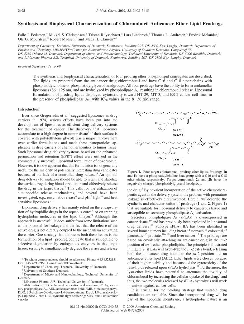
Synthesis and Biophysical Characterization of Chlorambucil Anticancer Ether Lipid Prodrugs
Palle J. Pedersen,† Mikkel S. Christensen,† Tristan Ruysschaert,‡ Lars Linderoth,† Thomas L. Andresen,§ Fredrik Melander,|
Ole G. Mouritsen,‡ Robert Madsen,† and Mads H. Clausen*,†
Department of Chemistry, Technical UniVersity of Denmark, KemitorVet, Building 201, DK-2800 Kgs. Lyngby, Denmark, Department ofPhysics and Chemistry, MEMPHYSsCenter for Biomembrane Physics, UniVersity of Southern Denmark, CampusVej 55,DK-5230 Odense M, Denmark, Department of Micro- and Nanotechnology, Technical UniVersity of Denmark, DK-4000 Roskilde, Denmark,and LiPlasome Pharma A/S, Technical UniVersity of Denmark, KemitorVet, Building 207, DK-2800 Kgs. Lyngby, Denmark
ReceiVed September 11, 2008
The synthesis and biophysical characterization of four prodrug ether phospholipid conjugates are described.The lipids are prepared from the anticancer drug chlorambucil and have C16 and C18 ether chains withphosphatidylcholine or phosphatidylglycerol headgroups. All four prodrugs have the ability to form unilamellarliposomes (86-125 nm) and are hydrolyzed by phospholipase A2, resulting in chlorambucil release. Liposomalformulations of prodrug lipids displayed cytotoxicity toward HT-29, MT-3, and ES-2 cancer cell lines inthe presence of phospholipase A2, with IC50 values in the 8-36 µM range.
Introduction
Ever since Gregoriadis et al.1 suggested liposomes as drugcarriers in 1974, serious efforts have been put into thedevelopment of liposomes as efficient drug delivery systemsfor the treatment of cancer. The discovery that liposomesaccumulate to a high degree in tumor tissue2 if their surface iscovered with poly(ethylene glycol) was a major improvementover earlier formulations and made these nanoparticles ap-plicable as drug carriers of chemotherapeutics to tumor tissue.Such liposomal drug delivery systems based on the enhancedpermeation and retention (EPRa) effect were utilized in thecommercially successful liposomal formulation of doxorubicin.However, it is now apparent that this formulation is not generallyuseful for the majority of potentially interesting drug candidatesbecause of the lack of a controlled drug release.3 An optimaldrug delivery formulation should be able to retain and stabilizethe carried drug during blood circulation and effectively releasethe drug in the target tissue.3 This calls for the utilization ofsite specific release mechanisms, and several have beeninvestigated, e.g., enzymatic release4 and pH,5 light,6 and heatsensitive liposomes.7
Liposomal drug delivery has mainly relied on the encapsula-tion of hydrophilic drugs in the aqueous core3,8 or on trappinghydrophobic molecules in the lipid bilayer.9 Although thisapproach is successful, it does suffer from some limitations suchas the potential for leakage and the fact that the release of theactive drug is not directly coupled to the mechanism activatingthe carrier. One strategy that addresses both these issues is theformulation of a lipid-prodrug conjugate that is susceptible toselective degradation by endogenous enzymes in the targettissue, serving to simultaneously degrade the carrier and release
the drug.3 By covalent incorporation of the active chemothera-peutic agent in the delivery system, the problem with prematureleakage is effectively circumvented. Herein, we describe thesynthesis and characterization of prodrugs (1 and 2, Figure 1)that are suitable for liposomal delivery to cancerous tissue andsusceptible to secretory phospholipase A2 activation.
Secretory phospholipase A2 (sPLA2) is overexpressed incancer tissue,10 and has previously been exploited in liposomaldrug delivery.11 Subtype sPLA2 IIA has been identified inseveral human tumors including breast,12 stomach,13 colorectal,14
pancreatic,15 prostate,10a,16 and liver cancer.17 The prodrugs arebased on covalently attaching an anticancer drug in the sn-2position of sn-1 ether phospholipids. The principle is illustratedin Figure 2: sPLA2 will hydrolyze the sn-2 ester bond, releasingboth the anticancer drug bound to the sn-2 position and ananticancer ether lipid (AEL). Ether lipids were chosen becauseof their higher stability and because of the cytotoxicity of thelyso-lipids released upon sPLA2 hydrolysis.11 Furthermore, thelyso-ether lipids have potential to attenuate the toxicity ofchlorambucil by increasing the cellular uptake of the drug,3 andthus, the two molecules released by sPLA2 hydrolysis will workin unison against cancer cells.
It is crucial for the prodrug strategy that suitable drugcandidates are available. Since the incorporated drug will bepart of the lipophilic membrane, a hydrophobic nature is an
* To whom correspondence should be addressed. Phone: +45 45252131.Fax: +45 45933968. E-mail: [email protected].
† Department of Chemistry, Technical University of Denmark.‡ University of Southern Denmark.§ Department of Micro- and Nanotechnology, Technical University of
Denmark.| LiPlasome Pharma A/S, Technical University of Denmark.a Abbrevations: EPR, enhanced permeation and retention; sPLA2, secre-
tory phospholipase A2; AEL, anticancer ether lipid; PMB, p-methoxybenzyl;DDQ, 2,3-dichloro-5,6-dicyano-1,4-benzoquinone; DBU, 1,8-diazabicyclo-[5.4.0]undec-7-ene; DLS, dynamic light scattering; SUV, small unilaminarvesicles.
Figure 1. Four target chlorambucil prodrug ether lipids. Prodrugs 1aand 1b have a phosphatidylcholine headgroup with a C16 and a C18ether chain, respectively. Target compounds 2a and 2b have thenegatively charged phosphatidylglycerol headgroup.
J. Med. Chem. 2009, 52, 3408–34153408
10.1021/jm900091h CCC: $40.75 2009 American Chemical SocietyPublished on Web 04/29/2009
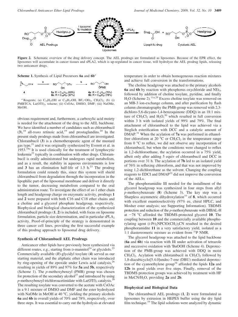
obvious requirement and, furthermore, a carboxylic acid moietyis needed for the attachment of the drug to the AEL backbone.We have identified a number of candidates such as chlorambucil(3),18 all-trans retinoic acid,19 and prostaglandins.20 In thepresent study prodrugs made from chlorambucil are investigated.Chlorambucil (3) is a chemotherapeutic agent of the mustardgas type,21 and it was originally synthesized by Everett et al. in1953.18a It is used clinically for the treatment of lymphocyticleukemia22 typically in combination with other drugs. Chloram-bucil is orally administrated but undergoes rapid metabolism,and as a result, the stability in aqueous environments is lowand 3 has an elimination half-life of 1.5 h.23 The prodrugformulation could remedy this, since this system will shieldchlorambucil from degradation through the incorporation in thelipophilic part of the liposomal membrane and deliver it directlyto the tumor, decreasing metabolism compared to the oraladministration route. To investigate the effect of sn-1 ether chainlength and headgroup charge on enzymatic activity, prodrugs 1and 2 were prepared with both C16 and C18 ether chains anda choline and a glycerol phosphate headgroup, respectively.Biophysical and biological characterization of the synthesizedchlorambucil prodrugs (1, 2) is included, with focus on liposomeformulation, particle size determination, and in particular sPLA2
activity. Proof-of-principle of the strategy is demonstrated inthree cancer cell lines, providing the first successful exampleof this prodrug approach to liposomal drug delivery.
Synthesis of Chlorambucil AEL Prodrugs
Anticancer ether lipids have previously been synthesized viadifferent routes, e.g., starting from D-mannitol24 or glycidols.25
Commercially available (R)-glycidyl tosylate (4) served as ourstarting material, and the aliphatic ether chain was introducedby ring-opening of the epoxide under Lewis acid catalysis,26
resulting in yields of 89% and 97% for 5a and 5b, respectively(Scheme 1). The p-methoxybenzyl (PMB) group was chosenfor protection of the secondary alcohol27 and introduced by usingp-methoxybenzyl trichloroacetimidate with La(OTf)3 catalysis.28
The resulting tosylate was converted to the acetate with CsOAcin a 9:1 mixture of DMSO and DMF and the ester hydrolyzedwith NaOMe in MeOH at 40 °C, yielding the primary alcohols6a and 6b in overall yields of 70% and 78%, respectively, overthree steps. It was essential to carry out the hydrolysis at elevated
temperature in order to obtain homogeneous reaction mixturesand achieve full conversion in the transformations.
The choline headgroup was attached to the primary alcohols6a and 6b by reaction with phosphorus oxychloride and NEt3,followed by addition of choline tosylate, pyridine, and finallyH2O (Scheme 2).11a,29 Excess choline tosylate was removed onan MB-3 ion-exchange column, and after purification by flashcolumn chromatography the PMB-group was removed with 2,3-dichloro-5,6-dicyano-1,4-benzoquinone (DDQ) in an 18:1 mix-ture of CH2Cl2 and H2O,30 which resulted in full conversionwithin 3 h with isolated yields of 99% and 79%. The finalattachment of chlorambucil to the lipid was achieved via aSteglich esterification with DCC and a catalytic amount ofDMAP.31 When the acylation of 7a was performed in ethanol-free chloroform at 20 °C or CH2Cl2 in the temperature rangefrom 0 °C to reflux, we did not observe any incorporation ofchlorambucil, but when the conditions were changed to refluxin 1,2-dichloroethane, the acylation occurred in a 75% yield,albeit only after adding 5 equiv of chlorambucil and DCC inportions over 31 h. The acylation of 7b led to an isolated yieldof 76% in refluxing chloroform, and that was not improved byusing 1,2-dichlorethane as the solvent. Changing the couplingreagents to EDCI and DMAP32 did not improve the conversionof the AELs.
The phosphoramidite 11 needed for the installment of theglycerol headgroup was synthesized in four steps from allylp-methoxybenzoate (8) (Scheme 3). The key step was aSharpless asymmetric dihydroxylation33 of 8, which occurredwith excellent enantioselectivity (97% ee, chiral HPLC, andMosher ester analysis; see Supporting Information). TBDMSprotection and reduction of the p-methoxybenzoate with DIBAL-Hat -78 °C afforded the TBDMS-protected glycerol 10. Thecoupling between 10 and the commercially available phospho-rylating agent (i-Pr)2NPClO(CH2)2CN resulted in the desiredphosphoramidite 11 in a very satisfactory yield, isolated as a1:1 diastereomeric mixture as evident from 31P NMR.
The glycerol headgroup was attached to the lipid backbone(6a and 6b) via reaction with 11 under activation of tetrazoleand successive oxidation with tBuOOH (Scheme 4). Deprotec-tion of the PMB-group was achieved with DDQ in moistCH2Cl2. Acylation with chlorambucil in CH2Cl2 followed by1,8-diazabicyclo[5.4.0]undec-7-ene (DBU) mediated deprotec-tion of the cyanoethylene group34 afforded the lipids 12a and12b in good yields over five steps. Finally, removal of theTBDMS protection groups was achieved by treatment with HFin MeCN/H2O, providing 2a and 2b.
Biophysical and Biological Data
The chlorambucil AEL prodrugs (1, 2) were formulated asliposomes by extrusion in HEPES buffer using the dry lipidfilm technique.35 The lipid solutions were analyzed by dynamic
Figure 2. Schematic overview of the drug delivery concept. The AEL prodrugs are formulated as liposomes. Because of the EPR effect, theliposomes will accumulate in cancer tissues and sPLA2, which is up-regulated in cancer tissue, will hydrolyze the AEL prodrug lipids, releasingtwo anticancer drugs.
Scheme 1. Synthesis of Lipid Precursors 6a and 6ba
a Reagents: (a) C16H33OH or C18H37OH, BF3 ·OEt2, CH2Cl2; (b) (i)PMBTCA, La(OTf)3, toluene; (ii) CsOAc, DMSO, DMF; (iii) NaOMe,MeOH.
Chlorambucil Anticancer Ether Lipid Prodrugs Journal of Medicinal Chemistry, 2009, Vol. 52, No. 10 3409
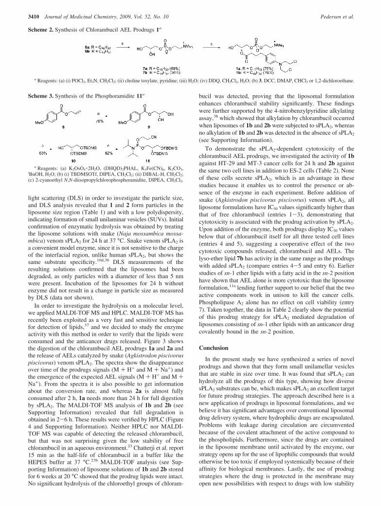
light scattering (DLS) in order to investigate the particle size,and DLS analysis revealed that 1 and 2 form particles in theliposome size region (Table 1) and with a low polydispersity,indicating formation of small unilaminar vesicles (SUVs). Initialconfirmation of enzymatic hydrolysis was obtained by treatingthe liposome solutions with snake (Naja mossambica mossa-mbica) venom sPLA2 for 24 h at 37 °C. Snake venom sPLA2 isa convenient model enzyme, since it is not sensitive to the chargeof the interfacial region, unlike human sPLA2, but shows thesame substrate specificity.10d,36 DLS measurements of theresulting solutions confirmed that the liposomes had beendegraded, as only particles with a diameter of less than 5 nmwere present. Incubation of the liposomes for 24 h withoutenzyme did not result in a change in particle size as measuredby DLS (data not shown).
In order to investigate the hydrolysis on a molecular level,we applied MALDI-TOF MS and HPLC. MALDI-TOF MS hasrecently been exploited as a very fast and sensitive techniquefor detection of lipids,37 and we decided to study the enzymeactivity with this method in order to verify that the lipids wereconsumed and the anticancer drugs released. Figure 3 showsthe digestion of the chlorambucil AEL prodrugs 1a and 2a andthe release of AELs catalyzed by snake (Agkistrodon pisciVoruspisciVorus) venom sPLA2. The spectra show the disappearanceover time of the prodrugs signals (M + H+ and M + Na+) andthe emergence of the expected AEL signals (M + H+ and M +Na+). From the spectra it is also possible to get informationabout the conversion rate, and whereas 2a is almost fullyconsumed after 2 h, 1a needs more than 24 h for full digestionby sPLA2. The MALDI-TOF MS analysis of 1b and 2b (seeSupporting Information) revealed that full degradation isobtained in 2-6 h. These results were verified by HPLC (Figure4 and Supporting Information). Neither HPLC nor MALDI-TOF MS was capable of detecting the released chlorambucil,but that was not surprising given the low stability of freechlorambucil in an aqueous environment.23 Chatterji et al. report15 min as the half-life of chlorambucil in a buffer like theHEPES buffer at 37 °C.23b MALDI-TOF analysis (see Sup-porting Information) of liposome solutions of 1b and 2b storedfor 6 weeks at 20 °C showed that the prodrug lipids were intact.No significant hydrolysis of the chloroethyl groups of chloram-
bucil was detected, proving that the liposomal formulationenhances chlorambucil stability significantly. These findingswere further supported by the 4-nitrobenzylpyridine alkylatingassay,38 which showed that alkylation by chlorambucil occurredwhen liposomes of 1b and 2b were subjected to sPLA2, whereasno alkylation of 1b and 2b was detected in the absence of sPLA2
(see Supporting Information).
To demonstrate the sPLA2-dependent cytotoxicity of thechlorambucil AEL prodrugs, we investigated the activity of 1bagainst HT-29 and MT-3 cancer cells for 24 h and 2b againstthe same two cell lines in addition to ES-2 cells (Table 2). Noneof these cells secrete sPLA2, which is an advantage in thesestudies because it enables us to control the presence or ab-sence of the enzyme in each experiment. Before addition ofsnake (Agkistrodon pisciVorus pisciVorus) venom sPLA2, allliposome formulations have IC50 values significantly higher thanthat of free chlorambucil (entries 1-3), demonstrating thatcytotoxicity is associated with the prodrug activation by sPLA2.Upon addition of the enzyme, both prodrugs display IC50 valuesbelow that of chlorambucil itself for all three tested cell lines(entries 4 and 5), suggesting a cooperative effect of the twocytotoxic compounds released, chlorambucil and AELs. Thelyso-ether lipid 7b has activity in the same range as the prodrugswith added sPLA2 (compare entries 4-5 and entry 6). Earlierstudies of sn-1 ether lipids with a fatty acid in the sn-2 positionhave shown that AEL alone is more cytotoxic than the liposomeformulation,11a lending further support to our belief that the twoactive components work in unison to kill the cancer cells.Phospholipase A2 alone has no effect on cell viability (entry7). Taken together, the data in Table 2 clearly show the potentialof this prodrug strategy for sPLA2 mediated degradation ofliposomes consisting of sn-1 ether lipids with an anticancer drugcovalently bound in the sn-2 position.
Conclusion
In the present study we have synthesized a series of novelprodrugs and shown that they form small unilamellar vesiclesthat are stable in size over time. It was found that sPLA2 canhydrolyze all the prodrugs of this type, showing how diversesPLA2 substrates can be, which makes sPLA2 an excellent targetfor future prodrug strategies. The approach described here is anew application of prodrugs in liposomal formulations, and webelieve it has significant advantages over conventional liposomaldrug delivery system, where hydrophilic drugs are encapsulated.Problems with leakage during circulation are circumventedbecause of the covalent attachment of the active compound tothe phospholipids. Furthermore, since the drugs are containedin the liposome membrane until activated by the enzyme, ourstrategy opens up for the use of lipophilic compounds that wouldotherwise be too toxic if employed systemically because of theiraffinity for biological membranes. Lastly, the use of prodrugstrategies where the drug is protected in the membrane mayopen new possibilities with respect to drugs with low stability
Scheme 2. Synthesis of Chlorambucil AEL Prodrugs 1a
a Reagents: (a) (i) POCl3, Et3N, CH2Cl2; (ii) choline tosylate, pyridine; (iii) H2O; (iv) DDQ, CH2Cl2, H2O; (b) 3, DCC, DMAP, CHCl3 or 1,2-dichloroethane.
Scheme 3. Synthesis of the Phosphoramidite 11a
a Reagents: (a) K2OsO4 ·2H2O, (DHQD)2PHAL, K3Fe(CN)6, K2CO3,tBuOH, H2O; (b) (i) TBDMSOTf, DIPEA, CH2Cl2; (ii) DIBAL-H, CH2Cl2;(c) 2-cyanoethyl N,N-diisopropylchlorophosphoramidite, DIPEA, CH2Cl2.
3410 Journal of Medicinal Chemistry, 2009, Vol. 52, No. 10 Pedersen et al.
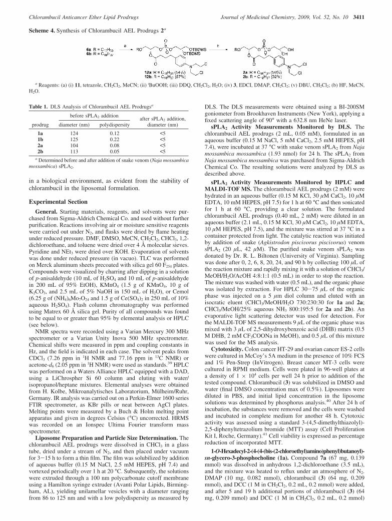
in a biological environment, as evident from the stability ofchlorambucil in the liposomal formulation.
Experimental Section
General. Starting materials, reagents, and solvents were pur-chased from Sigma-Aldrich Chemical Co. and used without furtherpurification. Reactions involving air or moisture sensitive reagentswere carried out under N2, and flasks were dried by flame heatingunder reduced pressure. DMF, DMSO, MeCN, CH2Cl2, CHCl3, 1,2-dichloroethane, and toluene were dried over 4 Å molecular sieves.Pyridine and NEt3 were dried over KOH. Evaporation of solventswas done under reduced pressure (in vacuo). TLC was performedon Merck aluminum sheets precoated with silica gel 60 F254 plates.Compounds were visualized by charring after dipping in a solutionof p-anisaldehyde (10 mL of H2SO4 and 10 mL of p-anisaldehydein 200 mL of 95% EtOH), KMnO4 (1.5 g of KMnO4, 10 g ofK2CO3, and 2.5 mL of 5% NaOH in 150 mL of H2O), or Cemol(6.25 g of (NH4)6Mo7O24 and 1.5 g of Ce(SO4)2 in 250 mL of 10%aqueous H2SO4). Flash column chromatography was performedusing Matrex 60 Å silica gel. Purity of all compounds was foundto be equal to or greater than 95% by elemental analysis or HPLC(see below).
NMR spectra were recorded using a Varian Mercury 300 MHzspectrometer or a Varian Unity Inova 500 MHz spectrometer.Chemical shifts were measured in ppm and coupling constants inHz, and the field is indicated in each case. The solvent peaks fromCDCl3 (7.26 ppm in 1H NMR and 77.16 ppm in 13C NMR) oracetone-d6 (2.05 ppm in 1H NMR) were used as standards.39 HPLCwas performed on a Waters Alliance HPLC equipped with a DAD,using a LiChrospher Si 60 column and eluting with water/isopropanol/heptane mixtures. Elemental analyses were obtainedfrom H. Kolbe, Mikroanalytisches Laboratorium, Mulheim/Ruhr,Germany. IR analysis was carried out on a Perkin-Elmer 1600 seriesFTIR spectrometer, as KBr pills or neat between AgCl plates.Melting points were measured by a Buch & Holm melting pointapparatus and given in degrees Celsius (°C) uncorrected. HRMSwas recorded on an Ionspec Ultima Fourier transform massspectrometer.
Liposome Preparation and Particle Size Determination. Thechlorambucil AEL prodrugs were dissolved in CHCl3 in a glasstube, dried under a stream of N2, and then placed under vacuumfor 3-15 h to form a thin film. The film was solubilized by additionof aqueous buffer (0.15 M NaCl, 2.5 mM HEPES, pH 7.4) andvortexed periodically over 1 h at 20 °C. Subsequently, the solutionswere extruded through a 100 nm polycarbonate cutoff membraneusing a Hamilton syringe extruder (Avanti Polar Lipids, Birming-ham, AL), yielding unilamellar vesicles with a diameter rangingfrom 86 to 125 nm and with a low polydispersity as measured by
DLS. The DLS measurements were obtained using a BI-200SMgoniometer from Brookhaven Instruments (New York), applying afixed scattering angle of 90° with a 632.8 nm HeNe laser.
sPLA2 Activity Measurements Monitored by DLS. Thechlorambucil AEL prodrugs (2 mL, 0.05 mM), formulated in anaqueous buffer (0.15 M NaCl, 5 mM CaCl2, 2.5 mM HEPES, pH7.4), were incubated at 37 °C with snake venom sPLA2 from Najamossambica mossambica (1.93 nmol) for 24 h. The sPLA2 fromNaja mossambica mossambica was purchased from Sigma-AldrichChemical Co. The resulting solutions were analyzed by DLS asdescribed above.
sPLA2 Activity Measurements Monitored by HPLC andMALDI-TOF MS. The chlorambucil AEL prodrugs (2 mM) werehydrated in an aqueous buffer (0.15 M KCl, 30 µM CaCl2, 10 µMEDTA, 10 mM HEPES, pH 7.5) for 1 h at 60 °C and then sonicatedfor 1 h at 60 °C, providing a clear solution. The formulatedchlorambucil AEL prodrugs (0.40 mL, 2 mM) were diluted in anaqueous buffer (2.1 mL, 0.15 M KCl, 30 µM CaCl2, 10 µM EDTA,10 µM HEPES, pH 7.5), and the mixture was stirred at 37 °C in acontainer protected from light. The catalytic reaction was initiatedby addition of snake (Agkistrodon pisciVorus pisciVorus) venomsPLA2 (20 µL, 42 µM). The purified snake venom sPLA2 wasdonated by Dr. R. L. Biltonen (University of Virginia). Samplingwas done after 0, 2, 6, 8, 20, 24, and 90 h by collecting 100 µL ofthe reaction mixture and rapidly mixing it with a solution of CHCl3/MeOH/H2O/AcOH 4:8:1:1 (0.5 mL) in order to stop the reaction.The mixture was washed with water (0.5 mL), and the organic phasewas isolated by extraction. For HPLC 30-75 µL of the organicphase was injected on a 5 µm diol column and eluted with anisocratic eluent (CHCl3/MeOH/H2O 730:230:30 for 1a and 2a;CHCl3/MeOH/25% aqueous NH3 800:195:5 for 2a and 2b). Anevaporative light scattering detector was used for detection. Forthe MALDI-TOF MS measurements 9 µL of the organic phase wasmixed with 3 µL of 2,5-dihydroxybenzoic acid (DHB) matrix (0.5M DHB, 2 mM CF3COONa in MeOH), and 0.5 µL of this mixturewas used for the MS analysis.
Cytotoxicity. Colon cancer HT-29 and ovarian cancer ES-2 cellswere cultured in McCoy’s 5A medium in the presence of 10% FCSand 1% Pen-Strep (InVitrogen). Breast cancer MT-3 cells werecultured in RPMI medium. Cells were plated in 96-well plates ata density of 1 × 10⁴ cells per well 24 h prior to addition of thetested compound. Chlorambucil (3) was solubilized in DMSO andwater (final DMSO concentration max of 0.5%). Liposomes werediluted in PBS, and initial lipid concentration in the liposomesolutions was determined by phosphorus analysis.40 After 24 h ofincubation, the substances were removed and the cells were washedand incubated in complete medium for another 48 h. Cytotoxicactivity was assessed using a standard 3-(4,5-dimethylthiazolyl)-2,5-diphenyltetrazolium bromide (MTT) assay (Cell ProliferationKit I, Roche, Germany).41 Cell viability is expressed as percentagereduction of incorporated MTT.
1-O-Hexadecyl-2-(4-(4-(bis-(2-chloroethyl)amino)phenyl)butanoyl)-sn-glycero-3-phosphocholine (1a). Compound 7a (67 mg, 0.139mmol) was dissolved in anhydrous 1,2-dichloroethane (3.5 mL),and the mixture was heated to reflux under an atmosphere of N2.DMAP (10 mg, 0.082 mmol), chlorambucil (3) (64 mg, 0.209mmol), and DCC (1 M in CH2Cl2, 0.2 mL, 0.2 mmol) were added,and after 5 and 19 h additional portions of chlorambucil (3) (64mg, 0.209 mmol) and DCC (1 M in CH2Cl2, 0.2 mL, 0.2 mmol)
Scheme 4. Synthesis of Chlorambucil AEL Prodrugs 2a
a Reagents: (a) (i) 11, tetrazole, CH2Cl2, MeCN; (ii) tBuOOH; (iii) DDQ, CH2Cl2, H2O; (iv) 3, EDCI, DMAP, CH2Cl2; (v) DBU, CH2Cl2; (b) HF, MeCN,H2O.
Table 1. DLS Analysis of Chlorambucil AEL Prodrugsa
before sPLA2 addition
prodrug diameter (nm) polydispersityafter sPLA2 addition,
diameter (nm)
1a 124 0.12 <51b 125 0.22 <52a 104 0.08 <52b 113 0.05 <5
a Determined before and after addition of snake venom (Naja mossambicamossambica) sPLA2.
Chlorambucil Anticancer Ether Lipid Prodrugs Journal of Medicinal Chemistry, 2009, Vol. 52, No. 10 3411
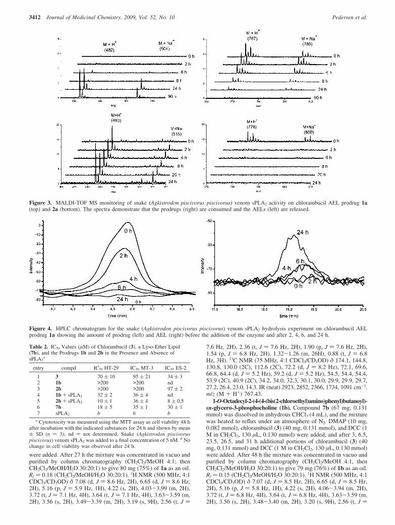
were added. After 27 h the mixture was concentrated in vacuo andpurified by column chromatography (CH2Cl2/MeOH 4:1; thenCH2Cl2/MeOH/H2O 30:20:1) to give 80 mg (75%) of 1a as an oil.Rf ) 0.18 (CH2Cl2/MeOH/H2O 30:20:1). 1H NMR (500 MHz, 4:1CDCl3/CD3OD) δ 7.08 (d, J ) 8.6 Hz, 2H), 6.65 (d, J ) 8.6 Hz,2H), 5.16 (p, J ) 5.9 Hz, 1H), 4.22 (s, 2H), 4.03-3.99 (m, 2H),3.72 (t, J ) 7.1 Hz, 4H), 3.64 (t, J ) 7.1 Hz, 4H), 3.63-3.59 (m,2H), 3.56 (s, 2H), 3.49-3.39 (m, 2H), 3.19 (s, 9H), 2.56 (t, J )
7.6 Hz, 2H), 2.36 (t, J ) 7.6 Hz, 2H), 1.90 (p, J ) 7.6 Hz, 2H),1.54 (p, J ) 6.8 Hz, 2H), 1.32-1.26 (m, 26H), 0.88 (t, J ) 6.8Hz, 3H). 13C NMR (75 MHz, 4:1 CDCl3/CD3OD) δ 174.1, 144.8,130.8, 130.0 (2C), 112.6 (2C), 72.2 (d, J ) 8.2 Hz), 72.1, 69.6,66.8, 64.4 (d, J ) 5.2 Hz), 59.2 (d, J ) 5.2 Hz), 54.5, 54.4, 54.4,53.9 (2C), 40.9 (2C), 34.2, 34.0, 32.3, 30.1, 30.0, 29.9, 29.9, 29.7,27.2, 26.4, 23.0, 14.3. IR (neat) 2923, 2852, 2366, 1734, 1091 cm-1.m/z (M + H+) 767.43.
1-O-Octadecyl-2-(4-(4-(bis(2-chloroethyl)amino)phenyl)butanoyl)-sn-glycero-3-phosphocholine (1b). Compound 7b (67 mg, 0.131mmol) was dissolved in anhydrous CHCl3 (4 mL), and the mixturewas heated to reflux under an atmosphere of N2. DMAP (10 mg,0.082 mmol), chlorambucil (3) (40 mg, 0.131 mmol), and DCC (1M in CH2Cl2, 130 µL, 0.130 mmol) were added, and after 3, 6.5,23.5, 26.5, and 31 h additional portions of chlorambucil (3) (40mg, 0.131 mmol) and DCC (1 M in CH2Cl2, 130 µL, 0.130 mmol)were added. After 48 h the mixture was concentrated in vacuo andpurified by column chromatography (CH2Cl2/MeOH 4:1, thenCH2Cl2/MeOH/H2O 30:20:1) to give 79 mg (76%) of 1b as an oil.Rf ) 0.15 (CH2Cl2/MeOH/H2O 30:20:1). 1H NMR (500 MHz, 4:1CDCl3/CD3OD) δ 7.07 (d, J ) 8.5 Hz, 2H), 6.65 (d, J ) 8.5 Hz,2H), 5.16 (p, J ) 5.8 Hz, 1H), 4.22 (s, 2H), 4.06-3.94 (m, 2H),3.72 (t, J ) 6.8 Hz, 4H), 3.64 (t, J ) 6.8 Hz, 4H), 3.63-3.59 (m,2H), 3.56 (s, 2H), 3.48-3.40 (m, 2H), 3.20 (s, 9H), 2.56 (t, J )
Figure 3. MALDI-TOF MS monitoring of snake (Agkistrodon pisciVorus pisciVorus) venom sPLA2 activity on chlorambucil AEL prodrug 1a(top) and 2a (bottom). The spectra demonstrate that the prodrugs (right) are consumed and the AELs (left) are released.
Figure 4. HPLC chromatogram for the snake (Agkistrodon pisciVorus pisciVorus) venom sPLA2 hydrolysis experiment on chlorambucil AELprodrug 1a showing the amount of prodrug (left) and AEL (right) before the addition of the enzyme and after 2, 4, 6, and 24 h.
Table 2. IC50 Values (µM) of Chlorambucil (3), a Lyso-Ether Lipid(7b), and the Prodrugs 1b and 2b in the Presence and Absence ofsPLA2
a
entry compd IC50 HT-29 IC50 MT-3 IC50 ES-2
1 3 70 ( 10 95 ( 21 34 ( 32 1b >200 >200 nd3 2b >200 >200 97 ( 24 1b + sPLA2 32 ( 2 36 ( 4 nd5 2b + sPLA2 10 ( 1 36 ( 4 8 ( 0,56 7b 18 ( 5 35 ( 1 30 ( 17 sPLA2 b b b
a Cytotoxicity was measured using the MTT assay as cell viability 48 hafter incubation with the indicated substances for 24 h and shown by mean( SD (n ) 3); nd ) not determined. Snake (Agkistrodon pisciVoruspisciVorus) venom sPLA2 was added to a final concentration of 5 nM. b Nochange in cell viability was observed after 24 h.
3412 Journal of Medicinal Chemistry, 2009, Vol. 52, No. 10 Pedersen et al.
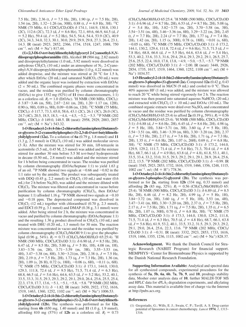
7.5 Hz, 2H), 2.36 (t, J ) 7.5 Hz, 2H), 1.90 (p, J ) 7.5 Hz, 2H),1.54 (m, 2H), 1.32-1.26 (m, 30H), 0.88 (t, J ) 6.9 Hz, 3H). 13CNMR (75 MHz, 4:1 CDCl3/CD3OD) δ 174.1, 144.9, 130.8, 130.1(2C), 112.6 (2C), 72.3 (d, J ) 8.6 Hz), 72.1, 69.6, 66.9, 64.5 (d, J) 5.2 Hz), 59.4 (d, J ) 5.2 Hz), 54.5, 54.4, 54.4, 53.9 (2C), 40.9(2C), 34.3, 34.0, 32.3, 30.1, 30.0, 29.9, 29.9, 29.8, 27.3, 26.4, 23.1,14.3. IR (neat) 2923, 2852, 2366, 1734, 1518, 1247, 1088, 750cm-1. m/z (M + Na+) 817.44.
(S)-(2,3-Di-O-tert-butyldimethylsilyl)glyceryl 2-Cyanoethyl-N,N-diisopropylphosphoramidite (11). Alcohol 10 (904 mg, 2.82 mmol)and diisopropylethylamine (1.0 mL, 5.92 mmol) were dissolved inanhydrous CH2Cl2 (10 mL) under an atmosphere of N2. 2-Cyano-ethyl-N,N-diisopropylchlorophosphoramidite (1.0 g, 4.22 mmol) wasadded dropwise, and the mixture was stirred at 20 °C for 1.5 h,after which EtOAc (20 mL) and saturated NaHCO3 (50 mL) wereadded and the organic layer was isolated by extraction with EtOAc(2 × 50 mL). The combined organic phases were concentrated invacuo, and the residue was purified by column chromatography(EtOAc) to give 1352 mg (92%) of 11 (two diastereoisomers, 1:1)as a colorless oil. Rf ) 1.0 (EtOAc). 1H NMR (300 MHz, CDCl3):δ 3.87-3.46 (m, 5H), 2.67-2.61 (m, 2H), 1.20-1.17 (m, 12H),0.90 (s, 9H), 0.89 (s, 9H), 0.09-0.06 (m, 12H). 13C NMR (75 MHz,CDCl3): δ 117.7, 73.3, 65.0, 58.6, 43.1 (2C), 26.1 (3C), 26.0 (3C),24.7 (4C), 20.5, 18.5, 18.3, -4.4, -4.5, -5.2, -5.3. 31P NMR (202MHz, CDCl3): δ 149.0, 148.5. IR (neat): 2958, 2929, 2883, 2857cm-1. m/z (M + Na+) 543.32.
1-O-Hexadecyl-2-(4-(4-(bis-(2-chloroethyl)amino)phenyl)butanoyl)-sn-glycero-3-(2-cyanoethylphospho)-(S)-2,3-di-O-tert-butyldimeth-ylsilylglycerol (12a). To a solution of 6a (0.79 g, 1.8 mmol) and 11(1.3 g, 2.5 mmol) in CH2Cl2 (10 mL) was added molecular sieves(3 Å). After the mixture was stirred for 30 min, 1H-tetrazole inacetonitrile (5.5 mL, 0.45 M, 2.5 mmol) was added and the mixturestirred for another 30 min before 5.5 M tert-butyl hydroperoxidein decane (0.50 mL, 2.8 mmol) was added and the mixture stirredfor 1 h before being concentrated in vacuo. The residue was purifiedby column chromatography (EtOAc/heptane 1:1) to yield 1.63 gof an oil. 31P NMR showed two signals at -0.66 and -0.82 in the1:1 ratio set by the amidite. The product was subsequently treatedwith DDQ (0.45 g, 2.2 mmol) in CH2Cl2 (10 mL) and water (0.6mL) for 2 h before Na2SO3 was added and the mixture diluted withCH2Cl2. The mixture was filtered and concentrated in vacuo beforepurification by column chromatography (CH2Cl2, then EtOAc/heptane 1:1) afforded 1.16 g. 31P NMR showed two signals at -0.10and -0.18 ppm. The deprotected compound was dissolved inCH2Cl2 (12 mL) together with chlorambucil (0.70 g, 2.3 mmol),and EDCI (0.59 g, 3.1 mmol) and DMAP (0.38 g, 3.1 mmol) wereadded. After being stirred for 2 h, the mixture was concentrated invacuo and purified by column chromatography (EtOAc/heptane 1:1)and the resulting 1.36 g product dissolved in CH2Cl2 (10 mL) andtreated with DBU (0.20 mL, 1.3 mmol) for 30 min. The reactionmixture was concentrated in vacuo and the residue was purified bycolumn chromatography (CH2Cl2/MeOH 9:1) to give the phospho-lipid (0.96 g, 54%). Rf ) 0.73 (CH2Cl2/MeOH/H2O 65:25:4). 1HNMR (500 MHz, CDCl3/CD3OD 3:1): δ 6.90 (d, J ) 8.5 Hz, 2H),6.47 (d, J ) 8.5 Hz, 2H), 5.00 (q, J ) 5 Hz, 1H), 4.08 (m, 1H),3.83-3.76 (m, 2H), 3.71-3.59 (m, 3H), 3.55-3.53 (m,4H), 3.47-3.38 (m, 8H), 3.30-3.21(m, 2H), 2.38 (t, J ) 7.5 Hz,2H), 2.19 (t, J ) 7.5 Hz, 2H), 1.73 (q, J ) 7.3 Hz, 2H), 1.36 (m,2H), 1.09 (s, 26 H), 0.72 (s, 18H), -0.06 (s, 6H), -0.11 (s, 6H).13C NMR (75 MHz, CDCl3/CD3OD 3:1): δ 173.1, 144.0, 130.0,129.3, 111.8, 72.4 (d, J ) 9.5 Hz), 71.5, 71.4 (d, J ) 6.3 Hz),68.8, 66.5 (d, J ) 5.6 Hz), 64.6, 63.5 (d, J ) 5.2 Hz), 53.2, 40.1,33.5, 33.3, 31.5, 29.3, 29.3, 29.2, 29.1, 29.0, 26.4, 25.6, 25.5, 25.4,22.3, 17.9, 17.7, 13.6, -5.1, -5.1, -5.8, -5.8. 31P NMR (202 MHz,CDCl3/CD3OD 3:1): δ -1.82. IR (neat): 3450, 2922, 1732, 1616,1519, 1463, 1360, 1252, 1102 cm-1. m/z (M + Na+) 1006.53.
1-O-Octadecyl-2-(4-(4-(bis-(2-chloroethyl)amino)phenyl)butanoyl)-sn-glycero-3-(2-cyanoethylphospho)-(S)-2,3-di-O-tert-butyldimeth-ylsilylglycerol (12b). The synthesis was performed as for 12a,starting from 6b (650 mg, 1.40 mmol) and 11 (1.0 g, 1.9 mmol),affording 810 mg (57%) of 12b as a colorless oil. Rf ) 0.73
(CH2Cl2/MeOH/H2O 65:25:4. 1H NMR (500 MHz, CDCl3/CD3OD3:1): δ 6.96 (d, J ) 8.7 Hz, 2H), 6.53 (d, J ) 8.7 Hz, 2H), 5.08 (q,J ) 5.4 Hz, 1H), 3.82-3.75 (m, 2H), 3.70-3.58 (m, 3H),3.54-3.51 (m, 4H), 3.46-3.36 (m, 8H), 3.29-3.22 (m, 2H), 2.45(t, J ) 7.7 Hz, 2H), 2.24 (J ) 7.7 Hz, 2H), 1.77 (q, J ) 7.4 Hz,2H), 1.42 (m, 2H), 1.15 (s, 30H), 0.78 (s, 18H), -0.01 (s, 6H),-0.05 (s, 6H). 13C NMR (75 MHz, CDCl3/CD3OD 3:1): δ 173.2,144.1, 130.2, 129.4, 111.8, 72.4 (d, J ) 9.6 Hz), 71.5, 71.5 (d, J )7.8 Hz), 68.8, 66.6 (d, J ) 5.5 Hz), 64.6, 63.6 (d, J ) 5.4 Hz),53.3, 40.2, 33.6, 33.3, 31.6, 29.4, 29.4, 29.3, 29.2, 29.1, 26.5, 25.7,25.6, 25.5, 22.4, 18.0, 17.8, 13.8, -4.9, -5.0, -5.7, -5.7. 31P NMR(202 MHz, CDCl3/CD3OD 3:1): δ -2.00. IR (neat): 3448, 2926,2854, 1735, 1617, 1519, 1464, 1360, 1252, 1108 cm-1. m/z (M +Na+) 1034.57.
1-O-Hexadecyl-2-(4-(4-(bis(2-chloroethyl)amino)phenyl)butanoyl)-sn-glycero-3-phospho-(S)-glycerol (2a). Compound 12a (0.42 g, 0.43mmol) was dissolved in MeCN (9 mL) and cooled to 0 °C. Then40% aqueous HF (1 mL) was added, and the mixture was allowedto reach 20 °C while being stirred vigorously for 2 h. The reactionmixture was then poured into saturated aqueous NaHCO3 (20 mL)and extracted with CH2Cl2 (3 × 10 mL) and EtOAc (10 mL). Thecombined organic extracts were dried over Na2SO4 and concentratedin vacuo and the residue was purified by column chromatography(CH2Cl2/MeOH/H2O 65:25:4) to afford 2a (0.19 g, 59%). Rf ) 0.56(CH2Cl2/MeOH/H2O 65:25:4). 1H NMR (500 MHz, CDCl3:CD3OD3:1): δ 6.88 (d, J ) 8.6 Hz, 2H), 6.45 (d, J ) 8.6 Hz, 2H), 4.98 (q,J ) 5 Hz, 1H), 3.84-3.71 (m, 3H), 3.60 (q, J ) 5 Hz, 1H),3.54-3.51 (m, 4H), 3.46-3.39 (m, 8H), 3.30-3.20 (m, 2H), 2.37(t, J ) 7.5 Hz, 2H), 2.17 (t, J ) 7.6 Hz, 2H), 1.71 (q, J ) 7.3 Hz,2H), 1.35 (t, J ) 6.6 Hz, 2H), 1.07 (s, 26H), 0.69 (t, J ) 6.8 Hz,3H). 13C NMR (75 MHz, CDCl3/CD3OD 3:1): δ 173.2, 144.0,129.9, 129.2, 111.7, 71.4 (d, J ) 5.4 Hz), 71.3, 70.4 (d, J ) 4.8Hz), 68.7, 66.1 (d, J ) 4.9 Hz), 63.8 (d, J ) 4.8), 61.8, 53.1, 40.1,33.5, 33.4, 33.2, 33.0, 31.5, 29.3, 29.2, 29.1, 29.1, 28.9, 26.4, 25.6,22.2, 13.5. 31P NMR (202 MHz, CDCl3/CD3OD 3:1): δ -0.99. IR(neat): 3345, 2923, 2853, 1732, 1616, 1519, 1466, 1356, 1248, 1115,1002 cm-1. m/z (M + H+) 778.36.
1-O-Octadecyl-2-(4-(4-(bis(2-chloroethyl)amino)phenyl)butanoyl)-sn-glycero-3-phospho-(S)-glycerol (2b). The synthesis was per-formed as for 2a, starting from 12b (334 mg, 0.33 mmol) andaffording 2b (83 mg, 32%). Rf ) 0.56 (CH2Cl2/MeOH/H2O 65:25:4). 1H NMR (500 MHz, CDCl3/CD3OD 3:1): δ 6.89 (d, J ) 8.5Hz, 2H), 6.46 (d, J ) 8.2 Hz, 2H), 4.99 (q, J ) 5 Hz, 1H),3.84-3.72 (m, 3H), 3.60 (q, J ) 5 Hz, 1H), 3.53 (m, 4H),3.47-3.41 (m, 8H), 3.30-3.20 (m, 2H), 2.37 (t, J ) 7.5 Hz, 2H),2.17 (t, J ) 7.5 Hz, 2H), 1.71 (q, J ) 7.5 Hz, 2H), 1.35 (t, J ) 6.5Hz, 2H), 1.07 (s, 30H), 0.70 (t, J ) 6.8 Hz, 3H). 13C NMR (75MHz, CDCl3/CD3OD 3:1): δ 173.3, 144.0, 130.0, 129.2, 111.8,71.5, 71.4 (d, J ) 6.3 Hz), 70.5 (d, J ) 4.8 Hz), 68.7, 66.1, 63.8(d, J ) 5.6 Hz), 61.9, 53.2, 40.1, 33.5, 33.2, 31.5, 29.3, 29.3, 29.2,29.1, 29.0, 26.4, 25.6, 22.3, 13.6. 31P NMR (202 MHz, CDCl3/CD3OD 3:1): δ -0.31. IR (neat): 3332, 2923, 2853, 1733, 1616,1519, 1466, 1355, 1236, 1115, 1002 cm-1. m/z (M + Na+) 828.37.
Acknowledgment. We thank the Danish Council for Stra-tegic Research (NABIIT Program) for financial support.MEMPHYSsCenter for Biomembrane Physics is supported bythe Danish National Research Foundation.
Supporting Information Available: Analytical and spectral datafor all synthesized compounds, experimental procedures for thesynthesis of 5a, 5b, 6a, 6b, 7a, 7b, 9, and 10, prodrugs stabilitydata, Mosher ester analysis data of 10, further MALDI-TOF MSand HPLC data for sPLA2 degradation experiments, and alkylatingassay data. This material is available free of charge via the Internetat http://pubs.acs.org.
References(1) Gregoriadis, G.; Wills, E. J.; Swain, C. P.; Tavill, A. S. Drug-carrier
potential of liposomes in cancer chemotherapy. Lancet 1974, 1, 1313–1316.
Chlorambucil Anticancer Ether Lipid Prodrugs Journal of Medicinal Chemistry, 2009, Vol. 52, No. 10 3413
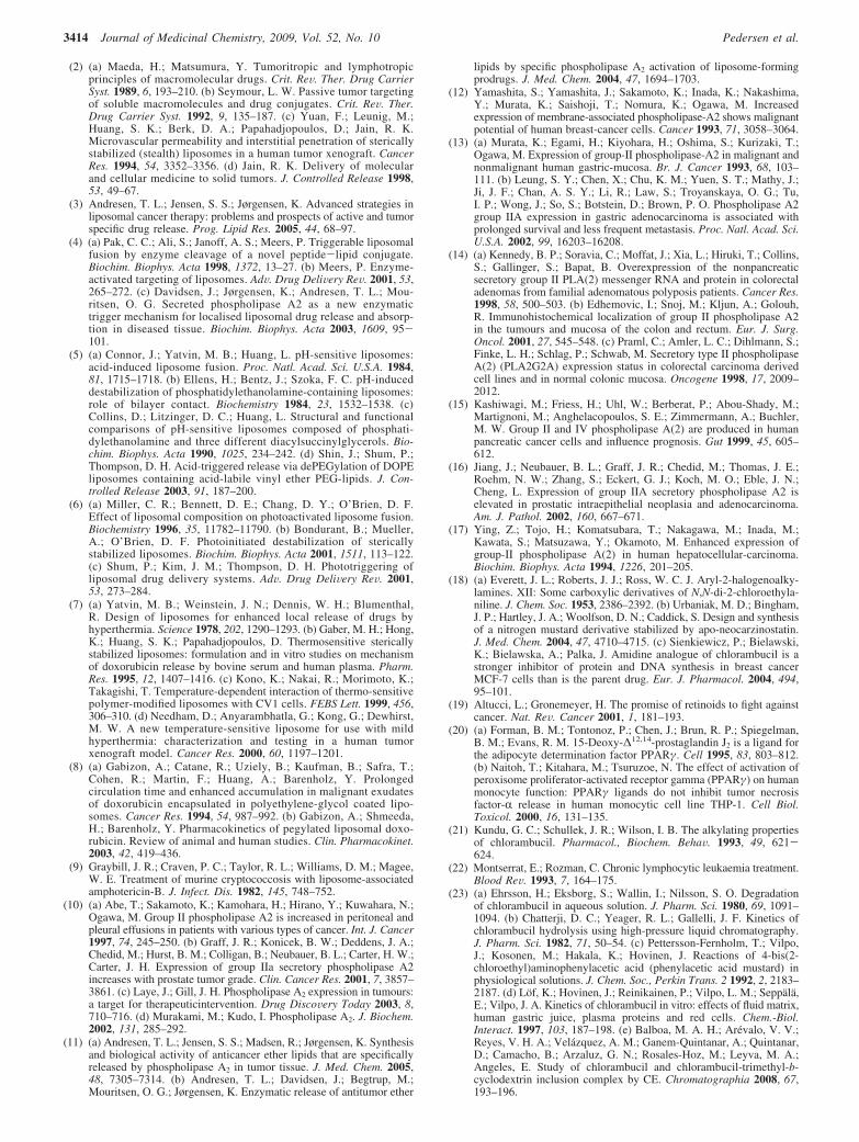
(2) (a) Maeda, H.; Matsumura, Y. Tumoritropic and lymphotropicprinciples of macromolecular drugs. Crit. ReV. Ther. Drug CarrierSyst. 1989, 6, 193–210. (b) Seymour, L. W. Passive tumor targetingof soluble macromolecules and drug conjugates. Crit. ReV. Ther.Drug Carrier Syst. 1992, 9, 135–187. (c) Yuan, F.; Leunig, M.;Huang, S. K.; Berk, D. A.; Papahadjopoulos, D.; Jain, R. K.Microvascular permeability and interstitial penetration of stericallystabilized (stealth) liposomes in a human tumor xenograft. CancerRes. 1994, 54, 3352–3356. (d) Jain, R. K. Delivery of molecularand cellular medicine to solid tumors. J. Controlled Release 1998,53, 49–67.
(3) Andresen, T. L.; Jensen, S. S.; Jørgensen, K. Advanced strategies inliposomal cancer therapy: problems and prospects of active and tumorspecific drug release. Prog. Lipid Res. 2005, 44, 68–97.
(4) (a) Pak, C. C.; Ali, S.; Janoff, A. S.; Meers, P. Triggerable liposomalfusion by enzyme cleavage of a novel peptide-lipid conjugate.Biochim. Biophys. Acta 1998, 1372, 13–27. (b) Meers, P. Enzyme-activated targeting of liposomes. AdV. Drug DeliVery ReV. 2001, 53,265–272. (c) Davidsen, J.; Jørgensen, K.; Andresen, T. L.; Mou-ritsen, O. G. Secreted phospholipase A2 as a new enzymatictrigger mechanism for localised liposomal drug release and absorp-tion in diseased tissue. Biochim. Biophys. Acta 2003, 1609, 95-101.
(5) (a) Connor, J.; Yatvin, M. B.; Huang, L. pH-sensitive liposomes:acid-induced liposome fusion. Proc. Natl. Acad. Sci. U.S.A. 1984,81, 1715–1718. (b) Ellens, H.; Bentz, J.; Szoka, F. C. pH-induceddestabilization of phosphatidylethanolamine-containing liposomes:role of bilayer contact. Biochemistry 1984, 23, 1532–1538. (c)Collins, D.; Litzinger, D. C.; Huang, L. Structural and functionalcomparisons of pH-sensitive liposomes composed of phosphati-dylethanolamine and three different diacylsuccinylglycerols. Bio-chim. Biophys. Acta 1990, 1025, 234–242. (d) Shin, J.; Shum, P.;Thompson, D. H. Acid-triggered release via dePEGylation of DOPEliposomes containing acid-labile vinyl ether PEG-lipids. J. Con-trolled Release 2003, 91, 187–200.
(6) (a) Miller, C. R.; Bennett, D. E.; Chang, D. Y.; O’Brien, D. F.Effect of liposomal composition on photoactivated liposome fusion.Biochemistry 1996, 35, 11782–11790. (b) Bondurant, B.; Mueller,A.; O’Brien, D. F. Photoinitiated destabilization of stericallystabilized liposomes. Biochim. Biophys. Acta 2001, 1511, 113–122.(c) Shum, P.; Kim, J. M.; Thompson, D. H. Phototriggering ofliposomal drug delivery systems. AdV. Drug DeliVery ReV. 2001,53, 273–284.
(7) (a) Yatvin, M. B.; Weinstein, J. N.; Dennis, W. H.; Blumenthal,R. Design of liposomes for enhanced local release of drugs byhyperthermia. Science 1978, 202, 1290–1293. (b) Gaber, M. H.; Hong,K.; Huang, S. K.; Papahadjopoulos, D. Thermosensitive stericallystabilized liposomes: formulation and in vitro studies on mechanismof doxorubicin release by bovine serum and human plasma. Pharm.Res. 1995, 12, 1407–1416. (c) Kono, K.; Nakai, R.; Morimoto, K.;Takagishi, T. Temperature-dependent interaction of thermo-sensitivepolymer-modified liposomes with CV1 cells. FEBS Lett. 1999, 456,306–310. (d) Needham, D.; Anyarambhatla, G.; Kong, G.; Dewhirst,M. W. A new temperature-sensitive liposome for use with mildhyperthermia: characterization and testing in a human tumorxenograft model. Cancer Res. 2000, 60, 1197–1201.
(8) (a) Gabizon, A.; Catane, R.; Uziely, B.; Kaufman, B.; Safra, T.;Cohen, R.; Martin, F.; Huang, A.; Barenholz, Y. Prolongedcirculation time and enhanced accumulation in malignant exudatesof doxorubicin encapsulated in polyethylene-glycol coated lipo-somes. Cancer Res. 1994, 54, 987–992. (b) Gabizon, A.; Shmeeda,H.; Barenholz, Y. Pharmacokinetics of pegylated liposomal doxo-rubicin. Review of animal and human studies. Clin. Pharmacokinet.2003, 42, 419–436.
(9) Graybill, J. R.; Craven, P. C.; Taylor, R. L.; Williams, D. M.; Magee,W. E. Treatment of murine cryptococcosis with liposome-associatedamphotericin-B. J. Infect. Dis. 1982, 145, 748–752.
(10) (a) Abe, T.; Sakamoto, K.; Kamohara, H.; Hirano, Y.; Kuwahara, N.;Ogawa, M. Group II phospholipase A2 is increased in peritoneal andpleural effusions in patients with various types of cancer. Int. J. Cancer1997, 74, 245–250. (b) Graff, J. R.; Konicek, B. W.; Deddens, J. A.;Chedid, M.; Hurst, B. M.; Colligan, B.; Neubauer, B. L.; Carter, H. W.;Carter, J. H. Expression of group IIa secretory phospholipase A2increases with prostate tumor grade. Clin. Cancer Res. 2001, 7, 3857–3861. (c) Laye, J.; Gill, J. H. Phospholipase A2 expression in tumours:a target for therapeuticintervention. Drug DiscoVery Today 2003, 8,710–716. (d) Murakami, M.; Kudo, I. Phospholipase A2. J. Biochem.2002, 131, 285–292.
(11) (a) Andresen, T. L.; Jensen, S. S.; Madsen, R.; Jørgensen, K. Synthesisand biological activity of anticancer ether lipids that are specificallyreleased by phospholipase A2 in tumor tissue. J. Med. Chem. 2005,48, 7305–7314. (b) Andresen, T. L.; Davidsen, J.; Begtrup, M.;Mouritsen, O. G.; Jørgensen, K. Enzymatic release of antitumor ether
lipids by specific phospholipase A2 activation of liposome-formingprodrugs. J. Med. Chem. 2004, 47, 1694–1703.
(12) Yamashita, S.; Yamashita, J.; Sakamoto, K.; Inada, K.; Nakashima,Y.; Murata, K.; Saishoji, T.; Nomura, K.; Ogawa, M. Increasedexpression of membrane-associated phospholipase-A2 shows malignantpotential of human breast-cancer cells. Cancer 1993, 71, 3058–3064.
(13) (a) Murata, K.; Egami, H.; Kiyohara, H.; Oshima, S.; Kurizaki, T.;Ogawa, M. Expression of group-II phospholipase-A2 in malignant andnonmalignant human gastric-mucosa. Br. J. Cancer 1993, 68, 103–111. (b) Leung, S. Y.; Chen, X.; Chu, K. M.; Yuen, S. T.; Mathy, J.;Ji, J. F.; Chan, A. S. Y.; Li, R.; Law, S.; Troyanskaya, O. G.; Tu,I. P.; Wong, J.; So, S.; Botstein, D.; Brown, P. O. Phospholipase A2group IIA expression in gastric adenocarcinoma is associated withprolonged survival and less frequent metastasis. Proc. Natl. Acad. Sci.U.S.A. 2002, 99, 16203–16208.
(14) (a) Kennedy, B. P.; Soravia, C.; Moffat, J.; Xia, L.; Hiruki, T.; Collins,S.; Gallinger, S.; Bapat, B. Overexpression of the nonpancreaticsecretory group II PLA(2) messenger RNA and protein in colorectaladenomas from familial adenomatous polyposis patients. Cancer Res.1998, 58, 500–503. (b) Edhemovic, I.; Snoj, M.; Kljun, A.; Golouh,R. Immunohistochemical localization of group II phospholipase A2in the tumours and mucosa of the colon and rectum. Eur. J. Surg.Oncol. 2001, 27, 545–548. (c) Praml, C.; Amler, L. C.; Dihlmann, S.;Finke, L. H.; Schlag, P.; Schwab, M. Secretory type II phospholipaseA(2) (PLA2G2A) expression status in colorectal carcinoma derivedcell lines and in normal colonic mucosa. Oncogene 1998, 17, 2009–2012.
(15) Kashiwagi, M.; Friess, H.; Uhl, W.; Berberat, P.; Abou-Shady, M.;Martignoni, M.; Anghelacopoulos, S. E.; Zimmermann, A.; Buchler,M. W. Group II and IV phospholipase A(2) are produced in humanpancreatic cancer cells and influence prognosis. Gut 1999, 45, 605–612.
(16) Jiang, J.; Neubauer, B. L.; Graff, J. R.; Chedid, M.; Thomas, J. E.;Roehm, N. W.; Zhang, S.; Eckert, G. J.; Koch, M. O.; Eble, J. N.;Cheng, L. Expression of group IIA secretory phospholipase A2 iselevated in prostatic intraepithelial neoplasia and adenocarcinoma.Am. J. Pathol. 2002, 160, 667–671.
(17) Ying, Z.; Tojo, H.; Komatsubara, T.; Nakagawa, M.; Inada, M.;Kawata, S.; Matsuzawa, Y.; Okamoto, M. Enhanced expression ofgroup-II phospholipase A(2) in human hepatocellular-carcinoma.Biochim. Biophys. Acta 1994, 1226, 201–205.
(18) (a) Everett, J. L.; Roberts, J. J.; Ross, W. C. J. Aryl-2-halogenoalky-lamines. XII: Some carboxylic derivatives of N,N-di-2-chloroethyla-niline. J. Chem. Soc. 1953, 2386–2392. (b) Urbaniak, M. D.; Bingham,J. P.; Hartley, J. A.; Woolfson, D. N.; Caddick, S. Design and synthesisof a nitrogen mustard derivative stabilized by apo-neocarzinostatin.J. Med. Chem. 2004, 47, 4710–4715. (c) Sienkiewicz, P.; Bielawski,K.; Bielawska, A.; Palka, J. Amidine analogue of chlorambucil is astronger inhibitor of protein and DNA synthesis in breast cancerMCF-7 cells than is the parent drug. Eur. J. Pharmacol. 2004, 494,95–101.
(19) Altucci, L.; Gronemeyer, H. The promise of retinoids to fight againstcancer. Nat. ReV. Cancer 2001, 1, 181–193.
(20) (a) Forman, B. M.; Tontonoz, P.; Chen, J.; Brun, R. P.; Spiegelman,B. M.; Evans, R. M. 15-Deoxy-∆12,14-prostaglandin J2 is a ligand forthe adipocyte determination factor PPARγ. Cell 1995, 83, 803–812.(b) Naitoh, T.; Kitahara, M.; Tsuruzoe, N. The effect of activation ofperoxisome proliferator-activated receptor gamma (PPARγ) on humanmonocyte function: PPARγ ligands do not inhibit tumor necrosisfactor-R release in human monocytic cell line THP-1. Cell Biol.Toxicol. 2000, 16, 131–135.
(21) Kundu, G. C.; Schullek, J. R.; Wilson, I. B. The alkylating propertiesof chlorambucil. Pharmacol., Biochem. BehaV. 1993, 49, 621-624.
(22) Montserrat, E.; Rozman, C. Chronic lymphocytic leukaemia treatment.Blood ReV. 1993, 7, 164–175.
(23) (a) Ehrsson, H.; Eksborg, S.; Wallin, I.; Nilsson, S. O. Degradationof chlorambucil in aqueous solution. J. Pharm. Sci. 1980, 69, 1091–1094. (b) Chatterji, D. C.; Yeager, R. L.; Gallelli, J. F. Kinetics ofchlorambucil hydrolysis using high-pressure liquid chromatography.J. Pharm. Sci. 1982, 71, 50–54. (c) Pettersson-Fernholm, T.; Vilpo,J.; Kosonen, M.; Hakala, K.; Hovinen, J. Reactions of 4-bis(2-chloroethyl)aminophenylacetic acid (phenylacetic acid mustard) inphysiological solutions. J. Chem. Soc., Perkin Trans. 2 1992, 2, 2183–2187. (d) Lof, K.; Hovinen, J.; Reinikainen, P.; Vilpo, L. M.; Seppala,E.; Vilpo, J. A. Kinetics of chlorambucil in vitro: effects of fluid matrix,human gastric juice, plasma proteins and red cells. Chem.-Biol.Interact. 1997, 103, 187–198. (e) Balboa, M. A. H.; Arevalo, V. V.;Reyes, V. H. A.; Velazquez, A. M.; Ganem-Quintanar, A.; Quintanar,D.; Camacho, B.; Arzaluz, G. N.; Rosales-Hoz, M.; Leyva, M. A.;Angeles, E. Study of chlorambucil and chlorambucil-trimethyl-b-cyclodextrin inclusion complex by CE. Chromatographia 2008, 67,193–196.
3414 Journal of Medicinal Chemistry, 2009, Vol. 52, No. 10 Pedersen et al.

(24) (a) Peters, U.; Bankova, W.; Welzel, P. Platelet-activating-factorsynthetic studies. Tetrahedron 1987, 43, 3803–3816. (b) Massing, U.;Eibl, H. Synthesis of enantiomerically pure 1-O-phosphocholine-2-O-acyl-octadecane and 1-O-phosphocholine-2-N-acyl-octadecane.Chem. Phys. Lipids 1994, 69, 105–120. (c) Massing, U.; Eibl, H. Newoptically pure dimethylacetals of glyceraldehydes and their applicationfor lipid and phospholipid synthesis. Chem. Phys. Lipids 1995, 76,211–224.
(25) (a) Hirth, G.; Barner, R. Synthesis of glyceryl etherphosphatides. 1.Preparation of 1-O-octadecyl-2-O-acetyl-sn-glyceryl-3-phosphoryl-choline (platelet activating factor), of its enantiomer and of someanalogous compounds. HelV. Chim. Acta 1982, 65, 1059–1084. (b)Guivisdalsky, P. N.; Bittman, R. Novel enantioselective synthesis ofplatelet activating factor and its enantiomer via ring-opening of glycidyltosylate with 1-hexadecanol. Tetrahedron Lett. 1988, 29, 4393–4396.(c) Guivisdalsky, P. N.; Bittman, R. An efficient stereocontrolled routeto both enantiomers of platelet activating factor and analogues withlong chain esters at C2. Saturated and unsaturated ether glycerolipidsby opening of glycidyl arenesulfonates. J. Org. Chem. 1989, 54, 4643–4648. (d) Lindberg, J.; Ekeroth, J.; Konradsson, P. Efficient synthesisof phospholipids from glycidyl phosphates. J. Org. Chem. 2002, 67,194–199.
(26) Guivisdalsky, P. N.; Bittman, R. Regiospecific opening of glycidylderivatives mediated by boron trifluoride. Asymmetric synthesis ofether-linked phospholipids. J. Org. Chem. 1989, 54, 4637–4642.
(27) Wang, P.; Blank, D. H.; Spencer, T. A. Synthesis of benzophenone-containing analogues of phosphatidylcholine. J. Org. Chem. 2004, 69,2693–2703.
(28) (a) Nakajima, N.; Horita, K.; Abe, R.; Yonemitsu, O. MPM (4-methoxybenzyl) protection of hydroxyl functions under mild acidicconditions. Tetrahedron Lett. 1988, 29, 4139–4142. (b) Rai, A. N.;Basu, A. An efficient method for para-methoxybenzyl ether formationwith lanthanum triflate. Tetrahedron Lett. 2003, 44, 2267–2269.
(29) (a) Hirth, G.; Barner, R. Synthesis of glyceryl etherphosphatides. 1.Preparation of 1-O-octadecyl-2-O-acetyl-sn-glyceryl-3-phosphoryl-choline (platelet activating factor), of its enantiomer and of someanalogous compounds. HelV. Chim. Acta 1982, 65, 1059–1084. (b)Chupin, V. V.; Ostapenko, O. V.; Klykov, V. N.; Anikin, M. V.;Serebrennikova, G. A. Formation of a structural isomer of platelet-activing-factor during 1-alkyl-sn-glycero-3-phosphocholine acetylation.Bioorg. Khim. 1993, 19, 1111–1121.
(30) Horita, K.; Yoshioka, T.; Tanaka, T.; Oikawa, Y.; Yonemitsu, O. Onthe selectivity of deprotection of benzyl, MPM (4-methoxybenzyl) andDMPM (3,4-dimethoxybenzyl) protecting groups for hydroxyl func-tions. Tetrahedron 1986, 42, 3021–3028.
(31) Neises, B.; Steglich, W. Simple method for the esterification ofcarboxylic acids. Angew. Chem., Int. Ed. Engl. 1978, 17, 522-524.
(32) Boden, E. P.; Keck, G. E. Proton-transfer steps in steglich esterification:A very practical new method for macrolactonization. J. Org. Chem.1985, 50, 2394–2395.
(33) (a) Kolb, H. C.; VanNieuwenhze, M. S.; Sharpless, K. B. Catalyticasymmetric dihydroxylation. Chem. ReV. 1994, 94, 2483–2547. (b)Corey, E. J.; Guzman-Perez, A.; Noe, M. C. The application of amechanistic model leads to the extension of the sharpless asymmetricdihydroxylation to allylic 4-methoxybenzoates and conformationallyrelated amine and homoallylic alcohol derivatives. J. Am. Chem. Soc.1995, 117, 10805–10816.
(34) Evans, D. A.; Gage, J. R.; Leighton, J. L. Asymmetric synthesis ofcalyculin A. 3. Assemblage of the calyculin skeleton and theintroduction of a new phosphate monoester synthesis. J. Org. Chem.1992, 571964–1966.
(35) (a) Lichtenberg, D.; Barenholz, Y. Liposomes. Preparation, Charac-terization and Preservation. Methods Biochem. Anal. 1988, 33, 337–462. (b) Tirrell, D. A.; Takigawa, D. Y.; Seki, K. Interactions ofsynthetic polymers with cell-membranes and model membrane sys-tems. 7. pH sensitization of phospholipid-vesicles via complexationwith synthetic poly(carboxylic acid)s. Ann. N.Y. Acad. Sci. 1985, 446,237–248.
(36) (a) Singer, A. G.; Ghomashchi, F.; Le Calvez, C.; Bollinger, J.;Bezzine, S.; Rouault, M.; Sadilek, M.; Nguyen, E.; Lazdunski, M.;Lambeau, G.; Gelb, M. H. Interfacial kinetic and binding propertiesof the complete set of human and mouse groups I, II, V, X, and XIIsecreted phospholipases A(2). J. Biol. Chem. 2002, 277, 48535–48549.(b) Bahnson, B. J. Structure, function and interfacial allosterism inphospholipase A2: insight from the anion-assisted dimer. Arch.Biochem. Biophys. 2005, 433, 96–106. (c) Gadd, M. E.; Biltonen, R. L.Characterization of the interaction of phospholipase A(2) withphosphatidylcholine-phosphatidylglycerol mixed lipids. Biochemistry2000, 39, 12312–12323. (d) Leidy, C.; Linderoth, L.; Andresen, T. L.;Mouritsen, O. G.; Jørgensen, K.; Peters, G. H. Domain-inducedactivation of human phospholipase A2 type IIA: local versus globallipid composition. Biophys. J. 2006, 90, 3165–3175. (e) Peters, G. H.;Møller, M. S.; Jørgensen, K.; Ronnholm, P.; Mikkelsen, M.; Andresen,T. L. Secretory phospholipase A(2) hydrolysis of phospholipidanalogues is dependent on water accessibility to the active site. J. Am.Chem. Soc. 2007, 129, 5451–5461.
(37) (a) Harvey, D. J. Matrix-assisted laser desorption/ionization massspectrometry of phospholipids. J. Mass Spectrom. 1995, 30, 1333–1346. (b) Schiller, J.; Arnhold, J.; Benard, S.; Muller, M.; Reichl, S.;Arnold, K. Lipid analysis by matrix-assisted laser desorption andionization mass spectrometry: a methodological approach. Anal.Biochem. 1999, 267, 46–56. (c) Petkovic, M.; Muller, J.; Muller, M.;Schiller, J.; Arnold, K.; Arnhold, J. Application of matrix-assisted laserdesorption/ionization time-of-flight mass spectrometry for monitoringthe digestion of phosphatidylcholine by pancreatic phospholipase A2.Anal. Biochem. 2002, 308, 61–70.
(38) (a) Epstein, J.; Rosenthal, R. W.; Ess, R. J. Use of p-(4-nitroben-zyl)pyridine as analytical reagent for ethylenimines and alkylatingagents. Anal. Chem. 1955, 27, 1435–1439. (b) Friedman, O. M.; Boger,E. Chlorimetric estimation of nitrogen mustard in aqueous media. Anal.Chem. 1961, 33, 906–910. (c) Genka, S.; Deutsch, J.; Shetty, U. H.;Stahle, P. L.; John, V.; Lieberburg, I. M.; Ali-Osmant, F.; Rapoport,S. I.; Greig, N. H. Development of lipophilic anticancer agents forthe treatment of brain tumors by the esterification of water-solublechlorambucil. Clin. Exp. Metastasis 1993, 11, 131–140.
(39) Gottlieb, H. E.; Kotlyar, V.; Nudelman, A. NMR chemical shifts ofcommon laboratory solvents as trace impurities. J. Org. Chem. 1997,62, 7512–7515.
(40) Chen, P. S.; Toribara, T. Y.; Warner, H. Microdetermination ofphosphorus. Anal. Chem. 1956, 28, 1756–1758.
(41) Carmichael, J.; DeGraff, W. G.; Gazdar, A. F.; Minna, J. D.; Mitchell,J. B. Evaluation of a tetrazolium-based semiautomated colorimetricassay: assessment of chemosensitivity testing. Cancer Res. 1987, 47,936–942.
JM900091H
Chlorambucil Anticancer Ether Lipid Prodrugs Journal of Medicinal Chemistry, 2009, Vol. 52, No. 10 3415

pubs.acs.org/jmc Published on Web 04/20/2010 r 2010 American Chemical Society
3782 J. Med. Chem. 2010, 53, 3782–3792
DOI: 10.1021/jm100190c
Liposomal Formulation of Retinoids Designed for Enzyme Triggered Release
Palle J. Pedersen,† Sidsel K. Adolph,| Arun K. Subramanian,† Ahmad Arouri,‡ Thomas L. Andresen,§ Ole G. Mouritsen,‡
Robert Madsen,† Mogens W. Madsen,| G€unther H. Peters,† and Mads H. Clausen*,†
†Department of Chemistry, Technical University of Denmark, Kemitorvet, Building 201 and 207, DK-2800 Kgs. Lyngby, Denmark,‡Department of Physics and Chemistry, MEMPHYS-Center for Biomembrane Physics, University of Southern Denmark, Campusvej 55,DK-5230OdenseM,Denmark, §Department ofMicro- andNanotechnology, Technical University ofDenmark, DK-4000Roskilde, Denmark, and|LiPlasome Pharma A/S, Technical University of Denmark, Diplomvej 378, DK-2800 Kgs. Lyngby, Denmark
Received February 12, 2010
The design of retinoid phospholipid prodrugs is described based onmolecular dynamics simulations andcytotoxicity studies of synthetic retinoid esters. The prodrugs are degradable by secretory phospholipaseA2 IIA and have potential in liposomal drug delivery targeting tumors. We have synthesized fourdifferent retinoid phospholipid prodrugs and shown that they form particles in the liposome size regionwith average diameters of 94-118 nm. Upon subjection to phospholipase A2, the lipid prodrugs werehydrolyzed, releasing cytotoxic retinoids and lysolipids. The formulated lipid prodrugs displayed IC50
values in the range of 3-19 μM toward HT-29 and Colo205 colon cancer cells in the presence ofphospholipase A2, while no significant cell death was observed in the absence of the enzyme.
Introduction
Retinoids,1 suchas all-trans retinoic acid (ATRA,aFigure 1),are known for their broad and diverse biological functions,and various strategies have been explored to make retinoidsapplicable as drugs in the treatment of diseases.2 One of thebiological functions of ATRA is its anticancer activity towarda broad range of cancer types, like breast, prostate, and coloncancer.3 For example, orally administrated ATRA is usedclinically in the treatment of leukemia.4 However, the oraladministration route is restricted by a relative low bioavail-ability4c and a fast clearance from the bloodstream,5 and thusalternative ways of administrating ATRA would be benefi-cial. Intravenous administration is hampered by the lowwatersolubility of ATRA, but this can be circumvented by formu-lating ATRA in liposome based drug delivery systems.6 Un-fortunately, the liposomal formulation strategies have oftenbeen plagued by problems with leakage, which have led to anuncontrolled release of ATRA from the carrier system. Re-cently, we have introduced new liposomal drug deliverysystems7 that address the formulation challenges of relativelyhydrophobic drugs. The drug delivery systems consist of
secretory phospholipase A2 (sPLA2) IIA sensitive phospholi-pids and are constructed of drug-lipid prodrugs, in which thelipophilic anticancer drugs are covalently attached to thephospholipids. Despite successful incorporation of the well-known anticancer drug chlorambucil in the sn-2 position,7a itwas later experienced that the desired delivery of free ATRAwas not feasible due to lack of sPLA2 activity on substrateslike 1 (Figure 1).7b ATRA is a rigid molecule and contains amethyl substituent in close proximity to the carboxylic acidmoiety. These features contrast with naturally occurring fattyacids, which are predominantly saturated and flexible mole-culeswithoutbranching.Knowing that some esters andamidesof ATRA retain their activity,8 we decided to incorporate analiphatic C6-linker between ATRA and the lipid backbone(Figure 2). To gain further insight into the substrate specificityof the sPLA2 enzyme, hydrolysis of the lipid prodrugs wasinvestigated by molecular dynamics (MD) simulation, whichhas earlier been demonstrated as a valuable tool for assessingsPLA2 activity toward a range of substrates.9
One major drawback in applying ATRA as a drug is itsnonselective activation of retinoic acid receptor (RAR) sub-types (RARs,R, β, γ), retinoic X receptor subtypes (RXRs,R,
Figure 1. Structure of the cytotoxic compounds ATRA and 2 and theATRA phospholipid prodrug (1) that is not hydrolyzed by sPLA2.
7b.
*To whom correspondence should be addressed. Phone: þ4545252131. Fax: þ45 45933968. E-mail: [email protected].
aAbbrevations: ATRA, all-trans retinoic acid; sPLA2, secretoryphospholipase A2; MD, molecular dynamics; RAR, retinoic acid recep-tor; RXR, retinoic X receptor; rmsd, root-mean-square deviation; S,substrate; PMB, p-methoxybenzyl; DDQ, 2,3-dichloro-5,6-dicyano-1,4-benzoquinone; DBU, 1,8-diazabicyclo[5.4.0]undec-7-ene; DIAD, diiso-propyl azodicarboxylate; NBS, N-bromosuccinimide; DLS, dynamiclight scattering; DPPG, 1,2-dipalmitoyl-sn-glycero-3-phosphoglycerol;DSPG, 1,2-distearoyl-sn-glycero-3-phosphoglycerol; 1-O-DSPG, 1-O-stearyl-2-stearoyl-sn-glycero-3-phosphoglycerol; POPG, 1-palmitoyl-2-oleoyl-sn-glycero-3-phosphoglycerol; NBD-DPPE, 1,2-dipalmitoyl-sn-glycero-3-phosphoethanolamine-N-[7-nitro-2-1,3-benzoxadiazol-4-yl];MALDI-TOF, matrix-assisted laser desorption/ionization time-of-flight;DHB, 2,5-dihydroxybenzoic acid; MTT, 3-(4,5-dimethylthiazolyl)-2,5-diphenyltetrazolium bromide.
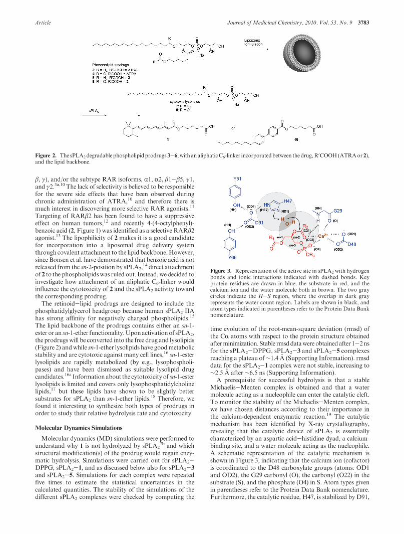
Article Journal of Medicinal Chemistry, 2010, Vol. 53, No. 9 3783
β, γ), and/or the subtype RAR isoforms, R1, R2, β1-β5, γ1,and γ2.3a,10 The lack of selectivity is believed to be responsiblefor the severe side effects that have been observed duringchronic administration of ATRA,10 and therefore there ismuch interest in discovering more selective RAR agonists.11
Targeting of RARβ2 has been found to have a suppressiveeffect on human tumors,12 and recently 4-(4-octylphenyl)-benzoic acid (2, Figure 1) was identified as a selective RARβ2agonist.13 The lipophilicity of 2 makes it is a good candidatefor incorporation into a liposomal drug delivery systemthrough covalent attachment to the lipid backbone.However,since Bonsen et al. have demonstrated that benzoic acid is notreleased from the sn-2-position by sPLA2,
14 direct attachmentof 2 to the phospholipidswas ruled out. Instead, we decided toinvestigate how attachment of an aliphatic C6-linker wouldinfluence the cytotoxicity of 2 and the sPLA2 activity towardthe corresponding prodrug.
The retinoid-lipid prodrugs are designed to include thephosphatidylglycerol headgroup because human sPLA2 IIAhas strong affinity for negatively charged phospholipids.15
The lipid backbone of the prodrugs contains either an sn-1-ester or an sn-1-ether functionality.Uponactivationof sPLA2,the prodrugswill be converted into the free drug and lysolipids(Figure 2) andwhile sn-1-ether lysolipids have goodmetabolicstability and are cytotoxic against many cell lines,16 sn-1-esterlysolipids are rapidly metabolized (by e.g., lysophospholi-pases) and have been dismissed as suitable lysolipid drugcandidates.16a Information about the cytotoxicityof sn-1-esterlysolipids is limited and covers only lysophosphatidylcholinelipids,17 but these lipids have shown to be slightly bettersubstrates for sPLA2 than sn-1-ether lipids.18 Therefore, wefound it interesting to synthesize both types of prodrugs inorder to study their relative hydrolysis rate and cytotoxicity.
Molecular Dynamics Simulations
Molecular dynamics (MD) simulations were performed tounderstand why 1 is not hydrolyzed by sPLA2
7b and whichstructural modification(s) of the prodrug would regain enzy-matic hydrolysis. Simulations were carried out for sPLA2-DPPG, sPLA2-1, and as discussed below also for sPLA2-3
and sPLA2-5. Simulations for each complex were repeatedfive times to estimate the statistical uncertainties in thecalculated quantities. The stability of the simulations of thedifferent sPLA2 complexes were checked by computing the
time evolution of the root-mean-square deviation (rmsd) ofthe CR atoms with respect to the protein structure obtainedafterminimization. Stable rmsddatawereobtainedafter 1-2nsfor the sPLA2-DPPG, sPLA2-3 and sPLA2-5 complexesreaching a plateau of∼1.4 A (Supporting Information). rmsddata for the sPLA2-1 complex were not stable, increasing to∼2.5 A after ∼6.5 ns (Supporting Information).
A prerequisite for successful hydrolysis is that a stableMichaelis-Menten complex is obtained and that a watermolecule acting as a nucleophile can enter the catalytic cleft.To monitor the stability of the Michaelis-Menten complex,we have chosen distances according to their importance inthe calcium-dependent enzymatic reaction.19 The catalyticmechanism has been identified by X-ray crystallography,revealing that the catalytic device of sPLA2 is essentiallycharacterized by an aspartic acid-histidine dyad, a calcium-binding site, and a water molecule acting as the nucleophile.A schematic representation of the catalytic mechanism isshown in Figure 3, indicating that the calcium ion (cofactor)is coordinated to the D48 carboxylate groups (atoms: OD1and OD2), the G29 carbonyl (O), the carbonyl (O22) in thesubstrate (S), and the phosphate (O4) in S. Atom types givenin parentheses refer to the Protein Data Bank nomenclature.Furthermore, the catalytic residue, H47, is stabilized by D91,
Figure 2. The sPLA2degradablephospholipidprodrugs3-6, withanaliphaticC6-linker incorporatedbetween the drug,R0COOH(ATRAor2),
and the lipid backbone.
Figure 3. Representation of the active site in sPLA2 with hydrogenbonds and ionic interactions indicated with dashed bonds. Keyprotein residues are drawn in blue, the substrate in red, and thecalcium ion and the water molecule both in brown. The two graycircles indicate the H-S region, where the overlap in dark grayrepresents the water count region. Labels are shown in black, andatom types indicated in parentheses refer to the Protein Data Banknomenclature.
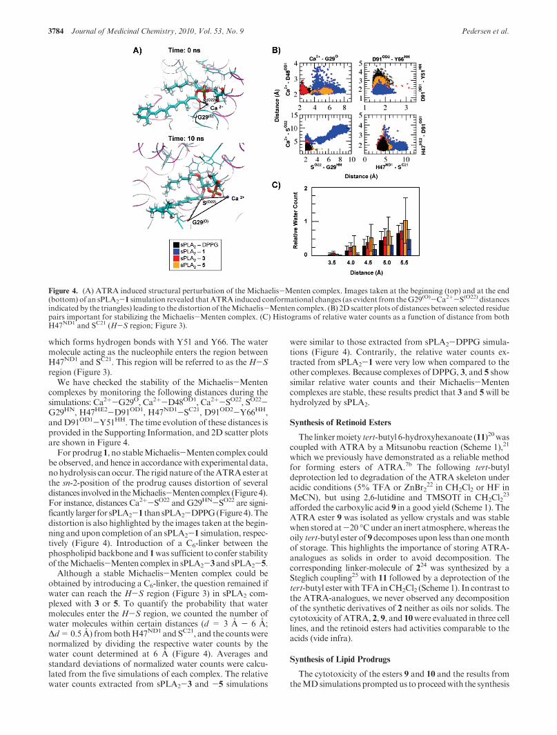
3784 Journal of Medicinal Chemistry, 2010, Vol. 53, No. 9 Pedersen et al.
which forms hydrogen bonds with Y51 and Y66. The watermolecule acting as the nucleophile enters the region betweenH47ND1 and SC21. This region will be referred to as theH-Sregion (Figure 3).
We have checked the stability of the Michaelis-Mentencomplexes by monitoring the following distances during thesimulations: Ca2þ-G29O, Ca2þ-D48OD1, Ca2þ-SO22, SO22-G29HN, H47HE2-D91OD1, H47ND1-SC21, D91OD2-Y66HH,andD91OD1-Y51HH. The time evolution of these distances isprovided in the Supporting Information, and 2D scatter plotsare shown in Figure 4.
For prodrug1, no stableMichaelis-Menten complex couldbe observed, and hence in accordance with experimental data,nohydrolysis canoccur.The rigid nature of theATRAester atthe sn-2-position of the prodrug causes distortion of severaldistances involved in theMichaelis-Mentencomplex (Figure4).For instance, distances Ca2þ-SO22 and G29HN-SO22 are signi-ficantly larger for sPLA2-1 than sPLA2-DPPG(Figure4).Thedistortion is also highlighted by the images taken at the begin-ning and upon completion of an sPLA2-1 simulation, respec-tively (Figure 4). Introduction of a C6-linker between thephospholipid backbone and 1was sufficient to confer stabilityof theMichaelis-Menten complex in sPLA2-3 and sPLA2-5.
Although a stable Michaelis-Menten complex could beobtained by introducing a C6-linker, the question remained ifwater can reach the H-S region (Figure 3) in sPLA2 com-plexed with 3 or 5. To quantify the probability that watermolecules enter the H-S region, we counted the number ofwater molecules within certain distances (d = 3 A - 6 A;Δd=0.5 A) frombothH47ND1 and SC21, and the counts werenormalized by dividing the respective water counts by thewater count determined at 6 A (Figure 4). Averages andstandard deviations of normalized water counts were calcu-lated from the five simulations of each complex. The relativewater counts extracted from sPLA2-3 and -5 simulations
were similar to those extracted from sPLA2-DPPG simula-tions (Figure 4). Contrarily, the relative water counts ex-tracted from sPLA2-1 were very low when compared to theother complexes. Because complexes of DPPG, 3, and 5 showsimilar relative water counts and their Michaelis-Mentencomplexes are stable, these results predict that 3 and 5 will behydrolyzed by sPLA2.
Synthesis of Retinoid Esters
The linkermoiety tert-butyl 6-hydroxyhexanoate (11)20wascoupled with ATRA by a Mitsunobu reaction (Scheme 1),21
which we previously have demonstrated as a reliable methodfor forming esters of ATRA.7b The following tert-butyldeprotection led to degradation of the ATRA skeleton underacidic conditions (5% TFA or ZnBr2
22 in CH2Cl2 or HF inMeCN), but using 2,6-lutidine and TMSOTf in CH2Cl2
23
afforded the carboxylic acid 9 in a good yield (Scheme 1). TheATRA ester 9 was isolated as yellow crystals and was stablewhen stored at-20 �Cunder an inert atmosphere, whereas theoily tert-butyl ester of 9decomposes upon less thanonemonthof storage. This highlights the importance of storing ATRA-analogues as solids in order to avoid decomposition. Thecorresponding linker-molecule of 224 was synthesized by aSteglich coupling25 with 11 followed by a deprotection of thetert-butyl esterwithTFA inCH2Cl2 (Scheme 1). In contrast tothe ATRA-analogues, we never observed any decompositionof the synthetic derivatives of 2 neither as oils nor solids. Thecytotoxicity of ATRA, 2, 9, and 10were evaluated in three celllines, and the retinoid esters had activities comparable to theacids (vide infra).
Synthesis of Lipid Prodrugs
The cytotoxicity of the esters 9 and 10 and the results fromtheMDsimulations promptedus to proceedwith the synthesis
Figure 4. (A) ATRA induced structural perturbation of the Michaelis-Menten complex. Images taken at the beginning (top) and at the end(bottom) of an sPLA2-1 simulation revealed thatATRA induced conformational changes (as evident from theG29(O)-Ca2þ-S(O22) distancesindicated by the triangles) leading to the distortion of theMichaelis-Menten complex. (B) 2D scatter plots of distances between selected residuepairs important for stabilizing the Michaelis-Menten complex. (C) Histograms of relative water counts as a function of distance from bothH47ND1 and SC21 (H-S region; Figure 3).
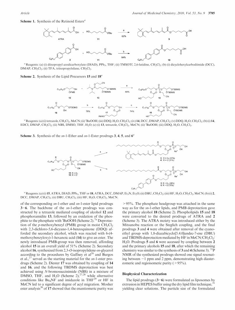
Article Journal of Medicinal Chemistry, 2010, Vol. 53, No. 9 3785
of the corresponding sn-1-ether and sn-1-ester lipid prodrugs3-6. The backbone of the sn-1-ether prodrugs was con-structed by a tetrazole mediated coupling of alcohol 12 andphosphoramidite 13, followed by an oxidation of the phos-phite to the phosphate with tBuOOH (Scheme 2).7a Deprotec-tion of the p-methoxybenzyl (PMB) group in moist CH2Cl2with 2,3-dichloro-5,6-dicyano-1,4-benzoquinone (DDQ) af-forded the secondary alcohol, which was reacted with 6-(4-methoxybenzyloxy)-1-hexanoic acid (14) to give an ester. Thenewly introduced PMB-group was then removed, affordingalcohol 15 in an overall yield of 51% (Scheme 2). Secondaryalcohol 16, synthesized from2,3-O-isopropylidene-sn-glycerolaccording to the procedures by Gaffney et al26 and Burgoset al.,27 served as the starting material for the sn-1-ester pro-drugs (Scheme 2). Diester 17 was obtained by coupling of 14and 16, and the following TBDMS deprotection was bestachieved using N-bromosuccinimide (NBS) in a mixture ofDMSO, THF, and H2O (Scheme 2),27,28 while alternativeconditions like Bu4NF and imidazole in THF29 or HF inMeCN led to a significant degree of acyl migration. Mosherester analysis30 of 17 showed that the enantiomeric purity was
>95%. The phosphate headgroup was attached in the sameway as for the sn-1-ether lipids, and PMB deprotection gavethe primary alcohol 18 (Scheme 2). Phospholipids 15 and 18
were converted to the desired prodrugs of ATRA and 2
(Scheme 3). The ATRA moiety was introduced either by theMitsunobu reaction or the Steglich coupling, and the finalprodrugs 3 and 4 were obtained after removal of the cyano-ethyl group with 1,8-diazabicyclo[5.4.0]undec-7-ene (DBU)andTBDMS-deprotectionmediatedbyHF inMeCN/CH2Cl2/H2O. Prodrugs 5 and 6 were accessed by coupling between 2
and the primary alcohols 15 and 18, after which the remainingchemistrywas similar to the synthesis of 3 and 4 (Scheme3). 31PNMR of the synthesized prodrugs showed one signal resonat-ing between -1 ppm and 2 ppm, demonstrating high diaster-eomeric and regioisomeric purity (>95%).
Biophysical Characterization
The lipid prodrugs (3-6) were formulated as liposomes byextrusion inHEPESbuffer using thedry lipid film technique,31
yielding clear solutions. The particle size of the formulated
Scheme 1. Synthesis of the Retinoid Estersa
aReagents: (a) (i) diisopropyl azodicarboxylate (DIAD), PPh3, THF; (ii) TMSOTf, 2,6-lutidine, CH2Cl2; (b) (i) dicyclohexylcarbodiimide (DCC),
DMAP, CH2Cl2; (ii) TFA, triisopropylsilane, CH2Cl2.
Scheme 2. Synthesis of the Lipid Precursors 15 and 18a
aReagents: (a) (i) tetrazole,CH2Cl2,MeCN; (ii) tBuOOH; (iii)DDQ,H2O,CH2Cl2; (iv) 14, DCC,DMAP,CH2Cl2; (v)DDQ,H2O,CH2Cl2; (b) (i) 14,
EDCI, DMAP, CH2Cl2; (ii) NBS, DMSO, THF, H2O; (c) (i) 13, tetrazole, CH2Cl2, MeCN; (ii) tBuOOH; (iii) DDQ, H2O, CH2Cl2.
Scheme 3. Synthesis of the sn-1-Ether and sn-1-Ester prodrugs 3, 4, 5, and 6a
aReagents: (a) (i) 15, ATRA,DIAD,PPh3, THFor 18, ATRA,DCC,DMAP,Et3N, Et2O; (ii)DBU,CH2Cl2; (iii)HF,H2O,CH2Cl2,MeCN; (b) (i) 2,
DCC, DMAP, CH2Cl2; (ii) DBU, CH2Cl2; (iii) HF, H2O, CH2Cl2, MeCN.

3786 Journal of Medicinal Chemistry, 2010, Vol. 53, No. 9 Pedersen et al.
lipids was measured by dynamic light scattering (DLS). TheDLS analysis revealed that all of the lipids were able to formparticles with a diameter around 100 nm (Table 1 and Sup-porting Information) andwith a lowpolydispersity, indicatingthe formation of unilamellar vesicles. The formation of a uni-lamellar bilayer is further supported by the fluorometricprocedure described by McIntyre et al.,32 which was used tostudy the permeability. Vesicles formed from prodrug 5 wascompared to vesicles of 1,2-dipalmitoyl-sn-glycero-3-phos-phoglycerol (DPPG) and 1-palmitoyl-2-oleoyl-sn-glycero-3-phosphoglycerol (POPG). The lipids were formulated with1,2-dipalmitoyl-sn-glycero-3-phosphoethanolamine-N-[7-ni-tro-2-1,3-benzoxadiazol-4-yl] (NBD-DPPE, 0.15mol%).Thefluorescence of the NBD group was monitored over timebefore and after the addition of dithionite (S2O4
-2), whichfunctions as a quenching agent. At the end of the experiment,the detergent Triton X-100 was added to solubilize the lipidvesicles and cause 100% quenching (see Figure 5). As evidentfrom the figure, the decrease in the fluorescence of the NBD-labeled gel-state DPPG occurs in two distinct steps, animmediate and fast one completed after 20 min followed bymuch slower decay of fluorescence. We anticipate that thequick drop in fluorescence signal corresponds to quenching ofthe dye in the outer leaflet and is dependent on the fraction oflipids exposed to the surrounding, i.e., vesicle size and lamel-larity. The slower second step is due to quenching of the dye inthe inner leaflet, which is controlled by membrane permeabil-ity to dithionite. In the case of the fluid POPG vesicles, thequenching of both leaflets occurs simultaneously, resultingeffectively in a single exponential-like decay in the NBDfluorescence. A comparable behavior for gel-state and fluidmembranes has been reported previously.32,33 The quenchingof the NBD-labeled vesicles for prodrug 5 resembles that ofDPPG, demonstrating that 5 forms a bilayer membrane,however seemingly more permeable to dithionite than thevesicles of DPPG.
Differential scanning calorimetry scans (15-65 �C) of thelipid solutions of 3 and 5 displayed no gel-to-liquid crystallineor any other thermotropic phase transition in the testedtemperature range (data not shown), indicating that the lipidbilayers are in a fluid state. This was not surprising taking intoaccount the bulky and stiff nature of the substituents in thesn-2-position, which hampers well-ordered chain packing.
The ability of sPLA2 to hydrolyze the formulated lipids at37 �C was investigated with matrix-assisted laser desorption/ionization time-of-flight (MALDI-TOF) MS. MALDI-TOFMS is a fast and sensitive technique which has been shown tobe a reliable tool for detection of lipids.34 In Figure 6, theobtained MS data for the sn-1-ether prodrug 3 subjected topurified sPLA2 from snake (Agkistrodon piscivorus piscivorus)venom is shown.Gratifyingly, and as evident from the spectra(Figure 6), the prodrug (MþHþ) is consumed by the enzymeand the desired constituents, lysolipid 7 (MþHþ) andATRAester 9 (M þ Naþ), are released. The MALDI-TOF MSanalysis of the sn-1-ether prodrugs 3 and 5 and the sn-1-esterprodrugs 4 and 6 revealed that all of the prodrugs weredegraded by the enzyme (Table 2) and the desired moleculeswere released (see Supporting Information). The observationthat the sn-1-ether prodrugs were consumed by sPLA2 to thesame extent as the sn-1-ester prodrugs illustrate and confirmthat sn-1-ether phospholipids are excellent substrates forsPLA2. On the basis of the MALDI-TOF MS analysis ofthe sPLA2 activity on the various prodrugs, we can concludethat the incorporation of an aliphatic C6-linker betweenATRA and the lipid backbone resolved the issue of sPLA2
activity on ATRA-lipid prodrugs. The experimental observa-tions are in agreement with the outcome from the MDsimulations, illustrating the power and potential of usingMD simulations in the design of sPLA2 degradable lipidprodrugs. To rule out that the degradation of the prodrugswas caused by nonenzymatic hydrolysis, a sample of eachprodrug was subjected to the reaction conditions in theabsence of sPLA2. As evident from the spectra in Figure 6(and in Supporting Information), no degradation of theprodrugs was observed within 48 h and none of the releaseproducts were detectable. Finally, we compared the enzymeactivity of the purified sPLA2 from snake (Agkistrodon pisci-vorus piscivorus) venom with the commercially availablesPLA2 from snake (Naja mossambica mossambica) venom,and as can be seen in Table 2, the two enzymes performequally well on all substrates.
Fawzy et al.35 and Hope et al.36 have demonstrated twodecades ago that ATRA is an inhibitor for sPLA2, reportingIC50 values of<50 and10μMrespectively.However,we havelater shown that DPPG is fully hydrolyzed by sPLA2 in thepresenceof 0.1 and1.0 equiv ofATRA,7bproving that the lackof hydrolysis for the ATRA-lipid prodrug 1 is not a conse-quence of inhibition by ATRA. These observations arefurther supported by the present work, in which it was foundthat 9 (IC50=8μM,Ki=6 μM, see Supporting Information)inhibits sPLA2 at the same level as ATRA (IC50 = 15 μM,Ki = 11 μM), and whereas the ATRA-lipid prodrug 1 is notconsumed by the enzyme, the prodrugs 3 and 4 are fullydegraded. This demonstrates that under the conditions used,the released ATRA ester 9 does not inhibit sPLA2, and thedifference in degradation of the prodrugs therefore solelyrelies on the ability of sPLA2 to hydrolyze the differentprodrugs. Additionally, Cunningham et al. have reported Ki
values <100 nM37 for potent sPLA2 inhibitors, illustratingthat ATRA and 9 are weak inhibitors.
Table 1. Measurement of Particle Size with DLS
particle size
prodrug diameter (nm) polydispersity index
3 94 0.08
4 97 0.13
5 118 0.16
6 118 0.05
Figure 5. Time-dependent fluorescence from NBD labeled lipidsquenched by dithionite.
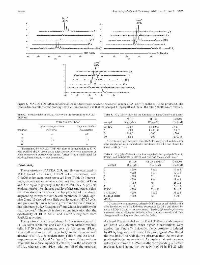
Article Journal of Medicinal Chemistry, 2010, Vol. 53, No. 9 3787
Cytotoxicity
The cytotoxicity of ATRA, 2, 9, and 10 were evaluated inMT-3 breast carcinoma, HT-29 colon carcinoma, andColo205 colon adenocarcinoma cell lines (Table 3). Interest-ingly, the retinoid esters were either more active than ATRAand 2 or equal in potency in the tested cell lines. A possibleexplanation for the enhancedactivityof thesemolecules is thatthe derivatization increases the lipophilicity of the drugs,augmenting transport over the cell membrane. RARβ2 ago-nists 2 and 10 showed very little activity against HT-29 cells,and presumably this is because growth inhibition in this cellline is induced byRARR agonists38 and 2has a lowaffinity forthat receptor.13 The result is also a strong indication that thecytotoxicity of 10 in MT-3 and Colo205 originate fromRARβ2 activation.
The cytotoxicity of the prodrugs 3-6 was investigated inHT-29 colon carcinoma and Colo205 colon adenocarcinomacells. HT-29 colon carcinoma cells do not secrete sPLA2,which allowed us to test the activity in the presence andabsence of sPLA2. As evident from Table 4 and the dose-response curve for 3 and 5 (Figure 7), none of the prodrugswere able to induce significant cell death in the absence ofsPLA2, whereas upon sPLA2 addition, all of the prodrugs
displayed IC50 values below10μMinHT-29 cells and completecell death was obtained when higher concentrations wereapplied (see Figure 7). Evidently, the cytotoxicity is inducedby sPLA2 triggeredbreakdownof the prodrugs into 9or 10 andthe lysolipids. Interestingly, we observed that the sn-1-esterprodrug 6 in the presence of sPLA2 displayed almost the samecytotoxicity towardHT-29 cells as the corresponding sn-1-etherprodrug 5, and taking the low activity of 10 in HT-29 cells
Figure 6. MALDI-TOF MS monitoring of snake (Agkistrodon piscivorus piscivorus) venom sPLA2 activity on the sn-1-ether prodrug 3. Thespectra demonstrate that the prodrug 3 (top left) is consumed and that the lysolipid 7 (top right) and the ATRA ester 9 (bottom) are released.
Table 2. Measurement of sPLA2 Activity on the Prodrugs byMALDI-TOF MS
hydrolysis by sPLA2a
prodrug
Agkistrodon piscivorus
piscivorus
Naja mossambica
mossambica
3 þ nd
4 þ þ5 þb þb
6 þ þaDetermined by MALDI-TOF MS after 48 h incubation at 37 �C
with purified sPLA2 from snake (Agkistrodon piscivorus piscivorus orNaja mossambica mossambica) venom. bAfter 48 h, a small signal forprodrug 5 remains; nd = not determined.
Table 3. IC50 (μM)Values for theRetinoids in Three CancerCell Linesa
compd
MT-3
IC50 (μM)
HT-29
IC50 (μM)
Colo205
IC50 (μM)
ATRA 30( 4 4.3 ( 0.2 37 ( 1
9 17( 1 3.6 ( 1.8 17 ( 2
2 51( 3 >200 >200
10 14( 1 >200 127 ( 18aCytotoxicitywasmeasured using theMTTassay as cell viability 48 h
after incubation with the indicated substances for 24 h and shown bymean ( SD (n = 3).
Table 4. IC50 (μM)Values for the Prodrugs 3-6, the Lysolipids 7 and 8,DSPG, and 1-O-DSPG in HT-29 and Colo205 Cancer Cell Linesa
compd
HT-29
IC50 (μM)
HT-29 þ sPLA2b
IC50 (μM)
Colo205
IC50 (μM)
3 >200 7 ( 2 16 ( 4
4 >200 6 ( 1 12 ( 3
5 >200 3 ( 1 7 ( 6
6 >200 8 ( 1 19 ( 4
7 11 ( 6 nd 25 ( 2
8 7 ( 1 nd 22 ( 3
DSPG >200 25 ( 11 54 ( 7
1-O-DSPG >200 9 ( 2 33 ( 4
C17H35COOH >200 nd >200
sPLA2 c c caCytotoxicitywasmeasured using theMTTassay as cell viability 48 h
after incubation with the indicated substances for 24 h and shown bymean( SD (ng 3); nd=not determined. bSnake (Agkistrodon piscivoruspiscivorus) venomsPLA2was added to a final concentrationof 5 nM. cNochange in cell viability was observed after 24 h.
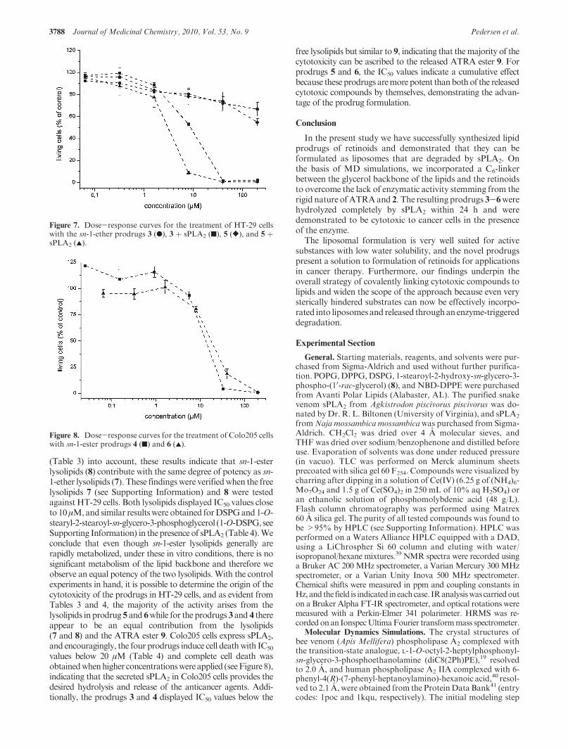
3788 Journal of Medicinal Chemistry, 2010, Vol. 53, No. 9 Pedersen et al.
(Table 3) into account, these results indicate that sn-1-esterlysolipids (8) contribute with the same degree of potency as sn-1-ether lysolipids (7). These findingswere verifiedwhen the freelysolipids 7 (see Supporting Information) and 8 were testedagainst HT-29 cells. Both lysolipids displayed IC50 values closeto 10μM,and similar resultswere obtained forDSPGand1-O-stearyl-2-stearoyl-sn-glycero-3-phosphoglycerol (1-O-DSPG, seeSupporting Information) in the presence of sPLA2 (Table 4).Weconclude that even though sn-1-ester lysolipids generally arerapidly metabolized, under these in vitro conditions, there is nosignificant metabolism of the lipid backbone and therefore weobserve an equal potency of the two lysolipids. With the controlexperiments in hand, it is possible to determine the origin of thecytotoxicity of the prodrugs in HT-29 cells, and as evident fromTables 3 and 4, the majority of the activity arises from thelysolipids in prodrug 5 and 6while for the prodrugs 3 and 4 thereappear to be an equal contribution from the lysolipids(7 and 8) and the ATRA ester 9. Colo205 cells express sPLA2,and encouragingly, the four prodrugs induce cell deathwith IC50
values below 20 μM (Table 4) and complete cell death wasobtainedwhenhigher concentrationswereapplied (seeFigure8),indicating that the secreted sPLA2 in Colo205 cells provides thedesired hydrolysis and release of the anticancer agents. Addi-tionally, the prodrugs 3 and 4 displayed IC50 values below the
free lysolipids but similar to 9, indicating that themajority of thecytotoxicity can be ascribed to the released ATRA ester 9. Forprodrugs 5 and 6, the IC50 values indicate a cumulative effectbecause these prodrugs aremorepotent thanbothof the releasedcytotoxic compounds by themselves, demonstrating the advan-tage of the prodrug formulation.
Conclusion
In the present study we have successfully synthesized lipidprodrugs of retinoids and demonstrated that they can beformulated as liposomes that are degraded by sPLA2. Onthe basis of MD simulations, we incorporated a C6-linkerbetween the glycerol backbone of the lipids and the retinoidsto overcome the lack of enzymatic activity stemming from therigid nature ofATRAand 2. The resulting prodrugs 3-6werehydrolyzed completely by sPLA2 within 24 h and weredemonstrated to be cytotoxic to cancer cells in the presenceof the enzyme.
The liposomal formulation is very well suited for activesubstances with low water solubility, and the novel prodrugspresent a solution to formulation of retinoids for applicationsin cancer therapy. Furthermore, our findings underpin theoverall strategy of covalently linking cytotoxic compounds tolipids and widen the scope of the approach because even verysterically hindered substrates can now be effectively incorpo-rated into liposomesand released throughanenzyme-triggereddegradation.
Experimental Section
General. Starting materials, reagents, and solvents were pur-chased from Sigma-Aldrich and used without further purifica-tion. POPG,DPPG,DSPG, 1-stearoyl-2-hydroxy-sn-glycero-3-phospho-(10-rac-glycerol) (8), and NBD-DPPE were purchasedfrom Avanti Polar Lipids (Alabaster, AL). The purified snakevenom sPLA2 from Agkistrodon piscivorus piscivorus was do-nated byDr. R. L. Biltonen (University of Virginia), and sPLA2
fromNaja mossambica mossambicawas purchased from Sigma-Aldrich. CH2Cl2 was dried over 4 A molecular sieves, andTHF was dried over sodium/benzophenone and distilled beforeuse. Evaporation of solvents was done under reduced pressure(in vacuo). TLC was performed on Merck aluminum sheetsprecoated with silica gel 60 F254. Compounds were visualized bycharring after dipping in a solution of Ce(IV) (6.25 g of (NH4)6-Mo7O24 and 1.5 g of Ce(SO4)2 in 250 mL of 10% aq H2SO4) oran ethanolic solution of phosphomolybdenic acid (48 g/L).Flash column chromatography was performed using Matrex60 A silica gel. The purity of all tested compounds was found tobe >95% by HPLC (see Supporting Information). HPLC wasperformed on a Waters Alliance HPLC equipped with a DAD,using a LiChrospher Si 60 column and eluting with water/isopropanol/hexane mixtures.39 NMR spectra were recorded usinga Bruker AC 200 MHz spectrometer, a Varian Mercury 300 MHzspectrometer, or a Varian Unity Inova 500 MHz spectrometer.Chemical shifts were measured in ppm and coupling constants inHz,and the field is indicated ineachcase. IRanalysiswas carriedouton a Bruker Alpha FT-IR spectrometer, and optical rotations weremeasured with a Perkin-Elmer 341 polarimeter. HRMS was re-cordedonan IonspecUltimaFourier transformmass spectrometer.
Molecular Dynamics Simulations. The crystal structures ofbee venom (Apis Mellifera) phospholipase A2 complexed withthe transition-state analogue, L-1-O-octyl-2-heptylphosphonyl-sn-glycero-3-phosphoethanolamine (diC8(2Ph)PE),19 resolvedto 2.0 A, and human phospholipase A2 IIA complexed with 6-phenyl-4(R)-(7-phenyl-heptanoylamino)-hexanoic acid,40 resol-ved to 2.1 A, were obtained from the ProteinData Bank41 (entrycodes: 1poc and 1kqu, respectively). The initial modeling step
Figure 7. Dose-response curves for the treatment of HT-29 cellswith the sn-1-ether prodrugs 3 (b), 3 þ sPLA2 (9), 5 ([), and 5 þsPLA2 (2).
Figure 8. Dose-response curves for the treatment of Colo205 cellswith sn-1-ester prodrugs 4 (9) and 6 (2).
Article Journal of Medicinal Chemistry, 2010, Vol. 53, No. 9 3789
involved placing diC8(2Ph)PE into the binding cleft of humanphospholipase A2 IIA, as described previously.18 A previouslyassembled sPLA2-substrate complex18was used as the templateto place lipid prodrugs into the active site. The optimized lipidprodrugs were manually overlapped with the pre-existing sub-strate, which was subsequently deleted. NAMD software42 withthe Charmm27 all-atom parameter set and TIP3 water modelwas used for all simulations.43 The lipid prodrugs were firstenergy-minimized for 1000 steps before using them in simula-tions. Missing force field parameters for the prodrug moleculeswere taken from similar atom types in the CHARMM27 forcefield. The sPLA2-lipid complexes were solvated using theprogram SOLVATE.44 Eighteen water molecules were ran-domly replaced with chloride ions to neutralize the systems.Ultimately, the system contained ∼4900 water molecules in asimulation cell of dimensions 52.7 � 51.7 � 67.3 A3. An initialenergy minimization of the complex was carried out for 1000steps, and the system was further minimized for 100, 200, 300,400, and 500 steps to generate different starting structures.Periodic boundary conditions were applied in x, y, and zdirections. Initial MD simulations were conducted for ∼100ps in which each systemwas slowly heated to 300K. Simulationswere carried out for 10 ns in the NPT ensemble, i.e., at constantnumber of atoms (N), pressure (P), and temperature (T). A timestep of 1 fs and an isotropic pressure of 1 atm using the Langevinpiston method45 were employed. Electrostatic forces were cal-culated using the particle mesh Ewald method with a uniformgrid spacing of ∼1 A.46 A 12 A cutoff was used for terminatingthe van der Waals interactions in combination with a switchingfunction starting at 10 A. Analysis of the trajectories were per-formed using the Visualization Molecular Dynamics softwaresuite.47
Liposome Preparation and Particle Size Determination. Thelipid prodrugs 3-6 were dissolved in CHCl3 in a glass tubeand dried under vacuum for 15 h to form a thin film. The lipidprodrugs (2mM)were solubilized by addition of aqueous buffer(0.15MKCl, 30 μMCaCl2, 10 μMEDTA, 10 mMHEPES, pH7.5) and vortexed periodically over 1 h at 20 �C. Subsequently,the solutions were extruded through a 100 nm polycarbonatecutoff membrane (20-30 repetitions) using a Hamilton syringeextruder (Avanti Polar Lipids, Birmingham, AL). The particlesize distribution of the formulated lipids was measured byDLS. The DLSmeasurements were obtained using a Zetasizernano particle analyzer (ZS ZEN3600, Malvern Instrument,Westborough, MA).
Permeability Assay. The permeability of the lipid vesicles wasexamined using the procedure described by McIntyre et al.32
The lipid vesicles were labeled with NBD-DPPE (0.15 mol %)and prepared in an aqueous buffer (10mMHEPES, 1M sucrose,150mMNaCl, pH7.4) by sonication for 30min at 45 �Cand thenextrusion through a 100 nm filter at 45 �C. Sucrose was addedto minimize the osmolarity difference between the external solu-tion and the vesicle interior that will arise after the addition ofdithionite.48 The experiments were performed in duplicates ormoreusingFLUOstarOmegamicroplate reader (BMGLABTECH)in a 96-well microplate at 20 �C (150 μL). The lipid samples(100 μM) were allowed to equilibrate at 20 �C before theaddition of sodium dithionite (Na2S2O4) solution (33 mM),which was used as a quenching agent. Because of the highinstability of dithionite,49 the dithionite solution was freshlyprepared prior to use. The NBD fluorescence was observed at520 nm (ex 485 nm). After 85 min, Triton X-100 was added tothe medium (0.3 wt %).
sPLA2 Activity Measurements Monitored by MALDI-TOFMS. The formulated lipid prodrugs (0.40 mL, 2 mM) werediluted in an aqueous buffer (2.1mL, 0.15MKCl, 30 μMCaCl2,10 μM EDTA, 10 mM HEPES, pH 7.5), and the mixture wasstirred at 37 �C in a container protected from light. The catalyticreaction was initiated by addition of snake (Agkistrodon pisci-vorus piscivorus (20 μL, 42 μM) orNaja mossambica mossambica
(12 μL, 71 μM)) venom sPLA2. Sampling was done after 0, 2, 24,and 48 h by collecting 100 μLof the reactionmixture and rapidlymixing it with a solution of CHCl3/MeOH/H2O/AcOH 4:8:1:1(0.5 mL) in order to stop the reaction. The mixture was washedwith water (0.5 mL), and the organic phase (80 μL) was isolatedby extraction and then concentrated in vacuo. The extract wasmixed with 9 μL of 2,5-dihydroxybenzoic acid (DHB) matrix(0.5 M DHB, 2 mM CF3COONa, 1 mg/mL DPPG in MeOH),and 0.5 μL of this mixture was used for the MS analysis.
Cytotoxicity. Colon cancer HT-29 cells were cultured inMcCoy’s 5A medium in the presence of 10% fetal calf serumand 1% Pen-Strep (InVitrogen). Breast cancer MT-3 and coloncancer Colo205 cells were cultured in RPMI 1640 supplementedwith 10% fetal calf serum and 1% Pen-Strep in a humidifiedatmosphere containing 5% CO2. Colo205 cells secrete sPLA2-IIa, however as the cytotoxicity assay was made with low celldensity and for a short incubation period, inadequate concen-trations were reached in the medium, which is why conditionedmedium was used. Conditioned medium was made as follows:Colo205 cells were grown to confluency for 72 h, at which pointthe cells were pelleted and themedium,which contained approx-imately 100 ng/mL sPLA2 IIA, was collected and used for theassay. Cells were plated in 96-well plates at a density of 1 � 104
cells per well for HT-29 andMT-3 cells and 2� 104 cells per wellfor Colo205 cells, 24 h prior to addition of the tested compound.The retinoids (ATRA, 2, 9, 10, and stearic acid) were solubilizedin DMSO and water (final DMSO concentration e0.5%).Liposomes were diluted in PBS, and initial lipid concentrationsin the liposome solutions were determined by phosphorusanalysis.50 After 24 h of incubation, the substances were removedand the cells were washed and incubated in complete medium foranother 48 h. Cytotoxic activity was assessed using a standard3-(4,5-dimethylthiazolyl)-2,5-diphenyltetrazoliumbromide (MTT)assay (Cell ProliferationKit I, Roche, Germany).51 Cell viability isexpressed as percentage reduction of incorporated MTT.
1-O-Octadecyl-2-(6-(all-trans-retinoyloxy)-hexanoyl)-sn-glycero-3-phospho-(S)-glycerol (3).Alcohol15 (13mg, 0.014mmol),ATRA(6mg, 0.019mmol), and PPh3 (9 mg, 0.043mmol) were dissolvedin THF (1.0mL).DIAD (11 μL, 0.043mmol) was added, and thereaction mixture was stirred at 20 �C for 1 h and then concen-trated in vacuo and purified by flash column chromatography(heptane/EtOAc 2:1 then heptane/EtOAc 1:2) to give 13 mg thatwas dissolved in CH2Cl2 (1.0 mL) and DBU (2 μL, 0.012 mmol)was added. The reaction mixture was stirred for 30 min at 20 �Cand then purified directly by flash column chromatography(heptane/EtOAc 1:1 then CH2Cl2/MeOH 9:1) to afford 8 mgthat was dissolved in MeCN (0.9 mL) and CH2Cl2 (0.3 mL) andcooled to 0 �C. Aqueous HF (40%, 30 μL) was added dropwise,and the reaction mixture was allowed to reach 20 �C. After 3.5 h,the reaction was quenched by dropwise addition of MeOSiMe3(0.3 mL) and the mixture was stirred for 30 min, after whichNaHCO3 (6 mg, 0.07 mmol) was added and the mixture wasconcentrated in vacuo and purified by flash column chromato-graphy (CH2Cl2/MeOH 10:1 then CH2Cl2/MeOH 4:1) to afford4 mg (31% over 3 step) of 3 as a yellow amorphous solid. Rf =0.44 (CH2Cl2/MeOH 4:1). 1H NMR (500MHz, CDCl3/CD3OD4:1): δ 7.02 (dd, J=15.0, 11.4 Hz, 1H), 6.32-6.27 (m, 2H), 6.15(d, J = 11.4 Hz, 1H), 6.14 (d, J = 16.2 Hz, 1H), 5.77 (s, 1H),5.18-5.13 (m, 1H), 4.11 (t, J=6.6 Hz, 2H), 4.03-3.94 (m, 2H),3.94-3.90 (m, 2H), 3.82-3.77 (m, 1H), 3.62 (t, J=5.2 Hz, 2H),3.60-3.57 (m, 2H), 3.49-3.39 (m, 2H), 2.39-2.33 (m, 5H),2.05-2.00 (m, 5H), 1.72 (s, 3H), 1.71-1.60 (m, 6H), 1.57-1.51(m, 2H), 1.50-1.45 (m, 2H), 1.45-1.40 (m, 2H), 1.31-1.23 (m,30H), 1.03 (s, 6H), 0.88 (t, J=6.9 Hz, 3H). 13C NMR (50MHz,CDCl3/CD3OD 4:1): δ 174.0, 168.0, 153.7, 140.0, 138.0, 137.7,135.3, 131.7, 130.3, 129.8, 129.2, 118.5, 72.2 (2C), 71.2, 69.4, 66.7,64.5, 64.1, 62.7, 40.0, 34.5 (2C), 33.4, 32.3, 30.1, 29.7 (12C), 29.2(2C), 28.7, 26.4, 25.9, 24.9, 23.0, 22.0, 19.6, 14.3, 14.1, 13.3. IR(neat) 3325, 2923, 2853, 1733, 1709, 1609, 1584, 1457, 1259, 1064,798 cm-1; m/z (M þ Hþ) 917.58.
3790 Journal of Medicinal Chemistry, 2010, Vol. 53, No. 9 Pedersen et al.
1-Octadecanoyl-2-(6-(all-trans-retinoyloxy)-hexanoyl)-sn-glycero-3-phospho-(S)-glycerol (4).Alcohol 18 (47mg, 0.052mmol), ATRA(31 mg, 0.104 mmol), DMAP (13 mg, 0.104 mmol), and Et3N(72μL, 0.52mmol)were dissolved inEt2O (1.2mL).DCC (27mg,0.13 mmol) was added, and the reaction mixture was stirred at20 �C for 22 h and then concentrated in vacuo and purified byflash column chromatography (toluene then toluene/EtOAc 5:1)to give 32 mg that was dissolved in CH2Cl2 (2.2 mL) and DBU(4.2μL, 0.028mmol) was added. The reactionmixturewas stirredfor 30 min at 20 �C and then purified directly by flash columnchromatography (heptane/EtOAc 1:1 then CH2Cl2/MeOH 9:1)to afford 21mg thatwas dissolved inMeCN(1.5mL) andCH2Cl2(0.5 mL) and cooled to 0 �C. Aqueous HF (40%, 90 μL) wasadded dropwise, and the reaction mixture was allowed to reach20 �C. After 3.5 h, the reaction was quenched by dropwiseaddition of MeOSiMe3 (0.3 mL) and the mixture was stirredfor 30 min, after which NaHCO3 (6 mg, 0.07 mmol) was addedand the mixture was concentrated in vacuo and purified by flashcolumn chromatography (CH2Cl2/MeOH 10:1 then CH2Cl2/MeOH 4:1 then CH2Cl2/MeOH/H2O 65:25:1) to afford 15 mg(31% over 3 step) of 4 as a yellow amorphous solid. Rf = 0.56(CH2Cl2/MeOH 5:1). 1HNMR (500MHz, CDCl3/CD3OD 4:1):δ 7.02 (dd, J = 15.0, 11.4 Hz, 1H), 6.32-6.27 (m, 2H), 6.15 (d,J = 11.4 Hz, 1H), 6.15 (d, J = 16.2 Hz, 1H), 5.77 (s, 1H),5.25-5.20 (m, 1H), 4.39 (dd, J=12.0, 3.3Hz, 1H), 4.16 (dd, J=12.0, 6.6 Hz, 1H), 4.11 (t, J = 6.6 Hz, 2H), 3.99-3.91 (m, 4H),3.83-3.78 (m, 1H), 3.63 (t, J=5.1 Hz, 2H), 2.36 (t, J=7.5 Hz,2H), 2.34 (s, 3H), 2.31 (t, J = 7.6 Hz, 2H), 2.05-2.00 (m, 5H),1.71 (s, 3H), 1.71-1.65 (m, 4H), 1.65-1.57 (m, 4H), 1.50-1.45(m, 2H), 1.45-1.41 (m, 2H), 1.34-1.21 (m, 28H), 1.03 (s, 6H),0.88 (t, J = 7.0 Hz, 3H). 13C NMR (50 MHz, CDCl3/CD3OD4:1): δ 174.2, 173.5, 168.0, 153.6, 140.1, 138.0, 137.6, 135.3, 131.6,130.3, 129.7, 129.1, 118.5, 71.2, 70.7, 66.8, 64.0 (2C), 62.6 (2C),39.9, 34.3 (3C), 33.4, 32.2, 29.9 (12C), 29.2 (2C), 28.7, 25.8, 25.1,24.8, 23.0, 21.9, 19.5, 14.3, 14.1, 13.1. 31P NMR (202 MHz,CDCl3/CD3OD 4:1): δ -0.08. IR (neat): 3390, 2924, 2853,1734, 1709, 1661, 1458, 1260, 1237, 1153, 1050, 805 cm-1;m/z (Mþ Hþ) 931.57.
1-O-Octadecyl-2-(6-(40-octyl-4-phenylbenzoyloxy)-hexanoyl)-sn-glycero-3-phospho-(S)-glycerol (5). Alcohol 15 (62 mg, 0.068mmol), carboxylic acid 2 (43mg, 0.14mmol), andDMAP(25mg,0.20mmol) were dissolved inCH2Cl2 (4.5mL).DCC (42mg, 0.20mmol) was added, and the reaction mixture was stirred at 20 �Cfor 18 h. The mixture was concentrated in vacuo and purified byflash column chromatography (toluene then toluene/EtOAc 5:1)to give 65 mg that was dissolved in CH2Cl2 (5.0 mL) and DBU(9 μL, 0.06 mmol) was added. The reaction mixture was stirredfor 35 min at 20 �C and then purified directly by flash columnchromatography (heptane/EtOAc 1:1 then CH2Cl2/MeOH 10:1)to afford58mg thatwas dissolved in amixtureofMeCN(4.2mL)and CH2Cl2 (1.4 mL) and cooled to 0 �C. Aqueous HF (40%,250μL)was addeddropwise, and the reactionmixturewas allowedto reach 20 �C. After 3.5 h, the reaction was quenched by drop-wise addition of MeOSiMe3 (0.93 mL), and the mixture wasstirred for 30 min, after which NaHCO3 (8 mg, 0.095 mmol) wasadded and themixturewas concentrated in vacuo and purified byflash columnchromatography (CH2Cl2/MeOH10:1 thenCH2Cl2/MeOH 4:1) to afford 24 mg (38% over 3 step) of 5 as a color-less amorphous solid.Rf = 0.23 (CH2Cl2/MeOH 4:1). 1H NMR(300MHz, CDCl3/CD3OD 4:1): δ 8.08 (d, J=8.3 Hz, 2H), 7.67(d, J=8.3Hz, 2H), 7.55 (d, J=8.1Hz, 2H), 7.29 (d, J=8.1Hz,2H), 5.19-5.14 (m, 1H), 4.34 (t, J= 6.6 Hz, 2H), 4.06-3.93 (m,4H), 3.85-3.80 (m, 1H), 3.66-3.61 (m, 2H), 3.60-3.56 (m, 2H),3.48-3.38 (m, 2H), 2.66 (t, J=7.7 Hz, 2H), 2.40 (t, J=7.5 Hz,2H), 1.86-1.79 (m, 2H), 1.76-1.70 (m, 2H), 1.69-1.62 (m, 2H),1.56-1.49 (m, 4H), 1.39-1.19 (m, 40H), 0.89 (t, J=6.8Hz, 3H),0.88 (t, J = 6.9 Hz, 3H). 13C NMR (50 MHz, CDCl3/CD3OD4:1): δ 173.9, 167.3, 146.1, 143.6, 137.5, 130.3 (2C), 129.3 (2C),128.9, 127.4 (2C), 127.1 (2C), 72.1 (2C), 71.3, 69.3, 66.7, 65.2,64.6, 62.6, 35.9, 34.5, 32.1 (2C), 31.8, 30.0, 29.7 (15C), 28.8, 26.3,
25.8, 24.9, 23.0 (2C), 14.3 (2C). 31P NMR (202 MHz, CDCl3/CD3OD 4:1): δ -1.02. IR (neat): 3320, 2920, 2851, 1717, 1276,1102, 1069 cm-1; m/z (M þ Hþ) 927.58.
1-Octadecanoyl-2-(6-(40-octyl-4-phenylbenzoyloxy)-hexanoyl)-sn-glycero-3-phospho-(S)-glycerol (6).The synthesiswasperformedas for 5, starting from alcohol 18 (70 mg, 0.077 mmol) and afford-ing 51 mg (70% over 3 step) of 6 as a colorless amorphous solid.Rf = 0.54 (CH2Cl2/MeOH 5:1). 1H NMR (500 MHz, CDCl3/CD3OD4:1): δ 8.08 (d, J=8.3Hz, 2H), 7.67 (d, J=8.3Hz, 2H),7.56 (d, J=8.1 Hz, 2H), 7.29 (d, J=8.1 Hz, 2H), 5.27-5.22 (m,1H), 4.39 (dd, J=12.0, 3.2Hz, 1H), 4.34 (d, J=6.6Hz, 2H), 4.17(dd, J=12.0, 6.6Hz, 1H), 4.05-4.00 (m, 2H), 3.98-3.91 (m, 2H),3.85-3.80 (m, 1H), 3.65-3.61 (m, 2H), 2.66 (t, J= 7.7 Hz, 2H),2.40 (t, J= 7.5 Hz, 2H), 2.31 (t, J= 7.6 Hz, 2H), 1.86-1.78 (m,2H), 1.76-1.69 (m, 2H), 1.68-1.62 (m, 2H), 1.62-1.56 (m, 2H),1.55-1.49 (m, 2H), 1.39-1.20 (m, 38H), 0.89 (t, J=6.7 Hz, 3H),0.88 (t, J=6.8Hz, 3H). 13CNMR(50MHz,CDCl3/CD3OD4:1):δ 174.4, 173.6, 167.4, 146.3, 143.7, 137.6, 130.4 (2C), 129.4 (2C),129.0, 127.5 (2C), 127.2 (2C), 71.2, 70.8, 67.1, 65.2, 64.4, 62.7 (2C),36.0, 34.4 (2C), 32.3 (2C), 31.8, 29.9 (15C), 28.8, 25.9, 25.2, 24.9,23.0 (2C), 14.3 (2C). 31P NMR (202MHz, CDCl3/CD3OD 4:1): δ-1.30. IR (neat): 3314, 2921, 2851, 1737, 1467, 1277, 1103 cm-1;m/z (M þ Hþ) 941.55.
Acknowledgment. We would like to acknowledge Prof.Helena Danielson (Department of Biochemistry and OrganicChemistry, Uppsala University) for her assistance with theenzyme inhibition assays. The technical help of Lars Duelund(MEMPHYS, University of Southern Denmark) is muchappreciated. We thank the Danish Council for StrategicResearch (NABIIT Program) for financial support. MEM-PHYS-Center for Biomembrane Physics is supported by theDanish National Research Foundation.
Supporting Information Available: Analytical and spectraldata for all synthesized compounds, experimental proceduresfor the synthesis of 2, 7, 9, 10, 14, 15, 17, 18, and 1-O-DSPG,Mosher ester analysis data of 17, DLS analysis, further MAL-DI-TOF MS data for sPLA2 degradation experiments, experi-mental procedures for the inhibition experiments and figuresfrom MD simulations. This material is available free of chargevia the Internet at http://pubs.acs.org.
References
(1) Barua, A. B.; Furr, H. C. Properties of Retinoids.Mol. Biotechnol.1998, 10, 167–182.
(2) Rolewski, S. L. Clinical Review: Topical Retinoids. Dermatol.Nurs. 2003, 15, 447–465.
(3) (a) Okuno, M.; Kojima, S.; Matsushima-Nishiwaki, R.; Tsurumi,H.; Muto, Y.; Friedman, S. L.; Moriwaki, H. Retinoids in CancerChemoprevention. Curr. Cancer Drug Targets 2004, 4, 285–298.(b) Freementle, S. J.; Spinella,M. J.; Dmitrovsky, E. Retinoids in cancertherapy and chemoprevention: promise meets resistance. Oncogene2003, 22, 7305–7315.
(4) (a) Huang, M. E.; Ye, Y. C.; Chen, S. R.; Chai, J.; Lu, J. X.; Zhoa,L.; Gu, L. J.; Wang, Z. Y. Use of all-trans retinoic acid in thetreatment of acute promyelocytic leukemia. Blood 1988, 72, 567–572. (b) Castaigne, S.; Chomienne, C.; Daniel, M. T.; Ballerini, P.;Berger, R.; Fenaux, P.; Degos, L. All-trans retinoic acid as a differentia-tion therapy for acute promyelocytic leukemia. I. Clinical results.Blood1990, 76, 1704–1709. (c) Regazzi, M. B.; Iacona, I.; Gervasutti, C.;Lazzarino, M.; Toma, S. Clinical Pharmacokinetics of Tritinoin. Clin.Pharmacokinet. 1997, 32, 382–402.
(5) Muindi, J. R. F.; Frankel, S. R.; Huselton, C.; DeGrazia, F.;Garland, W. A.; Young, C. W.; Warrell, R. P. Clinical pharmaco-logy of oral all-trans-retinoic acid in patients with acute promye-locytic leukemia. Cancer Res. 1992, 52, 2138–2142.
(6) (a) Shimizu, K.; Tamagawa, K.; Takahashi, N.; Takayama, K.;Maitani, Y. Stability and antitumor effects of all-trans retinoicacid-loaded liposomes contained sterylglucoside mixture. Int.J.Pharm.2003,258, 45–53. (b)Kawakami, S.;Opanasopit, P.;Yokoyama,M.; Chansri, N.; Yamamoto, T.; Okano, T.; Yamashita, F.; Hasdida, M.
Article Journal of Medicinal Chemistry, 2010, Vol. 53, No. 9 3791
Biodistribution Characteristics of All-trans Retinoic Acid Incorporated inLiposomes andPolymericMicelles Following IntravenousAdministration.J. Pharm. Sci. 2005, 94, 2606–2615. (c) Díaz, C.; Vargas, E.; G€atjens-Boniche,O.Cytotoxic effect induced by retinoic acid loaded into galatosyl-sphingosine containing liposomes on human hepatoma cell lines. Int.J. Pharm. 2006, 325, 208–115. (d) Suzuki, S.; Kawakami, S.; Chansri, N.;Yamashita, F.; Hasdida, M. Inhibition of pulmonary metastasis in mice byall-trans retinoic acid incorporated in cationic liposomes. J. ControlledRelease 2006, 116, 58–63.
(7) (a) Pedersen, P. J.; Christensen, M. S.; Ruysschaert, T.; Linderoth,L.; Andresen, T. L.; Melander, F.; Mouritsen, O. G.; Madsen, R.;Clausen, M. H. Synthesis and Biophysical Characterization ofChlorambucil Anticancer Ether Lipid Prodrugs. J. Med. Chem.2009, 52, 3408–3415. (b) Christensen,M. S.; Pedersen, P. J.; Andresen,T. L.; Madsen, R.; Clausen, M. H. Isomerization of all-(E)-RetinoicAcidMediated by CarbodiimideActivation;Synthesis of ATRAEtherLipid Conjugates. Eur. J. Org. Chem. 2009, 719–724. (c) Linderoth,L.; Peters, G. H.; Madsen, R.; Andresen, T. L. Drug Delivery by anEnzyme-Mediated Cyclization of a Lipid Prodrug with Unique Bilayer-Formation Properties. Angew. Chem., Int. Ed. 2009, 48, 1823–1826.
(8) (a) Newton, D. L.; Henderson, W. R.; Sporn, M. B. Structure-Activity Relationships of Retinoids in Hamster Tracheal OrganCulture.Cancer Res. 1980, 40, 3413–3425. (b) Um, S. J.; Kwon, Y. J.;Han, H. S.; Park, S. H.; Park, M. S.; Rho, Y. S.; Sin, H. S. Synthesis andBiological Activity of Novel Retinamide and Retinoate Derivatives.Chem. Pharm. Bull. 2004, 52, 501–506. (c) Gediya, L. K.; Khandelwal,A.; Patel, J.; Belosay, A.; Sabnis, G.; Mehta, J.; Purushottamachar, P.;Njar, V. C. O. Design, Synthesis, and Evaluation of Novel MutualProdrugs (Hybrid Drugs) of All-trans-Retinoic Acid and Histone Dea-cetylase Inhibitors with Enhanced Anticancer Activities in Breast andProstate Cancer Cells in Vitro. J. Med. Chem. 2008, 51, 3895–3904.(d) Ulukaya, E.;Wood, E. J. Fenretinide and its relation to cancer.CancerTreat. Rev. 1999, 25, 229–235.
(9) Linderoth, L.; Andresen, T. L.; Jørgensen, K.; Madsen, R.; Peters,G. H. Molecular Basis of Phospholipase A2 Activity towardPhospholipids with sn-1 Substitutions. Biophys. J. 2008, 94, 14–26.
(10) Camacho, L. H. Clinical applications of retinoids in cancer medi-cine. J. Biol. Regul. Homeostatic Agents 2003, 17, 98–114.
(11) (a) Maden, M. Retinoic Acid in the Development, RegenerationandMaintenance of theNervous System.Nat. Rev. Neurosci. 2007,8, 755–765. (b) Swift, C. B.; Hays, J. L.; Petty, J. W. Distinct Functionsof Retinoic Acid Receptor Beta Isoforms: Implications for TargetedTherapy. Endocr., Metab. Immune Disord.: Drug Targets 2008, 8,47–50.
(12) Zhuang, Y.; Faria, T. N.; Chambon, P.; Gudas, L. J. Identificationand characterization of retinoic acid receptor beta2 target genes inF9 teratocarcinoma cells. Mol. Cancer Res. 2003, 1, 619–630.
(13) (a) Piu, F.; Gauthier, N. K.; Olsson, R.; Currier, E. A.; Lund,B.W.; Croston, G. E.; Hacksell, U.; Brann,M. R. Identification ofnovel subtype selective RAR agonists. Biochem. Pharmacol. 2005,71, 156–162. (b) Lund, B.W.; Piu, F.; Gauthier, N. K.; Eeg, A.; Currier,E.; Sherbukhin, V.; Brann,M. R.; Hacksell, U.; Olsson, R. Discovery ofa Potent, Orally Available and Isoform-Selective Retinoic Acid β2Receptor Agonist. J. Med. Chem. 2005, 48, 7517–7519. (c) Lund, B.W.; Knapp, A. E.; Piu, F.; Gauthier, N. K.; Begtrup, M.; Hacksell, U.;Olsson, R. Design, Synthesis, and Structure-Activity Analysis ofIsoform-Selective Retinoic Acid Receptor β Ligands. J. Med. Chem.2009, 52, 1540–1545.
(14) Bonsen, P. P. M.; de Haas, G. H.; Pieterson, W. A.; van Deenen,L. L. Studies on Phospholipase A and its Zymogen from PorcinePancreas. IV. The Influence of Chemical Modification of theLecithin Structure on Substrate Properties. Biochim. Biophys. Acta1972, 270, 364–382.
(15) (a) Buckland, A. G.; Wilton, D. C. Anionic phospholipids, inter-facial binding and the regulation of cell functions. Biochim. Bio-phys. Acta 2000, 1483, 199–216. (b) Canaan, S.; Nielsen, R.;Ghomashchi, F.; Robinson, B. H.; Gelb, M. H. Unusual mode of bindingof human group IIA secreted phospholipase A2 to anionic interfaces asstudied by continuous wave and time domain electron paramagneticresonance spectroscopy. J. Biol. Chem. 2002, 277, 30984–30990.(c) Houlihan, W. J.; Lohmeyer, M.; Workman, P.; Cheon, S. H. Phos-pholipid antitumor agents.Med. Res. Rev. 1995, 15, 157–223.
(16) (a) Houlihan, W. J.; Lohmeyer, M.; Workman, P.; Cheon, S. H.Phospholipid antitumor agents.Med. Res. Rev. 1995, 15, 157–223.(b) Andresen, T. L.; Jensen, S. S.; Madsen, R.; Jørgensen, K. Synthesisand Biological Activity of Anticancer Ether Lipids that are SpecificallyReleased by Phospholipase A2 in Tumor Tissue. J. Med. Chem. 2005,48, 7305–7314.
(17) (a) Hsieh, C. C.; Yen, M. H.; Liu, H. W.; Lau, Y. T. Lysopho-sphatidylcholine induces apoptotic and non-apoptotic death invascular smooth muscle cells: in comparison with oxidized LDL.Atherosclerosis 2000, 151, 481–491. (b) Masamune, A.; Sakai, Y.;
Satoh, A.; Fujita, M.; Yoshida, M.; Shimosegawa, T. Lysophosphati-dylcholine induces apoptosis in AR42J cells. Pancreas 2001, 22,75–83. (c) Kogure, K.; Nakashima, S.; Tsuchie, A.; Tokumura, A.;Fukuzawa, K. Temporary membrane distortion of vascular smoothmuscle cells is responsible for their apoptosis induced by platelet-activating factor-like oxidized phospholipids and their degradationproduct, lysophosphatidylcholine. Chem. Phys. Lipids 2003, 126,29–38.
(18) Peters, G. H.; Møller, M. S.; Jørgensen, K.; R€onnholm, P.;Mikkelsen, M.; Andresen, T. L. Secretoty Phospholipase A2Hydrolysis of Phospholipid Analogues is Dependent on WaterAccessibility to the Active Site. J. Am. Chem. Soc. 2007, 129,5451–5461.
(19) Scott, D. L.; White, S.; Otwinowski, Z.; Yuan, W.; Gelb, M. H.;Sigler, P. B. Interfacial Catalysis;The Mechanism of Phospholi-pase-A2. Science 1990, 250, 1541–1546.
(20) tert-Butyl 6-hydroxyhexanoate (11) was synthesized according tothe litterature. Larock, R. C.; Leach, D. R. OrganopalladiumApproaches to Prostaglandins. 3. Synthesis of Bicyclic and Tricyclic7-Oxaprostaglandin Endoperoxide Analoguesnvia Oxypalladationof Norbornadiene. J. Org. Chem. 1984, 49, 2144–2148.
(21) Mitsunobu, O. The Use of Diethyl Azodicarboxylate and Triphe-nylphosphine in Synthesis and Transformation of Natural Pro-ducts. Synthesis 1981, 1–28.
(22) Kaul, R.; Brouillette, Y.; Sajjadi, Z.; Hansford, K. A.; Lubell,W. D. Selective tert-Butyl Ester Deprotection in the Presence ofAcid Labile ProtectionGroupswith ZnBr2. J. Org. Chem. 2004, 69,6131–6133.
(23) Borgulya, J.; Bernauer, K. Transformation of Carboxylic Acidt-Butyl Esters into the Corresponding Trimethylsilyl Esters or FreeAcids under Nonacidic Conditions. Synthesis 1980, 545–547.
(24) Compound 2 is commercially available (CAS no. 59662-49-6), butduring this work, 2 was synthesized in one step from the corres-ponding nitrile (CAS no. 52709-84-9) by a NaOH mediatedhydrolysis in 98% yield (see Supporting Information).
(25) Neises, B.; Steglich, W. Simple Method for the Esterification ofCarboxylic Acids. Angew. Chem., Int. Ed. Engl. 1978, 17, 522–524.
(26) Gaffney, P. R. J.; Reese, C. B. Synthesis of naturally occurringphosphatidylinositol 3,4,5-triphosphate [PtdIns(3,4,5)P3] and itsdiastereoisomers. J. Chem. Soc., Perkin Trans. 1 2001, 192–105.
(27) Burgos, C. E.; Ayer, D. E.; Johnson, R. A. A New, AsymmetricSynthesis of Lipids and Phospolipids. J. Org. Chem. 1987, 52,4973–4977.
(28) Batten, R. J.; Dixon, A. J.; Taylor, R. J. K. A New Method forRemoving the t-Butyldimethylsilyl Protecting Group. Synthesis1980, 234–236.
(29) Qin, D.; Byun,H. S.; Bittman, R. Synthesis of Plasmalogen via 2,3-Bis-O-(40-methoxybenzyl)-sn-glycerol. J. Am. Chem. Soc. 1999,121, 662–668.
(30) Dale, J.A.;Mosher,H.S.NuclearMagneticResonanceEnantiomerReagents. Configurational Correlations via Nuclear MagneticResonance Chemical Shifts of Diastereomeric Mandelate,O-Methylmandelate, and R-Methoxy-R-trifluoromethylphenyla-cetate (MTPA) Esters. J. Am. Chem. Soc. 1973, 95, 512–519.
(31) (a) Lichtenberg, D.; Barenholz, Y. Liposomes;Preparation,Characterization and Preservation. Methods Biochem. Anal.1988, 33, 337–462. (b) Tirrell, D. A.; Takigawa, D. Y.; Seki, K.Interactions of synthetic polymers with cell-membranes and modelmembrane systems 7. pH sensitization of phospholipid-vesicles viacomplexation with synthetic poly(carboxylic acid)s. Ann. N.Y. Acad.Sci. 1985, 446, 237–248.
(32) McIntyre, J. C.; Sleight, R.G. Fluorescence assay for phospholipidmembrane asymmetry. Biochemistry 1991, 30, 11819–11827.
(33) (a) Huster, D.; Muller, P.; Arnold, K.; Herrmann, A. Dynamics ofmembrane penetration of the fluorescent 7-nitrobenz-2-oxa-1,3-diazol-4-yl (NBD) group attached to an acyl chain of phosphati-dylcholine.Biophys. J. 2001, 80, 822–831. (b) Langner,M.; Hui, S.W.Dithionite penetration through phospholipid bilayers as a measure ofdefects in lipidmolecular packing.Chem. Phys. Lipids 1993, 65, 23–30.
(34) (a) Harvey, D. J. Matrix-Assisted Laser Desorption/IonizationMass Spectrometry of Phospholipids. J. Mass Spectrom. 1995, 30,1333–1346. (b) Schiller, J.; Arnhold, J.; Benard, S.; M€uller, M.; Reichl,S.; Arnold, K. Lipid Analysis byMatrix-Assisted Laser Desorption andIonization Mass Spectrometry: A Methodological Approach. Anal.Biochem. 1999, 267, 46–56. (c) Petkovi�c, M.; Muller, J.; Muller, M.;Schiller, J.; Arnold, K.; Arnhold, J. Application of matrix-assisted laserdesorption/ionization time-of-flight mass spectrometry for monitoringthe digestion of phosphatidylcholine by pancreatic phospholipase A2.Anal. Biochem. 2002, 308, 61–70.
(35) Fawzy, A. A.; Wishwanath, B. S.; Franson, R. C. Inhibition ofhuman nonpancreatic phospholipases A2 by retinoids and flavo-noids. Mechanism of action. Agents Actions 1988, 25, 394–400.
3792 Journal of Medicinal Chemistry, 2010, Vol. 53, No. 9 Pedersen et al.
(36) Hope, W. C.; Patel, B. J.; Fiedler-Nagy, C.; Wittreich, B. H.Retinoids inhibit Phospholipase A2 in human synovial fluid andarachidonic acid release from rat peritoneal macrophages. Inflam-mation 1990, 14, 543–559.
(37) Cunningham, T. J.; Maciejewski, J.; Yao, L. Inhibition of secretedphospholipase A2 by neuron survival and anti-inflammatory pep-tide CHEC-9. J. Neuroinflammation 2006, 3, 25.
(38) Nicke, B.; Kaiser, A.;Wiedernmann, B.; Riecken, E. O.; Rosewicz,S. Retinoic Acid Receptor R Mediates Growth Inhibition byRetinoids in Human Colon Carcinoma HT29 Cells. Biochem.Biophys. Res. Commun. 1999, 261, 572–577.
(39) (a) Geurts van Kassel, W. S. M.; Hax, W. M. A.; Demel, R. A.;deGier, J. High Performance Liquid Chromatographic Separationand Direct Ultraviolet Detection of Phospholipids. Biochim. Bio-phys. Acta 1977, 486, 524–530. (b) Rivnay, B. Combined Analysis ofPhospholipids by High-Performance Liquid Chromatography andThin-Layer Chromatography. J. Chromatogr. 1984, 294, 303–315.
(40) Hansford, K. A.; Reid, R. C.; Clark, C. I.; Tyndall, J. D. A.;Whitehouse, M. W.; Guthrie, T.; McGeary, R. P.; Schafer, K.;Martin, J. L.; Fairlie, D. P. D-Tyrosine as a chiral precusor topotent inhibitors of human nonpancreatic secretory phospholipaseA(2) (IIa) with antiinflammatory activity. ChemBioChem 2003, 4,181–185.
(41) Bernstein,F. C.;Koetzle, T. F.;Williams,G. J.;Meyer, E. E.; Brice,M. D.; Rodgers, J. R.; Kennard, O.; Shimanouchi, T.; Tasumi, M.Protein Data Bank;Computer-Based Archival File for Macro-molecular Structures. J. Mol. Biol. 1977, 112, 535–542.
(42) Phillips, J. C.; Braun, R.;Wang,W.; Gumbart, J.; Tajkhorshid, E.;Villa, E.; Chipot, C.; Skeel, R. D.; Kale, L.; Schulten, K. Scalablemolecular dynamics with NAMD. J. Comput. Chem. 2005, 26,1781–1802.
(43) Jorgensen,W. L.; Chandrasekhar, J.;Medura, J. D.; Impey, R.W.;Klein,M. L. Comparison of simple potential models for simulatingliquid water. J. Chem. Phys. 1983, 79, 926–935.
(44) Grubm€uller, H. Solvate: a program to create atomic solvent models.Electronic publication: http://www.mpibpc.gwdg.de/abteilungen/071/solvate/docu.html; accessed 02/11/2010.
(45) Feller, S. E.; Zhang, Y.; Pastor, R. W.; Brooks., B. R. Constantpressure molecular dynamics simulation: the Langevin pistonmethod. J. Chem. Phys. 1995, 103, 4613–4621.
(46) (a) Darden, T.; York, D.; Pedersen, L. Particle mesh Ewald anNlog(n) method for Ewald sums in large systems. J. Chem. Phys.1993, 98, 10089–10092. (b) Darden, T.; York, D.; Pedersen, L. Particlemesh Ewald: an N-log(N) method for Ewald sums in large systems.J. Chem. Phys. 1995, 103, 8577–8593.
(47) Humphrey, W.; Dalke, A.; Schulten, K. VMD;Visual MolecularDynamics. J. Mol. Graphics 1996, 14, 33–38.
(48) Mills, J. K.; Needham, D. Lysolipid incorporation in dipalmitoyl-phosphatidylcholine bilayer membranes enhances the ion perme-ability and drug release rates at the membrane phase transition.Biochim. Biophys. Acta 2005, 1716, 77–96.
(49) (a) de Carvalho, L. M.; Schwedt, G. Polarographic determinationof dithionite and its decomposition products: kinetic aspects,stabilizers, and analytical application. Anal. Chim. Acta 2001,436, 293–300. (b) Stutts, K. J. Liquid chromatographic assay ofdithionite and thiosulfate. Anal. Chem. 1987, 59, 543–544.
(50) Chen, P. S.; Toribara, T. Y.; Warner, H. Microdetermination ofphosphorus. Anal. Chem. 1956, 28, 1756–1758.
(51) Carmichael, J.; DeGraff, W. G.; Gazdar, A. F.; Minna, J. D.;Mitchell, J. B. Evaluation of a tetrazolium-based semiautomatedcolorimetric assay: assessment of chemosensitivity testing. CancerRes. 1987, 47, 936–942.