Embed Size (px)
Citation preview
Received: 21 November 2018 Revised: 4 March 2019 Accepted: 5 March 2019
DOI: 10.1111/obr.12862
OB E S I T Y COMORB I D I T Y / E T I O LOGY AND
P A THOPHY S I O LOGYLipotoxicity plays a key role in the development of both insulinresistance and muscle atrophy in patients with type 2 diabetes
Ruth C.R. Meex | Ellen E. Blaak | Luc J.C. van Loon
Department of Human Biology, NUTRIM
School of Nutrition and Translational Research
in Metabolism, Maastricht University,
Maastricht, The Netherlands
Correspondence
Ruth C.R. Meex, Department of Human
Biology, Maastricht University, 6229 ER
Maastricht, The Netherlands.
Email: [email protected]
Funding information
Horizon 2020 Framework Programme, Grant/
Award Number: MARIE SKŁODOWSKA‐CURIE INDIVIDUAL FELLOWSHIPS; European
Commission, Grant/Award Number: H2020‐MSCA‐IF
- - - - - - - - - - - - - - - - - - - - - - - - - - - - - - - - - - - - - - - -
This is an open access article under the terms of th
medium, provided the original work is properly cite
© 2019 The Authors. Obesity Reviews published b
Abbreviations: 1RM, one repetition maximum; DAG, di
growth factor 19; FGF‐21, fibroblasts growth factor 21;
CSF, granulocyte‐macrophage colony‐stimulating fa
intramyocellular lipid droplets; IκB, inhibitor of kappa
rapamycin; MuRF1, muscle RING finger 1; NAFLD, nonalc
nuclear factor kappa B; PA, phosphatidic acid; PEDF, pi
PGC1α, peroxisome proliferator‐activated receptor Gam
protein kinase C; PPARγ, peroxisome proliferator‐activated
X receptor; RBP4, retinol binding protein 4; ROS, reacti
diabetes; TNFα, tumour necrosis factor α
Obesity Reviews. 2019;20:1205–1217.
Summary
Insulin resistance and muscle mass loss often coincide in individuals with type 2 dia-
betes. Most patients with type 2 diabetes are overweight, and it is well established
that obesity and derangements in lipid metabolism play an important role in the
development of insulin resistance in these individuals. Specifically, increased adipose
tissue mass and dysfunctional adipose tissue lead to systemic lipid overflow and to
low‐grade inflammation via altered secretion of adipokines and cytokines. Further-
more, an increased flux of fatty acids from the adipose tissue may contribute to
increased fat storage in the liver and in skeletal muscle, resulting in an altered secre-
tion of hepatokines, mitochondrial dysfunction, and impaired insulin signalling in skel-
etal muscle. Recent studies suggest that obesity and lipid derangements in adipose
tissue can also lead to the development of muscle atrophy, which would make insulin
resistance and muscle atrophy two sides of the same coin. Unfortunately, the exact
relationship between lipid accumulation, type 2 diabetes, and muscle atrophy remains
largely unexplored. The aim of this review is to discuss the relationship between type
2 diabetes and muscle loss and to discuss some of the joint pathways through which
lipid accumulation in organs may affect peripheral insulin sensitivity and muscle mass.
KEYWORDS
insulin resistance, interorgan crosstalk, muscle atrophy, obesity
- - - - - - - - - - - - - - - - - - - - - - - - - -
e Creative Commons Attribution‐Nd and is not used for commercial
y John Wiley & Sons Ltd on beha
acylglycerol; FGF‐19, fibroblasts
FXR, farnesoid X receptor; GM‐
ctor; IL, interleukin; IMCL,
B; mTOR, mammalian target of
oholic fatty liver disease; NF‐κB,gment epithelium‐derived factor;
ma Coactivator 1‐alpha; PKC,
receptors gamma; PXR, pregane
ve oxygen species; T2D, type 2
1 | INTRODUCTION
The global prevalence of diabetes is rising rapidly. The number of
adults with diabetes in the world increased from 108 million in 1980
to 422 million in 2014,1 and by 2045, this number is expected to
increase to 693 million.2 Type 2 diabetes (T2D) is the most common
form of this disease and accounts for 85% to 95% of the cases.
- - - - - - - - - - - - - - - - - - - - - - - - - - - - - - - - - - - - - - - - - - - - - - - - - - - - - - - - - - - - - -
onCommercial License, which permits use, distribution and reproduction in any
purposes.
lf of World Obesity Federation
wileyonlinelibrary.com/journal/obr 1205

1206 MEEX ET AL.
Muscle insulin resistance is one of the key features of T2D. In
recent years, however, it has been increasingly recognized that there
is also a deterioration in muscle mass and muscle strength in patients
with T2D,3,4 and this is independent of the length of disease, meta-
bolic control, vitamin D status, and the presence of microvascular
complications and pain.5 In healthy individuals, muscle mass decreases
at an annual rate of 1% to 2% after the age of 50.6 This means that an
average male person of 80 kg with 35 kg of muscle mass would lose
350 to 700 g a year, which is the equivalent of 7 to 14 kg over
20 years. Individuals with T2D have an accelerated ageing process,
which places them at greater risk for developing frailty at an earlier
age. We, as well as others, have shown that the problem of muscle
loss is most striking in individuals with T2D who are older; it is esti-
mated that 30% to 50% of patients withT2D older than 65 suffer from
moderate to severe muscle loss, which is fourfold to fivefold higher
than the general population older than 65.4,7,8 Indeed, one of our pre-
vious studies showed that leg lean mass and appendicular skeletal
muscle mass were 3% lower in patients withT2D compared with con-
trol subjects.4 Muscle loss in patients with T2D is often not without
consequences and can result in poor physical performance and
decreased quality of life. A study performed in greater than 6000 par-
ticipants showed that diabetes was associated with a two to three
times increased odds of disability related to lower‐extremity mobility,
general physical activities, activities of daily living, instrumental activ-
ities of daily living, and leisure and social activities.9 Patients enter a
vicious cycle in which increased incidence of falls and hospitalization
lead to more muscle loss, a further deterioration in quality of life, and
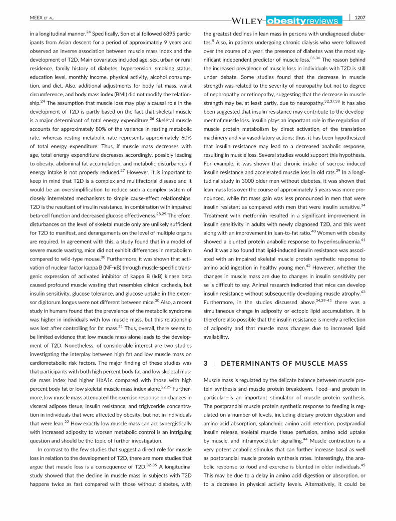
premature death (Figure 1).10-12 Given the steep rise in the number of
patients with T2D, the number of people that are affected by muscle
loss is expected to increase dramatically in the coming decades.
Studies estimated that approximately 80% of all individuals
with T2D suffer from overweight or obesity.13 This means there is
increased fat storage in subcutaneous and visceral fat depots,14,15 as
well as lipid accumulation in ectopic fat depots, including skeletal
FIGURE 1 Individuals with obesity and older individuals experience an inaffect metabolism in key organs including adipose tissue, liver, and skeletaresistance and a decrease in muscle mass. Patients often enter a vicious clead to more muscle loss, a deterioration in quality of life, and premature delipid metabolism. It will ameliorate muscle mass loss and the development oquality of life
muscle and liver.15-18 It is well established that obesity plays a crucial
role in the development of insulin resistance.19 In the past two
decades, an increasing number of papers also linked obesity to a
reduced muscle mass, and this phenomenon has been named
“sarcopenic obesity (Figure 1).”20 Sarcopenic obesity is now frequently
observed and led to the suggestion that obesity not only causes insulin
resistance but also plays a role in the development of muscle atro-
phy.21 If this is true, this would mean that insulin resistance and mus-
cle atrophy are “two sides of the same coin” and it would explain the
simultaneous occurrence of insulin resistance and muscle atrophy in
many patients with T2D. Notably, the combination of increased adi-
pose tissue mass and muscle atrophy may aggravate cardiometabolic
complications.22,23 In this review, we will discuss the relationship
between T2D and muscle mass loss, and we will discuss some of the
pathways throughwhich obesitymay affect insulin sensitivity andmuscle
mass. To date, data on the link betweenT2D and muscle loss is still in its
infancies, and a significant body of research comes from studies per-
formed in older individuals, who are also characterized by increased adi-
posity or ectopic fat accumulation, insulin resistance, and muscle loss.
Therefore, when information in patients with T2D is missing, observa-
tions made in older individuals will be reported. To conclude, this review
will also briefly discuss strategies as a way to counteract insulin resis-
tance and muscle mass loss.
2 | MUSCLE LOSS IN INDIVIDUALS WITHT2D: CAUSE OR CONSEQUENCE?
T2D and muscle atrophy develop hand in hand, and it has previously
been speculated that muscle loss might be a cause as well as a
consequence of T2D. Studies suggesting a causal role for muscle loss
in the development of metabolic disturbances are relatively scarce
though,24,25 and to our knowledge, there is only one study that evalu-
ated the association between low muscle mass and incidence of T2D
crease in lipid deposition in visceral and ectopic fat depots. This mayl muscle, which may result in the development of muscle insulinycle in which decreased activity levels and increased incidence of fallsath. Exercise (E) is an effective strategy to reduce obesity and improvef insulin resistance, and it will reduce the incidence of falls and improve
MEEX ET AL. 1207
in a longitudinal manner.24 Specifically, Son et al followed 6895 partic-
ipants from Asian descent for a period of approximately 9 years and
observed an inverse association between muscle mass index and the
development of T2D. Main covariates included age, sex, urban or rural
residence, family history of diabetes, hypertension, smoking status,
education level, monthly income, physical activity, alcohol consump-
tion, and diet. Also, additional adjustments for body fat mass, waist
circumference, and body mass index (BMI) did not modify the relation-
ship.24 The assumption that muscle loss may play a causal role in the
development of T2D is partly based on the fact that skeletal muscle
is a major determinant of total energy expenditure.26 Skeletal muscle
accounts for approximately 80% of the variance in resting metabolic
rate, whereas resting metabolic rate represents approximately 60%
of total energy expenditure. Thus, if muscle mass decreases with
age, total energy expenditure decreases accordingly, possibly leading
to obesity, abdominal fat accumulation, and metabolic disturbances if
energy intake is not properly reduced.27 However, it is important to
keep in mind that T2D is a complex and multifactorial disease and it
would be an oversimplification to reduce such a complex system of
closely interrelated mechanisms to simple cause‐effect relationships.
T2D is the resultant of insulin resistance, in combination with impaired
beta‐cell function and decreased glucose effectiveness.28,29 Therefore,
disturbances on the level of skeletal muscle only are unlikely sufficient
for T2D to manifest, and derangements on the level of multiple organs
are required. In agreement with this, a study found that in a model of
severe muscle wasting, mice did not exhibit differences in metabolism
compared to wild‐type mouse.30 Furthermore, it was shown that acti-
vation of nuclear factor kappa B (NF‐κB) throughmuscle‐specific trans-
genic expression of activated inhibitor of kappa B (IκB) kinase beta
caused profound muscle wasting that resembles clinical cachexia, but
insulin sensitivity, glucose tolerance, and glucose uptake in the exten-
sor digitorum longus were not different between mice.30 Also, a recent
study in humans found that the prevalence of the metabolic syndrome
was higher in individuals with low muscle mass, but this relationship
was lost after controlling for fat mass.31 Thus, overall, there seems to
be limited evidence that low muscle mass alone leads to the develop-
ment of T2D. Nonetheless, of considerable interest are two studies
investigating the interplay between high fat and low muscle mass on
cardiometabolic risk factors. The major finding of these studies was
that participants with both high percent body fat and low skeletal mus-
cle mass index had higher HbA1c compared with those with high
percent body fat or low skeletal muscle mass index alone.22,25 Further-
more, low muscle mass attenuated the exercise response on changes in
visceral adipose tissue, insulin resistance, and triglyceride concentra-
tion in individuals that were affected by obesity, but not in individuals
that were lean.22 How exactly low muscle mass can act synergistically
with increased adiposity to worsen metabolic control is an intriguing
question and should be the topic of further investigation.
In contrast to the few studies that suggest a direct role for muscle
loss in relation to the development of T2D, there are more studies that
argue that muscle loss is a consequence of T2D.32-35 A longitudinal
study showed that the decline in muscle mass in subjects with T2D
happens twice as fast compared with those without diabetes, with
the greatest declines in lean mass in persons with undiagnosed diabe-
tes.8 Also, in patients undergoing chronic dialysis who were followed
over the course of a year, the presence of diabetes was the most sig-
nificant independent predictor of muscle loss.35,36 The reason behind
the increased prevalence of muscle loss in individuals with T2D is still
under debate. Some studies found that the decrease in muscle
strength was related to the severity of neuropathy but not to degree
of nephropathy or retinopathy, suggesting that the decrease in muscle
strength may be, at least partly, due to neuropathy.32,37,38 It has also
been suggested that insulin resistance may contribute to the develop-
ment of muscle loss. Insulin plays an important role in the regulation of
muscle protein metabolism by direct activation of the translation
machinery and via vasodilatory actions; thus, it has been hypothesized
that insulin resistance may lead to a decreased anabolic response,
resulting in muscle loss. Several studies would support this hypothesis.
For example, it was shown that chronic intake of sucrose induced
insulin resistance and accelerated muscle loss in old rats.39 In a longi-
tudinal study in 3000 older men without diabetes, it was shown that
lean mass loss over the course of approximately 5 years was more pro-
nounced, while fat mass gain was less pronounced in men that were
insulin resistant as compared with men that were insulin sensitive.34
Treatment with metformin resulted in a significant improvement in
insulin sensitivity in adults with newly diagnosed T2D, and this went
along with an improvement in lean‐to‐fat ratio.40 Women with obesity
showed a blunted protein anabolic response to hyperinsulinaemia.41
And it was also found that lipid‐induced insulin resistance was associ-
ated with an impaired skeletal muscle protein synthetic response to
amino acid ingestion in healthy young men.42 However, whether the
changes in muscle mass are due to changes in insulin sensitivity per
se is difficult to say. Animal research indicated that mice can develop
insulin resistance without subsequently developing muscle atrophy.43
Furthermore, in the studies discussed above,34,39-42 there was a
simultaneous change in adiposity or ectopic lipid accumulation. It is
therefore also possible that the insulin resistance is merely a reflection
of adiposity and that muscle mass changes due to increased lipid
availability.
3 | DETERMINANTS OF MUSCLE MASS
Muscle mass is regulated by the delicate balance between muscle pro-
tein synthesis and muscle protein breakdown. Food—and protein in
particular—is an important stimulator of muscle protein synthesis.
The postprandial muscle protein synthetic response to feeding is reg-
ulated on a number of levels, including dietary protein digestion and
amino acid absorption, splanchnic amino acid retention, postprandial
insulin release, skeletal muscle tissue perfusion, amino acid uptake
by muscle, and intramyocellular signalling.44 Muscle contraction is a
very potent anabolic stimulus that can further increase basal as well
as postprandial muscle protein synthesis rates. Interestingly, the ana-
bolic response to food and exercise is blunted in older individuals.45
This may be due to a delay in amino acid digestion or absorption, or
to a decrease in physical activity levels. Alternatively, it could be

1208 MEEX ET AL.
caused by excess adiposity or circulating lipid levels. It is estimated
that approximately 80% of all individuals with T2D are affected by
overweight or obesity,13 which leads to increased lipid deposition in
visceral14,15 and ectopic fat depots, including skeletal muscle and
liver.15-18 In addition, many patients with T2D have decreased physi-
cal activity levels,46 which contributes to the accumulation of fat.
While ectopic fat accumulation in individuals with T2D is usually
the result of obesity, fat infiltration in organs in older individuals
can happen independent of weight changes.47 Nevertheless, there
are indications in both groups that expansion of adipose tissue, as
well as lipid accumulation in other organs, plays an important role in
the development of muscle atrophy. To test the hypothesis that the
development of anabolic resistance can be caused by excess lipids,
a study was performed in which young healthy volunteers received
a 7‐hour saline or intralipid infusion on two randomized occasions.42
The authors observed that excess lipid availability per se induced
insulin resistance of skeletal muscle glucose metabolism as well as
anabolic resistance of amino acid metabolism, and given the random-
ized crossover design, it could be concluded that this was indepen-
dently of any changes in amino acid handling or physical activity
levels.42 It is therefore likely that an increase in lipid levels in older
people and people with T2D contribute to the development of muscle
atrophy. In the following paragraphs, several possible pathways will
be discussed.
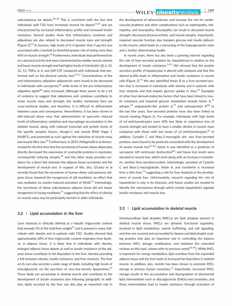
FIGURE 2 Schematic overview of interorgan crosstalk between adipose tdecrease of muscle mass. Increased lipid deposition in visceral and ectopiclead to a change in the secretion pattern of cytokines, which may lead to mcrosstalk. Lipids and cytokines may also affect mitochondrial function and varrows indicate pathways discussed in this review. Broken arrows indicate
3.1 | Expansion of the adipose tissue
Besides being a lipid buffering organ, adipose tissue is a major endo-
crine organ and is known to secrete hundreds of adipokines.48
Adipokines are important regulators of metabolism, and expansion of
adipose tissue leads to a change in the secretion of adipokines, which
have been linked to the development of insulin resistance in skeletal
muscle through interorgan crosstalk (Figure 2).49,50 Adipokines that
are well‐known to modulate metabolism and insulin sensitivity include
leptin,51 retinol binding protein 4 (RBP4),52 adiponectin,53 resistin,54
and pigment epithelium‐derived factor (PEDF).55 Macrophages are
also believed to be an important contributor of insulin resistance.56,57
Macrophages account for up to 40% of adipose cell content in indi-
viduals with obesity compared with only 10% in lean humans58 and
secrete pro‐inflammatory cytokines such as interleukin (IL) 6, IL1β,
granulocyte‐macrophage colony‐stimulating factor (GM‐CSF), and
tumour necrosis factor α (TNFα).59 In addition, macrophage infiltration
in adipose tissue leads to adipose tissue dysfunction, thereby contrib-
uting to the altered secretion pattern of adipokines. It is interesting to
note that the secretion pattern of adipokines and cytokines not only
changes with increased adiposity but also varies across fat depots.
Specifically, the secretion products of the mesenteric and omental adi-
pose tissue depots are more strongly associated with metabolic com-
plications of obesity compared with the secretion products of the
issue, liver, and skeletal muscle, leading to muscle insulin resistance andfat depots include skeletal muscle and liver. Increased lipid levels mayuscle insulin resistance and a decrease in muscle mass via interorgan
ascularization in skeletal muscle, which contributes to the problem. Fullpathways outside the scope of this review
MEEX ET AL. 1209
subcutaneous fat depots.60-62 This is consistent with the fact that
individuals with T2D have increased visceral fat depots14,15 and are
characterized by increased inflammatory profile and increased insulin
resistance. Several studies show that inflammatory cytokines and
adipokines are also related to decreased muscle mass and strength
(Figure 2).63 In humans, high levels of IL‐6 (greater than 5 pg/mL) was
associated with a twofold to threefold greater risk of losing more than
40% of muscle strength.64 Furthermore, individuals that performed low
on a physical activity test were characterized by smaller muscle volume
and lowermuscle strength and had higher levels of interleukin 1β, 6, 10,
12, 13, TNFα, IL‐6, and GM‐CSF compared with individuals that per-
formed well on the physical activity test.65-67 Concentrations of the
anti‐inflammatory adipokine adiponectin were found to be decreased
in individuals with sarcopenia,66 while levels of the pro‐inflammatory
adipokine leptin68 were increased. Although there seems to be a lot
of evidence to suggest that adipokines and cytokines contribute to
lower muscle mass and strength, the studies mentioned here are
cross‐sectional studies, and therefore, it is difficult to differentiate
between cause and consequence. Nevertheless, it has been shown in
diet‐induced obese mice that administration of quercetin reduced
levels of inflammatory cytokines and macrophage accumulation in the
skeletal muscle, along with reduced transcript and protein levels of
the specific atrophic factors, Atrogin‐1 and muscle RING finger 1
(MuRF1), and protected as such against the reduction of muscle mass
and muscle fibre size.69 Furthermore, in 2015, Pellegrinelli et al demon-
strated for the first time that the secretome of human obese adipocytes
directly decreased the expression of contractile proteins in myotubes,
consequently inducing atrophy,70 and this latter study provides evi-
dence for a direct link between the adipose tissue secretome and the
development of muscle loss. In support of this, also, O'Leary et al
recently found that the secretome of human obese subcutaneous adi-
pose tissue impaired the myogenesis of old myoblasts, an effect that
was mediated via resistin‐induced activation of NFκB.71 Interestingly,
the secretome of obese subcutaneous adipose tissue did not impair
myogenesis of young myoblasts,71 suggesting that the effect of obesity
on muscle mass may be particularly harmful in older individuals.
3.2 | Lipid accumulation in the liver
Liver steatosis is clinically defined as a hepatic triglyceride content
that exceeds 5% of the total liver weight72 and is present in many indi-
viduals with obesity and in patients with T2D. Studies showed that
approximately 60% of liver triglyceride content originates from lipoly-
sis in adipose tissue. It is likely that in individuals with obesity,
enlarged adipose tissue depots as well as insulin resistance of the adi-
pose tissue contribute to fat disposition in the liver, thereby providing
a link between obesity, insulin resistance, and liver steatosis. The liver
on its turn also secretes a wide range of lipids, including high levels of
triacylglycerols via the secretion of very‐low‐density lipoproteins.73
These lipids can accumulate in skeletal muscle and contribute to the
development of insulin resistance (see following paragraph). In addi-
tion, lipids secreted by the liver can also play an important role in
the development of atherosclerosis and increase the risk for cardio-
vascular problems and other complications such as nephropathy, reti-
nopathy, and neuropathy. Neuropathy can result in deceased muscle
strength, decreased physical activity, and muscle atrophy. Importantly,
impaired vascular function also hampers glucose and insulin delivery
to the muscle, which leads to a worsening of the hyperglycaemic state
and a further deteriorating health.
In recent years, there has also been a growing interest regarding
the role of liver‐secreted proteins (ie, hepatokines) in relation to the
development of insulin resistance.74,75 We showed that the protein
secretory profile of hepatocytes is altered with steatosis and that this
altered profile leads to inflammation and insulin resistance in muscle
cells (Figure 2).76 We also identified fetuin B as a liver‐secreted pro-
tein that is increased in individuals with obesity and in patients with
liver steatosis and that impairs glucose uptake in mice.76 Examples
of other liver‐derived endocrine factors that have been linked to insu-
lin resistance and impaired glucose metabolism include fetuin A,77
adropin,78 angiopoietin‐like protein 6,79 and selenoprotein P.80 In
the last few years, liver‐secreted proteins have also been linked to
muscle wasting (Figure 2). For example, individuals with high levels
of α1‐antichymotrypsin were 40% less likely to experience loss of
muscle strength and tended to have a smaller decline in muscle mass
compared with those with low levels of α1‐antichymotrypsin.64 In
addition, Cystatin C and Beta‐2‐macroglob ulin, two liver‐secreted
proteins, were found to be positively associated with the development
of severe muscle loss81-83; fetuin A was identified as a predictor of
sarcopenic left ventricular dysfunction84; and tissue iron levels were
elevated in muscle loss, which went along with an increase in transfer-
rin, another liver‐secreted protein. Interestingly, secretion of Cystatin
C and Beta‐2‐macroglobulin, fetuin A, and transferrin is increased
from a fatty liver,76 suggesting a role for liver steatosis in the develop-
ment of muscle loss. Unfortunately, research regarding the role of
hepatokines is only in its infancies, and future studies are needed to
identify the mechanisms through which certain hepatokines regulate
insulin resistance and muscle loss.
3.3 | Lipid accumulation in skeletal muscle
Intramyocellular lipid droplets (IMCLs) are lipid droplets present in
skeletal muscle tissue. IMCLs are dynamic functional organelles,
involved in lipid metabolism, vesicle trafficking, and cell signalling,
and they are covered and surrounded by lipases and lipid droplet coat-
ing proteins that play an important role in controlling the balance
between IMCL storage, mobilization, and oxidation (for extended
reviews on this topic, please refer to previous works85-89). While IMCL
is important for energy metabolism, lipid overflow from the expanded
adipose tissue and the liver leads to increased fat deposition in skeletal
muscle. In addition, also, resistin has been shown to promote IMCL
storage in primary human myotubes.71 Importantly, increased IMCL
storage results in the accumulation and dysregulation of detrimental
lipid intermediates such as diacylglycerols (DAGs) and ceramides, and
those intermediates lead to insulin resistance through activation of

1210 MEEX ET AL.
protein kinase C (PKC).90-100 High levels of DAG and ceramides have
also been suggested to play a role in the development of muscle
loss.16,101-103 In C2C12 and L6 myotubes, treatment with the fatty
acid palmitate induced ceramide accumulation, and this was associ-
ated with increased expression of pro‐atrophic genes such as
atrogin‐1/MAFbx, increased levels of FoxO3, upregulated eIF2α phos-
phorylation, and decreased protein synthesis.16,101,104 Conversely,
blocking ceramide synthesis prevented muscle atrophy, and this went
along with improved mammalian target of rapamycin (mTOR) signal-
ling, suppressed levels of Foxo3, and decreased atrogin‐1/MAFbx
expression.103 Rivas and colleagues found that obese animals had sig-
nificantly higher storage of ceramide and DAG compared with lean,
and there was an attenuated insulin response in components of the
mTOR anabolic signalling pathway.102 Interestingly, conversion of
DAG to phosphatidic acid (PA) activated the mTOR signalling pathway
and resulted in hypertrophy in isolated mouse extensor digitorum
longus muscle.105 In skeletal muscle of older humans, there were no
observable differences in total ceramide content; however, ceramide
species C16:0 was increased with 156% and C20:0‐ceramide was up
with 30%. In addition, there was a negative correlation between
C16:0‐ceramide content with lower leg lean mass and an attenuated
activation of anabolic signalling molecules such as Akt, FOXO1, and
S6K1 after an acute bout of high‐intensity resistance exercise.106
When ageing and obesity in mice occurred in combination, ceramide
accumulation was even more pronounced and negatively affected pro-
tein synthesis rate.16 Thus, there seems to be evidence that lipid inter-
mediates in skeletal muscle such as DAG's and ceramides negatively
affect insulin sensitivity as well as muscle mass. For completeness,
however, it should be mentioned that there are also studies that
report conflicting results regarding the role of lipid intermediates in
relation to insulin resistance as well as muscle loss. For example,
Turpin et al found that apoptosis in skeletal muscle myotubes was
induced by ceramides104 but could not find any signs of apoptosis,
autophagy, or proteolysis in high fat diet fed mice, in ob/ob mice, or
in mice after an intralipid infusion.107 Moreover, a number of studies
dissociated increased DAG levels from the development of insulin
resistance,108-110 suggesting that this relationship is not quite as
straightforward as previously thought. Possible explanations for these
inconsistencies include not taking into account the importance of
compartmentalization of the lipids, ignoring the structures, chain
lengths, and the degree of saturation of the lipids, or bypassing any
information on oxidation rates and fluxes. Either way, more research
will be needed to establish the exact role of IMCL and lipid intermedi-
ates in the development of insulin resistance and muscle loss.
3.4 | Mitochondrial dysfunction
Skeletal muscle mitochondrial dysfunction has previously been linked
to insulin resistance.111,112 Kelley et al observed mitochondrial abnor-
malities with respect to content, size, and morphology in patients with
T2D,113 while Mootha et al showed a lower gene expression of perox-
isome proliferator‐activated receptor gamma coactivator 1‐alpha
(PGC1α), a key transcriptional cofactor in mitochondrial biogenesis,
and a lower gene expression of its target genes encoding key enzymes
in oxidative mitochondrial metabolism.114 Decreased mitochondrial
function has also been reported in first degree relatives of patients
with T2D,111,115,116 suggesting that mitochondrial dysfunction may
actually be causally related to insulin resistance (Figure 2). It is inter-
esting to note that also age‐related muscle loss has also been associ-
ated with mitochondrial dysfunction. Specifically, age‐related muscle
loss has been linked to increased reactive oxygen species (ROS) pro-
duction, increased mitochondrial apoptotic susceptibility, decline in
mitochondrial respiratory chain function,117 reduced transcriptional
drive for mitochondrial biogenesis,118 and morphological changes in
mitochondria119; and also, most,120-124 but not all, studies125,126 state
that the capacity to mobilize and/or oxidize IMCL is substantially
impaired in older individuals. In congruence with this, it has recently
been suggested that accumulation of dysfunctional mitochondria initi-
ate a signalling cascade leading to motor neuron and muscle fibre
death and culminating in muscle loss (Figure 2).127 Not all studies
point in the same direction though, and a number of articles raised
doubts concerning mitochondrial dysfunction as a causal factor in
the development of insulin resistance and muscle atrophy.128-132 A
study reported normal in vivo mitochondrial function in Zucker dia-
betic rats throughout the pathogenesis of T2D130 and comparable
in vivo mitochondrial function was found between subjects with nor-
mal glycaemia, individuals with impaired glucose tolerance, and indi-
viduals with T2D.131 In addition to that, we showed that although
mitochondrial function was decreased in subjects with T2D, it could
be completely restored towards the levels of healthy control subjects
upon an exercise training programme, whereas insulin sensitivity could
only be partially restored.133 We also recently showed that 5 days of
muscle disuse was sufficient to induce substantial loss of both muscle
mass and strength in young and older subjects but did not reduce the
maximal activity of key mitochondrial enzymes.129 It seems fair to
state that mitochondrial dysfunction is present in many individuals
with T2D and may contribute to the development or progression of
insulin sensitivity and muscle atrophy. However, considering the mul-
tifactorial disease that T2D is, mitochondrial dysfunction cannot be
regarded as the main or sole causal factor in general.
4 | STRATEGIES TO COUNTERACT INSULINRESISTANCE AND MUSCLE LOSS
It may be clear that IMCL accumulation is strongly associated with the
severity of insulin resistance; however, this is only true for individuals
that are sedentary or untrained. Trained athletes are also character-
ized by elevated fat content in the muscle, but in contrast to their
sedentary counterparts, they are highly insulin sensitive.134-137
The precise mechanism by which trained persons are protected
from the insulin desensitizing effects of IMCL is still incompletely
understood, but here is substantial evidence that exercise enhances
the regulation of lipid droplet degradation and synthesis and increases
oxidative capacity, resulting in lower levels of detrimental lipid

MEEX ET AL. 1211
intermediates.138,139 Furthermore, exercise leads to improved insulin
sensitivity in adipose tissue, possibly resulting in lower levels of
uncontrolled fatty acid release,140 and thus less fatty acid spillover
to skeletal muscle and liver.141 Considering this, it is no wonder that
exercise is fundamental in the treatment of diabetes.142 Exercise can
delay and reduce the incidence of diabetes in persons at high risk by
58%143 and can improve (or even restore) abnormalities in oxidative
capacity, substrate selection (ie, metabolic flexibility),133 mitochondrial
function and density,133 capillary density, and lipid profile.144 Interest-
ing from a therapeutic point of view is the evidence that exercise is at
least as effective in older participants as it is in younger participants143
and that also patients on long‐standing insulin treatment can benefit
from exercise training.145 For example, in a group of patients with
long‐standingT2D on insulin therapy, 1 year of exercise training raised
in vivo skeletal muscle ATP production capacity by 21%, increased
expression levels of β‐oxidation, Krebs cycle, and oxidative phosphor-
ylation system‐related genes, and tended to increase mitochondrial
density and complex I activity,132 underscoring the importance of
exercise in the treatment of T2D. Also, nutritional intervention
programmes have been shown to be successful in preventing or
delaying the onset insulin resistance. Especially weight reduction has
been shown to lead to improved insulin sensitivity,146-148 suggesting
that nutritional treatment should primarily focus on achieving weight
loss. The underlying mechanism may differ from exercise though;
while exercise training leads to an improved mitochondrial function
but not to a decreased IMCL content, the opposite observation has
been made after weight loss.149 Importantly, exercise and nutritional
interventions are also key in the treatment of muscle loss. Muscle loss
is mainly attributed to a decrease in type 2 muscle fibers size,150-152
and it has been shown that 6 months of strength training can increase
type 2 fibers size up to 24%.150,153 In addition to that, resistance exer-
cise has been shown to prevent muscle atrophy and strength loss in
subjects undergoing step reduction154 and bed rest,155 while their
sedentary counterparts displayed a deterioration in muscle mass and
strength. Also, in very old adults in institutions, exercise has shown
to be effective156,157; a 6‐month exercise training programme
consisting of twice weekly resistance and balance exercises resulted
in a significant increase in grip strength, which may transfer to reduce
disability and muscle loss transition.156 Not only traditional high‐load
strength exercise has been shown to be effective in the treatment of
muscle loss. Low‐load, high‐volume exercise (30% one repetition max-
imum [1RM]) to volitional fatigue,158 low‐load blood flow restricted
exercise, and walking154,159 has also shown to stimulate muscle pro-
tein synthesis rate. Nutrition is often given in combination with exer-
cise to counter muscle loss, and interestingly, it has been shown that
they act synergistically to increase muscle protein synthesis.160 Impor-
tantly though, nutritional interventions should focus on obtaining fat
mass loss as well as on ways to improve protein synthesis and gain
lean mass. There are indications that older individuals and individuals
with obesity have a blunted muscle protein synthesis response upon
protein ingestion,161 and increasing both the quality and the dose of
protein might be an effective strategy to maximally stimulate
muscle protein synthesis.162-164 (For informative reviews regarding
the role of exercise and nutrition in relation muscle loss, see other
works165-167). It must be noted though that exercise as well as weight
loss can improve insulin sensitivity in patients with T2D without
increasing muscle mass.139,168 This is not necessarily surprising as it
is compatible with the multifactorial character of the disease. Apart
from ectopic lipid disposition, insulin sensitivity is also determined by
other parameters such as vascularization and oxidative capacity, and
these parameters can improve without absolute changes in muscle
mass. On a similar note, also, changes in muscle strength do not nec-
essarily go hand in hand with changes in muscle mass; longitudinal
studies previously showed that the decline in strength in patients with
T2D is much more rapid than the concomitant loss of muscle mass.169
In fact, changes in lean mass can only explain a small part (5%) of the
variability in strength decline, underscoring the importance of muscle
quality and the central neural and neuromuscular components deter-
mining muscle strength and function.169,170
Apart from exercise and nutrition, also, pharmacological interven-
tions are applied to treat patients with T2D. The use of metformin is
widespread among patients with T2D. Metformin ameliorates
hyperglycaemia via suppression of hepatic glucose production and
via improvements in peripheral glucose uptake, and although the exact
mode of action is not entirely clear, metformin is known for its weight‐
loss properties.171 Some mechanistic studies also demonstrated a pos-
itive effect on lipid accumulation in the liver. However, other studies
show that the effect on the liver was no longer present after correc-
tion for weight loss or that it could be attributed to the use of
suprapharmacological dosages of metformin.172 Also, multiple meta‐
analysis studies suggested that metformin has no effect on histological
responses in the liver.171,173,174 Nevertheless, due to its safe use and
its weight‐losing and insulin‐sensitizing properties, the use of metfor-
min remains the first‐line agent to treat insulin resistance in patients
withT2D. Another well‐known class of medication that improves insu-
lin sensitivity in individuals with T2D are the peroxisome proliferator‐
activated receptors gamma (PPARγ) agonists thiazolidinediones
(TZDs).175,176 Interestingly enough, TZD use leads to an increase in
extramyocellular lipid content and sometimes even weight gain.177,178
On the other hand, TZDs ameliorate ectopic fat storage and improve
lipid storage and metabolism in adipocytes,177 and this finding sup-
ports the notion that it is not the total amount of fat mass that is
important, but rather where and how the fat is stored. Unfortunately,
not all patients respond to TZDs, and its clinical use is also limited due
to adverse events,176 and therefore, other pharmacological therapies
should be explored. In the past few years, a lot of research has been
directed towards identifying new molecular mechanisms to target for
the treatment of nonalcoholic fatty liver disease (NAFLD). Potential
drug targets include microRNAs, incretin analogues/antagonists,
liver‐specific thyromimetics, AMP‐activated protein kinase activators,
farnesoid X receptor (FXR) and pregane X receptor (PXR) agonists,
fibroblasts growth factor 19 (FGF‐19) and fibroblasts growth factor
21 (FGF‐21) analogues, and antilipemic agents.179 There is evidence
that some of these drugs improve liver steatosis and fibrosis, and ame-
liorate lipid metabolism and insulin sensitivity in cellular and animal
models. However, (long‐term) efficiency has not yet been proven in

1212 MEEX ET AL.
humans, and some drugs come with severe side effects.179-181 There-
fore, more basic research and more randomized controlled trials of
adequate size and duration are needed in the search for novel treat-
ments. Also, pharmacological targets to treat muscle loss are sparse.
In 2014, a review summarized the impact of different therapeutic
interventions in relation to body composition changes in men and
females that were older and affected by obesity. The authors con-
cluded that weight loss based on diet combined with exercise—and
not drugs—represents the best strategy to treat phenotypic aspects
of sarcopenic obesity.182 A very recent study investigated systematic
reviews and meta‐analyses. The study included vitamin D, combined
oestrogen‐progesterone, dehydroepiandrosterone, growth hormone,
growth hormone‐releasing hormone, combined testosterone‐growth
hormone, insulin‐like growth factor‐1, pioglitazone, testosterone, and
angiotensin‐converting enzyme inhibitors. While the authors found
that testosterone and vitamin D mildly increased muscle mass in older
males and females respectively, this was only the case in individuals
with low starting values.183 Up to now, there is insufficient evidence
to recommend pharmacological interventions for the treatment of
muscle loss, and more research is needed in this field.183
5 | CONCLUSION
Many patientswithT2D are characterized by peripheral insulin resistance
and muscle atrophy. Derangements in lipid storage have been identified
as a hallmark of the development and progression of insulin resistance,
and an increasing amount of evidence suggest that it also plays an impor-
tant role in the development of muscle atrophy. The exact mechanisms
have not been established yet, but there is evidence that it may be via
the accumulation of harmful lipid intermediates in skeletal muscle,
decreased mitochondrial function, lipid accumulation in other organs, or
a combination of those. Unfortunately, research is often hampered by
the fact that many patients with T2D have decreased physical activity
levels compared with the rest of the population. In many studies, it is
therefore difficult to distinguish whether insulin resistance and muscle
loss are caused by the mechanisms mentioned in this review and/or by
a decrease in exercise per se. Furthermore, since insulin signalling plays
a permissive role in skeletal muscle amino acids delivery andmetabolism,
it is possible that insulin resistance may aggravate skeletal muscle ana-
bolic resistance. Future research will be needed to investigate the under-
lying mechanisms of muscle atrophy. This is important not only in the
setting of T2D but also in other conditions of lipid‐induced insulin resis-
tance, including ageing, disuse, and critical illness. With regard to drug
therapy, there are currently no well‐established guideline recommenda-
tions to reduce lipid accumulation in the liver or other organs. Results
of basic research over the past decade have been encouraging, and a
number of new molecular mechanisms have been explored. However,
most drugs pose long‐term safety issues, and so the biggest challenge lies
in the translation into the clinical setting. Up till now, nutritional and exer-
cise interventions are key in the treatment of T2D andmuscle loss, and if
possible, both should be applied to achieve optimal results.
CONFLICT OF INTEREST
The authors declare that there are no conflicts of interest regarding
the publication of this paper.
FINANCIAL SUPPORT
R.C.R.M. is supported by a “Marie Sklodowska Curie individual fellow-
ship” by the European Commission (H2020‐MSCA‐IF).
ORCID
Ruth C.R. Meex https://orcid.org/0000-0002-7247-9191
Ellen E. Blaak https://orcid.org/0000-0002-2496-3464
Luc J.C. van Loon https://orcid.org/0000-0002-6768-9231
REFERENCES
1. Collaboration NCDRF. Worldwide trends in diabetes since 1980: a
pooled analysis of 751 population‐based studies with 4.4 million par-
ticipants. Lancet. 2016;387:1513‐1530.
2. Cho NH, Shaw JE, Karuranga S, et al. IDF Diabetes Atlas: global esti-
mates of diabetes prevalence for 2017 and projections for 2045.
Diabetes Res Clin Pract. 2018;138:271‐281.
3. Park SW, Goodpaster BH, Strotmeyer ES, et al. Decreased muscle
strength and quality in older adults with type 2 diabetes: the health,
aging, and body composition study. Diabetes. 2006;55(6):1813‐1818.
4. Leenders M, Verdijk LB, van der Hoeven L, et al. Patients with type 2
diabetes show a greater decline in muscle mass, muscle strength,
and functional capacity with aging. J Am Med Dir Assoc.
2013;14(8):585‐592.
5. Guerrero N, Bunout D, Hirsch S, et al. Premature loss of muscle mass
and function in type 2 diabetes. Diabetes Res Clin Pract.
2016;117:32‐38.
6. von Haehling S, Morley JE, Anker SD. An overview of sarcopenia:
facts and numbers on prevalence and clinical impact. J Cachexia
Sarcopenia Muscle. 2010;1(2):129‐133.
7. Kim TN, Park MS, Yang SJ, et al. Prevalence and determinant factors
of sarcopenia in patients with type 2 diabetes: the Korean Sarcopenic
Obesity Study (KSOS). Diabetes Care. 2010;33(7):1497‐1499.
8. Park SW, Goodpaster BH, Strotmeyer ES, et al. Accelerated loss of
skeletal muscle strength in older adults with type 2 diabetes: the
health, aging, and body composition study. Diabetes Care.
2007;30(6):1507‐1512.
9. Kalyani RR, Saudek CD, Brancati FL, Selvin E. Association of diabetes,
comorbidities, and A1C with functional disability in older adults:
results from the National Health and nutrition examination survey
(NHANES), 1999‐2006. Diabetes Care. 2010;33(5):1055‐1060.
10. Landi F, Liperoti R, Russo A, et al. Sarcopenia as a risk factor for falls
in elderly individuals: results from the ilSIRENTE study. Clin Nutr.
2012;31(5):652‐658.
11. Fielding RA, Vellas B, Evans WJ, et al. Sarcopenia: an undiagnosed
condition in older adults. Current consensus definition: prevalence,
etiology, and consequences. International working group on
sarcopenia. J Am Med Dir Assoc. 2011;12(4):249‐256.
12. Ulley J, Abdelhafiz AH. Frailty predicts adverse outcomes in older
people with diabetes. Practitioner. 2017;261(1800):17‐20.
13. Lean ME. Obesity: burdens of illness and strategies for prevention or
management. Drugs Today (Barc). 2000;36(11):773‐784.

MEEX ET AL. 1213
14. Hirose H, Takayama M, IwaoY, Kawabe H. Effects of aging on visceral
and subcutaneous fat areas and on homeostasis model assessment of
insulin resistance and insulin secretion capacity in a comprehensive
health checkup. J Atheroscler Thromb. 2016;23(2):207‐215.
15. Cartwright MJ, Tchkonia T, Kirkland JL. Aging in adipocytes: potential
impact of inherent, depot‐specific mechanisms. Exp Gerontol.
2007;42(6):463‐471.
16. Tardif N, Salles J, Guillet C, et al. Muscle ectopic fat deposition con-
tributes to anabolic resistance in obese sarcopenic old rats through
eIF2α activation. Aging Cell. 2014;13(6):1001‐1011.
17. Zoico E, Rossi A, Di Francesco V, et al. Adipose tissue infiltration in
skeletal muscle of healthy elderly men: relationships with body com-
position, insulin resistance, and inflammation at the systemic and
tissue level. J Gerontol A Biol Sci Med Sci. 2010;65:295‐299.
18. Cree MG, Newcomer BR, Katsanos CS, et al. Intramuscular and liver
triglycerides are increased in the elderly. J Clin Endocrinol Metab.
2004;89(8):3864‐3871.
19. Abbasi F, Brown BW Jr, Lamendola C, McLaughlin T, Reaven GM.
Relationship between obesity, insulin resistance, and coronary heart
disease risk. J Am Coll Cardiol. 2002;40(5):937‐943.
20. Heber D, Ingles S, Ashley JM, Maxwell MH, Lyons RF, Elashoff RM.
Clinical detection of sarcopenic obesity by bioelectrical impedance
analysis. Am J Clin Nutr. 1996;64(3):472S‐477S.
21. Reinders I, Visser M, Schaap L. Body weight and body composition in
old age and their relationship with frailty. Curr Opin Clin Nutr Metab
Care. 2017;20(1):11‐15.
22. Terada T, Boule NG, Forhan M, et al. Cardiometabolic risk factors in
type 2 diabetes with high fat and low muscle mass: at baseline and
in response to exercise. Obesity (Silver Spring). 2017;25(5):881‐891.
23. Kim TN, Choi KM. The implications of sarcopenia and sarcopenic
obesity on cardiometabolic disease. J Cell Biochem. 2015;116(7):
1171‐1178.
24. Son JW, Lee SS, Kim SR, et al. Low muscle mass and risk of type 2 dia-
betes in middle‐aged and older adults: findings from the KoGES.
Diabetologia. 2017;60(5):865‐872.
25. Srikanthan P, Hevener AL, Karlamangla AS. Sarcopenia exacerbates
obesity‐associated insulin resistance and dysglycemia: findings from
the National Health and Nutrition Examination Survey III. PLoS ONE.
2010;5(5):e10805.
26. Ravussin E, Lillioja S, Anderson TE, Christin L, Bogardus C. Determi-
nants of 24‐hour energy expenditure in man. Methods and results
using a respiratory chamber. J Clin Invest. 1986;78(6):1568‐1578.
27. Karakelides H, Nair KS. Sarcopenia of aging and its metabolic impact.
Curr Top Dev Biol. 2005;68:123‐148.
28. Boston RC, Stefanovski D, Moate PJ, Sumner AE, Watanabe RM,
Bergman RN. MINMOD Millennium: a computer program to calculate
glucose effectiveness and insulin sensitivity from the frequently sam-
pled intravenous glucose tolerance test. Diabetes Technol Ther.
2003;5(6):1003‐1015.
29. Best JD, Kahn SE, Ader M, Watanabe RM, Ni TC, Bergman RN. Role
of glucose effectiveness in the determination of glucose tolerance.
Diabetes Care. 1996;19(9):1018‐1030.
30. Cai D, Frantz JD, Tawa NE Jr, et al. IKKbeta/NF‐kappaB activation
causes severe muscle wasting in mice. Cell. 2004;119(2):285‐298.
31. Koo HS, Kim MJ, Kim KM, Kim YS. Decreased muscle mass is not an
independent risk factor for metabolic syndrome in Korean population
aged 70 or older. Clin Endocrinol. 2015;82(4):509‐516.
32. Andersen H, Nielsen S, Mogensen CE, Jakobsen J. Muscle strength in
type 2 diabetes. Diabetes. 2004;53(6):1543‐1548.
33. Workeneh B, Bajaj M. The regulation of muscle protein turnover in
diabetes. Int J Biochem Cell Biol. 2013;45(10):2239‐2244.
34. Lee CG, Boyko EJ, Strotmeyer ES, et al. Association between insulin
resistance and lean mass loss and fat mass gain in older men without
diabetes mellitus. J Am Geriatr Soc. 2011;59(7):1217‐1224.
35. Pupim LB, Heimburger O, Qureshi AR, Ikizler TA, Stenvinkel P. Accel-
erated lean body mass loss in incident chronic dialysis patients with
diabetes mellitus. Kidney Int. 2005;68(5):2368‐2374.
36. Pupim LB, Flakoll PJ, Majchrzak KM, Aftab Guy DL, Stenvinkel P,
Ikizler TA. Increased muscle protein breakdown in chronic hemodialy-
sis patients with type 2 diabetes mellitus. Kidney Int.
2005;68(4):1857‐1865.
37. Kalyani RR, Metter EJ, Egan J, Golden SH, Ferrucci L. Hyperglycemia
predicts persistently lower muscle strength with aging. Diabetes Care.
2015;38(1):82‐90.
38. Andreassen CS, Jakobsen J, Ringgaard S, Ejskjaer N, Andersen H.
Accelerated atrophy of lower leg and foot muscles—a follow‐up study
of long‐term diabetic polyneuropathy using magnetic resonance
imaging (MRI). Diabetologia. 2009;52(6):1182‐1191.
39. Gatineau E, Savary‐Auzeloux I, Migne C, Polakof S, Dardevet D,
Mosoni L. Chronic intake of sucrose accelerates sarcopenia in older
male rats through alterations in insulin sensitivity and muscle protein
synthesis. J Nutr. 2015;145(5):923‐930.
40. Aghili R, Malek M, Valojerdi AE, Banazadeh Z, Najafi L, Khamseh ME.
Body composition in adults with newly diagnosed type 2 diabetes:
effects of metformin. J Diabetes Metab Disord. 2014;13:88.
41. Chevalier S, Marliss EB, Morais JA, Lamarche M, Gougeon R. Whole‐body protein anabolic response is resistant to the action of insulin in
obese women. Am J Clin Nutr. 2005;82:355‐365.
42. Stephens FB, Chee C, Wall BT, et al. Lipid‐induced insulin resistance
is associated with an impaired skeletal muscle protein synthetic
response to amino acid ingestion in healthy young men. Diabetes.
2015;64(5):1615‐1620.
43. Ostler JE, Maurya SK, Dials J, et al. Effects of insulin resistance on
skeletal muscle growth and exercise capacity in type 2 diabetic mouse
models. Am J Physiol Endocrinol Metab. 2014;306(6):E592‐E605.
44. Gorissen SH, Remond D, van Loon LJ. The muscle protein synthetic
response to food ingestion. Meat Sci. 2015;109:96‐100.
45. Burd NA, Gorissen SH, van Loon LJ. Anabolic resistance of muscle
protein synthesis with aging. Exerc Sport Sci Rev. 2013;41:169‐173.
46. Crespo CJ, Keteyian SJ, Heath GW, Sempos CT. Leisure‐time physical
activity among US adults: results from the Third National Health and
Nutrition Examination Survey. Arch Intern Med. 1996;156:93‐98.
47. Delmonico MJ, HarrisTB, Visser M, et al. Longitudinal study of muscle
strength, quality, and adipose tissue infiltration. Am J Clin Nutr.
2009;90(6):1579‐1585.
48. Lehr S, Hartwig S, Lamers D, et al. Identification and validation of
novel adipokines released from primary human adipocytes. Mol Cell
Proteomics. 2012;11(1):M111 010504.
49. Ebert T, Roth I, Richter J, et al. Different associations of adipokines in
lean and healthy adults. Horm Metab Res. 2014;46(1):41‐47.
50. Fasshauer M, Bluher M. Adipokines in health and disease. Trends
Pharmacol Sci. 2015;36(7):461‐470.
51. Friedman JM, Halaas JL. Leptin and the regulation of body weight in
mammals. Nature. 1998;395:763‐770.
52. Yang Q, Graham TE, Mody N, et al. Serum retinol binding protein 4
contributes to insulin resistance in obesity and type 2 diabetes.
Nature. 2005;436(7049):356‐362.

1214 MEEX ET AL.
53. Yamauchi T, Kamon J, Minokoshi Y, et al. Adiponectin stimulates glu-
cose utilization and fatty‐acid oxidation by activating AMP‐activatedprotein kinase. Nat Med. 2002;8(11):1288‐1295.
54. Steppan CM, Bailey ST, Bhat S, et al. The hormone resistin links obe-
sity to diabetes. Nature. 2001;409(6818):307‐312.
55. Crowe S, Wu LE, Economou C, et al. Pigment epithelium‐derivedfactor contributes to insulin resistance in obesity. Cell Metab.
2009;10(1):40‐47.
56. Boutens L, Stienstra R. Adipose tissue macrophages: going off track
during obesity. Diabetologia. 2016;59(5):879‐894.
57. Xu H, Barnes GT, Yang Q, et al. Chronic inflammation in fat plays a
crucial role in the development of obesity‐related insulin resistance.
J Clin Invest. 2003;112(12):1821‐1830.
58. Weisberg SP, McCann D, Desai M, Rosenbaum M, Leibel RL, Ferrante
AW Jr. Obesity is associated with macrophage accumulation in adi-
pose tissue. J Clin Invest. 2003;112:1796‐1808.
59. Heilbronn LK, Liu B. Do adipose tissue macrophages promote insulin
resistance or adipose tissue remodelling in humans? Horm Mol Biol
Clin Invest. 2014;20:3‐13.
60. Mazaki‐Tovi M, Bolin SR, Schenck PA. Differential secretion of
adipokines from subcutaneous and visceral adipose tissue in healthy
dogs: association with body condition and response to troglitazone.
Vet J. 2016;216:136‐141.
61. Kranendonk ME, van Herwaarden JA, Stupkova T, et al. Inflammatory
characteristics of distinct abdominal adipose tissue depots relate
differently to metabolic risk factors for cardiovascular disease: dis-
tinct fat depots and vascular risk factors. Atherosclerosis.
2015;239(2):419‐427.
62. Fried SK, Bunkin DA, Greenberg AS. Omental and subcutaneous adi-
pose tissues of obese subjects release interleukin‐6: depot differenceand regulation by glucocorticoid. J Clin Endocrinol Metab.
1998;83:847‐850.
63. Kalinkovich A, Livshits G. Sarcopenic obesity or obese sarcopenia: a
cross talk between age‐associated adipose tissue and skeletal muscle
inflammation as a main mechanism of the pathogenesis. Ageing Res
Rev. 2017;35:200‐221.
64. Schaap LA, Pluijm SM, Deeg DJ, Visser M. Inflammatory markers and
loss of muscle mass (sarcopenia) and strength. Am J Med.
2006;119(6):526.e9‐526.e17.
65. Calvani R, Marini F, Cesari M, et al. Systemic inflammation, body com-
position, and physical performance in old community‐dwellers. J
Cachexia Sarcopenia Muscle. 2017;8(1):69‐77.
66. Can B, Kara O, Kizilarslanoglu MC, et al. Serum markers of inflamma-
tion and oxidative stress in sarcopenia. Aging Clin Exp Res.
2017;29(4):745‐752.
67. Toth MJ, Ades PA, Tischler MD, Tracy RP, LeWinter MM. Immune
activation is associated with reduced skeletal muscle mass and phys-
ical function in chronic heart failure. Int J Cardiol. 2006;109:179‐187.
68. Kohara K, Ochi M, Tabara Y, Nagai T, Igase M, Miki T. Leptin in
sarcopenic visceral obesity: possible link between adipocytes and
myocytes. PLoS ONE. 2011;6:e24633.
69. Le NH, Kim CS, Park T, et al. Quercetin protects against obesity‐induced skeletal muscle inflammation and atrophy. Mediat Inflamm.
2014;2014:834294.
70. Pellegrinelli V, Rouault C, Rodriguez‐Cuenca S, et al. Human adipo-
cytes induce inflammation and atrophy in muscle cells during
obesity. Diabetes. 2015;64(9):3121‐3134.
71. O'Leary MF, Wallace GR, Davis ET, et al. Obese subcutaneous adi-
pose tissue impairs human myogenesis, particularly in old skeletal
muscle, via resistin‐mediated activation of NFκB. Sci Rep.
2018;8(1):15360.
72. Kleiner DE, Brunt EM, van Natta M, et al. Design and validation of a
histological scoring system for nonalcoholic fatty liver disease.
Hepatology. 2005;41(6):1313‐1321.
73. Peng KY, Watt MJ, Rensen S, et al. Mitochondrial dysfunction‐relatedlipid changes occur in nonalcoholic fatty liver disease progression. J
Lipid Res. 2018;59(10):1977‐1986.
74. Meex RCR, Watt MJ. Hepatokines: linking nonalcoholic fatty liver dis-
ease and insulin resistance. Nat Rev Endocrinol. 2017;13:509‐520.
75. Stefan N, Haring HU. The role of hepatokines in metabolism. Nat Rev
Endocrinol. 2013;9:144‐152.
76. Meex RC, Hoy AJ, Morris A, et al. Fetuin B is a secreted hepatocyte
factor linking steatosis to impaired glucose metabolism. Cell Metab.
2015;22(6):1078‐1089.
77. Pal D, Dasgupta S, Kundu R, et al. Fetuin‐A acts as an endogenous
ligand of TLR4 to promote lipid‐induced insulin resistance. Nat Med.
2012;18(8):1279‐1285.
78. Kumar KG, Trevaskis JL, Lam DD, et al. Identification of adropin as a
secreted factor linking dietary macronutrient intake with energy
homeostasis and lipid metabolism. Cell Metab. 2008;8(6):468‐481.
79. OikeY, Akao M, Yasunaga K, et al. Angiopoietin‐related growth factor
antagonizes obesity and insulin resistance. Nat Med.
2005;11(4):400‐408.
80. Misu H, TakamuraT, Takayama H, et al. A liver‐derived secretory pro-
tein, selenoprotein P, causes insulin resistance. Cell Metab.
2010;12(5):483‐495.
81. Kim H, Suzuki T, Kim M, et al. Incidence and predictors of sarcopenia
onset in community‐dwelling elderly Japanese women: 4‐year follow‐up study. J Am Med Dir Assoc. 2015;16(1):85.e1‐85.e8.
82. Kim JK, Choi SR, Choi MJ, et al. Prevalence of and factors associated
with sarcopenia in elderly patients with end‐stage renal disease. Clin
Nutr. 2014;33(1):64‐68.
83. Lippi G, Sanchis‐Gomar F, Montagnana M. Biological markers in older
people at risk of mobility limitations. Curr Pharm Des.
2014;20(19):3222‐3244.
84. Chang WT, Tsai WC, Wu CH, et al. Fetuin‐A as a predicator of
sarcopenic left ventricular dysfunction. Sci Rep. 2015;5(1):12078.
85. Badin PM, Langin D, Moro C. Dynamics of skeletal muscle lipid pools.
Trends Endocrinol Metab. 2013;24(12):607‐615.
86. Bosma M. Lipid droplet dynamics in skeletal muscle. Exp Cell Res.
2016;340:180‐186.
87. Meex RC, Schrauwen P, Hesselink MK. Modulation of myocellular fat
stores: lipid droplet dynamics in health and disease. Am J Physiol Regul
Integr Comp Physiol. 2009;297:R913‐R924.
88. MacPherson RE, Peters SJ. Piecing together the puzzle of perilipin
proteins and skeletal muscle lipolysis. Appl Physiol Nutr Metab.
2015;40:641‐651.
89. Stinkens R, Goossens GH, Jocken JW, Blaak EE. Targeting fatty acid
metabolism to improve glucose metabolism. Obes Rev.
2015;16(9):715‐757.
90. Conte M, Vasuri F, Bertaggia E, et al. Differential expression of
perilipin 2 and 5 in human skeletal muscle during aging and their asso-
ciation with atrophy‐related genes. Biogerontology. 2015;16(3):
329‐340.
91. Aquilano K, Baldelli S, La Barbera L, Lettieri Barbato D, Tatulli G,
Ciriolo MR. Adipose triglyceride lipase decrement affects skeletal
muscle homeostasis during aging through FAs‐PPARα‐PGC‐1α anti-
oxidant response. Oncotarget. 2016;7:23019‐23032.

MEEX ET AL. 1215
92. Cho KA, Kang PB. PLIN2 inhibits insulin‐induced glucose uptake in
myoblasts through the activation of the NLRP3 inflammasome. Int J
Mol Med. 2015;36:839‐844.
93. Szendroedi J, Yoshimura T, Phielix E, et al. Role of diacylglycerol acti-
vation of PKCθ in lipid‐induced muscle insulin resistance in humans.
Proc Natl Acad Sci U S A. 2014;111(26):9597‐9602.
94. Boon J, Hoy AJ, Stark R, et al. Ceramides contained in LDL are ele-
vated in type 2 diabetes and promote inflammation and skeletal
muscle insulin resistance. Diabetes. 2013;62(2):401‐410.
95. Badin PM, Louche K, Mairal A, et al. Altered skeletal muscle lipase
expression and activity contribute to insulin resistance in humans.
Diabetes. 2011;60(6):1734‐1742.
96. Straczkowski M, Kowalska I, Baranowski M, et al. Increased skeletal
muscle ceramide level in men at risk of developing type 2 diabetes.
Diabetologia. 2007;50(11):2366‐2373.
97. Thrush AB, Brindley DN, Chabowski A, Heigenhauser GJ, Dyck DJ.
Skeletal muscle lipogenic protein expression is not different between
lean and obese individuals: a potential factor in ceramide accumula-
tion. J Clin Endocrinol Metab. 2009;94(12):5053‐5061.
98. Liu L, Zhang Y, Chen N, Shi X, Tsang B, Yu YH. Upregulation of
myocellular DGAT1 augments triglyceride synthesis in skeletal muscle
and protects against fat‐induced insulin resistance. J Clin Invest.
2007;117:1679‐1689.
99. Shulman GI. Cellular mechanisms of insulin resistance. J Clin Invest.
2000;106:171‐176.
100. Jocken JW, Goossens GH, Boon H, et al. Insulin‐mediated suppres-
sion of lipolysis in adipose tissue and skeletal muscle of obese type
2 diabetic men and men with normal glucose tolerance. Diabetologia.
2013;56(10):2255‐2265.
101. Bryner RW, Woodworth‐Hobbs ME, Williamson DL, Alway SE.
Docosahexaenoic acid protects muscle cells from palmitate‐inducedatrophy. ISRN Obes. 2012;2012:647348.
102. Rivas DA, McDonald DJ, Rice NP, Haran PH, Dolnikowski GG, Fiel-
ding RA. Diminished anabolic signaling response to insulin induced
by intramuscular lipid accumulation is associated with inflammation
in aging but not obesity. Am J Physiol Regul Integr Comp Physiol.
2016;310(7):R561‐R569.
103. de Larichaudy J, Zufferli A, Serra F, et al. TNF‐α‐ and tumor‐inducedskeletal muscle atrophy involves sphingolipid metabolism. Skelet Mus-
cle. 2012;2(1):2.
104. Turpin SM, Lancaster GI, Darby I, Febbraio MA, Watt MJ. Apoptosis
in skeletal muscle myotubes is induced by ceramides and is positively
related to insulin resistance. Am J Physiol Endocrinol Metab. 2006;291:
E1341‐E1350.
105. You JS, Lincoln HC, Kim CR, et al. The role of diacylglycerol kinase
zeta and phosphatidic acid in the mechanical activation of mammalian
target of rapamycin (mTOR) signaling and skeletal muscle hypertro-
phy. J Biol Chem. 2014;289(3):1551‐1563.
106. Rivas DA, Morris EP, Haran PH, et al. Increased ceramide content and
NFκB signaling may contribute to the attenuation of anabolic signal-
ing after resistance exercise in aged males. J Appl Physiol (1985).
2012;113(11):1727‐1736.
107. Turpin SM, Ryall JG, Southgate R, et al. Examination of ‘lipotoxicity’ inskeletal muscle of high‐fat fed and ob/ob mice. J Physiol.
2009;587(7):1593‐1605.
108. Amati F. Revisiting the diacylglycerol‐induced insulin resistance
hypothesis. Obes Rev. 2012;13(Suppl 2):40‐50.
109. Amati F, Dube JJ, Alvarez‐Carnero E, et al. Skeletal muscle triglycer-
ides, diacylglycerols, and ceramides in insulin resistance: another
paradox in endurance‐trained athletes? Diabetes. 2011;60(10):
2588‐2597.
110. Dube JJ, Amati F, Toledo FG, et al. Effects of weight loss and exercise
on insulin resistance, and intramyocellular triacylglycerol, diacylglyc-
erol and ceramide. Diabetologia. 2011;54(5):1147‐1156.
111. Phielix E, Schrauwen‐Hinderling VB, Mensink M, et al. Lower intrinsic
ADP‐stimulated mitochondrial respiration underlies in vivo mitochon-
drial dysfunction in muscle of male type 2 diabetic patients. Diabetes.
2008;57(11):2943‐2949.
112. Mogensen M, Sahlin K, Fernstrom M, et al. Mitochondrial respiration
is decreased in skeletal muscle of patients with type 2 diabetes.
Diabetes. 2007;56(6):1592‐1599.
113. Kelley DE, He J, Menshikova EV, Ritov VB. Dysfunction of mitochon-
dria in human skeletal muscle in type 2 diabetes. Diabetes.
2002;51:2944‐2950.
114. Mootha VK, Lindgren CM, Eriksson KF, et al. PGC‐1α‐responsivegenes involved in oxidative phosphorylation are coordinately down-
regulated in human diabetes. Nat Genet. 2003;34(3):267‐273.
115. Petersen KF, Dufour S, Befroy D, Garcia R, Shulman GI. Impaired
mitochondrial activity in the insulin‐resistant offspring of patients
with type 2 diabetes. N Engl J Med. 2004;350:664‐671.
116. Befroy DE, Petersen KF, Dufour S, et al. Impaired mitochondrial sub-
strate oxidation in muscle of insulin‐resistant offspring of type 2
diabetic patients. Diabetes. 2007;56(5):1376‐1381.
117. Cooper JM, Mann VM, Schapira AH. Analyses of mitochondrial respi-
ratory chain function and mitochondrial DNA deletion in human
skeletal muscle: effect of ageing. J Neurol Sci. 1992;113(1):91‐98.
118. Chabi B, Ljubicic V, Menzies KJ, Huang JH, Saleem A, Hood DA.
Mitochondrial function and apoptotic susceptibility in aging skeletal
muscle. Aging Cell. 2008;7(1):2‐12.
119. Leduc‐Gaudet JP, Picard M, St‐Jean Pelletier F, et al. Mitochondrial
morphology is altered in atrophied skeletal muscle of aged mice.
Oncotarget. 2015;6(20):17923‐17937.
120. Blaak EE, van Baak MA, Saris WH. Beta‐adrenergically stimulated fat
oxidation is diminished in middle‐aged compared to young subjects. J
Clin Endocrinol Metab. 1999;84(10):3764‐3769.
121. Sial S, Coggan AR, Carroll R, Goodwin J, Klein S. Fat and carbohydrate
metabolism during exercise in elderly and young subjects. Am J Phys.
1996;271:E983‐E989.
122. Toth MJ, Tchernof A. Lipid metabolism in the elderly. Eur J Clin Nutr.
2000;54(Suppl 3):S121‐S125.
123. Calles‐Escandon J, Arciero PJ, Gardner AW, Bauman C, Poehlman ET.
Basal fat oxidation decreases with aging in women. J Appl Physiol
(1985). 1995;78(1):266‐271.
124. Calles‐Escandon J, Poehlman ET, Garcia‐Rubi E. Lipolysis in elderly
postmenopausal women. Metabolism. 1997;46(11):1312‐1315.
125. Melanson EL, Donahoo WT, Grunwald GK, Schwartz R. Changes in
24‐h substrate oxidation in older and younger men in response to
exercise. J Appl Physiol (1985). 2007;103:1576‐1582.
126. Bonadonna RC, Groop LC, Simonson DC, DeFronzo RA. Free fatty
acid and glucose metabolism in human aging: evidence for operation
of the Randle cycle. Am J Phys. 1994;266(3 Pt 1):E501‐E509.
127. Alway SE, Mohamed JS, Myers MJ. Mitochondria initiate and regulate
sarcopenia. Exerc Sport Sci Rev. 2017;45:58‐69.
128. Barrientos A, Casademont J, Rotig A, et al. Absence of relationship
between the level of electron transport chain activities and aging in
human skeletal muscle. Biochem Biophys Res Commun.
1996;229(2):536‐539.

1216 MEEX ET AL.
129. Dirks ML, Wall BT, Nilwik R, Weerts DH, Verdijk LB, van Loon LJ.
Skeletal muscle disuse atrophy is not attenuated by dietary protein
supplementation in healthy older men. J Nutr. 2014;144:1196‐1203.
130. de Feyter HM, Lenaers E, Houten SM, et al. Increased
intramyocellular lipid content but normal skeletal muscle mitochon-
drial oxidative capacity throughout the pathogenesis of type 2
diabetes. FASEB J. 2008;22(11):3947‐3955.
131. de Feyter HM, van den Broek NM, Praet SF, Nicolay K, van Loon LJ,
Prompers JJ. Early or advanced stage type 2 diabetes is not accompa-
nied by in vivo skeletal muscle mitochondrial dysfunction. Eur J
Endocrinol. 2008;158:643‐653.
132. van Tienen FH, Praet SF, de Feyter HM, et al. Physical activity is the
key determinant of skeletal muscle mitochondrial function in type 2
diabetes. J Clin Endocrinol Metab. 2012;97(9):3261‐3269.
133. Meex RC, Schrauwen‐Hinderling VB, Moonen‐Kornips E, et al. Resto-ration of muscle mitochondrial function and metabolic flexibility in
type 2 diabetes by exercise training is paralleled by increased
myocellular fat storage and improved insulin sensitivity. Diabetes.
2009;59:572‐579.
134. Goodpaster BH, He J, Watkins S, Kelley DE. Skeletal muscle lipid con-
tent and insulin resistance: evidence for a paradox in endurance‐trained athletes. J Clin Endocrinol Metab. 2001;86:5755‐5761.
135. Schrauwen‐Hinderling VB, Schrauwen P, Hesselink MK, et al. The
increase in intramyocellular lipid content is a very early response to
training. J Clin Endocrinol Metab. 2003;88(4):1610‐1616.
136. Tarnopolsky MA, Rennie CD, Robertshaw HA, Fedak‐TarnopolskySN, Devries MC, Hamadeh MJ. Influence of endurance exercise train-
ing and sex on intramyocellular lipid and mitochondrial ultrastructure,
substrate use, and mitochondrial enzyme activity. Am J Physiol Regul
Integr Comp Physiol. 2007;292:R1271‐R1278.
137. van Loon LJ, Koopman R, Manders R, van der Weegen W, van
Kranenburg GP, Keizer HA. Intramyocellular lipid content in type 2
diabetes patients compared with overweight sedentary men and
highly trained endurance athletes. Am J Physiol Endocrinol Metab.
2004;287:E558‐E565.
138. Zacharewicz E, Hesselink MKC, Schrauwen P. Exercise counteracts
lipotoxicity by improving lipid turnover and lipid droplet quality. J
Intern Med. 2018;284:505‐518.
139. Meex RC, Schrauwen‐Hinderling VB, Moonen‐Kornips E, et al. Resto-ration of muscle mitochondrial function and metabolic flexibility in
type 2 diabetes by exercise training is paralleled by increased
myocellular fat storage and improved insulin sensitivity. Diabetes.
2010;59(3):572‐579.
140. Hickner RC, Racette SB, Binder EF, Fisher JS, Kohrt WM. Effects of
10 days of endurance exercise training on the suppression of whole
body and regional lipolysis by insulin. J Clin Endocrinol Metab.
2000;85:1498‐1504.
141. Brouwers B, Schrauwen‐Hinderling VB, Jelenik T, et al. Exercise train-
ing reduces intrahepatic lipid content in people with and people
without nonalcoholic fatty liver. Am J Physiol Endocrinol Metab.
2018;314(2):E165‐E173.
142. Albright A, Franz M, Hornsby G, et al. American College of Sports
Medicine position stand. Exercise and type 2 diabetes. Med Sci Sports
Exerc. 2000;32(7):1345‐1360.
143. Knowler WC, Barrett‐Connor E, Fowler SE, et al. Reduction in the
incidence of type 2 diabetes with lifestyle intervention or metformin.
N Engl J Med. 2002;346:393‐403.
144. Sigal RJ, Kenny GP, Boule NG, et al. Effects of aerobic training, resis-
tance training, or both on glycemic control in type 2 diabetes: a
randomized trial. Ann Intern Med. 2007;147(6):357‐369.
145. Praet SF, Jonkers RA, Schep G, et al. Long‐standing, insulin‐treatedtype 2 diabetes patients with complications respond well to short‐term resistance and interval exercise training. Eur J Endocrinol.
2008;158(2):163‐172.
146. Weyer C, Hanson K, Bogardus C, Pratley RE. Long‐term changes in
insulin action and insulin secretion associated with gain, loss, regain
and maintenance of body weight. Diabetologia. 2000;43:36‐46.
147. Schenk S, Harber MP, Shrivastava CR, Burant CF, Horowitz JF.
Improved insulin sensitivity after weight loss and exercise training is
mediated by a reduction in plasma fatty acid mobilization, not
enhanced oxidative capacity. J Physiol. 2009;587:4949‐4961.
148. Penn L, White M, Lindstrom J, et al. Importance of weight loss main-
tenance and risk prediction in the prevention of type 2 diabetes:
analysis of European diabetes prevention study RCT. PLoS ONE.
2013;8(2):e57143.
149. Toledo FG, Menshikova EV, Azuma K, et al. Mitochondrial capacity in
skeletal muscle is not stimulated by weight loss despite increases in
insulin action and decreases in intramyocellular lipid content. Diabe-
tes. 2008;57(4):987‐994.
150. Nilwik R, Snijders T, Leenders M, et al. The decline in skeletal muscle
mass with aging is mainly attributed to a reduction in type II muscle
fiber size. Exp Gerontol. 2013;48(5):492‐498.
151. Larsson L, Sjodin B, Karlsson J. Histochemical and biochemical
changes in human skeletal muscle with age in sedentary males, age
22‐‐65 years. Acta Physiol Scand. 1978;103:31‐39.
152. Lexell J, Henriksson‐Larsen K, Winblad B, Sjostrom M. Distribution of
different fiber types in human skeletal muscles: effects of aging stud-
ied in whole muscle cross sections. Muscle Nerve. 1983;6:588‐595.
153. Leenders M, Verdijk LB, van der Hoeven L, van Kranenburg J, Nilwik
R, van Loon LJ. Elderly men and women benefit equally from
prolonged resistance‐type exercise training. J Gerontol A Biol Sci
Med Sci. 2013;68:769‐779.
154. Devries MC, Breen L, von Allmen M, et al. Low‐load resistance train-
ing during step‐reduction attenuates declines in muscle mass and
strength and enhances anabolic sensitivity in older men. Physiol Rep.
2015;3(8):e12493.
155. Bamman MM, Clarke MS, Feeback DL, et al. Impact of resistance
exercise during bed rest on skeletal muscle sarcopenia and myosin
isoform distribution. J Appl Physiol (1985). 1998;84(1):157‐163.
156. Hassan BH, Hewitt J, Keogh JW, Bermeo S, Duque G, Henwood TR.
Impact of resistance training on sarcopenia in nursing care facilities: a
pilot study. Geriatr Nurs. 2015.
157. Stewart VH, Saunders DH, Greig CA. Responsiveness of muscle size
and strength to physical training in very elderly people: a systematic
review. Scand J Med Sci Sports. 2014;24:e1‐e10.
158. Burd NA, West DW, Staples AW, et al. Low‐load high volume resis-
tance exercise stimulates muscle protein synthesis more than high‐load low volume resistance exercise in young men. PLoS ONE.
2010;5(8):e12033.
159. Breen L, Stokes KA, Churchward‐Venne TA, et al. Two weeks of
reduced activity decreases leg lean mass and induces “anabolic resis-
tance” of myofibrillar protein synthesis in healthy elderly. J Clin
Endocrinol Metab. 2013;98(6):2604‐2612.
160. Churchward‐Venne TA, Burd NA, Mitchell CJ, et al. Supplementation
of a suboptimal protein dose with leucine or essential amino acids:
effects on myofibrillar protein synthesis at rest and following resis-
tance exercise in men. J Physiol. 2012;590(11):2751‐2765.
161. Murphy J, Chevalier S, Gougeon R, Goulet ED, Morais JA. Effect of
obesity and type 2 diabetes on protein anabolic response to insulin
in elderly women. Exp Gerontol. 2015;69:20‐26.
MEEX ET AL. 1217
162. Pennings B, Groen B, de Lange A, et al. Amino acid absorption and
subsequent muscle protein accretion following graded intakes of
whey protein in elderly men. Am J Physiol Endocrinol Metab.
2012;302(8):E992‐E999.
163. Wall BT, Hamer HM, de Lange A, et al. Leucine co‐ingestion improves
post‐prandial muscle protein accretion in elderly men. Clin Nutr.
2013;32(3):412‐419.
164. Pennings B, Boirie Y, Senden JM, Gijsen AP, Kuipers H, van Loon LJ.
Whey protein stimulates postprandial muscle protein accretion more
effectively than do casein and casein hydrolysate in older men. Am J
Clin Nutr. 2011;93:997‐1005.
165. Churchward‐Venne TA, Breen L, Phillips SM. Alterations in human
muscle protein metabolism with aging: protein and exercise as coun-
termeasures to offset sarcopenia. Biofactors. 2014;40:199‐205.
166. Phillips SM. Nutritional supplements in support of resistance exercise
to counter age‐related sarcopenia. Adv Nutr. 2015;6(4):452‐460.
167. Wall BT, Cermak NM, van Loon LJ. Dietary protein considerations to
support active aging. Sports Med. 2014;44(Suppl 2):S185‐S194.
168. Roberts MD, Haun CT, Mobley CB, et al. Physiological differences
between low versus high skeletal muscle hypertrophic responders
to resistance exercise training: current perspectives and future
research directions. Front Physiol. 2018;9:834.
169. Goodpaster BH, Carlson CL, Visser M, et al. Attenuation of skeletal
muscle and strength in the elderly: The Health ABC Study. J Appl
Physiol (1985). 2001;90(6):2157‐2165.
170. Goodpaster BH, Park SW, Harris TB, et al. The loss of skeletal muscle
strength, mass, and quality in older adults: the health, aging and body
composition study. J Gerontol A Biol Sci Med Sci. 2006;61(10):
1059‐1064.
171. Musso G, Cassader M, Rosina F, Gambino R. Impact of current treat-
ments on liver disease, glucose metabolism and cardiovascular risk in
non‐alcoholic fatty liver disease (NAFLD): a systematic review and
meta‐analysis of randomised trials. Diabetologia. 2012;55:885‐904.
172. Erion DM, Park HJ, Lee HY. The role of lipids in the pathogenesis and
treatment of type 2 diabetes and associated co‐morbidities. BMB Rep.
2016;49:139‐148.
173. Li Y, Liu L, Wang B, Wang J, Chen D. Metformin in non‐alcoholic fattyliver disease: a systematic review and meta‐analysis. Biomed Rep.
2013;1:57‐64.
174. Rakoski MO, Singal AG, Rogers MA, Conjeevaram H. Meta‐analysis:insulin sensitizers for the treatment of non‐alcoholic steatohepatitis.
Aliment Pharmacol Ther. 2010;32:1211‐1221.
175. Nolan JJ, Ludvik B, Beerdsen P, Joyce M, Olefsky J. Improvement in
glucose tolerance and insulin resistance in obese subjects treated
with troglitazone. N Engl J Med. 1994;331:1188‐1193.
176. Cariou B, Charbonnel B, Staels B. Thiazolidinediones and PPARγ ago-
nists: time for a reassessment. Trends Endocrinol Metab. 2012;23:
205‐215.
177. Mayerson AB, Hundal RS, Dufour S, et al. The effects of rosiglitazone
on insulin sensitivity, lipolysis, and hepatic and skeletal muscle triglyc-
eride content in patients with type 2 diabetes. Diabetes.
2002;51(3):797‐802.
178. Kelly IE, Han TS, Walsh K, Lean ME. Effects of a thiazolidinedione
compound on body fat and fat distribution of patients with type 2
diabetes. Diabetes Care. 1999;22(2):288‐293.
179. Musso G, Gambino R, Cassader M. Emerging molecular targets for
the treatment of nonalcoholic fatty liver disease. Annu Rev Med.
2010;61:375‐392.
180. Fiorucci S, Biagioli M, Distrutti E. Future trends in the treatment of
non‐alcoholic steatohepatitis. Pharmacol Res. 2018;134:289‐298.
181. Musso G, Gambino R, Cassader M, Pagano G. A meta‐analysis of ran-domized trials for the treatment of nonalcoholic fatty liver disease.
Hepatology. 2010;52:79‐104.
182. Poggiogalle E, Migliaccio S, Lenzi A, Donini LM. Treatment of body
composition changes in obese and overweight older adults: insight
into the phenotype of sarcopenic obesity. Endocrine.
2014;47:699‐716.
183. de Spiegeleer A, Beckwee D, Bautmans I, Petrovic M. Sarcopenia
guidelines development group of the Belgian Society of G, geriatrics.
Pharmacological interventions to improve muscle mass, muscle
strength and physical performance in older people: an umbrella
review of systematic reviews and meta‐analyses. Drugs Aging.
2018;35(8):719‐734.
How to cite this article: Meex RCR, Blaak EE, van Loon LJC.
Lipotoxicity plays a key role in the development of both
insulin resistance and muscle atrophy in patients with type 2
diabetes. Obesity Reviews. 2019;20:1205–1217. https://doi.
org/10.1111/obr.12862