Embed Size (px)
Citation preview
LIS1 controls mitosis and mitotic spindleorganization via the LIS1–NDEL1–dynein complex
Hyang Mi Moon1,2,{, Yong Ha Youn1,{, Hayley Pemble3,4, Jessica Yingling6, Torsten Wittmann3
and Anthony Wynshaw-Boris1,5,∗,§
1Department of Pediatrics, Institute for Human Genetics, 2Biomedical Sciences Graduate Program, 3Department of Cell
and Tissue Biology, 4Tetrad Graduate Program and 5Eli and Edythe Broad Center of Regenerative Medicine and Stem
CellResearch,UniversityofCalifornia,SanFrancisco,CA94143,USA 6DepartmentofPediatrics,UniversityofCalifornia,
San Diego, CA 92122, USA
Received September 2, 2013; Revised and Accepted September 4, 2013
Heterozygous LIS1 mutations are responsible for the human neuronal migration disorder lissencephaly. Mitoticfunctions of LIS1 have been suggested from many organisms throughout evolution. However, the cellular func-tionsof LIS1 at distinct intracellular compartments such as the centrosome and the cell cortexhavenot beenwelldefinedespeciallyduringmitoticcelldivision.Here,weuseddetailedcellularapproaches andtime-lapse livecellimaging of mitosis from Lis1 mutant mouse embryonic fibroblasts to reveal critical roles of LIS1 in mitotic spin-dle regulation. We found that LIS1 is required for the tight control of chromosome congression and segregationto dictate kinetochore–microtubule (MT) interactions and anaphase progression. In addition, LIS1 is essentialfor the establishment of mitotic spindle pole integrity by maintaining normal centrosome number. Moreover,LIS1 plays crucial roles in mitotic spindle orientation by increasing the density of astral MT plus-end movementstoward the cell cortex, which enhances cortical targeting of LIS1–dynein complex. Overexpression of NDEL1–dynein and MT stabilization rescues spindle orientation defects in Lis1 mutants, demonstrating that mouse LIS1acts via the LIS1–NDEL1–dynein complex to regulate astral MT plus-ends dynamics and establish proper con-tacts of MTs with the cell cortex to ensure precise cell division.
INTRODUCTION
Mitotic cell divisions are essential for the accurate partitioning ofgenetic material into two daughter cells. Inappropriate segrega-tion of chromosomes during mitosis leads to aneuploidy andgenomic instability (1). During the mitotic phase of the cellcycle (M phase), microtubules (MTs) undergo dynamic reorgan-ization to coordinate chromosome separation. Mitotic spindlesare assembled by dramatic MT remodeling and emanate fromthe centrosome, an MT-organizing center, also called thespindle pole (2). The centrosome participates in MT nucleationand anchoring MT minus-ends. The core component of thecentrosome is a centriole pair composed of a mother centrioleand a daughter centriole, which recruits pericentriolar materialcomponents (3,4). The centrosome duplication cycle is precisely
controlled to preserve centrosome number and proper centrioleassembly (5). Importantly, mammalian cell division planes aremainly determined by the positioning of bipolar mitotic spindles(6). In addition, the spatiotemporal interactions between the cellcortex and astral MT plus-ends have critical roles in mitoticspindle regulation (7–9). Several MT plus-end binding proteinsmediate dynamic contacts of astral MT plus-ends to the cellcortex by interacting with cortical force generators on themembrane (10).
Many of the proteins important for mitosis have been discov-ered, although much of the detailed mechanisms employed byeach protein involved in cell division remains to be understood.Among those mitotically important proteins, LIS1 is part of acomplex that interacts with diverse cortical factors and centroso-mal proteins at kinetochores on the chromosomes, the mitotic
†Present address: Department of Neurosurgery, Stanford University, Stanford CA 94305, USA.‡Present address: Department of Developmental Neurobiology, St Jude Children’s Research Hospital, Memphis TN 38105, USA.
∗To whom correspondence should be addressed. Tel: +1 2163680581; Fax: +1 2163683832; Email: [email protected]
§Present address: Department of Genetics and Genome Sciences, Case Western Reserve University, School of Medicine, 10900 Euclid Avenue, BRB731Cleveland OH 44106-4955, USA.
# The Author 2013. Published by Oxford University Press. All rights reserved.For Permissions, please email: [email protected]
Human Molecular Genetics, 2014, Vol. 23, No. 2 449–466doi:10.1093/hmg/ddt436Advance Access published on September 12, 2013
Downloaded from https://academic.oup.com/hmg/article-abstract/23/2/449/663111by gueston 06 April 2018
spindles and the cell cortex, and it has been implicated in theregulation of the mitotic spindles and chromosome segregationduring mitosis (11–13). Human LIS1 was first identified as acausative gene of human lissencephaly (‘smooth brain’), asevere neuro-developmental disease (14,15). Heterozygous mu-tation or deletion of human LIS1 leads to this brain malformationdue to defects in neuronal migration. LIS1 is also part of a highlyconserved protein complex first discovered in Aspergillus nidu-lans that is responsible for nuclear distribution (NUD) and func-tions in cytoplasmic dynein regulation (16,17). LIS1homologues from Aspergillus to mammals form a complexwith cytoplasmic dynein and NUD proteins (18–20). Cytoplas-mic dynein is a MT minus-end-directed motor involved inmitotic spindle assembly by regulating MT dynamics especiallyat astral MTs and mediating poleward transport of spindle as-sembly checkpoint proteins (21–25). Through its motor activity,cytoplasmic dynein exerts pulling forces on the chromosomes.Dynactin, an accessory linker protein complexed with dyneinsubunits, also contributes to these cellular functions by assistingcargo loading and increasing processivity (26). Corticallyanchored cytoplasmic dynein/dynactin complexes are importantcortical force generators along MTs (24,27) that are essential formitotic spindle formation and positioning in M phase (21,28),and LIS1 has been implicated in dynein targeting at MTplus-ends along astral MTs during cell division of various celltypes (11,20,29). In addition, several NUD family proteins asso-ciate with both LIS1 and cytoplasmic dynein. Two mammalianNudE homologues, NDE1 and NDEL1, interact with LIS1/cyto-plasmic dynein complex (19,30–34). NDE1 and NDEL1 displayprominent centrosomal localization, as does LIS1 (19,32,35).NDE1 is required for targeting of LIS1 to the cytoplasmicdynein complex to generate persistent motor forces (36,37),while NDEL1 has been implicated in the process of LIS1/dynein recruitment, serving as a scaffold (11,38,39). In addition,a subset of NDE1 and NDEL1 proteins is observed in close prox-imity to the cell cortex where LIS1 accumulates (13,40,41).These previous studies support the notion that the LIS1–NDE1/NDEL1–dynein/dynactin complex is likely part of theprotein machinery needed to coordinate various signals fromthe cell cortex to the mitotic spindles by generating pullingforces on spindle MTs. Despite these studies, the precise func-tions of LIS1 and its complex during mitosis remain elusive. Fur-thermore, it is unclear whether other components of LIS1 proteincomplex are involved in LIS1-dependent mitotic spindle regula-tion during mammalian cell division.
We took advantage of genetic null (knock-out, KO) andhypomorphic-conditional (HC) alleles of Lis1 (42,43) touncover the critical dose-dependent roles of LIS1 in mouse em-bryonic fibroblasts (MEFs) and mouse neural progenitors (NPs).Previously, we found that Lis1 deficiency in mouse brainsresulted in apoptosis and mitotic spindle orientation defects inNPs, while in MEFs loss of Lis1 led to severe defects in cellgrowth and MT capture at the cell cortex in interphase cells(13). In the current study, we used Lis1 mutant MEFs and per-formed time-lapse live cell imaging of mitotic progression com-pared with WT MEFs, to examine the functions of LIS1 duringmitosis. We also analyzed mitotic spindle organization indetail by examining centrosome integrity and number. Toaddress the mechanism of LIS1-dependent spindle regulation,we overexpressed several candidate protein complexed with
LIS1 in Lis1 mutant MEFs and tested whether any of them canrescue spindle misorientation defects caused by Lis1-deficiency.Importantly, we performed time-lapse live cell imaging of MTplus-ends in Lis1 mutant MEFs to explore changes in the dynam-ics of astral MTs reaching to the cell cortex. Thus, by using bothgenetic and conditional Lis1 mutant MEFs with reduced LIS1protein amount, we investigated essential functions of LIS1complex to form proper mitotic spindle during mammaliancell division.
RESULTS
Perturbed mitotic progression in mitosis of Lis1 mutantMEFs
To investigate cellular functions of mouse LIS1 during mitoticcell division, we performed time-lapse live cell imaging ofmitosis of Lis1 mutant MEFs isolated from Lis1 mutant condi-tional knock-out (CKO) mice that harbor HC alleles of mouseLis1. Tamoxifen (TM)-inducible Cre (44) mice were mated toproduce homozygous Lis1 CKO mice (CreERTM;Lis1hc/hc, here-after termed Lis1-CKO-Cre) to acutely delete the Lis1 gene uponTM treatment, which reduced the LIS1 protein level to ,10% ofwild-type (WT) levels after 72 h incubation, as described in ourprevious study (13). Control mice contained two normal WTLis1 alleles with the Cre transgene (CreERTM;Lis1+/+, hereaftertermed WT-Cre). To visualize dynamic movements of chromo-somes and MTs simultaneously during mitotic progression, weinfected MEFs with retroviruses encoding histone 2B(H2B)-GFP (45) and mCherry–a-tubulin, followed by treat-ment with 4-hydroxy-TM for 12 h. Live cell imaging revealedthat Lis1-CKO-Cre MEFs treated with TM (CreERTM;Lis1hc/hc
+ TM) exhibited a high frequency of abnormalities duringmitosis that were not observed in WT-Cre MEFs (Supplemen-tary Material, Video S1). In prometaphase, Lis1-CKO-CreMEFs often formed extra centrosomes (e.g. four centrosomes)(Fig. 1A and Supplementary Material, Video S2) (See below).Loss of LIS1 frequently resulted in a kinked and curved morph-ology of the mitotic spindle. As the mitotic cell cycle eventuallyproceeded into metaphase, Lis1-CKO-Cre MEFs displayedchromosomes roughly aligned near the metaphase plate alongwith pseudo-bipolar spindles. In anaphase, Lis1-CKO-CreMEFs retained several misaligned and unattached chromo-somes, which ultimately resulted in chromosome missegrega-tion and lagging chromosomes in telophase. The average timefrom nuclear envelope breakdown to metaphase plate formationwas not significantly extended in Lis1-CKO-Cre MEFs (17.2+0.8 min) compared with those of WT-Cre MEFs (16.2+0.9 min), indicating that acute loss of LIS1 did not severelyimpair the timing of metaphase plate formation (Fig. 1B).However, the average time from metaphase to anaphase onsetwas significantly delayed from Lis1-CKO-Cre MEFs (9.9+0.9 min) compared with those of WT-Cre MEFs (6.1+0.9 min) (Fig. 1C). We obtained similar results of mitoticdelay from a different analysis of anaphase onset timing thatdetermines the accumulative percentage of cells that enter ana-phase. The time when 50% cells entered anaphase was longerin Lis1-CKO-Cre MEFs (t50% ¼ �10 min) than WT-CreMEFs (t50% ¼ �5 min) by 2-fold (Fig. 1E). The total durationof mitosis from nuclear envelope breakdown to anaphase onset
450 Human Molecular Genetics, 2014, Vol. 23, No. 2
Downloaded from https://academic.oup.com/hmg/article-abstract/23/2/449/663111by gueston 06 April 2018
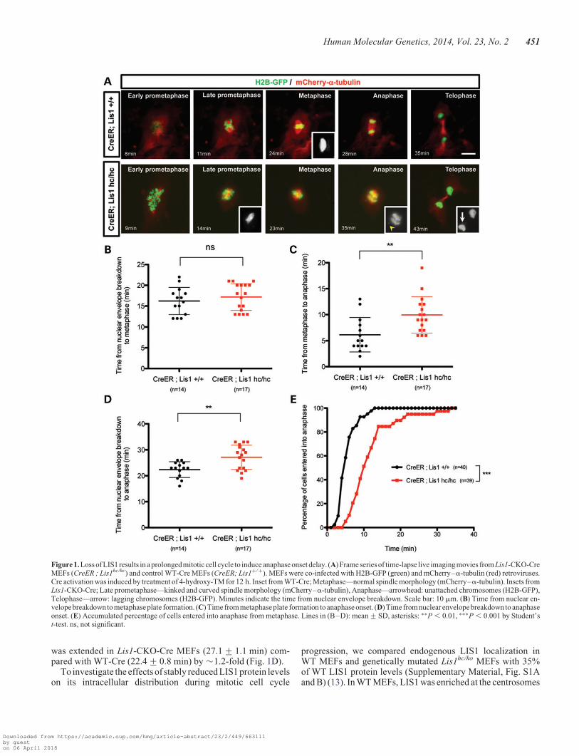
was extended in Lis1-CKO-Cre MEFs (27.1+ 1.1 min) com-pared with WT-Cre (22.4+ 0.8 min) by �1.2-fold (Fig. 1D).
To investigate the effects of stably reduced LIS1 protein levelson its intracellular distribution during mitotic cell cycle
progression, we compared endogenous LIS1 localization inWT MEFs and genetically mutated Lis1hc/ko MEFs with 35%of WT LIS1 protein levels (Supplementary Material, Fig. S1Aand B) (13). In WT MEFs, LIS1 was enriched at the centrosomes
Figure 1. Loss of LIS1 results in a prolongedmitotic cell cycle to induce anaphase onset delay. (A) Frame series of time-lapse live imaging movies from Lis1-CKO-CreMEFs (CreER ; Lis1hc/hc) and control WT-Cre MEFs (CreER; Lis1+/+). MEFs were co-infected with H2B-GFP (green) and mCherry–a-tubulin (red) retroviruses.Cre activation was induced by treatment of 4-hydroxy-TM for 12 h. Inset from WT-Cre; Metaphase—normal spindle morphology (mCherry–a-tubulin). Insets fromLis1-CKO-Cre; Late prometaphase—kinked and curved spindle morphology (mCherry–a-tubulin), Anaphase—arrowhead: unattached chromosomes (H2B-GFP),Telophase—arrow: lagging chromosomes (H2B-GFP). Minutes indicate the time from nuclear envelope breakdown. Scale bar: 10 mm. (B) Time from nuclear en-velopebreakdown to metaphase plate formation. (C) Time from metaphase plate formation to anaphase onset. (D) Time from nuclear envelope breakdown to anaphaseonset. (E) Accumulated percentage of cells entered into anaphase from metaphase. Lines in (B–D): mean+SD, asterisks: ∗∗P , 0.01, ∗∗∗P , 0.001 by Student’st-test. ns, not significant.
Human Molecular Genetics, 2014, Vol. 23, No. 2 451
Downloaded from https://academic.oup.com/hmg/article-abstract/23/2/449/663111by gueston 06 April 2018
and the kinetochores from prometaphase to metaphase. A major-ity of LIS1 was cytosolic, but a small fraction of LIS1 was alsoobserved near the cell cortex as puncta. From metaphase to ana-phase, LIS1 immunostaining overlapped with MTs along themitotic spindles. Prior to telophase, a majority of LIS1co-localized with the centrosomes in WT MEFs. In contrast,Lis1hc/ko MEFs displayed significant reduction in LIS1 distribu-tion in the cytoplasm, although centrosome-specific localizationof LIS1 appeared to be maintained. In metaphase, LIS1 punctaaccumulated at several kinetochores near the metaphase platein Lis1hc/ko MEFs. Importantly, mitotic spindle-associatedLIS1 mostly disappeared in these Lis1hc/ko MEFs (Supplemen-tary Material, Fig. S1A). Together, these observations supportcritical mitotic functions of mouse LIS1 at the centrosome, themitotic spindle and the cell cortex in M phase (12), suggestingthat the LIS1 protein complex may mediate mitotic functionsin these specific intracellular compartments.
Formation of extra centrosomes caused by loss of Lis1
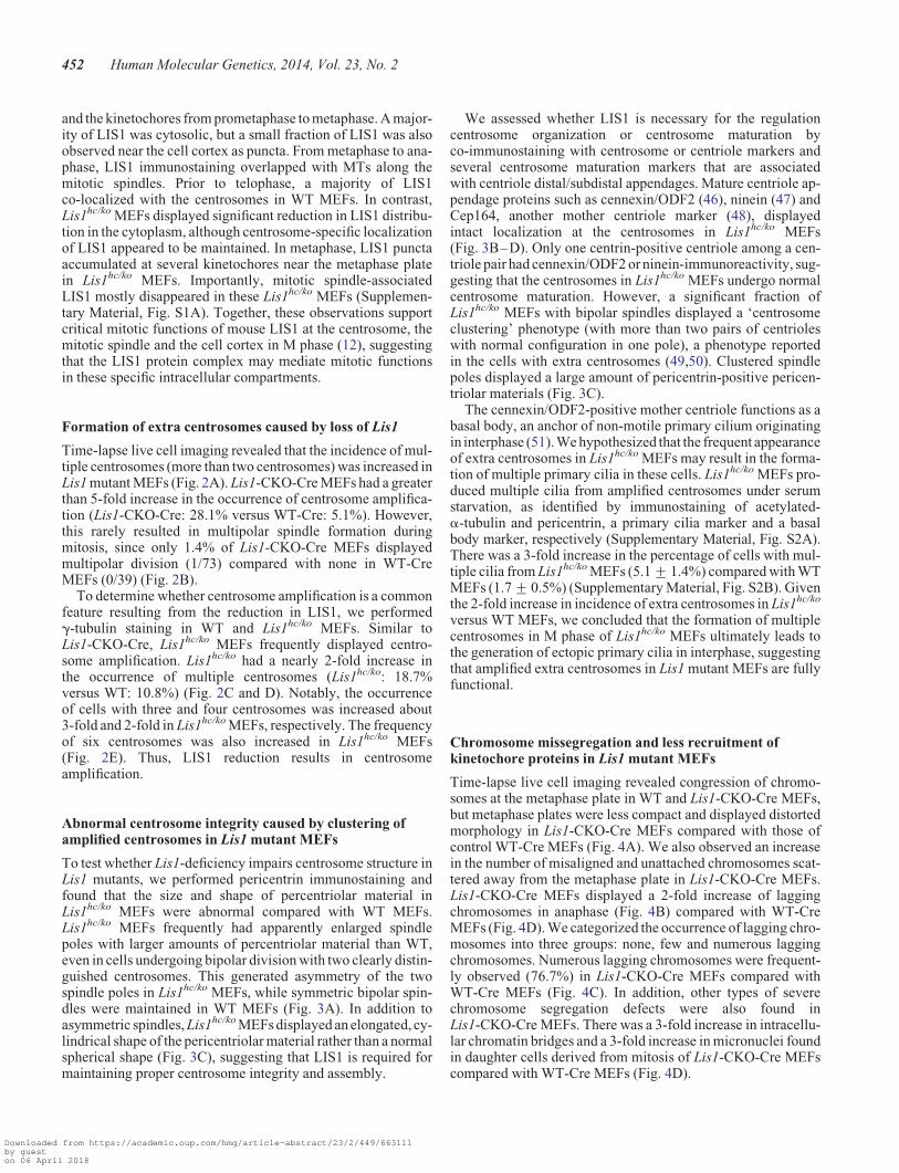
Time-lapse live cell imaging revealed that the incidence of mul-tiple centrosomes (more than two centrosomes) was increased inLis1 mutant MEFs (Fig. 2A). Lis1-CKO-Cre MEFs had a greaterthan 5-fold increase in the occurrence of centrosome amplifica-tion (Lis1-CKO-Cre: 28.1% versus WT-Cre: 5.1%). However,this rarely resulted in multipolar spindle formation duringmitosis, since only 1.4% of Lis1-CKO-Cre MEFs displayedmultipolar division (1/73) compared with none in WT-CreMEFs (0/39) (Fig. 2B).
To determine whether centrosome amplification is a commonfeature resulting from the reduction in LIS1, we performedg-tubulin staining in WT and Lis1hc/ko MEFs. Similar toLis1-CKO-Cre, Lis1hc/ko MEFs frequently displayed centro-some amplification. Lis1hc/ko had a nearly 2-fold increase inthe occurrence of multiple centrosomes (Lis1hc/ko: 18.7%versus WT: 10.8%) (Fig. 2C and D). Notably, the occurrenceof cells with three and four centrosomes was increased about3-fold and 2-fold in Lis1hc/ko MEFs, respectively. The frequencyof six centrosomes was also increased in Lis1hc/ko MEFs(Fig. 2E). Thus, LIS1 reduction results in centrosomeamplification.
Abnormal centrosome integrity caused by clustering ofamplified centrosomes in Lis1 mutant MEFs
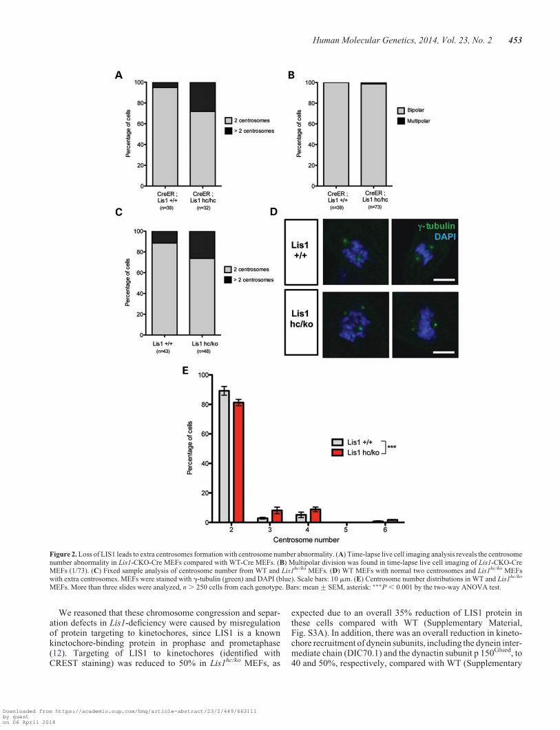
To test whether Lis1-deficiency impairs centrosome structure inLis1 mutants, we performed pericentrin immunostaining andfound that the size and shape of percentriolar material inLis1hc/ko MEFs were abnormal compared with WT MEFs.Lis1hc/ko MEFs frequently had apparently enlarged spindlepoles with larger amounts of percentriolar material than WT,even in cells undergoing bipolar division with two clearly distin-guished centrosomes. This generated asymmetry of the twospindle poles in Lis1hc/ko MEFs, while symmetric bipolar spin-dles were maintained in WT MEFs (Fig. 3A). In addition toasymmetric spindles, Lis1hc/ko MEFs displayed an elongated, cy-lindrical shape of the pericentriolar material rather than a normalspherical shape (Fig. 3C), suggesting that LIS1 is required formaintaining proper centrosome integrity and assembly.
We assessed whether LIS1 is necessary for the regulationcentrosome organization or centrosome maturation byco-immunostaining with centrosome or centriole markers andseveral centrosome maturation markers that are associatedwith centriole distal/subdistal appendages. Mature centriole ap-pendage proteins such as cennexin/ODF2 (46), ninein (47) andCep164, another mother centriole marker (48), displayedintact localization at the centrosomes in Lis1hc/ko MEFs(Fig. 3B–D). Only one centrin-positive centriole among a cen-triole pair had cennexin/ODF2 or ninein-immunoreactivity, sug-gesting that the centrosomes in Lis1hc/ko MEFs undergo normalcentrosome maturation. However, a significant fraction ofLis1hc/ko MEFs with bipolar spindles displayed a ‘centrosomeclustering’ phenotype (with more than two pairs of centrioleswith normal configuration in one pole), a phenotype reportedin the cells with extra centrosomes (49,50). Clustered spindlepoles displayed a large amount of pericentrin-positive pericen-triolar materials (Fig. 3C).
The cennexin/ODF2-positive mother centriole functions as abasal body, an anchor of non-motile primary cilium originatingin interphase (51). We hypothesized that the frequent appearanceof extra centrosomes in Lis1hc/ko MEFs may result in the forma-tion of multiple primary cilia in these cells. Lis1hc/ko MEFs pro-duced multiple cilia from amplified centrosomes under serumstarvation, as identified by immunostaining of acetylated-a-tubulin and pericentrin, a primary cilia marker and a basalbody marker, respectively (Supplementary Material, Fig. S2A).There was a 3-fold increase in the percentage of cells with mul-tiple cilia from Lis1hc/ko MEFs (5.1+ 1.4%) compared with WTMEFs (1.7+ 0.5%) (Supplementary Material, Fig. S2B). Giventhe 2-fold increase in incidence of extra centrosomes in Lis1hc/ko
versus WT MEFs, we concluded that the formation of multiplecentrosomes in M phase of Lis1hc/ko MEFs ultimately leads tothe generation of ectopic primary cilia in interphase, suggestingthat amplified extra centrosomes in Lis1 mutant MEFs are fullyfunctional.
Chromosome missegregation and less recruitment ofkinetochore proteins in Lis1 mutant MEFs
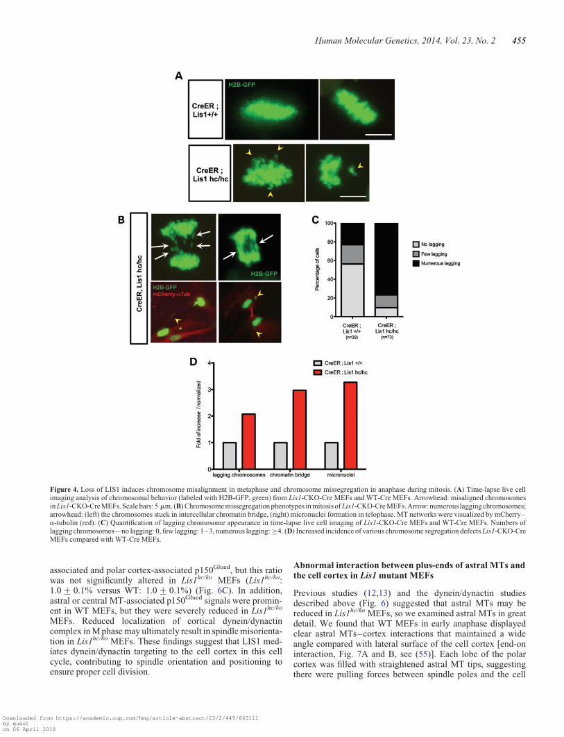
Time-lapse live cell imaging revealed congression of chromo-somes at the metaphase plate in WT and Lis1-CKO-Cre MEFs,but metaphase plates were less compact and displayed distortedmorphology in Lis1-CKO-Cre MEFs compared with those ofcontrol WT-Cre MEFs (Fig. 4A). We also observed an increasein the number of misaligned and unattached chromosomes scat-tered away from the metaphase plate in Lis1-CKO-Cre MEFs.Lis1-CKO-Cre MEFs displayed a 2-fold increase of laggingchromosomes in anaphase (Fig. 4B) compared with WT-CreMEFs (Fig. 4D). We categorized the occurrence of lagging chro-mosomes into three groups: none, few and numerous laggingchromosomes. Numerous lagging chromosomes were frequent-ly observed (76.7%) in Lis1-CKO-Cre MEFs compared withWT-Cre MEFs (Fig. 4C). In addition, other types of severechromosome segregation defects were also found inLis1-CKO-Cre MEFs. There was a 3-fold increase in intracellu-lar chromatin bridges and a 3-fold increase in micronuclei foundin daughter cells derived from mitosis of Lis1-CKO-Cre MEFscompared with WT-Cre MEFs (Fig. 4D).
452 Human Molecular Genetics, 2014, Vol. 23, No. 2
Downloaded from https://academic.oup.com/hmg/article-abstract/23/2/449/663111by gueston 06 April 2018
We reasoned that these chromosome congression and separ-ation defects in Lis1-deficiency were caused by misregulationof protein targeting to kinetochores, since LIS1 is a knownkinetochore-binding protein in prophase and prometaphase(12). Targeting of LIS1 to kinetochores (identified withCREST staining) was reduced to 50% in Lis1hc/ko MEFs, as
expected due to an overall 35% reduction of LIS1 protein inthese cells compared with WT (Supplementary Material,Fig. S3A). In addition, there was an overall reduction in kineto-chore recruitment of dynein subunits, including the dynein inter-mediate chain (DIC70.1) and the dynactin subunit p 150Glued, to40 and 50%, respectively, compared with WT (Supplementary
Figure 2. Loss of LIS1 leads to extra centrosomes formation with centrosome number abnormality. (A) Time-lapse live cell imaging analysis reveals the centrosomenumber abnormality in Lis1-CKO-Cre MEFs compared with WT-Cre MEFs. (B) Multipolar division was found in time-lapse live cell imaging of Lis1-CKO-CreMEFs (1/73). (C) Fixed sample analysis of centrosome number from WT and Lis1hc/ko MEFs. (D) WT MEFs with normal two centrosomes and Lis1hc/ko MEFswith extra centrosomes. MEFs were stained with g-tubulin (green) and DAPI (blue). Scale bars: 10 mm. (E) Centrosome number distributions in WT and Lis1hc/ko
MEFs. More than three slides were analyzed, n . 250 cells from each genotype. Bars: mean+SEM, asterisk: ∗∗∗P , 0.001 by the two-way ANOVA test.
Human Molecular Genetics, 2014, Vol. 23, No. 2 453
Downloaded from https://academic.oup.com/hmg/article-abstract/23/2/449/663111by gueston 06 April 2018
Material, Fig. S3A and B). Targeting of CLIP170 to kineto-chores was also significantly reduced in Lis1hc/ko MEFs to 30%of WT levels (Supplementary Material, Fig. S3C). Next, wedetermined interkinetochore distances that reflect the strengthof tension from kinetochore-bound MTs to the spindle poles.Lis1hc/ko MEFs displayed a decrease in interkinetochore distance(Lis1hc/ko: 0.48+ 0.01 mm versus WT: 0.65+ 0.17 mm) (Sup-plementary Material, Fig. S3D). These data suggest that aberrantand/or unstable MT attachments to kinetochores in Lis1hc/ko
MEFs may result from a depletion of kinetochore-targetedproteins such as LIS1, the dynein/dynactin complex andCLIP170.
Impairment of mitotic spindle formation and spindlemisorientation in Lis1 mutant MEFs
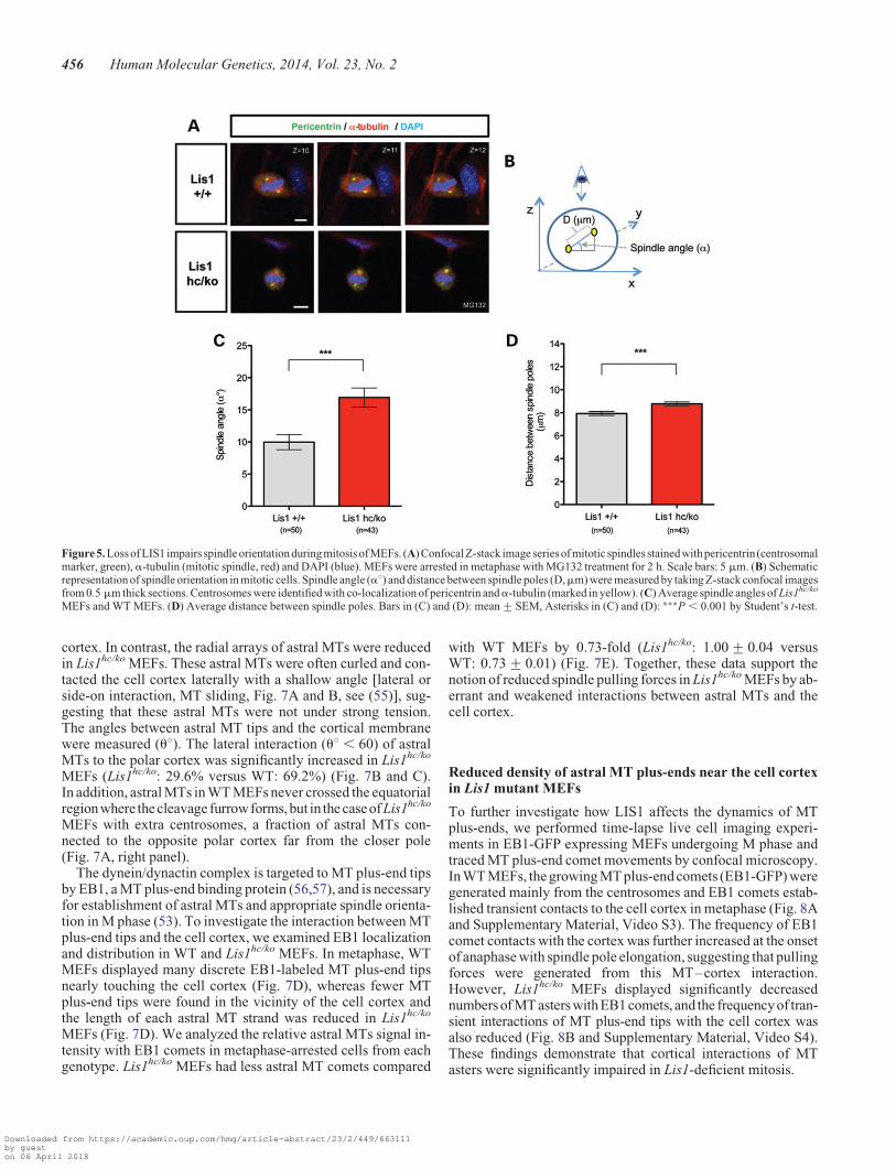
Previously, it was shown that LIS1 overexpression results inspindle misorientation in MDCK cells (12), and that DrosophilaDlis1 mutants as well as mouse Lis1 mutants display mitoticspindle defects in NP division (13,52). Consistent with thesefindings, we uncovered defects in spindle formation and posi-tioning from time-lapse live cell imaging of Lis1-CKO-CreMEFs (Fig. 1A). To determine mitotic spindle orientation paral-lel to the cell-substrate adhesion plane, which occurs in adherentcells (53), we arrested MEFs in metaphase by treatment withMG132. Pericentrin, a pericentriolar marker of centrosomes(54), was used to identify spindle poles. Confocal images fromWT MEFs displayed a relatively narrow centrosomal distribu-tion in the same or adjacent confocal planes along the cell axis.In contrast, Lis1hc/ko MEFs exhibited a high degree of spindletilting compared with the substrate plane (Fig. 5A). Spindle
angle (a8, measured by an amplitude of angle between centro-somes and the plane of cell substrate, shown in Fig. 5B) was sig-nificantly increased in Lis1hc/ko MEFs. The average spindleangle in Lis1hc/ko MEFs (a8 ¼ 16.9+ 1.5) was increased about2-fold compared with WT MEFs (a8 ¼ 10.0+ 1.2) (Fig. 5C).Since Drosophila Dlis1 mutants displayed defects in centrosomeseparation during mitosis of NPs (52), we expected that the dis-tance between the two spindle poles may be impaired in Lis1hc/ko
MEFs. Lis1hc/ko MEFs displayed a moderate change in pole dis-tance (8.8+ 0.2 mm) compared with WT MEFs (7.9+0.2 mm), reflecting alteration in the pulling forces on twospindle poles (Fig. 5D).
To determine whether spindle misorientation in Lis1hc/ko
MEFs results from cell shape changes, we analyzed the cellheight and the longest cell axis of the metaphase cells fromLis1hc/ko MEFs and WT MEFs. Neither was significantlyaltered in Lis1hc/ko MEFs compared with WT MEFs (cellheight Lis1hc/ko: 12.7+ 0.6 mm versus WT: 13.7+ 0.4 mm,and long axis Lis1hc/ko: 17.9+ 0.9 mm versus WT: 17.5+0.7 mm) (Supplementary Material, Fig. S4A and B).
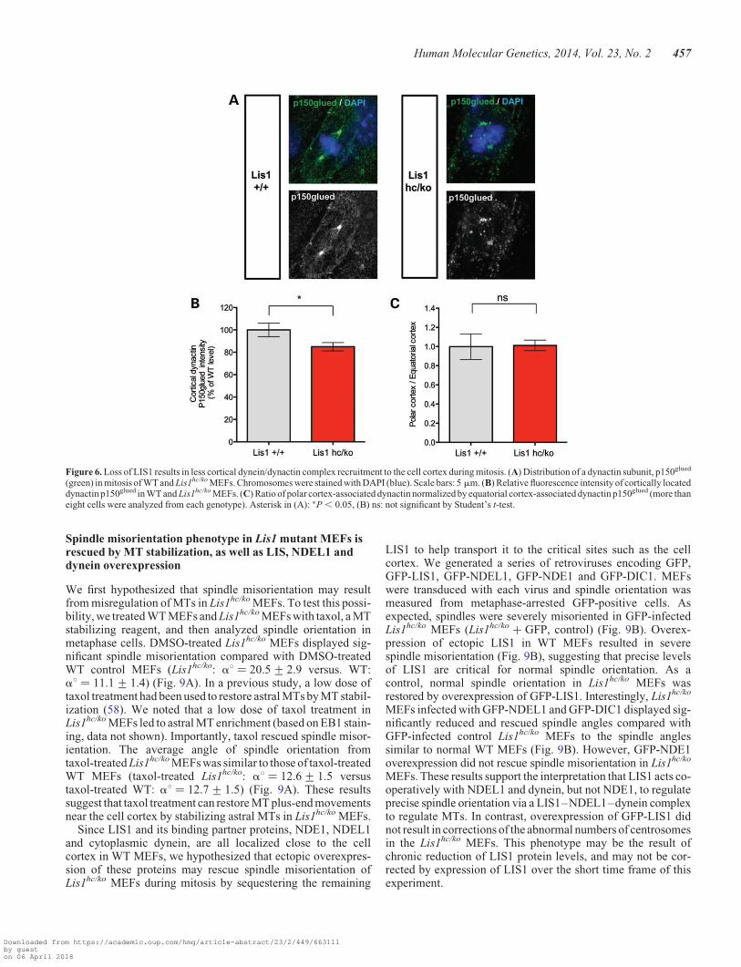
Reduced cortical dynein/dynactin complex in Lis1 mutantMEFs during mitosis
To examine cortically located dynein/dynactin complexes inLis1hc/ko MEFs, we performed immunostaining with thep150Glued subunit of dynactin. Cortical staining of p150Glued
was significantly reduced in Lis1hc/ko MEFs to 85% comparedwith WT MEFs 100% (Lis1hc/ko: 85+ 3.7% versus WT:100+ 6.1%) (Fig. 6A and B). We also examined the distributionratio of dynactin subunit pools between equatorial cortex-
Figure 3. Lis1 mutant MEFs exhibit centrosome clustering phenotype during mitosis. (A–D) Centrosome clustering phenotype with normal centrosome maturationin Lis1hc/ko MEFs. (A) Co-staining with pericentrin (pericentriolar material marker) and centrin (each centriole). Co-staining with centrin and several mature centriolemarkers: (B) cennexin/ODF2, (C) ninein, (D) Cep164, respectively. Insets in (A–D): high magnification images of centrosomes. Scale bars: 5 mm.
454 Human Molecular Genetics, 2014, Vol. 23, No. 2
Downloaded from https://academic.oup.com/hmg/article-abstract/23/2/449/663111by gueston 06 April 2018
associated and polar cortex-associated p150Glued, but this ratiowas not significantly altered in Lis1hc/ko MEFs (Lis1hc/ko:1.0+ 0.1% versus WT: 1.0+ 0.1%) (Fig. 6C). In addition,astral or central MT-associated p150Glued signals were promin-ent in WT MEFs, but they were severely reduced in Lis1hc/ko
MEFs. Reduced localization of cortical dynein/dynactincomplex in M phase may ultimately result in spindle misorienta-tion in Lis1hc/ko MEFs. These findings suggest that LIS1 med-iates dynein/dynactin targeting to the cell cortex in this cellcycle, contributing to spindle orientation and positioning toensure proper cell division.
Abnormal interaction between plus-ends of astral MTs andthe cell cortex in Lis1 mutant MEFs
Previous studies (12,13) and the dynein/dynactin studiesdescribed above (Fig. 6) suggested that astral MTs may bereduced in Lis1hc/ko MEFs, so we examined astral MTs in greatdetail. We found that WT MEFs in early anaphase displayedclear astral MTs–cortex interactions that maintained a wideangle compared with lateral surface of the cell cortex [end-oninteraction, Fig. 7A and B, see (55)]. Each lobe of the polarcortex was filled with straightened astral MT tips, suggestingthere were pulling forces between spindle poles and the cell
Figure 4. Loss of LIS1 induces chromosome misalignment in metaphase and chromosome missegregation in anaphase during mitosis. (A) Time-lapse live cellimaging analysis of chromosomal behavior (labeled with H2B-GFP, green) from Lis1-CKO-Cre MEFs and WT-Cre MEFs. Arrowhead: misaligned chromosomesin Lis1-CKO-Cre MEFs. Scale bars: 5 mm. (B) Chromosome missegregation phenotypes in mitosis of Lis1-CKO-Cre MEFs. Arrow: numerous lagging chromosomes;arrowhead: (left) the chromosomes stuck in intercellular chromatin bridge, (right) micronuclei formation in telophase. MT networks were visualized by mCherry–a-tubulin (red). (C) Quantification of lagging chromosome appearance in time-lapse live cell imaging of Lis1-CKO-Cre MEFs and WT-Cre MEFs. Numbers oflagging chromosomes—no lagging: 0, few lagging: 1–3, numerous lagging: ≥4. (D) Increased incidence of various chromosome segregation defects Lis1-CKO-CreMEFs compared with WT-Cre MEFs.
Human Molecular Genetics, 2014, Vol. 23, No. 2 455
Downloaded from https://academic.oup.com/hmg/article-abstract/23/2/449/663111by gueston 06 April 2018
cortex. In contrast, the radial arrays of astral MTs were reducedin Lis1hc/ko MEFs. These astral MTs were often curled and con-tacted the cell cortex laterally with a shallow angle [lateral orside-on interaction, MT sliding, Fig. 7A and B, see (55)], sug-gesting that these astral MTs were not under strong tension.The angles between astral MT tips and the cortical membranewere measured (u8). The lateral interaction (u8 , 60) of astralMTs to the polar cortex was significantly increased in Lis1hc/ko
MEFs (Lis1hc/ko: 29.6% versus WT: 69.2%) (Fig. 7B and C).In addition, astral MTs in WT MEFs never crossed the equatorialregionwhere thecleavagefurrowforms,but in the caseofLis1hc/ko
MEFs with extra centrosomes, a fraction of astral MTs con-nected to the opposite polar cortex far from the closer pole(Fig. 7A, right panel).
The dynein/dynactin complex is targeted to MT plus-end tipsby EB1, a MT plus-end binding protein (56,57), and is necessaryfor establishment of astral MTs and appropriate spindle orienta-tion in M phase (53). To investigate the interaction between MTplus-end tips and the cell cortex, we examined EB1 localizationand distribution in WT and Lis1hc/ko MEFs. In metaphase, WTMEFs displayed many discrete EB1-labeled MT plus-end tipsnearly touching the cell cortex (Fig. 7D), whereas fewer MTplus-end tips were found in the vicinity of the cell cortex andthe length of each astral MT strand was reduced in Lis1hc/ko
MEFs (Fig. 7D). We analyzed the relative astral MTs signal in-tensity with EB1 comets in metaphase-arrested cells from eachgenotype. Lis1hc/ko MEFs had less astral MT comets compared
with WT MEFs by 0.73-fold (Lis1hc/ko: 1.00+ 0.04 versusWT: 0.73+ 0.01) (Fig. 7E). Together, these data support thenotion of reduced spindle pulling forces in Lis1hc/ko MEFs by ab-errant and weakened interactions between astral MTs and thecell cortex.
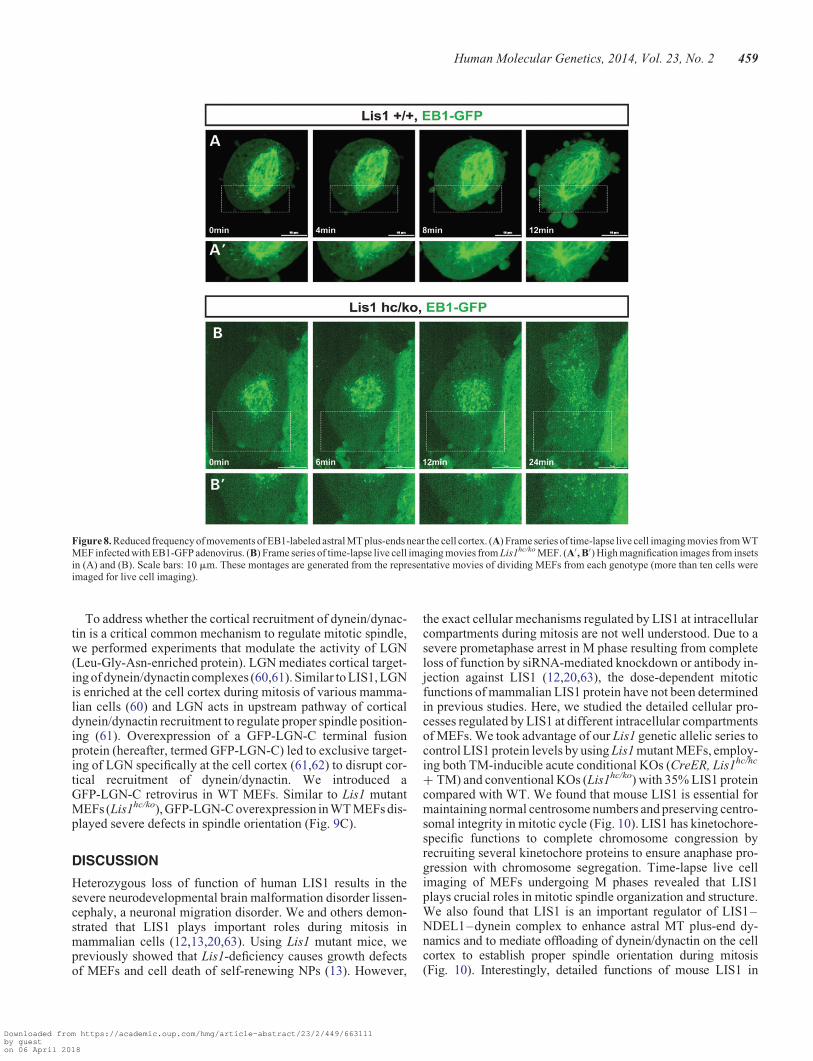
Reduced density of astral MT plus-ends near the cell cortexin Lis1 mutant MEFs
To further investigate how LIS1 affects the dynamics of MTplus-ends, we performed time-lapse live cell imaging experi-ments in EB1-GFP expressing MEFs undergoing M phase andtraced MT plus-end comet movements by confocal microscopy.In WT MEFs, the growing MT plus-end comets (EB1-GFP) weregenerated mainly from the centrosomes and EB1 comets estab-lished transient contacts to the cell cortex in metaphase (Fig. 8Aand Supplementary Material, Video S3). The frequency of EB1comet contacts with the cortex was further increased at the onsetof anaphase with spindle pole elongation, suggesting that pullingforces were generated from this MT–cortex interaction.However, Lis1hc/ko MEFs displayed significantly decreasednumbers of MT asters with EB1 comets, and the frequencyof tran-sient interactions of MT plus-end tips with the cell cortex wasalso reduced (Fig. 8B and Supplementary Material, Video S4).These findings demonstrate that cortical interactions of MTasters were significantly impaired in Lis1-deficient mitosis.
Figure 5. Loss of LIS1 impairs spindle orientation during mitosisof MEFs. (A) ConfocalZ-stack image seriesof mitotic spindles stained with pericentrin (centrosomalmarker, green), a-tubulin (mitotic spindle, red) and DAPI (blue). MEFs were arrested in metaphase with MG132 treatment for 2 h. Scale bars: 5 mm. (B) Schematicrepresentation of spindle orientation in mitotic cells. Spindle angle (a8) and distance between spindle poles (D,mm) were measured by taking Z-stack confocal imagesfrom 0.5 mm thick sections. Centrosomes were identified with co-localization of pericentrin anda-tubulin (marked in yellow). (C) Average spindle angles of Lis1hc/ko
MEFs and WT MEFs. (D) Average distance between spindle poles. Bars in (C) and (D): mean+SEM, Asterisks in (C) and (D): ∗∗∗P , 0.001 by Student’s t-test.
456 Human Molecular Genetics, 2014, Vol. 23, No. 2
Downloaded from https://academic.oup.com/hmg/article-abstract/23/2/449/663111by gueston 06 April 2018
Spindle misorientation phenotype in Lis1 mutant MEFs isrescued by MT stabilization, as well as LIS, NDEL1 anddynein overexpression
We first hypothesized that spindle misorientation may resultfrom misregulation of MTs in Lis1hc/ko MEFs. To test this possi-bility, we treated WT MEFs and Lis1hc/ko MEFs with taxol, a MTstabilizing reagent, and then analyzed spindle orientation inmetaphase cells. DMSO-treated Lis1hc/ko MEFs displayed sig-nificant spindle misorientation compared with DMSO-treatedWT control MEFs (Lis1hc/ko: a8 ¼ 20.5+ 2.9 versus. WT:a8 ¼ 11.1+ 1.4) (Fig. 9A). In a previous study, a low dose oftaxol treatment had been used to restore astral MTs by MT stabil-ization (58). We noted that a low dose of taxol treatment inLis1hc/ko MEFs led to astral MT enrichment (based on EB1 stain-ing, data not shown). Importantly, taxol rescued spindle misor-ientation. The average angle of spindle orientation fromtaxol-treated Lis1hc/ko MEFs was similar to those of taxol-treatedWT MEFs (taxol-treated Lis1hc/ko: a8 ¼ 12.6+ 1.5 versustaxol-treated WT: a8 ¼ 12.7+ 1.5) (Fig. 9A). These resultssuggest that taxol treatment can restore MT plus-end movementsnear the cell cortex by stabilizing astral MTs in Lis1hc/ko MEFs.
Since LIS1 and its binding partner proteins, NDE1, NDEL1and cytoplasmic dynein, are all localized close to the cellcortex in WT MEFs, we hypothesized that ectopic overexpres-sion of these proteins may rescue spindle misorientation ofLis1hc/ko MEFs during mitosis by sequestering the remaining
LIS1 to help transport it to the critical sites such as the cellcortex. We generated a series of retroviruses encoding GFP,GFP-LIS1, GFP-NDEL1, GFP-NDE1 and GFP-DIC1. MEFswere transduced with each virus and spindle orientation wasmeasured from metaphase-arrested GFP-positive cells. Asexpected, spindles were severely misoriented in GFP-infectedLis1hc/ko MEFs (Lis1hc/ko + GFP, control) (Fig. 9B). Overex-pression of ectopic LIS1 in WT MEFs resulted in severespindle misorientation (Fig. 9B), suggesting that precise levelsof LIS1 are critical for normal spindle orientation. As acontrol, normal spindle orientation in Lis1hc/ko MEFs wasrestored by overexpression of GFP-LIS1. Interestingly, Lis1hc/ko
MEFs infected with GFP-NDEL1 and GFP-DIC1 displayed sig-nificantly reduced and rescued spindle angles compared withGFP-infected control Lis1hc/ko MEFs to the spindle anglessimilar to normal WT MEFs (Fig. 9B). However, GFP-NDE1overexpression did not rescue spindle misorientation in Lis1hc/ko
MEFs. These results support the interpretation that LIS1 acts co-operatively with NDEL1 and dynein, but not NDE1, to regulateprecise spindle orientation via a LIS1–NDEL1–dynein complexto regulate MTs. In contrast, overexpression of GFP-LIS1 didnot result in corrections of the abnormal numbers of centrosomesin the Lis1hc/ko MEFs. This phenotype may be the result ofchronic reduction of LIS1 protein levels, and may not be cor-rected by expression of LIS1 over the short time frame of thisexperiment.
Figure 6. Loss of LIS1 results in less cortical dynein/dynactin complex recruitment to the cell cortex during mitosis. (A) Distribution of a dynactin subunit, p150glued
(green) in mitosis of WT and Lis1hc/ko MEFs. Chromosomes were stained with DAPI (blue). Scale bars: 5 mm. (B) Relative fluorescence intensity of cortically locateddynactin p150glued in WT and Lis1hc/ko MEFs. (C) Ratio of polar cortex-associated dynactin normalized by equatorial cortex-associated dynactin p150glued (more thaneight cells were analyzed from each genotype). Asterisk in (A): ∗P , 0.05, (B) ns: not significant by Student’s t-test.
Human Molecular Genetics, 2014, Vol. 23, No. 2 457
Downloaded from https://academic.oup.com/hmg/article-abstract/23/2/449/663111by gueston 06 April 2018
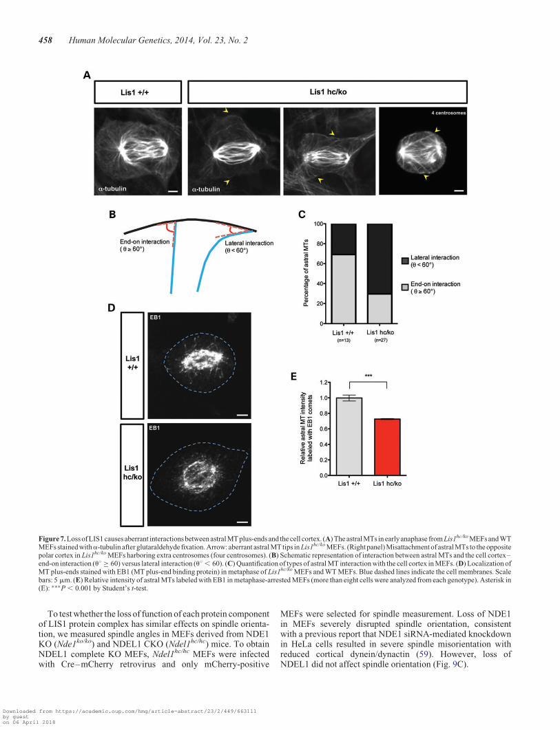
To test whether the loss of function of each protein componentof LIS1 protein complex has similar effects on spindle orienta-tion, we measured spindle angles in MEFs derived from NDE1KO (Nde1ko/ko) and NDEL1 CKO (Ndel1hc/hc) mice. To obtainNDEL1 complete KO MEFs, Ndel1hc/hc MEFs were infectedwith Cre–mCherry retrovirus and only mCherry-positive
MEFs were selected for spindle measurement. Loss of NDE1in MEFs severely disrupted spindle orientation, consistentwith a previous report that NDE1 siRNA-mediated knockdownin HeLa cells resulted in severe spindle misorientation withreduced cortical dynein/dynactin (59). However, loss ofNDEL1 did not affect spindle orientation (Fig. 9C).
Figure 7. Loss of LIS1 causes aberrant interactions between astral MT plus-ends and the cell cortex. (A) The astral MTs in early anaphase from Lis1hc/ko MEFs and WTMEFs stained witha-tubulin after glutaraldehyde fixation. Arrow: aberrant astral MT tips in Lis1hc/ko MEFs. (Right panel) Misattachment of astral MTs to the oppositepolar cortex in Lis1hc/ko MEFs harboring extra centrosomes (four centrosomes). (B) Schematic representation of interaction between astral MTs and the cell cortex–end-on interaction (u8 ≥ 60) versus lateral interaction (u8 , 60). (C) Quantification of types of astral MT interaction with the cell cortex in MEFs. (D) Localization ofMT plus-ends stained with EB1 (MT plus-end binding protein) in metaphase of Lis1hc/ko MEFs and WT MEFs. Blue dashed lines indicate the cell membranes. Scalebars: 5 mm. (E) Relative intensity of astral MTs labeled with EB1 in metaphase-arrested MEFs (more than eight cells were analyzed from each genotype). Asterisk in(E): ∗∗∗P , 0.001 by Student’s t-test.
458 Human Molecular Genetics, 2014, Vol. 23, No. 2
Downloaded from https://academic.oup.com/hmg/article-abstract/23/2/449/663111by gueston 06 April 2018
To address whether the cortical recruitment of dynein/dynac-tin is a critical common mechanism to regulate mitotic spindle,we performed experiments that modulate the activity of LGN(Leu-Gly-Asn-enriched protein). LGN mediates cortical target-ing of dynein/dynactin complexes (60,61). Similar to LIS1, LGNis enriched at the cell cortex during mitosis of various mamma-lian cells (60) and LGN acts in upstream pathway of corticaldynein/dynactin recruitment to regulate proper spindle position-ing (61). Overexpression of a GFP-LGN-C terminal fusionprotein (hereafter, termed GFP-LGN-C) led to exclusive target-ing of LGN specifically at the cell cortex (61,62) to disrupt cor-tical recruitment of dynein/dynactin. We introduced aGFP-LGN-C retrovirus in WT MEFs. Similar to Lis1 mutantMEFs (Lis1hc/ko), GFP-LGN-C overexpression in WT MEFs dis-played severe defects in spindle orientation (Fig. 9C).
DISCUSSION
Heterozygous loss of function of human LIS1 results in thesevere neurodevelopmental brain malformation disorder lissen-cephaly, a neuronal migration disorder. We and others demon-strated that LIS1 plays important roles during mitosis inmammalian cells (12,13,20,63). Using Lis1 mutant mice, wepreviously showed that Lis1-deficiency causes growth defectsof MEFs and cell death of self-renewing NPs (13). However,
the exact cellular mechanisms regulated by LIS1 at intracellularcompartments during mitosis are not well understood. Due to asevere prometaphase arrest in M phase resulting from completeloss of function by siRNA-mediated knockdown or antibody in-jection against LIS1 (12,20,63), the dose-dependent mitoticfunctions of mammalian LIS1 protein have not been determinedin previous studies. Here, we studied the detailed cellular pro-cesses regulated by LIS1 at different intracellular compartmentsof MEFs. We took advantage of our Lis1 genetic allelic series tocontrol LIS1 protein levels by using Lis1 mutant MEFs, employ-ing both TM-inducible acute conditional KOs (CreER, Lis1hc/hc
+ TM) and conventional KOs (Lis1hc/ko) with 35% LIS1 proteincompared with WT. We found that mouse LIS1 is essential formaintaining normal centrosome numbers and preserving centro-somal integrity in mitotic cycle (Fig. 10). LIS1 has kinetochore-specific functions to complete chromosome congression byrecruiting several kinetochore proteins to ensure anaphase pro-gression with chromosome segregation. Time-lapse live cellimaging of MEFs undergoing M phases revealed that LIS1plays crucial roles in mitotic spindle organization and structure.We also found that LIS1 is an important regulator of LIS1–NDEL1–dynein complex to enhance astral MT plus-end dy-namics and to mediate offloading of dynein/dynactin on the cellcortex to establish proper spindle orientation during mitosis(Fig. 10). Interestingly, detailed functions of mouse LIS1 in
Figure 8. Reduced frequency of movements of EB1-labeled astral MT plus-ends near the cell cortex. (A) Frame series of time-lapse live cell imaging movies from WTMEF infected with EB1-GFP adenovirus. (B) Frame series of time-lapse live cell imaging movies from Lis1hc/ko MEF. (A′, B′) High magnification images from insetsin (A) and (B). Scale bars: 10 mm. These montages are generated from the representative movies of dividing MEFs from each genotype (more than ten cells wereimaged for live cell imaging).
Human Molecular Genetics, 2014, Vol. 23, No. 2 459
Downloaded from https://academic.oup.com/hmg/article-abstract/23/2/449/663111by gueston 06 April 2018
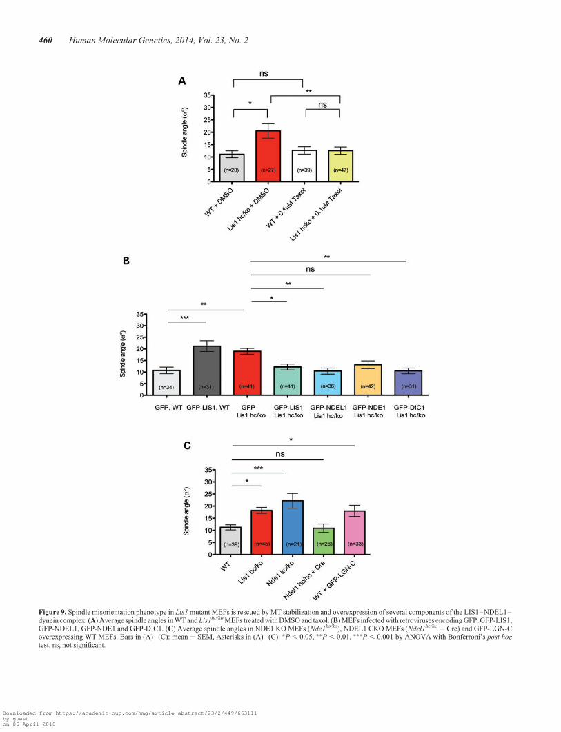
Figure 9. Spindle misorientation phenotype in Lis1 mutant MEFs is rescued by MT stabilization and overexpression of several components of the LIS1–NDEL1–dynein complex. (A) Average spindle angles in WT and Lis1hc/ko MEFs treated with DMSO and taxol. (B) MEFs infected with retroviruses encoding GFP, GFP-LIS1,GFP-NDEL1, GFP-NDE1 and GFP-DIC1. (C) Average spindle angles in NDE1 KO MEFs (Nde1ko/ko), NDEL1 CKO MEFs (Ndel1hc/hc + Cre) and GFP-LGN-Coverexpressing WT MEFs. Bars in (A)–(C): mean+SEM, Asterisks in (A)–(C): ∗P , 0.05, ∗∗P , 0.01, ∗∗∗P , 0.001 by ANOVA with Bonferroni’s post hoctest. ns, not significant.
460 Human Molecular Genetics, 2014, Vol. 23, No. 2
Downloaded from https://academic.oup.com/hmg/article-abstract/23/2/449/663111by gueston 06 April 2018
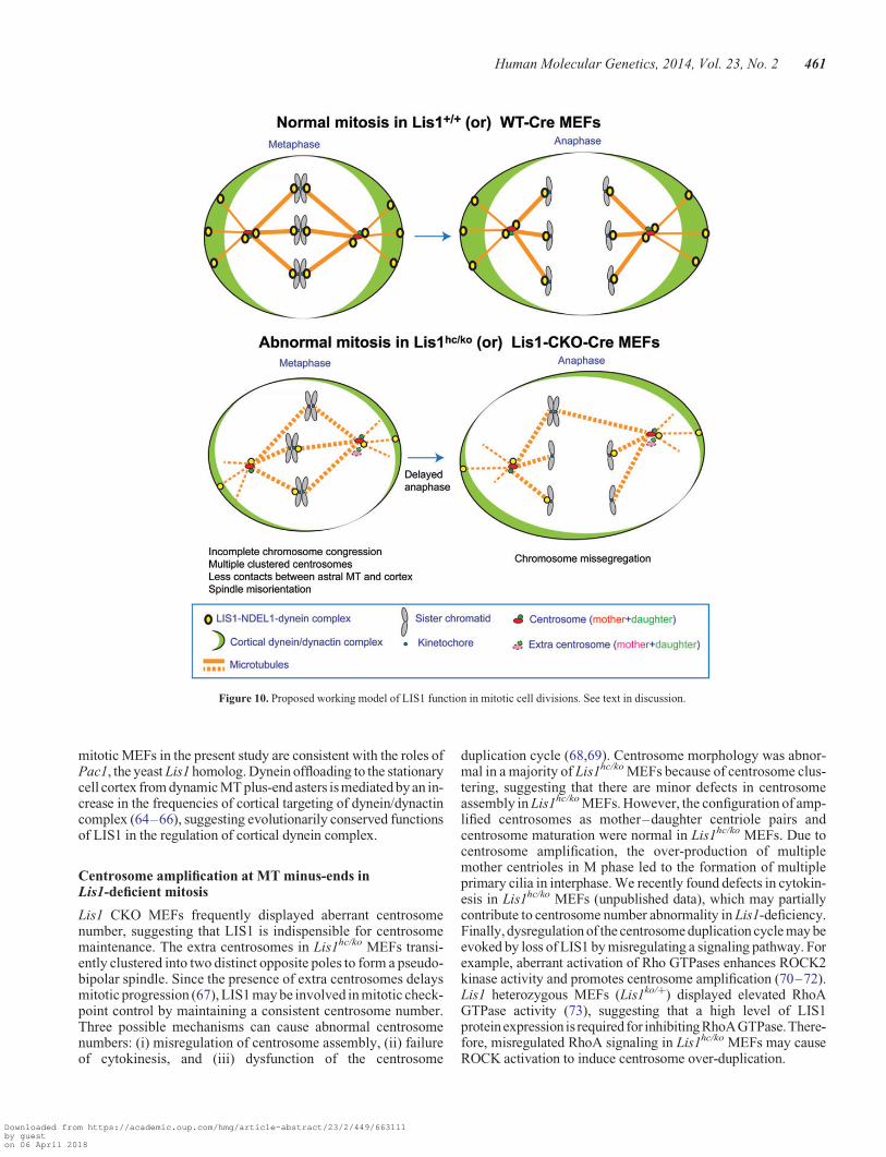
mitotic MEFs in the present study are consistent with the roles ofPac1, the yeast Lis1 homolog. Dynein offloading to the stationarycell cortex from dynamic MT plus-end asters is mediated by an in-crease in the frequencies of cortical targeting of dynein/dynactincomplex (64–66), suggesting evolutionarily conserved functionsof LIS1 in the regulation of cortical dynein complex.
Centrosome amplification at MT minus-ends inLis1-deficient mitosis
Lis1 CKO MEFs frequently displayed aberrant centrosomenumber, suggesting that LIS1 is indispensible for centrosomemaintenance. The extra centrosomes in Lis1hc/ko MEFs transi-ently clustered into two distinct opposite poles to form a pseudo-bipolar spindle. Since the presence of extra centrosomes delaysmitotic progression (67), LIS1 may be involved in mitotic check-point control by maintaining a consistent centrosome number.Three possible mechanisms can cause abnormal centrosomenumbers: (i) misregulation of centrosome assembly, (ii) failureof cytokinesis, and (iii) dysfunction of the centrosome
duplication cycle (68,69). Centrosome morphology was abnor-mal in a majority of Lis1hc/ko MEFs because of centrosome clus-tering, suggesting that there are minor defects in centrosomeassembly in Lis1hc/ko MEFs. However, the configuration of amp-lified centrosomes as mother–daughter centriole pairs andcentrosome maturation were normal in Lis1hc/ko MEFs. Due tocentrosome amplification, the over-production of multiplemother centrioles in M phase led to the formation of multipleprimary cilia in interphase. We recently found defects in cytokin-esis in Lis1hc/ko MEFs (unpublished data), which may partiallycontribute to centrosome number abnormality in Lis1-deficiency.Finally, dysregulation of the centrosome duplication cycle may beevoked by loss of LIS1 by misregulating a signaling pathway. Forexample, aberrant activation of Rho GTPases enhances ROCK2kinase activity and promotes centrosome amplification (70–72).Lis1 heterozygous MEFs (Lis1ko/+) displayed elevated RhoAGTPase activity (73), suggesting that a high level of LIS1proteinexpression is required for inhibitingRhoAGTPase.There-fore, misregulated RhoA signaling in Lis1hc/ko MEFs may causeROCK activation to induce centrosome over-duplication.
Figure 10. Proposed working model of LIS1 function in mitotic cell divisions. See text in discussion.
Human Molecular Genetics, 2014, Vol. 23, No. 2 461
Downloaded from https://academic.oup.com/hmg/article-abstract/23/2/449/663111by gueston 06 April 2018
Missegregation of chromosomes and fate determinants inLis1-deficient mitosis
Appropriate levels of LIS1 protein at kinetochores are requiredfor spindle checkpoint control to inhibit improper kinetochore-MT attachments and to execute chromosome segregation and in-heritance by recruiting other kinetochore proteins (dynein andCLIP170). Extra centrosomes in Lis1hc/ko MEFs may lead tomerotelic kinetochore-chromosome attachments by interferingwith kinetochore-MT capture (74). Improper kinteochore-MTattachments and extra centrosomes promote genomic instabilityby generating aneuploid daughter cells. The frequent formationof chromatin bridges and micronuclei in Lis1-deficient mitosisreflect severe chromosome segregation defects. It has beenshown that a human hepatocellular carcinoma cell type displaysdownregulation of LIS1 mRNA and protein, and the severity oftumor formation is strongly correlated with LIS1 downregula-tion (75). This suggests that LIS1 may act as a tumor suppressorunder certain conditions. Together with our data, this study sup-ports the notion that LIS1 is required for maintaining genomicstability to prevent the loss of chromosomes during mitosis.
LIS1-dependent centrosome number maintenance may playcritical roles in the maintenance of genomic stability essentialfor asymmetric NP division during development. In Drosophila,PLK4 homologue mutants induce extra centrosome formationand misalignment of apical cell fate determinants during NPdivisions (76). Multiple centrosomes-harboring NPs misorientthe mitotic spindle and the relationship between mitotic spindlesand apical markers was disrupted. We also frequently observedthree centrosomes in apical NPs from Lis1 mutant embryonicmouse brains (unpublished data), suggesting that mammalianNP cells may employ similar mechanisms.
Spindle misorientation and reduced interaction betweenastral MT plus-ends and the cell cortex during mitosis inLis1 mutant MEFs
Mitotic spindle orientation is critical for the proper segregationof chromosomes to the daughter cells. Here, we demonstratethat Lis1 mutant MEFs showed significant spindle misorienta-tion with severe tilting compared with adhesion plane. Similarly,we previously found spindle orientation defects in NPs from Lis1mutants (13). NDEL1- (77) and NDE1-depleted NPs (78) alsodisplayed severe spindle misorientation. In the present study,we showed that Lis1-deficiency results in reduced corticaldynactin p150Glued in M phase of MEFs. The number of astralMTs and the frequency of movements of EB1-labeled MTplus-ends reaching to the cortex were significantly reduced inLis1hc/ko MEFs. Due to the loss of LIS1, a key component ofthe LIS1–NDEL1–dynein/dynactin complex, MT asters maybecome destabilized at the cell cortex. By overexpressionexperiments, we confirmed that NDEL1 and dynein are thecore mediators acting in LIS1-dependent pathways of mitoticspindle regulation. Most recently, it was shown that stationarybarrier- or bead-attached dynein controls the dynamics of MTplus-ends in vitro (79,80). In addition, it has been shown thatLGN functions as an upstream regulator to target corticaldynein/dynactin in HeLa cells (61). Taken together, these dataindicate that LIS1- (or LGN-) mediated cortical targeting ofdynein/dynactin complex is a key cellular mechanism to regulate
contacts between astral MT plus-ends and the cell cortex duringmitosis.
In addition to the function of the LIS1–NDEL1–dyneincomplex near the cell cortex that controls MT dynamics inmitotic spindles, other cytoskeletal components may contributeto the mitotic function of LIS1. Subcortical actin cytoskeletalcomponents as well as actin retraction fibers at the adhesionsites have been implicated in mitotic spindle formation(81,82). Interestingly, Lis1 heterozygous mutant neurons(Lis1ko/+) displayed misregulated actin fibers at the leadingedges during cell migration (83). This finding raises the possibil-ity that dysfunction of the actin cytoskeleton affected by reducedLIS1 has at least an indirect impact on actin-dependent mitoticspindle regulation. The cell cortex is a critically important sitefor vigorous crosstalk and interactions between MTs and theactin cytoskeleton to integrate signal cues into mitotic spindleregulatory pathway. In the current study, we conclude thatLIS1, as a central component of LIS1–NDEL1–dyneincomplex, participates in the regulation of MT plus-ends dynam-ics on astral MTs near the cell cortex to ensure mitotic spindlepositioning and orientation. In future studies, it will be interest-ing to determine whether LIS1 directly regulates the actin cyto-skeleton to promote actin fiber assembly at the corticaladhesion-binding sites during mitosis.
MATERIALS AND METHODS
Mice
Lis1 null (Lis1ko, formerly referred to as Pafah1b1tm1Awb) andHC (Lis1hc, formerly referred to as Pafah1b1tm2Awb(loxP))alleles were used, which were described previously (13,42). Pre-viously reported Nde1 null (Nde1ko, formerly referred to asNde1tm1Caw) allele (78) and Ndel1 HC KO (Ndel1hc, formerly re-ferred to as Ndel1tm1Shr) allele (84) were used for spindle orien-tation analysis. To induce Cre recombinase activation to deleteLis1 conditional allele, the TM-inducible CreERTM line (44)was used to mate with Lis1 mutant mice.
Cell culture
Primary MEFs were derived from E14.5 embryos and were cul-tured in DMEM (Mediatech) supplemented with 12.5% FBS(Gibco), penicillin/streptomycin and L-glutamine at 378C in a5% CO2 incubator. All MEFs used in this study were less thanpassage 4 (13). CreERTM recombinase activity was induced byadministrating 4-hydroxy TM (Sigma, 100 nM) dissolved inculture media. H293T cells for viral packaging were maintainedin 10% FBS (Gibco) in DMEM medium (Mediatech).
Retrovirus production and MEF infection
To produce retroviruses, pMV-GP (gag, pol), pCMV-G (VSV-Genv) and pCX retroviral vectors were purified by EndofreePlasmid Maxiprep kit (Qiagen) and transfected in H293T cellswith TransIT-LT1 transfection reagent (Mirus). Packagingvectors (pMV-GP, pCMV-G) were gifts from Atsushi Miyano-hara (UCSD). pCLNR-H2BG (H2B-GFP) was a gift from Geof-frey Wahl (Salk institute, Addgene plasmid #17735). Toconstruct pCX–mCherry–a-tubulin retroviral vector, cDNA
462 Human Molecular Genetics, 2014, Vol. 23, No. 2
Downloaded from https://academic.oup.com/hmg/article-abstract/23/2/449/663111by gueston 06 April 2018
of mCherry was amplified by PCR from pcDNA3.1-mCherryderived from an mCherry expression vector (a gift from RogerTsien, UCSD). The mCherry cDNA PCR product was subclonedinto pEGFP-a-tubulin (Clontech) digested with SalI/NotI togenerate mCherry–a-tubulin. Then, the PCR product ofcDNA encoding mCherry–a-tubulin was ligated into the linear-ized pCX backbone vector derived from pCX-Centrin2-DsRed(gift from Joseph Gleeson, UCSD) with BamHI/PacI. OtherGFP–fusion proteins used for rescue experiments (pCX-GFP-LIS1, pCX-GFP-NDEL1, pCX-GFP-NDE1 and pCX-GFP-DIC1) were generated by cDNA amplification from mammalianexpression vectors pCMV-GFP-NDEL1 [described in (85)],pCMV-GFP-LIS1, pCMV-GFP-NDE1 [gifts from Shinji Hirot-sune, Osaka City University, Japan, described in (84)] andpCMV-GFP-DIC1 [gifts from Shinji Hirotsune, described in(86)], followed by BamHI/PacI digestion for cloning into thepCX retroviral vector. To construct the pCX–Cre–mCherryretroviral vector, Cre cDNA PCR product was amplified frompBS500-CreGFP (a gift from Brian Sauer, NIH, Addgeneplasmid #11920) and inserted into pcDNA3.1-mCherry withNheI/KpnI. Then Cre–mCherry fusion cDNA was derivedfrom this construct and subcloned into pCX vector withEcoRI/PacI to generate pCX–Cre–mCherry. Protein expres-sion of both Cre and mCherry was confirmed by Cre antibodystaining and mCherry fluorescence. To construct pCX-GFP-LGN-C, GFP-LGN-C fragment cDNA was amplified frompIC389 [pBabe-GFP-LGN-C, a gift from Iain Cheeseman,MIT, described in Kiyomitsu and Cheeseman (61)]. We gener-ated pCX-GFP-LGN-C construct by serial introductions ofPCR products digested with BamHI/PacI and BamHI, respect-ively. Viral supernatants (in DMEM without antibiotics andFBS) were collected at 48 h post-transfection and filteredthrough 0.45 mm filter (Sartorius). MEFs were infected withretrovirus by co-incubating the mixture of viral supernatantand fresh media for 24 h. During infection, 12.5% FBS was sup-plemented in the viral supernatant media along with 4 mg/mlpolybrene (Sigma).
Western blotting
MEFs were lysed in Tris-Triton buffer (10 mM Tris pH 7.4,100 mM NaCl, 1 mM EDTA, 1 mM EGTA, 1% Triton X-100,10% glycerol, 0.1% SDS, 0.5% deoxycholate) with protease/phosphatase inhibitors. Lysates were collected on ice and centri-fuged. Supernatants with protein extracts were transferred intonew tubes, boiled at 958C for 5 min and then stored at 2208Cbefore use. Protein concentrations were determined by theBCA protein assay kit (Pierce) and equal amounts were loadedon a 10% SDS–PAGE resolving gel (Bio-Rad). After electro-phoresis, proteins were transferred to a nitrocellulose membrane(Bio-Rad). The membrane was blocked in TBST (TBS with0.1% Tween 20) with 2.5% skim milk for 1 h at RT and incubatedwith the following primary antibodies at 48C overnight: rabbitanti-LIS1 (1:1000, a gift from Shinji Hirotsune), mouseanti-a-tubulin (Sigma, 1:8000) and mouse anti-b-actin (Sigma,1:5000). Secondary antibodies used were HRP-conjugated goatanti-rabbit and goat anti-mouse (Jackson Lab, 1:10 000) whichwere incubated for 1 h. Antibody binding was detected via theECL kit (Pierce). Relative protein amount was measured usingthe ImageJ software after background subtraction.
Immunocytochemistry
MEFs were grown on acid-washed and 0.2% gelatin (Millipore)-coated glass cover slips and fixed with 4% PFA in PBS for20 min. For centrosomal protein immunostaining and EB1 stain-ing, MEFs were fixed with 2208C cold methanol for 2 min.2.5% normal goat serum (or FBS) and 0.1% Triton X-100 inPBS were used for blocking for 1 h at RT. The primary anti-bodies were diluted in blocking buffer and incubated overnightat 48C. The primary antibodies used were: rabbit anti-LIS1(Abcam, 1:250); mouse anti-a-tubulin (Sigma, 1:500); ratanti-a-tubulin (AbD Serotec, 1:1000); mouse anti-g-tubulin(Sigma, 1:500); rabbit anti-pericentrin (Covance, 1:1000);mouse anti-p150Glued (BD bioscience, 1:200); mouse anti-EB1(BD bioscience, 1:200); mouse anti-GFP (Invitrogen, 1:400);rabbit anti-GFP (Invitrogen, 1:400); mouse anti-centrin (Milli-pore, 1:200); rabbit anti-cennexin/ODF2 (Abcam, 1:200);rabbit anti-ninein (Abcam, 1:200); and rabbit anti-Cep164(1:1000, a gift from Erich Nigg, University of Basel, Switzer-land). The following goat secondary antibodies were incubatedfor 1 h at RT: anti-mouse AlexaFluor-488; anti-mouse AlexaFluor-594; anti-rabbit AlexaFluor-488; anti-rabbit Alexafluor-568; anti-rat AlexaFluor-488; and anti-rat AlexaFluor-647(Invitrogen). ProLong gold antifade reagent with DAPI (Invitro-gen) was used to counterstain the nucleus and as mountingmedium. All the fixed sample images were captured using aNikon C1si laser-scanning confocal microscope with a ×601.4 NA PlanApo oil objective lense (Nikon).
For primary cilia staining, MEFs were serum-starved for 24 hwith 0.5% FBS in DMEM and fixed in 4% PFA in PBS. Primarymouse anti-acetylated a-tubulin (Sigma, 1:1000) was used toidentify primary cilia. For visualization of astral MTs, MEFswere fixed in 0.25% glutaraldehyde in BRB80 buffer (80 mM
PIPES pH 6.8, 1 mM MgCl2, 1 mM EGTA) for 10 min, andthen treated with 0.2% sodium borohydride in PBS for 20 min,changing the borohydride solution two times. Primary ratanti-a-tubulin antibody was diluted in blocking buffer (2%FBS, 0.1% Triton X-100 in PBS). To visualize cortical dynactinp150Glued, MEFs were pre-extracted with 0.5% Triton X-100 inPHEM buffer (120 mM PIPES, 50 mM HEPES, 20 mM EGTA,8 mM MgSO4) with 5 mM taxol (Sigma) for 1 min and thenfixed with 2208C cold methanol for 2 min. To compare corticaldynactin p150Glued, we quantified the amount of staining inmetaphase cells from each genotype by measuring fluorescenceintensity at two equatorial cortex locations and four polar cortexlocations per cell.
Mitotic spindle analysis and cell shape analysis
For analysis of the mitotic spindle, MEFs were arrested in meta-phase by treatment with proteosome inhibitor, 10 mM MG132(EMD biosciences) for 2 h. From the cover slips of MEFs, aseries of Z stack images (0.5 mm apart) of metaphase mitoticspindles stained with anti-pericentrin, anti-a-tubulin antibodieswere obtained using the Nikon C1si laser-scanning confocalmicroscope (Biological Imaging Development Center, UCSF).The linear (x–y plane) and vertical (z-axis) distance betweenspindle poles was measured from Nikon EZ-C1 imaging soft-ware. Mitotic spindle length (D) and spindle angles (a8) werecalculated by 3D trigometric functions. The drug treatment in
Human Molecular Genetics, 2014, Vol. 23, No. 2 463
Downloaded from https://academic.oup.com/hmg/article-abstract/23/2/449/663111by gueston 06 April 2018
MEFs was performed in following conditions: 1 mM taxol(Sigma) were added to the medium and incubated for 30 minafter the treatment of 10 mM MG132 for 2 h. Cell shape was ana-lyzed from the MEFs incubated with lipophilic dye, DHCC(3,3′-dihexyoxacarbo cyanine inodide, Sigma) for 10 min andthen treated with 10 mM MG132 for 2 h.
Kinetochore staining and quantification
MEFs were treated with 10 mM nocodazole (Sigma) for 1 h undernormal incubation conditions to accumulate proteins at the kine-tochores. Then MEFs were fixed with 2208C cold methanol for2 min. Confocal images were obtained from an OlympusFV1000 laser scanning confocal microscope. From each geno-type, 10 cells were analyzed and compared in softWoRx Explor-er (Applied Precision) using 10 × 10 window in Data Inspector.Interkinetochore distance was determined using the line segmenttool across Z-stacks. The total immunofluorescence of 10 kine-tochores was averaged for each cell. Primary antibodies were:goat anti-LIS1 (Abcam, 1:200); mouse anti-p150glued (BD bio-science, 1:200); rabbit anti-CLIP170 (Holly Goodson, Univer-sity of Norte Dame, 1:200); mouse anti-DIC70.1 (Sigma,1:25); human SH-CREST autoimmune serum (a gift fromWilliam Brinkley, Baylor College of Medicine, 1:10 000); andMad2 (a gift from Don Cleveland, UCSD, 1:200). Secondaryantibodies were: donkey anti-rabbit FITC; donkey anti-goatCy3; donkey anti-human Cy2, and donkey anti-human Cy3(Jackson Laboratory).
Time-lapse live cell imaging
Glass-bottom six-well tissue culture plates (MatTek) were usedfor live cell imaging of mitosis. The plates were pre-incubatedwith 0.2% gelatin (Millipore) solution before plating MEFs onglass-bottom dishes 24 h prior to live cell imaging. Retroviruseswere transduced into primary MEFs by co-incubating with viralsupernatants for 24 h. 4-hydroxy TM (100 nM, Sigma) was usedfor 12 h for CreERTM for Cre transgene activation. MEFs expres-sing both H2B-GFP and mCherry–a-tubulin retroviruses wererecorded with a time-lapse Nikon Ti microscope equippedwith 5% CO2 and a 378C temperature-controlled chamber(Nikon imaging center, UCSF). To monitor long-term mitosisof MEFs, multiple points were selected for acquiring each fluor-escence image (GFP/mCherry) with a motorized stage andperfect focus function. Fluorescence was captured with a Cool-snap camera (Roper Scientific) with exposure time of 100 ms forGFP and 200 ms for mCherry. Time-lapse images of cell divisionwere taken every 1 min (up to 12 h). Filters and multipoint scan-ning were controlled by NIS-Elements imaging software(Nikon).
Tracking EB1-GFP
EB1-GFP was amplified using oligos (fwd: caccatggcagtgaacg-tatactca and rev: ttacttgtacagctcgtccat) and inserted into pTOPOvector and subsequently cloned into pAd/CMV/V5-DEST withGateway cloning (Invitrogen). Adenovirus particles were puri-fied as described previously (87). One microliter of EB1-GFPadenovirus was added to MEF growth media 24 h prior toimaging. Cells were treated with 9 mM RO-3306 (EMD
bioscience), a CDK1 inhibitor, for 18 h to arrest cells at theG2/M transition of the cell cycle. Mitosis of MEFs was imagedafter drug release by changing to fresh media containing20 mM HEPES (Sigma).
EB1-GFP protein dynamics were imaged at 378C with a ×100NA 1.49 oil-immersed objective lens (CFI Apo TIRF, Nikon)equipped with a spinning-disk confocal scanning unit (Borealis-modified CSU-X1, Spectral Applied Research) using a Nikon Timicroscope (Nikon) with a 488 nm laser, electronic shutters, acool charged-coupled device camera (CoolSnap HQ, Photo-metrics) and controlled by NIS-Elements software (Nikon).MT plus-ends were tracked in time-lapse images fromEB1-GFP expressing MEFs acquired every 30 s using perfectfocus function. Three sections of Z stacks (2 mm apart) wereacquired at every time point.
Online supplementary materials
Supplementary Material, Figure S1 shows LIS1 localizationduring mitosis from WT and Lis1 mutant MEFs and theamount of protein expression in each genotype. SupplementaryMaterial, Figure S2 shows primary cilia formation from WT andLis1 mutant MEFs and quantification of multiple cilia. Supple-mentary Material, Figure S3 shows kinetochore protein stainingin WT and Lis1 mutant MEFs and quantification of fluorescenceintensity at kinetochores. Supplementary Material, Figure S4shows quantification of metaphase cell morphology in WT andLis1 mutant MEFs. Supplementary Material, Video S1 showsthe time-lapse live cell imaging of mitotic cell division fromCreERTM;Lis1+/+ MEFs treated with 4-hydroxy TM.H2B-GFP and mCherry–a-tubulin labeled fluorescencesignals were acquired with a 1 min interval (as shown inFig. 1A, upper panel). Supplementary Material, Video S2shows the time-lapse live cell imaging of mitotic cell divisionfrom CreERTM;Lis1hc/hc MEFs treated with 4-hydroxy TM.H2B-GFP and mCherry-a-tubuin labeled fluorescence signalswere acquired with a 1 min interval (as shown in Fig. 1A,lower panel). Supplementary Material, Video S3 shows thedynamic movements of MT plus-ends comets labeled withEB1-GFP in WT MEFs. Confocal images were acquired with30 s interval (as shown in Fig. 8A). Supplementary Material,Video S4 shows the dynamic movements of MT plus-endscomets labeled with EB1-GFP in Lis1hc/ko mutant MEFs. Con-focal images were acquired with 30 s interval (as shown inFig. 8B).
SUPPLEMENTARY MATERIAL
Supplementary Material is available at HMG online.
Conflict of Interest statement. None declared.
FUNDING
This work was supported by National Institutes of Health, R01NS41030 and National Institutes of Health, R01 HD047380.H.M.M. was supported by a UCSF Graduate Student ResearchAward.
464 Human Molecular Genetics, 2014, Vol. 23, No. 2
Downloaded from https://academic.oup.com/hmg/article-abstract/23/2/449/663111by gueston 06 April 2018
REFERENCES
1. Holland, A.J. and Cleveland, D.W. (2009) Boveri revisited: chromosomalinstability, aneuploidy and tumorigenesis. Nat. Rev.. Mol. Cell Biol., 10,478–487.
2. Bornens, M. (2002) Centrosome composition and microtubule anchoringmechanisms. Curr. Opin. Cell Biol., 14, 25–34.
3. Zimmerman, W. and Doxsey, S.J. (2000) Construction of centrosomes andspindle poles by molecular motor-driven assembly of protein particles.Traffic, 1, 927–934.
4. Doxsey, S., McCollum, D. and Theurkauf, W. (2005) Centrosomes incellular regulation. Ann. Rev. Cell Dev. Biol., 21, 411–434.
5. Nigg, E.A. and Stearns, T. (2011) The centrosome cycle: centriolebiogenesis, duplication and inherent asymmetries. Nat. Cell Biol., 13,1154–1160.
6. Glotzer, M. (1996) Mitosis: don’t get mad, get even. Curr. Biol., 6,1592–1594.
7. Kirschner, M.W. and Mitchison, T. (1986) Microtubule dynamics. Nature,324, 621.
8. Kline-Smith, S.L. and Walczak, C.E. (2004) Mitotic spindle assembly andchromosome segregation: refocusing on microtubule dynamics. Mol. Cell,15, 317–327.
9. Glotzer, M. (2009) The 3Ms of central spindle assembly: microtubules,motors and MAPs. Nat. Rev. Mol. Cell Biol., 10, 9–20.
10. Moore, J.K. and Cooper, J.A. (2010) Coordinating mitosis with cell polarity:molecular motors at the cell cortex. Semin. Cell Dev. Biol., 21, 283–289.
11. Coquelle, F.M., Caspi, M., Cordelieres, F.P., Dompierre, J.P., Dujardin,D.L., Koifman, C., Martin, P., Hoogenraad, C.C., Akhmanova, A., Galjart,N. et al. (2002) LIS1, CLIP-170’s key to the dynein/dynactin pathway. Mol.
Cell Biol., 22, 3089–3102.12. Faulkner, N.E., Dujardin, D.L., Tai, C.Y., Vaughan, K.T., O’Connell, C.B.,
Wang, Y. and Vallee, R.B. (2000) A role for the lissencephaly gene LIS1 inmitosis and cytoplasmic dynein function. Nat. Cell Biol., 2, 784–791.
13. Yingling, J., Youn, Y.H., Darling, D., Toyo-Oka, K., Pramparo, T.,Hirotsune, S. and Wynshaw-Boris, A. (2008) Neuroepithelial stem cellproliferation requires LIS1 for precise spindle orientation and symmetricdivision. Cell, 132, 474–486.
14. Reiner, O., Carrozzo, R., Shen, Y., Wehnert, M., Faustinella, F., Dobyns,W.B., Caskey, C.T. and Ledbetter, D.H. (1993) Isolation of a Miller-Diekerlissencephaly gene containing G protein beta-subunit-like repeats. Nature,364, 717–721.
15. Hattori, M., Adachi, H., Tsujimoto, M., Arai, H. and Inoue, K. (1994)Miller-Dieker lissencephaly gene encodes a subunit of brainplatelet-activating factor acetylhydrolase [corrected]. Nature, 370,216–218.
16. Xiang, X., Osmani, A.H., Osmani, S.A., Xin, M. and Morris, N.R. (1995)Nudf, a nuclear migration gene in Aspergillus nidulans, is similar to thehuman LIS-1 gene required for neuronal migration. Mol. Biol. Cell, 6, 297–310.
17. Morris, N.R., Efimov, V.P. and Xiang, X. (1998) Nuclear migration,nucleokinesis and lissencephaly. Trends Cell Biol., 8, 467–470.
18. Smith, D.S., Niethammer, M., Ayala, R., Zhou, Y., Gambello, M.J.,Wynshaw-Boris, A. and Tsai, L.H. (2000) Regulation of cytoplasmic dyneinbehaviourand microtubuleorganization by mammalian Lis1. Nat.CellBiol.,2, 767–775.
19. Sasaki, S., Shionoya, A., Ishida, M., Gambello, M.J., Yingling, J.,Wynshaw-Boris, A. and Hirotsune, S. (2000) A LIS1/NUDEL/cytoplasmicdynein heavy chain complex in the developing and adult nervous system.Neuron, 28, 681–696.
20. Tai, C.Y., Dujardin, D.L., Faulkner, N.E. and Vallee, R.B. (2002) Role ofdynein, dynactin, and CLIP-170 interactions in LIS1 kinetochore function.J. Cell Biol., 156, 959–968.
21. Busson, S., Dujardin, D., Moreau, A., Dompierre, J. and De Mey, J.R. (1998)Dynein and dynactin are localized to astral microtubules and at cortical sitesin mitotic epithelial cells. Curr. Biol., 8, 541–544.
22. Howell, B.J., McEwen, B.F., Canman, J.C., Hoffman, D.B., Farrar, E.M.,Rieder, C.L. and Salmon, E.D. (2001) Cytoplasmic dynein/dynactin driveskinetochore protein transport to the spindle poles and has a role in mitoticspindle checkpoint inactivation. J. Cell Biol., 155, 1159–1172.
23. Merdes, A., Ramyar, K., Vechio, J.D. and Cleveland, D.W. (1996) Acomplex of NuMA and cytoplasmic dynein is essential for mitotic spindleassembly. Cell, 87, 447–458.
24. Nguyen-Ngoc, T., Afshar, K. and Gonczy, P. (2007) Coupling of corticaldynein and G alpha proteins mediates spindle positioning in Caenorhabditiselegans. Nat. Cell Biol., 9, 1294–1302.
25. O’Connell, C.B. and Wang, Y.L. (2000) Mammalian spindle orientation andposition respond to changes in cell shape in a dynein-dependent fashion.Mol.
Biol. Cell, 11, 1765–1774.26. Schroer, T.A. (2004) Dynactin. Ann. Rev. Cell Dev. Biol., 20, 759–779.27. Gonczy, P. (2002) Mechanisms of spindle positioning: focus on flies and
worms. Trends Cell Biol., 12, 332–339.28. Dujardin,D.L. and Vallee, R.B. (2002) Dynein at the cortex. Curr.Opin.Cell
Biol., 14, 44–49.29. Sheeman, B., Carvalho, P., Sagot, I., Geiser, J., Kho, D., Hoyt, M.A. and
Pellman, D. (2003) Determinants of S. cerevisiae dynein localization andactivation: implications for the mechanism of spindle positioning. Curr.
Biol., 13, 364–372.30. Derewenda, U., Tarricone, C., Choi, W.C., Cooper, D.R., Lukasik, S.,
Perrina, F., Tripathy, A., Kim, M.H., Cafiso, D.S., Musacchio, A. et al.
(2007) The structure of the coiled-coil domain of Ndel1 and the basis of itsinteraction with Lis1, the causal protein of Miller-Dieker lissencephaly.Structure, 15, 1467–1481.
31. Efimov, V.P. and Morris, N.R. (2000) The LIS1-related NUDF protein ofAspergillus nidulans interacts with the coiled-coil domain of the NUDE/RO11 protein. J. Cell Biol., 150, 681–688.
32. Niethammer, M., Smith, D.S., Ayala, R., Peng, J., Ko, J., Lee, M.S.,Morabito, M. and Tsai, L.H. (2000) NUDEL Is a novel Cdk5 substrate thatassociates with LIS1 and cytoplasmic dynein. Neuron, 28, 697–711.
33. Liang, Y., Yu, W., Li, Y., Yang, Z., Yan, X., Huang, Q. and Zhu, X. (2004)Nudel functions in membrane traffic mainly through association with Lis1and cytoplasmic dynein. J. Cell Biol., 164, 557–566.
34. Stehman, S.A., Chen, Y., McKenney, R.J. and Vallee, R.B. (2007) Nude andNudEL are required for mitotic progression and are involved in dyneinrecruitment to kinetochores. J. Cell Biol., 178, 583–594.
35. Feng, Y., Olson, E.C., Stukenberg, P.T., Flanagan, L.A., Kirschner, M.W.and Walsh, C.A. (2000) LIS1 regulates CNS lamination by interacting withmNudE, a central component of the centrosome. Neuron, 28, 665–679.
36. McKenney, R.J., Vershinin, M., Kunwar, A., Vallee, R.B. and Gross, S.P.(2010) LIS1 and NudE induce a persistent dynein force-producing state.Cell, 141, 304–314.
37. McKenney, R.J., Weil, S.J., Scherer, J. and Vallee, R.B. (2011) Mutuallyexclusive cytoplasmic dynein regulation by NudE-Lis1 and dynactin.J. Biol. Chem., 286, 39615–39622.
38. Li, J., Lee, W.L. and Cooper, J.A. (2005) NudEL targets dynein tomicrotubule ends through LIS1. Nat. Cell Biol., 7, 686–690.
39. Zylkiewicz, E., Kijanska, M., Choi, W.C., Derewenda, U., Derewenda, Z.S.and Stukenberg, P.T. (2011) The N-terminal coiled-coil of Ndel1 is aregulated scaffold that recruits LIS1 to dynein. J. Cell Biol., 192, 433–445.
40. Lam, C., Vergnolle, M.A., Thorpe, L., Woodman, P.G. and Allan, V.J.(2010) Functional interplay between LIS1, NDE1 and NDEL1 indynein-dependent organelle positioning. J. Cell Sci., 123, 202–212.
41. Sumigray, K.D., Chen, H. and Lechler, T. (2011) Lis1 is essential for corticalmicrotubule organization and desmosome stability in the epidermis. J. Cell
Biol., 194, 631–642.42. Hirotsune, S., Fleck, M.W., Gambello, M.J., Bix, G.J., Chen, A., Clark, G.D.,
Ledbetter, D.H., McBain, C.J. and Wynshaw-Boris, A. (1998) Gradedreduction of Pafah1b1 (Lis1) activity results in neuronal migration defectsand early embryonic lethality. Nat. Genet., 19, 333–339.
43. Gambello, M.J., Darling, D.L., Yingling, J., Tanaka, T., Gleeson, J.G. andWynshaw-Boris, A. (2003) Multiple dose-dependent effects of Lis1 oncerebral cortical development. J. Neurosci., 23, 1719–1729.
44. Hayashi, S. and McMahon, A.P. (2002) Efficient recombination in diversetissues by a tamoxifen-inducible form of Cre: a tool for temporally regulatedgene activation/inactivation in the mouse. Dev. Biol., 244, 305–318.
45. Kanda, T., Sullivan, K.F. and Wahl, G.M. (1998) Histone-GFP fusionprotein enables sensitive analysis of chromosome dynamics in livingmammalian cells. Curr. Biol., 8, 377–385.
46. Nakagawa, Y., Yamane, Y., Okanoue, T., Tsukita, S. and Tsukita, S. (2001)Outer dense fiber 2 is a widespread centrosome scaffold componentpreferentially associated with mother centrioles: its identification fromisolated centrosomes. Mol. Biol. Cell, 12, 1687–1697.
47. Mogensen, M.M., Malik, A., Piel, M., Bouckson-Castaing, V. and Bornens,M. (2000) Microtubule minus-end anchorage at centrosomal andnon-centrosomal sites: the role of ninein. J. Cell Sci., 113, 3013–3023.
Human Molecular Genetics, 2014, Vol. 23, No. 2 465
Downloaded from https://academic.oup.com/hmg/article-abstract/23/2/449/663111by gueston 06 April 2018
48. Graser, S., Stierhof, Y.D., Lavoie, S.B., Gassner, O.S., Lamla, S., Le Clech,M. and Nigg, E.A. (2007) Cep164, a novel centriole appendage proteinrequired for primary cilium formation. J. Cell Biol., 179, 321–330.
49. Quintyne, N.J., Reing, J.E., Hoffelder, D.R., Gollin, S.M. and Saunders,W.S. (2005) Spindle multipolarity is prevented by centrosomal clustering.Science, 307, 127–129.
50. Kwon, M., Godinho, S.A., Chandhok, N.S., Ganem, N.J., Azioune, A.,Thery, M. and Pellman, D. (2008) Mechanisms to suppress multipolardivisions in cancer cells with extra centrosomes. Genes Dev., 22,2189–2203.
51. Ishikawa, H., Kubo, A., Tsukita, S. and Tsukita, S. (2005) Odf2-deficientmother centrioles lack distal/subdistal appendages and the ability to generateprimary cilia. Nat. Cell Biol., 7, 517–524.
52. Siller, K.H., Serr, M., Steward, R., Hays, T.S. and Doe, C.Q. (2005) Liveimaging of Drosophila brain neuroblasts reveals a role for Lis1/dynactin inspindle assembly and mitotic checkpoint control. Mol. Biol. Cell, 16,5127–5140.
53. Toyoshima, F. and Nishida,E. (2007) Integrin-mediated adhesion orients thespindle parallel to the substratum in an EB1- and myosin X-dependentmanner. EMBO J., 26, 1487–1498.
54. Zimmerman, W.C., Sillibourne, J., Rosa, J. and Doxsey, S.J. (2004)Mitosis-specific anchoring of gamma tubulin complexes by pericentrincontrols spindle organization and mitotic entry. Mol. Biol. Cell, 15, 3642–3657.
55. Gusnowski, E.M. and Srayko, M. (2011) Visualization of dynein-dependentmicrotubule gliding at the cell cortex: implications for spindle positioning.J. Cell Biol., 194, 377–386.
56. Schuyler, S.C. and Pellman, D. (2001) Microtubule "plus-end-trackingproteins": the end is just the beginning. Cell, 105, 421–424.
57. Mimori-Kiyosue, Y., Shiina, N. and Tsukita, S. (2000) The dynamicbehavior of the APC-binding protein EB1 on the distal ends of microtubules.Curr. Biol., 10, 865–868.
58. Thoma, C.R., Toso, A., Gutbrodt, K.L., Reggi, S.P., Frew, I.J., Schraml, P.,Hergovich, A., Moch, H., Meraldi, P. and Krek, W. (2009) VHL loss causesspindle misorientation and chromosome instability. Nat. Cell Biol., 11,994–1001.
59. Chan, Y.W., Fava, L.L., Uldschmid, A., Schmitz, M.H., Gerlich, D.W.,Nigg, E.A. and Santamaria, A. (2009) Mitotic control ofkinetochore-associated dynein and spindle orientation by human Spindly. J.
Cell Biol., 185, 859–874.60. Kaushik, R., Yu, F., Chia, W., Yang, X. and Bahri, S. (2003) Subcellular
localization of LGN during mitosis: evidence for its cortical localization inmitotic cell culture systems and its requirement for normal cell cycleprogression. Mol. Biol. Cell, 14, 3144–3155.
61. Kiyomitsu, T. and Cheeseman, I.M. (2012) Chromosome- andspindle-pole-derived signals generate an intrinsic code for spindle positionand orientation. Nat. Cell Biol., 14, 311–317.
62. Du, Q. and Macara, I.G. (2004) Mammalian Pins is a conformational switchthat links NuMA to heterotrimeric G proteins. Cell, 119, 503–516.
63. Tsai, J.W., Chen, Y., Kriegstein, A.R. and Vallee, R.B. (2005) LIS1 RNAinterference blocks neural stem cell division, morphogenesis, and motility atmultiple stages. J. Cell Biol., 170, 935–945.
64. Lee, W.L., Kaiser, M.A. and Cooper, J.A. (2005) The offloading model fordynein function: differential function of motor subunits. J. Cell Biol., 168,201–207.
65. Markus, S.M., Punch, J.J. and Lee, W.L. (2009) Motor- and tail-dependenttargeting of dynein to microtubule plus ends and the cell cortex. Curr. Biol.,19, 196–205.
66. Markus, S.M., Plevock, K.M., St Germain, B.J., Punch, J.J., Meaden, C.W.and Lee, W.L. (2011) Quantitative analysis of Pac1/LIS1-mediated dyneintargeting: implications for regulation of dynein activity in budding yeast.Cytoskeleton (Hoboken), 68, 157–174.
67. Yang, Z., Loncarek, J., Khodjakov, A. and Rieder, C.L. (2008) Extracentrosomes and/or chromosomes prolong mitosis in human cells. Nat. Cell
Biol., 10, 748–751.
68. Nigg, E.A. (2002) Centrosome aberrations: cause or consequence of cancerprogression? Nat. Rev.. Cancer, 2, 815–825.
69. Kramer, A., Maier, B. and Bartek, J. (2011) Centrosome clustering andchromosomal (in)stability: a matter of life and death. Mol. Oncol., 5,324–335.
70. Ma, Z., Kanai, M., Kawamura, K., Kaibuchi, K., Ye, K. and Fukasawa, K.(2006) Interaction between ROCK II and nucleophosmin/B23 in theregulation of centrosome duplication. Mol. Cell. Biol., 26, 9016–9034.
71. Kanai, M., Crowe, M.S., Zheng, Y., Vande Woude, G.F. and Fukasawa, K.(2010) Rhoa and RhoC are both required for the ROCK II-dependentpromotion of centrosome duplication. Oncogene, 29, 6040–6050.
72. Fukasawa, K. (2011) Aberrant activation of cell cycle regulators,centrosome amplification, and mitotic defects. Horm. Cancer, 2, 104–112.
73. Kholmanskikh, S.S., Koeller, H.B., Wynshaw-Boris, A., Gomez, T.,Letourneau, P.C. and Ross, M.E. (2006) Calcium-dependent interaction ofLis1 with IQGAP1 and Cdc42 promotes neuronal motility. Nat. Neurosci., 9,50–57.
74. Ganem, N.J., Godinho, S.A. and Pellman, D. (2009) A mechanism linkingextra centrosomes to chromosomal instability. Nature, 460, 278–282.
75. Xing, Z., Tang, X., Gao, Y., Da, L., Song, H., Wang, S., Tiollais, P., Li, T. andZhao, M. (2011) The human LIS1 is downregulated in hepatocellularcarcinoma and plays a tumor suppressor function. Biochem. Biophys. Res.Commun., 409, 193–199.
76. Basto, R., Brunk, K., Vinadogrova, T., Peel, N., Franz, A., Khodjakov, A.and Raff, J.W. (2008) Centrosome amplification can initiate tumorigenesisin flies. Cell, 133, 1032–1042.
77. Pramparo, T., Youn, Y.H., Yingling, J., Hirotsune, S. and Wynshaw-Boris,A. (2010) Novel embryonic neuronal migration and proliferation defectsin Dcx mutant mice are exacerbated by Lis1 reduction. J. Neurosci., 30,3002–3012.
78. Feng,Y. and Walsh, C.A. (2004) Mitotic spindle regulation by Nde1 controlscerebral cortical size. Neuron, 44, 279–293.
79. Laan, L., Pavin, N., Husson, J., Romet-Lemonne, G., van Duijn, M., Lopez,M.P., Vale, R.D., Julicher, F., Reck-Peterson, S.L. and Dogterom, M. (2012)Cortical dynein controls microtubule dynamics to generate pulling forcesthat position microtubule asters. Cell, 148, 502–514.
80. Hendricks, A.G., Lazarus, J.E., Perlson, E., Gardner, M.K., Odde, D.J.,Goldman, Y.E. and Holzbaur, E.L. (2012) Dynein tethers and stabilizesdynamic microtubule plus ends. Curr. Biol., 22, 632–637.
81. Thery, M., Jimenez-Dalmaroni, A., Racine, V., Bornens, M. and Julicher, F.(2007) Experimental and theoretical study of mitotic spindle orientation.Nature, 447, 493–496.
82. Fink, J., Carpi, N., Betz, T., Betard, A., Chebah, M., Azioune, A., Bornens,M., Sykes, C., Fetler, L., Cuvelier, D. et al. (2011) External forces controlmitotic spindle positioning. Nat. Cell Biol., 13, 771–778.
83. Kholmanskikh, S.S., Dobrin, J.S., Wynshaw-Boris, A., Letourneau, P.C. andRoss, M.E. (2003) Disregulated RhoGTPases and actin cytoskeletoncontribute to the migration defect in Lis1-deficient neurons. J. Neurosci., 23,8673–8681.
84. Sasaki, S., Mori, D., Toyo-oka, K., Chen, A., Garrett-Beal, L., Muramatsu,M.,Miyagawa,S.,Hiraiwa,N., Yoshiki, A., Wynshaw-Boris, A. et al. (2005)Complete loss of Ndel1 results in neuronal migration defects and earlyembryonic lethality. Mol. Cell. Biol., 25, 7812–7827.
85. Toyo-oka, K., Shionoya, A., Gambello, M.J., Cardoso, C., Leventer, R.,Ward, H.L., Ayala, R., Tsai, L.H., Dobyns, W., Ledbetter, D. et al. (2003)14–3–3epsilon Is important for neuronal migration by binding to NUDEL: amolecular explanation for Miller-Dieker syndrome. Nat. Genet., 34,274–285.
86. Yamada, M., Toba, S., Yoshida, Y., Haratani, K., Mori, D., Yano, Y.,Mimori-Kiyosue, Y., Nakamura, T., Itoh, K., Fushiki, S. et al. (2008) LIS1and NDEL1 coordinate the plus-end-directed transport of cytoplasmicdynein. EMBO J., 27, 2471–2483.
87. Kumar, P., Lyle, K.S., Gierke, S., Matov, A., Danuser, G. and Wittmann, T.(2009) GSK3beta phosphorylation modulates CLASP-microtubuleassociation and lamella microtubule attachment. J. Cell. Biol., 184,895–908.
466 Human Molecular Genetics, 2014, Vol. 23, No. 2
Downloaded from https://academic.oup.com/hmg/article-abstract/23/2/449/663111by gueston 06 April 2018