Embed Size (px)
Citation preview

http://www.eurekalert.org/pub_releases/2011-04/sfgm-fhs040611.phpFlu helps spread pneumonia
Bacteria that cause pneumonia and meningitis are only able to spread when individuals are infected with flu, says a scientist reporting at the Society for General
Microbiology's Spring Conference in Harrogate.The work could have implications for the management of influenza pandemics and could help reduce
incidence of pneumococcal infections in very young children, who are more susceptible to disease.Streptococcus pneumoniae normally lives harmlessly in the nasal passage. Up to 80% of young children
carry the bacterium in their nose. It is already known that if a colonized individual is infected with influenza virus, the bacterium is more likely to spread to other parts of the body and may cause potentially life-threatening infections such as pneumonia, sepsis or meningitis. Young children, the elderly and the immunocompromised are most vulnerable to these secondary bacterial infections. S. pneumoniae kills more than one million children under the age of five each year.
Dr Dimitri Diavatopoulos from the Radboud University Nijmegen Medical Centre in The Netherlands explains how infection with the flu virus is also necessary for transmitting S. pneumoniae between individuals. His work has shown that in infant mice, all mice had to be infected with flu for pneumococcal bacteria to efficiently spread between them. Blocking influenza infection in these mice effectively prevented the spread of the bacterium.
Viral infection is likely to encourage the spread of pneumonia through a combination of factors, suggested Dr Diavatopoulos. "We think that the flu virus increases the bacterial load in the nose of colonized individuals but also makes uncolonized individuals more susceptible to pneumococcal infection by altering host immunity."
Dr Diavatopoulos believes that learning how viral infections affect not only the development but also the spread of bacterial pathogens will be clinically beneficial. "If we know that the flu virus - and potentially other respiratory viruses – allows the transmission of S. pneumoniae, then targeting these viruses may represent a novel therapeutic strategy to reduce pneumococcal diseases," he said. "During influenza pandemic planning, when a high proportion of the population is infected with the virus, this is important. The findings are particularly relevant to childcare centres as up to 80% of children are asymptomatic carriers of S. pneumonia and are more vulnerable to developing serious infections such as pneumonia or meningitis."http://www.eurekalert.org/pub_releases/2011-04/dumc-eda040811.php
Experimental drug achieves unprecedented weight lossDURHAM, N.C.- An investigational combination of drugs already approved to treat
obesity, migraine and epilepsy produced up to a 10 percent weight loss in obese individuals participating in a one-year clinical trial, according to researchers at Duke
University Medical Center.Appearing online in The Lancet today, the study found that treatment with the controlled-release
combination therapy consisting of phentermine and topiramate also achieved significant reductions in blood pressure and hemoglobin A1C. Study participants also experienced improvements in cholesterol, triglycerides and inflammatory markers, including C-reactive protein, when taking either of two doses of the combination when compared to placebo.
"Patients receiving this combination experienced 8.6 percent greater weight loss, on average, compared to those patients receiving placebo," says Kishore M. Gadde, M.D., director of Duke's obesity clinical trials program. "This kind of weight loss, coupled with significant reductions in cardiometabolic risk factors represents a potentially important advancement in the management of obesity."
Currently, orlistat is the only drug available for the long-term treatment of obesity. It is marketed in prescription strength as Xenical, and available over the counter as Alli. Meta-analysis studies have shown that treatment with orlistat, at maximum strength, can lead to approximately seven-pound greater weight loss compared to treatment with placebo after one year. "The combination drug achieves about 19 pounds of weight loss relative to placebo at one year," Gadde says.
The 56-week, phase 3 study was conducted in 93 U.S. centers with 2487 patients who had a BMI of 27-45kg/m2, and two or more co-morbidities such as diabetes or heart disease. Patients were randomly assigned to receive either a placebo or one of two low-dose drug combinations. The study tested phentermine, a short-term obesity treatment available since 1959, and topiramate, marketed under the trade name Topamax, in doses up to 400mg to treat epilepsy and prevent migraines. Patients in the study also received diet and exercise advice.
2023/05/14 1 Name Student Number

The study was funded by Vivus, which is seeking FDA approval to market the combination therapy under the trade name Qnexa. In October 2010, the FDA ruled that the New Drug Application could not be approved in its current form, and asked the company for more safety data.
In March, the FDA issued a warning regarding the use of topiramate during pregnancy stating that pregnant women who take the drug are at considerably increased risk of having babies born with cleft lip and/or cleft palate. Topiramate has also been associated with memory problems and mood changes, including depression and anxiety.
Gadde says 34 women became pregnant while in Qnexa clinical trials, and "no birth defects were reported for the babies born." Even so, Gadde says pregnant women would not be candidates for use of this drug because "there is no reason for women to use weight loss drugs while they are pregnant or trying to become pregnant."
The study used once-daily oral doses of the combined drugs. Phentermine 7.5mg plus topiramate CR 46mg achieved 7.8 percent weight loss (p<0.0001 vs placebo). Phentermine 15mg plus topiramate CR 92mg achieved 9.8 percent weight loss (p<0.0001 vs placebo). The placebo group experienced 1.2 percent weight loss.
The greatest improvement in cardiovascular disease and diabetes were seen in high risk patients among the groups given placebo, phentermine 7.5mg plus topiramate 40mg, or phentermine 15mg plus topiramate 92mg respectively:Systolic blood pressure: -4.9mmHg, -6.9mmHg, -9.1mmHg Total cholesterol: -4.9%, -5.7%, -7.8%Diastolic blood pressure: -3.9mmHg, -5.2mmHg, -5.8mmHg LDL: -3.6%, 0.7%, -4.3%Hemoglobin A1C in diabetics: -0.1%, -0.4%, -0.4% HDL: 2.8%, 9.5%, 10.7%Fasting insulin in pre-diabetics: 6.0pmol/L, -29.2pmol/L, -31.9pmol/LM Triglycerides: -8.8%, -24.1%, -25.6%
The most common adverse events in the groups given placebo, phentermine 7.5mg plus topiramate 40mg, or phentermine 15mg plus topiramate 92mg, respectively were:Dry mouth (2%, 13% and 21%) insomnia (5%, 6%, 10%)parethesia (numbness or tingling), (2%, 14%, 21%) dizziness (3%, 7%, 10%constipation (6%, 15%, 17%) dysgeusia (distorted sense of taste) (1%, 7%, 10%)
Depression-related events were found in 4 percent assigned to placebo, 4 percent assigned to phentermine 7.5mg plus topiramate 46mg and 7 percent to phentermine 15mg plus topiramate 92mg. Anxiety-related events were reported in 3 percent, 5 percent and 8 percent respectively.
"Although the overall incidence of these events was relatively small," Gadde says, "clearly there is a dose-related increase in risk."
The drug combination works in two ways, he says. Phentermine increases the release of norepinephrine, a brain chemical that may influence hunger and satiety. Topiramate has numerous mechanisms of action including effects on sodium channels, glutamate and GABA transmission, and carbonic anhydrase inhibition, although the mechanism responsible for weight loss is not clearly known.
"We believe it mainly works by reducing hunger and increasing satiety, but may have an independent effect on glucose control," Gadde says. "More patients on placebo developed diabetes during one year than patients who were on the combination drug. More patients on the combination drug were also able to go off their diabetes and blood pressure medicines."
Gadde believes more treatments are needed for obesity as rates increase. "Two thirds of Americans are overweight or obese. For obese patients who have failed to achieve meaningful weight loss with diet and exercise, we have just one treatment before jumping to bariatric surgery. We need more treatment options."Dr. Gadde has received funding from Vivus for conduct of research studies. He was a paid consultant to Vivus until 2008.http://www.eurekalert.org/pub_releases/2011-04/uouh-sfp040811.php
Scientists find potential benefit of hypericin for recurrent brain tumorsEarly research shows synthetic hypericin to be well-tolerated as a salvage therapy for
malignant gliomasSALT LAKE CITY – Researchers have found that a synthetic version of hypericin, a compound naturally found in St. John's wort, may be a promising treatment for patients with recurrent malignant brain tumors. Their findings were published online on March 31, 2011 in the journal Cancer.
Malignant gliomas, tumors that arise in the brain or spine, are largely incurable cancers with a poor prognosis. An estimated 10,000 Americans are diagnosed each year with malignant gliomas, and their average one-year survival is approximately 50 percent. Laboratory studies have shown that synthetic hypericin strongly inhibits the growth of gliomas, due in part to its inhibitory effect on protein kinase C, a family of enzymes that promotes tumor proliferation.
"Because hypericin has shown dramatic results in stopping tumor growth in gliomas in the laboratory, we wanted to examine the safety and potential antitumor activity of synthetic hypericin in patients with recurrent
2023/05/14 2 Name Student Number

malignant gliomas," says William T. Couldwell, MD, PhD, professor and chairman of neurosurgery at the University of Utah School of Medicine, and lead author on the study.
In this study, Couldwell and a team of scientists from across the US and Canada administered oral synthetic hypericin to patients with two types of gliomas, anaplastic astrocytoma and glioblastoma, whose tumors had recurred or progressed despite standard treatment. In order to evaluate the safety and tolerability of the drug, the researchers gave the patients gradually increasing dosages of synthetic hypericin and monitored them for adverse effects. Forty percent of the study participants were able to complete a three-month treatment regimen, demonstrating that hypericin is well-tolerated as an oral medication in this patient group.
Couldwell and his colleagues also examined response to treatment among this group of glioma patients. They found that 22 percent of all study participants achieved either stable disease or a partial response during treatment with hypericin. Of the 18 patients who completed at least 60 days of hypericin treatment, 50 percent achieved either stable disease or a partial response.
"The patients enrolled in our study were all individuals whose tumors had recurred or progressed after extensive prior therapy," says Couldwell. "Finding evidence of potential antitumor activity among this very ill population of patients who had failed conventional treatment is a promising sign that hypericin could be useful as an adjunct to the current standard of care."
Gliomas are typically treated with a combination of surgery, radiation therapy, and chemotherapy. The investigators suggest that the future of hypericin in the treatment of malignant gliomas will most likely focus on the use of the synthetic compound either in conjunction with radiation therapy or other chemotherapeutic agents or in patients with resistant tumors.
"Despite advances in care, the prognosis for patients with malignant glioma remains poor. The next step is to examine the effect of hypericin if given earlier in the course of therapy," says Couldwell. "Since different chemotherapy agents have different mechanisms of action, it would be interesting to see if adding hypericin to existing treatment regimens for malignant glioma would have an additive or synergistic effect."http://www.eurekalert.org/pub_releases/2011-04/uog-otw041111.php
Estrogen treatment with no side-effects in sightOestrogen treatment for osteoporosis has often been associated with serious side-
effects.Researchers at the Sahlgrenska Academy, University of Gothenburg, Sweden, have now, in mice, found a
way of utilising the positive effects of oestrogen in mice so that only the skeleton is acted on, current research at the Academy shows.
The study is presented in the respected journal PNAS (Proceedings of the National Academy of Sciences).Many women are affected by osteoporosis after the menopause, when the body's production of oestrogen
decreases. Oestrogen is the hormone that principally strengthens the bone mass in women, and it is also of significance for the skeleton in men. Treatment of osteoporosis with oestrogens is, however, associated with serious side-effects such as breast cancer and blood clots. In order to develop an oestrogen treatment that utilises the favourable effects of the oestrogen but not its side-effects, the researchers have analysed which parts of the oestrogen receptor is most important in enabling oestrogen to act on bone tissue and other tissues.Oestrogen has recipient molecules known as oestrogen receptors, which cause the body to respond to oestrogen.
"This is the first study to analyse the significance of different parts of a particular type of oestrogen receptor through studies in mice. It enables us to differentiate the favourable effects of oestrogen in bone tissue from the adverse effects in other tissues," says Anna Börjesson, a PhD student at the Centre for Bone and Arthritis Research at the Sahlgrenska Academy.This knowledge improves the prospects of being able to develop new, safer oestrogen treatments in the future.
"The development of special oestrogens that are tailored to bone and only affect a particular part of this type of oestrogen receptor may lead to a more targeted and effective treatment for osteoporosis with minimal side-effects," Professor Claes Ohlsson explains.http://www.eurekalert.org/pub_releases/2011-04/mcsc-tnt041111.php
The nauseating taste of bitterThe wisdom of the body helps protect against accidental poisoning
PHILADELPHIA – Swallow the good, spit out the bad. A new study from the Monell Center highlights the vital role taste plays as the body's gatekeeper. The research shows that strong bitter taste in and of itself can cause people to both report the sensation of nausea and display a pattern of stomach activity characteristic of actual nausea.
"Nausea is a huge negative modulator of quality of life for many people, including pregnant women, patients undergoing chemotherapy, and virtually all types of GI patients," said senior author Paul A.S. Breslin, Ph.D., a 2023/05/14 3 Name Student Number
sensory scientist at Monell. "Our findings may help clinicians ease suffering in these patients by advising them to avoid strongly bitter foods."
The findings demonstrate that our bodies anticipate the consequences of food we eat. It was already known that the taste of nutrients such as sugars and fats causes the body to release hormones in preparation for digestion and metabolism. The current study reveals that the body also responds to the taste of possible toxins.
Bitter taste is thought to have evolved to signal the potential presence of toxins, which are abundantly present in plants. Breslin believes that strong bitter taste causes the bad feeling of nausea "to punish us so that we won't eat that toxin again." Thus nausea serves to distinguish the everyday bitterness of foods like coffee, chocolate, and beer from the very strong bitterness of potentially poisonous substances.
In the study, published in Current Biology, 63 subjects sampled an intensely bitter but non-toxic solution known as sucrose octa-acetate (SOA). After holding the solution in their mouths for three minutes, they were asked to rate the degree of perceived nausea. Sixty-five percent were at least mildly to moderately nauseated and 20 percent indicated that they were strongly nauseated. A different bitter solution produced the same results. The findings were specifically related to bitter taste, as sweet, salty or umami taste did not cause nausea.
To illustrate how bitter taste affected gastric motility – the rhythm of stomach muscular activity – the researchers first simulated motion-related nausea. Stomach motor activity was recorded from subjects sitting in a drum with vertical black stripes painted inside while the drum rotated around their heads. All but one were strongly nauseated.
The scientists then measured stomach activity from 23 subjects who were holding SOA in their mouths. Individuals who described feeling nauseous also had a pattern of stomach activity that was very similar to that recorded from those in the drum.
"This is a wonderful example of what is called 'the wisdom of the body,'" said Breslin. "The findings show that taste detects toxins before they enter our bodies. Further, their ingestion is punished by the feeling of nausea and our gastric function is disturbed to minimize their entry into our blood."
Future studies will explore the effectiveness of bitter blockers in reducing nausea in clinical populations.Also contributing to the study were first author Catherine Peyrot des Gachons and Gary K. Beauchamp, both of Monell; Robert M. Stern from The Pennsylvania State University; and Kenneth L. Koch from Wake Forest University School of Medicine. Dr. Breslin is also faculty at Rutgers University School of Environmental and Biological Sciences. The research was funded by the National Institute on Deafness and Other Communication Disorders.http://www.eurekalert.org/pub_releases/2011-04/jaaj-rlt040711.phpRoutine lab test data predicts progression to kidney failure for chronic kidney
disease patientsA prediction model that included data on measures of several routinely obtained
laboratory tests including blood levels of calcium, phosphate and albumin accurately predicted the short-term risk of kidney failure for patients with moderate to severe chronic kidney disease, according to a study that will appear in the April 20 issue of
JAMA.The study is being published early online to coincide with its presentation at the World Congress of
Nephrology."An estimated 23 million people in the United States (11.5 percent of the adult population) have chronic
kidney disease (CKD) and are at increased risk for cardiovascular events and progression to kidney failure," according to background information in the article. "Accurate prediction of risk could facilitate individualized decision making, enabling early and appropriate patient care. Currently, there are no widely accepted predictive instruments for CKD progression; therefore, physicians must make ad hoc decisions about which patients to treat, risking delays in treatment in those who ultimately progress to kidney failure, or unnecessary treatment in those who do not progress."
Navdeep Tangri, M.D., F.R.C.P.C., of Tufts Medical Center, Boston, and colleagues conducted a study to develop and externally validate an accurate but simple prediction model for progression of CKD, with a goal being to use variables routinely measured in patients with CKD to create a model that could be easily implemented in clinical practice. The researchers used demographic, clinical, and laboratory data from 2 independent Canadian groups of patients with CKD stages 3 to 5 (moderate to severe) who were referred to nephrologists between April 2001 and December 2008. The primary outcome measured was kidney failure, defined as need for dialysis or pre-emptive kidney transplantation.
The development and validation groups included 3,449 patients (386 with kidney failure [11 percent]) and 4,942 patients (1,177 with kidney failure [24 percent]), respectively. The researchers found that the most accurate model included age, sex, estimated glomerular filtration rate (GFR), albuminuria, serum calcium,
2023/05/14 4 Name Student Number
serum phosphate, serum bicarbonate, and serum albumin. In the validation cohort, this model was more accurate than a simpler model that included age, sex, estimated GFR, and albuminuria.
"Our risk prediction models have important implications for clinical practice, research, and public health policy. For example, in CKD stage 3, the relative contribution of the nephrologist vs. the primary care physician to CKD care is uncertain. Using our models, lower-risk patients could be managed by the primary care physician without additional testing or treatment of CKD complications; whereas, higher-risk patients could receive more intensive testing, intervention, and early nephrology care. Similarly, in CKD stage 4, the timing of appropriate predialysis interventions remains uncertain. Using our models, different risk thresholds could be used to triage patients for decisions regarding dialysis modality education, vascular access creation, and pre-emptive transplantation. Furthermore, our models could be used to select higher-risk patients for enrollment in clinical trials and for evaluation of risk-treatment interactions. In addition, our models may also be useful for identifying high-risk patients with CKD stage 3 for public health interventions, thereby improving the cost-effectiveness of CKD care," the authors write.
"In conclusion, we have developed and validated highly accurate predictive models for progression of CKD to kidney failure. Our best model uses routinely available laboratory data and can predict the short-term risk of kidney failure with accuracy and could be easily implemented in a laboratory information system or an electronic health record. External validation in multiple diverse CKD cohorts and evaluation in clinical trials are needed."(JAMA. 2011;305[15]:doi.10.1001/jama.2011.451; Available pre-embargo to the media at www.jamamedia.org)Editor's Note: Please see the article for additional information, including other authors, author contributions and affiliations, financial disclosures, funding and support, etc.Editorial: Supplementing Creatinine-Based Estimates of Risk in Chronic Kidney Disease - Is It Time?
In an accompanying editorial, Marcello Tonelli, M.D., S.M., F.R.C.P.C., of the University of Alberta, Edmonton, Canada, and Braden Manns, M.D., M.Sc., F.R.C.P.C., of the University of Calgary, Alberta, Canada, comment on the studies in this week's JAMA on risk predictors for patients with kidney disease.
"Developing optimal risk prediction tools is only part of the challenge ahead. Data are urgently needed to clarify how better prognostic information can be incorporated into routine care to improve patient outcomes rather than simply increasing physician workload, the costs of laboratory testing, and the complexity of risk instruments. This seems particularly important given studies showing that routine estimated GFR reporting increased the likelihood of specialist referrals but did not improve outcomes. Achieving this objective will require studies that use other metrics besides discrimination and calibration - such as assessing the acceptability of risk stratification schemes to primary care physicians and the feasibility of implementation in diverse clinical settings - and will require studies that demonstrate that using better risk prediction tools will lead to clinically meaningful benefit for patients."(JAMA. 2011;305[15]:doi.10.1001/jama.2011.502; Available pre-embargo to the media at www.jamamedia.org)http://www.nature.com/news/2011/110406/full/news.2011.215.html
Stem cells make 'retina in a dish'Mouse cells have been coaxed into forming a retina, the most complex tissue yet
engineered.Ewen Callaway
A retina made in a laboratory in Japan could pave the way for treatments for human eye diseases, including some forms of blindness.
Created by coaxing mouse embryonic stem cells into a precise three-dimensional assembly, the 'retina in a dish' is by far and away the most complex biological tissue engineered yet, scientists say.
"There's nothing like it," says Robin Ali, a human molecular geneticist at the Institute of Ophthalmology in London who was not involved in the study. "When I received the manuscript, I was stunned, I really was. I never though I'd see the day where you have recapitulation of development in a dish."If the technique, published today in Nature1, can be adapted to human cells and proved safe for transplantation - which will take years - it could offer an unlimited well of tissue to replace damaged retinas. More immediately, the synthetic retinal tissue could help scientists in the study of eye disease and in identifying therapies.
The work may also guide the assembly of other organs and tissues, says Bruce Conklin, a stem-cell biologist at the Gladstone Institute of Cardiovascular Disease in San Francisco, who was not involved in the work. "I think it really reveals a larger discovery that's coming upon all of us: that these cells have instructions that allow them to self-organize."Cocktail recipe
In hindsight, previous work had suggested that, given the right cues, stem cells could form eye tissue spontaneously, Ali says. A cocktail of genes is enough to induce frog embryos to form form eyes on other parts
2023/05/14 5 Name Student Number
of their body2, and human embryonic stem cells in a Petri dish can be coaxed into making the pigmented cells that support the retina, sheets of cells that resemble lenses and light-sensing retinal cells themselves3.
However, the eye structure created by Yoshiki Sasai at the RIKEN Center for Developmental Biology in Kobe and his team is much more complex.
The optic cup is brandy-snifter-shaped organ that has two distinct cell layers. The outer layer - that nearest to the brain - is made up of pigmented retinal cells that provide nutrients and support the retina. The inner layer is the retina itself, and contains several types of light-sensitive neuron, ganglion cells that conduct light information to the brain, and supporting glial cells.
To make this organ in a dish, Sasai's team grew mouse embryonic stem cells in a nutrient soup containing proteins that pushed stem cells to transform into retinal cells. The team also added a protein gel to support the cells. "It's a bandage to the tissue. Without that, cells tend to fall apart," Sasai says.
At first, the stem cells formed blobs of early retinal cells. Then, over the next week, the blobs grew and began to form a structure, seen early in eye development, called an optic vesicle. Just as it would in an embryo, the laboratory-made optic vesicle folded in on itself over the next two days to form an optic cup, with its characteristic brandy-snifter shape, double layer and the appropriate cells.
Even though the optic cups look and develop like the real thing, "there may be differences between the synthetic retina and what happens normally," Ali says.
Sasai's team has not yet tested whether the optic cups can sense light or transmit impulses to the mouse brain. "That's what we are now trying," he says. However, previous studies have suggested that embryonic retinas can be transplanted into adult rodents4, so Sasai is hopeful.
Sasai, Ali and others expect that human retinas, which develop similarly to those of mice, could eventually be created in the lab. "In terms of regenerative medicine, we have to go beyond mouse cells. We have to make human retinal tissue from human embryonic stem cells and investigation is under way," Sasai says.The eyes have it
Synthetic human retinas could provide a source of cells to treat conditions such as retinitis pigmentosa, in which the retina's light-sensing cells atrophy, eventually leading to blindness. In 2006, Ali's team found that retinal cells from newborn mice work when transplanted into older mice5. Synthetic retinas, he says, "provide a much more attractive, more practical source of cells".
David Gamm, a stem-cell biologist at the University of Wisconsin, Madison, says that transplanting entire layers of eye tissue, rather than individual retinal cells, could help people with widespread retinal damage. But, he adds, diseases such as late-stage glaucoma, in which the wiring between the retina and brain is damaged, will be much tougher to fix.
When and whether such therapies will make it to patients is impossible to predict. However, in the nearer term, synthetic retinas will be useful for unpicking the molecular defects behind eye diseases, and finding treatments for them, Sasai says. Retinas created from reprogrammed stem cells from patients with eye diseases could, for instance, be used to screen drugs or test gene therapies, Ali says.
Robert Lanza, chief scientific officer of the biotechnology company Advanced Cell Technology, based in Santa Monica, California, says the paper has implications far beyond treating and modelling eye diseases. The research shows that embryonic stem cells, given the right physical and chemical surroundings, can spontaneously transform into intricate tissues. "Stem cells are smart," Lanza says. "This is just the tip of the iceberg. Hopefully it's the beginning of an important new phase of stem-cell research." References Eiraku, M. et al. Nature 472, 51-56 (2011). Zuber, M. E., Gestri, G., Viczian, A. S., Barsacchi, G. & Harris, W. A. Development 130, 5155-5167 (2003). Osakada, F. et al. Nature Biotechnol. 26, 215-224 (2008). Tansley, K. J. Exp. Biol. 22, 221-233 (1946). MacLaren, R. E. et al. Nature 444, 203-207 (2006).http://www.eurekalert.org/pub_releases/2011-04/uota-aht041211.php
Alcohol helps the brain remember, says new studyRepeated ethanol exposure enhances synaptic plasticity in a key area in the brainAUSTIN, Texas-Drinking alcohol primes certain areas of our brain to learn and remember better, says a new
study from the Waggoner Center for Alcohol and Addiction Research at The University of Texas at Austin.The common view that drinking is bad for learning and memory isn't wrong, says neurobiologist Hitoshi
Morikawa, but it highlights only one side of what ethanol consumption does to the brain."Usually, when we talk about learning and memory, we're talking about conscious memory," says
Morikawa, whose results were published last month in The Journal of Neuroscience. "Alcohol diminishes our ability to hold on to pieces of information like your colleague's name, or the definition of a word, or where you parked your car this morning. But our subconscious is learning and remembering too, and alcohol may actually increase our capacity to learn, or 'conditionability,' at that level."2023/05/14 6 Name Student Number
Morikawa's study, which found that repeated ethanol exposure enhances synaptic plasticity in a key area in the brain, is further evidence toward an emerging consensus in the neuroscience community that drug and alcohol addiction is fundamentally a learning and memory disorder.
When we drink alcohol (or shoot up heroin, or snort cocaine, or take methamphetamines), our subconscious is learning to consume more. But it doesn't stop there. We become more receptive to forming subsconscious memories and habits with respect to food, music, even people and social situations.
In an important sense, says Morikawa, alcoholics aren't addicted to the experience of pleasure or relief they get from drinking alcohol. They're addicted to the constellation of environmental, behavioral and physiological cues that are reinforced when alcohol triggers the release of dopamine in the brain. "People commonly think of dopamine as a happy transmitter, or a pleasure transmitter, but more accurately it's a learning transmitter," says Morikawa. "It strengthens those synapses that are active when dopamine is released."
Alcohol, in this model, is the enabler. It hijacks the dopaminergic system, and it tells our brain that what we're doing at that moment is rewarding (and thus worth repeating).
Among the things we learn is that drinking alcohol is rewarding. We also learn that going to the bar, chatting with friends, eating certain foods and listening to certain kinds of music are rewarding. The more often we do these things while drinking, and the more dopamine that gets released, the more "potentiated" the various synapses become and the more we crave the set of experiences and associations that orbit around the alcohol use.
Morikawa's long-term hope is that by understanding the neurobiological underpinnings of addiction better, he can develop anti-addiction drugs that would weaken, rather than strengthen, the key synapses. And if he can do that, he would be able to erase the subconscious memory of addiction.
"We're talking about de-wiring things," says Morikawa. "It's kind of scary because it has the potential to be a mind controlling substance. Our goal, though, is to reverse the mind controlling aspects of addictive drugs."http://www.eurekalert.org/pub_releases/2011-04/ksu-wli041211.php
Weight loss improves memory, according to Kent State researcherJohn Gunstad, an associate professor in Kent State University's Department of
Psychology, and a team of researchers have discovered a link between weight loss and improved memory and concentration.
The study shows that bariatric surgery patients exhibited improved memory function 12 weeks after their operations.
The findings will be published in an upcoming issue of Surgery for Obesity and Related Diseases, the Official Journal of the American Society for Metabolic and Bariatric Surgery. The research report is also available online at www.soard.org/article/S1550-7289(10)00688-X/abstract.
"The initial idea came from our clinical work," Gunstad said. "I was working at Brown Medical School in Rhode Island at the time and had the chance to work with a large number of people who were looking to lose weight through either behavioral means or weight loss surgery."
Gunstad said he kept noticing that these patients would make similar mistakes. "As a neuropsychologist who is focused on how the brain functions, I look for these little mental errors all the time," Gunstad explained.
The research team studied 150 participants (109 bariatric surgery patients and 41 obese control subjects) at Cornell Medical College and Weill Columbia University Medical Center, both in New York City, and the Neuropsychiatric Research Institute in Fargo, N.D. Many bariatric surgery patients exhibited impaired performance on cognitive testing, according to the study's report.
The researchers discovered that bariatric surgery patients demonstrated improved memory and concentration 12 weeks after surgery, improving from the slightly impaired range to the normal range.
"The primary motivation for looking at surgery patients is that we know they lose a lot of weight in a short amount of time, so it was a good group to study," Gunstad said. "This is the first evidence to show that by going through this surgery, individuals might improve their memory, concentration and problem solving."
Gunstad thinks the study is reason for optimism. "One of the things about obesity, relative to other medical conditions, is that something can be done to fix it," Gunstad said. "Our thought was, if some of these effects are reversible, then we're really on to something - that it might be an opportunity for individuals who have memory or concentration problems to make those things better in a short amount of time. And that's what we found."
The team is following study participants for two years. They tested subjects before surgery, 12 weeks after surgery and one year after surgery, and will also test at the two-year mark.
Gunstad wasn't surprised by the study's findings. "A lot of the factors that come with obesity – things such as high blood pressure, type 2 diabetes and sleep apnea - that might damage the brain are somewhat reversible," Gunstad said. "As those problems go away, memory function gets better."2023/05/14 7 Name Student Number
The team's next project will examine whether people who experience behavioral weight loss see the same effects as those who have had bariatric surgery. Gunstad said he expects to see similar results.
"One of the things we know is that as individuals become more cardiovascular fit and their heart health gets better, their brain health also improves," Gunstad added. "Even if we take young adults and put them through an exercise program, their memory and their concentration get better by the end of the program."Gunstad was the principal investigator for the team, which included Gladys Strain, Ph.D., of Cornell Medical College in New York City; Michael Devlin, M.D., of Weill Columbia University Medical Center in New York City; Rena Wing, Ph.D., and Ronald Cohen, Ph.D., of the Warren Albert Medical School of Brown University in Providence, R.I.; Robert Paul, Ph.D., of the University of Missouri-St. Louis in St. Louis, Miss.; and Ross Crosby, Ph.D., and James Mitchell, M.D., of the Neuropsychiatric Research Institute in Fargo, N.D.The cost for the research project was approximately $1.5 million, and was funded by a grant from the National Institute of Health.To view a video of Gunstad discussing his research, visit www.youtube.com/watch?v=gsFP2zAkStU.http://www.latimes.com/health/la-he-coffee-heart-disease-20110410,0,7175647.story
Coffee studies should warm your heartRecent studies say coffee may be good for the cardiovascular system and might help
prevent strokes - a repudiation of previous research.By Elena Conis, Special to the Los Angeles Times
Looking for a reason to not give up your coffee habit? Here's one possibility: heart health.Numerous studies in recent years have reported that drinking coffee may be good for the cardiovascular
system and might even help prevent strokes. Just last month, Swedish researchers announced results of a large study showing that coffee seemed to reduce the risk of stroke in women by up to 25%.
Not long ago, researchers thought quite the opposite about coffee and the heart, says Dr. Thomas Hemmen, director of the UC San Diego Stroke Center: "Coffee is fun and it tastes good, so people assumed for many years that it would be bad for you."
Studies conducted in the 1970s and 1980s offered little in the way of confirmation or refutation. Several suggested an increased risk of heart attack among coffee drinkers. Others showed a lowered risk of heart attack and stroke. Still others found no connection at all.
Many of these early studies were criticized for being too small or too brief. In response, researchers at the Harvard School of Public Health decided to look at coffee consumption, heart disease and stroke risk among more than 45,000 healthy men enrolled in the school's ongoing Health Professionals Follow-Up Study. Their analysis, published in the New England Journal of Medicine in 1990, found that coffee drinking had no effect on the men's risk of heart attack or stroke.
But in the last few years, a spate of studies has revisited the question, and many of them have found - unexpectedly - that coffee drinking is linked to a decreased stroke risk.
A 2008 study of more than 26,000 male smokers in Finland found that the men who drank eight or more cups of coffee a day had a 23% lower risk of stroke than the men who drank little or no coffee. And a few other reports suggest the effect applies to healthy nonsmokers too. Researchers at UCLA and USC examined data on coffee consumption and stroke prevalence among more than 9,000 participants in the National Health and Nutrition Examination Survey. At a 2009 conference, they reported that the likelihood of stroke was highest among people who didn't drink coffee and lowest among those who drank the most coffee: 5% of people who drank one or two cups a day suffered strokes, whereas 2.9% of people who drank six or more cups suffered strokes. The study will be published in a few months.
Results from an even larger study of coffee drinking and stroke risk were published in the journal Circulation in 2009: Among the 83,000 women enrolled in Harvard's ongoing Nurses' Health Study, those who drank two to four cups of coffee a day had a 19% to 20% lower risk of stroke than women who drank less than one cup a month.
And this year, a study of more than 81,000 men and women in Japan showed that drinking one or two cups of coffee a day reduced the risk of death from cardiovascular disease by up to 23%. The findings were published in the Journal of Epidemiology and Community Health.
Such studies reveal that coffee isn't harmful, as once thought, and might even be beneficial, says Dr. Larry Goldstein, professor of medicine and director of the Duke University Stroke Center. But while they show an association between coffee drinking and lower stroke risk, they still don't prove that coffee is the cause, he says.
"People who drink coffee are different in many ways from those who don't drink coffee," says Dr. Nerses Sanossian, one of the authors of the UCLA-USC study and a professor of neurology at USC.
Any one of those differences, or more than one of them, could be behind the apparently lower stroke risk. Some of the studies that show a link between coffee drinking and reduced stroke risk have also shown that 2023/05/14 8 Name Student Number
coffee drinkers are more likely to smoke, have lower education levels and have diets higher in potassium. And although it's unlikely that smoking, for instance, is behind their reduced stroke risk, it's possible that something else is. "It may be due to some other factors we haven't even taken into consideration," Sanossian says.
Even though coffee is considered safe, even in large amounts, you shouldn't rush to take up the habit, says Mark Urman, a cardiologist at the Cedars-Sinai Heart Institute. "If you're not a coffee drinker, don't start drinking to prevent a stroke or otherwise," he says. Coffee can cause heart palpitations in some people, and withdrawal symptoms in those who try to skip their daily cups for a day or two. And many people, he adds, like to load their coffees with cream and sugar, which could very well counteract any advantage coffee has for the blood vessels and heart.
Definitive proof that coffee is good for the blood vessels is unlikely to emerge anytime soon, Hemmen says. Such studies would need to randomly select people to drink either a lot of coffee or a little coffee, and then researchers would have to closely monitor their coffee intake and health for decades.
And that, says Hemmen, would be "very difficult, and really expensive."http://www.eurekalert.org/pub_releases/2011-04/acs-gft041311.php
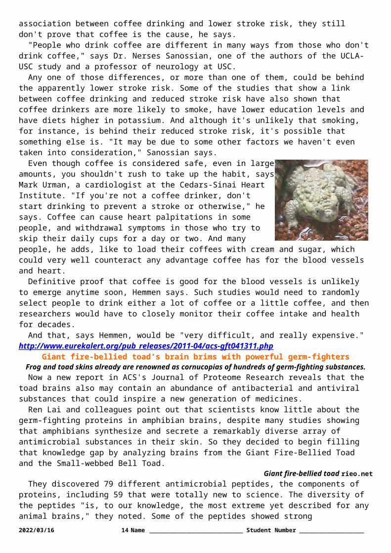
Giant fire-bellied toad's brain brims with powerful germ-fightersFrog and toad skins already are renowned as cornucopias of
hundreds of germ-fighting substances. Now a new report in ACS's Journal of Proteome Research reveals that the
toad brains also may contain an abundance of antibacterial and antiviral substances that could inspire a new generation of medicines.
Ren Lai and colleagues point out that scientists know little about the germ-fighting proteins in amphibian brains, despite many studies showing that amphibians synthesize and secrete a remarkably diverse array of antimicrobial substances in their skin. So they decided to begin filling that knowledge gap by analyzing brains from the Giant Fire-Bellied Toad and the Small-webbed Bell Toad.
Giant fire-bellied toad rieo.netThey discovered 79 different antimicrobial peptides, the components of proteins, including 59 that were
totally new to science. The diversity of the peptides "is, to our knowledge, the most extreme yet described for any animal brains," they noted. Some of the peptides showed strong antimicrobial activity, crippling or killing strains of staph bacteria, E. coli, and the fungus that causes yeast infections in humans. These promising findings suggest that the toad brains might be a valuable source for developing new antibacterial and antiviral drugs.The researchers acknowledge funding from the Chinese National Natural Science Foundation and the Ministry of Science and Technology.http://www.eurekalert.org/pub_releases/2011-04/jhmi-ead041311.php
Experimental Alzheimer's disease drugs might help patients with nerve injuries
Compounds helped nerve extensions re-grow faster in mouse studiesDrugs already in development to treat Alzheimer's disease may eventually be tapped for a different purpose
altogether: re-growing the ends of injured nerves to relieve pain and paralysis. According to a new Johns Hopkins study, experimental compounds originally designed to combat a protein that builds up in Alzheimer's-addled brains appear to make crushed or cut nerve endings grow back significantly faster, a potential boon for those who suffer from neuropathies or traumatic injuries.
The new drugs target a protein known as "-Site amyloid precursor protein cleaving enzyme 1," or BACE1, which plays a key role in generating the amyloid protein plaques that are thought to gum up normal nerve signaling in the brain. Previous laboratory research showed that BACE1 also is involved in creating the insulation material known as myelin, which coats the projections that nerve cells extend to connect with each other, as well as generating a molecular cascade that causes these projections to degenerate when they're injured.
Based on these earlier findings, assistant professor of neurology Mohamed Farah, Ph.D., professor of neurology John Griffin, M.D., and their colleagues tried blocking the action of BACE1 to analyze the effect on injured axon projections. The researchers started their experiments with mice whose ability to make BACE1 had been genetically knocked out. After these animals' sciatic nerves were cut or crushed, the scientists closely watched what happened as the axons regenerated.
2023/05/14 9 Name Student Number
Compared to normal mice that make BACE1, the animals lacking this protein cleaned up the debris around the injury site significantly faster. Since this debris can inhibit regeneration, Farah and his colleagues expected that the axons would re-grow faster. Sure enough, the cut ends of the animals' nerve cells generated more new sprouts, which grew into extensions that reached their targets - muscles or other nerve cells - days faster than the mice that made BACE1.
Hopeful that compounds able to block BACE1 activity would have a similar effect, Farah and Griffin's team worked with two experimental drugs already developed to target Alzheimer's disease (BACE1 inhibitor IV, produced by Calbiochem, and WAY 258131, a Wyeth compound that was synthesized by researchers at Johns Hopkins Brain Science Institute for this study). Mice given either of the two drugs systemically after nerve injuries had a similar increase in re-growth, though less pronounced. This was expected, explains Farah, since the drugs dampen the effect of BACE1 without removing it entirely as in the genetic knockout mice.
The Hopkins researchers said their proof of the principle work, published in the Journal of Neuroscience on April 13, was reason to celebrate. "Anything that speeds nerve re-growth could be enormously helpful to people with nerve injuries caused by a range of injuries and diseases, from diabetic neuropathy to motorcycle accidents," says Farah.
"After an injury, the environment around nerves and their target tissue sometimes degenerates before the nerves can heal, which kills the chances that the nerve will re-grow," he explains. "If we can help nerves re-grow faster, we increase the chances that they can reach their target and become healthy again after injury."
As a next step, the researchers plan to test the experimental compounds in other animal models of nerve injury, including neuropathies and spinal cord injuries.
"BACE1 inhibitors are a major drug target for many drug companies for Alzheimer's," says Griffin. "Our work may suggest that these drugs could have great utility in a very large clinical population with tremendous unmet need. Validation of our early research in other animal models of nerve injury will set the stage for further clinical investigation."Other Johns Hopkins researchers who participated in this study include Bao Han Pan, Ph.D., Paul N. Hoffman, M.D., Ph.D., Dana Ferraris, Ph.D., Takashi Tsukamoto, Ph.D., Thien Nguyen, M.D., Ph.D., Philip C. Wong, Ph.D., Donald L. Price, M.D., and Barbara S. Slusher, Ph.D., M.B.A.http://www.eurekalert.org/pub_releases/2011-04/uocp-cdi041311.php
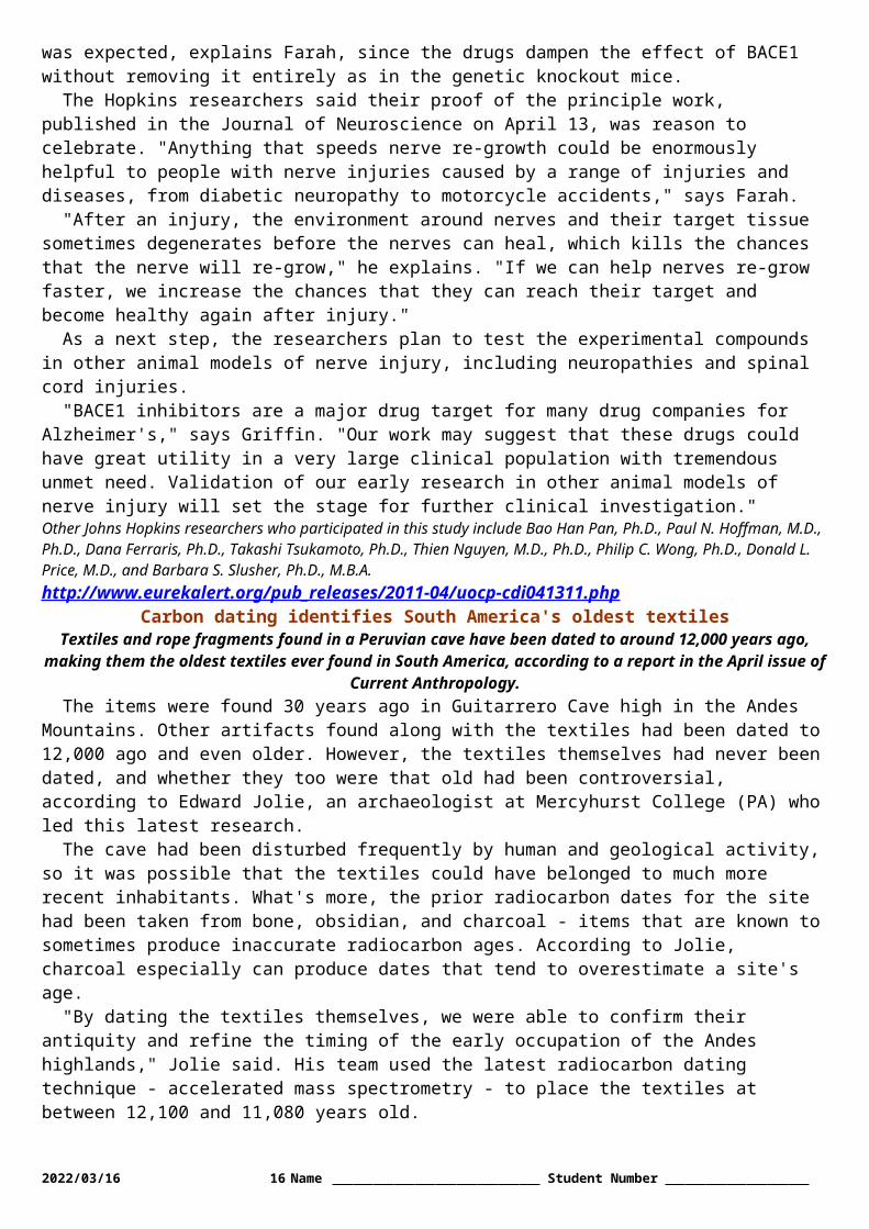
Carbon dating identifies South America's oldest textilesTextiles and rope fragments found in a Peruvian cave have been dated to around 12,000 years ago, making them the oldest textiles ever found in South America,
according to a report in the April issue of Current Anthropology.The items were found 30 years ago in Guitarrero Cave high in the Andes Mountains. Other artifacts found
along with the textiles had been dated to 12,000 ago and even older. However, the textiles themselves had never been dated, and whether they too were that old had been controversial, according to Edward Jolie, an archaeologist at Mercyhurst College (PA) who led this latest research.
The cave had been disturbed frequently by human and geological activity, so it was possible that the textiles could have belonged to much more recent inhabitants. What's more, the prior radiocarbon dates for the site had been taken from bone, obsidian, and charcoal - items that are known to sometimes produce inaccurate radiocarbon ages. According to Jolie, charcoal especially can produce dates that tend to overestimate a site's age.
"By dating the textiles themselves, we were able to confirm their antiquity and refine the timing of the early occupation of the Andes highlands," Jolie said. His team used the latest radiocarbon dating technique - accelerated mass spectrometry - to place the textiles at between 12,100 and 11,080 years old.
The textile items include fragments of woven fabrics possibly used for bags, baskets, wall or floor coverings, or bedding. They were likely left by settlers from lower altitude areas during "periodic forays" into the mountains, the researchers say. "Guitarrero Cave's location at a lower elevation in a more temperate environment as compared with the high Andean [plain] made it an ideal site for humans to camp and provision themselves for excursions to even higher altitudes," Jolie and his colleagues write.These early mountain forays set the stage for the permanent settlements that came later - after 11,000 years ago - when the climate had warmed, glaciers receded, and settlers had a chance to adapt to living at higher altitudes.
Jolie's research also suggests that women were among these earliest high altitude explorers. Bundles of processed plant material found in the cave indicate that textile weaving occurred on site. "Given what we know about textile and basket production in other cultures, there's a good possibility that it would have been women doing this work," Jolie said.
2023/05/14 10 Name Student Number
"There's an assumption that these early forays into the mountains must have been made exclusively by men," he added. "It appears that might not be the case, though more work needs to be done to prove it."Edward A. Jolie, Thomas F. Lynch, Phil R. Geib, and J. M. Adovasio, "Cordage, Textiles, and the Late Pleistocene Peopling of the Andes." Current Anthropology 42:2 (April 2011).http://www.eurekalert.org/pub_releases/2011-04/amon-lfm041111.php
Long-sought fossil mammal with transitional middle ear foundFossil from China suggests mammalian ear of monotremes evolved separately from
that of marsupials and placentalsPaleontologists from the American Museum of Natural History and the Chinese
Academy of Sciences announce the discovery of Liaoconodon hui, a complete fossil mammal from the Mesozoic found in China that includes the long-sought transitional middle ear. The specimen shows the bones associated with hearing in mammals - the malleus, incus, and ectotympanic - decoupled from the lower jaw, as had been predicted, but were held in place by an ossified cartilage that rested in a groove on the lower jaw. The new research, published in Nature this week, also suggests that the middle ear evolved at least twice in mammals, for monotremes and for the marsupial-placental group.
"People have been looking for this specimen for over 150 years since noticing a puzzling groove on the lower jaw of some early mammals, " says Jin Meng, curator in the Division of Paleontology at the Museum and first author of the paper. "Now we have cartilage with ear bones attached, the first clear paleontological evidence showing relationships between the lower jaw and middle ear."
Mammals - the group of animals that includes egg-laying monotremes like the platypus, marsupials like the opossum, and placentals like mice and whales - are loosely united by a suite of characteristics, including the middle ear ossicles. The mammalian middle ear, or the area just inside the ear drum, is ringed in shape and includes three bones, two of which are found in the joint of the lower jaw of living reptiles. This means that during the evolutionary shift from the group that includes lizards, crocodilians, and dinosaurs to mammals, the quadrate and articular plus prearticular bones separated from the posterior lower jaw and became associated with hearing as the incus and malleus.
This is Liaoconodon hui, a fossil mammal from China. Credit: Meng, et al 2011 ( Nature)The transition from reptiles to mammals has long been an open question, although studies of developing
embryos have linked reptilian bones of the lower jaw joint to mammalian middle ear bones. Previously discovered fossils have filled in parts of the mammalian middle-ear puzzle. An early mammal, Morganucodon that dates to about 200 million years ago, has bones more akin to a reptilian jaw joint but with a reduction in these bones, which functioned for both hearing and chewing. Other fossils described within the last decade have expanded information about early mammals - finding, for example, that ossified cartilage still connected to the groove was common on the lower jaws of early mammals. But these fossils did not include the bones of the middle ear.
The new fossil described this week, Liaoconodon hui, fills the gap in knowledge between the basal, early mammaliaforms like Morganucodon, where the middle ear bones are part of the mandible and the definitive middle ear of living and fossil mammals. Liaoconodon hui is a medium-sized mammal for the Mesozioc (35.7 cm long from nose to tip of tail, or about 14 inches) and dates from 125 to 122 million years. It is named in part for the bountiful fossil beds in Liaoning, China, where it was found. The species name, hui, honors paleontologist Yaoming Hu who graduated from the American Museum of Natural History-supported doctoral program and recently passed away. The fossil is particularly complete, and its skull was prepared from both dorsal and ventral sides, allowing Meng and colleagues to see that the incus and malleus have detached from the lower jaw to form part of the middle ear. These bones remain linked to the jaw by the ossified Meckel's cartilage that rests in the groove on the lower jaw. The team hypothesizes that in this early mammal, the ear drum was stabilized with the ossified cartilage as a supporting structure.
"Before we did not know the detailed morphology of how the bones of the middle ear detached, or the purpose of the ossified cartilage," says Meng. "Liaoconodon hui changes previous interpretations because we now know the detailed morphology of the transitional mammal and can propose that the ossified cartilage is a stabilizer."
Also presented in the new research paper is a detailed phylogenetic analysis of some features of living and fossil mammals. Looking at features associated with bones and the groove on the lower jaw, which indicated
2023/05/14 11 Name Student Number

the presence of ossified Meckel's cartilage, it appears that the middle ear probably evolved twice, in monotremes and in placentals and marsupials.
"I've always dreamed of a fossil with a good ear ossicle," says Meng. "Now, we have had this once in a lifetime discovery."In addition to Meng, authors of the paper include Wang Yuanqing and Li Chuankui, both of the Institute of Vertebrate Paleontology and Paleoanthropology, Chinese Academy of Sciences in Beijing. The research (doi:10.1038/nature09921) was funded by Major Basic Research Project of the Ministry of Science and Technology, China, the National Science Foundation of China, the Special Fund for Fossil Excavation and Preparation of the Chinese Academy of Sciences, and the National Science Foundation of USA.http://www.eurekalert.org/pub_releases/2011-04/ps-srb040811.php
Scientists recreate brain cells from skin cells to study schizophrenia safelyA team of scientists at Penn State University, the Salk Institute for Biological Studies,
and other institutions have developed a method for recreating a schizophrenic patient's own brain cells, which then can be studied safely and effectively in a Petri
dish.The method brings researchers a step closer to understanding the biological underpinnings of schizophrenia.
The method also is expected to be used to study other mysterious diseases such as autism and bipolar disorder, and the researchers hope that it will open the door to personalized medicine -- customized treatments for individual sufferers of a disease based on genetic and cellular information. The study will be published in a future edition of the journal Nature and will be posted on the journal's advance online website on 13 April 2011.
Gong Chen, an associate professor of biology at Penn State and one of the study's authors, explained that the team first took samples of skin cells from schizophrenic patients. Then, using molecular-biology techniques, they reprogrammed these original skin cells to become unspecialized or undifferentiated stem cells called induced pluripotent stem cells (iPSCs). "A pluripotent stem cell is a kind of blank slate," Chen explained. "During development, such stem cells differentiate into many diverse, specialized cell types, such as a muscle cell, a brain cell, or a blood cell."
After generating iPSCs from skin cells, the authors cultured them to become brain cells, or neurons. They then compared the neurons derived from schizophrenic patients to the neurons created from the iPSCs of healthy individuals. They found that the neurons generated from schizophrenic patients were, in fact, distinct: compared with healthy neurons, they made fewer connections with each other. Kristen Brennand, a Salk researcher and one of the study's authors, then administered a number of frequently prescribed antipsychotic medications to test the drugs' ability to improve how neurons communicate with neighboring cells. "Now, for the very first time, we have a model system that allows us to study how antipsychotic drugs work in live, genetically identical neurons from patients with known clinical outcomes, and we can start correlating pharmacological effects with symptoms," Brennand said.
Chen, who contributed to the study by using electrophysiology techniques to test the function of the iPSC-derived neurons, described the new method as "patient specific," offering a step toward personalized medicine for sufferers of schizophrenia and potentially other diseases. "What's so exciting about this approach is that we can examine patient-derived neurons that are perhaps equivalent to a particular patient's own neural cells," Chen said. "Obviously, we don't want to remove someone's brain cells to experiment on, so recreating the patient's brain cells in a Petri dish is the next best thing for research purposes. Using this method, we can figure out how a particular drug will affect that particular patient's brain cells, without needing the patient to try the drug, and potentially, to suffer the side effects. The patient can be his or her own guinea pig for the design of his or her own treatment, without having to be experimented on directly."
Lead author Fred Gage, a professor at Salk's Laboratory of Genetics and holder of the Vi and John Adler Chair for Research on Age-Related Neurodegenerative Diseases, explained that schizophrenia exemplifies many of the research challenges posed by complex psychiatric disorders. "This model not only affords us the opportunity to look at live neurons from schizophrenia patients and healthy individuals to understand more about the disease mechanism, but also it allows us to screen for drugs that may be effective in reversing it," Gage said.
Schizophrenia, which is defined by a combination of paranoid delusions, auditory hallucinations, and diminished cognitive function, afflicts one percent of the population worldwide, corresponding to nearly three million people in the United States alone. Genetic evidence indicates that many different combinations of genetic lesions -- some of them affecting the susceptibility to environmental influences -- may lead to a variety of signs and symptoms collectively labeled schizophrenia.
2023/05/14 12 Name Student Number

"Nobody knows how much the environment contributes to the disease," said Brennand. "By growing neurons in a dish, we can take the environment out of the equation and start focusing on the underlying biological problems." In another part of the study, Brennand used a modified rabies virus, developed by Salk professors Edward Callaway and John Young, to highlight the connections between neurons. The viral tracer made it apparent that the schizophrenic neurons connected less frequently with each other and had fewer projections growing out from their cell bodies. In addition, gene-expression profiles identified almost 600 genes whose activity was misregulated in these neurons; 25 percent of those genes had been implicated in schizophrenia before.
Gage added that, for many years, mental illness has been thought of as a strictly social or environmental disease. "Many people believed that if affected individuals just worked through their problems, they could overcome them," he said. "But we are showing real biological dysfunctions in neurons that are independent of the environment."In addition to Gage, Brennand, and Chen, other researchers who contributed to the study include Anthony Simone, Jessica Jou, Chelsea Gelboin-Burkhart, Ngoc Tran, Sarah Sangar, Yan Li, Yanglin Mu and Diana Yu in the Gage Laboratory; Shane McCarthy at the Cold Spring Harbor Laboratory in New York; and Jonathan Sebat at the University of California at San Diego.The work was funded, in part, by the California Institute for Regenerative Medicine, the Lookout Foundation, the Mathers Foundation, and the Helmsley Foundation. [ Katrina Voss / Gina Kirchweger ]http://www.eurekalert.org/pub_releases/2011-04/uos-lfs041211.php
Loch fossils show life harnessed sun and sex early onRemote lochs along the west coast of Scotland are turning up new evidence about the
origins of life on land.A team of scientists exploring rocks around Loch Torridon have discovered the remarkably preserved
remains of organisms that once lived on the bottom of ancient lake beds as long as a billion (1000 million) years ago.
These fossils illuminate a key moment in the history of evolution when life made the leap from tiny, simple bacterial (prokaryote) cells towards larger, more complex (eukaryotic) cells which would make photosynthesis and sexual reproduction possible.
The team, from Oxford University, the University of Sheffield and Boston College, report their findings in this week's Nature.
"These new fossils show that the move toward complex algal cells living in lakes on land had started over a billion years ago, much earlier than had been thought," said Professor Martin Brasier of Oxford University's Department of Earth Sciences, an author of the paper.
"These new cells differ from their bacterial ancestors in that they have specialised structures including a nucleus, as well as mitochondria and chloroplasts – which are vital for photosynthesis. They also undergo sexual reproduction, leading to much more rapid rates of evolutionary turnover."
Some of these ancient fossils are so finely ornamented, and so large and complex, that they are evidence for a surprisingly early start for the emergence of complex eukaryote cells on land. The researchers believe that it was from complex cells such as these that green algae and green land plants – everything from lettuce to larch trees – were able to evolve and colonise the land.
Dr Charles Wellman, Reader in Palaeobiology in the Department of Animal and Plant Sciences at the University of Sheffield, an author of the paper, said: "It is generally considered that life originated in the ocean and that the important developments in the early evolution of life took place in the marine environment: the origin of prokaryotes, eukaryotes, sex, multicellularity etc. During this time the continents are often considered to have been essentially barren of life - or at the most with an insignificant microbial biota dominated by cyanobacteria.
"We have discovered evidence for complex life on land from 1 billion year old deposits from Scotland. This suggests that life on land at this time was more abundant and complex than anticipated. It also opens the intriguing possibility that some of the major events in the early history of life may have taken place on land and not entirely within the marine realm."
Professor Brasier said: "It may even be that the sort of conditions found in the ancient lakes around Loch Torridon favoured a key step in this transformation, which involved the incorporation of symbiotic bacteria into the cell to form chloroplasts, rather than this occurring in the sea as usually envisaged."Around 500 million years after the emergence of these complex cells, the surface of the land was starting to become covered in simple vegetation like lichens, mosses and liverworts, and the first animals were able to take their chance and leave the sea behind. These pioneers were followed by the first fish and ferns, reptiles and conifers, mammals and flowering plants – and, eventually, humans.2023/05/14 13 Name Student Number
Professor Brasier adds: "None of this would have been possible without advances long ago made by these little microbes, now entombed within phosphate from the Torridon lakes. It was arguably these organisms that helped to turn our landscape from a harsh and rocky desert into a green and pleasant place."
http://www.physorg.com/news/2011-04-climate-seismic-shifts.htmlMonsoons spinning the Earth's plates: study
Scientists have for the first time shown a link between intensifying climate events and tectonic plate movement in findings that could provide a valuable insight into
why huge tremors occur.(PhysOrg.com) -- Scientists have for the first time shown a link between intensifying climate events and
tectonic plate movement in findings that could provide a valuable insight into why huge tremors occur.A new study from The Australian National University has for the first time confirmed that long-term climate
change has the potential to spin the Earth’s tectonic plates.Dr Giampiero Iaffaldano from the ANU Research School of Earth Sciences and colleagues in France and
Germany have established a link between the motion of the Indian plate over the past 10 million years and a specific climate change event over the same period: the intensification of the Indian monsoon.
Dr Iaffaldano said that the monsoon, which increased rainfall in northeast Indian by four metres annually, sped up motion in the Indian plate by almost one centimetre per year.“The 100km-thick outer shell of Earth, the lithosphere, is divided into pieces called tectonic plates. Plates
move in different directions at speeds in the order of centimetres per year, comparable to the speed of fingernail growth in humans.“The significance of this finding lies in recognising for the first time that long-term climate changes have the
potential to act as a force and influence the motion of tectonic plates. It is known that certain geologic events caused by plate motions – for example the drift of continents, the closure of ocean basins and the building of large mountain belts – have the ability to influence climate patterns over a period of a million years.“Now we know that the opposite holds as well: long-term climate change, or the natural changes in climate
patterns over millions of years, can modify the motion of plates in a feedback mechanism.”Dr Iaffaldano added that the finding could help unlock the causes of plate-motion events like large-scale
earthquakes.“When forces moving plates along their boundaries reach certain thresholds, earthquakes occur and energy
is released. This happens cyclically, typically every several hundred years in the case of large earthquakes. However it appears that the seismic potential of plate boundaries, which is an indication of how prone these are to large earthquakes, depends, among other factors, also on how strong or weak these forces have been in the past. In other words, it depends also on the history of plates over millions of years.“In order to understand the seismic potential of plate boundaries it is important to identify all the possible
factors that caused plate motion to change in the past. In that respect we have discovered that climate change could in fact be one possible candidate, something we did not consider until now.“This new knowledge shall be used to analyse the past behaviour of plates in the Earth’s crust. Ultimately
we aim at understanding what caused plate motions to change and which regions are currently more prone to large earthquakes. To that end, we may also have to consider the history of climate over the past million years.”More information: Monsoon speeds up Indian plate motion, Earth and Planetary Science Letters, Volume 304, Issues 3-4, 15 April 2011, Pages 503-510. doi:10.1016/j.epsl.2011.02.026 http://dx.doi.org/ … .2011.02.026Provided by Australian National Universityhttp://www.newscientist.com/article/mg21028083.100-genes-from-algae-allow-blind-mice-to-see.html
Genes from algae allow blind mice to see* Updated 17:17 14 April 2011 by Rowan Hooper
BLIND people could one day have their sight restored thanks to a treatment that borrows a gene from an unlikely source - algae - and inserts it into the retina.
The technique has succeeded in restoring the ability to sense light and dark to blind mice, and clinical trials in humans could begin in as little as two years.
"The idea is to develop a treatment for blindness," says Alan Horsager, a neuroscientist at the Institute of Genetic Medicine at the University of Southern California, Los Angeles, who leads the research. "We introduce a gene that encodes a light-sensitive protein, and we target the expression of that gene to a subset of retinal cells."
2023/05/14 14 Name Student Number
Some 15 million people worldwide have some form of blindness, such as retinitis pigmentosa (RP) or age-related macular degeneration (AMD). In people with these conditions the photoreceptors, which transform light hitting the eye into electrical impulses, are damaged, preventing the brain from receiving image information.
As the global population ages, it is thought that the number of people affected will increase. There are experimental attempts to develop electronic implantsMovie Camera and to use stem cells to grow new retinal tissues to restore sight, but there is currently no commercial treatment available.
Horsager hopes his work will change that. His team's approach is based on gene therapy, where a "tame" virus is harnessed to transfer a gene into target cells in the recipient. In this case the gene of interest is one that makes Channelrhodopsin-2 (ChR2), a photosensitive protein used by unicellular algae to help them move towards light. The target cells are bipolar cells in the retina.
The retina contains three cellular layers that work together to detect and transmit light signals to the brain (see diagram). The first layer contains the photoreceptors - the rods and cones that detect light. The second layer is made of bipolar cells that act as a conduit between the photoreceptor and the third type of cell, the ganglion, which transmits the light signals to the brain.
In people with RP and AMD, the photoreceptors have been damaged and lost, so the ganglion cells do not receive signals and the brain cannot produce an image. The idea behind the gene therapy is to make the bipolar cells function as photoreceptors by producing ChR2. The modified bipolar cell would then be able to sense light and transmit a signal to the ganglion.
Horsager's team tested their technique using three groups of mice: one with normal vision, and two groups of mouse strains that naturally become blind with age in a similar way to people with RP and AMD. One blind group was treated with the gene therapy, while the other two groups were not.
Treated mice received a sub-retinal injection of the virus containing the algal gene. Ten weeks after the injection, the team dissected some of the mice and used immunolabelling to see whether ChR2 was being expressed in the retina. They found that the protein was being made by the bipolar cells.
But the strongest evidence of the treatment's success came when treated mice were put in the centre of a water maze with six possible corridors, only one of which led to a ledge that the mice could clamber out of the water onto. With a guiding light shining at the end of the corridor which contained the ledge, the gene-therapy mice were able to find the escape platform 2.5 times faster, on average, than the untreated blind mice. The work will appear in Molecular Therapy.
Repeating the test 10 months later, the team found that the treated mice were still showing significant improvements in vision compared with the untreated blind mice. "Our expectation is that this would be a one-time treatment that is permanent or semi-permanent," says Horsager.
Concerns have been raised about the safety of gene therapy in the past, not least about links between the viruses used to transfer the genes and disease. Horsager says the algal genes were only expressed in the target cells, and that there is no evidence of an immune response in the mice, suggesting that the transfer of the foreign gene has been restricted to the bipolar cells.
However, small amounts of ChR2 DNA were found in other tissues. "Regulatory agencies would be very concerned that ChR2 DNA was found in tissues outside of the treated eye," says Robert Lanza, of Advanced Cell Technology in Worcester, Massachusetts. Horsager's team believe the rogue DNA is due to cross-contamination during the analysis process.
"It's a good paper, and it's clear that they are heading towards a clinical trial with the information they are gathering," says Pete Coffey of the department of ophthalmology at University College London. But he points out that although there is a statistical difference between the performance of the treated and untreated mice, that difference is small.
2023/05/14 15 Name Student Number
Coffey also adds that, as Horsager and colleagues admit, the mice seem to be seeing the difference between light and dark, but not much more. Nevertheless, he thinks this sort of technology will be seen in the clinic before a treatment based on a stem cell replacement for photoreceptors. That's because stem cells must be connected to existing neural networks - something that's not yet possible - whereas gene therapy simply involves making what is left in a diseased eye photosensitive.
"The question," says Coffey, "is how good is it going to be? Just light/dark or are people going to be able to read large texts?"
Horsager's team is trying to go beyond simple light/dark discrimination by precisely activating particular cells in the retinal system. However, the tests used so far don't say much about visual acuity.
"If you can get acuity back it would be phenomenal for anyone who's been blind," says Coffey.The last sentence of the paragraph beginning: "However, small amounts of ChR2 DNA…" was added after
this article was first posted. In addition, the penultimate paragraph initially stated, incorrectly, that Horsager's method treats all bipolar cells equally.http://www.physorg.com/news/2011-04-higher-ccsvi-prevalence-ms-unclear.html
Higher CCSVI prevalence confirmed in MS, but meaning of findings remains unclear
A just released study on the relationship between multiple sclerosis (MS) and chronic cerebral venous insufficiency (CCSVI), a narrowing of the extracranial veins that
restricts the normal outflow of blood from the brain, found that CCSVI may be a result of MS, not a cause.
The study, conducted by University at Buffalo researchers, appears in the current issue of Neurology, the journal of the American Academy of Neurology.
Robert Zivadinov, MD, PhD, associate professor of neurology in the UB School of Medicine and Biomedical Sciences and president of the International Society for Neurovascular Disease, is first author on the paper. Zivadinov says of the findings: "Given the intense interest in the hypothesis that CCSVI is a possible cause of MS, independent evaluation of CCSVI was identified as an urgent need.
"Our results indicate that only 56.1 percent of MS patients and 38.1 percent of patients with a condition known as clinically isolated syndrome (CIS), an individual's first neurological episode, had CCSVI.
"While this may suggest an association between the MS and CCSVI, association does not imply causality. In fact, 42.3 percent of participants classified as having other neurological diseases (OND), as well as 22.7 percent of healthy controls involved in the study, also presented with CCSVI.
"These findings indicate that CCSVI does not have a primary role in causing MS," says Zivadinov. "Our findings are consistent with increased prevalence of CCSVI in MS, but substantially lower than the sensitivity and specificity rates in MS reported originally by the Italian investigators."
CCSVI is a complex vascular condition discovered and described by Paolo Zamboni, MD, from Italy's University of Ferrara. It is characterized by narrowing of vessels draining blood from the cranium. Zamboni hypothesized that this narrowing restricts the normal outflow of blood from the brain, resulting in alterations in the blood flow patterns within the brain that eventually cause injury to brain tissue and degeneration of neurons, leading to MS.
Zamboni's original investigation in a group of 65 patients and 235 controls showed that CCSVI appeared to be strongly associated with MS, increasing the risk of having MS by 43 fold.
The results of the UB study are based on 499 participants in the Combined Transcranial and Extracranial Venous Doppler Evaluation (CTEVD) study, which began at the university in April 2009.
The study group consisted of 289 persons with MS, 163 healthy controls, 26 with OND and 21 with CIS.MS patients also were defined by disease type: relapsing-remitting (RR), secondary progressive (SP),
primary-progressive (PP), progressive-relapsing (PR) and MS with neuromyelitis optical (NMO) -- a type of MS that affects the optic nerves and spinal cord exclusively.
All patients underwent transcranial and extracranial echo-Doppler scans of the head and neck. Persons were considered "CCSVI-positive" if they met two or more of five venous hemodynamic (VH) criteria.Prevalence rates were calculated in three groupings: only subjects with positive and negative CCSVI diagnoses; only borderline cases included in the negative group; and subjects who fulfilled any of the five criteria.
When only positive and negative CCSVI cases were considered, results showed a CCSVI prevalence of 62.5 percent in MS patients, 45.8 percent in those with OND, 42.1 percent in CIS, and 25.5 percent in healthy controls. When borderline cases were included as negative for CCSVI, prevalence figures were 56.1 percent in MS patients, 42.3 percent in those with OND, 38.1 percent with CIS and 22.7 percent in healthy controls.
2023/05/14 16 Name Student Number
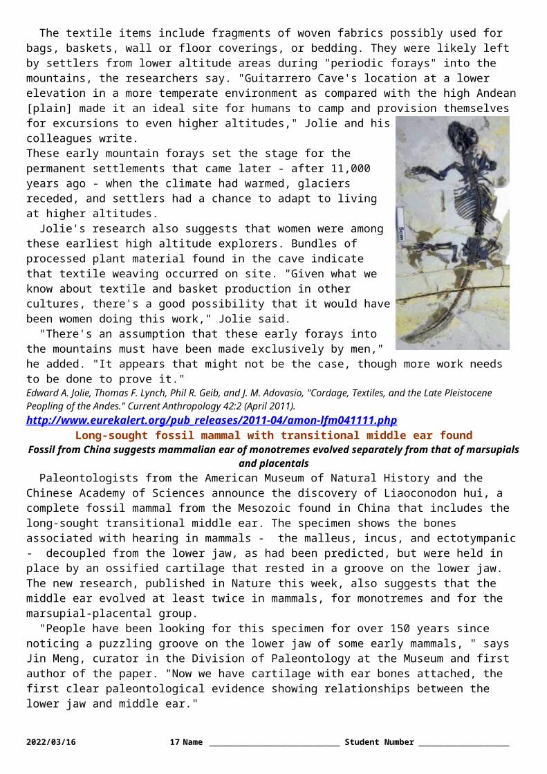
When all cases that met at least one of the five VH criteria were included in the analysis, CCSVI prevalence was 81.3 percent in MS cases, 76.2 percent in CIS patients, 65.4 percent in OND cases and 55.2 percent in healthy controls.
The highest prevalence was seen in relapsing primary-progressive MS (89.4 percent), followed by non-relapsing secondary-progressive MS (67.2 percent), NMO (66.6 percent), primary-progressive MS (54.5 percent) and relapsing-remitting MS (49.2 percent). CCSVI prevalence was substantially higher in progressive MS than in non-progressive MS patients. In addition, patients with a progressive MS disease subtype had higher CCSVI prevalence than those with non-progressive MS.
"The higher prevalence of CCSVI in progressive MS patients suggests that CCSVI may be a consequence, rather than a cause, of MS," says Bianca Weinstock-Guttman, MD, co-principal investigator of the study and UB professor of neurology. Therefore, the possibility that CCSVI may be a consequence of MS progression cannot be excluded and should be further investigated.
"Several studies have reported that patients with progressive MS show decreased blood flow through the brain's neuronal tissue, indicating that CCSVI may be secondary to reduced perfusion," says Weinstock-Guttman. "In addition, we recently showed an association between the severity of CCSVI and reduced cerebral blood flow in brain parenchyma of MS patients in an published pilot study."
E. Ann Yeh, MD, UB assistant professor of neurology and a major collaborator on the study, noted that of the 10 pediatric MS patients who participated in the study, five presented with CCSVI (50 percent), yielding prevalence similar to that in adult MS patients. "Although the sample size was too small to draw any firm conclusions, these results suggest that CCSVI is also present in children and is not the result of aging," she says.Concludes Zivadinov: "The differences between our study, the original Italian CCSVI study and other recently published studies also emphasize the need for a multimodal approach for the assessment of CCSVI. In addition to Doppler sonography, use of selective venography, magnetic resonance venography and intraluminal Doppler methods can provide more evidence for the true prevalence of CCSVI in MS." Provided by University at Buffalohttp://www.eurekalert.org/pub_releases/2011-04/e-sa041411.php
Serotonin: A critical chemical for human intimacy and romancePhiladelphia, PA - The judgments we make about the intimacy of other couples'
relationships appear to be influenced by the brain chemical serotonin, reports a new study published in Biological Psychiatry.
Healthy adult volunteers, whose levels of serotonin activity had been lowered, rated couples in photos as being less intimate and less romantic than volunteers with normal serotonin activity.
The approach involved giving amino acid drinks to two groups of volunteers in order to manipulate blood concentrations of the amino acid tryptophan, which is a vital ingredient in the synthesis of serotonin. One group received drinks that contained tryptophan. The other group received drinks that did not contain tryptophan. They were then asked to make judgments about sets of photographs of couples. Differences in the judgments made by the two groups reflected changes in their serotonin activity.
"Serotonin is important in social behavior, and also plays a significant role in psychological disorders such as depression," explained Professor Robert Rogers of Oxford University, who led the research. "We wanted to see whether serotonin activity influences the judgments we make about peoples' close personal relationships."
The volunteers who received the drink without tryptophan consistently rated the couples in the photos as being less 'intimate' and 'romantic' than the participants who received the control drink.
This finding is an important reminder that our relationships with other people are influenced by processes beyond our awareness and control. But we should not be surprised by this revelation. Serotonin function drops in association with episodes of depression, where the capacity for intimacy also is often compromised.
Understanding the powerful influence of these chemicals is important as supportive close relationships are known to protect against the development of mental illnesses and to promote recovery in those affected by psychiatric conditions. The opposite is also true: dysfunctional relationships can be triggers for those at risk of these conditions.
The results raise the possibility that lower serotonin activity in people with depression and other psychiatric conditions could contribute to changes in the way they perceive personal relationships, or even in their ability to maintain positive personal relationships.
"Although this is only a small study, the same patterns may well extend to the way we perceive our own relationships," said Professor Rogers.
"The ability to chemically influence the capacity for intimacy could be very important. Reduced capacity for intimacy can be a vexing symptom of many psychiatric disorders and an important target for treatment," noted 2023/05/14 17 Name Student Number

Dr. John Krystal, Editor of Biological Psychiatry. "Drugs that ameliorate the impact of serotonin deficits might play a role in the treatment of this symptom."
Although much more research is necessary before a drug might come to market that can help promote intimacy, it is clear for now that our chemistry has an impact on nearly aspect of our lives, from our most public actions to our most private, as we see here with human intimacy and romantic feelings.Notes to Editors: The article is "Serotonergic Activity Influences the Cognitive Appraisal of Close Intimate Relationships in Healthy Adults" by Amy C. Bilderbeck, Ciara McCabe, Judi Wakeley, Francis McGlone, Tirril Harris, Phillip J. Cowen, and Robert D. Rogers. Bilderbeck, McCabe, Wakeley, Cowen, and Rogers are affiliated with Oxford University, Oxford, United Kingdom. McGlone is affiliated with University of Liverpool, Liverpool, United Kingdom. Harris and Cowen are from King's College, London, United Kingdom. The article appears in Biological Psychiatry, Volume 69, Number 8 (April 15, 2011), published by Elsevier.http://www.eurekalert.org/pub_releases/2011-04/uoc--eor041411.php
Eyes of rock let chitons see predatorsSanta Barbara, Calif. – Using eyes made of a calcium carbonate crystal, a simple mollusk
may have evolved enough vision to spot potential predators, scientists say.Daniel Speiser, a postdoctoral fellow in the Department of Ecology Evolution and Marine Biology at UC
Santa Barbara, studied mollusks that he collected in the Florida Keys. His research of their vision, performed during his graduate studies at Duke University, resulted in a study published today by Current Biology.
The three-inch-long mollusks, called chitons, have hundreds of eye-like structures with lenses made of aragonite, a type of rock. It's the first time scientists have found an animal that makes eye lenses from aragonite and not the rock's close cousin, calcite. This lined chiton, whose anterior end is to the right, lives about 50 feet below the water's surface near Whidbey Island,
Washington. Credit: Kirt L. OnthankScientists discovered the chiton's unique eyes decades ago. But it wasn't clear whether chitons used these
eyes to see objects overhead, or simply to sense changes in light. "Turns out they can see objects, though probably not well," said Speiser.
"It's surprising how these creatures make their eyes from rocks," said Sönke Johnsen, associate professor of biology at Duke. Most animals make their eyes from cells with proteins and chitin. "But it seems like an easy way to evolve eyes, by using what you've already got," he said. Chitons also make their shells from aragonite.
Johnsen and Speiser studied West Indian fuzzy chitons, or Acanthopleura granulata, which have flat shells made of eight separate plates. Hundreds of tiny lenses on the surface of the plates cover clusters of light-sensitive cells beneath.
To test the creature's vision, Speiser placed individual chitons on a slate slab. When left undisturbed, they lift part of their armored, oval-shaped body to breathe. At this point, Speiser showed them either a black disk ranging from .35 centimeters to 10 centimeters in diameter or a corresponding gray slide that blocked the same amount of light. The disk or slide appeared 20 centimeters above the chitons.
When shown the gray screens, the chitons did not respond. But they clamped down when shown a black disk three centimeters or larger in diameter. That would be the equivalent of humans looking in the sky and seeing a disk the diameter of 20 moons, making human vision about a thousand times sharper than chiton vision, Johnsen said.
Because the chitons responded to the larger disks and not the gray slides, they seem to be seeing the disk and not simply responding to a change in light, said University of Sussex biologist Michael Land, an expert on animal vision who was not involved in the research. It's not yet clear if they respond only to the removal of light by the disk as opposed to added light.
This drawing shows the way light would bend and focus in the chiton's eye depending on whether the animal was above or below water. Credit: Daniel Speiser and Current Biology
Land also said it's not likely that the chitons' eyes were part of the evolutionary route to human eyes.Chitons are an ancient, primitive species that first appeared on Earth more than 500 million years ago. But
the oldest chitons with eyes only began to appear in the fossil record in the last 25 million years, making their
2023/05/14 18 Name Student Number
eyes among the most recent to evolve in animals. Speiser said chitons probably evolved to have eyes with lenses so they could see their predators and defend themselves against being eaten.
Speiser and his colleagues also tested whether the chitons' eyes work in both air and water, since some species spend time in both. The experiments made a strong case for the chiton lens being able to focus light differently, depending on whether the animal is above or below water, Land said.
He added that chiton eyes are still an anomaly in the evolution of vision. The retinas are structurally similar to snail and slug retinas. But snail and slug retinas respond to the appearance of light, while chiton retinas may only respond to the removal of light, a difference that might be worth another look, Land said.
http://www.nytimes.com/2011/04/14/world/asia/14quake.htmlIn Japan, Aftershocks Are Also Felt From Within
By ANDREW POLLACK and KEITH BRADSHERTOKYO — Aguri Suzuki, a 44-year-old real estate agent, says she sometimes thinks the ground is shaking even when it is not. When she sees a tree branch swaying in the
wind, she worries there has been an earthquake.Doctors here say they are seeing more people who are experiencing such phantom quakes, as well as other
symptoms of “earthquake sickness” like dizziness and anxiety.And it is no wonder. As if the threat of radiation from a crippled nuclear power plant were not enough,
Tokyo and the region to its northeast have been under a constant barrage of aftershocks since the magnitude 9.0 earthquake that set off a devastating tsunami on March 11. Two earthquakes were felt in Tokyo on Wednesday morning, three on Tuesday, a large one on Monday and a very large one of magnitude 7.1 last Thursday.
Over all, there have been 400 aftershocks of magnitude 5.0 or greater in northeastern Japan since March 11. That is as many sizable quakes in one month as Japan typically experiences in two and a half years, according to the Japan Meteorological Agency.
The quakes are complicating efforts to control the Fukushima Daiichi nuclear power plant. For instance, the quake on Monday knocked out cooling at the Fukushima plant for nearly an hour.
Every time a sizable quake occurs, the first question on many people’s minds is whether the nuclear plant has been further damaged and whether a new cloud of radiation is on the way. A spokesman for the Tokyo Electric Power Company, the plant’s owner, is then hustled onto television to reassure viewers.
Government officials are becoming concerned that in the rush to cool the reactors and prevent hydrogen explosions, the plant’s vulnerability to another tsunami has been overlooked.“A week ago we thought the major risk was a hydrogen explosion,” a senior official in the office of the
prime minister said Tuesday. “I think the major risk at the moment is an aftershock and tsunami.”Hidehiko Nishiyama, the deputy director general of Japan’s nuclear regulator, the Nuclear and Industrial
Safety Agency, said at a news conference on Wednesday evening that three measures are being considered that would allow electricity and cooling at the plant to remain intact even after a tsunami measuring 15 meters, or 49 feet. Right now the site can withstand a tsunami of only about 18 feet, he said. One measure is to interconnect the external power lines that have been built to the power plant, so that if one power line is broken, the others can still carry electricity to the various reactors. A second measure is to put a generator on a small hill inside the plant site, and the third is to place a fire pumper engine on the hill that could send water into the reactors and spent fuel pools even if electricity was interrupted.
Japan, which sits atop four colliding tectonic plates, has a long history of earthquakes and some sophisticated technology to deal with them. A detection system transmits warnings of some pending quakes a few seconds in advance to television broadcasters and to many cellphones. In recent weeks it has not been unusual to see nearly all the people in a restaurant or a train suddenly look at their cellphones at the same time.
Yurekuru, a free app for the iPhone that delivers such warnings (its name might be translated as “the shaking is coming”), now has 1.5 million users, compared with only 100,000 before the March 11 quake, according to RC Solution, the app’s developer.
Geologists say the frequency of the aftershocks has declined since March 11 and will continue to decline, but will still remain higher than normal for a long time. “There is an increased frequency and it will last for at least five or ten years,” said Ross S. Stein, a geophysicist at the U.S. Geological Survey in Menlo Park, Calif., who has studied the situation in Japan.
2023/05/14 19 Name Student Number

The March 11 quake was so strong that a Japan Coast Guard monitoring instrument on the floor of the Pacific Ocean near the epicenter moved 24 meters, or about 79 feet, eastward. The city of Sendai, whose airport was inundated by the tsunami, moved about 13 feet, according to Shinji Toda, a professor at Kyoto University.
Such large movements have shifted stresses in the earth, increasing the likelihood of quakes on some fault lines while reducing the likelihood on others, including the one involved in the 1923 Tokyo earthquake.
But over all, Dr. Stein said, the risks have increased. “There’s this very broad turn-on of seismicity that extends 300 miles from the rupture zone,” he said.
Satoko Oki, an assistant professor at the University of Tokyo’s earthquake research institute, said that an aftershock of the March 11 quake could reach magnitude 8.0.
There is some precedent. The 2004 quake of magnitude 9.1 near Sumatra, Indonesia, which spawned a tsunami that killed more than 200,000 people, was followed three months later by one measuring 8.6 and later by four more huge ones. But the 8.8 magnitude earthquake in Chile in early 2010 has not yet produced an aftershock larger than 7.1, Dr. Stein said.
To be sure, the spate of earthquakes has not caused the same panic and mass exodus as the fears of radiation in the first week after the nuclear crisis began. Still, with levels of radiation in the Tokyo air having sharply fallen since then, some people interviewed on the street said they worried about the aftershocks more than radiation.
Dr. Hideaki Sakata, director of the Mejiro University Clinic, who is treating Ms. Suzuki, said that feeling the ground shaking when it is not is similar to the continued feeling of swaying when one first gets off a boat onto solid ground.
Dr. Kazuhiro Soeda, an ear, nose and throat specialist in Utsunomiya, outside Tokyo, who also treats patients having trouble dealing with the aftershocks, said: “People are getting too sensitive. This is something we’ve never experienced before.” Makiko Inoue, Kantaro Suzuki and Ken Ijichi contributed reporting.http://www.physorg.com/news/2011-04-chance-discovery-revolutionize-hydrogen-production.html
A chance discovery may revolutionize hydrogen productionProducing hydrogen in a sustainable way is a challenge and production cost is too
high.A team led by EPFL Professor Xile Hu has discovered that a molybdenum based catalyst is produced at
room temperature, inexpensive and efficient. The results of the research are published online in Chemical Science Thursday the 14th of April. An international patent based on this discovery has just been filled.
Existing in large quantities on Earth, water is composed of hydrogen and oxygen. It can be broken down by applying an electrical current; this is the process known as electrolysis. To improve this particularly slow reaction, platinum is generally used as a catalyst. However, platinum is a particularly expensive material that has tripled in price over the last decade. Now EPFL scientists have shown that amorphous molybdenum sulphides, found abundantly, are efficient catalysts and hydrogen production cost can be significantly lowered.Industrial prospects
The new catalysts exhibit many advantageous technical characteristics. They are stable and compatible with acidic, neutral or basic conditions in water. Also, the rate of the hydrogen production is faster than other catalysts of the same price. The discovery opens up some interesting possibilities for industrial applications such as in the area of solar energy storage.
It's only by chance that Daniel Merki, Stéphane Fierro, Heron Vrubel and Xile Hu made this discovery during an electrochemical experience. "It's a perfect illustration of the famous serendipity principle in fundamental research", as Xile Hu emphasizes: "Thanks to this unexpected result, we've revealed a unique phenomenon", he explains. "But we don't yet know exactly why the catalysts are so efficient."
The next stage is to create a prototype that can help to improve sunlight-driven hydrogen production. But a better understanding of the observed phenomenon is also required in order to optimize the catalysts.More information: Daniel Merki, Stéphane Fierro, Heron Vrubel and Xile Hu, "Amorphous Molybdenum Sulfide Films as Catalysts for Electrochemical Hydrogen Production in Water," Chemical Science, 2011.Provided by Ecole Polytechnique Federale de Lausannehttp://www.newscientist.com/article/mg21028083.200-why-30-years-of-aids-is-only-the-tip-of-an-iceberg.html
Why 30 years of AIDS is only the tip of an icebergby Hayley Crawford
JUNE marks 30 years since the first report of AIDS - a syndrome that has killed an estimated 25 million people worldwide.
2023/05/14 20 Name Student Number

Yet this year's anniversary is somewhat arbitrary: the virus responsible for AIDS has probably been circulating within human populations for 100 years. Why did it take so long to detect it?
In June 1981, doctors reported an unusual type of pneumonia in five previously healthy young homosexual men in Los Angeles. Two years later the cause of their immunodeficiency was identified: a retrovirus that targets white blood cells, subsequently named human immunodeficiency virus (HIV).
Similarities with the simian immunodeficiency virus (SIV) that infects chimpanzees in west-central Africa suggest the original source of the infection, which probably spread to people who hunted and ate the apes.
Thanks to two chance finds in medical samples collected 50 years ago in what is now the Democratic Republic of the Congo (DRC), we even know roughly when HIV arrived. In 1998, researchers found HIV in a blood sample collected in 1959. This was followed in 2008 by a second discovery of the virus in a sample collected from a woman's lymph node in 1960. The two viruses were subtly different due to their independent evolutionary histories. Comparing their gene sequences established that they likely diverged from a single common ancestor between 1902 and 1921, suggesting HIV has been in human populations for at least that long. Gene sequences also reveal that HIV spread from Africa to Haiti - probably shortly after what is now the DRC gained independence from Belgium in 1960 - and arrived in the US around 1969.
For Paul Sharp at the University of Edinburgh, UK, it is obvious why HIV went undetected for 70 years. If infection follows an exponential curve, he says, there may have been just 4000 cases in west-central Africa in 1960. The researchers who found the two samples can count themselves "very lucky" to have done so, he says.
Michael Worobey at the University of Arizona in Tucson is one of those researchers. "Finding those specimens did involve luck - but also time, energy and perseverance," he says. He thinks HIV evaded detection for other reasons. There is a delay of about 10 years between infection and onset of symptoms. And AIDS isn't associated with a specific suite of symptoms. "HIV causes you to die from any number of other infections." In an area like sub-Saharan Africa, where a number of fatal diseases are already rife, it is only with hindsight that some of those deaths can be attributed to HIV.
Worobey says that what is truly telling is not that HIV circulated unnoticed in Africa for 70 years, but that it went undetected in the US between 1969 and 1981. With an exponential rate of spread, there may have been about 100,000 infections by the time the first cases were reported in 1981. "It took 12 years and 100,000 cases in a developed country to detect HIV, so it's not a mystery that it remained hidden for so long in sub-Saharan Africa," he says. "The past 30 years really is just the tip of the iceberg in the history of HIV."http://news.discovery.com/earth/antarctic-lake-stromatolites-110414.html
Antarctic Lake Hides Bizarre EcosystemBacterial colonies form cones like those on early Earth
content provided by Alexandra WitzeIn the eerie bluish-purple depths of an Antarctic lake, scientists have discovered otherworldly mounds that
tell tales of the planet’s early days. Bacteria slowly built the mounds, known as stromatolites, layer by layer on the lake bottom. The lumps, which look like over-sized traffic cones, resemble similar structures that first appeared billions of years ago and remain in fossil form as one of the oldest widespread records of ancient life.
The Antarctic discovery could thus help scientists better understand the conditions under which primitive life-forms thrived. “It’s like going back to early Earth,” says Dawn Sumner, a geobiologist at the University of California, Davis.
East Antarctica’s ice-covered Lake Untersee is home to bacteria mounds of stromatolites. Dale AndersenSumner and her colleagues, led by Dale Andersen of the SETI Institute in Mountain View, Calif., describe
the discovery in an upcoming issue of Geobiology. “These are just incredibly beautiful microbial landscapes,” she says. Researchers have probed many Antarctic lakes to study the weird and wonderful microbes that live there; Andersen alone has dived into at least eight such lakes. But he says the discovery of the stromatolites rocketed East Antarctica’s Lake Untersee “to the top of my list.”
Researchers study fossil stromatolites, from 3 billion years ago or more, to understand how life got a foothold on Earth. Today, stromatolites actively form in only a few spots in the ocean, like off the western coast of Australia and in the Bahamas. They also grow in some freshwater environments, like super-salty lakes high
2023/05/14 21 Name Student Number

in the Andes and in a few of Antarctica’s other freshwater lakes. But scientists have never seen anything like the size and shape of Untersee’s stromatolites.
Drawn by its extremely alkaline waters and high amounts of dissolved methane, Andersen and his colleagues traveled to Untersee in 2008 to drill through its permanent ice cover and collect water samples.
Andersen was used to finding mats of bacterial growth in other Antarctic lakes, but nothing like the big mounds he saw when he dived under the ice at Untersee. Up to half a meter high, these purplish piles studded the lake’s bottom like barnacles clinging to a ship hull. “It totally blew us away,” Andersen says. “We had never seen anything like that.”
Samples of one of the mounds showed that it was made mostly of long, stringy cyanobacteria, ancient photosynthetic organisms. The bacteria may take decades to build each layer in Untersee’s frigid waters, Sumner says, so the mounds may have taken thousands of years to accumulate.
Oddly, the stromatolite mounds sat next to smaller, pinnacle-shaped lumps that researchers had seen in many other lakes. And the stromatolites were made mostly of Phormidium bacteria, while the pinnacles were made of another group, Leptolyngbya.
To Sumner, that sharp distinction between bacterial composition on different shaped lumps says something significant about Untersee. “Everywhere else that we’ve looked you have a gradation between the structures,” like in bacterial mats sprawling around Yellowstone’s hot springs, she says. “There’s something very special about this particular example that’s allowing these large conical stromatolites to form.”
But scientists aren’t sure yet what that something special is. Andersen’s team has recently studied two other ice-covered Antarctic lakes, Vanda and Joyce, without finding large conical stromatolites there. Conditions vary from lake to lake, making each of them unique in their own frigid way; Lake Vanda, for instance, has a more transparent ice cover that lets more light penetrate. Lake Joyce has thicker ice, which constrains how far down photosynthesizing organisms can grow.
Understanding what makes Untersee different would help scientists better figure out the limits on life, both today and in the long-distant past. “It’s a real challenge to our understanding of how these communities developed,” says Ian Hawes, a polar limnologist at the University of Canterbury in Christchurch, New Zealand.
More answers should come this November, when Andersen’s team is scheduled to return to Untersee to scrape up more samples of the ghostly blue mounds.http://www.newscientist.com/article/dn20348-mind-controls-magnetic-relief-for-depression.html
Mind controls: Magnetic relief for depression* 17:07 14 April 2011 by Clare Wilson
Anyone who remembers high-school physics knows that a fluctuating magnetic field can induce an electrical current. That's the principle behind transcranial magnetic
stimulation (TMS), where an electromagnet is held over the head and pulsed rapidly.Depending on the frequency of the pulses, this can either enhance or suppress activity in neurons a few
centimetres under the skull.TMS is seen as one of the safer forms of brain stimulation, as it requires no surgery. Yet it is not completely
risk free: some people experience pain in the scalp, headaches or facial spasms. More concerning were the 10 cases of seizures triggered by TMS in the first few years of its use.
Fortunately these became very rare once those administering TMS learned to limit the intensity and frequency of the stimulation and give patients regular breaks in treatment. TMS was leapt on as the perfect research tool. Much knowledge of the brain has come from people who have had a stroke or head injury - the mental abilities they lack reveal the role of the damaged area. TMS allows researchers to disable parts of the brain at will in a way that is completely reversible.
The method has also been tried out in numerous medical conditions and forms of enhancement. But many of the studies are regarded sceptically, because it is hard to control for the placebo effect. Researchers have typically tried to give half the volunteers fake therapy with the TMS machine turned off, but people often know if they are getting real treatment or not by the presence or absence of the characteristic physical signs.
TMS has now been approved in the US for treating severe depression. The downside is that patients need to go to a hospital to receive TMS for about 35 minutes a day, five days a week, for four to six weeks. "There's a big schlepp factor," admits Mark George, a neuropsychiatrist at the Medical University of South Carolina.
Last year, however, a group at Emory University in Atlanta found that a month's worth of treatment could be crammed into a few days with no apparent ill effects (Depression and Anxiety, vol 27, p 960). That approach might lead to wider use.
2023/05/14 22 Name Student Number

The big drawback of TMS is that at the moment it is only offered at major hospitals. But US firm Neuralieve is developing a handheld device for people to use at home for treating migraine. It delivers only a single pulse, but if its safety is proven, who knows how long that will be the case?VERDICT Approved for depression, but impractical for less serious illnesses or enhancement until equipment becomes more portable.http://www.newscientist.com/article/dn20384-evolutionary-babel-was-in-southern-africa.html
Evolutionary Babel was in southern Africa* 19:00 14 April 2011 by Ferris Jabr
Where did humanity utter its first words? A new linguistic analysis attempts to rewrite the story of Babel by borrowing from the methods of genetic analysis – and finds that modern language originated in sub-Saharan Africa and spread across the
world with migrating human populations.Quentin Atkinson of the University of Auckland in New Zealand designed a computer program to analyse
the diversity of 504 languages. Specifically, the program focused on phonemes – the sounds that make up words, like "c", "a", and "tch" in the word "catch".
Earlier research has shown that the more people speak a language, the higher its phonemic diversity. Large populations tend to draw on a more varied jumble of consonants, vowels and tones than smaller .
Africa turned out to have the greatest phonemic diversity – it is the only place in the world where languages incorporate clicks of the tongue into their vocabularies, for instance – while South America and Oceania have the smallest. Remarkably, this echoes genetic analyses showing that African populations have higher genetic diversity than European, Asian and American populations.
This is generally attributed to the "serial founder" effect: it's thought that humans first lived in a large and genetically diverse population in Africa, from which smaller groups broke off and migrated to what is now Europe. Because each break-off group carried only a subset of the genetic diversity of its parent group, this migration was, in effect, written in the migrants' genes.Mother language
Atkinson argues that the process was mirrored in languages: as smaller populations broke off and spread across the world, human language lost some of its phonemic diversity, and sounds that humans first spoke in the African Babel were left behind. To test this, Atkinson compared the phoneme content of languages around the world and used this analysis to determine the most likely origin of all language. He found that sub-Saharan Africa was a far better fit for the origin of modern language than any other location.
"One of the big questions is whether there was a single origin of language", or if it emerged in parallel in different locations, says Atkinson. "This suggests there was one major origin in Africa."
"It's a compelling idea," says Sohini Ramachandran of Brown University in Providence, Rhode Island, who studies population genetics and human evolution. "Language is such an adaptive thing that it makes sense to have a single origin before the diaspora out of Africa. It's also a nice confirmation of what we have seen in earlier genetic studies. The processes that shaped genetic variation of humans may also have shaped cultural traits."Fighting talk
The findings are likely to create something of a stir among linguists, who have typically been reluctant to draw conclusions about how languages were evolving at the dawn of humanity. "Most linguists do not think it's possible to trace linguistic history past 10,000 years," says Merritt Ruhlen of Stanford University, California. "There is a lot of anger and tension surrounding that kind of analysis."
"The study deals with something that happened maybe 50,000 to 100,000 years ago, which is not even close to the time span that most linguists are comfortable with," agrees his colleague Brenna Henn, who nonetheless agrees with its conclusions. "Most linguists say you can't possibly provide evidence of how languages were related to each other that long ago." Journal reference: Science, DOI: 10.1126/science.1199295http://www.bbc.co.uk/news/science-environment-13049700
Language universality idea tested with biology methodBy Jason Palmer Science and technology reporter, BBC News
Language brain centres during MRI The study challenges the idea that the "language centres" of our brains are the sole driver of language
A long-standing idea that human languages share universal features that are dictated by human brain structure has been cast into doubt.
2023/05/14 23 Name Student Number
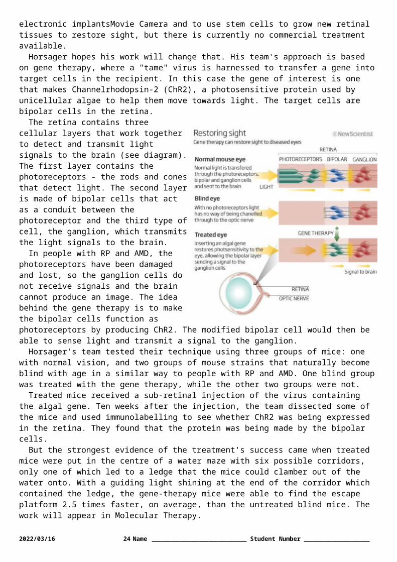
A study reported in Nature has borrowed methods from evolutionary biology to trace the development of grammar in several language families. The results suggest that features shared across language families evolved independently in each lineage. The authors say cultural evolution, not the brain, drives language development.
At the heart of both studies is a method based on what are known as phylogenetic studies.Lead author Michael Dunn, an evolutionary linguist at the Max Planck Institute for Psycholinguistics in the
Netherlands, said the approach is akin to the study of pea plants by Gregor Mendel, which ultimately led to the idea of heritability of traits.
"By looking at variation amongst the descendant plants and knowing how they were related to each other, [Mendel] could work out the mechanisms that must govern that variation," Dr Dunn explained to BBC News.
"He inferred the existence of some kind of information transfer just from knowing family trees and observing variation, and that's exactly the same thing we're doing."Family trees
Modern phylogenetics studies look at variations in animals that are known to be related, and from those can work out when specific structures evolved. For their studies, the team studied the characteristics of word order in four language families: Indo-European, Uto-Aztec, Bantu and Austronesian.
They considered whether what we call prepositions occur before or after a noun ("in the boat" versus "the boat in") and how the word order of subject and object work out in either case ("I put the dog in the boat" versus "I the dog put the canoe in").
The method starts by making use of well-established linguistic data on words and grammar within these language families, and building "family trees" of those languages. "Once we have those trees we look at distribution of these different word order features over the descendant languages, and build evolutionary models for what's most likely to produce the diversity that we observe in the world," Dr Dunn said.
The models revealed that while different language structures in the family tree could be seen to evolve along the branches, just how and when they evolved depended on which branch they were on.
"We show that each of these language families evolves according to its own set of rules, not according to a universal set of rules," Dr Dunn explained. "That is inconsistent with the dominant 'universality theories' of grammar; it suggests rather that language is part of not a specialised module distinct from the rest of cognition, but more part of broad human cognitive skills."
The paper asserts instead that "cultural evolution is the primary factor that determines linguistic structure, with the current state of a linguistic system shaping and constraining future states".
However, co-author and evolutionary biologist Russell Gray of the University of Auckland stressed that the team was not pitting biology against culture in a mutually exclusive way. "We're not saying that biology is irrelevant - of course it's not," Professor Gray told BBC News. "But the clumsy argument about an innate structure of the human mind imposing these kind of 'universals' that we've seen in cognitive science for such a long time just isn't tenable."Steven Pinker, a cognitive scientist at Harvard University, called the work "an important and welcome study".
However, Professor Pinker told BBC News that the finer details of the method need bearing out in order to more fully support their hypothesis that cultural boundaries drive the development of language more than biological limitations do.
"The [authors] suggest that the human mind has a tendency to generalise orderings across phrases of different types, which would not occur if the mind generated every phrase type with a unique and isolated rule.
"The tendency may be partial, and it may be elaborated in different ways in differently language families, but it needs an explanation in terms of the working of the mind of language speakers."http://www.eurekalert.org/pub_releases/2011-04/tju-jru041211.phpJefferson researchers unlock key to personalized cancer medicine using tumor
metabolismThomas Jefferson University researchers used gene signatures and energy
metabolism to predict clinical outcome, rather than gene mutationsPHILADELPHIA—Identifying gene mutations in cancer patients to predict clinical outcome has been the cornerstone of cancer research for nearly three decades, but now researchers at the Kimmel Cancer Center at Jefferson have invented a new approach that instead links cancer cell metabolism with poor clinical outcome. This approach can now be applied to virtually any type of human cancer cell.
The researchers demonstrate that recurrence, metastasis, and poor clinical outcome in breast cancer patients can be identified by simply gene profiling cancer cells that are using ketones and lactate as a food supply.
These findings are reported in the April 15th online issue of Cell Cycle. The investigators are calling this new approach to personalized cancer medicine "Metabolo-Genomics."2023/05/14 24 Name Student Number

High-energy metabolites have long been suspected to "fuel" aggressive tumor cell behavior. The researchers used this premise to generate a gene expression signature from genetically identical cancer cells, but one cell group was fed a diet of high-energy metabolites. These lactate- and ketone-induced "gene signatures" then predicted recurrence, metastasis, and poor survival. So, it appears that what cancer cells are eating determines clinical outcome, not necessarily new gene mutations.
Michael P. Lisanti, M.D., Ph.D., Professor and Chair of Stem Cell Biology & Regenerative Medicine at Jefferson Medical College of Thomas Jefferson University and a member of the Kimmel Cancer Center at Jefferson, together with other researchers, found that treatment of human breast cancer cells with high-energy metabolites increases the expression of genes associated with normal stem cells, including genes upregulated in embryonic and neural stem cells.
What's more, lactate and ketones were found to promote the growth of normal stem cells, which has critical applications for stem cell transplantation and for a host of different human diseases. It appears that these metabolites increase "stemness" in cancer cells, which drives poorer outcomes.
"Tumors that are using the body's own nutrients (lactate and ketones) as "fuel" have a poorer outcome for patient survival, a behavior that now can be used to predict if a patient is at a high-risk for recurrence or metastasis," Dr. Lisanti said. "This is getting to the heart of personalized cancer medicine. Now, we have identified a panel of biomarkers that directly links cancer metabolism with targeted cancer therapy."
These findings suggest, according to the authors, that high-risk cancer patients (those whose cancer cells use high-energy metabolites) can be treated with new therapeutics that target oxidative mitochondrial metabolism, such as the antioxidant metformin, a drug that is also used to treat diabetes.
"Knowing the gene signatures of patients whose cancer cells are "eating" these metabolites (lactate and ketones) for fuel is a pivotal piece of new information that we can use to diagnose and treat cancer patients," said Martinez-Outschoorn, M.D., of the department of Medical Oncology at Thomas Jefferson University, and the lead author of the paper. "It's not just that we know those patients will have poor survival; we know that those patients are using mitochondrial metabolism, which is the type of energy metabolism that we should be targeting with new anti-cancer drugs."
The researchers propose that this new approach to diagnosis and subsequent treatment be called "Metabolo-Genomics" since it incorporates both cell metabolism and gene transcriptional profiling. This strategy could now be used to direct which patients receive a particular "tailored" anti-metabolic therapy.
Genetic markers, like expression of the mutationally activated HER2 gene, provide biomarkers that can be used to identify breast cancer patients at high-risk for recurrence or metastasis, and to modify their subsequent treatment with targeted therapies (i.e., herceptin, a drug used in aggressive breast cancers). But with "Metabolo-Genomics," it is now about using "global" cancer cell metabolism for these predictions.
"Just by feeding cancer cells a particular energy-rich diet, it changes their character, without introducing mutations or altering their genetic profile," Dr. Lisanti said. "We've only fed them high energy nutrients that help them to use their mitochondria, and this changes their transcriptional profile. It's a new biomarker for "lethal" cancers that we can now treat with the right drugs, such as the anti-oxidant metformin."
Dr. Lisanti and his colleagues believe that tumor metabolism is the new big picture for understanding how cancers undergo recurrence and metastasis.http://www.physorg.com/news/2011-04-neurological-basis.html
Neurological basis for embarrassment describedRecording people belting out an old Motown tune and then asking them to listen to
their own singing without the accompanying music seems like an unusually cruel form of punishment.
But for a team of scientists at the University of California, San Francisco and University of California, Berkeley, this exact Karaoke experiment has revealed what part of the brain is essential for embarrassment.
The twist to the experiment was that most of the subjects had neurodegenerative diseases, which helped scientists identify a thumb-sized bit of tissue in the right hemisphere of the front part of the brain called the "pregenual anterior cingulate cortex" as integral to embarrassment. The degree to which the singers were embarrassed in hearing themselves sing "My Girl" – the 1964 hit by the Temptations – depended on the integrity of this particular region.
"In healthy people, watching themselves sing elicits a considerable embarrassment reaction," said Virginia Sturm, a postdoctoral fellow at UCSF. Their blood pressure goes up, their heart rate increases, and their breathing changes, she explained. People who had neurological damage in the medial frontal cortex, however, responded more indifferently. "This brain region predicted the behavior," said Sturm. "The smaller the region, the less embarrassed the people were."2023/05/14 25 Name Student Number

Knowing that people lose their ability to be embarrassed and which part of the brain governs that ability may suggest ways to help diagnose people with certain neurodegenerative diseases earlier.
The work, presented today at the 63rd Annual Meeting of the American Academy of Neurology in Hawaii, is part of a larger body of work at UCSF's Memory and Aging Center examining emotion and social behavior in neurodegenerative diseases and searching for better ways to predict, prevent and treat them.
How Neurodegeneration Changes BehaviorNeurologists at UCSF and elsewhere in the country have documented for years how people with a group of
related neurodegenerative conditions called frontotemporal dementia act in ways that would be embarrassing to healthy people. These conditions result from progressive degeneration of the temporal and frontal lobes of the brain, which play a significant role in decision-making, behavior, and understanding and expression of emotion and language – including complex emotions like embarrassment.
As these parts of the brain deteriorate, people lose their ability to interact with others and may behave strangely. A growing body of work at UCSF and other medical centers has linked the loss of certain brain structures and neuronal networks to specific behavioral changes.In their Karaoke experiment, Sturm and her colleagues took 79 people – most with neurodegenerative diseases – and asked them to sing while probes measured their vital signs and cameras videotaped their expressions.
They sang. Their songs were recorded, and then they were played back at normal speed without the accompanying music. Sturm and her colleagues assessed how embarrassing this was for the participants based on facial expressions and physiological markers, such as sweating and heart rate.
Next, all the people sat for MRIs, which made extremely accurate maps of their brains. Sturm and her colleagues used these maps to measure the volumes of the different regions of the brain and considered whether the sizes of those regions could predict embarrassment.
They found that people who had significant neurodegeneration in the pregenual anterior cingulate cortex were less likely to be embarrassed. In fact, the more deterioration of tissue this part of the brain, the less embarrassed people were about their own singing. The same group was also subjected to a simple "startle" test of emotional reactivity in which they sat quietly until a loud gunshot sound crashed through the room.
"They do jump, and they are afraid," said Sturm, "so it's not like they don't have any emotional reactions at all. But patients with loss in this brain region seem to lose these more complicated social emotions. Emotions like embarrassment are particularly vulnerable in neurodegenerative diseases that target the frontal lobes."
While changes in thinking and memory are easily identified by family members and clinicians, changes in emotion and social behavior can be more subtle and easily missed. A better understanding of the neural basis of social emotions like embarrassment may also help family members and caregivers better comprehend their loved ones' more severe behavioral changes. Provided by University of California, San Franciscohttp://www.physorg.com/news/2011-04-fuel-cell-pocket.html
Putting a fuel cell 'in your pocket'(PhysOrg.com) -- Technology using catalysts which make hydrogen from formic acid
could eventually replace lithium batteries and power a host of mobile devices.Edman Tsang of Oxford University’s Department of Chemistry and colleagues are developing new catalysts
which can produce hydrogen at room temperature without the need for solvents or additives. Their initial results, reported in a recent paper in Nature Nanotechnology, are promising and suggest that a hydrogen fuel cell in your pocket might not be that far away.
The new approach involves placing a single atomic layer of palladium atoms onto silver nanoparticles. ‘The structural and electronic effects from the underlying silver greatly enhance the catalytic properties of palladium, giving impressive activity for the conversion of formic acid to hydrogen and carbon dioxide at room temperature,’ Edman told us.He explains that the storage and handling of organic liquids, such as formic acid, is much easier and safer than storing hydrogen. The catalysts would enable the production of hydrogen from liquid fuel stored in a disposable or recycled cartridge, creating miniature fuel cells to power everything from mobile phones to laptops.
Another advantage of the new technology is that the gas stream generated from the reaction is mainly composed of hydrogen and carbon dioxide but virtually free from catalyst-poisoning carbon monoxide; removing the need for clean-up processes and extending the life of the fuel cells.
The chemists have worked closely with George Smith, Paul Bagot and co-workers at Oxford University’s Department of Materials to characterise the catalysts using atom probe tomography. The underlying technology is the subject of a recent Isis Innovation patent application.
2023/05/14 26 Name Student Number

‘There are lots of hurdles before you can get a real device, but we are looking at the possibility of using this new technology to replace lithium battery technology with an alternative which has a longer lifespan and has less impact on the environment,’ explains Edman. More informationhttp://www.physorg.com/news/2011-04-procedure-breakthrough-lung-transplants.html
New procedure promises to be a breakthrough in lung transplantsFor decades, heart and lung transplant surgeons have followed a strict directive: Get
the donor organ into the recipient as soon as possible.That practice may be changing. In a study published Wednesday in the New England Journal of Medicine,
researchers said both the number of donor lungs and successful transplants may be dramatically increased by treating the organs on a perfusion machine for several hours before transplantation.
The technique marks a paradigm shift in the transplantation field, experts noted. About 85 percent of lungs made available for donation are not used due to tissue damage that potentially could be repaired with perfusion or other techniques. "We won't just transplant an organ," said Dr. Shaf Keshavjee, the senior author of the study and director of the Toronto Lung Transplant Program at Toronto General Hospital. "We will diagnose it, fix it, make it OK and then transplant it."
Variations on the pre-transplant treatment concept are also being tried on kidneys, livers and hearts. Lungs, however, provide a unique opportunity because they are greatly needed and appear so amenable to perfusion treatment, said John Dark, a professor of cardiothoracic surgery at Newcastle University, U.K., and president of the International Society for Heart & Lung Transplantation. "This is the most exciting advance in lung transplantation since we first started 25 years ago," said Dark, who was not involved in the study. "It's converting lungs you can't use into lungs you can use. At the moment, we are only using about 20 percent, worldwide, of the lungs that are offered to us."
In the United States, 1,786 people are on the waiting list for a lung transplant, according to the Department of Health and Human Services' Organ Procurement and Transplantation Network. About one-third of these patients wait at least three years for a suitable pair of donor lungs, and about 10 percent to 15 percent of people die before receiving a transplant. Lungs must be able to function immediately once transplanted. For that reason, surgeons are very selective about which lungs they use and accept only those in the most pristine condition, Keshavjee said. That poses a challenge because lungs are easily damaged from injury or in the final stages of terminal illness.
The new technique pumps a liquid consisting of oxygen, proteins and nutrients into the donor lungs after they've been removed and transported to the recipient's hospital. Keshavjee and his team used the Toronto XVIVO Lung Perfusion System, which was designed for this purpose. The system is being used around the world but is not yet approved by the Food and Drug Administration for use in the United States.
Researchers treated 23 sets of lungs that were impaired in some manner - and would have been rejected for transplant - with perfusion for four hours and transplanted 20 of them that looked viable after the treatment. Surgery outcomes were compared to 116 patients who received conventional donor lungs.
After 72 hours, 15 percent of the patients who received treated lungs suffered primary graft dysfunction, a potentially fatal complication, compared to 30 percent of the patients in the control group. After 30 days, the rate of deaths, complications and hospital stays were similar between the two groups.
The perfusion technique essentially repairs damage caused by swelling or inflammation in the lung, Dark said. That opens the door to other types of treatments for organs after they have been removed from a donor's body, including antibiotics, gene therapy or even immunosuppressant medications that might lower the risk of rejection. But estimates on how the technology will impact organ transplantation vary.
"There is still some discrepancy from the transplant community on whether this will really bring significant benefit," said Dr. Christian A. Bermudez, associate director of heart and lung transplantation at the University of Pittsburgh School of Medicine. "There is no doubt it would increase the complexity of the procurement. But it may be a good strategy that would allow some increase in the organ utilization."
Pre-transplant treatment of an organ would add significant cost to transplant surgeries. And, if the organ was treated and still deemed unsuitable for transplant, it's not clear who would pay for the failed treatment, Keshavjee said. "There are things to be ironed out," he said. "The evidence is mounting in favor of it, but it's radically different."http://www.physorg.com/news/2011-04-kilo-person-plane.html
The 70 kilo single person plane(PhysOrg.com) -- Aki Suokas, a Finnish aeronautical engineer, has just finished creating
a unique single-seat aircraft this week. The project was completed at Aero Friedrichshafen, and it has been dubbed the FlyNano.
2023/05/14 27 Name Student Number
The FlyNano is made up completely of a carbon fiber composite material that makes this plane very light. The whole of the plane weighs only 70 kilograms in total. This weight class may actually make this plane light enough that the pilot may not need a license to fly it, depending on your flight path. The plane both takes off and lands on water, and comes in three versions.
The first version, one electric only, and two fuel based versions. The electric-only version has a 20kW engine. The two petrol-based engined run on 24 bhp and 35 bhp, respectively. The top speed on these planes is roughly 140 km/h, with a minimum speed of 70 km/h . The FlyNano has a wingspan of five meters. The plan can support a take off weight of 200 kg, as well as a take off speed of about 70km/h. In theory, the plane can travel about up to 70 kilometers in a single fueling, or charging, depending on the model that you have chosen to fly.
The 70 kilo single person planeIf you want to own one of these planes it will cost you about $39,000, for the least expensive of these three
models. Orders on the FlyNano will be taken in three months from now, so you have some time to save up the cash if you really want to have this one man craft for yourself.
The FlyNano has been officially launched at Aero 2011 show at Friedrichshafen, Germany, 13 – 16 April.More information: http://www.flynano.com/
2023/05/14 28 Name Student Number