Embed Size (px)
Citation preview
Citation: Takamatsu A, Nagai Y and Constantine SH. Liver Abscesses with Bacteria-Associated Hemophagocytic Syndrome. Ann Hematol Oncol. 2015; 2(9): 1064.
Ann Hematol Oncol - Volume 2 Issue 9 - 2015ISSN : 2375-7965 | www.austinpublishinggroup.com Takamatsu et al. © All rights are reserved
Annals of Hematology & OncologyOpen Access
Abstract
A 31-year-old man was diagnosed with multiple liver abscesses. Despite 4 weeks of antibiotic treatment, his fever persisted with associated pancytopenia and increased liver enzymes. A bone marrow biopsy with aspiration revealed hemophagocytosis, and the diagnosis of Bacteria-Associated Hemophagocytic Syndrome (BAHS) was made. Administration of corticosteroids and Intravenous Immunoglobulin (IVIG) resulted in significant improvement of symptoms. BAHS accounts for 9% of Hemophagocytic Lymphohistiocytosis (HLH) and has a poor prognosis. It can be a diagnostic challenge because it can mimic or coexist with sepsis. Along with proper antibiotic treatment, early co-therapy with immunosuppressive agents may be life saving.
Keywords: Liver abscess; Bacteria-associated hemophagocytic syndrome; Hemophagocytic lymphohistiocytosis; Sepsis
IntroductionHemophagocytic Lymphohistiocytosis (HLH) is a life-
threatening disease characterized by extreme inflammation due to an uncontrolled and ineffective immune response [1]. It is caused by a dysregulation in natural killer T-cell function, resulting in activation and proliferation of lymphocytes or histiocytes with uncontrolled hemophagocytosis and cytokine overproduction [2]. HLH can be either primary, with a genetic etiology or secondary, associated with malignancies, autoimmune diseases, or infections. HLH can occur at any age; however, most of published studies are focused on pediatric patients. There are no precise data regarding the incidence of HLH in adults. An estimate of HLH in children <18 years old across ethnicities and races is approximately 1 in 100,000 per year [3]. In a nationwide survey in Japan that included both adult and pediatric patients, the annual incidence of HLH was estimated to be 1 in 800,000 per year [4]. In adults, most of HLH is secondary. HLH related to infections accounts for 40% of the cases, of which 9% is Bacteria-Associated Hemophagocytic Syndrome (BAHS) [5]. A standard treatment of BAHS, however, has not been established. We describe a case of a patient with BAHS induced by liver abscesses. Corticosteroids and Intravenous Immunoglobulin (IVIG) proved to be useful in treating this patient.
Case PresentationA 31-year-old Japanese man presented to the hospital with one-
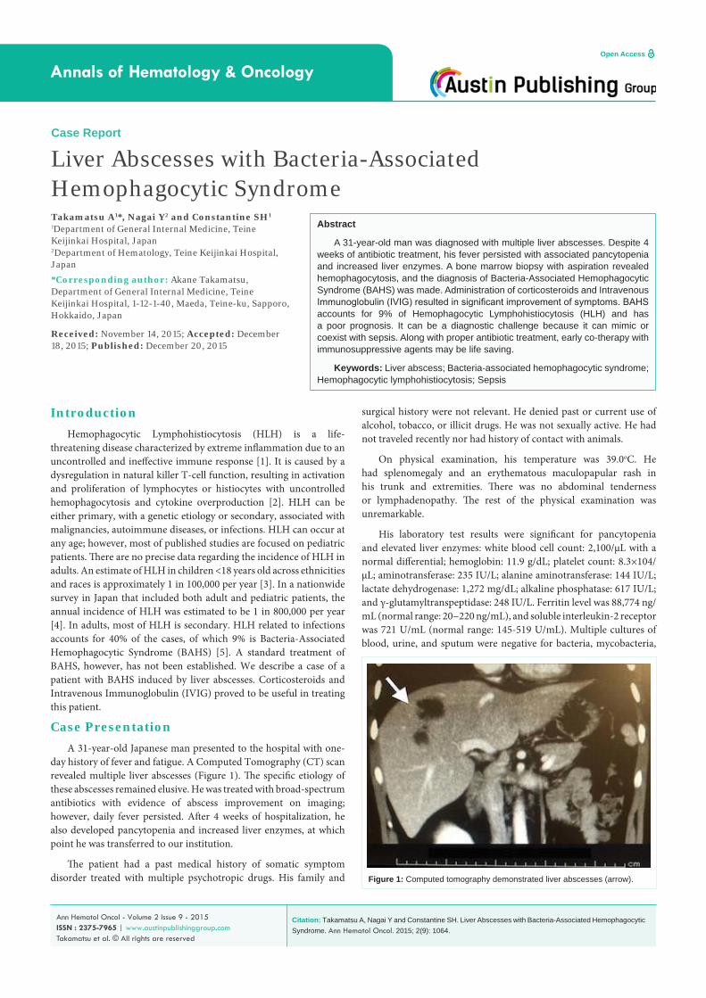
day history of fever and fatigue. A Computed Tomography (CT) scan revealed multiple liver abscesses (Figure 1). The specific etiology of these abscesses remained elusive. He was treated with broad-spectrum antibiotics with evidence of abscess improvement on imaging; however, daily fever persisted. After 4 weeks of hospitalization, he also developed pancytopenia and increased liver enzymes, at which point he was transferred to our institution.
The patient had a past medical history of somatic symptom disorder treated with multiple psychotropic drugs. His family and
Case Report
Liver Abscesses with Bacteria-Associated Hemophagocytic SyndromeTakamatsu A1*, Nagai Y2 and Constantine SH1
1Department of General Internal Medicine, Teine Keijinkai Hospital, Japan2Department of Hematology, Teine Keijinkai Hospital, Japan
*Corresponding author: Akane Takamatsu, Department of General Internal Medicine, Teine Keijinkai Hospital, 1-12-1-40, Maeda, Teine-ku, Sapporo, Hokkaido, Japan
Received: November 14, 2015; Accepted: December 18, 2015; Published: December 20, 2015
surgical history were not relevant. He denied past or current use of alcohol, tobacco, or illicit drugs. He was not sexually active. He had not traveled recently nor had history of contact with animals.
On physical examination, his temperature was 39.0oC. He had splenomegaly and an erythematous maculopapular rash in his trunk and extremities. There was no abdominal tenderness or lymphadenopathy. The rest of the physical examination was unremarkable.
His laboratory test results were significant for pancytopenia and elevated liver enzymes: white blood cell count: 2,100/μL with a normal differential; hemoglobin: 11.9 g/dL; platelet count: 8.3×10⁴/μL; aminotransferase: 235 IU/L; alanine aminotransferase: 144 IU/L; lactate dehydrogenase: 1,272 mg/dL; alkaline phosphatase: 617 IU/L; and γ-glutamyltranspeptidase: 248 IU/L. Ferritin level was 88,774 ng/mL (normal range: 20−220 ng/mL), and soluble interleukin-2 receptor was 721 U/mL (normal range: 145-519 U/mL). Multiple cultures of blood, urine, and sputum were negative for bacteria, mycobacteria,
Figure 1: Computed tomography demonstrated liver abscesses (arrow).
Ann Hematol Oncol 2(9): id1064 (2015) - Page - 02
Takamatsu A Austin Publishing Group
Submit your Manuscript | www.austinpublishinggroup.com
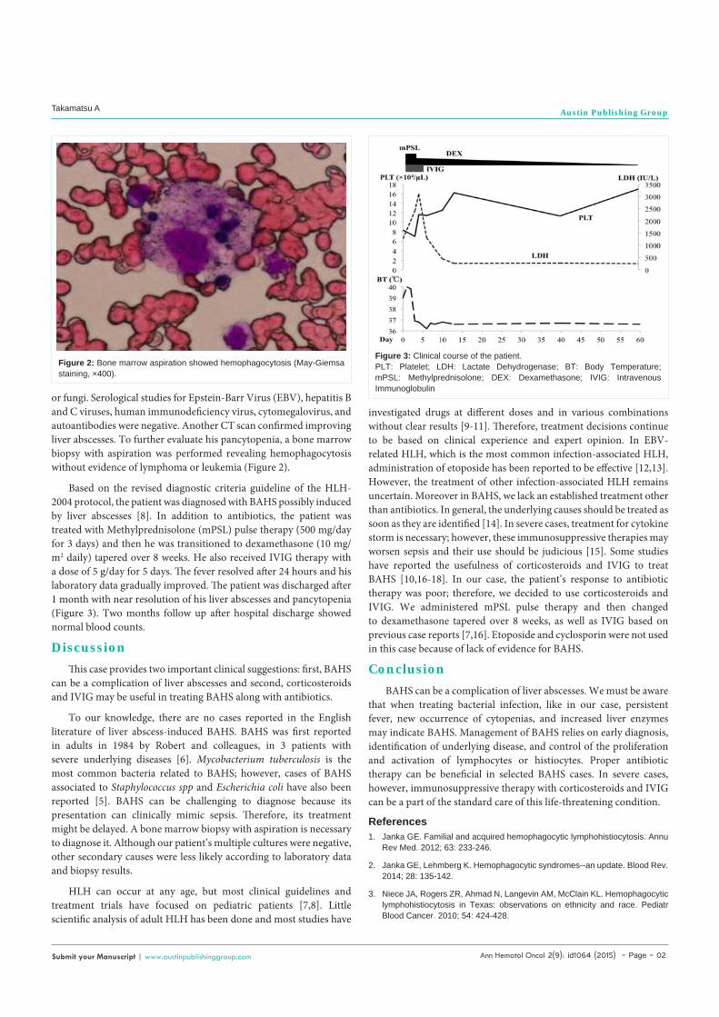
or fungi. Serological studies for Epstein-Barr Virus (EBV), hepatitis B and C viruses, human immunodeficiency virus, cytomegalovirus, and autoantibodies were negative. Another CT scan confirmed improving liver abscesses. To further evaluate his pancytopenia, a bone marrow biopsy with aspiration was performed revealing hemophagocytosis without evidence of lymphoma or leukemia (Figure 2).
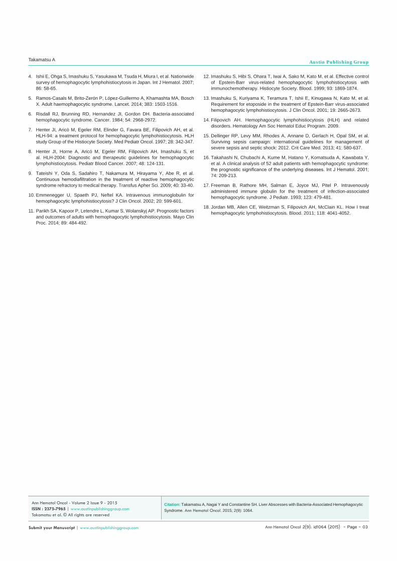
Based on the revised diagnostic criteria guideline of the HLH-2004 protocol, the patient was diagnosed with BAHS possibly induced by liver abscesses [8]. In addition to antibiotics, the patient was treated with Methylprednisolone (mPSL) pulse therapy (500 mg/day for 3 days) and then he was transitioned to dexamethasone (10 mg/m2 daily) tapered over 8 weeks. He also received IVIG therapy with a dose of 5 g/day for 5 days. The fever resolved after 24 hours and his laboratory data gradually improved. The patient was discharged after 1 month with near resolution of his liver abscesses and pancytopenia (Figure 3). Two months follow up after hospital discharge showed normal blood counts.
DiscussionThis case provides two important clinical suggestions: first, BAHS
can be a complication of liver abscesses and second, corticosteroids and IVIG may be useful in treating BAHS along with antibiotics.
To our knowledge, there are no cases reported in the English literature of liver abscess-induced BAHS. BAHS was first reported in adults in 1984 by Robert and colleagues, in 3 patients with severe underlying diseases [6]. Mycobacterium tuberculosis is the most common bacteria related to BAHS; however, cases of BAHS associated to Staphylococcus spp and Escherichia coli have also been reported [5]. BAHS can be challenging to diagnose because its presentation can clinically mimic sepsis. Therefore, its treatment might be delayed. A bone marrow biopsy with aspiration is necessary to diagnose it. Although our patient’s multiple cultures were negative, other secondary causes were less likely according to laboratory data and biopsy results.
HLH can occur at any age, but most clinical guidelines and treatment trials have focused on pediatric patients [7,8]. Little scientific analysis of adult HLH has been done and most studies have
investigated drugs at different doses and in various combinations without clear results [9-11]. Therefore, treatment decisions continue to be based on clinical experience and expert opinion. In EBV-related HLH, which is the most common infection-associated HLH, administration of etoposide has been reported to be effective [12,13]. However, the treatment of other infection-associated HLH remains uncertain. Moreover in BAHS, we lack an established treatment other than antibiotics. In general, the underlying causes should be treated as soon as they are identified [14]. In severe cases, treatment for cytokine storm is necessary; however, these immunosuppressive therapies may worsen sepsis and their use should be judicious [15]. Some studies have reported the usefulness of corticosteroids and IVIG to treat BAHS [10,16-18]. In our case, the patient’s response to antibiotic therapy was poor; therefore, we decided to use corticosteroids and IVIG. We administered mPSL pulse therapy and then changed to dexamethasone tapered over 8 weeks, as well as IVIG based on previous case reports [7,16]. Etoposide and cyclosporin were not used in this case because of lack of evidence for BAHS.
ConclusionBAHS can be a complication of liver abscesses. We must be aware
that when treating bacterial infection, like in our case, persistent fever, new occurrence of cytopenias, and increased liver enzymes may indicate BAHS. Management of BAHS relies on early diagnosis, identification of underlying disease, and control of the proliferation and activation of lymphocytes or histiocytes. Proper antibiotic therapy can be beneficial in selected BAHS cases. In severe cases, however, immunosuppressive therapy with corticosteroids and IVIG can be a part of the standard care of this life-threatening condition.
References1. Janka GE. Familial and acquired hemophagocytic lymphohistiocytosis. Annu
Rev Med. 2012; 63: 233-246.
2. Janka GE, Lehmberg K. Hemophagocytic syndromes--an update. Blood Rev. 2014; 28: 135-142.
3. Niece JA, Rogers ZR, Ahmad N, Langevin AM, McClain KL. Hemophagocytic lymphohistiocytosis in Texas: observations on ethnicity and race. Pediatr Blood Cancer. 2010; 54: 424-428.
Figure 2: Bone marrow aspiration showed hemophagocytosis (May-Giemsa staining, ×400).
Figure 3: Clinical course of the patient.PLT: Platelet; LDH: Lactate Dehydrogenase; BT: Body Temperature; mPSL: Methylprednisolone; DEX: Dexamethasone; IVIG: Intravenous Immunoglobulin
Ann Hematol Oncol 2(9): id1064 (2015) - Page - 03
Takamatsu A Austin Publishing Group
Submit your Manuscript | www.austinpublishinggroup.com
4. Ishii E, Ohga S, Imashuku S, Yasukawa M, Tsuda H, Miura I, et al. Nationwide survey of hemophagocytic lymphohistiocytosis in Japan. Int J Hematol. 2007; 86: 58-65.
5. Ramos-Casals M, Brito-Zerón P, López-Guillermo A, Khamashta MA, Bosch X. Adult haemophagocytic syndrome. Lancet. 2014; 383: 1503-1516.
6. Risdall RJ, Brunning RD, Hernandez JI, Gordon DH. Bacteria-associated hemophagocytic syndrome. Cancer. 1984; 54: 2968-2972.
7. Henter JI, Aricò M, Egeler RM, Elinder G, Favara BE, Filipovich AH, et al. HLH-94: a treatment protocol for hemophagocytic lymphohistiocytosis. HLH study Group of the Histiocyte Society. Med Pediatr Oncol. 1997; 28: 342-347.
8. Henter JI, Horne A, Aricó M, Egeler RM, Filipovich AH, Imashuku S, et al. HLH-2004: Diagnostic and therapeutic guidelines for hemophagocytic lymphohistiocytosis. Pediatr Blood Cancer. 2007; 48: 124-131.
9. Tateishi Y, Oda S, Sadahiro T, Nakamura M, Hirayama Y, Abe R, et al. Continuous hemodiafiltration in the treatment of reactive hemophagocytic syndrome refractory to medical therapy. Transfus Apher Sci. 2009; 40: 33-40.
10. Emmenegger U, Spaeth PJ, Neftel KA. Intravenous immunoglobulin for hemophagocytic lymphohistiocytosis? J Clin Oncol. 2002; 20: 599-601.
11. Parikh SA, Kapoor P, Letendre L, Kumar S, Wolanskyj AP. Prognostic factors and outcomes of adults with hemophagocytic lymphohistiocytosis. Mayo Clin Proc. 2014; 89: 484-492.
12. Imashuku S, Hibi S, Ohara T, Iwai A, Sako M, Kato M, et al. Effective control of Epstein-Barr virus-related hemophagocytic lymphohistiocytosis with immunochemotherapy. Histiocyte Society. Blood. 1999; 93: 1869-1874.
13. Imashuku S, Kuriyama K, Teramura T, Ishii E, Kinugawa N, Kato M, et al. Requirement for etoposide in the treatment of Epstein-Barr virus-associated hemophagocytic lymphohistiocytosis. J Clin Oncol. 2001; 19: 2665-2673.
14. Filipovich AH. Hemophagocytic lymphohistiocytosis (HLH) and related disorders. Hematology Am Soc Hematol Educ Program. 2009.
15. Dellinger RP, Levy MM, Rhodes A, Annane D, Gerlach H, Opal SM, et al. Surviving sepsis campaign: international guidelines for management of severe sepsis and septic shock: 2012. Crit Care Med. 2013; 41: 580-637.
16. Takahashi N, Chubachi A, Kume M, Hatano Y, Komatsuda A, Kawabata Y, et al. A clinical analysis of 52 adult patients with hemophagocytic syndrome: the prognostic significance of the underlying diseases. Int J Hematol. 2001; 74: 209-213.
17. Freeman B, Rathore MH, Salman E, Joyce MJ, Pitel P. Intravenously administered immune globulin for the treatment of infection-associated hemophagocytic syndrome. J Pediatr. 1993; 123: 479-481.
18. Jordan MB, Allen CE, Weitzman S, Filipovich AH, McClain KL. How I treat hemophagocytic lymphohistiocytosis. Blood. 2011; 118: 4041-4052.
Citation: Takamatsu A, Nagai Y and Constantine SH. Liver Abscesses with Bacteria-Associated Hemophagocytic Syndrome. Ann Hematol Oncol. 2015; 2(9): 1064.
Ann Hematol Oncol - Volume 2 Issue 9 - 2015ISSN : 2375-7965 | www.austinpublishinggroup.com Takamatsu et al. © All rights are reserved