Embed Size (px)
Citation preview

Synapse® Liver Analysis (CT/MR) extracts the liver and nearby vessel regions from contrast-enhanced CT and MR images and displays the results as a combined 3D mask for preoperative analysis. Hepatectomy simulation is also featured.
Synapse 3D Clinical Application
Liver Analysis (CT/MR)
REGISTRATION
AUTOMATED/SEMIAUTOMATED ORGAN EXTRACTION
Recommended image type u Contrast-enhanced CT, cholangiography MR, SPECTAnalysis target u Liver
n Rigid (linear)
n Liver
n Portal vein (PV)
n Non-rigid (deformed)
n Hepatic artery (HA)
n Hepatic vein (HV)
n Bile duct and gallbladder*
n Inferior vena cava (IVC)
• Register multiphase CT, MRCP, and SPECT to improve assessment accuracy.
*Using MRCP (MR) images
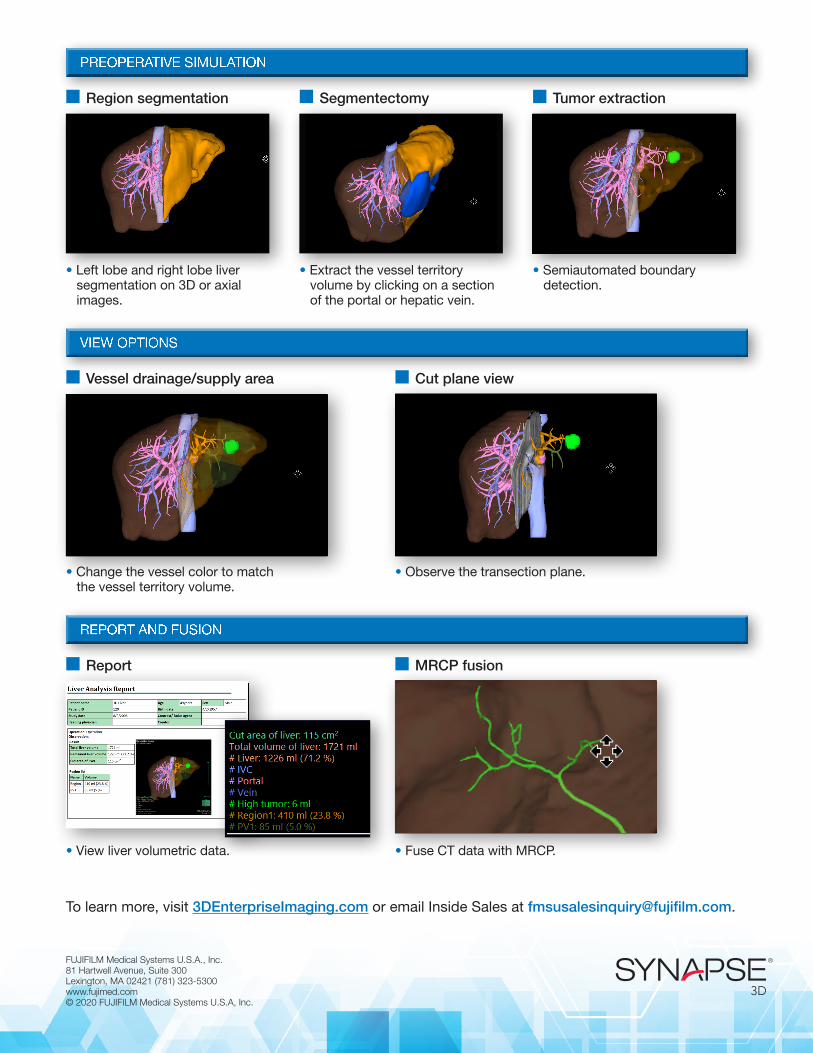
PREOPERATIVE SIMULATION
VIEW OPTIONS
REPORT AND FUSION
FUJIFILM Medical Systems U.S.A., Inc. 81 Hartwell Avenue, Suite 300Lexington, MA 02421 (781) 323-5300 www.fujimed.com © 2020 FUJIFILM Medical Systems U.S.A, Inc.
• Left lobe and right lobe liver segmentation on 3D or axial images.
• Change the vessel color to match the vessel territory volume.
• View liver volumetric data.
• Extract the vessel territory volume by clicking on a section of the portal or hepatic vein.
• Observe the transection plane.
• Fuse CT data with MRCP.
• Semiautomated boundary detection.
To learn more, visit 3DEnterpriseImaging.com or email Inside Sales at [email protected].
n Region segmentation
n Vessel drainage/supply area
n Report
n Segmentectomy
n Cut plane view
n MRCP fusion
n Tumor extraction