Embed Size (px)
Citation preview
Open Access
Arroyo et al., J Drug Metab Toxicol 2012, S5DOI: 10.4172/2157-7609.S5-001
Open Access
J Drug Metab Toxicol ISSN: 2157-7609 JDMT, an open access journalHeavy Metal Drug Toxicology
Keywords: Cadmium; Liver; Hepatocytes; Cadmium-inducedhepatotoxicity mechanism
IntroductionCadmium (Cd) is an important industrial and environmental
toxicant with many industrial applications. Cd emissions to the atmospheric, aquatic, and terrestrial environment have increased during the last century. Since Cd is not degraded in the environment, the risk of human exposure is constantly increased due to Cd also enters to the food chain [1]. Humans are generally exposed to Cd by two main routes, inhalation and ingestion. Absorption of Cd by skin is relatively insignificant [2]. In humans and other mammals, Cd exposure can result in a variety of adverse effects, such as testicular damage, pulmonary edema, renal and hepatic dysfunction, osteomalacia, etc. In addition to the direct cytotoxic effects that could lead to apoptotic or necrotic event, Cd has been implicated in the development of cancer and it has been classified as a type I carcinogen by the International Agency for Cancer Research [3]. Emerging evidence suggests that Cd exposure affects Cytochrome P 450 1A1 (Cyp1A1). Cyp1A1 has been always associated with the transformation of pro-carcinogenic compounds to highly carcinogenic metabolites [4]. Chronic Cd intoxication results mainly in renal disease [5,6] but acute Cd exposure primarily results in liver accumulation and hepatocellular damage. After acute exposure to inorganic forms of Cd, the preponderance of the dose accumulates in the liver [7]. Cadmium retention is generally higher in women rather than men [8]. Gender differences in susceptibility, at lower exposure, are uncertain, but some data indicate that Cd has estrogenic effects and affects female offspring [8]. Humans are susceptible to Cd toxicity primarily through the ingestion of contaminated food or water and the inhalation of cigarette smoke. Today, nanotechnology with the development of Cd-containing nanoparticules, could be also a source of metal exposure [9]. Gastrointestinal ingestion of Cd through food or drinking water is the major route of intake for this metal in non-smoking and non-occupationally exposed populations [10]. Cigarette smoke is the largest source of Cd exposure in general population. Each cigarette could contain up to 6.67 µg Cd, and 40-60% of it generally passes through the pulmonary epithelium into systemic circulation.
Cadmium DispositionThe mechanism by which Cd is absorbed, transported and taken
up by cells is still not fully understood. As a toxic metal, there is unlikely to be specific transport proteins for Cd. Rather, because Cd has similar chemical and physical properties to essential metals such as iron (Fe), zinc (Zn) or calcium (Ca), it can be transported and taken up into the cells by a process referred to as “ionic and molecular mimicry” [11]. As Cd avidly binds to sulfhydryl-amino acids and many proteins containing sulfhydryl groups, it is important to consider it, to understand how the body handles this toxic metal. Intestinal absorption of Cd is characterized by a rapid rate of metal accumulation within the intestinal mucosa and a low rate of diffusive transfer into systemic circulation [12]. The intestinal uptake of Cd and its subsequent distribution to target tissues is greatly dependent of the cadmium chemical form in the intestinal epithelium. Foulkes [13] have proposed a two step process from the absorptive Cd movement from intestinal lumen into enterocytes, the first one consists of a non-specific binding of Cd to the luminal plasma membrane, and the second one the transport across the luminal plasma membrane into enterocytes. Some studies have demonstrated that Cd ions are taken up by the divalent metal transporter 1 (DMT1), and the metal transporter protein 1 (MTP1), which are located in the basolateral and the apical membranes of the enterocytes respectively [14]. A channel-like calcium transporter (CaT1) that mediates intestinal Ca absorption has also been involved in the absorption and tissue distribution of Cd [15]. It also seems possible that the Ca-binding protein, calbindin-D, may facilitate the absorption of Cd. Metallothionein (MT) is induced in the enterocytes by Cd. This response plays an important role in the retention of Cd
*Corresponding author: Ma. Concepción Gutiérrez-Ruiz, Universidad Autónoma Metropolitana-Iztapalapa, Laboratorio de Fisiología Celular, Departamento de Ciencias de la Salud, División de Ciencias Biológicas y de la Salud. Avenida San Rafael Atlixco 186, Colonia Vicentina, México, D.F. 09340, México. Tel: 52-55-58046451; Fax: 52-55-58044730; E-mail: [email protected]
Received November 18, 2011; Accepted December 27, 2011; Published January 02, 2012
Citation: Arroyo VS, Flores KM, Ortiz LB, Gómez-Quiroz LE, Gutiérrez-Ruiz MC (2012) Liver and Cadmium Toxicity. J Drug Metab Toxicol S5:001. doi:10.4172/2157-7609.S5-001
Copyright: © 2012 Arroyo VS, et al. This is an open-access article distributed under the terms of the Creative Commons Attribution License, which permits unrestricted use, distribution, and reproduction in any medium, provided the original author and source are credited.
AbstractCadmium (Cd) is an important environmental pollutant. This metal presents a serious threat for both, humans
and animals health. The environmental risk can lead to the absorption of large quantities of Cd and its toxic action on the organism. It adversely affects some organs in humans and animals, including the liver, kidneys, lungs, pancreas, and testis. The liver and kidneys, which are the primary organs involved in the elimination of this metal, are especially sensitive to its toxic effects. The liver is the main target organ of Cd toxicity following both acute and chronic exposure. This review presents the current state of knowledge related to the cellular mechanisms of Cd toxicity in the liver. Different mechanisms are discussed: the disruption of the cellular antioxidant system and the decrease in thiol status, the generation of reactive oxygen species and oxidative stress, the interference of biological metal homeostasis, involvement of inflammatory mediators, disruption of cell adhesion and cell damage leading to apoptosis.
Liver and Cadmium ToxicityVerónica Souza Arroyo, Karina Martínez Flores, Leticia Bucio Ortiz, Luis Enrique Gómez-Quiroz and María Concepción Gutiérrez-Ruiz*
Laboratorio de Fisiología Celular, Departamento de Ciencias de la Salud, División de Ciencias Biológicas y de la Salud. Universidad Autónoma Metropolitana-Iztapalapa, Avenida San Rafael Atlixco 186, Colonia Vicentina, México, D.F. 09340, México
Journal of Drug Metabolism &Toxicology
Review Article

Citation: Arroyo VS, Flores KM, Ortiz LB, Gómez-Quiroz LE, Gutiérrez-Ruiz MC (2012) Liver and Cadmium Toxicity. J Drug Metab Toxicol S5:001. doi:10.4172/2157-7609.S5-001
Page 2 of 7
J Drug Metab Toxicol ISSN: 2157-7609 JDMT, an open access journalHeavy Metal Drug Toxicology
within the small intestine mucosa after Cd ingestion. Retention of Cd by MT in the enterocytes would reduce the amount of Cd that could enter into systemic circulation. There is not enough information regarding metal excretion by the enterocytes [16], some data involve the transporter MTP1, which is a Fe transporter homologous to DMT1, in the enterocytes basolateral exportation of Cd. Following Cd gastrointestinal or pulmonary absorption into systemic circulation, it is delivered to target organs. Much of the Cd absorbed in the intestines is delivered first to the liver via portal circulation, bound mainly to albumin, where it is taken up from the sinusoidal capillaries to the hepatocytes. Transport of Cd into hepatocytes occurs by a two-phase process, involving the binding to the plasma membrane following internalization [17,18], playing membrane transporters present in the sinusoidal plasma membrane an important role in the second phase process. Our group reported some years ago [18], that Cd transport occurred by temperature-insensitive processes, temperature-sensitive processes, probably Ca channel, and carriers that involved interaction with sulfhydryl groups. There are some reports that involve specific sinusoidal membrane transporters as DMT1, ZIP8 and ZIP14 that participate in Cd uptake into hepatocytes [19,20]. In the hepatocyte, Cd forms complexes with small peptides and proteins via sulfhydryl groups, including glutathione (GSH) or the high affinity metal binding protein family MT-I and MT-II. Canalicular transport of Cd is linked to biliary GSH excretion. Cd may be transported as GSH complex by the canalicular gluthathione transport system(s). In mammals, beside GSH, Cd can be detoxified after association with MT. Nonetheless, MT plays an important role in tissue Cd retention, and is responsible for the long biological half-life of Cd in the body. Cd-induced acute hepatotoxicity is enhanced by the presence of MT-III but greatly inhibited by that of f MT-I and MT-II. The mechanism involved remains unclear as MT-III expression mainly occurs in the brain and it is not expressed in the liver [21].
Hepatic response
Regardless of oral, pulmonary or parenteral exposure, the liver is by far the primary organ that takes up the greatest quantity of Cd during initial hours after exposure. The studies of Cd-induced hepatotoxicity in experimental animals and in vitro cellular models have yielded a substantial amount of information to metal toxicity as well as mechanistic relevant information. It has been proposed that acute hepatotoxicity injury, involves a direct toxic effect of the metal, ischemia due to endothelial cell injury, and the latter inflammatory injury, in which Kupffer cell activation and neutrophil infiltration play a major role through a cascade of inflammatory mediators [22]. Some histopathological changes such as loss of normal architecture of the parenchymatous tissue, cytoplasmic vacuolization, cellular degeneration and necrosis, congested blood vessels, destructed mitochondria cristae, fat globules, severe glycogen depletion, lipofuscin pigments, and collagenous fibers formation are observed in liver tissue from rats exposed to Cd for 22 days [23]. These cellular changes may result in both apoptosis and necrosis. Several mechanisms have been postulated for Cd-induced hepatotoxicity. Its injury appears to be associated with sulfhydryl groups binding, implicating membrane proteins, cytoplasmic proteins and enzymes. Furthermore, Cd is a non-redox metal that can indirectly cause oxidative stress by depleting cellular levels of GSH. Also it competes with essential metals such as Zn, selenium (Se), cupper (Cu) and Ca [24] interfering with various cellular processes such as metal membrane transport and energy metabolism. Also, it affects several cellular functions, such as enzyme
activities, DNA repair systems, redox state of the cell and signal transduction [25]. Cd also modifies cell-cell adhesion by disassembling E-cadherin/beta-catenin complex [26].
Involvement of inflammatory mediators and adhesion molecules in Cd-induced hepatotoxicity
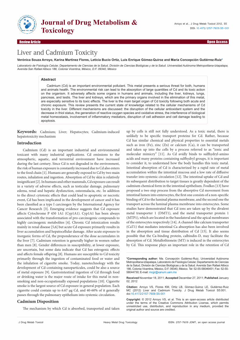
Cd hepatotoxicity is closely related to inflammation, since after acute exposure, the damaged liver is often infiltrated by polymorphonuclear neutrophils (PMN), which, in addition to Kupffer cells, contribute to the hepatotoxicity by enhancing inflammatory mediators and promoting necrosis [27]. Previous studies demonstrate that activated Kupffer cells release a number of inflammatory mediators that subsequently enhance the expression of adhesion molecules that initiate a cascade of cellular and humoral responses leading to inflammation and secondary liver damage during Cd-induced hepatotoxicity [22]. When Kupffer cells are selectively destroyed, inhibited or suppressed, the hepatotoxicity of Cd is dramatically reduced. Although the mechanism by which Kupffer cells contribute to Cd hepatotoxicity is not fully understood, it is known that activated Kupffer cells, release a variety of cytotoxic mediators that can directly damage hepatocytes. These mediators include reactive oxygen species (ROS), nitric oxide, and cytokines. Particular attention has been paid to the role of tumor necrosis factor α (TNF-α) in Cd hepatotoxicity [28]. It has been shown that anti-TNF-α antibodies prevented Cd-induced secretion of acute phase proteins and gene expression of interleukin-1β (IL-1β), interleukin-6 (IL-6) and interleukin-8 (IL-8) in human hepatoma cell line HepG2 [29]. In addition, increased expression of IL-1β, TNF-α, IL-6 and IL-8 have been demonstrated in hepatocyte [30] or HepG2 cells exposed to Cd [29]. Moreover, TNF-α and IL-1β are recognized to stimulate the production of adhesion molecules, such as intercellular adhesion molecule-1 (IMAC-1), vascular cell adhesion molecule-1 (VCAM-1), E-selectin, P-selectin, β2-integrin Mac-1 in the liver, that induce the recruitment, adherence and activation of circulating inflammatory cells [31]. Rikans and Yamano [32], suggested that the hepatic endothelial cells might be the first cellular target for Cd-induced hepatocellular injury. Cd-induced degeneration of the hepatic endothelium is indicated by the extrusion of damage cells into the capillary lumen, producing a local ischemia in the parenchyma. Damage to hepatic endothelial cells was observed within 3 h after Cd (3 mg/kg CdCl2) administration in rats. There was extensive destruction of fenestrations on the luminal surface of endothelial cells [33]. Once damaged, the injured endothelial cells obstruct the capillary lumen and produce local ischemia, which may then initiate a number of molecular and cellular events, which result in a subsequent activation of Kupffer cells, release of inflammatory mediators and recruitment of inflammatory cells, mainly PMN and leukocytes. In liver, the inflammatory cells accumulate in sinusoids and adhere to endothelial cells. This step is mediated by E-selectin, Mac-1 and ICAM-1. The final step is transmigration, which is mediated by platelet–endothelial adhesion molecule-1 (PECAM-1) suggesting an important role for these adhesion molecules during Cd hepatoxicity [31]. In addition, ROS promote inflammation by enhancing the activation of the transcription factors, such as nuclear factor-κB (NF-κB) and the activator protein-1 (AP-1), which provide signals for the expression of proinflammatory and adhesion molecules genes [34,29]. This idea is supported by fact that N-acetyl cysteine (NAC), a GSH precursor, could inhibit cadmium-induced AP-1 activation. Although Cd affects multiple sites in the endothelium, few data are available. Currently, it is considered an important area of research to understand cellular toxicity induced by Cd (Figure 1).
Citation: Arroyo VS, Flores KM, Ortiz LB, Gómez-Quiroz LE, Gutiérrez-Ruiz MC (2012) Liver and Cadmium Toxicity. J Drug Metab Toxicol S5:001. doi:10.4172/2157-7609.S5-001
Page 3 of 7
J Drug Metab Toxicol ISSN: 2157-7609 JDMT, an open access journalHeavy Metal Drug Toxicology
Cadmium hepatotoxicity related to the homeostasis of essential metals
Maintaining metal homeostasis is essential for the function of many important enzymes, transcription factors, and other subcellular proteins. Disruption of metal homeostasis can lead to disease, and even death. Deregulation of the homeostasis of essential metals by Cd, and cellular Cd traffic via essential metals pathways contribute to Cd toxicity. Although many Cd-bound species can be obtained in vitro in the presence of large metal excess with pure biomolecules, demonstration of their occurrence in vivo, in mammalian cells in particular, is not an easy task [35]. The function of many metalloproteins critically depends on their interaction with a metal, usually a transition one, such as Cu, Fe, Zn, particularly the last one. Cd and Zn belong to the same chemical group, and both do not change oxidation states. Also they occur as divalent cations in biological environments. For instance, Cd in MT is exclusively bound to sulfur, but Cd coordination is mixed in one Zn site of alcohol dehydrogenase, or several Zn finger proteins. In this way, substitution of native metals by Cd is very often proposed as the leading molecular mechanism of Cd hepatotoxicity. Besides Zn involvement at the active site of enzymes, Zn has been more recently associated with an increasing number of regulatory functions [35]. MT’s are involved in Zn buffering and Zn exchange between proteins. Considering the tight binding of Cd by MT and the sensitivity of the expression of MT genes to stressful conditions, these proteins may mediate Cd toxicity in various ways [36], including the decrease of Zn buffering ability, by changing the dynamics of Zn exchanges, and by decreasing the cellular antioxidant defense. Cd accumulation could disrupt Zn homeostasis. Cd can redistribute Zn from non-hepatic tissues to liver and the increase in hepatic Zn deposition can account for the increase in hepatic d-aminolevulinate-dehydratase (d-ALA-D). Cd is not an essential element and it has been presumed that the transport systems for other essential elements are used for Cd uptake. Fe, Ca, Zn, Mn, and magnesium (Mg) transporters have been involved in Cd uptake in hepatoma cell lines [37,24] and mouse liver [38]. Cd uptake and accumulation in hepatocytes are principally due by calcium channels and carriers that involved interactions with sulfhydryl groups [18,39]. No evidence for Cd transport by any Zn transporter has been
published. Due to the binding affinity between Fe-containing proteins and Cd, it has been shown that Cd-ferritin complexes may serve as via of entry of Cd into hepatocytes by receptor–mediated endocytosis. Specific membrane transporters, DMT1, ZIP8 and ZIP14, present in the sinusoidal plasma membrane, play a role in Cd uptake into hepatocytes [19,20]. To confirm whether this transport system play a role in Cd toxicity, Himeno et al. [40], examined the protective effects of Mn and Zn in Cd cytotoxicity in MT-I/II-null Cd-resistant cells. Addition of Mn suppressed dose-dependently Cd cytotoxicity due to decrease Cd accumulation. In vivo interactions between Se and Cd have been reported already long ago. Different studies have demonstrated that Se compounds protect against acute Cd toxicity. Also, Cd has been found to abolish the cancer-protective effects of Se. Kotyzová et al. [41], reported that Se-deficient rats showed significantly lower accumulation of Cd in the liver, compared to Se-adequate rats. Zn and Fe levels were not affected while a significant elevation of copper was found. Se deficiency was also found to influence the effectiveness of Cd mobilization in male rats. The ionic radius of Cd is almost the same of Ca (0.99 Å and 0.97 Å, respectively). This favors the exchange of the two metals in calcium-binding proteins and facilitates the entry of Cd into the cells through Ca channels. Once accumulated in the hepatocytes, Cd can mobilize Ca from cellular storages sites and alter the intracellular concentration of Ca2+ [42]. Another mechanism by which Cd interferes with Ca homeostasis is by modulating extracellular calcium-sensing receptors (CaSR). CaSR are G proteins-coupled receptors that are expressed in hepatocytes [43]. Their activation can lead to cell signaling via more than one pathway. Coupling to a G protein type activates phospholipase C affecting membrane phosphatidyl inositol 1,4,5-triphosphate (IP3) and subsequent mobilization of Ca2+ from intracellular stores, as well as production of diacylglycerol and activation of protein kinase C (PKC). Either acutely or chronically applied, Cd potently modulates the activity of CaSR [44]. Cd also inhibits Ca-ATPase activity and the formation of the enzyme phosphorylated intermediates modifying essential sulfhydryl groups. Na+, K+-ATPase activity of liver decreases after Cd administration, which may lead to general deficit in cell membrane transport [45]. Homeostasis alteration of essential metals like Ca, Zn, or Cu, individually or more probably collectively, plays a key role in the toxicological action of Cd.
Sulfhydryl group inactivation and oxidative stress
Cd binds to sulfhydryl groups with higher affinity than phosphate, chloride, carboxyl, or amino groups. The importance of sulfhydryl group reactions to Cd-induced hepatotoxicity is in part, the protection provided by MT and GSH, compounds that are rich in cysteine residues [32]. The inactivation of protein and non-protein thiols might also produce toxicity by disrupting the intracellular redox state, which in turn, could affect a number of important biological processes. A shift in the redox balance toward the oxidative state, that is, the induction of oxidative stress, may have a number of deleterious effects. Oxidative stress, defined as the state of imbalance between the concentrations of ROS and the antioxidant defense mechanism, contributes to the development of liver damage. Cd has been widely reported to induce oxidative stress in both, in vitro and in vivo studies. It produces lipid peroxidation (LPO) damage and also disturbs the prooxidant-antioxidant balance indirectly by damaging the antioxidant barrier. Cd decreases the level of non-enzymatic antioxidants, including GSH and the total sulfhydryl groups, and inactivates antioxidant enzymes such as superoxide dismutase (SOD), catalase (CAT), glutathione reductase (GR) and glutathione peroxidase (GPx). Moreover, Cd
CollagenFibrogenicResponse
HSC Cd Kupffer
Inflammatory response
NOROS
Cytokines(IL-8,TNF,IL-1)
IL-8,IMAC-1,VCAM-1
NeutrophilsandLekocybes
SustainedinfldmmatoryresponseRos,NO,etc
Hepatocytes
Liver dysfunction
Figure 1: General mechanism of cadmium-induced liver damage. Different hepatic cell types are involved in cadmium toxic response resulting in liver dysfunction.
Citation: Arroyo VS, Flores KM, Ortiz LB, Gómez-Quiroz LE, Gutiérrez-Ruiz MC (2012) Liver and Cadmium Toxicity. J Drug Metab Toxicol S5:001. doi:10.4172/2157-7609.S5-001
Page 4 of 7
J Drug Metab Toxicol ISSN: 2157-7609 JDMT, an open access journalHeavy Metal Drug Toxicology
may increase the amount of unbound free or chelated Cu and Fe ions that can promote oxidative stress via Fenton reaction. By these ways, Cd induces prooxidative state in biological systems, resulting in the overproduction of ROS such as superoxide anion (O2
•), hydroxyl radical (OH) and hydrogen peroxide (H2O2). However, little is known about direct evidence and mechanism for Cd-induced free radicals generation.
GSH is considered to be the major thiol-disulphide redox buffer in the cell [46] and the first line of defense against oxidative damage and free radical generation. Complex formation between heavy metal ion and GSH has been implicated as the initial step in biological detoxification processes, prior to transfer of the heavy metal to cysteine-rich peptides such as MT. The capacity of GSH to regenerate the most important antioxidants is linked to the redox state of the GSSG/2GSH. It has been shown that depletion of hepatic GSH, increases in GSSG and lowering of GSH/GSSG ratios significantly enhances Cd-induced mortality and hepatotoxicity. In HepG2 cells and primary rat hepatocytes, Cd induced loss of GSH and cell death [47], indicating that disruption of the cellular GSH system is a key element in the mechanism of Cd-induced liver damage. In normal conditions GSSG is regenerated to GSH by the action of the flavo protein glutathione reductase, which uses the reduced nicotinamide adenine dinucleotide phosphate (NADPH) as a donor of electrons. Tandogan and Ulusu [48], reported that Cd inhibits GR activity. GR inhibition is non-competitive with respect to both, GSSG and NADPH, and significantly enhances Cd-induced mortality and hepatotoxicity. A major function of GSH is detoxification of xenobiotics and their metabolites. These compounds form conjugates with GSH either spontaneously or enzymatically by glutathione S-transferase (GST). GST catalyses the reaction of alkylating agents with the thiol group of GSH, thereby neutralizing their electrophilic sites and rendering the products more water-soluble. Cd decreases the activity of GST in the liver [49,50]. The decline in GST activity could be explained due to the effect of Cd on GSH because of its high affinity to this molecule. Reactions of Cd with GSH may lead to either the formation of complexes [51] or the oxidation of GSH. Cd induces oxidative stress also by decreasing the activities of some antioxidant enzymes in the liver such as SOD, CAT and GPx [50,52-54], which play an important role in the elimination of free radicals. SOD, which converts O2
• into H2O2 could be found as three different forms , a CuZn SOD, present mainly in cytosol and nucleus, a manganese (Mn) SOD (mitochondrial SOD) and an extracellular CuZn-containing SOD (ecSOD), also known as SOD1, 2 and 3, respectively. High Cd concentrations (100-300 µM), reduce the activities of CAT, GR, SOD, GPx as well as the metabolites GSH and oxidized GSSG after 4 h exposure in rat liver cells [55].
Cd exposure leads to an increase of LPO associated with a decrease in the activity of SOD, CAT and GPx [53] as well as an increase of free radicals. The inhibition of CuZn SOD by Cd is considered to be as a result of a direct interaction between Cd and Zn in the enzyme, resulting in its inactivation. Therefore, Cd-induces the lost of Zn may be via a mechanism of competitive inhibition or an alternative biological pathway in the protein instead of chemical substitution. In the case of SOD2, it has been proposed a mechanism mediated by the substitution of Mn by Cd, due to Cd-induced SOD2 inhibition is completely restored by incubating mitochondria with Cd plus Mn (II) [56]. As Se is an essential component of GPx, the reduction in activity of GPx might be due to depletion of Se by Cd [57] or by the formation of the chemical complex between Cd and Se at the active site of the enzyme. Regarding CAT, as the presence of Cd in the organism decreases the level of Fe
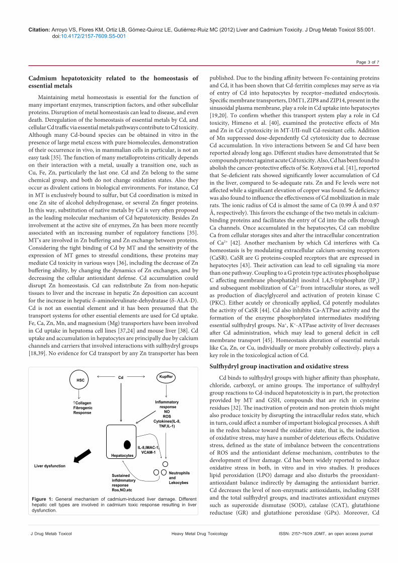
in blood, kidney and liver and since CAT contains Fe in its catalytic center, the decreased activity of the enzyme in the liver exposed to Cd might be a result of Fe deficiency. It has also been proposed that Cd could replace Fe and Cu in various cytoplasmic and membrane proteins, such as SOD or CAT, increasing the concentration of ionic Cu and Fe, which could catalyze the breakdown of H2O2 via Fenton reaction resulting in oxidative stress [58]. Djukic-Cosic et al. [59], have shown that Cd exposure induce a significant increase in Fe content and MDA, a well known secondary product of LPO, in mouse liver after 4 and 6 h treatment. These results indicate a positive correlation between hepatic Fe and MDA content. A possible mechanism that could explain the increase in Fe caused by Cd is the redistribution of Fe in the organism. Cd has been shown to bind to both ferritin and apoferritin, and to displace Fe from various intracellular sites [58]. Recently, Lai and Loo [60], reported that cells without adequate Fe would be unable to fully express the stress gene, heme oxygenase-1, when confronted with the toxic metal, Cd (Figure 2).
Mitochondria and CadmiumSeveral evidences prove that mitochondria can play as key target
in Cd exposure [61-63], and a major source of Cd-induced ROS production, mainly blocking the mitochondrial electron transport chain [64] and by uncoupling oxidative phosphorylation by the acceleration of H+ influx through the Pi/H+ symporter. This organelle exerts an important role in cellular energy production, and the interference with the normal oxidative metabolism induced by Cd, results in energetic deficits affecting crucial cell functions. In HepG2 cells and isolated rat liver mitochondria, Cd modifies mitochondrial function by inhibiting oxidative phosphorylation and affecting membrane structure-function relationships by changing lipid/phospholipids profiles [65,66]. It has been proposed two sets of events following Cd intoxication, the first one, characterized by transmembrane electrical potential (∆Ψ) dissipation, membrane permeability transition (MPT) and basal respiration stimulation, followed by depression of ATP levels. The second one involves respiration inhibition, Fe mobilization and mitochondrial membrane LPO. It has been proposed that Cd initially binds to protein thiols in mitochondrial membrane. This degree of interaction would, however be sufficient to produce the conformational changes required for activation of the Ca uniporter-mediated cation uptake, and affects mitochondrial permeability transition. The hepatocyte mitochondrion has been considered a major source for Cd-induced ROS production [67]. ROS produced include O2
•, H2O2, and OH• and it has been proposed that complex III of the electron transport chain could be responsible for the production of these ROS [68,69]. Even more, these studies indicate, that Cd may bind between semiubiquinone and cytochrome b566 of the Q0 site of cytochrome b of complex III (cytochrome bc1 oxidoreductase), resulting in accumulation of semiubiquinones at the
Antioxidantenzymesinactivation
GSH depletion
Mitochondria-derived RoSProduction
Oxidative stressCdLipids,DNA andProtein oxidation
hepatocellularDamage
Figure 2: Cadmium-induced oxidative stress. Proposed pathways for cadmium induced oxidative stress damage.
Citation: Arroyo VS, Flores KM, Ortiz LB, Gómez-Quiroz LE, Gutiérrez-Ruiz MC (2012) Liver and Cadmium Toxicity. J Drug Metabol Toxicol S5:001. doi:10.4172/2157-7609.S5-001
Page 5 of 7
J Drug Metabol Toxicol ISSN: 2157-7609 JDMT, an open access journalHeavy Metal Drug Toxicology
Q0 site. The semiubiquinones, being unstable, are prone to transfer one electron to molecular oxygen to form O2
• and thus producing oxidative stress. In addition, Cd induces Fe mobilization from the iron-containing respiratory chain proteins. It is well established that respiratory chain inhibition increases generation of ROS that, in the presence of Fe2+, gives rise via the Fenton reaction to the highly reactive OH• causing LPO. Several studies demonstrate a relationship between Cd exposure and LPO [52,54,70,71] showing that Cd increased the hepatic level of malondialdehyde (MDA). Cd, through binding to the inner membrane, enhances LPO and disturbs the integrity of mitochondrial membranes. Breakdown products of lipid peroxides may also react with DNA amino bases and proteins. Cd-generated radical adducts can be detected by electron spin resonance (ESR) [72]. The ESR studies provide direct evidence to identify Cd-generated free radicals as O2
•, H2O2, OH• and lipid radicals. The relevance in LPO as a key determinant in hepatocellular damage was confirmed by the effect of vitamin E alone or in combination of beta-carotene that reduced the harmful effects of Cd in rats [49].
Cadmium-induced apoptosis
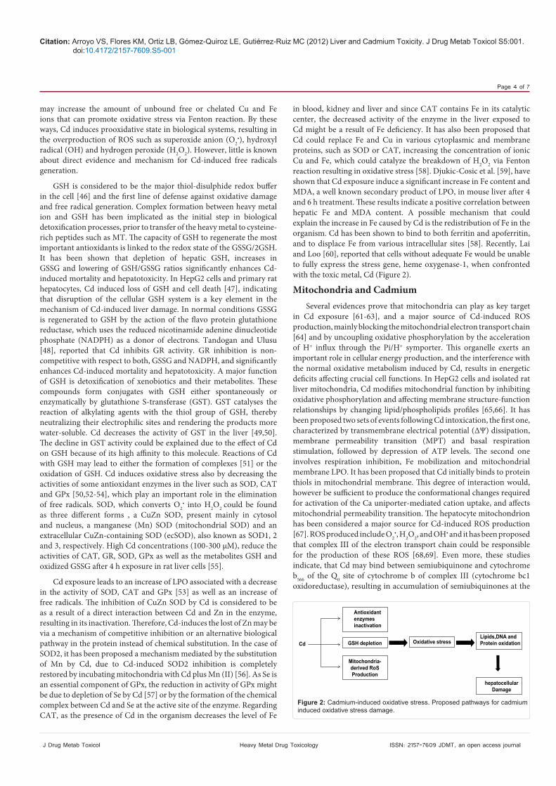
Apoptosis is a genetically-regulated form of cell death, which play an important role in the development and maintenance of tissue homeostasis in multicellular organism. In vitro studies in rat, mouse or human hepatocytes show that apoptosis plays a predominant role in Cd hepatoxicity [73-75]. However, the apoptosis signaling induced by Cd is still unclear. Apoptosis mediated by mitochondria may be more relevant in metal-induced cell death. Toxicity primarily appears to be caused by the binding of Cd to thiol groups in mitochondrial resulting in mitochondria dysfunction and subsequent injury. In isolated mitochondria from mouse liver, it has been showed that Cd could directly lead to the dysfunction of mitochondria, including the inhibition of respiration, the opening of membrane permeability transition pore, MPT, loss of transmembrane potential, and the release of Cyt c. Furthermore, Cd may enter mitochondria via Ca uniporter and interact with thiol groups of adenine nucleotide translocator (ANT) to induce the MPT, Cyt c release and apoptosis. In addition, when thiol-containing reagents are pre-incubated with Cd, dysfunction of mitochondria and Cyt c release can delay and suppress the apoptosis [76]. Pham et al. [74], have suggested the presence of mitochondrial-mediated caspase-independent pathways in Cd-induced apoptosis in primary cultures of rat hepatocytes, as the pre-treatment with a caspase inhibitor does not prevent apoptosis. The mitochondria derived protein, endonuclease G (Endo G) has been identified as potential caspase-independent apoptotic mediator in human hepatoma cell line Hep3B. The mechanism involves Ca and ROS related alteration of mitochondrial homeostasis and subsequent release of Endo G and AIF [77]. Cd has been demonstrated to interfere with Ca by inhibiting Ca-dependent ATPases in nuclei and endoplasmic reticulum, probably through interaction with protein thiol groups. A rise in [Ca]i, has been suggested to cause apoptosis by inducing dissipation of mitochondrial membrane potential (MMP) and oxidative phosphorylation uncoupling. Wang et al. [42], have reported that a major part of [Ca]i elevation in Cd-exposed hepatocytes, may be related to extracellular Ca entry, whereas the initial [Ca]i elevation is associated with an excessive release of Ca from intracellular stores. However data reported by Xie et al. [78], show that Cd-induced [Ca]i elevation was attenuated in cells HL-7702 co-incubated with a Ca chelator. So, Cd-induced apoptosis was mediated by the release of Ca from intracellular Ca storage but not an influx of extracellular Ca. Cadmium causes ER stress in vitro
and in vivo and mediates induction of apoptosis in some tissues [79]. However, to date no conclusive data are available in Cd-induced ER stress and its relation with hepatocyte apoptosis [80,81] (Figure 3).
SummaryIn conclusion, many studies have shown that Cd induces
hepatocellular damage by an imbalance in the cellular redox status which leads to oxidative stress, although there are many questions pending to address regarding the exact mechanism of Cd-induced damage, we know that Cd compete with metal sites in many protective or antioxidants proteins inducing an improper activity and even the total loss of the antioxidant cellular defense, in addition the disturb in Ca homeostasis lead to the activation of PKC which drives many of the noxious effects initiated by ROS. Moreover, Cd depletes GSH and other sulfhydryl groups, which aggravates the oxidative stress and the cellular damage resulting in apoptosis. Finally all liver cells are involved in the Cd-induced damage; the inflammatory response elicited by Cd generates the infiltration and activation of phagocytic cells releasing more inflammatory mediators such as cytokines, or ROS. Further deeply research is needed to figure out the complete mechanism of hepatocellular damage induced by Cd in order to set up proper therapeutics approaches for chronic and acute Cd intoxication.
Conflict of Interest
There are no conflicts of interest.
Acknowledgements
This work was partly supporter by grants SEP-CONACYT # 106194 and by the Universidad Autónoma Metropolitana–Iztapalapa.
Referencs
1. ATSDR (Agency for Toxic Substance and Disease Registry) Draft Toxicological Profile for Cadmium (2005) Department of Health and Humans Services, Public Health Service, Centers for Disease Control, Atlanta, GA, USA.
2. Mead MN (2010) Cadmium confusion: do consumers need protection?. Environ Health Perspect 118: 528-534.
3. IARC (International agency for research on cancer) (1993) Monographs on the Evaluation of the Carcinogenic Risks to Humans Beryllium, Cadmium, Mercury and Exposures in the Glass Manufacturing Industry. IARC Scientific Publications, Lyon, France, pp.119-238.
4. Anwar-Mohamed A, Elbekai RH, EI-Kadi AO (2009) Regulation of CYP1A1 by heavy metals and consequences for drug metabolism. Expert Opin Drug Metab Toxicol 5: 501-521.
5. Diamond GL, Thayer WC, Choudhury H (2003) Pharmacokinetics pharmacodynamics (PK/PD) modeling of risks of kidney toxicity from
l.X lll
ROS
Cd
Cd
Ca
Cd
Oxidative Strress
ApoptosisCytcAlFEndo G
Cd
GsH
Figure 3: Cadmium-induced apoptosis.

Citation: Arroyo VS, Flores KM, Ortiz LB, Gómez-Quiroz LE, Gutiérrez-Ruiz MC (2012) Liver and Cadmium Toxicity. J Drug Metab Toxicol S5:001. doi:10.4172/2157-7609.S5-001
Page 6 of 7
J Drug Metab Toxicol ISSN: 2157-7609 JDMT, an open access journalHeavy Metal Drug Toxicology
exposure to cadmium: estimates of dietary risks in the US population. J Toxicol Environ Health 66: 2141-2164.
6. Jin T, Nordberg G, Ye T, Bo M, Wang H, et al. (2004) Osteoporosis and renal dysfunction in a general population exposed to cadmium in China. Environ Res 96: 353-359.
7. Zalups RK (2000) Evidence for basolateral uptake of cadmium in the kidney of rats. Toxicol Appl Pharmacol 164: 15-23.
8. Vahter M, Akesson A, Lidén C, Ceccatelli S, Berglund M (2007) Gender differences in the disposition and toxicity of metals. Environ Res 104: 85-95.
9. Rzigalinski BA, Strobl JS (2009) Cadmium-containing nanoparticles: perspectives on pharmacology and toxicology of quantum dots. Toxicol Appl Pharmacol 238: 280-288.
10. Godt J, Scheidig F, Grosse-Siestrup C, Esche V, Brandenburg P, et al. (2006) The toxicity of cadmium and resulting hazards for human health. J Occup Med Toxicol 10: 1-22.
11. Vesey DA (2010) Transport pathways for cadmium in the intestine and kidney proximal tubule: Focus on the interaction with essential metals. Toxicol Lett 198: 13-19.
12. Zalups RK, Ahmad S (2003) Molecular handling of cadmium in transporting epithelia. Toxicol Appl Pharmacol 186: 163-188.
13. Foulkes EC (2000) Transport of toxic heavy metals across cell membranes. Proc Soc Exp Biol Med 223: 234-240.
14. Ryu DY, Lee SJ, Park DW, Choi BS, Klaassen CD, et al. (2004) Dietary iron regulates intestinal cadmium absorption through iron transporters in rats. Toxicol Lett 152: 19–25.
15. Min KS, Ueda H, Tanaka K (2008) Involvement of intestinal transporter 1 and metallothionein in cadmium accumulation in the liver and kidney of mice fed a low-calcium diet. Toxicol Lett 176: 85-92.
16. Carriere P, Mantha M, Champagne-Paradis S, Jumarie C (2011) Characterization of basolateral-to-apical transepithelial transport of cadmium in intestinal TC7 cell monolayers. Biometals 24: 857-874.
17. DelRaso NJ, Foy BD, Gearhart JM, Frazier JM (2003) Cadmium uptake kinetics in rat hepatocytes: Correlation for albumin binding. Toxicol Sci 72: 19-30.
18. Souza V, Bucio L, Gutiérrez-Ruiz MC (1997) Cadmium uptake by a human hepatic cells line (WRL-68). Toxicology 120: 215-220.
19. Fujishiro H, Okugaki S, Kubota K, Fujiyama T, Miyataka H, et al. (2009) The role of ZIP8 down-regulation in cadmium-resistant metallothionein–null cells. J Appl Toxicol 29: 367-373.
20. Liuzzi JP, Aydemir F, Nam H, Knutson MD, Cousing RJ (2006) Zip14 (Slc39a14) mediates non-transferrin-bound iron uptake into cells. Proc Natl Acad Sci 103: 13612-13617.
21. Honda A, Komuro H, Hasegawa T, Seko Y, Shimada A, et al. (2010) Resistance of metallothionein-III null mice to cadmium-induced acute hepatotoxicity. J Toxicol Sci 35: 209-215.
22. Yamano T, DeCicco LA, Rikans LE (2000) Attenuation of cadmium-induced liver injury in senescent male fischer 344 rats: role of Kupffer cells and inflammatory cytokines. Toxicol Appl Pharmacol 162: 68-75.
23. El-Sokkary GH, Nafady AA, Shabash EH (2010) Melatonin administration ameliorates cadmium-induced oxidative stress and morphological changes in the liver of rat. Ecotoxicol Environ Saf 73: 456-463.
24. Martelli A, Rousselet E, Dycke C, Bouron A, Moulis JM (2006) Cadmium toxicity in animal cells by interference with essential metals. Biochimie 88: 1807-1814.
25. Van Kerkhove E, Pennemans V, Swennen Q (2010) Cadmium and transport of ions and substances across cell membranes and epithelia. Biometals 23: 823-855.
26. Bruscalupi G, Massimi M, Devirgiliis LC, Leoni S (2009) Multiple parameters are involved in the effects of cadmium on prenatal hepatocytes. Toxicol In Vitro 23: 1311-8.
27. Horiguchi H, Harada A, Oguma E, Sato M, Homma Y, et al. (2000) Cadmium-induced acute hepatic injury is exacerbated in human interleukin-8 transgenic mice. Toxicol Appl Pharmacol 163: 231–239.
28. Marth E, Burt S, Jelovcan S (2000) Influence of cadmium on the immune system Description of stimulating reactions. Cent Eur J Public Health 8: 40-44.
29. Souza V, Escobar M, Gómez-Quiroz L, Bucio L, Hernández E, et al. (2004) Acute cadmium exposure enhances AP-1 DNA binding and induces cytokines expression and heat shock protein 70 in HepG2 cells. Toxicology 197: 213-228.
30. Harstad EB, Klaassen CD (2002) Gadolinium chloride pretreatment prevents cadmium chloride-induced liver damage in both wild-type and MT-null mice. Toxicol Appl Pharmacol 180: 178-185.
31. Mousa SA (2004) Expression of adhesion molecules during cadmium hepatotoxicity. Life Sc 75: 93-105.
32. Rikans LE, Yamano T (2000) Mechanisms of cadmium-mediated acute hepatotoxicity. J Biochem Mol Toxicol 14: 110–117.
33. Kuester RK, Waalkes MP, Goering PL, Fisher BL, McCuskey RS, et al. (2002) Differential hepatotoxicity induced by cadmium in Fischer 344 and Sprague-Dawley rats. Toxicol Sci 65: 151-159.
34. Jaeschke H (2000) Reactive oxygen and mechanism of inflammatory liver injury. J Gastroenterology 15: 718-724.
35. Moulis JM (2010) Cellular mechanisms of cadmium toxicity related to the homeostasis of essential metals. Biometals 23: 877-896.
36. Sabolic I, Breliak D, Skarica M, Herak-kramberger CM (2010) Role of metallothionein in cadmium traffic and toxicity in kidneys and other mammalian organs. Biometals 23: 897-926.
37. Fotakis G, Timbrell JA (2006a) Role of trace elements in cadmium chloride uptake in hepatoma cells lines. Toxicol Lett 164: 97–103.
38. Zhang Y, Li B, Chen C, Gao Z (2009) Hepatic distribution of iron, copper, zinc and cadmium-containing proteins in normal and iron overload mice. Biometals 22: 251-259.
39. Fotakis G,Timbrell JA (2006b) Modulation of cadmium chloride toxicity by sulphur amino acids in hepatoma cells. Toxicol In Vitro 20: 641–648.
40. Himeno S, Yanagiya T, Fujishiro H (2009) The role of zinc transporters in cadmium and manganeso transport in mammalian cells. Biochimie 91: 1218-1222.
41. Kotyzová D, Cerná P, Leseticky L, Eybl V (2010) Trace elements status in selenium-deficient rats-Interaction with cadmium. Biol Trace Elem Res 136: 287-293.
42. Wang SS, Chen L, Xia SK (2007) Cadmium is acutely toxic for murine hepatocytes: Effects on intracellular free Ca2+ homeostasis. Physiol Res 56: 193-201.
43. Canaff L, Petit JL, Kisiel M, Watson PH, Gascon-Barré M, et al. (2001) Extracellular calcium-sensing receptor is expressed in rat hepatocytes. J Biol Chem 276: 4070-4079.
44. Chang W, Shoback D (2004) Extracellular Ca2+-sensing receptors-an overview. Cell Calcium 35: 183-196.
45. Karthikeyan J, Bavani G (2009) Effect of cadmium on lactate dehydrogenase isoenzyme, succinate dehydrogenase and Na (+)-K(+)-ATPase in liver tissue of rat. J Environ Biol 31: 895-898.
46. Masella R, Benedetto Di, Vari R, Filesi C, Giovannini C (2005) Novel mechanisms of natural antioxidant compounds in biological systems: involvement of glutathione and glutathione-related enzymes. J Nutr Biochem 16: 577–586.
47. Gebhardt R (2009) Prevention of cadmium-induced toxicity in liver-derived cells by the combination Heppeel. Environ Toxicol Pharm 26: 402-409.
48. Tandogan B, Ulusu NN (2010) Inhibition of purified bovine liver glutathione reductase with some metal ions. J Enzyme Inhib Med Chem 25: 68-73.
Citation: Arroyo VS, Flores KM, Ortiz LB, Gómez-Quiroz LE, Gutiérrez-Ruiz MC (2012) Liver and Cadmium Toxicity. J Drug Metab Toxicol S5:001. doi:10.4172/2157-7609.S5-001
Page 7 of 7
J Drug Metab Toxicol ISSN: 2157-7609 JDMT, an open access journalHeavy Metal Drug Toxicology
49. El-Demerdash FM, Yousef MI, Kedwany FS, Baghdadi HH (2004) Cadmium-induced changes in lipid peroxidation, blood hematology, biochemical parameters and semen quality of male rats: protective role of vitamin E and β-carotene. Food Chem Toxicol 42: 1563-1571.
50. Renugadevi J, Prabu MS (2010) Cadmium-induced hepatotoxicity in rats and the protective effect of naringenin. Exp Toxicol Pathol 62: 171-181.
51. Mah V, Jalilehvand F (2010) Cadmium (II) complex formation with glutathione. J Biol Inorg Chem 15: 441-458.
52. Koyu A, Gokcimen A, Ozguner F, Senal Bayram D, Kocak A (2006) Evaluation of the effects of cadmium on rat liver. Mol Cell Biochem 284: 81–85.
53. Newairy AA, El-Sharaky AS, Badreldeen MM, Eweda SM, Sheweita SA (2007) The hepatoprotective effects of selenium against cadmium toxicity in rats. Toxicology 242: 23-30.
54. Sinha M, Manna P, Sil PC (2009) Induction of necrosis in cadmium-induced hepatic oxidative stress and its prevention by the prophylactic properties of taurine. J Trace Elem Med Biol 23: 300-313.
55. Ikediobi CO, Badisa VL, Ayuk-Takem LT, Laatinwo LM, West J (2004) Response of antioxidant enzymes and redox metabolites to cadmium-induced oxidative stress in CRL-1439 normal rat liver cells. Int J Mol 14: 87-92.
56. Casalino E, Calzaretti G, Sblano C, Landriscina C (2002) Molecualr inhibitory mechanisms of antioxidant enzymes in rat liver and kidney by cadmium. Toxicology 30: 37-50.
57. Lazarus M, Orct T, Blanusa M, Kostial K, Pirsljin J, et al. (2006) Effect of selenium pre-treatment of cadmium content and enzymatic antioxidants in tissues of surckling rat. Toxicol Lett 164: S191.
58. Waisberg M, Joseph P, Hale B, Beyersmann D (2003) Molecular and cellular mechanisms of cadmium carcinogenesis. Toxicology 192: 95–117.
59. Djukic-Cosic D, Jovanovic MC, Bulat ZP, Ninkovic M, Malicevic Z, et al. (2008) Relation between lipid peroxidation and iron concentration in mouse liver after acute and subacute cadmium intoxication. J Trace Elem Med Biol 22: 66-72.
60. Lai C, Loo G (2011) Cellular iron depletion weakens induction of heme oxygenase-1 by cadmium. Int J Biochem Cell Biol 43: 88-97.
61. Belyaeva EA, Korotkov SM, Saris NE (2011) In vitro modulation of heavy metal-induced rat liver mitochondria dysfunction: A comparison of copper and mercury with cadmium. J Trace Elem Med Biol 25S: S63-S73.
62. Diep PTN, Denizeau F, Jumarie C (2005) Kinetics of the early subcellular distribution of cadmium in rat hepatocytes. BioMetals 18: 255-267.
63. Zhang Y, Li JH, Liu XR, Jiang FL, Tian FF, et al. (2011) Spectroscopic and microscopic studies on the mechanisms of mitochondrial toxicity induced by different concentrations of cadmium. J Membr Biol 241: 39-49.
64. Ciapaite J, Nauciene Z, Baniene R, Wagner MJ, Krab K, et al. (2009) Modular kinetic analysis reveals differences in Cd2+ and Cu2+ ion-induced impairment of oxidative phosphorylation in liver. FEBS J 276: 3656-3668.
65. Modi HR, Katyare SS (2009) Effect of treatment with cadmium on structure-function relationships in rat liver mitochondria: Studies on oxidative energy metabolism and lipid/phospholipids profiles. J Membr Biol 232: 47-57.
66. Yang MS, Yu LC, Gupta RC (2004) Analysis of changes in energy and redox states in HepG2 hepatoma and C6 glioma cells upon exposure to cadmium. Toxicology 201: 105-113.
67. Belyaeva EA, Dymkowska D, Wieckowski MR, Wojtczak L (2008) Mitochondria as an important target in heavy metals toxicity in rat hepatoma AS-30D cells. Toxicol Appl Pharmacol 231: 34-42.
68. Belyaeva EA, Dymkowska D, Wieckowski MR, Wojtczak L (2006) Reactive oxygen species produced by the mitochondria respiratory chain are involved
in Cd2+ induced injury of rat ascites hepatoma AS-30D cells. Biochim Biophys Acta 1757: 1568-1574.
69. Wang Y, Fang J, Leonard SS, Rao KM (2004) Cadmium inhibits the electron transfer chain and induces reactive oxygen species. Free Radic Biol Med 36: 1434-1443.
70. Bucio L, Souza V, Albores A, Sierra A, Chávez E, et al. (2005) Cadmium and mercury toxicity in a human fetal hepatic cell line (WRL-68 cells). Toxicology 102: 285-99.
71. Thijssen S, Cuypers A, Maringwa J, Smeets K, Horemans N, et al. (2007) Low cadmium exposure triggers a biphasic oxidative stress response in mice kidney. Toxicology 236: 29-41.
72. Liu J, Qian SY, Guo Q, Jiang J, Waalkes MP, et al. (2008) Cadmium generated reactive oxygen and carbon centered radical species in rat: insights from in vitro spin-trapping studies. Free Radic Biol Med 45: 475-481.
73. Lasfer M, Vadrot N, Aoudjehane L, Conti F, Bringuier AF, et al. (2008) Cadmium induces mitochondria-dependent apoptosis of normal human hepatocytes. Cell Biol Toxicol 24: 55-62.
74. Pham TND, Marion M, Denizeau F, Jumarie C (2006) Cadmium-induced apoptosis in rat hepatocytes does not necessarily involve caspase-dependent pathways. Toxicol In Vitro 20: 1331-1342.
75. Yu X, Sidhu JS, Hong S, Robinson JF, Ponce RA, et al. (2011) Cadmium induced p53-dependent activation of stress signaling, accumulation of ubiquitinated proteins, and apoptosis in mouse embryonic fibroblast cells. Toxicol Sci 120: 403-412.
76. Oh SH, Lim SC (2006) A rapid and transient ROS generation by cadmium triggers apoptosis via caspase-dependent pathway in HepG2 cells and this is inhibited through N-acetylcysteine-mediated catalase regulation. Toxicol Appl Pharmacol 212: 212-223.
77. Lemarie A, Legadic-Gossmann D, Morzadec C, Allain N, Fardel O, et al. (2004) Cadmium induces caspase-independent apoptosis in liver Hep3B cells: role for calcium in signaling oxidative stress-related impairment of mitochondria and relocation of endonuclease G and apoptosis-inducing factor. Free Radic Biol Med 36: 1517-1531.
78. Xie Z, Zhang Y, Li A, Li P, Ji W, et al. (2010) Cd-induced apoptosis was mediated by the release of Ca2+ from intracellular Ca storage. Toxicol Lett 192: 115-118.
79. Kitamura M, Hiramatsu N (2010) The oxidative stress: endoplasmic reticulum stress axis in cadmium toxicity. Biometals 23: 941-950.
80. Biagioli M, Pifferi S, Ragghianti M, Bucci S, Rizzuto R, et al. (2008) Endoplasmic reticulum stress and alteration in calcium homeostasis are involved in cadmium-induced apoptosis. Cell Calcium 43: 184-195.
81. Hiramatsu N, Kasai A, Du S, Takeda M, Hayakawa K, et al. (2007) Rapid, transient induction of ER stress in the liver and kidney after acute exposure to heavy metals: Evidence from transgenic sensor mice. FEBS Lett 581: 2055-2059.
Thisarticlewasoriginallypublishedinaspecialissue,Heavy Metal Drug ToxicologyhandledbyEditor(s).Dr.RakeshK.Srivastava,KansasMedicalCenter,UnitedStates