Embed Size (px)
Citation preview
Liver – Fatty change
1

Liver – Fatty change
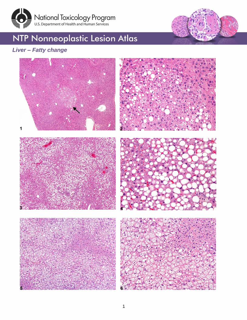
Figure Legend: Figure 1 Fatty change–focal fatty change (arrow) in a B6C3F1 male mouse
from a chronic study. Figure 2 Fatty change–focal fatty change in a B6C3F1 male mouse from
a chronic study (higher magnification of Figure 1). Figure 3 Fatty change–macrovesicular fatty
change in a male F344/N rat from a chronic study. Figure 4 Fatty change–macrovesicular fatty
change in a male F344/N rat from a chronic study (higher magnification of Figure 3). Figure 5
Fatty change–microvesicular fatty change in a male P53+/- (C57Bl/6) mouse from a subchronic
study. Figure 6 Fatty change–microvesicular fatty change in a male P53+/- (C57Bl/6) mouse
from a subchronic study (higher magnification of Figure 5). Figure 7 Fatty change–hepatocyte
cytoplasmic vacuolation consistent with glycogen and macrovesicular fatty change in a male
B6C3F1 mouse from a chronic study. Figure 8 Fatty change–hepatocyte cytoplasmic
vacuolation consistent with glycogen and macrovesicular fatty change in a male B6C3F1 mouse
from a chronic study (higher magnification of Figure 7).
Comment: Fatty change can be focal (Figure 1, arrow; Figure 2) or diffuse (Figure 3, Figure 4,
Figure 5, and Figure 6) and macrovesicular (Figure 3 and Figure 4) or microvesicular (Figure 5
and Figure 6). Macrovesicular fatty change is often associated with metabolic disturbances and
is generally readily reversible, whereas microvesicular fatty change is more likely a reflection of
toxicity, possibly involving mitochondrial disturbances. Figure 1 and Figure 2 represent a focal
lesion in which the morphologic features of the vacuolated hepatocytes are consistent with both
macrovesicular and microvesicular fatty change. Focal fatty change can be a spontaneous
lesion and may be more common in some strains than others, whereas diffuse or zonal (e.g.,
2

Liver – Fatty change
centrilobular or periportal) fatty change is more likely to be treatment related. The cytoplasmic
vacuolization in Figure 3 and Figure 4 is diffuse and consists of both large and small single or
double cytoplasmic vacuoles within hepatocytes. The morphologic features are consistent with
macrovesicular fatty change, with compression and displacement of the nuclei to the periphery
of affected hepatocytes. In contrast, Figure 5 and Figure 6 are representative of microvesicular
fatty change, with a more prominent response in periportal and midlobular areas. The
cytoplasmic change consists of multiple small vacuoles filling the cytoplasm without prominent
compression or displacement of hepatocyte nuclei. Sinusoids may be collapsed by the
hypertrophic hepatocytes. Figure 7 and Figure 8 represent a mixture of both macrovesicular
fatty change and glycogen accumulation in hepatocytes. Mixtures of glycogen and both
macrovesicular and microvesicular fatty change may be seen. Phospholipidosis is another form
of hepatocyte cytoplasmic vacuolization that is associated with exposure to cationic amphophilic
xenobiotics and requires special staining and/or electron microscopy for definitive confirmation.
Recommendation: The morphologic appearance of fatty change is distinctive enough to allow
its diagnosis in the absence of confirmatory special stains or other procedures. Whenever
present, diagnosis of hepatocellular fatty change should be recorded. Since some degree of
fatty change may occur in untreated or vehicle controls, severity grading is recommended to
document potential treatment-related effects. Because focal fatty change may be spontaneous,
the distribution modifier “focal” should be included in the diagnosis when the lesion is focal.
When the lesion is diffuse, no distribution modifier should be used (i.e., the lesion is assumed to
be diffuse when the distribution is not specified in the diagnosis). The morphologic features of
the fatty change (e.g., macrovesicular, microvesicular, or both) should be described in the
pathology narrative. Other distinctive features, such as the pattern of distribution, should also be
described in the pathology narrative.
Fatty change is frequently accompanied by other cytoplasmic alterations, such as glycogen
accumulation or depletion, and it can be difficult to distinguish the different types of alterations in
cytoplasmic morphology. In those situations, a diagnosis of hepatocyte cytoplasmic
vacuolization is appropriate, along with a severity grade, but the morphology of the cytoplasmic
3

Liver – Fatty change
changes in the hepatocytes should be thoroughly described in the pathology narrative. The
pathology narrative should also include the pathologist’s opinions regarding the contents of the
cytoplasmic vacuoles.
References:
Cattley RC, Popp JA. 2002. Liver. In: Handbook of Toxicologic Pathology (Haschek WM, Rousseaux CG, Wallig MA, eds). Academic Press, San Diego, 2:187–225. Abstract: http://www.sciencedirect.com/science/book/9780123302151
Eustis SL, Boorman GA, Harada T, Popp JA. 1990. Liver. In: Pathology of the Fischer Rat (Boorman GA, Eustis SL, Elwell MR, Montgomery CA, MacKenzie WF, eds). Academic Press, San Diego, 71–94. Abstract: http://www.ncbi.nlm.nih.gov/nlmcatalog/9002563
Evans JG, Lake BG. 1998. The digestive system II. Hepatobiliary system. In: Target Organ Pathology (Turton J, Hooson J, eds). Taylor and Francis, London, 61–98. Abstract: http://www.amazon.com/Target-Organ-Pathology-Basic-Text/dp/0748401571
Greaves P. 2007. Histopathology of Preclinical Toxicity Studies: Interpretation and Relevance in Drug Safety Evaluation, 3rd ed. Elsevier, Amsterdam. Abstract: http://www.sciencedirect.com/science/book/9780444527714
Harada T, Enomoto A, Boorman GA, Maronpot RR. 1999. Liver and gallbladder. In: Pathology of the Mouse: Reference and Atlas (Maronpot RR, Boorman GA, Gaul BW, eds). Cache River Press, Vienna, IL, 119–83. Abstract: http://www.cacheriverpress.com/books/pathmouse.htm
Hardisty JF, Brix AE. 2005. Comparative hepatic toxicity: Prechronic/chronic liver toxicity in rodents. Toxicol Pathol 33:35–40. Full-Text: http://tpx.sagepub.com/content/33/1/35.full.pdf
Haschek WM, Rousseaux CG, Wallig MA. 2010. Fundamentals of Toxicologic Pathology, 2nd ed. Academic Press, San Diego, 197–235. Abstract: http://www.sciencedirect.com/science/book/9780123704696
Thoolen B, Maronpot RR, Harada T, Nyska A, Rousseaux C, Nolte T, Malarkey D, Kaufmann W, Kutter K, Deschl U, Nakae D, Gregson R, Winlove M, Brix A, Singl B, Belpoggi F, Ward JM. 2010. Hepatobiliary lesion nomenclature and diagnostic criteria for lesions in rats and mice (INHAND). Toxicol Pathol 38:5S–81S. Full-Text: http://tpx.sagepub.com/content/38/7_suppl/5S.full
4

Liver – Fatty change
Author:
Robert R. Maronpot, DVM, MS, MPH, DACVP, DABT, FIATP Senior Pathologist Experimental Pathology Laboratories, Inc. Research Triangle Park, NC
5