Embed Size (px)
Citation preview
Liver
Last time we talked about the liver and the doctor started by revising some
information about it:
It has five surfaces.
It reaches the 5th
intercostal space ; some books write that it reaches the 4th
intercostal space and that is also correct due to variation ; but in most cases it
ascends above the 5th space so logically the 5
th space is the best answer since
it’s rare for it to reach the 4th
space.
The physiological function of the liver lobes :
a. The right lobe receives blood from the right hepatic artery and give
rise to right hepatic vein.
b. The left lobe along with caudate and quadrate lobes acts as a one
physiological unit.
The free edge of lesser omentum carries all the blood that’s going to the
liver:
a. Portal: Constitutes (70-80) % of the blood that reaches the liver.
b. Hepatic: Constitutes (20-30) %.
So by clamping the free edge of the lesser omentum we can stop the bleeding in
the liver. The bleeding can be due to car accidents that result in a trauma of the
liver and the clamping is only temporary till we clean the wound and find where
the trauma formed laceration in the liver and stitch it and stop the bleeding.
-----------------------------------------------------------------------------
Common Bile Duct (CBD)
The cystic duct its 4 cm long, it’s the duct of the gallbladder and it joins the
common hepatic duct to form the CBD.
The CBD its 10 cm long, about 3 inches (The “4 cm” in the slide refers to
the upper part that’s above the duodenum).
Its divided into 3 parts:
a. Supra duodenal: Above duodenum, about 4 cm.
b. Retro duodenal: Behind the 1st part of duodenum.
c. Retro pancreatic or
infra duodenal:
below the
duodenum.
It descends in the free
edge of lesser omentum
(1stpart) then it passes
behind duodenum (2nd
part) and end in its retro
pancreatic position (3rd
part).
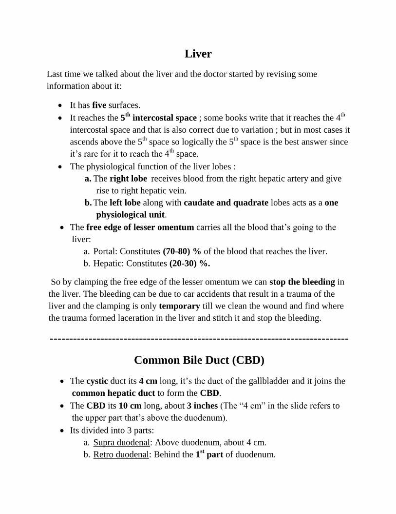
Parts of CBD:
o First part is found in the free edge of the lesser omentum. It’s related
to hepatic artery and portal vein, it’s found to the right side of the
artery and posterior to it we find the portal vein.
o Second part is found behind the 1st part of duodenum at the right to
the gastroduodenal artery.
o Third part lies at the posterior surface of the head of pancreas and
here the CBD meets the main pancreatic duct. In this part the duct is
related to IVC, at on its left we find the gastroduodenal artery and
portal vein.
The CBD ends in the middle of the 2nd
part of duodenum and it’s closed
by the sphincter of Oddi which opens inside the duodenum. Its location in
the duodenum is called the major duodenal papilla or ampulla of Vater
(Can be noticed in the ERCP picture below).
The meeting of the CBD and the pancreatic duct has variations and these
variations must be considered while performing ERCP.
o Sometimes a common sphincter can’t be found but we find two
separate openings, one for the common bile duct and another for the
main pancreatic duct; each duct has its own sphincter.
o Sometimes they meet before the sphincter and have one duct; in this
case they have one sphincter, we deal with it as the normal case.
The blood supply of the CBD arises from the cystic artery which is usually a
branch from the right hepatic artery and sometimes it can take a branch from the
superior pancreaticoduodenal artery which also comes from the celiac trunk, so
it follows the foregut.
In the past the CBD was very important clinically because a stone can descend
and block the duct; resulting in jaundice (االصفرار) and sometimes pancreatitis.
So they used to remove it with an open surgery, and after removing the stone, they
had to do radiography to make sure that there are no other stones.
Nowadays they perform a procedure called ERCP where they enter through the
mouth by a scope and clean the stone via a small basket and pull it to the
duodenum or by irrigation by saline which is mostly used in case of mud since the
saline will dissolve the mud and open the duct; the patient will recover in 6 hours.
Gall stones have different sizes and shapes, they look like mosaic. The
doctor once found 120 small stones in a single patient but it can occur as a
single big stone that fills Hartman’s pouch.
Cholelithiasis: means stones in the gallbladder.
Cholecystitis: mean inflammation.
Cholecystectomy: means an operation.
Gangrene (cut of blood supply) happens in appendicitis but not in
cholecystitis.
Obstructive jaundice: the closure of the CBD by a stone or more, or mud
(Thickening of the secretion) and results in obstruction. Most common in
Ramadan due to dehydration.
o The bacteria are present as part of the normal flora but they are not
pathogenic, Dehydration will decrease the immunity making them
more active (Similar to the
H. Pylori activation in
stomach).
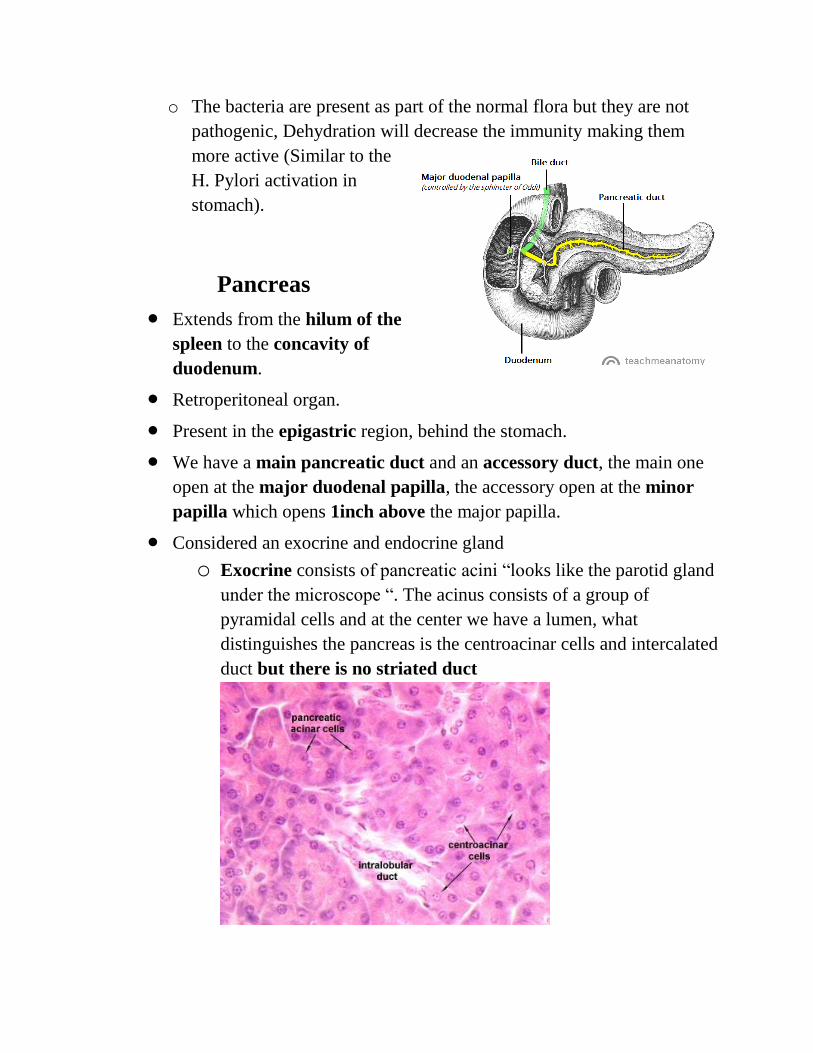
Pancreas
Extends from the hilum of the
spleen to the concavity of
duodenum.
Retroperitoneal organ.
Present in the epigastric region, behind the stomach.
We have a main pancreatic duct and an accessory duct, the main one
open at the major duodenal papilla, the accessory open at the minor
papilla which opens 1inch above the major papilla.
Considered an exocrine and endocrine gland
o Exocrine consists of pancreatic acini “looks like the parotid gland
under the microscope “. The acinus consists of a group of
pyramidal cells and at the center we have a lumen, what
distinguishes the pancreas is the centroacinar cells and intercalated
duct but there is no striated duct
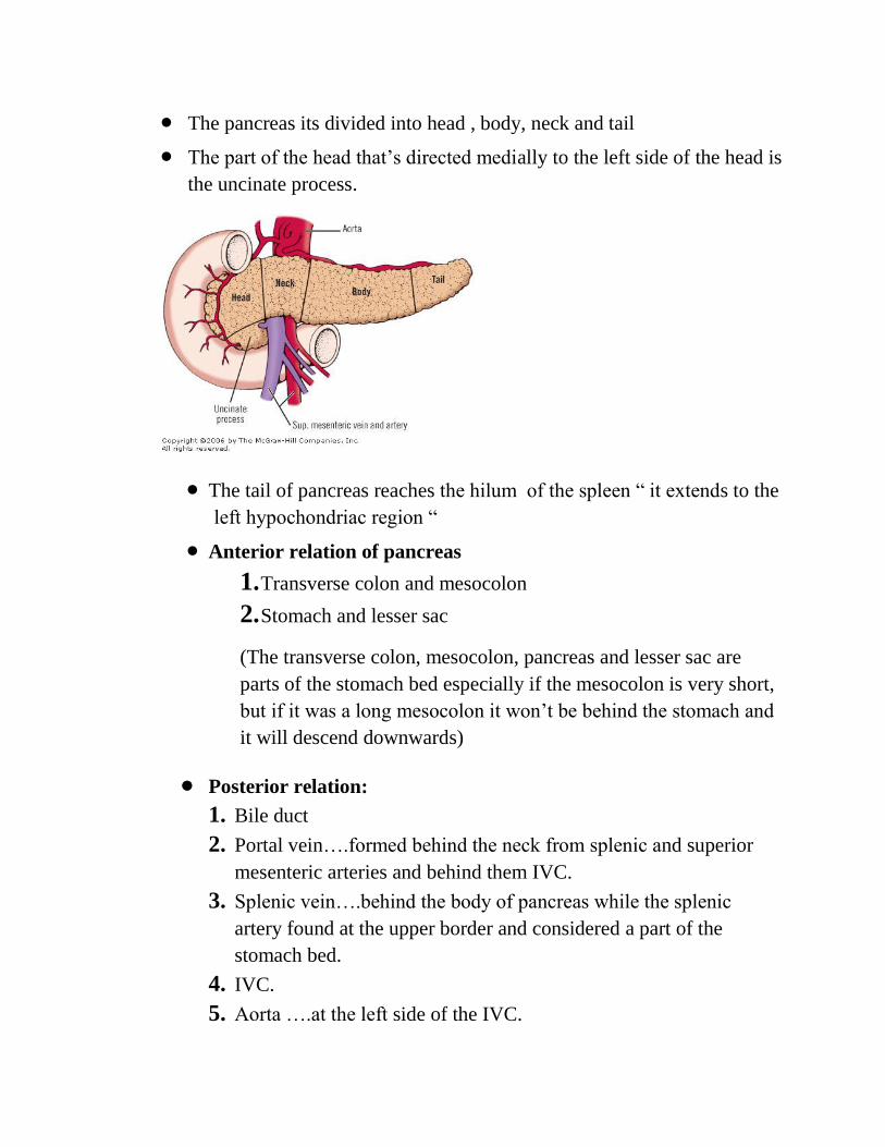
The pancreas its divided into head , body, neck and tail
The part of the head that’s directed medially to the left side of the head is
the uncinate process.
The tail of pancreas reaches the hilum of the spleen “ it extends to the
left hypochondriac region “
Anterior relation of pancreas
1. Transverse colon and mesocolon
2. Stomach and lesser sac
(The transverse colon, mesocolon, pancreas and lesser sac are
parts of the stomach bed especially if the mesocolon is very short,
but if it was a long mesocolon it won’t be behind the stomach and
it will descend downwards)
Posterior relation:
1. Bile duct
2. Portal vein….formed behind the neck from splenic and superior
mesenteric arteries and behind them IVC.
3. Splenic vein….behind the body of pancreas while the splenic
artery found at the upper border and considered a part of the
stomach bed.
4. IVC.
5. Aorta ….at the left side of the IVC.
6. The Origin of superior mesenteric artery….behind the body of
pancreas.
7. The left psoas major muscle.
8. The left kidney and left suprarenal gland.
9. Hilum of the spleen is found behind the tail, the tail of pancreas
forms an impression at the visceral surface of spleen.
The doctor said that we can consider the tail of pancreas intra
peritoneal because it’s found between the two layers of splenorenal
ligament, and add to the tail the splenic vessels (in the ligament).
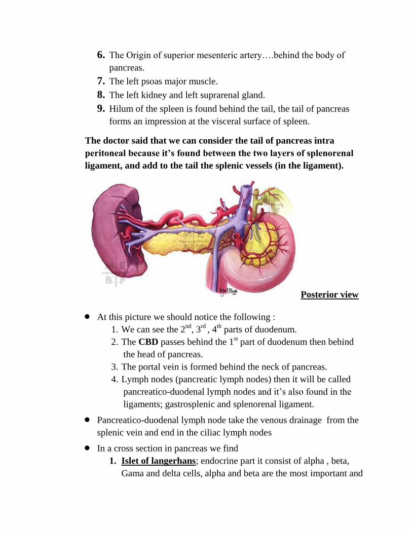
Posterior view
At this picture we should notice the following :
1. We can see the 2nd
, 3rd
, 4th parts of duodenum.
2. The CBD passes behind the 1st part of duodenum then behind
the head of pancreas.
3. The portal vein is formed behind the neck of pancreas.
4. Lymph nodes (pancreatic lymph nodes) then it will be called
pancreatico-duodenal lymph nodes and it’s also found in the
ligaments; gastrosplenic and splenorenal ligament.
Pancreatico-duodenal lymph node take the venous drainage from the
splenic vein and end in the ciliac lymph nodes
In a cross section in pancreas we find
1. Islet of langerhans; endocrine part it consist of alpha , beta,
Gama and delta cells, alpha and beta are the most important and
we can distinguish between them under the light microscope;
the alpha cells produce glucagon, and beta cells give insulin
2. Pancreativ Acini; the cells of pancreas have polarity in which
the cell has a basophilic base and acidophilic apex (contains
granules for secretion) , exocrine part
Glucagon is produced when glucose level in blood drops below the
normal “normal level (70-90) mg/dL, although some books state the
normal level is up to 100 and it’s also true” during the exam the
glucose level drops because the brain cells consume a lot of energy
and as a result, the glucagon is produced to breakdown glycogen and
rise the glucose level in the blood to normal ; in contrast if you eat
mansaf the glucose level will rise to 200-300 so the beta cells will
produce insulin that will decrease the glucose level by forming
glycogen or consuming it as energy.
*any defect in the beta cells will result in abnormality in insulin
producing (diabetes mellitus)
There is a balance between the weight and the efficiency of the pancreas
A person height is 170cm the perfect weight is 70kg, so the pancreas will
work well at 70kg, if he was overweight this will create pressure at the
beta cells to produce more insulin, and after a while this will result in
diabetes mellitus, because the cells can’t work enough in a person with
extra weight. (The organs are prepared to work in an ideal wt.)
The superior mesenteric vessels are found in front of the uncinate
process, but they originate behind the body of pancreas from the
abdominal aorta.
Superior mesenteric vessels pass between the head and uncinate process,
and pass in front of the horizontal part of duodenum.
Uncinate process is an extension of the head to the left.
Neck of pancreas:
1. Portal vein form behind the neck from splenic and the superior
mesenteric.
2. The neck is between the head and the body.
3. Pancreatic duct and CBD open into the major duodenal papilla.
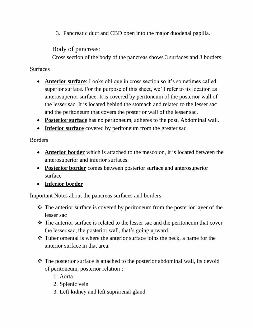
Body of pancreas:
Cross section of the body of the pancreas shows 3 surfaces and 3 borders:
Surfaces
Anterior surface: Looks oblique in cross section so it’s sometimes called
superior surface. For the purpose of this sheet, we’ll refer to its location as
anterosuperior surface. It is covered by peritoneum of the posterior wall of
the lesser sac. It is located behind the stomach and related to the lesser sac
and the peritoneum that covers the posterior wall of the lesser sac.
Posterior surface has no peritoneum, adheres to the post. Abdominal wall.
Inferior surface covered by peritoneum from the greater sac.
Borders
Anterior border which is attached to the mescolon, it is located between the
anterosuperior and inferior surfaces.
Posterior border comes between posterior surface and anterosuperior
surface
Inferior border
Important Notes about the pancreas surfaces and borders:
The anterior surface is covered by peritoneum from the posterior layer of the
lesser sac
The anterior surface is related to the lesser sac and the peritoneum that cover
the lesser sac, the posterior wall, that’s going upward.
Tuber omental is where the anterior surface joins the neck, a name for the
anterior surface in that area.
The posterior surface is attached to the posterior abdominal wall, its devoid
of peritoneum, posterior relation :
1. Aorta
2. Splenic vein
3. Left kidney and left suprarenal gland
4. The origin of Superior mesenteric artery
5. The crura of the diaphragm
The inferior surface is narrow on the right but broader on the left, this
surface is covered by peritoneum from the greater omentum; when the
greater omentum descends as two layers and then ascends as two layers, it
attaches to the inferior surface and to the anterior border (as mesocolon),
which means that the anterior border separates the inferior surface from the
anterior surface.
The anterior border separates the antero-superior surface from the inferior
surface.
To conclude things….the anterior surface covered by posterior layer of the
lesser sac and the inferior surface covered by peritoneum from the greater
omentum ( الدكتور عاد هاي المعلومة مليون مرة)
We have two surfaces that are covered by peritoneum antero-superior and
inferior surface, Anterior border separate the anterior surface from the
inferior surface, and its attached to mesocolon, the greater omentum covers
the inferior surface and then it attaches to the anterior border with the
mesocolon.
The superior border is blunt and flat at the right but narrow and sharp to
the left
It commences on the right in the omental tuberosity.
The ciliac artery is found above the pancreas so it’s related to the superior
surface, the hepatic artery is the same since it is a branch from the celiac, the
splenic artery runs tortuous on the upper border (superior surface) going
towards the hilum of the spleen, at the end of its course it’s related to the
inferior border.
The anterior border separate the anterior surface from the inferior, and along
this border the two layers of mesocolon diverge from one another; passing
upward to the anterior surface and the other downward to cover the inferior
surface.
The inferior border separates the inferior surface from the posterior.
The superior mesenteric vessels emerge under the right extremity and end its
course related to the inferior border.
Tail of pancreas
It’s one of the contents of the splenicorenal ligament.
The splenicorenal ligament: two layers of peritoneums that contain the tail
of pancreas and the splenic vessels.
It forms an impression on the lower part of the hilum of the spleen
In spleen-ectomy we ligate the splenic vessels and we should be careful not
to injure the tail of pancreas, in case of injury the secretion will go to the
abdomen and it causes peritonitis
Pancreatic duct
We have the main pancreatic and accessory duct, the accessory opens 1
inch above the main pancreatic duct and both of them open in the middle of
the 2nd
part of duodenum,
Pancreas blood supply
It takes blood from the splenic artery and from the superior and inferior
pancreatico-duodenal artery, the pancreas follow the foregut and midgut
because it’s supplied by both branches from the ciliac and from the superior
mesenteric artery
Vein
The veins drain to the splenic vein and superior mesenteric that form the
portal vein
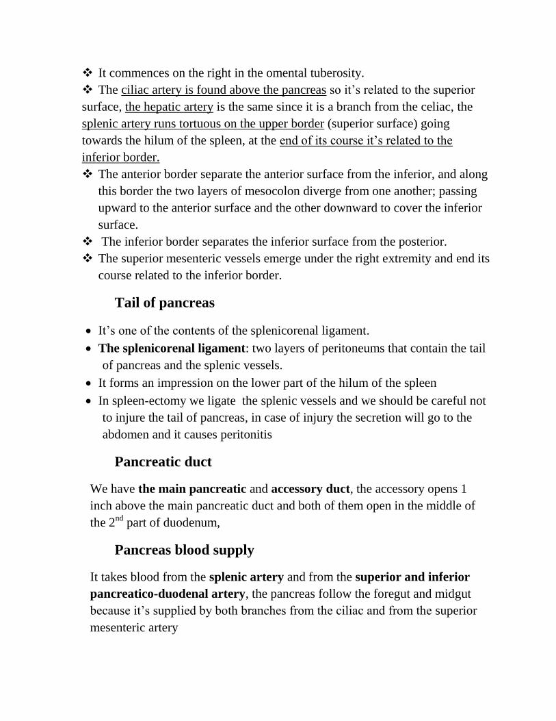
The lymphatic drainage
Pancreaticoduodenal lymph nodes that drain to ciliac or superior mesenteric
lymph nodes and both of them end in
the thoracic duct.
Nerve supply
Sympathetic and parasympathetic
chain
**The congenital defect in pancreas
will be discussed in embryology;
annular pancreas and ectopic pancreas,
ectopic mean not in its place in the abdominal cavity, the annular pancreas
may result in duodenal obstruction
Clinical cases
Cancer head of pancreas : one of the complication is obstructive jaundice
because it blocks the common bile duct and the pancreatic duct.
Cancer of the body of pancreas : will form pressure on IVC, that results in
congestion of blood especially at the lower part.
Acute pancreatitis: inflammation of the pancreas.
----------------------------------------------------------------------------------------------
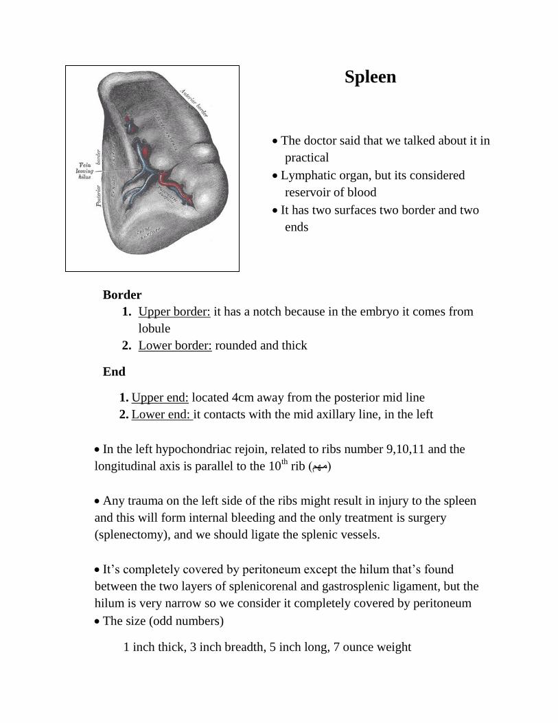
Spleen
The doctor said that we talked about it in
practical
Lymphatic organ, but its considered
reservoir of blood
It has two surfaces two border and two
ends
Border
1. Upper border: it has a notch because in the embryo it comes from
lobule
2. Lower border: rounded and thick
End
1. Upper end: located 4cm away from the posterior mid line
2. Lower end: it contacts with the mid axillary line, in the left
In the left hypochondriac rejoin, related to ribs number 9,10,11 and the
longitudinal axis is parallel to the 10th rib (مهم)
Any trauma on the left side of the ribs might result in injury to the spleen
and this will form internal bleeding and the only treatment is surgery
(splenectomy), and we should ligate the splenic vessels.
It’s completely covered by peritoneum except the hilum that’s found
between the two layers of splenicorenal and gastrosplenic ligament, but the
hilum is very narrow so we consider it completely covered by peritoneum
The size (odd numbers)
1 inch thick, 3 inch breadth, 5 inch long, 7 ounce weight
The splenic vessels give 5-7 branches at the hilum.
The spleen is related to the stomach, although it’s found at the lateral side
with greater curvature, considered one of the stomach bed because a part of it
is behind the stomach.
Surfaces
1. Diaphragmatic: related to ribs 9,10,11 , and it’s also related to left
pleura and lung , located above the colicophrenic ligament.
2. Visceral surface : contains gastric impression (above the hilum of
spleen) , renal impression ( below the hilum ) , colic impression
(inferiorly) and impression for the tail of pancreas.
The lower extremity contacts with the mid axillary line
Blood supply
The splenic artery (branch from the ciliac trunk) that runs tortuous along the
upper border of pancreas and at the hilum it gives 5-7 branches
The veins drain to the portal vein
The lymphatic drainage at the end will go to the ciliac lymph nodes
It take sympathetic and parasympathetic
“I believe that laughing is the best calorie burner, I believe in kissing, kissing a lot, I
believe in being strong when everything seems to be going wrong, I believe that
happy girls are the prettiest, I believe that tomorrow is another day, I believe in
miracles “


![CHOLELITHIASIS [Autosaved]](https://img.pdfslide.net/doc/110x75/577ce5051a28abf1038fa5b3/cholelithiasis-autosaved.jpg)