Embed Size (px)
Citation preview
Annals of the Rheumatic Diseases 1989; 48: 912-917
Localisation of lysozyme mRNA in rheumatoidsynovial membrane by in situ hybridisationYRJO T KONTTINEN,i VILLE BERGROTH,i MARKKU KULOMAA,3DAN NORDSTROM,1 MARGARETHA SEGERBERG-KONTTINEN,iRIITTA KEINANEN,3 PERTTI KEMPPINEN,' MIKA HUKKANEN,1 ANDMATS GRONBLAD2
From the 'Fourth Department of Medicine and the 2Department of Physical Medicine, Helsinki UniversityCentral Hospital, Helsinki, Finland; and the 3Department of Biochemical Sciences, University of Tampere,Tampere, Finland
SUMMARY Type A synovial lining cells have been shown to contain lysozyme in their lysosomes.This might be phagocytosed because synovial fluid contains lysozyme originating from tissuemacrophages and articular cartilage but in arthritides, in particular, from neutrophils. In situhybridisation with 35S labelled cDNA was used to detect mRNA for lysozyme over synovial liningin patients with rheumatoid arthritis. No hybridisation was found with lactoferrin cDNA, whichwas used as a negative control. Computer search against the EMBL gene bank (release 14) didnot show any significant cross hybridisation to a known sequence. In cytological specimens35S-cDNA:mRNA hybrids were observed in positive but not in negative control cells. Thepresence of lysozyme and its mRNA suggests that type A synovial lining cells are of mononuclearphagocyte lineage.
Chronic synovitis is characterised by lining cellhyperplasia and villus formation often covered by afibrin layer and containing areas of necrosis andextravasation.1 Chronic inflammatory cell infil-trates, in particular, lymphocytes, have been a focusof research,2 3 although cells of the macrophage andfibroblast lineage are also present.47 In particular,the macrophage-like type A and fibroblast-like typeB lining cells as well as intermediate type liningcells8-- have been under scrutiny, also because oftheir role in pannus tissue formation.iiFrom various animal and human studies it seems
that type A synovial lining cells are peroxidasenegative, 12 non-specific esterase positive13 Fc andC3b receptor carrying'2 13 macrophages 14 whichprobably originate from the bone marrow.i5
Recently, an interesting report giving furthersupport to the mononuclear phagocyte origin of thetype A lining cells was published. In an elegantimmunoelectron microscopic study lysozyme (EC3.2.1.17) was shown to be present in lysosomes ofcells displaying macrophage-like ultramorphologicalAccepted for publication 9 March 1989.Correspondence to Dr Yrjo T Konttinen, Fourth Department ofMedicine, Helsinki University Central Hospital, Unioninkatu 38,SF-00170 Helsinki 17, Finland.
characteristics.16 This double identification seemsquite convincing at first glance. Rheumatoidsynovial fluid, however, contains high concentra-tions of lysozyme,17 which may originate frommononuclear phagocytes or articular cartilage,18 butprobably is mainly derived from neutrophils. Inrheumatoid effusions containing 25 X 109/lneutrophils the breakdown in the synovial cavitymay exceed one billion cells a day.19 Furthermore,synovial fluid neutrophils are very actively involvedin the phagocytosis of, for example, immunoglobulin-rheumatoid factor complexes and regurgitationinto the extracellular environment during feedingwill greatly contribute to high synovial fluidlysozyme concentrations. Therefore, it is quitepossible that lysozyme, ultrastructurally localised inthe lysosomes of macrophage-like type A synoviallining cells, has been phagocytosed from thesynovial fluid. There are many examples of the wayin which a phagocytosing cell may, in its lysosomes,contain immunoreactive substances phagocytosedbut not synthesised by the cell. To take an examplefrom the above, it has been shown that rheumatoidarthritis synovial fluid neutrophils with immuno-histochemical staining often contain immunoglobu-lin, though they do not synthesise it.20 Similarly,
912
copyright. on 16 S
eptember 2018 by guest. P
rotected byhttp://ard.bm
j.com/
Ann R
heum D
is: first published as 10.1136/ard.48.11.912 on 1 Novem
ber 1989. Dow
nloaded from
Lysozyme mRNA in rheumatoid synovial membrane 913
macrophage-like type A lining cells might phago-cytose synovial fluid lysozyme, though they wouldnot synthesise it.To further evaluate the question 'What are
synoviocytes?', tentatively put forward by Revelland coworkers,21 we decided to extend their workand determine whether lysozyme in synovial lining isonly phagocytosed or also synthesised in situ.
Patients and methods
PATIENTS
Synovial membrane biopsy samples obtained fromthree patients (mean age 54 years) fulfilling the 1987revised American Rheumatism Association criteriafor rheumatoid arthritis,22 and from two patients(mean age 41 .years) with a traumatic joint lesion(meniscal cartilage rupture) were studied.
TISSUE PREPARATION AND FIXATIONThe synovial membrane tissue samples were snapfrozen in OCT compound (Lab-Tek, Naperville, IL)and stored at -20°C until sectioning on a cryostat.Sections were cut at 6 lim onto microscope slidescoated with 0.5% gelatin, 0 05% CrK(SO4)2, and0-02% diethylpyrocarbonate (RNase inhibitor;DEP; Sigma, St Louis, MO). To prevent RNasecontamination gloves were worn when handlingtissue and slides. Only RNase-free glassware andplasticware were used. They were treated with0.02% DEP in water and then rinsed with distilled,deionised water before use. After air drying thecryostat sections were fixed in freshly prepared3-5% neutral buffered paraformaldehyde containing0-02% DEP for five minutes at room temperature.After fixation the slides were washed twice in2xSSC (SSC=0.15 M sodium chloride, 0-015 Msodium citrate) for five minutes. To remove basicproteins, which may bind cDNA probes non-specifically, the sections were incubated in 0-2 MHCI for 20 minutes at room temperature after twofive minute washes in 2xSSC. The sections werethen dehydrated in 70%, 95%, and 100% ethanol,two minutes in each.
HYBRIDISATION PROBESA recombinant plasmid, pLZM, containing a cDNAinsert covering the coding sequence for the chickenegg white lysozyme was grown in Escherichia coliRRI cells and purified by standard procedures.23The cDNALZM insert was isolated from the vectorDNA (pBR322) by digestion with Pst I restrictionendonuclease (Boehringer Mannheim, WestGermany) followed by preparative 4% -poly-acrylamide gel electrophoresis. The cDNALZM frag-ment (593 bp) was labelled with alpha-35S-dCTP
and alpha-35S-dLTP (Amersham International,UK) to a specific activity of 5-9 x10⁷ cpm/,ug ofDNA by a nick translation method.24A recombinant plasmid, phLFfl containing a
partial cDNA (990 bp) for the human lactoferrin25was prepared and labelled as described above. Thespecific activity of the 35S labelled insert was0-5-1 x 10⁸ cpm/[tg DNA.
PREHYBRIDISATION AND HYBRIDISATIONBUFFERSPrehybridisation was carried out in 100 [tm TRIS-HCl (pH 7.5), 144 mM NaCl, 50% deionizedformamide, Denhart's solution (0.02% Ficoll 400,0-02% polyvinylpyrrolidine, and 0-02% bovineserum albumin), 7 ,uM EDTA, 0-05% yeast totalRNA (type III), 0-005% t-RNA (type X), 0405%herring sperm DNA, and 0-05% inorganic sodiumpyrophosphate. Hybridisation was performed in theprehybridisation buffer, which was supplementedwith 10% dextran sulphate (mol.wt 8000) and0 005% polyadenylic acid.26 The probe was heatdenaturated at 95°C for 10 minutes and quicklycooled in ice.
PREHYBRIDISATIONThe slides were incubated in prehybridisation bufferat room temperature for two hours, gently rinsed in2x SSC, and air dried.
HYBRIDISATIONThe hybridisation buffer was prepared so that eachsection received 3-5 x10⁴ cpm in 50 ,Il of buffer.Hybridisation was allowed to proceed in a sealedmoist chamber at 37°C for 48 hours. After hybridisa-tion the unhybridised probe was removed by rinsingtwo times, 10 minutes each, in 2xSSC with 0 05%inorganic sodium pyrophosphate (NaPPi) at roomtemperature, followed by a wash in 0 5xSSC with0-05% NaPPi for 24-48 hours (two changes).
AUTORADIOGRAPHYThe dried slides were dipped in NTB2 nuclear trackemulsion gel (Eastman Kodak Co, Rochester, NY)exposed in darkness for 12 days, developed for twominutes (Rodinal; Agfa-Gevaert, Leverkusen,FRG), fixed (Rapid Fix, Agfa-Gevaert), counter-stained with haematoxylin, and mounted.
CONTROL EXPERIMENTSThe cDNA probe was from the nucleotide sequenceencoding for chicken egg white lysozyme. Thereactivity of the probe with mRNA for human LZMhad therefore to be tested. Human peripheral bloodleucocytes were cytocentrifuged onto microscopeslides and hybridisation was performed as in the
copyright. on 16 S
eptember 2018 by guest. P
rotected byhttp://ard.bm
j.com/
Ann R
heum D
is: first published as 10.1136/ard.48.11.912 on 1 Novem
ber 1989. Dow
nloaded from
914 Konttinen, Bergroth, Kulomaa, et al
+sw+S
.
-_
'_
I:l. w
0~ ~ ~ _
4%
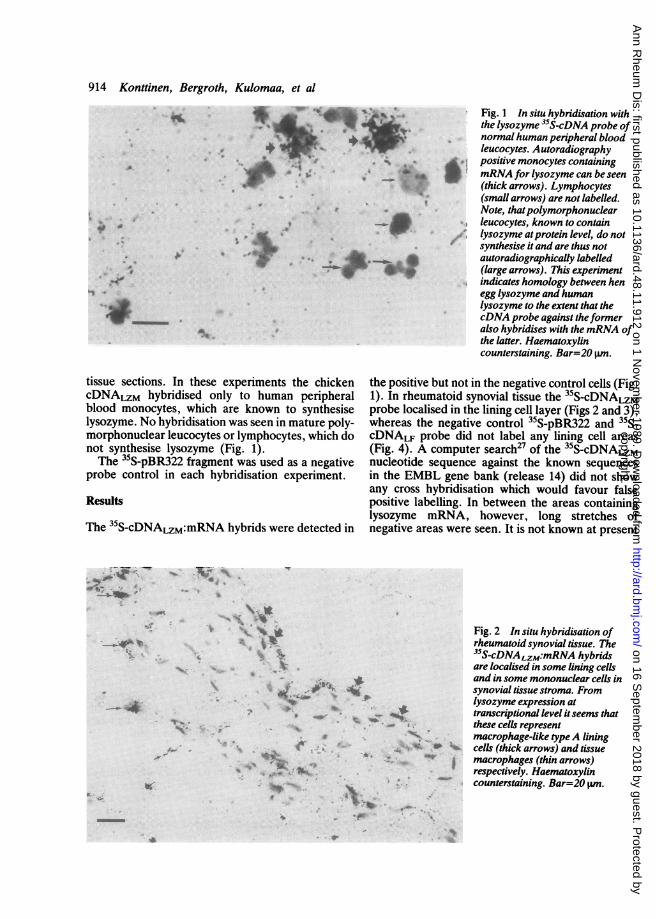
tissue sections. In these experiments the chickencDNALzM hybridised only to human peripheralblood monocytes, which are known to synthesiselysozyme. No hybridisation was seen in mature poly-morphonuclear leucocytes or lymphocytes, which donot synthesise lysozyme (Fig. 1).The 35S-pBR322 fragment was used as a negative
probe control in each hybridisation experiment.
Results
The 35S-cDNALZM:mRNA hybrids were detected in
--_- i_ _rft
Fig. 1 In situ hybridisation withthe lysozyme 35S-cDNA probe ofnormal human peripheral bloodleucocytes. Autoradiographypositive monocytes containingmRNA for lysozyme can be seen(thick arrows). Lymphocytes(small arrows) are not labelled.Note, thatpolymorphonuclearleucocytes, known to containlysozyme atprotein level, do notsynthesise it and are thus notautoradiographically labelled(large arrows). This experimentindicates homology between henegg lysozyme and humanlysozyme to the extent that thecDNA probe against theformeralso hybridises with themRNA ofthe latter. Haematoxylincounterstaining. Bar=20 pmn.
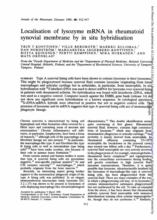
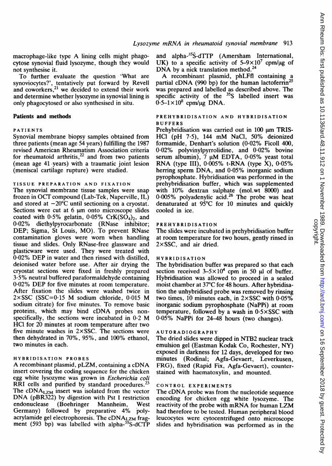
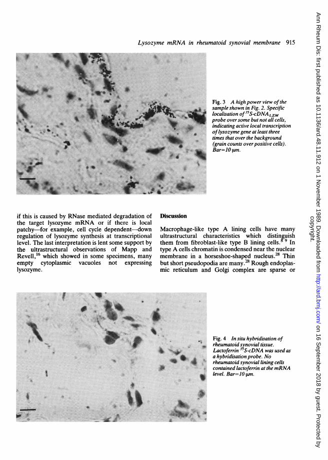
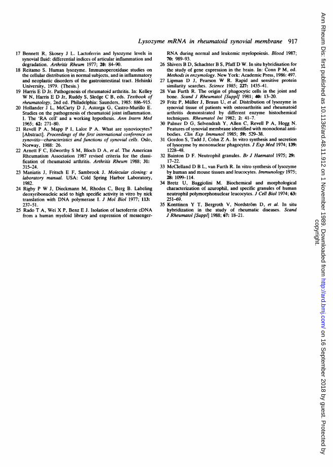
the positive but not in the negative control cells (Fig.1). In rheumatoid synovial tissue the 35S-cDNALzMprobe localised in the lining cell layer (Figs 2 and 3),whereas the negative control 35S-pBR322 and 35S-cDNALF probe did not label any lining cell areas(Fig. 4). A computer search27 of the 35S-cDNALzMnucleotide sequence against the known sequencesin the EMBL gene bank (release 14) did not showany cross hybridisation which would favour falsepositive labelling. In between the areas containinglysozyme mRNA, however, long stretches ofnegative areas were seen. It is not known at present
S. a,
V*Y
+S 4
4#_4._4
A~~~~~~_X t.~~~~~~~4
a.
"W ¾"1104
Fig. 2 In situ hybridisation ofrheumatoid synovial tissue. The35S-cDNALzM:mRNA hybridsare localised in some lining cellsand in some mononuclear cells insynovial tissue stroma. Fromlysozyme expression attranscriptional level itseems thatthese cells represent
* macrophage-like typeA liningcells (thick arrows) and tissuenacrophages (thin arrows)respectively. Haematoxylin
- counterstaining. Bar=20 pm.
t. .
la
I'llp40, r -,
ow lb-I
I
copyright. on 16 S
eptember 2018 by guest. P
rotected byhttp://ard.bm
j.com/
Ann R
heum D
is: first published as 10.1136/ard.48.11.912 on 1 Novem
ber 1989. Dow
nloaded from
Lysozyme mRNA in rheumatoid synovial membrane 915
*_
~~~~ -~
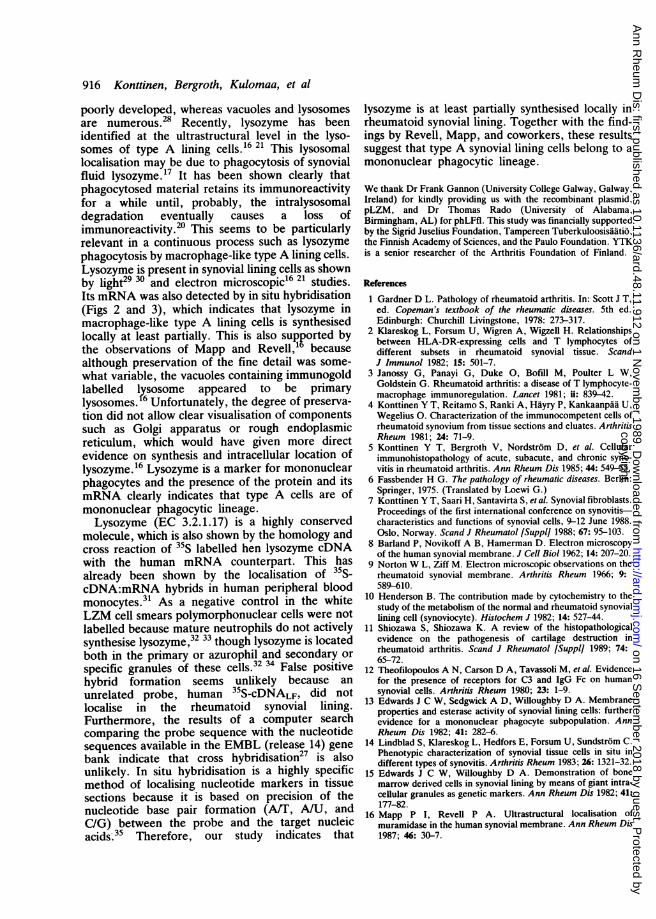
Fig. 3 A high power view ofthesample shown in Fig. 2. Specificlocalisation of35S-cDNA LZMprobe over some but not all cells,indicating active local transcriptionoflysozyme gene at least threetimes that over the background(grain counts overpositive cells).Bar=10 wn.
4 *1
t S.& " a S's v
if this is caused by RNase mediated degradation ofthe target lysozyme mRNA or if there is localpatchy-for example, cell cycle dependent-downregulation of lysozyme synthesis at transcriptionallevel. The last interpretation is lent some support bythe ultrastructural observations of Mapp andRevell,16 which showed in some specimens, manyempty cytoplasmic vacuoles not expressinglysozyme.
_
_~~_k~~~_
. g
N'
A
I4i
Discussion
Macrophage-like type A lining cells have manyultrastructural characteristics which distinguishthem from fibroblast-like type B lining cells.Y9 Intype A cells chromatin is condensed near the nuclearmembrane in a horseshoe-shaped nucleus.28 Thinbut short pseudopodia are many.28 Rough endoplas-mic reticulum and Golgi complex are sparse or
Fig. 4 In situ hybridisation of- rheumatoid synovial tissue.
*+̂ Lactoferrin 35S-cDNA was used as* a hybridisation probe. No
*1 4 rheumatoid synovial lining cellscontained lactoferrin at the mRNA-level. Bar=10 Wn.
0 4 I
.4-It
copyright. on 16 S
eptember 2018 by guest. P
rotected byhttp://ard.bm
j.com/
Ann R
heum D
is: first published as 10.1136/ard.48.11.912 on 1 Novem
ber 1989. Dow
nloaded from
916 Konttinen, Bergroth, Kulomaa, et al
poorly developed, whereas vacuoles and lysosomesare numerous.28 Recently, lysozyme has beenidentified at the ultrastructural level in the lyso-somes of type A lining cells.16 21 This lysosomallocalisation may be due to phagocytosis of synovialfluid lysozyme.17 It has been shown clearly thatphagocytosed material retains its immunoreactivityfor a while until, probably, the intralysosomaldegradation eventually causes a loss ofimmunoreactivity.20 This seems to be particularlyrelevant in a continuous process such as lysozymephagocytosis by macrophage-like type A lining cells.Lysozyme is present in synovial lining cells as shownby light29 30 and electron microscopic16 21 studies.Its mRNA was also detected by in situ hybridisation(Figs 2 and 3), which indicates that lysozyme inmacrophage-like type A lining cells is synthesisedlocally at least partially. This is also supported bythe observations of Mapp and Revell," becausealthough preservation of the fine detail was some-what variable, the vacuoles containing immunogoldlabelled l?rsosome appeared to be primarylysosomes. Unfortunately, the degree of preserva-tion did not allow clear visualisation of componentssuch as Golgi apparatus or rough endoplasmicreticulum, which would have given more directevidence on synthesis and intracellular location oflysozyme.16 Lysozyme is a marker for mononuclearphagocytes and the presence of the protein and itsmRNA clearly indicates that type A cells are ofmononuclear phagocytic lineage.Lysozyme (EC 3.2.1.17) is a highly conserved
molecule, which is also shown by the homology andcross reaction of 35S labelled hen lysozyme cDNAwith the human mRNA counterpart. This hasalready been shown by the localisation of 35S-cDNA:mRNA hybrids in human peripheral bloodmonocytes.31 As a negative control in the whiteLZM cell smears polymorphonuclear cells were notlabelled because mature neutrophils do not activelysynthesise lysozyme,32 33 though lysozyme is locatedboth in the primary or azurophil and secondary orspecific granules of these cells.32 34 False positivehybrid formation seems unlikely because anunrelated probe, human 35S-cDNALF, did notlocalise in the rheumatoid synovial lining.Furthermore, the results of a computer searchcomparing the probe sequence with the nucleotidesequences available in the EMBL (release 14) genebank indicate that cross hybridisation27 is alsounlikely. In situ hybridisation is a highly specificmethod of localising nucleotide markers in tissuesections because it is based on precision of thenucleotide base pair formation (AST, A/U, andC/G) between the probe and the target nucleicacids.35 Therefore, our study indicates that
lysozyme is at least partially synthesised locally inrheumatoid synovial lining. Together with the find-ings by Revell, Mapp, and coworkers, these resultssuggest that type A synovial lining cells belong to amononuclear phagocytic lineage.
We thank Dr Frank Gannon (University College Galway, Galway,Ireland) for kindly providing us with the recombinant plasmid,pLZM, and Dr Thomas Rado (University of Alabama,Birmingham, AL) for phLFfl. This study was financially supportedby the Sigrid Juselius Foundation, Tampereen Tuberkuloosisaatio,the Finnish Academy of Sciences, and the Paulo Foundation. YTKis a senior researcher of the Arthritis Foundation of Finland.
References
1 Gardner D L. Pathology of rheumatoid arthritis. In: Scott J T,ed. Copeman's textbook of the rheumatic diseases. 5th ed.Edinburgh: Churchill Livingstone, 1978: 273-317.
2 Klareskog L, Forsum U, Wigren A, Wigzell H. Relationshipsbetween HLA-DR-expressing cells and T lymphocytes ofdifferent subsets in rheumatoid synovial tissue. ScandJ Immunol 1982; 15: 501-7.
3 Janossy G, Panayi G, Duke 0, Bofill M, Poulter L W,Goldstein G. Rheumatoid arthritis: a disease of T lymphocyte-macrophage immunoregulation. Lancet 1981; ii: 839-42.
4 Konttinen Y T, Reitamo S, Ranki A, Hayry P, Kankaanpaa U,Wegelius 0. Characterization of the immunocompetent cells ofrheumatoid synovium from tissue sections and eluates. ArthritisRheum 1981; 24: 71-9.
5 Konttinen Y T, Bergroth V, Nordstrom D, et al. Cellularimmunohistopathology of acute, subacute, and chronic syno-vitis in rheumatoid arthritis. Ann Rheum Dis 1985; 44: 549-55.
6 Fassbender H G. The pathology of rheumatic diseases. Berlin:Springer, 1975. (Translated by Loewi G.)
7 Konttinen Y T, Saari H, Santavirta S, et al. Synovial fibroblasts.Proceedings of the first international conference on synovitis-characteristics and functions of synovial cells, 9-12 June 1988.Oslo, Norway. Scand J Rheumatol [Suppli 1988; 67: 95-103.
8 Barland P, Novikoff A B, Hamerman D. Electron microscopyof the human synovial membrane. J Cell Biol 1962; 14: 207-20.
9 Norton W L, Ziff M. Electron microscopic observations on therheumatoid synovial membrane. Arthritis Rheum 1966; 9:589-610.
10 Henderson B. The contribution made by cytochemistry to thestudy of the metabolism of the normal and rheumatoid synoviallining cell (synoviocyte). Histochem J 1982; 14: 527-44.
11 Shiozawa S, Shiozawa K. A review of the histopathologicalevidence on the pathogenesis of cartilage destruction inrheumatoid arthritis. Scand J Rheumatol [Suppl] 1989; 74:65-72.
12 Theofilopoulos A N, Carson D A, Tavassoli M, et al. Evidencefor the presence of receptors for C3 and IgG Fc on humansynovial cells. Arthritis Rheum 1980; 23: 1-9.
13 Edwards J C W, Sedgwick A D, Willoughby D A. Membraneproperties and esterase activity of synovial lining cells: furtherevidence for a mononuclear phagocyte subpopulation. AnnRheum Dis 1982; 41: 282-6.
14 Lindblad S, Klareskog L, Hedfors E, Forsum U, Sundstrom C.Phenotypic characterization of synovial tissue cells in situ indifferent types of synovitis. Arthritis Rheum 1983; 26: 1321-32.
15 Edwards J C W, Willoughby D A. Demonstration of bonemarrow derived cells in synovial lining by means of giant intra-cellular granules as genetic markers. Ann Rheum Dis 1982; 41:177-82.
16 Mapp P I, Revell P A. Ultrastructural localisation ofmuramidase in the human synovial membrane. Ann Rheum Dis1987; 46: 30-7.
copyright. on 16 S
eptember 2018 by guest. P
rotected byhttp://ard.bm
j.com/
Ann R
heum D
is: first published as 10.1136/ard.48.11.912 on 1 Novem
ber 1989. Dow
nloaded from
Lysozyme mRNA in rheumatoid synovial membrane 917
17 Bennett R, Skosey J L. Lactoferrin and lysozyme levels insynovial fluid: differential indices of articular inflammation anddegradation. Arthritis Rheum 1977; 20: 84-90.
18 Reitamo S. Human lysozyme. Immunoperoxidase studies onthe cellular distribution in normal subjects, and in inflammatoryand neoplastic disorders of the gastrointestinal tract. HelsinkiUniversity, 1979. (Thesis.)
19 Harris E D Jr. Pathogenesis of rheumatoid arthritis. In: KelleyW N, Harris E D Jr, Ruddy S, Sledge C B, eds. Textbook ofrheumatology, 2nd ed. Philadelphia: Saunders, 1985: 886-915.
20 Hollander J L, McCarty D J, Astorga G, Castro-Murillo E.Studies on the pathogenesis of rheumatoid joint inflammation.I. The 'RA cell' and a working hypothesis. Ann Intern Med1965; 62: 271-80.
21 Revell P A, Mapp P I, Lalor P A. What are synoviocytes?[Abstract]. Proceedings of the first international conference onsynovitis-characteristics and functions of synovial cells. Oslo,Norway, 1988: 26.
22 Arnett F C, Edworthy S M, Bloch D A, et al. The AmericanRheumatism Association 1987 revised criteria for the classi-fication of rheumatoid arthritis. Arthritis Rheum 1988; 31:315-24.
23 Maniatis J, Fritsch E F, Sambrook J. Molecular cloning: alaboratory manual. USA: Cold Spring Harbor Laboratory,1982.
24 Rigby P W J, Dieckmann M, Rhodes C, Berg B. Labelingdeoxyribonucleic acid to high specific activity in vitro by nicktranslation with DNA polymerase I. J Mol Biol 1977; 113:237-51.
25 Rado T A, Wei X P, Benz E J. Isolation of lactoferrin cDNAfrom a human myeloid library and expression of messenger-
RNA during normal and leukemic myelopoiesis. Blood 1987;70: 989-93.
26 Shivers B D, Schachter B S, Pfaff D W. In situ hybridisation forthe study of gene expression in the brain. In: Conn P M, ed.Methods in enzymology. New York: Academic Press, 1986: 497.
27 Lipman D J, Pearson W R. Rapid and sensitive proteinsimilarity searches. Science 1985; 227: 1435-41.
28 Van Furth R. The origin of phagocytic cells in the joint andbone. Scand J Rheumatol [SupplI 1981; 40: 13-20.
29 Fritz P, Muller J, Braun U, et al. Distribution of lysozyme insynovial tissue of patients with osteoarthritis and rheumatoidarthritis demonstrated by different enzyme histochemicaltechniques. Rheumatol Int 1982; 2: 41-7.
30 Palmer D G, Selvendrah Y, Allen C, Revell P A, Hogg N.Features of synovial membrane identified with monoclonal anti-bodies. Clin Exp Immunol 1985; 59: 529-38.
31 Gordon S, Tadd J, Cohn Z A. In vitro synthesis and secretionof lysozyme by mononuclear phagocytes. J Exp Med 1974; 139:1228-48.
32 Bainton D F. Neutrophil granules. Br J Haematol 1975; 29:17-22.
33 McClelland D B L, van Furth R. In vitro synthesis of lysozymeby human and mouse tissues and leucocytes. Immunology 1975;28: 1099-114.
34 Bretz U, Baggiolini M. Biochemical and morphologicalcharacterization of azurophil, and specific granules of humanneutrophil polymorphonuclear leucocytes. J Cell Biol 1974; 63:251-69.
35 Konttinen Y T, Bergroth V, Nordstrom D, et al. In situhybridization in the study of rheumatic diseases. ScandJ Rheumatol [Suppl] 1988; 67: 18-21. copyright.
on 16 Septem
ber 2018 by guest. Protected by
http://ard.bmj.com
/A
nn Rheum
Dis: first published as 10.1136/ard.48.11.912 on 1 N
ovember 1989. D
ownloaded from