Embed Size (px)
Citation preview
147
femoral and inguinal groups. There was always pyrexia,sometimes up to 101-102°F. Other symptoms includedsoreness of the eyes and general aching. These symptomssubsided after a few days and the patients could thentolerate much higher doses-e.g., 12 mg. per kg. daily.The severity of the reaction seemed to be proportionateto the number of micronlariae initially present in the skinrather than to the level of hetrazan dosage. Apparentlythe reaction is mostly an allergic one, excited bythe breakdown products of microfilari2e injured or
destroyed by the drug. This allergic reaction wouldmake hetrazan unpopular if it were used for the masstreatment of onchocerciasis, although it might be quiteacceptable if employed for the mass treatment offilariasis due to W. bancrofti. In one patient who haddimness of vision before he was treated for onchocerciasis,the vision deteriorated further during treatment andadministration of hetrazan was discontinued. ’
Much histological material was collected from the
patients with onchocerciasis. Full publication will bedelayed until this has been cut and examined.
SUMMARY
Hetrazan is very rapid and effective in removingmicronlariae from the blood of patients infected withWuchereria bancrofti. It does not remove microfilariaefrom hydroceles and it has no direct toxic action onmicronlarise in vitro. The clinical observations are com-
patible with the hypothesis that hetrazan acts by modifyingthe micronlarise so that they are seized by phagocytes.
Hetrazan removes microfilariae from the skin in
patients with onchocerciasis but apparently it does notkill the adult worms. It is well tolerated by patientswith W. bancrofti but it provokes violent allergic reactionsin patients with onchocerciasis.
Grateful acknowledgments are due to the Directors, andmany members, of the Medical Services of Tanganyika andUganda, without whose kind support this work would nothave been possible ; and to Mr. D. Garlick for technicalassistance..
REFERENCES
Hawking, F., Sewell, P., Thurston, J. P. (1948) Lancet, ii, 730.Santiago-Stevenson, D., Oliver-Gonzalez, J., Hewitt, R. I. (1947)
J. Amer. med. Ass. 135, 708.
LOIASIS
TREATED WITH HETRAZAN (BANOCIDE)F. MURGATROYD
M.D. Lpool, F.R.C.P., D.T.M.PHYSICIAN, HOSPITAL FOR TROPICAL DISEASES, UNIVERSITYCOLLEGE HOSPITAL, LONDON; DEPUTY DIRECTOR, DIVISIONOF CLINICAL TROPICAL MEDICINE, LONDON SCHOOL OF
HYGIENE AND TROPICAL MEDICINE; CONSULTINGPHYSICIAN TO THE COLONIAL OFFICE
A. W. WOODRUFFM.D. Durh., M.R.C.P., D.T.M. & H.
FIRST ASSISTANT, HOSPITAL FOR TROPICAL DISEASES,UNIVERSITY COLLEGE HOSPITAL, LONDON; LECTURERIN CLINICAL TROPICAL MEDICINE, LONDON SCHOOL
OF HYGIENE AND TROPICAL MEDICINE
THis paper records observations at the Hospital forTropical Diseases, London, on seventeen Europeanpatients (eight males and nine females) with loiasistreated with l-diethylcarbamyl-4-methylpiperazine, thefilaricidal drug marketed as ’ Hetrazan ’ (CyanamidProducts Ltd.) and ’ Banocide ’ (Burroughs Wellcome& Co.).The lengths of residence of the patients in endemic
areas of Loa loa infection ranged from 1 to 24 years,and the times between last leaving an infected area andcoming under the present observation ranged similarlyfrom a few days to 6 years.
In each case, in addition to the clinical assessment,the blood was examined for micronlariae, both with wetfilms and with stained thick films, at least three prepara-
tions of each being examined where no microfiiaria wasfound ; total and differential leucocyte-counts were made ;and an intradermal filarial sensitivity test and a filarialcomplement-fixation test were also performed. Theseexaminations were made before treatment and were
repeated at intervals during treatment and subsequently.BEFORE TREATMENT
Calabar b’,;elli7ags.-Each patient was subject tocalabar swellings, and had been so for periods varyingfrom 6 months to 14 years, the average being 5 years.The frequency of the swellings varied from one every2 or 3 days in two recently infected persons to one every4-8 months in a patient who had been infected for 8years ; the average frequency of the swellings was oneevery 6 weeks.
Microfilariae were found in only three patients ; inthe remainder films were repeatedly negative despite___.a.._.i.+..a 1 --- I -- -- --V -r - -
loa infection, including intwo patients the presenceof worms under the con-
junctivse and in anotherthree patients the appear-ance of dead worms underthe skin during treatment.Eosinophilia.-The total
leucocyte-counts variedfrom patient to patientfrom 5200 to 37,000 perc.mm., with an average of
13,080 per c.mm. ; the
eosinophilia similarlyvaried from 2 to 59%, withan average of 23%.
Immunology.—A stand-ardised intradermal filarialsensitivity test 1 was posi-tive in each patient, andsera of nine of the seven-
Fig. I-Dead adult Loa loa underskin after treatment.
teen patients gave positive complement-fixation reactionswith filarial antigen.
Diagnostic Criteria.-A diagnosis of loiasis was acceptedif microfilariæ of L. loa were found or an adult L. loawas seen. Failing either of these criteria the diagnosiswas accepted if all the following conditions were satisfied:(1) the recent history or presence of calabar swellings wasbeyond doubt ; (2) there was an otherwise unexplainedeosinophilia of more than 10% ; and (3) there wasimmunological evidence of filarial infection shown byintradermal or complement-fixation tests.
TREATMENT
Dosage.-Owing to limited supplies, each of the firstsix patients was treated with daily doses of only 2 mg.per kg. of body-weight, but later the daily dose wasincreased to 4 mg. per kg. in two cases, and then to 6 mg.per kg. for each of the remaining nine patients. Thedaily dose was divided into three equal portions givenby mouth after breakfast, luncheon, and supper. Twopatients were treated for 10 days, eight for 14 days, andthe remaining seven for 21 days ; the total amounts givenvaried from 1-2 to 10-5 g.
General.—The patients were normally kept in hospitaluntil at least the first week of treatment had been
completed, after which, provided they had no toxic
symptoms and felt otherwise well, they were treated asoutpatients. They were requested to attend the outpatientdepartment at monthly or two-monthly intervals aftertreatment ; in this way one patient has been underobservation for 14 months after finishing treatment, onefor 12 months, one for 8 months, one for 6 months, twofor 3 months, and six for 2 months or less. Questionaries
1. Fairley, N. H. Trans. R. Soc. trop Med. Hyg. 1931, 24, 635.
148
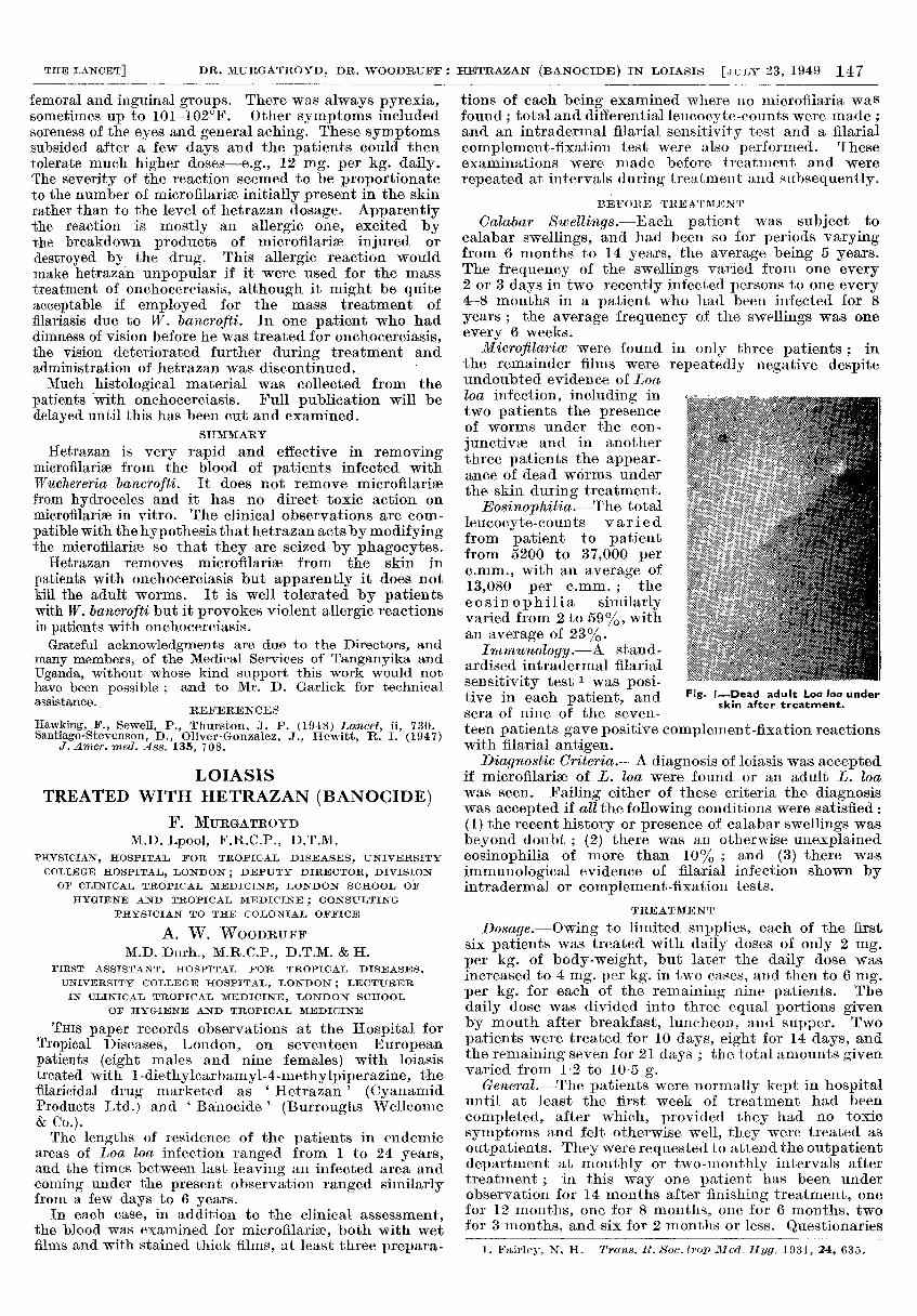
Fig. 2-Disappearance of microfitarise of Loa loa from blood and insensi-tivity of those of Acanthocheilonema perstans under treatment in apatient with a double infection.
were also sent to five patients who went abroad soon aftertreatment.
IMMEDIATE EFFECTS
Cutaneous Reactions.—On the day after the start oftreatment six patients complained of itching, and inthree of them the pruritus was accompanied by rashes.In one the rash was morbilliform and diffusely distributedover the trunk, in another it was urticarial and affectedonly the left arm, and in the third it was papulo-erythematous and affected only the neck and upper chest ;the rashes were transitory and lasted only 48 hours. Theirritation was more protracted, taking usually 3 or 4
days to disappear, though in one case it lasted 16 days ;its intensity seemed less than that of the itching andburning experienced by some patients with onchocerciasistreated with the same drug, and it was alleviated by, Benadryl’ 50 mg. or ’Anthisan’ 100 mg. thrice daily.In three patients, one of whom had no other cutaneousreactions to treatment, cutaneous thickenings or nodulesappeared ; these were about 0-5 cm. in diameter anddisappeared in 2 or 3 days. In three further patientscutaneous serpiginous linear swellings appeared, in onepatient within 24 hours and in the other two betweenthe 3rd and 4th days from the start of treatment. Theappearance, size, and shape of the swellings suggestedreactions round adult worms (fig. 1), and in two patientsspecimens of dead adult E. loa were extracted from theswellings ; Prof. J. C. Buckley kindly confirmed theiridentity. Five of the seventeen patients showed nocutaneous reactions during treatment.
Effect on Microfilariœ.—The embryos of L. loa rapidlydisappeared from the peripheral blood of treated patients.
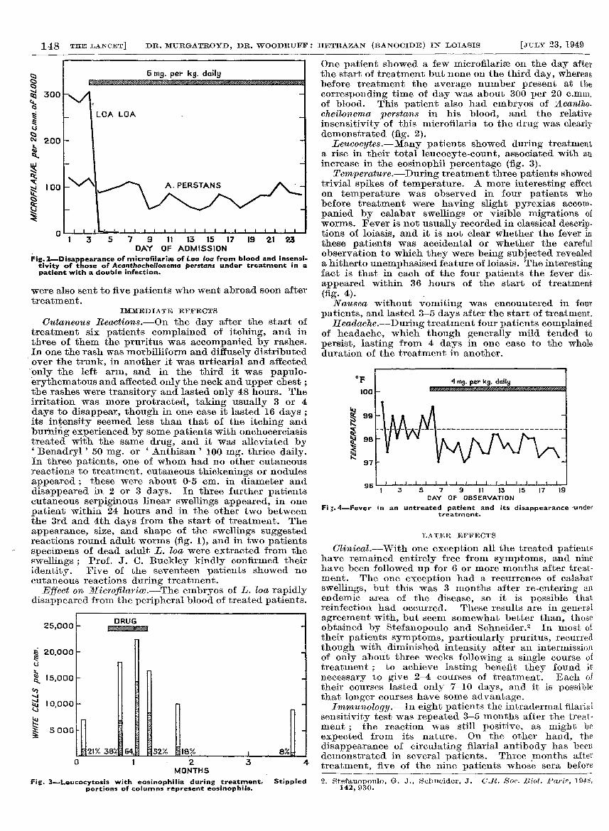
Fig. 3-Leucocytosis with eosinophilia during treatment, Stippledportions of columns represent eosinophils.
One patient showed a few microfilariæ on the day afterthe start of treatment but none on the third day, whereasbefore treatment the average number present at thecorresponding time of day was about 300 per 20 c.mm.of blood. This patient also had embryos of Acantho-cheilonema perstans in his blood, and the relativeinsensitivity of this microfilaria to the drug was clearlydemonstrated (fig. 2).Leucocytes.-Many patients showed during treatment
a rise in their total leucocyte-count, associated with anincrease in the eosinophil percentage (fig. 3).
Temperature.-During treatment three patients showedtrivial spikes of temperature. A more interesting effecton temperature was observed in four patients whobefore treatment were having slight pyrexias accom.
panied by calabar swellings or visible migrations ofworms. Fever is not usually recorded in classical descrip-tions of loiasis, and it is not clear whether the fever inthese patients was accidental or whether the carefulobservation to which they were being subjected revealeda hitherto unemphasised feature of loiasis. The interestingfact is that in each of the four patients the fever dis-
appeared within 36 hours of the start of treatment
(fig. 4). ,
Nausea without vomiting was encountered in fourpatients, and lasted 3-5 days after the start of treatment.
Headache.—During treatment four patients complainedof headache, which though generally mild tended to
persist, lasting from 4 days in one case to the wholeduration of the treatment in another.
DAY Ur UBSERVAIION
Fi.-.4-Fever in an untreated patient and its disappearance undertreatment.
LATER EFFECTS
Clinical.—With one exception all the treated patientshave remained entirely free from symptoms, and ninehave been followed up for 6 or more months after treat-ment. The one exception had a recurrence of calabarswellings, but this was 3 months after re-entering anendemic area of the disease, so it is possible thatreinfection had occurred. These results are in generalagreement with, but seem somewhat better than, thoseobtained by Stefanopoulo and Schneider.2 In most oftheir patients symptoms, particularly pruritus, recurredthough with diminished intensity after an intermissionof only about three weeks following a single course oftreatment ; to achieve lasting benefit they found it
necessary to give 2-4 courses of treatment. Each oftheir courses lasted only 7-10 days, and it is possiblethat longer courses have some advantage.
Immunology.—In eight patients the intradermal filarialsensitivity test was repeated 3-5 months after the treat-ment ; the reaction was still positive, as might be
expected from its nature. On the other hand, the
disappearance of circulating filarial antibody has beendemonstrated in several patients. Three months aftertreatment, five of the nine patients whose sera before
2. Stefanopoulo, G. J., Schneider, J. C.R. Soc. Biol. Paris, 1948,142, 930.
149
treatment gave positive complement-fixation reactionswith filarial antigen were retested. In four the reaction
had’changed from a strong positive to a negative, whilein the fifth, though the reaction remained positive, theamount of complement fixed had decreased from 8 to5 M.H.D. In another patient, re-tested only 2 monthsafter treatment, the reaction had also changed from astrong positive to a weak positive.
COMMENT
Neither symptoms nor alterations in total and differen-tial leucocyte-counts were found in two healthy personsto whom the drug was given in doses of 6 mg. per kg. ofbody-weight daily for 14 days. It seems likely that thereactions in patients with loiasis given the drug are dueto the death of the parasites, since embryos disappearfrom the blood, cutaneous lesions containing dead adultworms are found, calabar swellings and other manifes-tations of the disease cease, and circulating filarialantibody disappears.
SUMMARY
In loiasis, 1 - diethylcarbamyl - 4 - methylpiperazinebrought about disappearance of micronlariae from theblood and death of adult worms. Cutaneous reactions,headache, nausea, transient fever, and leucocytosis witheosinophilia occurred at times during treatment. Insome patients mild fever present before treatment
(possibly a hitherto overlooked feature of loiasis)disappeared on treatment.
All the patients except one have remained free fromsymptoms of loiasis for 1-14 months since treatment ;the exception may have been reinfected. Complement-fixation tests showed disappearance of circulating filarialantibody from treated patients.The findings suggest that the drug is a valuable
filaricidal agent in loiasis.Our thanks are due to the Medical Research Council,
Lederle Laboratories Division American Cyanamid Company,and Burroughs Wellcome & Co. for supplies; and to theWellcome Museum of Medical Science for the photograph.
SEQUELÆ OF MENINGOCOCCALMENINGITIS IN CHILDREN
J. D. MATTHEWSM.B. N.Z., M.R.C.P., D.C.H.
LATE HOUSE-PHYSICIAN, HOSPITAL FOR SICK CHILDREN,GREAT ORMOND STREET, LONDON
THOUGH it is widely recognised that chemotherapyhas greatly reduced the mortality from meningococcalmeningitis in children, it is not so clear from publishedreports whether or not there is a heavy incidence of
sequelæ among the survivors. The fact that some ofwhat may be termed borderline patients who mighthave died but for modern therapy now survive madeit possible that serious sequelæ might even haveincreased. This study was made to investigate this
point.Between October, 1938, and May, 1948, 50 children
with meningococcal meningitis were admitted to the
Hospital for Sick Children ; 43 were aged 2 years orless, and 29 of these were aged a year or less. Therewas only 1 child over the age of 5 years. They wereall treated with one of the sulphonamides, with or
without the addition of antimeningococcal serum or
penicillin. The following is the result of an attemptedfollow-up of these children.
_
Of the 50 children, 7 (14%) died in hospital of themeningitis ; 1 died eleven months later in the RadcliffeInfirmary, Oxford, with pneumonia and bilateral mastoid-itis following whooping-cough. At necropsy the onlystigmata of the meningitis eleven months before were
slight thickening of the arachnoid in the interpeduncularfossa, adhesions and thickening round the base of thebrain, and some thickening of the spinal-cord arachnoid.
Of the remaining 42 children, 35 were seen andexamined personally ; 3 were followed up by letterand a doctor’s report obtained where the child wasnot normal; and 4 could not be traced ; but all of themhad been seen at various periods after discharge andfound to be normal:
Total Died Seen Traced Not traced50 .. 7 .. 35 .. 4 .. 4
PROGNOSIS .
The prognosis was worst in the youngest age-group.Thus, of the 29 infants aged a year or less, 6 (21%)died. Of the 14 children aged more than a year andless than 2 years, only 1 (7%) died. There were nodeaths among the children aged more than 2 years.The details according to age-group were as follows :Age (yr.) Number Alive Dead0-1 ...... 29 .. 23 (79%) .. 6 (21%)1-2 ..... 14 .. 13 (93%) .. 1 (7%)Over 2..... 7 .. 7 (100 %) .. 0Over 5 .... 1 .. 1 (100%) .. 0
Total.. 50 .. 43 (86%) .. 7 (14%)
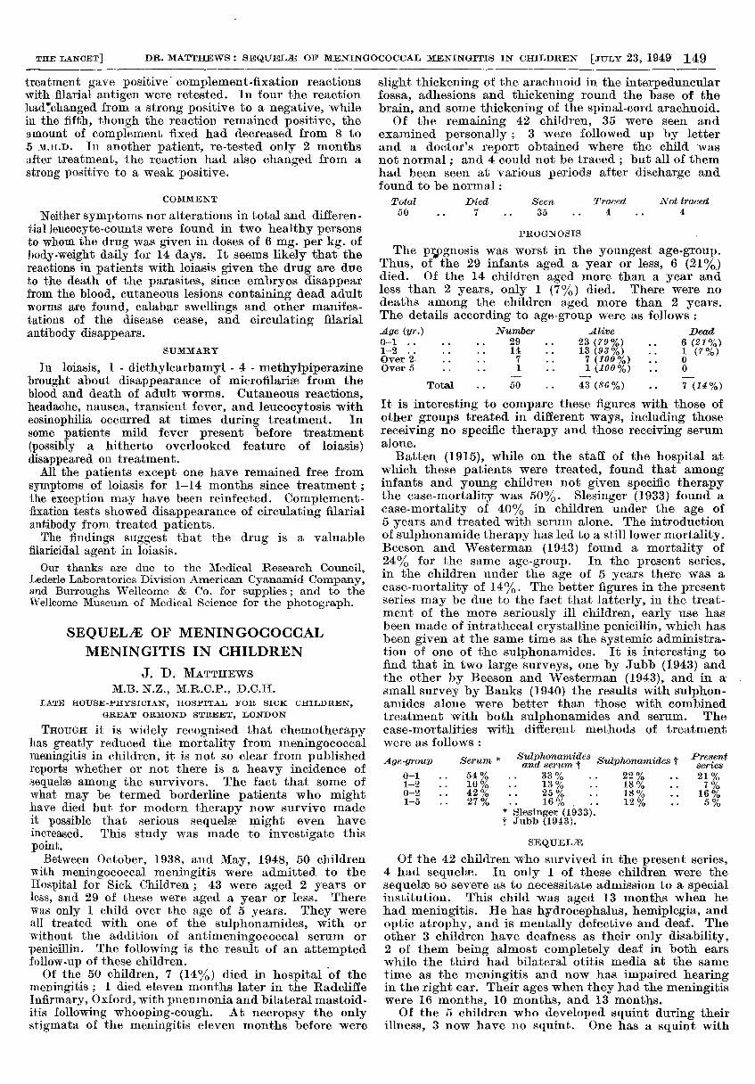
It is interesting to compare these figures with those ofother groups treated in different ways, including thosereceiving no specific therapy and those receiving serumalone.
Batten (1915), while on the staff of the hospital atwhich these patients were treated, found that amonginfants and young children not given specific therapythe case-mortality was 50%. Slesinger (1933) found acase-mortality of 40% in children under the age of5 years and treated with serum alone. The introductionof sulphonamide therapy has led to a still lower mortality.Beeson and Westerman (1943) found a mortality of
24% for the same age-group. In the present series,in the children under the age of 5 years there was acase-mortality of 14%. The better figures in the presentseries may be due to the fact that latterly, in the treat-ment of the more seriously ill children, early use hasbeen made of intrathecal crystalline penicillin, which hasbeen given at the same time as the systemic administra-tion of one of the sulphonamides. It is interesting tofind that in two large surveys, one by Jubb (1943) andthe other by Beeson and Westerman (1943), and in asmall survey by Banks (1940) the results with sulphon-amides alone were better than those with combinedtreatment with both sulphonamides and serum. Thecase-mortalities with different methods of treatmentwere as follows :
Age-group Serum * Sulphonamides suiphonamides † Presentand serum f series
0-1 .. 54% .. 33% .. 22% .. 21%1-2 .. 10% .. 13% .. 18% .. 7%0-2 .. 42% .. 25% .. 18% .. 16%1-5 .. 27% .. 16% ... 12% .. 5%
* Slesinger (1933).t Jubb (1943).
SEQUELAE
Of the 42 children who survived in the present series,4 had sequelae. In only 1 of these children were thesequelae so severe as to necessitate admission to a specialinstitution. This child was aged 13 months when hehad meningitis. He has hydrocephalus, hemiplegia, andoptic atrophy, and is mentally defective and deaf. Theother 3 children have deafness as their only disability,2 of them being almost completely deaf in both earswhile the third had bilateral otitis media at the sametime as the meningitis and now has impaired hearingin the right ear. Their ages when they had the meningitiswere 16 months, 10 months, and 13 months.
Of the 5 children who developed squint during theirillness, 3 now have no squint. One has a squint with