Embed Size (px)
Citation preview
78 RETINA TODAY JULY/AUGUST 2014
COVER STORY
Long-Acting Anti-VEGF Delivery
The potential of a posterior segment delivery system was evaluated in a phase 1 trial.
BY ROMAN G. RUBIO, MD
The pivotal phase 3 clinical trials MARINA and ANCHOR demonstrated that excellent visual acuity and anatomic results can be achieved in individuals with neovascular age-related macu-
lar degeneration (AMD) through a monthly schedule of intravitreal ranibizumab (Lucentis, Genentech) injection.1,2 In the real world setting, it has become increasingly appar-ent that continuous monthly intravitreal injections can be challenging for providers and patients. As a result, there is growing interest in alternative dosing strategies, namely as-needed, or PRN, therapy, and treat-and-extend protocols.
Although these strategies to increase dosing inter-vals can be advantageous, they still may fall short of the ideal in terms of optimal dose delivery and patient convenience. PRN treatment, when guided by opti-cal coherence tomography and visual acuity results obtained at monthly visits, can deliver efficacy similar to monthly treatments with fewer ranibizumab injections.3-6 However, this approach still requires frequent visits to the physician’s office.
At Genentech, we are pursuing the development of a ranibizumab port delivery system (PDS) that is implanted in the eye, which allows the surgeon to refill the device as needed while delivering therapeutic concentrations of ranibizumab into the vitreous over an extended period of time. The first-in-human results of the refillable drug deliv-ery implant with ranibizumab in patients with neovascular AMD were presented in 2012 at the American Academy of Ophthalmology7 and recently presented at the World Ophthalmology Conference in Tokyo.8 This article sum-marizes parts of the latter presentation.
SUSTAINED DELIVERYA number of approaches have been developed to
achieve sustained drug delivery to the back of the eye, most often with small molecules, such as corticosteroids. These drug-delivery devices have primarily taken the form of biodegradable depots and nonbiodegradable implants or devices.
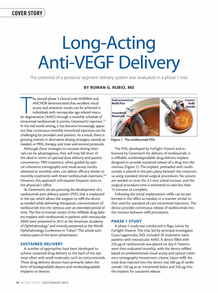
The PDS, developed by ForSight Vision4 and in-licensed by Genentech for delivery of ranibizumab, is a refillable, nonbiodegradable drug delivery implant designed to provide sustained release of a drug into the vitreous (Figure 1). The implant, preloaded with ranibi-zumab, is placed in the pars plana beneath the conjuncti-va using standard retinal surgical procedures. No sutures are needed to close the 3.2-mm scleral incision, and the surgical procedure time is estimated to take less than 15 minutes to complete.
Following the initial implantation, refills can be per-formed in the office as needed, in a manner similar to that used for standard-of-care intravitreal injections. The device provides continuous release of ranibizumab into the vitreous between refill procedures.
PHASE 1 STUDYA phase 1 study was conducted in Riga, Latvia, by
ForSight Vision4. The trial, led by principal investigator Guna Laganovska, MD, included 20 treatment-naïve patients with neovascular AMD. A device filled with 250 µg of ranibizumab was placed on day 0. Patients were then evaluated monthly, with the device refilled based on predetermined visual acuity and optical coher-ence tomography retreatment criteria. Upon refill, the total dose injected into the device was 500 µg of ranibi-zumab: 250 µg as an intravitreal bolus and 250 µg into the implant for sustained release.
Figure 1. The ranibizumab PDS.
JULY/AUGUST 2014 RETINA TODAY 79
COVER STORY
The primary endpoint of the study was at 1 year, fol-lowed by an observation period which extended the study to 36 months. Treatment refills were discontinued at month 12, and only safety observations were per-formed during years 2 and 3.
The primary objective of this phase 1 study was to demonstrate safety, including the incidence and sever-ity of adverse events (AEs), and the occurrence of any
complications related to implan-tation, refill, or explantation. Secondary objectives of the study included preliminary evaluations of efficacy, with measures includ-ing visual acuity and anatomic outcomes.
SAFETYA total of 77 study-related AEs
occurred, most of which were mild and transient in nature. The most common was conjunctival hyperemia. There were 3 serious AEs (SAEs), all associated with device placement. One occur-rence of endophthalmitis was successfully treated within the first month, after which refills were resumed; this patient went on to gain 3 letters of visual acu-ity from baseline at month 12. The other 2 SAEs were cases of persistent vitreous hemorrhage; of these, 1 patient gained 4 let-ters from baseline at month 12, and the second had hand motion visual acuity at month 12.
Another significant AE was occurrence of a traumatic cataract related to the implantation pro-cedure. After phacoemulsification and IOL implantation, this patient gained 29 letters at month 12.
The 14 patients who did not have their implants explanted were evaluated for safety every 3 months between months 12 and 36 dur-ing the observation period of the phase 1 study. These patients were transitioned to standard of care intravitreal injections, and the device was no longer refilled.
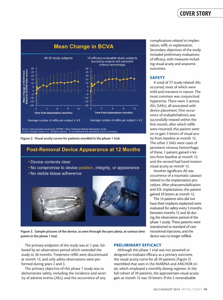
PRELIMINARY EFFICACYAlthough this phase 1 trial was not powered or
designed to evaluate efficacy as a primary outcome, the visual acuity curve for all 20 patients (Figure 2) resembled that seen in the MARINA and ANCHOR tri-als, which employed a monthly dosing regimen. In the full cohort of 20 patients, the approximate visual acuity gain at month 12 was 10 letters. If the 2 nonevaluable
Figure 2. Visual acuity curves for patients enrolled in the phase 1 trial.
Figure 3. Sample pictures of the device, as seen through the pars plana, at various time
points in the phase 1 trial.
80 RETINA TODAY JULY/AUGUST 2014
COVER STORY
patients with persistent vitreous hemorrhage are exclud-ed, the gain at month 12 was 15 letters.
The average number of refills for the full 20-patient cohort was 4.8, and, with the 2 nonevaluable patients excluded, the average was 4.2.
At 12 months, most patients achieved significant gains in visual acuity from baseline: 10 patients (50%) gained 3 lines or more, and 2 (10%) lost 3 lines or more. Of the 2 patients who lost vision, 1 was due to persistent vitre-ous hemorrhage, and 1 was a patient who developed progression of subretinal fibrosis.
Visual acuity improvement was paralleled by an improvement in anatomic outcomes, with a decrease in mean central retinal thickness through month 12.
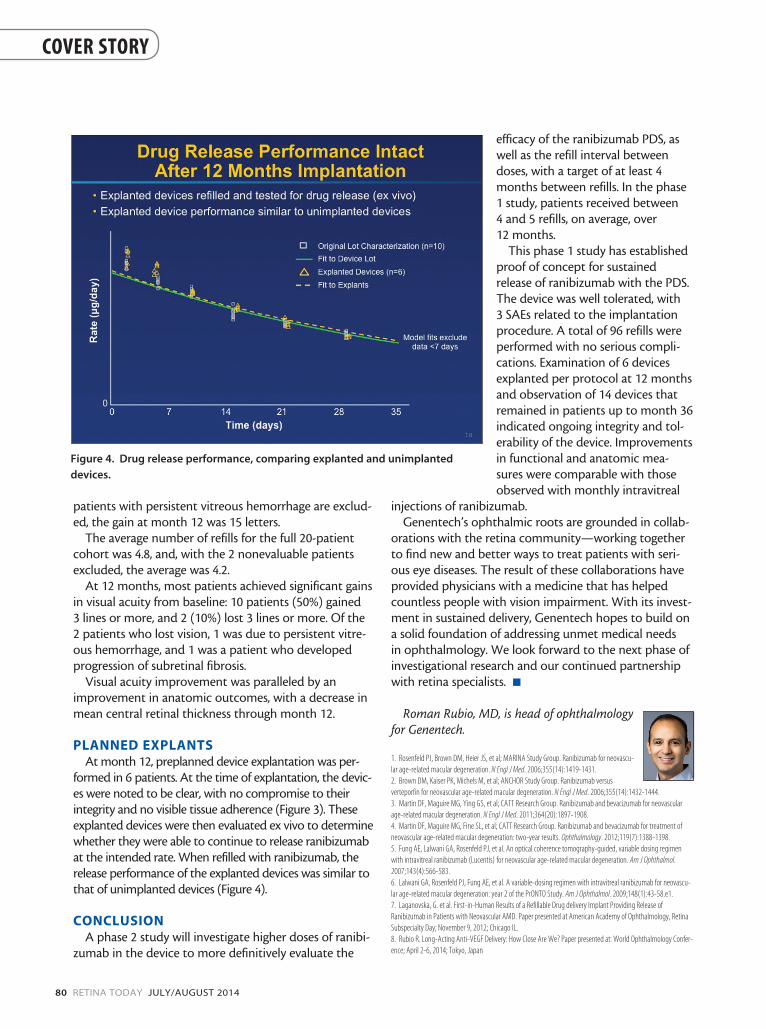
PLANNED EXPLANTSAt month 12, preplanned device explantation was per-
formed in 6 patients. At the time of explantation, the devic-es were noted to be clear, with no compromise to their integrity and no visible tissue adherence (Figure 3). These explanted devices were then evaluated ex vivo to determine whether they were able to continue to release ranibizumab at the intended rate. When refilled with ranibizumab, the release performance of the explanted devices was similar to that of unimplanted devices (Figure 4).
CONCLUSIONA phase 2 study will investigate higher doses of ranibi-
zumab in the device to more definitively evaluate the
efficacy of the ranibizumab PDS, as well as the refill interval between doses, with a target of at least 4 months between refills. In the phase 1 study, patients received between 4 and 5 refills, on average, over 12 months.
This phase 1 study has established proof of concept for sustained release of ranibizumab with the PDS. The device was well tolerated, with 3 SAEs related to the implantation procedure. A total of 96 refills were performed with no serious compli-cations. Examination of 6 devices explanted per protocol at 12 months and observation of 14 devices that remained in patients up to month 36 indicated ongoing integrity and tol-erability of the device. Improvements in functional and anatomic mea-sures were comparable with those observed with monthly intravitreal
injections of ranibizumab.Genentech’s ophthalmic roots are grounded in collab-
orations with the retina community—working together to find new and better ways to treat patients with seri-ous eye diseases. The result of these collaborations have provided physicians with a medicine that has helped countless people with vision impairment. With its invest-ment in sustained delivery, Genentech hopes to build on a solid foundation of addressing unmet medical needs in ophthalmology. We look forward to the next phase of investigational research and our continued partnership with retina specialists. n
Roman Rubio, MD, is head of ophthalmology for Genentech.
1. Rosenfeld PJ, Brown DM, Heier JS, et al; MARINA Study Group. Ranibizumab for neovascu-lar age-related macular degeneration. N Engl J Med. 2006;355(14):1419-1431.2. Brown DM, Kaiser PK, Michels M, et al; ANCHOR Study Group. Ranibizumab versus verteporfin for neovascular age-related macular degeneration. N Engl J Med. 2006;355(14):1432-1444.3. Martin DF, Maguire MG, Ying GS, et al; CATT Research Group. Ranibizumab and bevacizumab for neovascular age-related macular degeneration. N Engl J Med. 2011;364(20):1897-1908. 4. Martin DF, Maguire MG, Fine SL, et al; CATT Research Group. Ranibizumab and bevacizumab for treatment of neovascular age-related macular degeneration: two-year results. Ophthalmology. 2012;119(7):1388-1398. 5. Fung AE, Lalwani GA, Rosenfeld PJ, et al. An optical coherence tomography-guided, variable dosing regimen with intravitreal ranibizumab (Lucentis) for neovascular age-related macular degeneration. Am J Ophthalmol. 2007;143(4):566-583.6. Lalwani GA, Rosenfeld PJ, Fung AE, et al. A variable-dosing regimen with intravitreal ranibizumab for neovascu-lar age-related macular degeneration: year 2 of the PrONTO Study. Am J Ophthalmol. 2009;148(1):43-58.e1.7. Laganovska, G. et al. First-in-Human Results of a Refillable Drug delivery Implant Providing Release of Ranibizumab in Patients with Neovascular AMD. Paper presented at American Academy of Ophthalmology, Retina Subspecialty Day; November 9, 2012; Chicago IL.8. Rubio R. Long-Acting Anti-VEGF Delivery: How Close Are We? Paper presented at: World Ophthalmology Confer-ence; April 2-6, 2014; Tokyo, Japan
Figure 4. Drug release performance, comparing explanted and unimplanted
devices.