Embed Size (px)
Citation preview

Long-Range Intra-Protein Communication Can BeTransmitted by Correlated Side-Chain Fluctuations AloneKateri H. DuBay1,2¤, Jacques P. Bothma3, Phillip L. Geissler1,2,3*
1Department of Chemistry, University of California at Berkeley, Berkeley, California, United States of America, 2Chemical Sciences, Physical Biosciences, and Materials
Sciences Divisions, Lawrence Berkeley National Lab, Berkeley, California, United States of America, 3 Biophysical Graduate Group, University of California at Berkeley,
Berkeley, California, United States of America
Abstract
Allosteric regulation is a key component of cellular communication, but the way in which information is passed from onesite to another within a folded protein is not often clear. While backbone motions have long been considered essential forlong-range information conveyance, side-chain motions have rarely been considered. In this work, we demonstrate theirpotential utility using Monte Carlo sampling of side-chain torsional angles on a fixed backbone to quantify correlationsamongst side-chain inter-rotameric motions. Results indicate that long-range correlations of side-chain fluctuations canarise independently from several different types of interactions: steric repulsions, implicit solvent interactions, or hydrogenbonding and salt-bridge interactions. These robust correlations persist across the entire protein (up to 60 A in the case ofcalmodulin) and can propagate long-range changes in side-chain variability in response to single residue perturbations.
Citation: DuBay KH, Bothma JP, Geissler PL (2011) Long-Range Intra-Protein Communication Can Be Transmitted by Correlated Side-Chain FluctuationsAlone. PLoS Comput Biol 7(9): e1002168. doi:10.1371/journal.pcbi.1002168
Editor: Eugene I. Shakhnovich, Harvard University, United States of America
Received December 1, 2010; Accepted July 5, 2011; Published September 29, 2011
Copyright: ! 2011 DuBay et al. This is an open-access article distributed under the terms of the Creative Commons Attribution License, which permitsunrestricted use, distribution, and reproduction in any medium, provided the original author and source are credited.
Funding: Support for this work was provided by DOE, UC Berkeley, and the NSF. All computational work was enabled through funding by the Director, Office ofScience, Office of Basic Energy Sciences, Materials Sciences and Engineering Division, of the U.S. Department of Energy under Contract No. DE-AC02-05CH11231.KHD was also supported by the Director, Office of Science, Office of Basic Energy Sciences, Materials Sciences and Engineering Division, of the U.S. Department ofEnergy under Contract No. DE-AC02-05CH11231, the Berkeley Fellowship, and a NSF GRF. JPB was supported by the Berkeley Fellowship. The funders had no rolein study design, data collection and analysis, decision to publish, or preparation of the manuscript.
Competing Interests: The authors have declared that no competing interests exist.
* E-mail: [email protected]
¤ Current address: Department of Chemistry, Columbia University, New York, New York, United States of America
Introduction
Allostery is an essential feature of protein regulation andfunction. Allosteric regulation acts by linking distant sites of aprotein together in such a way that information about one site istransmitted to and influences the behavior of another. Chemicalmodifications as subtle as the phosphorylation of a serine residuecan cause dramatic changes in protein function [1], and shifts instructure as small as 1 A have even been shown to modifybehavior in a domain up to 100 A away [2]. Traditionally,allostery has been understood as a feature of symmetric, multi-subunit proteins where the binding of a ligand to one subunitfacilitates the binding of similar ligands to the other subunits,resulting in cooperative binding transitions [3]. However, allostericbehavior has now been observed within a single protein domain[4] and its definition extended to include any shift in proteinstructure and function at one site resulting from modification atanother.Moreover, it was proposed some time ago that information
regarding the binding of a ligand or other modification at oneprotein site could be transmitted through altered proteinfluctuations, even if the protein’s average structure remainsunaffected [5]. Two particularly clear examples of this kind ofdynamic allostery have been recently observed in the binding ofcAMP to the CAP dimer and in the subsequent binding of thecAMP-activated CAP dimer to DNA [6,7]. In the first step, thebinding of cAMP to one monomer of CAP lowers the binding
affinity of cAMP to the second even though no structural changesare observed, and calorimetric analysis suggests that the negativecooperativity results entirely from entropic effects [6].The observed allosteric effect of protein fluctuations has led to
the idea that allostery may be present in all proteins [8–10], andthat functional allostery simply exploits and refines pre-existinglong-range correlations and interaction networks. In fact, suchnetworks are to be expected given the physical constraints of thedensely-folded, yet fluctuating, protein. Just as in any condensedphase, significant fluctuations in this packed environment arepermitted through correlated motions.Qualitative experimental evidence for long-range correlation
abounds in studies demonstrating allosteric regulation, as exem-plified in [1] and [2]. However, attempts to quantify these long-range correlations using NMR techniques have proven difficult[11–13], and much of our current understanding of correlatedmotions has come from analyses of molecular dynamics (MD)simulations. Traditional MD trajectories evaluated with covari-ance matrices and principle component analyses [14] have shedlight on important features of intra-protein correlations, such ashow backbone motions tend to be significantly correlated withinsecondary structural units [14] and how a few flexible hingeresidues can cause large motions within otherwise stable folds [15].Energy-perturbative MD simulations, such as anisotropic thermaldiffusion [16] and pump-probe MD [17], have been used toobserve the rapid anisotropic diffusion of an energy perturbationwithin the protein. However, these MD studies are limited in their
PLoS Computational Biology | www.ploscompbiol.org 1 September 2011 | Volume 7 | Issue 9 | e1002168
ability to characterize sluggish rearrangements and have largelyneglected the contributions of correlated side-chain fluctuations.Within the folded protein, side-chains are significantly less
ordered than the backbone [18], and alternative side-chainconfigurations in protein crystals are more prevalent than previouslythought [19]. The thermodynamic importance of this side-chainvariability in calmodulin-ligand binding has been highlightedin Refs. [20–22]. In addition, the participation of side-chainfluctuations in long-range networks has been demonstrated throughNMR mutational studies [9,23]. In one MD simulation designed toincorporate data from NMR experiments, correlations were evenobserved between side-chains whose motions appeared decoupledfrom those of their backbone atoms [24].Double mutant cycles [25] have also been applied to examine
the dependence of folding and binding on interactions betweenspecific residue pairs. While such mutational studies candemonstrate the interactions of certain residue pairs, they areexperimentally demanding, making it difficult to obtain acomprehensive picture of any long-range side-chain interactionspresent, in particular those involving residues essential for foldingstability. As an alternative, an evolutionary statistical networkanalysis method has been developed to determine networks ofcorrelated residues that are common to evolutionarily relatedproteins [26]. Although this method has had some success inidentifying allosterically-related regions within proteins [27], itsrobustness has been challenged in a study on artificially-generatedsequences [28]. In principle, it is also limited to detectingcorrelated changes in residues during evolution, presumablyhighlighting only correlated networks with a selected functionand can therefore say little about the presence or absence of othercorrelations.In this study, we employed an atomistically detailed model to
examine the kinds of correlations that emerge among side-chainfluctuations within the natively-folded protein. The computation-ally inexpensive nature of our model energy function [21],together with a variety of advanced Monte Carlo sampling
techniques, allowed an unprecedentedly thorough investigation ofthe correlations among these fluctuations that result from differenttypes of interactions. Keeping the backbone fixed, we find thatlong-range correlation of side-chain fluctuations can emerge fromdifferent types of atomic interactions, that significant correlationspersist across the entire folded protein, and that these correlationsalone can propagate changes in structure and mobility over scalesas large as 50 A.
Results
A simple model was used to explore side-chain rotationsIn order to investigate the correlated rearrangements that arise
from side-chain fluctuations alone, it was necessary to isolate thesemotions from other sources of configurational change. For thisreason, we held the backbone fixed in its folded conformationthroughout the calculations described in this paper. Whilefluctuations resulting from bond stretching and angle bendingare important and likely to give rise to a great deal of correlatedmotion, we focused here instead on side-chains’ torsional degreesof freedom, as these rotations give rise to the changes in atomicconfigurations that are largest in magnitude.This study made use of a model we designed to roughly capture
the essential physical determinants of side-chain behavior withinthe folded protein, namely, steric repulsions, van der Waalsattractions, hydrogen bonding, salt-bridge interactions, andsolvation [21]. While not fully realistic in every particular (e.g.,resolving the positions only of nuclei heavier than hydrogen), themodel properly represents the variety, strength, and anisotropy ofthe side-chain interactions and the physical constraints of thefolded backbone on which they reside.We explored this model with Monte Carlo (MC) sampling (see
Methods). Each MC step consisted of the proposed rotation of asingle randomly-chosen side-chain dihedral angle. To promotebroad sampling of thermally accessible configurations, wepermitted moves through sterically disallowed regions of statespace. Using exact correction methods, we constructed equilibri-um averages with contributions only from sterically allowedstructures (those in which each heavy atom excludes a sphericalvolume with radius 0.75 times its van der Waals radius). See [21]for details.Boltzmann-weighted ensembles of the side-chain configurations
determined using this sampling procedure include a diverse set ofrotamer states and correlate well with experimental observations ofside-chain fluctuations and changes in entropy upon ligandbinding [21]. We therefore applied this method to investigatecorrelations among the diverse set of rotamer states.
Single residue perturbations effected changes in side-chain fluctuations throughout the proteinSeveral experimental approaches that probe correlations within
proteins mutate single residues and observe the resulting changesin structure, function, or dynamics [9,23,29,30]. We began ourexamination of side-chain correlations in a similar way bymodeling the changes in torsional variability that occuredthroughout a small globular protein, barstar, as a result ofperturbations to a single side-chain. Previous MD simulations ofbarstar suggested a relatively rigid backbone, as well as significantvariability in side-chain packing [31]. Additional results from 19FNMR experiments showed that the P27A mutation results indetectable dynamic changes even in residues more than 12 Aaway from the mutation site, and suggested that the motion ofbarstar’s side-chains gives rise to a network of correlated residues[32].
Author Summary
Allosteric regulation occurs when the function of one partof a protein changes in response to a signal recognized byanother part of the protein. Such intra-protein communi-cation is essential for many biochemical processes,allowing the cell to adapt its behavior to a dynamicenvironment. Most studies of the information conveyanceunderlying allostery have to date focused on the role ofbackbone motions in mediating large structural changes.Here we focus instead on more subtle contributions,arising from fluctuations of side-chain torsions. Using amodel for side-chain bond rotations in the tightly packedenvironment imposed by native backbone conformations,we observed significant sensitivity of side-chain organiza-tion to small, localized perturbations. This susceptibilityarises from correlations among side-chain motions thatcan propagate information within a protein in complex,heterogeneous ways. Specifically, we found appreciablecorrelations even between side-chains distant from oneanother, so that the effect of a minor perturbation at onesite on the protein could be observed in the alteredfluctuations of side-chains throughout the protein. Inconclusion, we have demonstrated that the statisticalmechanics of correlated side-chain fluctuations within amodel of the folded protein provides the basis for anunconventional but potentially important means ofallostery.
Correlated Side-Chain Motions
PLoS Computational Biology | www.ploscompbiol.org 2 September 2011 | Volume 7 | Issue 9 | e1002168
Quantifying such correlated fluctuations requires a metric thatcan report on the extent of local variability at the single residuescale. For this purpose, we calculated the Gibbs entropy for eachresidue, !SS(res), associated with occupying distinct rotameric states.
!SS(res)i ~{kB
X
Hi
p(Hi)ln p(Hi), !1"
where Hi~fh(i)1 , . . . ,h(i)Nig denotes the set of torsional variables for
each of the Ni rotatable sp3-sp3 hybridized bonds belonging to
residue i, and h(i)n denotes the set of ideal torsional angle values for
the n th torsional angle in residue i. While sp3-sp2 hybridizedbonds were allowed to rotate, they were also excluded from thestatistical analysis due the difficulty in determining ideal dihedral
angles [33]. The probabilities p(Hi) of these 3Ni states werecalculated in simulations by constructing histograms over thecourse of importance sampling from the Boltzmann distribution ofside-chain configurations. In doing so, we focused on the inter-rotameric rearrangements (those between the three most likely
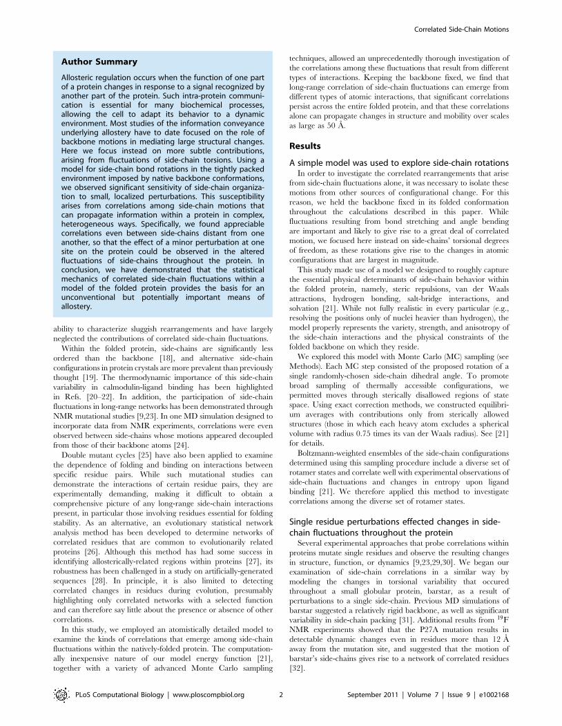
energy basins for the torsional angle of an sp3-sp3 hybridizedbond), which allowed the calculation of absolute local entropiesthat would have been impractical at a higher level of resolution.However, intra-rotameric fluctuations (those within a singletorsional energy basin) are sensitive to the structural perturbationswe applied, and it is necessary to allow deviations from these idealangles, w~x{h, in order to fully account for the variety ofpossible side-chain configurations [34,35]. A quadratic energy isassociated with these deviations w (see Methods).Fig. 1 shows the change in !SS(res)
i that resulted from a single-residue perturbation. Residues shown in red demonstrated astatistically-significant increase in side-chain variability, while thevariability of those shown in blue decreased (see Methods). Theperturbations shown, a mutation of isoleucine to glycine at position86 (Fig. 1(a)) and a constraint of the glutamate in position 46 to itscrystalline configuration (Fig. 1(b)), were chosen to demonstrate thetypes of changes possible. (A comparison to the previously studiedP27A [32] was not possible, since neither proline nor alanineresidues have rotatable side-chains in our model.) Surprisingly,removing the isoleucine side-chain at position 86 (circled in Fig. 1(a))not only affected the local entropy of a few neighboring residues, butalso altered the side-chain variability of residues much farther fromthe site of mutation. Motions of even distant residues must thereforebe linked to those of residue 86. Because the interaction potentials inour model are short in range, the changes in fluxionality thatresulted from this mutation must propagate through neighboringresidues to those farther away. Fig. 1(b) shows analogous changesthat resulted when a residue, E46 (circled), is merely frozen into itscrystalline conformation. Such a reduction in motion of one side-chain might be expected to result in the increased variability of itsnearest neighbors. However, we found that even so subtle aconstraint resulted in unexpected and wide-spread changes in theside-chain fluctuations. Some residues near the frozen amino acideven became slightly more constrained while the variability of a fewresidues farther away increased. A similar effect was observed inNMR experiments upon ligand-binding in stromelysin 1, where thefew residues participating in strong interactions with the ligand lostmobility, but the order parameters of those farther away actuallydecreased upon binding, indicating an increase in their entropy[36]. It was suggested that the increased fluctuations far from thebinding site may counter the loss of entropy at the binding site itselfand therefore assist in modulating the thermodynamics of binding.
The changes in side-chain statistics we observed as a result ofthese single residue perturbations are not readily intuited.Increasing or decreasing disorder at one site may result in thesame or opposite effect in other regions of the folded protein, andthe effects cannot be easily predicted from the spatial arrangementof the residues.
Figure 1. Single-residue perturbations in barstar. Changes D!SS(res)i
in the Gibbs entropy of each residue i in barstar (1a19 [47]) that resultedfrom perturbations to single side-chains. Residues whose entropychanges by a significant amount, according to Student’s t-test at the90% level, are shown in color. Red indicates increased entropy, blueindicates decreased entropy (see scale bar). Although side-chains aredepicted in their crystallographic arrangements for graphical simplicity,note that !SS(res)
i is a measure of the extent of fluctuations among a widevariety of distinct packings. For the results presented in panel (a), I86(shown in black and circled) was mutated to G. For those of panel (b)E46 (shown in black and circled) was constrained to its crystallographicconfiguration.doi:10.1371/journal.pcbi.1002168.g001
Correlated Side-Chain Motions
PLoS Computational Biology | www.ploscompbiol.org 3 September 2011 | Volume 7 | Issue 9 | e1002168

Correlated fluctuations result from several types ofinteractions and persist throughout proteinCorrelated fluctuations within the folded protein are commonly
quantified using Pearson correlation coefficients [14]. Despite theirlimitations in detecting nonlinear correlations and correlationsbetween the motions of particles moving orthogonally to oneanother [37], Pearson coefficients have yielded importantinformation regarding correlated motions. These coefficients aremost appropriate for backbone motions as these motions areexpected to be correlated in similar directions and to be linear innature due to the stiffness and collective motions of varioussecondary structural elements [14]. However, in a study analyzingthe results of molecular dynamics simulations of protein G andlysozyme, a generalized correlation measurement based on mutualinformation was able to detect significantly more correlation thanthe Pearson coefficient [37]. Side-chain motions are even morelikely to fall outside the purview of the Pearson coefficient,dominated as they are by dihedral angle rotations. A parameterbased on mutual information is able to provide a more robustmeasurement of correlated side-chain fluctuations [37], and can bereadily derived from simulation data in a similar way to theentropies calculated in the preceding section. We therefore choseto consider the mutual information associated with each pair ofresidues within a folded protein.Pairwise mutual information is a measure of the correlation
between two random variables. In our case it reports on the degreeof correlation between the rotameric state populations of tworesidues. The mutual information Iij between residues i and j canbe calculated as
Iij~{kBX
Hi
X
Hj
p(Hi,Hj)ln#p(Hi,Hj)
p(Hi)p(Hj)$, !2"
where p(Hi,Hj) denotes the probability of each of the 3Ni :3Nj joint
states of residues i and j, and Ni is the number of rotatable sp3-sp3
hybridized bonds in residue i. After rearranging Eq. 2 andsubstituting in Eq. 1, this becomes
Iij~!SS(res)ij {(!SS(res)
i z!SS(res)j ) !3"
where !SS(res)ij is the Gibbs entropy associated with the discrete
rotameric states for residues i and j considered jointly. Thus whenthe fluctuations of the two residues are completely independent of
one another, !SS(res)ij ~!SS(res)
i z!SS(res)j and Iij~0. However, when the
residues are correlated, their entropies are inseparable, and Iijw0.One difficult feature of mutual information is that a numeri-
cally-calculated estimate of two completely uncorrelated variablesonly approaches zero at the limit of infinite sampling. For anyfinite sampling, a small amount of spurious mutual informationwill be observed, regardless of the actual coupling between the twovariables [38]. When calculating I numerically, this inherent biasin the noise must be accounted for in order to determine themutual information’s statistical significance. We used twoapproaches to address this bias. In the first, we subtracted outthe expected spurious mutual information to estimate the trueamount of correlation between the two variables. The resultingexcess mutual information, I ’, between residues i and j is definedas
I ’ij~Iij(n){I (ref)ij (n): !4"
Iij(n) is the numerically-calculated mutual information measured
over a finite sampling period consisting of n MC steps. I (ref)ij (n) is
the same measurement, but this time computed within a non-interacting reference state, where no correlations are possible (seeMethods for details). I ’ is then an estimate of the mutualinformation of the infinitely-sampled ensemble. In the secondapproach, we focused on the robustness of the mutual information
measurement, calculating its signal-to-noise ratio, Iij(n)=I (ref)(n).The extended structure of calmodulin (3cln [39]), as shown in
Fig. 2(a), provides an exemplary test case for examining how side-
Figure 2. Structural representations of extended crystallinecalmodulin. The crystal structure (a) and contact map (b) of calcium-bound calmodulin (3cln [39]). The calcium ions are shown in yellow, andseveral residues are labeled in both panels for reference. The distancebetween each pair of Ca atoms is indicated by color (see scale bar) in(b), where x- and y-axes run over residue labels. The residue labelingcorresponds to the full sequence, however residues that do not possesstorsional degrees of freedom in our model (A, G, P, and all residuesbound to the calcium ions) are excluded from the contact map.doi:10.1371/journal.pcbi.1002168.g002
Correlated Side-Chain Motions
PLoS Computational Biology | www.ploscompbiol.org 4 September 2011 | Volume 7 | Issue 9 | e1002168

chain fluctuations are correlated within the folded protein.Although in solution this chain collapses, the structure of thecrystal is extended, featuring two globular regions connected by anextended a-helix. Any information shared between the two lobesmust pass through this extended a-helix, since the pairwiseinteractions in our model largely decay by 7 A. We calculated thepairwise excess mutual information, I ’ij , for all residue pairs ij inCa2z-bound calmodulin, as well as the ratio of Iij=I
(ref)ij in order to
gauge the significance of the measured correlations. Bothquantities are shown in Fig. 3 as functions of the residues’ positionalong the backbone. For reference, we present in Fig. 2(b) thespatial distance between residues in the native structure as afunction of the same indices. Panels (b)–(e) of Fig. 3 indicatemutual information resulting from various interaction typesconsidered in isolation. Panel (f) gives results for the full model.Different types of inter-atomic interactions in our model gave
rise to different patterns of correlated fluctuations. In Fig. 3(b),correlations that result solely from steric repulsions are shown.While the signature of calmodulin’s a-helical structure can beclearly seen along the diagonal, where residues i and iz3 or i andiz4 are often highly correlated, many other residue pairs appearsignificantly correlated as well, even those that are spatially distant.In Fig. 3(c), the correlations that result from the implicit solventalone are shown. These correlations are more limited, restrictedalmost completely to residues that are nearby in space, as can be
seen when comparing Fig. 3(c) to Fig. 2(b). Again the a-helicalresidues display appreciable correlation, even more than thatresulting from the repulsive sterics, as might be expected fromtheir high degree of solvent exposure. The correlations that resultfrom considering van der Waals attractions along with therepulsive sterics is shown in Fig. 3(d). While the correlations alongthe a-helix remain strong, many other correlations emerge as aresult of these attractions. Hydrogen bonding and salt bridgeinteractions, taken alone, generate highly significant correlationsthroughout the entire structure (see Fig. 3(e)), which appearremarkably insensitive to spatial distance. Since only a subset ofthe residues participate in such interactions, the fluctuations of theremaining residues are completely uncorrelated in this restrictedversion of our model. The full potential, used to generate the datain Fig. 3(f), results in both the most significant signal-to-noise ratiosand the largest excess mutual information values, indicating alarge degree of correlation that spans the full range of inter-residuedistances while retaining features of the dominant a-helicalstructure.To further explore how different interactions give rise to long-
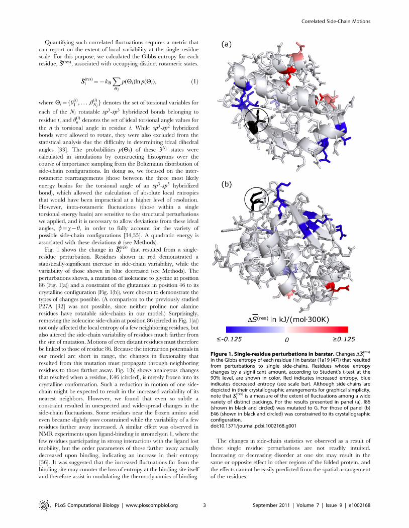
range correlations in both a small globular protein as well as theextended calmodulin structure, we calculated the average excessmutual information per residue pair for all residue pairs incalmodulin and barstar, resolved by the spatial inter-residuedistance between Ca atoms. (See Fig. 4.) In both proteins, steric
Figure 3. Mutual information of residue pairs in calmodulin. The mutual information, Iij , associated with side-chain fluctuations of residuepairs in calmodulin. Plots (b)–(f) display the mutual information signal:noise ratio, Iij=I
(ref)ij (upper left triangles) and the excess mutual information I ’ij
(lower right triangles), as indicated in (a). The x- and y-axes run over labels, i and j respectively, of residues in the amino acid sequence, excludingthose lacking rotameric freedom in our model. Scale bars for the signal:noise ratio and the excess mutual information are presented on the top andbottom left, respectively. Results are shown for the following combinations of interactions: (b) repulsive sterics (S), (c) implicit solvent (IS) (d) Lennard-Jones (LJ) interaction comprising repulsive sterics and van der Waals attractions, (e) hydrogen bonding and salt-bridges (HBSB), and (f) the fullpotential (LJ+HBSB+IS). Residue 30K, which we scrutinize in detail later (see Fig. 5), is highlighted in (f) for reference.doi:10.1371/journal.pcbi.1002168.g003
Correlated Side-Chain Motions
PLoS Computational Biology | www.ploscompbiol.org 5 September 2011 | Volume 7 | Issue 9 | e1002168
repulsions alone give rise to small, but significant, correlations thatpersist across the entire protein structure. The same is true for theimplicit solvent interactions and their combination, S+IS.However, much larger correlations emerge when van der Waalsattractions are considered in addition to the steric repulsions.Hydrogen bonding and salt bridge interactions are clearly themost correlating types of interactions considered. However, the fullpotential, which combines all these interaction types, results in thelargest overall correlation.An additional feature within these plots deserves mention; in both
proteins, correlation is at a maximum around 6 A for all subsets ofinteractions excepting hydrogen bonding and salt bridges. Thisshort-distance peak indicates that residue pairs adjacent in theamino acid sequence (whose a-carbons are separated by &3:8 A)do not interact as strongly on average as do residue pairs that arepositioned slightly farther apart. In a-helices, neighboring residuespoint in different directions and, while still likely to interact with
their sequential nearest neighbor, are more likely to interact stronglywith their iz3 and iz4 neighbors. In b-sheets, however, residuesalternately point towards different faces of the sheet, so that the side-chains on residues i and iz2 are much more likely to interact withone another than do those on i and iz1. Residues influenced onlyby hydrogen bonding and salt bridge interactions, when artificiallyfreed of the steric constraints that would keep them from collapsingback on themselves, still correlate most strongly with their nearestneighbors.Substantial long-range correlation is seen throughout both
barstar and calmodulin. Moreover, the fact that so many subsetsof the full potential independently give rise to long-rangecorrelations suggests that correlated side-chain fluctuations shouldbe a robust characteristic of most protein sequences and nearly anyglobular fold.
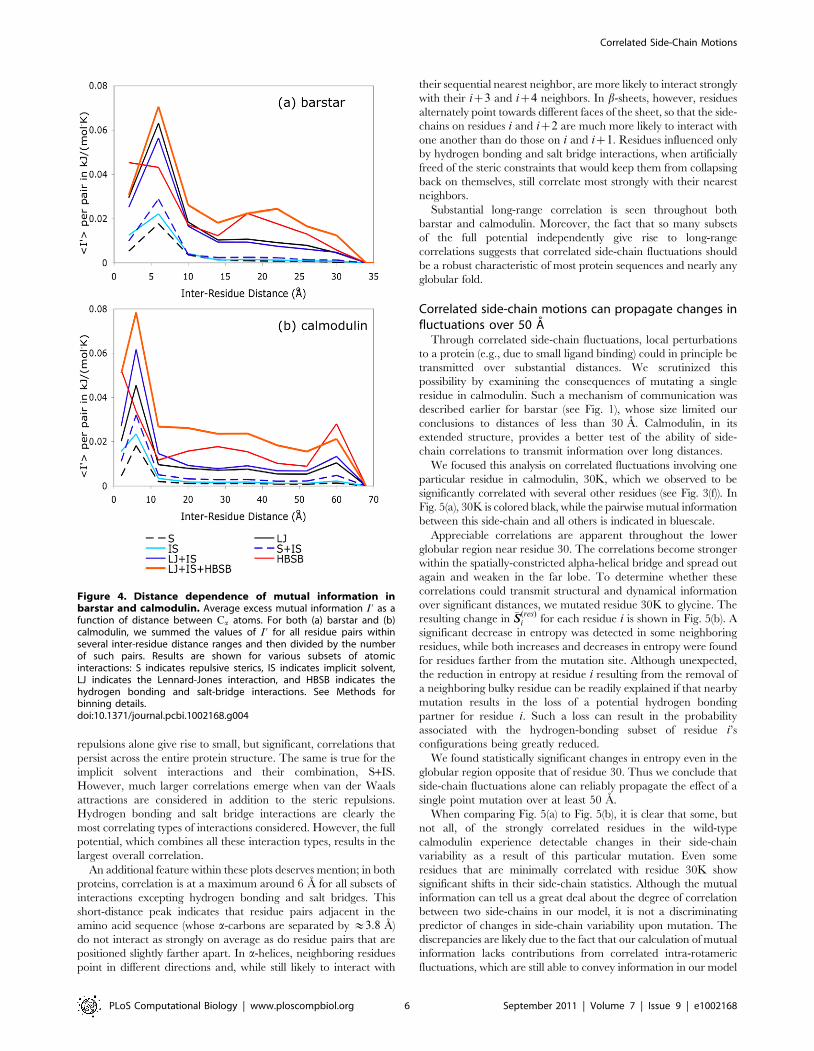
Correlated side-chain motions can propagate changes influctuations over 50 AThrough correlated side-chain fluctuations, local perturbations
to a protein (e.g., due to small ligand binding) could in principle betransmitted over substantial distances. We scrutinized thispossibility by examining the consequences of mutating a singleresidue in calmodulin. Such a mechanism of communication wasdescribed earlier for barstar (see Fig. 1), whose size limited ourconclusions to distances of less than 30 A. Calmodulin, in itsextended structure, provides a better test of the ability of side-chain correlations to transmit information over long distances.We focused this analysis on correlated fluctuations involving one
particular residue in calmodulin, 30K, which we observed to besignificantly correlated with several other residues (see Fig. 3(f)). InFig. 5(a), 30K is colored black, while the pairwise mutual informationbetween this side-chain and all others is indicated in bluescale.Appreciable correlations are apparent throughout the lower
globular region near residue 30. The correlations become strongerwithin the spatially-constricted alpha-helical bridge and spread outagain and weaken in the far lobe. To determine whether thesecorrelations could transmit structural and dynamical informationover significant distances, we mutated residue 30K to glycine. Theresulting change in !SS(res)
i for each residue i is shown in Fig. 5(b). Asignificant decrease in entropy was detected in some neighboringresidues, while both increases and decreases in entropy were foundfor residues farther from the mutation site. Although unexpected,the reduction in entropy at residue i resulting from the removal ofa neighboring bulky residue can be readily explained if that nearbymutation results in the loss of a potential hydrogen bondingpartner for residue i. Such a loss can result in the probabilityassociated with the hydrogen-bonding subset of residue i’sconfigurations being greatly reduced.We found statistically significant changes in entropy even in the
globular region opposite that of residue 30. Thus we conclude thatside-chain fluctuations alone can reliably propagate the effect of asingle point mutation over at least 50 A.When comparing Fig. 5(a) to Fig. 5(b), it is clear that some, but
not all, of the strongly correlated residues in the wild-typecalmodulin experience detectable changes in their side-chainvariability as a result of this particular mutation. Even someresidues that are minimally correlated with residue 30K showsignificant shifts in their side-chain statistics. Although the mutualinformation can tell us a great deal about the degree of correlationbetween two side-chains in our model, it is not a discriminatingpredictor of changes in side-chain variability upon mutation. Thediscrepancies are likely due to the fact that our calculation of mutualinformation lacks contributions from correlated intra-rotamericfluctuations, which are still able to convey information in our model
Figure 4. Distance dependence of mutual information inbarstar and calmodulin. Average excess mutual information I ’ as afunction of distance between Ca atoms. For both (a) barstar and (b)calmodulin, we summed the values of I ’ for all residue pairs withinseveral inter-residue distance ranges and then divided by the numberof such pairs. Results are shown for various subsets of atomicinteractions: S indicates repulsive sterics, IS indicates implicit solvent,LJ indicates the Lennard-Jones interaction, and HBSB indicates thehydrogen bonding and salt-bridge interactions. See Methods forbinning details.doi:10.1371/journal.pcbi.1002168.g004
Correlated Side-Chain Motions
PLoS Computational Biology | www.ploscompbiol.org 6 September 2011 | Volume 7 | Issue 9 | e1002168
and will therefore influence the detected changes upon side-chainmutation. Furthermore, observing the statistically significantchanges in Fig. 5(b) requires a great deal of sampling – were moresampling feasible, additional changes would likely be detected.
Magnitude of side-chain correlations is substantialIf the side-chain motions of a protein’s N different residues were
negligibly correlated, then the total entropy S associated withtransitions among distinct rotameric states could be calculated as asimple sum of single-residue contributions, S&
PNj~1 sj . The
excess mutual information, summed over all residue pairs,provides a rough measure of the error in such a mean-fieldestimate. Correspondingly, the quantity
Pij I ’ij characterizes the
global thermodynamic significance of inter-residue correlations.For crystalline barstar modeled with the full potential,
Pij I ’ij is
calculated to be 72 kJ/(mol:300 K). The higher-order correlationsexpected in such a dense environment [40] (see Fig. 3 where asingle residue is often significantly correlated to several others)make this value an overestimate of the total correlation. Even so,its magnitude is noteworthy. In addition, while allowing intra-rotameric fluctuations, this calculation neglects their contributionto the total correlation, which were found to be essential inreproducing the calorimetric TDSbinding of calmodulin with itsligands in [21] and are likely to be substantial.
Long-range correlations are present within severaldifferent backbone models of folded barstarThe rigidity of the peptide backbone in these calculations justifies
to some extent our schematic model of side chain interactions: Forour purposes the potential energy function need not resolve subtle
thermodynamic differences among diverse chain conformations,but instead serves to establish basic length and energy scales forrearrangements within the native state’s basin of attraction.In considering the biological relevance of our results, backbone
rigidity is in part justified empirically by the observation that onlyweak correlations exist between backbone NMR order parame-ters, S2, and their associated side-chain order parameters, S2
axis[41]. This weak correlation is likely due to the fact that side-chainand backbone fluctuations largely occur on different time-scales[42], with typical side-chain fluctuations ranging from picosecondsto nanoseconds, while typical collective backbone fluctuationsrange from nanoseconds to seconds and longer.However, it is important to assess how variations in backbone
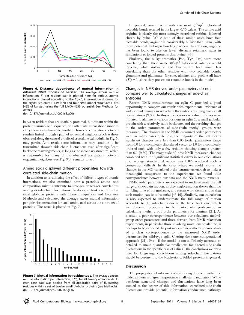
configuration of the folded protein might influence the side-chaincorrelations we have calculated. Toward this end we examinedfour different structural models from an NMR structure of barstar(1btb [43]). These four conformations were chosen to represent therange of models included in the NMR structure (see Methods). Ineach case plots of SI ’T per pair vs. inter-residue distance for thefull potential closely resemble results for the crystal structure (seeFig. 6). Since the statistics of side-chain rotations in a fluctuatingbackbone environment can be rigorously decomposed intocontributions from sub-ensembles in which the backbone is heldfixed, the consistent nature of the observed long-range correlationfrom one backbone structure to another establishes theirrobustness to typical backbone motions.Larger backbone fluctuations, however, such as partial unfolding
events or the motions of hinged regions, are certain to disrupt manyof these correlations and may limit their role in conveying allostericinformation. In particular those correlations arising from contact
Figure 5. Correlation between residue 30 and other residues in calmodulin. The extent of correlation between residue 30 (shown in blackand circled) and all other side-chains in calmodulin (3cln [39]) is shown here. In (a) each residue i is colored according to the magnitude of its excessmutual information I ’i,30 with 30K (see left scale bar and Fig. 3). Coloring in (b) indicates the change D!SSres in each residue’s side chain entropyeffected by the mutation K30G. Here, red represents increased entropy and blue decreased entropy (see right scale bar). See Methods for details.doi:10.1371/journal.pcbi.1002168.g005
Correlated Side-Chain Motions
PLoS Computational Biology | www.ploscompbiol.org 7 September 2011 | Volume 7 | Issue 9 | e1002168
between residues that are spatially proximal, but distant within theprotein’s amino acid sequence, will attenuate as backbone motionscarry them away from one another. However, correlations betweenresidues linked through a path of sequential neighbors, such as thoseobserved along the central a-helix of crystalline calmodulin in Fig. 5,may persist. As a result, some information may continue to betransmitted through side-chain fluctuations even after significantbackbone rearrangements, as long as the secondary structure, whichis responsible for many of the observed correlations betweensequential neighbors (see Fig. 3(f)), remains intact.
Amino acids displayed different propensities towardscorrelated side-chain motionIn addition to scrutinizing the effect of different types of atomic
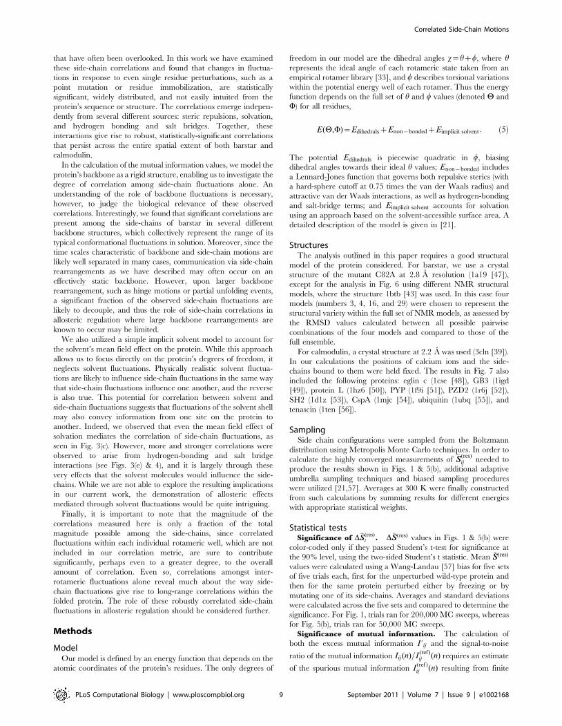
interactions, we also examined how a protein’s amino acidcomposition might contribute to stronger or weaker correlationsamong its side-chain fluctuations. To do so, we took a set of twelvesmall globular proteins with different sequences and folds (seeMethods) and calculated the average excess mutual informationper pairwise interaction for each amino acid across the entire set ofproteins. The result is plotted in Fig. 7.
In general, amino acids with the most sp3-sp3 hybridizedrotatable bonds resulted in the largest SI ’T values. The amino acidarginine is clearly the most strongly correlated residue, followedclosely by lysine. While both of these amino acids have fourrotatable bonds, arginine is considerably bulkier than lysine, withmore potential hydrogen bonding partners. In addition, argininehas been found to take on fewer alternate rotameric states insimulations of folded proteins than lysine [44].Similarly, the bulky aromatics (Phe, Tyr, Trp) were more
correlating than their single sp3-sp3 hybridized rotamer wouldindicate, while isoleucine and leucine are both much lesscorrelating than the other residues with two rotatable bonds:glutamine and glutamate. Glycine, alanine, and proline all haveSI ’T~0, since they possess no rotatable bonds in the model.
Changes in NMR-derived order parameters do notcompare well to calculated changes in side-chainentropiesRecent NMR measurements on eglin C provided a good
opportunity to compare our results with experimental evidence ofwide-spread changes in side-chain fluctuations resulting from smallperturbations [9,30]. In this work, a series of valine residues weremutated to alanine at various positions in eglin C, a small globularprotein with a relatively static backbone, and the resulting changesin the order parameters of side-chain methyl groups weremeasured. The changes in the NMR-measured order parameterswere in many cases quite low; the majority of the statisticallysignificant changes were less than 0.05 (order parameters rangefrom 0.0 for a completely disordered vector to 1.0 for a completelyordered one), with only a few residues showing changes greaterthan 0.1 [9,30]. The magnitude of these NMR-measured changescombined with the significant statistical errors in our calculations(the average standard deviation was 0.02) rendered such acomparison difficult. In the cases where we could resolve thechanges in our MC-calculated order parameters enough to make ameaningful comparison to the experiments we found littlecorrespondence between our data and the NMR measurements.NMR order parameters are expected to underestimate the full
range of side-chain motion, as they neglect motion slower than thetumbling time of the molecule, and recent work demonstrates thatsuch motion can be substantial [45,46]. Similarly, our calculationis also expected to underestimate the full range of motionaccessible to the side-chains due to the fixed backbone, whichwe observed previously to be particularly problematic incalculating methyl group order parameters for alanines [21]. Asa result, a poor correspondence between our calculated methyl-group order parameters and those derived from NMR relaxationexperiments, in particular those involving mutations to alanine, isperhaps to be expected. In past work we nevertheless demonstrat-ed a clear correspondence to the measured NMR orderparameters for wild-type eglin C using the same computationalapproach [21]. Even if the model is not sufficiently accurate ordetailed to make quantitative predictions for altered side-chainfluctuations in the specific case of eglin C, the conclusions we drawhere for long-range correlations among side-chain fluctuationsshould be pertinent to the biophysics of folded proteins in general.
Discussion
The propagation of information across long distances within thefolded protein is of great importance in allosteric regulation. Whilebackbone structural changes and fluctuations have long beenstudied as the bearer of this information, correlated side-chainfluctuations provide potential information conductance pathways
Figure 6. Distance dependence of mutual information indifferent NMR models of barstar. The average excess mutualinformation I ’ per residue pair is plotted here for various atomicinteractions, binned according to the Ca-Ca inter-residue distance, forthe crystal structure (1a19 [47]) and four NMR model structures (1btb[43]) of barstar, using the full LJ+IS+HBSB potential. See Methods fordetails.doi:10.1371/journal.pcbi.1002168.g006
Figure 7. Mutual information by residue type. The average excessmutual information per interaction, SI ’T, for all twenty amino acids. Ineach case data was pooled from all applicable pairs of fluctuatingresidues within a set of twelve small globular proteins (see Methods).doi:10.1371/journal.pcbi.1002168.g007
Correlated Side-Chain Motions
PLoS Computational Biology | www.ploscompbiol.org 8 September 2011 | Volume 7 | Issue 9 | e1002168
that have often been overlooked. In this work we have examinedthese side-chain correlations and found that changes in fluctua-tions in response to even single residue perturbations, such as apoint mutation or residue immobilization, are statisticallysignificant, widely distributed, and not easily intuited from theprotein’s sequence or structure. The correlations emerge indepen-dently from several different sources: steric repulsions, solvation,and hydrogen bonding and salt bridges. Together, theseinteractions give rise to robust, statistically-significant correlationsthat persist across the entire spatial extent of both barstar andcalmodulin.In the calculation of the mutual information values, we model the
protein’s backbone as a rigid structure, enabling us to investigate thedegree of correlation among side-chain fluctuations alone. Anunderstanding of the role of backbone fluctuations is necessary,however, to judge the biological relevance of these observedcorrelations. Interestingly, we found that significant correlations arepresent among the side-chains of barstar in several differentbackbone structures, which collectively represent the range of itstypical conformational fluctuations in solution. Moreover, since thetime scales characteristic of backbone and side-chain motions arelikely well separated in many cases, communication via side-chainrearrangements as we have described may often occur on aneffectively static backbone. However, upon larger backbonerearrangement, such as hinge motions or partial unfolding events,a significant fraction of the observed side-chain fluctuations arelikely to decouple, and thus the role of side-chain correlations inallosteric regulation where large backbone rearrangements areknown to occur may be limited.We also utilized a simple implicit solvent model to account for
the solvent’s mean field effect on the protein. While this approachallows us to focus directly on the protein’s degrees of freedom, itneglects solvent fluctuations. Physically realistic solvent fluctua-tions are likely to influence side-chain fluctuations in the same waythat side-chain fluctuations influence one another, and the reverseis also true. This potential for correlation between solvent andside-chain fluctuations suggests that fluctuations of the solvent shellmay also convey information from one site on the protein toanother. Indeed, we observed that even the mean field effect ofsolvation mediates the correlation of side-chain fluctuations, asseen in Fig. 3(c). However, more and stronger correlations wereobserved to arise from hydrogen-bonding and salt bridgeinteractions (see Figs. 3(e) & 4), and it is largely through thesevery effects that the solvent molecules would influence the side-chains. While we are not able to explore the resulting implicationsin our current work, the demonstration of allosteric effectsmediated through solvent fluctuations would be quite intriguing.Finally, it is important to note that the magnitude of the
correlations measured here is only a fraction of the totalmagnitude possible among the side-chains, since correlatedfluctuations within each individual rotameric well, which are notincluded in our correlation metric, are sure to contributesignificantly, perhaps even to a greater degree, to the overallamount of correlation. Even so, correlations amongst inter-rotameric fluctuations alone reveal much about the way side-chain fluctuations give rise to long-range correlations within thefolded protein. The role of these robustly correlated side-chainfluctuations in allosteric regulation should be considered further.
Methods
ModelOur model is defined by an energy function that depends on the
atomic coordinates of the protein’s residues. The only degrees of
freedom in our model are the dihedral angles x~hzw, where hrepresents the ideal angle of each rotameric state taken from anempirical rotamer library [33], and w describes torsional variationswithin the potential energy well of each rotamer. Thus the energyfunction depends on the full set of h and w values (denoted H andW) for all residues,
E(H,W)~EdihedralszEnon{bondedzEimplicit solvent: !5"
The potential Edihedrals is piecewise quadratic in w, biasingdihedral angles towards their ideal h values; Enon{bonded includesa Lennard-Jones function that governs both repulsive sterics (witha hard-sphere cutoff at 0.75 times the van der Waals radius) andattractive van der Waals interactions, as well as hydrogen-bondingand salt-bridge terms; and Eimplicit solvent accounts for solvationusing an approach based on the solvent-accessible surface area. Adetailed description of the model is given in [21].
StructuresThe analysis outlined in this paper requires a good structural
model of the protein considered. For barstar, we use a crystalstructure of the mutant C82A at 2.8 A resolution (1a19 [47]),except for the analysis in Fig. 6 using different NMR structuralmodels, where the structure 1btb [43] was used. In this case fourmodels (numbers 3, 4, 16, and 29) were chosen to represent thestructural variety within the full set of NMR models, as assessed bythe RMSD values calculated between all possible pairwisecombinations of the four models and compared to those of thefull ensemble.For calmodulin, a crystal structure at 2.2 A was used (3cln [39]).
In our calculations the positions of calcium ions and the side-chains bound to them were held fixed. The results in Fig. 7 alsoincluded the following proteins: eglin c (1cse [48]), GB3 (1igd[49]), protein L (1hz6 [50]), PYP (1f9i [51]), PZD2 (1r6j [52]),SH2 (1d1z [53]), CspA (1mjc [54]), ubiquitin (1ubq [55]), andtenascin (1ten [56]).
SamplingSide chain configurations were sampled from the Boltzmann
distribution using Metropolis Monte Carlo techniques. In order tocalculate the highly converged measurements of !SS(res)
ij needed toproduce the results shown in Figs. 1 & 5(b), additional adaptiveumbrella sampling techniques and biased sampling procedureswere utilized [21,57]. Averages at 300 K were finally constructedfrom such calculations by summing results for different energieswith appropriate statistical weights.
Statistical testsSignificance of D!SS(res)
i . D!SS(res) values in Figs. 1 & 5(b) werecolor-coded only if they passed Student’s t-test for significance atthe 90% level, using the two-sided Student’s t statistic. Mean !SS(res)
values were calculated using a Wang-Landau [57] bias for five setsof five trials each, first for the unperturbed wild-type protein andthen for the same protein perturbed either by freezing or bymutating one of its side-chains. Averages and standard deviationswere calculated across the five sets and compared to determine thesignificance. For Fig. 1, trials ran for 200,000 MC sweeps, whereasfor Fig. 5(b), trials ran for 50,000 MC sweeps.
Significance of mutual information. The calculation ofboth the excess mutual information I ’ij and the signal-to-noise
ratio of the mutual information Iij(n)=I(ref)ij (n) requires an estimate
of the spurious mutual information I (ref)ij (n) resulting from finite
Correlated Side-Chain Motions
PLoS Computational Biology | www.ploscompbiol.org 9 September 2011 | Volume 7 | Issue 9 | e1002168
sampling (see Eq. 4). We constructed I (ref)ij (n) by samplingrotameric states from a non-interacting reference system definedby the energy function
E(H)~{kBTX
i
ln p(hi)
Here, p(hi) is the probability of observing rotamer state hi in thefully interacting model. By construction, these single-residuedistributions are then identical in the reference system,
p(ref)(hi)~p(hi). Rotameric fluctuations of distinct residues,however, are statistically independent in the reference system,
p(ref)(hi,hj)~p(hi)p(hj), so that I (ref)ij (n) vanishes in the limit of
complete sampling, n??.For each interacting trial run, the probabilities p(hi) associated
with each rotameric state were recorded and used to bias itscorresponding non-interacting reference run. Excess mutualinformation values and signal-to-noise ratios were then calculatedindependently for each pair of interacting and non-interacting, butbiased, trial runs. Presented results are averages of these I ’ij andIij(n)=I
(ref)ij (n) values.
For the results shown in Figs. 3, 4, 5(a), & 6, Iij was calculated infive trial runs of 50,000 MC sweeps initiated from randomly-chosen side-chain configurations. Subsequently, I (ref)ij was com-puted from five independent runs of a biased, noninteractingreference system, also initiated from randomly-chosen side-chain
configurations. As described above, the lengths and biases of thesereference runs were chosen to produce samples equivalent in sizeand in single-rotamer distribution to the number and distributionof sterically valid configurations generated in the correspondingsimulation of the interacting system. The signal:noise ratio and theexcess mutual information were calculated independently for eachtrial; averages over those trials are presented in the figures. Theresults shown in Fig. 5(a) and Fig. 6 were calculated using the fullLJ+IS+HBSB potential.For Figs. 4 & 6, the above results were collected into inter-
residue distance bins of 4 A wide for barstar and 8 A wide forcalmodulin, except for the first two bins which were kept 4 A widein order to highlight the peak at short distances of &6 A.All images were made using MacPyMOL [58].
Acknowledgments
We would like to thank John D. Chodera (UCB) and Gavin E. Crooks(LBNL) for their helpful discussions and insight.
Author Contributions
Conceived and designed the experiments: KHD PLG. Performed theexperiments: KHD. Analyzed the data: KHD PLG. Contributed reagents/materials/analysis tools: KHD. Wrote the paper: KHD PLG. Suggestedand assisted in the analysis of correlations in different NMR modelstructures of barstar: JPB.
References
1. Barford D, Johnson LN (1989) The allosteric transition of glycogenphosphorylase. Nature 340: 609–616.
2. Ottemann KM, Xiao W, Shin YK, Koshland DE (1999) A piston model fortransmembrane signaling of the aspartate receptor. Science 285: 1751–1754.
3. Perutz MF (1970) Stereochemistry of cooperative effects in haemoglobin. Nature228: 726–739.
4. Volkman BF, Lipson D, Wemmer DE, Kern D (2001) Two-state allostericbehavior in a singledomain signaling protein. Science 291: 2429–2433.
5. Cooper A, Dryden DT (1984) Allostery without conformational change. aplausible model. Eur Biophys J 11: 103–109.
6. Popovych N, Sun S, Ebright RH, Kalodimos CG (2006) Dynamically drivenprotein allostery. Nat Struct Mol Biol 13: 831–838.
7. Tzeng SR, Kalodimos CG (2009) Dynamic activation of an allosteric regulatoryprotein. Nature 462: 368–372.
8. Gunasekaran K, Ma B, Nussinov R (2004) Is allostery an intrinsic property of alldynamic proteins? Proteins 57: 433–443.
9. Clarkson MW, Gilmore SA, Edgell MH, Lee AL (2006) Dynamic coupling andallosteric behavior in a nonallosteric protein. Biochemistry 45: 7693–7699.
10. Petit CM, Zhang J, Sapienza PJ, Fuentes EJ, Lee AL (2009) Hidden dynamicallostery in a pdz domain. Proc Natl Acad Sci USA 106: 18249–18254.
11. Mayer KL, Earley MR, Gupta S, Pichumani K, Regan L, et al. (2003)Covariation of backbone motion throughout a small protein domain. Nat StructBiol 10: 962–965.
12. Bouvignies G, Bernado P, Meier S, Cho K, Grzesiek S, et al. (2005)Identification of slow correlated motions in proteins using residual dipolar andhydrogen-bond scalar couplings. Proc Natl Acad Sci USA 102: 13885–13890.
13. Lange OF, Grubmuller H, de Groot BL (2005) Molecular dynamics simulationsof protein g challenge nmr-derived correlated backbone motions. Angew ChemInt Ed 44: 3394–3399.
14. Ichiye T, Karplus M (1991) Collective motions in proteins: a covariance analysisof atomic fluctuations in molecular dynamics and normal mode simulations.Proteins 11: 205–217.
15. Henzler-Wildman KA, Lei M, Thai V, Kerns SJ, Karplus M, et al. (2007) Ahierarchy of timescales in protein dynamics is linked to enzyme catalysis. Nature450: 913–916.
16. Ota N, Agard DA (2005) Intramolecular signaling pathways revealed bymodeling anisotropic thermal diffusion. J Mol Biol 351: 345–354.
17. Sharp K, Skinner JJ (2006) Pump-probe molecular dynamics as a tool forstudying protein motion and long range coupling. Proteins 65: 347–361.
18. Igumenova TI, Frederick KK, Wand AJ (2006) Characterization of the fastdynamics of protein amino acid side chains using nmr relaxation in solution.Chem Rev 106: 1672–1699.
19. Lang PT, Ng HL, Fraser JS, Corn JE, Echols N, et al. (2010) Automatedelectron-density sampling reveals widespread conformational polymorphism inproteins. Protein Sci 19: 1420–1431.
20. Frederick KK, Marlow MS, Valentine KG, Wand AJ (2007) Conformationalentropy in molecular recognition by proteins. Nature 448: 325–329.
21. DuBay KH, Geissler PL (2009) Calculation of proteins’ total side-chain torsionalentropy and its influence on protein-ligand interactions. J Mol Biol 391:484–497.
22. Marlow MS, Dogan J, Frederick KK, Valentine KG, Wand AJ (2010) The roleof conformational entropy in molecular recognition by calmodulin. Nat ChemBiol 6: 352–358.
23. Millet O, Mittermaier A, Baker D, Kay LE (2003) The effects of mutations onmotions of side-chains in protein l studied by 2 h nmr dynamics and scalarcouplings. J Mol Biol 329: 551–563.
24. Dhulesia A, Gsponer J, Vendruscolo M (2008) Mapping of two networks ofresidues that exhibit structural and dynamical changes upon binding in a pdzdomain protein. J Am Chem Soc 130: 8931–8939.
25. Fersht A (1999) Structure and Mechanism in Protein Science W.H. Freemanand Company.
26. Lockless SW, Ranganathan R (1999) Evolutionarily conserved pathways ofenergetic connectivity in protein families. Science 286: 295–299.
27. Suel GM, Lockless SW, Wall MA, Ranganathan R (2003) Evolutionarilyconserved networks of residues mediate allosteric communication in proteins.Nat Struct Biol 10: 59–69.
28. Noivirt O, EisensteinM,Horovitz A (2005) Detection and reduction of evolutionarynoise in correlated mutation analysis. Protein Eng Des Sel 18: 247–253.
29. Zıdek L, Novotny MV, Stone MJ (1999) Increased protein backbone conforma-tional entropy upon hydrophobic ligand binding. Nat Struct Biol 6: 1118–1121.
30. Clarkson MW, Lee AL (2004) Long-range dynamic effects of point mutationspropagate through side chains in the serine protease inhibitor eglin c.Biochemistry 43: 12448–12458.
31. Wong KB, Daggett V (1998) Barstar has a highly dynamic hydrophobic core:evidence from molecular dynamics simulations and nuclear magnetic resonancerelaxation data. Biochemistry 37: 11182–11192.
32. Li H, Frieden C (2007) Comparison of c40/82a and p27a c40/82a barstarmutants using 19f nmr. Biochemistry 46: 4337–4347.
33. Lovell SC, Word JM, Richardson JS, Richardson DC (2000) The penultimaterotamer library. Proteins 40: 389–408.
34. Kussell E, Shimada J, Shakhnovich EI (2001) Excluded volume in protein side-chain packing. J Mol Biol 311: 183–193.
35. Shetty RP, Bakker PIWD, DePristo MA, Blundell TL (2003) Advantages of fine-grained side chain conformer libraries. Protein Eng 16: 963–669.
36. Arumugam S, Gao G, Patton BL, Semenchenko V, Brew K, et al. (2003)Increased backbone mobility in beta-barrel enhances entropy gain drivingbinding of n-timp-1 to mmp-3. J Mol Biol 327: 719–734.
37. Lange OF, Grubmuller H (2006) Generalized correlation for biomoleculardynamics. Proteins 62: 1053–1061.
38. Roulston MS (1999) Estimating the errors on measured entropy and mutualinformation. Physica D 125: 285–294.
Correlated Side-Chain Motions
PLoS Computational Biology | www.ploscompbiol.org 10 September 2011 | Volume 7 | Issue 9 | e1002168
39. Babu YS, Bugg CE, Cook WJ (1988) Structure of calmodulin refined at 2.2 aresolution. J Mol Biol 204: 191–204.
40. Killian BJ, Kravitz JY, Gilson MK (2007) Extraction of configurational entropyfrom molecular simulations via an expansion approximation. J Chem Phys 127:024107.
41. Mittermaier A, Kay LE, Forman-Kay JD (1999) Analysis of deuteriumrelaxation-derived methyl axis order parameters and correlation with localstructure. J Biomol NMR 13: 181–185.
42. Petsko GA, Ringe D (2004) Protein Structure and Function. New Science PressLtd, London, UK.
43. Lubienski MJ, Bycroft M, Freund SM, Fersht AR (1994) Three-dimensionalsolution structure and 13c assignments of barstar using nuclear magneticresonance spectroscopy. Biochemistry 33: 8866–8877.
44. Berezovsky IN, Chen WW, Choi PJ, Shakhnovich EI (2005) Entropicstabilization of proteins and its proteomic consequences. PLoS Comput Biol1: 0322–0332.
45. Maragakis P, Lindorff-Larsen K, Eastwood MP, Dror RO, Klepeis JL, et al.(2008) Microsecond molecular dynamics simulation shows effect of slow loopdynamics on backbone amide order parameters of proteins. J Phys Chem B 112:6155–6158.
46. Fares C, Lakomek NA, Walter KFA, Frank BTC, Meiler J, et al. (2009)Accessing ns-micros side chain dynamics in ubiquitin with methyl rdcs. J BiomolNMR 45: 23–44.
47. Ratnaparkhi GS, Ramachandran S, Udgaonkar JB, Varadarajan R (1998)Discrepancies between the nmr and x-ray structures of uncomplexed barstar:analysis suggests that packing densities of protein structures determined by nmrare unreliable. Biochemistry 37: 6958–6966.
48. Bode W, Papamokos E, Musil D (1987) The high-resolution x-ray crystalstructure of the complex formed between subtilisin carlsberg and eglin c, an
elastase inhibitor from the leech hirudo medicinalis. structural analysis, subtilisinstructure and interface geometry. Eur J Biochem 166: 673–692.
49. Derrick JP, Wigley DB (1994) The third igg-binding domain from streptococcalprotein g. an analysis by x-ray crystallography of the structure alone and in acomplex with fab. J Mol Biol 243: 906–918.
50. O’Neill JW, Kim DE, Baker D, Zhang KY (2001) Structures of the b1 domain ofprotein l from peptostreptococcus magnus with a tyrosine to tryptophansubstitution. Acta Crystallogr D 57: 480–487.
51. Brudler R, Meyer TE, Genick UK, Devanathan S, Woo TT, et al. (2000)Coupling of hydrogen bonding to chromophore conformation and function inphotoactive yellow protein. Biochemistry 39: 13478–13486.
52. Kang BS, Devedjiev Y, Derewenda U, Derewenda ZS (2004) The pdz2 domainof syntenin at ultrahighresolution: bridging the gap between macromolecularand small molecule crystallography. J Mol Biol 338: 483–493.
53. Poy F, Yaffe MB, Sayos J, Saxena K, Morra M, et al. (1999) Crystal structures ofthe xlp protein sap reveal a class of sh2 domains with extended, phosphotyr-osine-independent sequence recognition. Mol Cell 4: 555–561.
54. Schindelin H, Jiang W, Inouye M, Heinemann U (1994) Crystal structure ofcspa, the major cold shock protein of escherichia coli. P Natl Acad Sci USA 91:5119–5123.
55. Vijay-Kumar S, Bugg CE, Cook WJ (1987) Structure of ubiquitin refined at 1.8a resolution. J Mol Biol 194: 531–544.
56. Leahy DJ, Hendrickson WA, Aukhil I, Erickson HP (1992) Structure of afibronectin type iii domain from tenascin phased by mad analysis of theselenomethionyl protein. Science 258: 987–991.
57. Wang F, Landau DP (2001) Efficient, multiple-range random walk algorithm tocalculate the density of states. Phys Rev Lett 86: 2050–2053.
58. DeLano W (2007) MacPyMOL: A PyMOL-based Molecular GraphicsApplication for MacOS X. DeLano Scientific LLC, Palo Alto, CA, USA.
Correlated Side-Chain Motions
PLoS Computational Biology | www.ploscompbiol.org 11 September 2011 | Volume 7 | Issue 9 | e1002168