Embed Size (px)
Citation preview

www.sciencedirect.com
c o r t e x 4 8 ( 2 0 1 2 ) 3 1 7e3 3 2
Available online at
Journal homepage: www.elsevier.com/locate/cortex
Research report
Long-term accelerated forgetting of verbal and non-verbalinformation in temporal lobe epilepsy
Heather Wilkinson a, Juliet S. Holdstock b, Gus Baker c, Andrea Herbert d, Fiona Clague e,f
and John J. Downes b,*aDepartment of Psychology, University of Chester, UKb School of Psychology, University of Liverpool, UKcUniversity Department of Neurosciences, Walton Centre for Neurology and Neurosurgery, Liverpool, UKdDepartment of Clinical and Community Psychology, Devon Partnership NHS Trust, Exeter, UKeClinical Psychology Department, Astley Ainslie Hospital, Edinburgh, UKfDepartment of Clinical and Health Psychology, School of Health in Social Science, The University of Edinburgh, UK
a r t i c l e i n f o
Article history:
Received 18 December 2009
Revised 24 February 2010
Accepted 15 December 2010
Action editor Sergio Della Sala
Published online 11 February 2011
Keywords:
Memory
Recall
Consolidation
Seizures
Hippocampus
* Corresponding author. School of PsychologE-mail address: [email protected] (J.J
0010-9452/$ e see front matter ª 2011 Elsevdoi:10.1016/j.cortex.2011.01.002
a b s t r a c t
Introduction: We investigated whether pre-surgical patients with temporal lobe epilepsy
(TLE) forget verbal and non-verbal material faster than healthy controls over retention
intervals of an hour and 6 weeks, and whether any observed memory loss was associated
with structural changes to the hippocampus and/or seizure frequency.
Methods: A mixed factorial design compared the performance of 27 patients with TLE and
22 healthy control participants, matched for IQ, age and gender, on tests of story recall and
complex figure recall at three delays: immediate, 1 h and 6 weeks. Performance of the
patient and control groups was matched at the immediate delay, which enabled compar-
isons of forgetting rate over the longer delays.
Results: We found that TLE can affect the acquisition and retention of new memories over
a relatively short delay of 1 h. This deficit was associated with structural hippocampal
abnormality, with a material-specific effect that was particularly evident for the verbal
task. We also found evidence of accelerated long-term forgetting in both patient groups, for
the verbal and non-verbal tasks. It was demonstrated most strongly on the verbal task by
the patients with right lateralized hippocampal sclerosis whose verbal recall was normal at
the 1-h delay. Accelerated long-term forgetting was not associated with hippocampal
pathology, but was associated with the frequency of epileptic seizures.
Discussion: The findings from the verbal task in particular provide evidence consistent with
an extended period of memory consolidation that can be disrupted by both left and right
TLE. The material-specific effects at the 1-h delay only, suggest that the initial consolida-
tion of verbal and non-verbal, information depends on the integrity of the left and right
hippocampus, respectively.
ª 2011 Elsevier Srl. All rights reserved.
y, University of Liverpool, Liverpool L69 7ZA, UK.. Downes).ier Srl. All rights reserved.

c o r t e x 4 8 ( 2 0 1 2 ) 3 1 7e3 3 2318
1. Introduction memory test delay (Helmstaedter et al., 1998; Mameniskiene
Patients with temporal lobe epilepsy (TLE) often report expe-
riencing problems with memory in everyday life and yet in
many cases can perform normally on standardized neuro-
psychological tests of memory used in clinical assessment
(Blake et al., 2000). The standardized tests used for clinical
purposes are, however, limited in the durations over which
memory is tested, the longest retention delay being 30 min. It
has been argued that acquisition and initial consolidation of
long-term memories can sometimes be spared in patients
with TLE, enabling them to perform normally on standardized
memory tests, but that an extended period of consolidation,
that occurs over days, weeks, months or even years, and is
required for long-term maintenance of the memories, is dis-
rupted by this disorder (Blake et al., 2000). Indeed, a number of
single cases have been reported of individuals with TLEwhose
memory is unimpaired after the kind of short delays used in
clinical testing, but who show more rapid forgetting of infor-
mation than healthy individuals over longer delays of several
weeks (Kapur et al., 1997; O’Connor et al., 1997; Mayes et al.,
2003). This pattern has been termed long-term amnesia.
None of these cases, however, have had typical mesial TLE:
one had treatment for testicular cancer and very frequent
complex partial seizures (O’Connor et al., 1997), another had
seizures secondary to a traumatic head injury that resulted in
considerable bilateral damage to the frontal and temporal
lobes [but excluded the hippocampus (HC)] (Mayes et al., 2003),
and the third had a late seizure onset which occurred at 56
years of age (Kapur et al., 1997). The results from these case
studies may not therefore generalize to more typical cases of
TLE, but may reflect idiosyncratic seizure type and location.
The possibility that long-term forgetting may be a more
general characteristic of TLE has been investigated by a small
number of group studies, with mixed results (see Bell and
Giovagnoli, 2007; Butler and Zeman, 2008a, 2008b for reviews).
Studies using the Selective Reminding Test (Buschke, 1973;
Buschke and Fuld, 1974) have found that patients with TLE
were impaired relative to controls on tests of learning and at
memory after delays of both 30 min, and a longer delay of 24 h
and 13 days, respectively (Giovagnoli et al., 1995; Bell et al.,
2005). Similarly, recall of the story from the logical memory
subtest of the Wechsler Adult Intelligence Scale III (Wechsler,
1997) was impaired after both short and long delays in
patients with TLE (Bell, 2006). None of these studies found an
interaction between group (patients vs controls) and memory
test delay and therefore found no evidence for accelerated
long-term forgetting in the patient group. However, the lack of
an interaction is difficult to interpret in these studies because
thememory performance of the patients and controls was not
equated at the short delay (see Huppert and Piercy, 1978; Isaac
and Mayes, 1999). So, although these studies show clear
memory deficits in these patients at both short and long
delays, they provide no strong evidence concerning the rela-
tive rates of forgetting over long delays of the patients and
healthy controls. Similarly, two further studies have reported
deficits in verbal and non-verbal memory in TLE over both
short and long delays, but neither study examined whether
there was an interaction between participant group and
et al., 2006). Thus, although these latter two studies show
that the patients’ memories were very impaired at the longest
delays, again, they provide no information about the relative
rates of forgetting of the patients and healthy controls.
There are two studies in the literature that have matched
the memory performance of the patients and controls on the
initial memory test and found accelerated forgetting in the
patient group over delays of 24 h to 8 weeks (Martin et al.,
1991; Blake et al., 2000). Martin et al. (1991) used the Selec-
tive Reminding Test to compare free recall of verbal material
(unrelated word lists) by 21 patients with TLE and 21 tension
headache control subjects at delays of 30 min and 24 h. They
found a significant group by delay interaction, with the TLE
and tension headache control groups performing at an equal
level during the final learning and the 30 min delayedmemory
trials, but the TLE patients performing significantly worse
than the controls at the 24-h delay. There was no significant
interaction with laterality of epileptic focus.
One limitation of the study of Martin et al. (1991) was that
the TLE group consisted of both pre- andpost-surgical patients
and so it is difficult to infer whether the accelerated forgetting
is a function of temporal lobe resection or of TLE per se. In the
subsequent study conducted by Blake et al. (2000), only pre-
surgical patients were included. Forgetting was assessed over
delays of up to 8 weeks in 21 consecutive partial seizure
patients, which included 14 pre-surgical TLE patients with
either a right or left epileptic focus. The patients’ performance
on standardized tests of memory (WMS-R) and object naming
(Graded Naming Test) did not differ significantly from the
healthy controls, but, despite this, almost half of the patients
reported that their everyday memory was moderately or
severely impaired. To explore accelerated forgetting, the
patients were repeatedly presented with a short story until
their recall reached 90% correct or for amaximumof ten trials.
The number of learning trials required to reach criterion did
not differ significantly between the patient and control groups
indicating intact learning in the patient group. Nevertheless
the patients with a left hemispheric epileptic focus recalled
significantly less of the story than healthy controls and the
patients with a right hemispheric focus after an 8-week delay.
Therecall of the threegroupsdidnotdifferaftera30-mindelay.
Similar findings were reported for recognition, which was
significantly poorer for the left hemisphere group thanhealthy
controls, although the difference between the left and right
hemisphere groups did not quite reach statistical significance.
In summary, Blake et al. (2000) showed normal learning
and short-term retention (30 min) in TLE, but accelerated loss
of this normally learnt information over an 8-week period.
Furthermore, it was found that the long-term forgetting of the
verbal material used in this study was associated with a left
rather than right seizure focus, consistent with reports of
lateralization of verbal memory to the left temporal lobe (e.g.,
Milner, 1971).
Our study investigatedwhyTLEcanresult inaccelerated loss
of memories over short and extended time periods. We were
interested in whether the same or different variables were
associated with forgetting over relatively short and long reten-
tion intervals. To do this, following the methodology of Blake

c o r t e x 4 8 ( 2 0 1 2 ) 3 1 7e3 3 2 319
et al. (2000), we matched the performance of pre-operative
patients with TLE that had resulted in left or right lateralized
hippocampal sclerosis and healthy controls on immediate
verbal and non-verbal, visuospatial, recall and then investi-
gated forgettingoverdelays of 1 h and 6weeks. Inparticular,we
investigated whether forgetting over these delays was associ-
ated with measures of hippocampal sclerosis (hippocampal
volume, T2 relaxation time) or seizure frequency or both.
Wewere interested in the possible contribution of theHC to
long-termforgettingbecause it is believed tohavea central role
in current models of memory consolidation (e.g., Alvarez and
Squire, 1994). It could therefore be predicted that the integrity
of this structure may be important for long-term retention.
Indeed, hippocampal size, as determined by magnetic reso-
nance imaging (MRI), in healthy individuals has been found to
predict verbal recall over a long retention interval of approxi-
mately 11 weeks (Walhovd et al., 2004). In that study, hippo-
campal volume was more important for recall after long than
short delays of up to 30 min. An investigation of transient
epileptic amnesia (TEA) however, found significant or close to
significant correlations between verbal and non-verbal recall
and hippocampal volume after a 30-min delay but not after
a long, 3-week delay (Butler et al., 2009). Considering TLE, there
is strong evidence that theHC is involved inmemory retention
over30 mindelaysused inclinical assessment (e.g., Baxendale,
1995; Baxendale et al., 1998), but it has not been investigated to
date whether hippocampal atrophy is associated with long-
term accelerated forgetting in TLE. This therefore remains to
be determined and was a main focus of our study.
More recent studies have shown that other scan protocol
parameters may offer greater potential in identifying
pathology within the HC and related structures. One such
parameter isT2 relaxation time,which isbelieved toprovidean
index of structural integrity. Bernasconi et al. (2000) have
shown that T2 relaxation times are more sensitive than T1
volumetry analysis in detecting hippocampal sclerosis. A
numberof studieshavealso lookedat the relationshipbetween
T2 relaxation times and memory measures. For example,
Lillywhite et al. (2007), reported negative correlations between
left hippocampal T2 relaxation time and verbal memory in
newly diagnosed patients with left TLE, whereas Baxendale
et al. (1998) reported a relationship between right-sided
hippocampal T2 relaxation time and non-verbal memory
performance in a group of TLE patients. In the present study,
we selected TLE patients who had left or right lateralized
structural hippocampal abnormality, which enabled us to
determine whether structural hippocampal integrity, as
measured using both T1 volumetry and T2 relaxometry, was
associated with verbal and non-verbal recall over both
a shorter 1-h delay, and a long, 6-week delay.
Hippocampal atrophy is not the only factor that may lead
to acceleratedmemory loss in TLE. In the group study reported
by Blake et al. (2000), accelerated long-term forgetting was
found despite very few of the patients having structural
changes to the temporal lobe as indicated by MRI. Blake et al.
attributed the long-term verbal memory deficits associated
with left TLE in that study to the effect of seizure activity
during the 8-week retention interval. This is consistent with
the finding that higher seizure frequency and particularly the
occurrence of seizures with loss of consciousness were
associated with poorer verbal memory in TLE patients after
a 4-week delay (Mameniskiene et al., 2006). Furthermore,
accelerated forgetting has been reported to be associated with
other disorders involving seizure activity such as TEA (Butler
et al., 2007; Butler and Zeman, 2008a, 2008b; Butler et al.,
2009; Manes et al., 2008), and idiopathic generalized epilepsy
(Davidson et al., 2007), although in TEA, accelerated long-term
forgetting appears to be independent of the frequency of overt
seizures (Butler et al., 2009). In our study, seizure frequency
was recorded during the 6 week retention interval to deter-
mine whether there was an association between seizure
frequency and memory performance in our patient groups.
In summary, we matched the performance of pre-
operative patients with TLE that had resulted in left or right
lateralized hippocampal sclerosis and healthy controls on
immediate verbal and non-verbal, visuospatial, recall and
then investigated forgetting over delays of 1 h and 6 weeks. In
addition, we investigated whether forgetting over the 1 h and
6 week delays in our patients correlated with lateralized
neuroanatomical measures, including hippocampal volume,
and with clinical measures, which included seizure frequency
to determine whether any accelerated forgetting over these
time intervals was associated with structural brain changes,
seizure activity, or both.
2. Methods
2.1. Participants
Twenty seven patients with TLE were recruited from The
Walton Centre for Neurology and Neurosurgery (WCNN), Liv-
erpool, who met the following criteria: (a) they were being
considered for epilepsy surgery; (b) they had been scanned
using the MRI volumetric protocol developed at the Magnetic
Resonance and Image Analysis Research Centre (MARIARC),
University of Liverpool; (c) they met the criteria described
below for having either left or right hippocampal volume
abnormality; (d) there was no evidence of current or recent
psychiatric illness (depression, anxiety) measured using The
Hospital Anxiety and Depression Scale (HADS; Snaith and
Zigmond, 1986); (e) there was no history of head injury or
neurological illness other than epilepsy, no signs of drug
abuse, or Non-Epileptic Attack Disorder (NEAD).
The focus of this study was the investigation of the rela-
tionship between lateralized hippocampal sclerosis and
memory performance in TLE. It was therefore not appropriate
to select patients on the basis of lateralization of seizure
activity as determined by electroencephalography (EEG), as
this may not correspond to lateralization of hippocampal
sclerosis. Rather, the patients were selected purely on the
basis of lateralization of hippocampal sclerosis as determined
by the neuroimaging procedures described below. The
patients were classified as having either left or right hippo-
campal volume abnormality according to whether their scan
results revealed: (a) abnormal hippocampal volume in one
hemisphere; and/or (b) abnormal relative hippocampal
volumes i.e., an abnormal asymmetry score (see below). These
criteria were based on comparisons with Cezayirli’s (2000)
healthy control data set and the actual cut-offs used were:

c o r t e x 4 8 ( 2 0 1 2 ) 3 1 7e3 3 2320
(1) Abnormal HC volumes: right <1.99 ml left <1.94 ml.
(2) Abnormal relative HC volume: right asymmetry score
<�.06, left asymmetry score >.22.
Fifteen of the patients had left hippocampal volume
abnormality (LHS group) and 12 patients had right hippo-
campal volume abnormality (RHS group). All patients apart
from two were on polytherapy regimes, and it is worth noting
that for most, their seizures were relatively uncontrolled,
which is one of the reasons they were being considered for
surgical intervention.
Twenty-two healthy control participants who had no
previous history of epilepsy, neurological or psychiatric illness
were recruited from the areas of Liverpool, the Wirral and
West Lancashire. The control group was closely matched to
the two patient groups for age, gender and estimated IQ (see
Table 1). Using the National Adult Reading Test (NART; Nelson
and Willison (1982)), error scores were used to derive esti-
mates of full-scale, verbal, and performance IQ (FSIQ, VIQ, and
PIQ respectively) for all participants from tables provided in
themanual. One-way analysis of variances (ANOVAs) failed to
detect any reliable group differences on the three scores [FSIQ,
F(2,48)¼ 2.31, p¼ .11; VIQ, F(2,48)¼ 2.28, p¼ .11; PIQ, F(2,48)¼2.16, p¼ .13]. Due to limited testing time, measures of perfor-
mance on standardized tests of language, executive functions
and memory are not available for the participants of this
study.
As shown in Table 1, the RHS and LHS patients were well
matched on clinical variables. Between group t-tests
revealed no reliable differences between the groups for the
following variables: age at which seizures started, t(25)¼1.39, p> .05; number of seizure-related episodes, t(24)¼1.78, p> .05; duration of epilepsy, t(25)< 1; and HADS
anxiety and depression scores for the two test sessions
[HADS anxiety: session 1, t(24)< 1, session 2 t(24)< 1; HADS
depression: session 1, t(24)< 1, session 2 t(24)¼ 1.16,
p> .05].
The study was approved by the South Sefton Research
Ethics Committee. All participants gave informed consent
prior to taking part in the research.
Table 1 e Clinical characteristics and neuropsychological datagroups and healthy control participants. Mean and SD of each
LHS (n¼ 15)
Age 34.80 (10.13)
NART FS IQ 106.00 (10.95)
NART VIQ 104.47 (10.10)
NART PIQ 105.73 (9.73)
Years education 12.36a (2.13)
Age seizure onset 11.47 (9.34)
No. seizure-related episodes 21.40 (20.59)
Duration 23.33 (11.74)
Session 1 HADS anxiety 7.80 (3.78)
Session 1 HADS depression 4.87 (3.44)
Session 2 HADS anxiety 6.80 (3.71)
Session 2 HADS depression 3.27 (2.40)
No. days between test sessions 43.67 (6.06)
Notes: a¼Mean based on 14 patients, b¼Mean based on 11 patients; n/a
2.2. MRI protocol
2.2.1. AcquisitionThe patients were scanned using the 1.5 T SIGNA whole body
MR system at MARIARC (University of Liverpool). None of the
control participantswere scanned.Theproceduredescribedby
Mackay et al. (2000)was followed. A sagittal spin echo localiser
(TE¼ 16 msec, TR¼ 500 msec) was used to prescribe the scans.
Images were acquired using a spoiled gradient echo (SPGR)
pulse sequence (TE¼ 9 sec, TR¼ 34 sec, flip angle¼ 30�). 124coronal T1 weighted images were acquired. The Field of View
(FOV) of the images was 20 cm and each image refers to
a contiguous section of tissue of 1.6 mmthickness. Acquisition
time was 13 min and 56 sec for a 1NEX scan.
For T2 mapping, images were acquired at four different
echo times using an FSE sequence (TR¼ 4000 msec, TE¼ 25,
50,75,100 msec, FOV¼ 20 cm). This sequence collected two
echos (TE¼ 25 and 50 msec) from two slices in one 6 min 24 sec
scan, and was repeated (TE¼ 75 and 100 msec) so that images
were obtained for all four TEs. Two sliceswere prescribed from
the sagittal localiser on a slice showing the long axis of the HC
so that anterior and central slices could be positioned
perpendicular to the structure to reduce partial voluming
effects from surrounding tissue and cerebrospinal fluid (CSF).
2.2.2. Post-acquisition reformattingThe MR images were reformatted using ANALYZE software
(MAYO Foundation, Minnessota, USA) running on a SPARC 10
workstation (SUN Microsystems, CA, USA). The 256� 256� 124
acquired voxels of side .78 mm� .78 mm� 1.6 mmwere linearly
interpolated to 256� 256� 254 cubic voxels of side .78mm. The
HCandamygdalaareoptimallyvisualised,andtheirvolumesbest
measured, on image sections oriented perpendicular to the long
axisof theHC(Bartzokisetal., 1993).Thesesectionswereobtained
by reformatting oblique sections through the cubic voxel data
within theANALYZE software (seeMackay et al., 2000 for details).
2.2.3. Structure identificationPoint counting procedures were used to estimate the volumes
of the HC, amygdala, and temporal lobe (see Mackay et al.,
for left (LHS) and right hippocampal sclerosis (RHS) patientgroup are provided.
RHS (n¼ 12) Control (n¼ 22)
38.67 (8.07) 41.14 (12.24)
103.92 (9.01) 111.14 (10.10)
102.83 (8.43) 109.32 (9.20)
104.25 (8.13) 110.36 (9.04)
13.08 (2.81) 13.41 (2.15)
17.29 (12.07) n/a
41.09b (35.58) n/a
21.38 (12.13) n/a
7.36b (3.14) 6.27 (3.53)
5.55b (3.45) 2.91 (2.54)
7.27b (3.44) 6.14 (3.54)
4.64b (3.64) 2.14 (2.83)
44.00 (4.07) 42.45 (6.83)
not applicable

c o r t e x 4 8 ( 2 0 1 2 ) 3 1 7e3 3 2 321
2000). The structure identification procedures used are
described below.
2.2.3.1. HC. The anterior boundary of the HC is the posterior
boundary of the amygdala, which was taken to be the anterior
limit of thealveus.Theposteriorboundary is reachedwhen the
lateral ventricles divide into the frontal and temporal horns.
2.2.3.2. AMYGDALA. At its anterior boundary the amygdala
merges with the white matter of the temporal pole, therefore
its anterior limit was the last slice on which the boundary of
the amygdala is distinguishable.
2.2.3.3. TEMPORAL LOBE. The posterior boundary was the slice
marking the anterior limit of the division of the lateral
ventricles into their frontal and temporal horns.
Absolute volumes were measured in millilitres (mls). Total
intracranial volumes (icv’s) were not available, so the volumes
reported below are unadjusted. Although initial studies in the
field (e.g., Barr et al., 1990) reported correlations between
unadjusted neuroanatomical variables and memory perfor-
mance, the use ofmeasures adjusted for head size and a variety
ofother relevant factors (e.g., ageandsex)hasbecomethenorm.
However, theuseofadjustedmeasures isnotwithoutproblems.
Arndt et al. (1991) showed that the commonly usedmethods of
head-size correction resulted in less reliablemeasures than the
rawvolumes. This conclusionwas confirmedbyMathalon et al.
(1993), although they also suggested that any change in reli-
ability associated with the use of corrected measures resulted
from two influences: an increase in error variance and a reduc-
tion in true score variance. These authors found that the
correlations between brain structural volumes and age gener-
ally improved with the adjusted measures. However, in most
cases,where the latter correlationswere significant, so toowere
the correlations using the unadjusted measures. Furthermore,
improvements in correlations were only found when the
correlation between icv and the structure of interest was itself
significant. In this respect, it is worth noting that the robust
correlation between hippocampal volume and icv typically
found in healthy samples, has been reported as non-significant
in the case of TLE (see e.g., Briellmannet al., 1998). Thus, theuse
of non-corrected neuroanatomical measures is not without
precedent,andatworst, is likely toprovideaconservative testof
the relationshipbetweenbrain structure volumes andmemory.
A measure of asymmetry was also derived for the hippo-
campal volumes. This was computed by subtracting the left
from right hippocampal volumes and expressing this differ-
ence as a proportion of their mean volume, i.e., (R� L)/[(Rþ L)/
2]. More positive values therefore indicate a relatively larger
left hippocampal volume, whereas more negative values
indicate a relatively larger right HC.
3. Materials
3.1. Verbal recall test
Theverbal recall testusedashort story,whichhadbeenpiloted
on volunteer undergraduates in the School of Psychology at
The University of Liverpool. The story itself was one that had
originally been derived from Isaac and Mayes (1999), but had
been increased in length to improve test sensitivity and diffi-
culty,whilst also attempting to avoid floor and ceiling levels on
initial learning. The story consisted of a section of prose that
was divided into 73 consecutive idea units for the purposes of
scoring. Each idea unit comprised an element of information
such as a person’s name or action from the story.
3.2. Non-verbal recall test
A standard copy of the Rey-Osterreith Complex Figure Recall
Test (ROCFT; Osterrieth, 1944), printed on an A4 sheet, was
administered to the participants.
4. Procedure
Testing was conducted in either the participant’s home or in
the School of Psychology at the University of Liverpool. For
TLE patients, the time of the last seizure was recorded at the
start of each session, and none of the patients were post-ictal
at the time of testing (i.e., had not had a seizure within the
preceding 24 h).
In session 1, participants were first read the short prose
passage, comprising the verbal recall test, andwere instructed
to try to remember asmuch of it as possible for a latermemory
test. Immediately following presentation of the story partici-
pants were asked to recount the passage in as much detail as
possible. Recall was scored by assigning one point for each
correctly recalled unit of information and half a point for each
unit that was partially recalled. All participants were required
to recall up to a learning criterion of 75% of the story units (55
points). If the overall score fell below the 55 points required to
reach 75% criterion, the passage was read again and the
process was repeated until the performance criterion was
achieved or a maximum of five presentations had been given.
Immediately after completion of the story recall task,
participants were instructed to copy the Rey Complex
Figure and were instructed that they would later be asked to
draw it from memory. When participants had completed this
task, both the copy and the original figure were removed from
viewandafter a 30 secunfilleddelay, participantswereasked to
drawit frommemory inasmuchdetail aspossible.A learning to
criterionprocedurewasnot used for this test because, aswill be
seen in the Results section, control and patient performance
was good, and well matched at the 30 sec test, after a single
exposure. At 1 h and6weeks later, participantswere onceagain
asked to recall the story in as much detail as they could
remember and to draw the Rey Figure frommemory.
In order to determine whether there was a relationship
between seizure activity and memory performance after the
6-week delay, the patients were asked to record the date, time
and type of any seizures they experienced in epileptic
symptom diaries of the sort used in the WCNN, Liverpool,
during the intervening time between the 1-h and 6-week test
session. The patients were asked to rate their seizures using
an AeD scale: A¼ Strange taste in the mouth, B¼ Become
vague, mumble, search around floor, C¼Aþ B followed by
collapse and convulsion, D¼Absence or muscle jerks or
seizures involving the whole body (tonic clonic). They were
c o r t e x 4 8 ( 2 0 1 2 ) 3 1 7e3 3 2322
also asked to number how many times the seizure occurred,
e.g., they reported ‘3A’ in the diary for three seizures with
a strange taste etc. They also recorded the time that the
seizure occurred and whether it occurred when they were
asleep or awake and any triggers that they may have experi-
enced before the seizure, e.g., ‘missed medication’.
5. Results
5.1. Dependent variables
Recall performance for both the verbal and non-verbal tasks
was recorded initially as raw scores and then transformed to
percent correct within the SPSS 16 software. This applied also
to the verbal task acquisition variable, trial 1 recall, whereas
the second acquisition variable, trials to criterion, was recor-
ded in its raw form. Additional, proportional measures of
forgetting were derived from this data using the following
formulae: immediate recall (imm) to 1-h recall (1 h) for-
getting¼ (imme1 h)/(imm); 1-h recall to 6 weeks recall (6 w)
forgetting¼ (1 he6 w)/(1 h). These proportional measures,
because they correct, to some extent, for potentially differing
absolute performance levels, provide a more robust measure
of forgetting, particularly for the 1 he6 w interval.
For the correlational analyses, the above memory
measures were used together with two additional sets of
variables, one neuroanatomical and one clinical. The set of
neuroanatomical variables comprised the following: Left and
right hippocampal, amygdala and temporal lobe volumes
(mls), and left and right anterior (hippocampal) T2 relaxation
times. In addition, total (leftþ right) hippocampal volume, and
the derived measure of hippocampal asymmetry were
included. The set of clinical variables included: age of seizure
onset; duration of epiliepsy; days elapsed between test
sessions; number of seizure-related episodes between the two
test sessions; and the four scores from the HADS (anxiety and
depression at each test session).
6. Data analysis
The analyses are reported in three sections, broadly following
the template provided by Blake et al. (2000). In the first, the
combined TLE group is compared with controls, whereas for
the second, the combined group is divided into their respec-
tive (RHS and LHS) subgroups. Analysis of the combined TLE
group and controls was conducted partly to establish the
comparability of the broad pattern of results with that re-
ported by Blake et al. (2000), in addition to providing a guide for
the subsequent subgroup analyses. As such, only main effect
and interaction terms are reported. For both the combined
and subgroup analyses, a mixed factorial ANOVA with one
between-subjects factor (group, with either two or three
levels), and one within-subjects factor (delay, with three
levels) was performed on the raw recall data. Principally, this
allowed us to determine whether recall performance across
the three delays was matched in the groups being compared.
A significant interaction would indicate otherwise, in other
words, differential forgetting across the sampled time periods.
For the subgroup analyses, this was followed by simple main
effects analyses following the procedures laid out in Kirk
(1988). Where the interaction using the RHS/LHS subgroups
was unreliable, but that using the combined TLE group was
significant, the same follow-up analyses were used for the
former. In this case, it was assumed that smaller group sizes
led to a reduction in power, and the significant interaction
detected in the combined TLE group analysis provided a priori
justification for further analyses. In situations where a factor
comprised three levels, the simple main effect, if significant,
was partitioned into single degree of freedom (single df)
contrasts. Where delay was the factor of interest, the
‘repeated’ contrasts available in SPSS were used: thus,
performance at the first level (imm) was compared with
performance at the second level (1 h), which in turn was
compared with performance at the third level (6 w), allowing
us to determine whether performance dropped significantly
for each pair of consecutive delays. Where group was the
factor of interest, user-defined contrasts were used, allowing
pairwise comparisons between each of the three groups (this
type of analysis was also used for the two verbal acquisition
variables: trial 1 recall, and trials to criterion). In addition, the
two derived measures of forgetting were analysed using one-
way ANOVAs with the same user-defined contrasts.
In the third analysis section, the relationships between
memory scores and both MRI volumetric measures and clin-
ical variables were examined using Pearson’s r. Where
appropriate, the relationships between learning (i.e., trials to
criterion), memory over short delays, and memory over long
delays were examined in for the combined TLE group and the
RHS and LHS subgroups separately. For the correlational
analyses, the criterion for significancewas set at p< .01 to take
into account, to some extent, multiple comparisons, whereas
the conventional .05 level was used in all other cases.
6.1. Combined TLE group results
Mixed factorial ANOVAs revealed that for both the verbal and
non-verbal tasks there were significant main effects of group
[verbal: F(1,47)¼ 24.39, p< .01; non-verbal: F(1,47)¼ 9.99,
p< .01] and delay [verbal: F(2,94)¼ 475.09, p< .01; non-verbal:
F(2,94)¼ 228.08, p< .01] and significant group by delay inter-
actions [verbal: F(2,94)¼ 17.14, p< .01; non-verbal task:
F(2,94)¼ 3.524, p< .05]. For both tasks the effect of group was
due to the poorer performance, averaged over the three
delays, of the patients relative to controls, whereas the main
effect of delay was due to a decrease in recall performance
with increasing delay. As can be seen in Tables 2 and 3, the
significant group by delay interactions are the result of faster
forgetting of both types of material in the patients than the
controls. The nature of this accelerated forgetting was
explored in more detail in the RHS/LHS subgroup analysis
below.
6.2. RHS/LHS subgroup results
6.2.1. Verbal task6.2.1.1. ACQUISITION VARIABLES. As can be seen in Table 2 the
performance of both groups of patients was poorer than the
controls on the two acquisition measures. One-way between

Table 2e Story Recall performance for the TLE group as awhole, the subgroup of patientswith left lateralized hippocampalsclerosis (LHS), the subgroup with right lateralized hippocampal sclerosis (RHS) and healthy controls. Shown are meansand SDs (in parentheses) for recall on the first learning trial, trials to criterion, criterion recall, and recall at 1 h and 6weeks,together with the imme1 h and 1 he6 week measures of forgetting (see text for an explanation of these measures).
Group Learning trial 1 Trials to criterion Criterion recall 1-h delay 6-week delay Imme1 h 1 he6 w
All TLE patients (n¼ 27) 37.74 (19.66) 3.37 (1.18) 82.33 (3.97) 66.89 (12.45) 20.41 (13.03) .19 (.15) .69 (.19)
LHS (n¼ 15) 40.80 (19.67) 3.33 (1.29) 82.53 (4.68) 63.11 (12.57) 19.20 (14.02) .23 (.15) .69 (.22)
RHS (n¼ 12) 33.92 (19.82) 3.42 (1.08) 82.08 (3.03) 71.62 (10.99) 21.92 (12.11) .13 (.13) .71 (.16)
Controls (n¼ 22) 66.39 (22.09) 1.95 (.95) 83.97 (4.73) 71.49 (9.03) 41.18 (9.01) .15 (.09) .42 (.14)
c o r t e x 4 8 ( 2 0 1 2 ) 3 1 7e3 3 2 323
subject ANOVAs revealed a significant effect of group on trial 1
recall and the number of trials to reach criterion, [F(2,48)¼11.81, p¼<.01] and [F(2,48)¼ 10.14, p¼<.01], respectively.
Further contrast analyses revealed that, in both cases, the TLE
subgroupswerematched, but bothwere significantly different
from the control group (Trial 1: RHSeLHS p¼ .40, RHSecontrol
p< .01, LHSecontrol p< .01; Trials to criterion: RHSeLHS
p¼ .84, RHSecontrols p< .01, LHSecontrols p< .01).
6.2.1.2. RETENTION VARIABLES. The initial mixed factorial ANOVA
revealed significant main effects of group [F(2,46)¼ 13.60,
p< .01] and delay [F(2,92)¼ 518.60, p< .01], and a significant
group by delay interaction [F(4,92)¼ 9.66, p< .01].
The interaction, which is shown in Fig. 1, was explored
using the procedures described earlier. Simple main effects
analyses of delay showed that forgetting was significant for
each of the three groups ( p< .01 in each case). Furthermore,
there was a significant reduction in recall from immediate to
1 h, and from 1 h to 6w for each of the three groups ( p< .01 for
all contrasts).
Group simplemain effects revealed a non-significant effect
at immediate testing [F(2,48)< 1], confirming that the learning
to criterionmethodology had been successful in matching the
performance of the three groups. In contrast, significant
effects of groupwere found at 1 h [F(2,48)¼ 3.23, p< .05] and at
6 w [F(2,48)¼ 20.02, p< .01]. Subsequent contrast analyses
revealed that for the 1-h delay, the effect of group was due to
the poor performance of the LHS patients relative to the other
two groups (the performance of the RHS group was actually
numerically greater than the controls at 1 h; see Fig. 1 and
Table 2). The differences in 1-h recall between the LHS and
RHS patients and the LHS patients and controls were both
significant [t(46)¼ 2.06, p< .05 and t(46)¼�2.34, p¼ .02
respectively] but this was not the case for the RHSecontrol
contrast [t(46)< 1]. At 6 w story recall performances of both
TLE subgroups were reliably different from the controls
Table 3e Rey Complex Figure recall performance for the TLE grohippocampal sclerosis (LHS), the subgroupwith right lateralizeare themeans and SDs (in parentheses) for copy performance, rand 1 he6 week measures of forgetting (see text for an explan
Group Copy 30 sec
All TLE patients (n¼ 27) 96.91 (4.64) 64.31 (10.53) 4
LHS (n¼ 15) 98.33 (3.12) 64.58 (11.44) 5
RHS (n¼ 12) 95.14 (5.69) 63.98 (9.76) 4
Controls (n¼ 22) 94.45 (7.82) 68.65 (15.16) 6
[LHSecontrols, t(46)¼�5.72, p< .01; RHSecontrols, t(46)¼�4.67, p< .01], whereas the difference between patient groups
was unreliable, t(46)< 1.
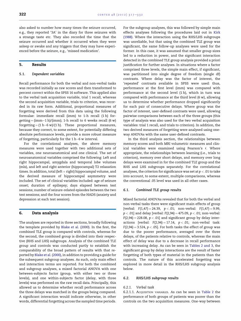
The forgetting measures were analysed using one-way
ANOVAs with the afore-mentioned user-defined contrast
analyses. These analyses confirmed that the LHS patients
showed more accelerated forgetting over the imme1-h
interval (see Fig. 2). The imme1-h measure differed signifi-
cantly between the three participant groups [F(2,48)¼ 3.26,
p< .05]. The contrast analyses revealed that the LHS group
showed significantly greater forgetting from immediate to 1-h
testing than both the RHS and control groups [t(46)¼�2.29,
p¼ .03 and t(46)¼�2.14, p¼ .04 respectively], and that the
difference between the RHS and control groupswas unreliable
[t(46)< 1; as with the raw recall data, the RHS group actually
performed best on this measure, in that they showed the least
forgetting]. There was also a significant group difference for
the 1 he6 w forgetting measure, F(2,48)¼ 15.82, p< .01, but in
this case both patient groups showed equivalent and signifi-
cantly faster forgetting than the controls [RHSeLHS, t(46)< 1;
RHSecontrol, t(46)< 4.65, p< .01; LHSecontrol, t(46)¼ 4.69,
p< .01].
6.2.2. Non-verbal task6.2.2.1. COPY PERFORMANCE. The three groups were matched on
copy performance for the Rey Figure [F(2,48)¼ 1.84, p¼ .17]
and performed effectively at ceiling on this task (see Table 3).
Furthermore, none of the single df contrasts revealed signifi-
cant pairwise differences between the three groups ( p> .05
for each contrast). We therefore found no evidence of visuo-
perceptive/constructive problems in our patient groups.
6.2.2.2. RETENTION VARIABLES. As with the verbal memory data,
a mixed factorial 3� 3 ANOVA with one between subject
factor of group (left hippocampal abnormality, right hippo-
campal abnormality, controls) and delay (30 sec, 1 h, 6 weeks),
up as awhole, the subgroup of patientswith left lateralizedd hippocampal sclerosis (RHS) and healthy controls. Shownecall at 30 sec, 1 h and 6weeks, together with the imme1 hation of these measures).
1 h 6 weeks Imme1 h 1 he6 w
9.72 (13.80) 19.07 (10.81) .23 (.19) .61 (.21)
1.50 (14.57) 19.88 (10.46) .21 (.20) .57 (.21)
7.50 (13.05) 18.07 (11.61) .25 (.19) .67 (.21)
1.20 (18.21) 33.45 (12.55) .11 (.17) .42 (.24)
0
10
20
30
40
50
60
70
80
90
100
Imm 1hr 6 weeks
ll
ac
eR
tc
er
ro
Ct
ne
cr
eP
na
eM
Test Delay Interval
Left
Right
Control
Fig. 1 e Story recall performance (mean percent correct and
S.E’s) for patients with left and right hippocampal sclerosis
and controls at immediate test, and 1 h and 6 week delays.
0
10
20
30
40
50
60
70
80
90
100
Copy 30 Sec 1 hour 6 weeks
ll
ac
eR
tc
er
ro
Ct
ne
cr
eP
na
eM
Test Delay Interval
Left
Right
Control
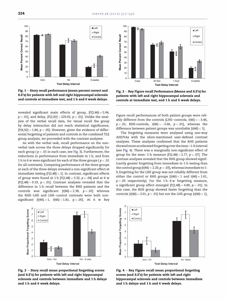
Fig. 3 e Rey Figure recall Performance (Means and S.E’s) for
patients with left and right hippocampal sclerosis and
controls at immediate test, and 1 h and 6 week delays.
c o r t e x 4 8 ( 2 0 1 2 ) 3 1 7e3 3 2324
revealed significant main effects of group, [F(2,46)¼ 5.04,
p< .01], and delay, [F(2,92)¼ 229.02, p< .01]. Unlike the anal-
ysis of the verbal recall data, for visual recall the group
by delay interaction did not reach statistical significance,
[F(4,92)¼ 1.84, p> .05]. However, given the evidence of differ-
ential forgetting of patients and controls in the combined TLE
group analysis, we proceeded with the contrast analyses.
As with the verbal task, recall performance on the non-
verbal task across the three delays dropped significantly for
each group ( p< .01 in each case, see Fig. 3). Furthermore, the
reductions in performance from immediate to 1 h, and from
1 h to 6 wwere significant for each of the three groups ( p< .01
for all contrasts). Comparing performance of the three groups
at each of the three delays revealed a non-significant effect at
immediate testing [F(2,48)< 1]. In contrast, significant effects
of group were found at 1 h [F(2,48)¼ 3.32, p¼ .04] and at 6 w
[F(2,48)¼ 9.19, p< .01]. Contrast analyses revealed that the
difference in 1-h recall between the RHS patients and the
controls was significant [t(46)¼ 2.38, p¼ .02] whereas
the RHSeLHS and LHSecontrol contrasts were both non-
significant [t(46)< 1, t(46)¼ 1.81, p> .05]. At 6 w Rey
0
.1
.2
.3
.4
.5
.6
.7
.8
Imm -1hr 1hr - 6 wks
se
ro
cS
gn
it
te
gr
oF
la
no
it
ro
po
rP
na
eM
Test Delay Interval
Left
Right
Control
Fig. 2 e Story recall mean proportional forgetting scores
(and S.E’s) for patients with left and right hippocampal
sclerosis and controls between immediate and 1 h delays
and 1 h and 6 week delays.
Figure recall performances of both patient groups were reli-
ably different from the controls [LHSecontrols, t(46)¼�3.46,
p< .01; RHS-controls, t(46)¼�3.66, p< .01], whereas the
difference between patient groups was unreliable [t(46)< 1].
The forgetting measures were analysed using one-way
ANOVAs with the afore-mentioned user-defined contrast
analyses. These analyses confirmed that the RHS patients
showedmoreaccelerated forgettingover the imme1-h interval
(see Fig. 4). There was a marginally non-significant effect of
group for the imme1-h measure [F(2,48)¼ 2.77, p< .07]. The
contrast analyses revealed that the RHS group showed signif-
icantly greater forgetting from immediate to 1-h testing than
thecontrol group [t(46)¼ 2.20,p¼ .03],whereas immediate to1-
h forgetting for the LHS group was not reliably different from
either the control or RHS groups [t(46)< 1 and t(46)¼ 1.61,
p> .05 respectively]. For the 1 he6 w forgetting measure,
a significant group effect emerged [F(2,48)¼ 4.89, p¼ .01]. In
this case, the RHS group showed faster forgetting than the
controls [t(46)¼ 3.01, p< .01] but not the LHS group [t(46)< 1],
0
.1
.2
.3
.4
.5
.6
.7
.8
Imm -1hr 1hr -6wks
se
ro
cS
gn
it
te
gr
oF
la
no
it
ro
po
rP
na
eM
Test Delay Interval
Left
Right
Control
Fig. 4 e Rey Figure recall mean proportional forgetting
scores (and S.E’s) for patients with left and right
hippocampal sclerosis and controls between immediate
and 1 h delays and 1 h and 6 week delays.

wn
.T2
.517)
.426)
.293)
.960)
.375)
.830
are
.T2
.991)
.567)
.110)
.519)
.083)
c o r t e x 4 8 ( 2 0 1 2 ) 3 1 7e3 3 2 325
and the LHS group showed faster forgetting than the controls
but at a marginally non-significant level [t(46)¼ 1.91, p¼ .06].
Table
4ePearsonrco
rrelationsbetw
eenstory
reca
llandMRI-derivedneuro
anatom
icalm
easu
resin
theco
mbin
edTLEgro
up(n
[27).Exact
pro
babilityvaluesare
sho
inparenth
ese
swithsignifica
ntco
rrelationshighlightedin
bold.
L.hipp.vol.
R.hipp.vol.
Totalhipp.vol.
Hipp.asy
mm
etry
L.amyg.vol.
R.amyg.vol.
L.temp.vol.
R.temp.vol.
L.ant.T2
R.ant
Trials
tocriterion
L.501(.008)
�.050(.805)
�.358(.067)
.369(.058)
�.164(.425)
�.084(.683)
�.107(.594)
�.025(.900)
.385(.052)
�.133(
Criterionreca
ll�.
142(.479)
�.181(.367)
�.219(.273)
�.012(.951)
�.138(.501)
�.189(.354)
�.011(.956)
�.278(.161)
�.046(.822)
.163(
1-h
reca
ll.536(.004)
�.368(.059)
.088(.662)
L.688(<
.001)
.073(.724)
�.208(.307)
.064(.753)
�.319(.104)
L.598(<
.001)
.214(
6-w
reca
ll.302(.126)
�.155(.441)
.086(.668)
�.308(.118)
�.069(.736)
�.082(.691)
�.285(.149)
�.255(.200)
�.147(.474)
.010(
Imme
1hforgetting
L.588(.001)
.329(.093)
�.149(.459)
.702(<
.001)
�.112(.585)
.167(.414)
�.065(.748)
.249(.210)
.603(<
.001)
�.181(
1he6w
forgetting
�.133(.510)
.030(.880)
�.064(.750)
.083(.682)
.043(.833)
�.083(.685)
.329(.094)
.157(.436)
�.081(.694)
.044(
Table
5e
Pearsonrco
rrelationsbetw
eenReyFigure
reca
llandMRI-derivedneuro
anatom
icalm
easu
resin
theco
mbin
edTLEgro
up(n
[27).Exact
pro
babilityvalues
shownin
parenth
ese
swithsignifica
ntco
rrelationshighlightedin
bold.
L.hipp.vol.
R.hipp.vol.
Totalhipp.vol.
Hipp.asy
mmetry
L.amyg.vol.
R.amyg.vol.
L.tem
p.vol.
R.temp.vol.
L.ant.T2
R.ant
30se
c(imm)reca
ll.152(.450)
�.129(.520)
.007(.971)
�.147(.466)
�.261(.197)
�.198(.332)
�.132(.511)
�.121(.548)
�.214(.293)
.002(
1-h
reca
ll.177(.376)
.338(.085)
.352(.072)
.122(.544)
.123(.548)
.307(.128)
�.101(.617)
�.067(.740)
.096(.640)
.118(
6-w
reca
ll.054(.789)
.108(.590)
.111(.581)
.067(.740)
�.261(.197)
�.112(.587)
�.209(.295)
�.005(.980)
.151(.461)
�.321(
Imm
e1hforgetting
�.102(.614)
L.504(.007)
�.419(.029)
�.259(.192)
�.353(.077)
L.528(.006)
.099(.625)
�.033(.870)
�.262(.196)
�.132(
1he6w
forgetting
.210(.292)
�.063(.754)
.091(.650)
�.252(.206)
.359(.072)
.124(.547)
.211(.291)
�.141(.484)
�.284(.160)
.346(
7. Correlational analyses
Correlations were conducted between: (i) the set of neuro-
anatomical measures and verbal and non-verbal memory
measures; (ii) the set of clinical variables and verbal and non-
verbal memory measures; and (iii) the memory measures
themselves. For each set of correlations, we report those for
the verbal followed by those for the non-verbal memory
measures for the combined TLE group first. Where appro-
priate we also include analyses for the RHS/LHS subgroups.
This was not attempted with the neuroanatomical measures
because these were used as criterion variables in the
subgroup allocation. Any correlations, therefore, are likely to
be attenuated because of the artificially reduced ranges of
values, and therefore obfuscate the trends existing in the
complete patient sample. As noted above, given the number
of correlations being performed at each of these stages, pwas
set to .01, rather than the conventional .05.
7.1. Memory-neuroanatomical correlations
7.1.1. Verbal memoryThe results of the correlations between MRI brain measures
and verbal memory performance for the combined group are
showninTable 4. Significant correlationswere foundbetween
left hippocampal volumeand trials to criterion, 1-h recall, and
the imme1 h forgetting measure, whereas the correlations
with recall at 6 w and the 1 he6 w forgetting measure were
unreliable. A similar pattern emerged for the sets of correla-
tions involvinghippocampal asymmetry andhippocampal T2
relaxation time. In both cases, the correlationswith 1-h recall,
and the imme1 h forgetting measure were significant,
whereas the remaining ones were unreliable. Thus, the prin-
cipal significant correlations to emerge involved the 1 h
memorymeasures and the left hippocampal measures. None
of the correlations involving the remaining recall memory
measures or involving right-sided and/or non-hippocampal
neuroanatomical measures were found to be reliable.
7.1.2. Non-verbal memoryThe results of the correlations between the neuroanatomical
measures and non-verbal memory performance are shown
in Table 5. As can be seen, only two of these were significant.
In both cases, the relevant memory variable was the
imme1 h forgetting measure, and the two structures
involved were both right-sided: right hippocampal volume
and right amygdala volume. Thus, none of the correlations
between the non-verbal memory variables and left-sided
neuroanatomical measures were significant.
7.2. Memory-clinical correlations
7.2.1. Verbal memoryThe results of the correlations between clinical measures
and verbal memory performance in the combined TLE group
are shown in Table 6. Marginally non-significant correlations

Table
6ePearsonrco
rrelationsbetw
eenstory
reca
llandth
eclin
icalv
ariablesin
theco
mbin
edTLEgro
up(n
[27).Exact
pro
babilityvaluesare
shownin
parenth
ese
swith
significa
ntco
rrelationshighlightedin
bold.
No.se
izure-related
episodes
Agese
izure
onse
tDuration
Daysbetween
test
sess
ions
Sess
ion1HADS
anxiety
score
Ses
sion1HADS
depress
ionsc
ore
Sess
ion2HADS
anxiety
score
Ses
sion2HADS
depress
ionsc
ore
Trials
tocriterion
.088(.669)
�.107(.595)
.260(.190)
.314(.111)
�.227(.265)
�.122(.553)
�.019(.927)
�.107(.603)
Criterionreca
ll�.
020(.924)
.003(.990)
.036(.858)
�.248(.213)
.177(.386)
�.155(.449)
.291(.149)
�.110(.593)
1-h
reca
ll�.
045(.827)
.466(.014)
�.356(.068)
�.217(.277)
�.038(.856)
.209(.306)
�.220(.279)
�.040(.846)
6-w
reca
llL.547(.004)
.179(.371)
.147(.463)
�.232(.245)
.330(.099)
.306(.129)
.292(.147)
.291(.149)
Imm
e1hforgetting
.046(.823)
�.477(.012)
.364(.062)
.147(.465)
.077(.708)
�.253(.213)
.292(.148)
.007(.972)
1he6w
forgetting
.591(<
.001)
�.076(.706)
�.246(.216)
.181(.366)
�.377(.057)
�.300(.137)
�.421(.032)
�.335(.095)
c o r t e x 4 8 ( 2 0 1 2 ) 3 1 7e3 3 2326
were found between age at seizure onset and both 1-h recall
( p¼ .014) and the imme1 h forgetting measure ( p¼ .012). No
significant correlations were found between age at seizure
onset and memory performance at the other delays. Given
that 1-h recall and imme1 h forgetting correlated significantly
with left hippocampal volume and left anterior hippocampal
T2 relaxation time, we also investigated whether age of
seizure onset correlated with these MRI measures. Age of
seizure onset was found to correlate significantly with left
hippocampal volume (r¼ .535, p¼ .004), but not anterior
hippocampal T2 relaxation time (r¼�.329, p¼ .1). Significant
correlations were also found between the number of seizure-
related episodes and both 6-w recall, and 1 he6 w forgetting.
The correlation between number of seizure-related
episodes and 1 he6 w forgetting was also significant when
considering LHS patients (r¼ .739, p¼ .002), but failed to
reach the adopted significance threshold (r¼ .602, p¼ .05) for
RHS patients. As shown in Table 1, the mean number of
seizure-related episodes in the RHS group was almost twice
that seen in the LHS group, and the weaker effect is therefore
most likely due to the smaller number of patients in that
group.
7.2.2. Non-verbal memoryThe results of the correlations between clinical measures and
verbal memory performance in the combined TLE group are
shown in Table 7. No significant correlations were found
between age of seizure onset and visual memory measures.
However, as with the analysis of the verbal memory data,
significant correlations were found between the number of
seizure-related incidents and both 6-week recall and 1 he6 w
forgetting.
For the RHS and LHS subgroups, the correlations between
number of seizure-related episodes and 6-w recall and
1 he6 w forgetting were unreliable (RHS r¼ .568, p¼ .068; LHS
r¼ .444, p¼ .097). As noted above, the contrasting patterns of
correlations found in the combined TLE and subgroup anal-
yses are most likely due to the resulting small group sizes for
the latter.
7.3. Correlations between memory measures
These correlations were conducted separately for the RHS and
LHS subgroups given that different patterns of memory
performance were found between groups in the ANOVA
analyses reported above.
7.3.1. Verbal memoryFor the LHS subgroup, significant correlations were found
between trials to criterion and both 1-h recall and imme1 h
forgetting, but not with the 6-wmemory measures. Recall at 6
weeks and 1 he6 w forgetting correlated significantly with
each other but not with any of the other memory measures.
For patients with right lateralized hippocampal sclerosis,
there were no significant correlations between trials to crite-
rion or immediate recall and any of the other memory
measures. Recall at 6 w and 1 he6 w forgetting correlated
significantly with each other but not with any of the other
memory measures.

Table
7e
Pearsonrco
rrelationsbetw
eenReyFigure
reca
llandth
eclin
icalm
easu
resin
theco
mbin
edTLEgro
up(n
[27).Exact
pro
babilityvaluesare
shownin
parenth
ese
swithsignifica
ntco
rrelationshighlightedin
bold.
No.se
izure-related
episodes
Agese
izure
onse
tDuration
Daysbetween
test
sessions
Sess
ion1HADS
anxiety
score
Sess
ion1HADS
depress
ionsc
ore
Sess
ion2HADS
anxiety
score
Ses
sion2HADS
depress
ionsc
ore
30se
c(imm)reca
ll�.
243(.231)
.169(.398)
�.127(.529)
�.142(.480)
�.021(.921)
.194(.342)
�.110(.593)
�.241(.236)
1-h
reca
ll�.
259(.201)
�.184(.359)
.223(.264)
.116(.563)
.288(.154)
.211(.301)
.206(.313)
�.076(.713)
6-w
reca
llL.636(<
.001)
�.093(.645)
.172(.392)
�.033(.872)
.221(.277)
.393(.047)
.020(.925)
.085(.681)
Imm
e1hforgetting
.100(.626)
.326(.097)
�.300(.128)
�.308(.118)
�.250(.218)
�.047(.821)
�.222(.276)
.012(.953)
1he6w
forgetting
.541(.004)
.066(.745)
�.049(.810)
.089(.659)
.068(.740)
�.264(.192)
.075(.715)
�.009(.965)
c o r t e x 4 8 ( 2 0 1 2 ) 3 1 7e3 3 2 327
7.3.2. Non-verbal memoryFor the LHS subgroup, correlations between immediate recall
and each of the other memory measures were unreliable.
Recall at 6 w and 1 he6 w forgetting correlated significantly
with each other, but not with any of the other memory
measures, although there was a trend for a correlation
between imme1 h and 1 he6 w forgetting ( p¼ .019).
Likewise, for the RHS subgroup, all correlations involving
immediate recall were non-significant. Recall at 6 w and
1 he6 w forgetting correlated significantly with each other,
and there was a trend between 1 h and 6-w recall ( p¼ .012),
but none of the other correlations betweenmemorymeasures
reached or approached statistical significance.
8. Discussion
In our study we found that patients with TLE showed deficits
in both the acquisition of new memories and recall of
successfully acquired memories over delays of 1 h and 6 w.
Our investigation of the specific patterns of memory loss
shown by patients with left or right lateralized hippocampal
pathology on tests of verbal and visuospatial memory sug-
gested that the deficit in memory acquisition and accelerated
memory loss over the 1-h delay may be mediated, at least in
part, by a different mechanism to that underlying the accel-
erated loss of memory over the longer 6-w delay. In support of
this, we found that verbal recall was lost at an accelerated rate
in the patients with right lateralized hippocampal sclerosis
despite their normal retention of verbal material over the 1-h
delay, and that the long-term verbal retention of both groups
of patients did not correlate significantly with measures of
acquisition or short-term retention. Furthermore, we showed
that retention over the relatively short 1-h delay, but not the 6-
w delay, was associated with the presence of hippocampal
pathology in either the left or right HC. Specifically, pathology
in the left HC was associated with verbal memory deficits in
acquisition and 1 h, but not 6-w retention, and pathology in
the right HCwas associated with visuospatial memory deficits
at only the 1-h delay. We therefore suggest that the presence
of hippocampal pathology in patients with TLE can result in
deficits in acquiring newmemories and retaining successfully
acquired memories over relatively short delays, but does not
underlie accelerated forgetting over longer delays of several
weeks.
Rather,we found that accelerated forgettingover the longer
6-w delay was associated with the frequency of seizures
during that period. This finding is consistent with other work
that has suggested that long-term forgetting in TLE results
from the disruption of consolidation processes by the occur-
rence of epileptic seizures (Mameniskiene et al., 2006). Each
aspect of our findings is discussed in greater detail below.
8.1. Acquisition
Our patients with TLE required more trials to learn the verbal
recall task to criterion level than healthy controls suggesting
a deficit in the acquisition of new verbal memories. This is
consistent with some previous studies (e.g., Giovagnoli et al.,
1995; Bell et al., 2005; Bell, 2006; Davidson et al., 2007), but

c o r t e x 4 8 ( 2 0 1 2 ) 3 1 7e3 3 2328
inconsistent with the findings of Blake et al. (2000). The
difference between our findings and those of Blake et al. most
likely relates to the presence of hippocampal pathology in our
patients. Whereas few of Blake et al.’s patients had structural
changes within the medial temporal lobe, our patients were
specifically selected according to the presence of lateralized
structural changes (atrophy or abnormal T2 relaxation times)
within the HC. Indeed, we found a significant relationship
between left hippocampal volume and the number of trials
required to reach criterion (patients with smaller left hippo-
campal volume required more learning trials) and a trend for
a relationship between left anterior hippocampal T2 relaxa-
tion time and number of trials to reach criterion (r¼ .386,
p¼ .052). Our findings therefore suggest that, although deficits
in the acquisition of new memories may not occur in TLE
patients when medial temporal lobe pathology is absent (e.g.,
Blake et al., 2000), such deficits are seen in patients whose
epilepsy is accompanied by structural hippocampal abnor-
mality, and that these deficits become more severe as the
severity of hippocampal pathology increases.
8.2. Accelerated forgetting
A different pattern of retention was found for the verbal and
non-verbal tasks. Considering the combined TLE group, there
was evidence of accelerated forgetting of both verbal and
non-verbal material (i.e., significant group by delay interac-
tions). When considering the more detailed subgroup anal-
yses, the LHS subgroup forgot verbal material at an
accelerated rate (relative to controls) over both the 1-h and
6 w delays, whereas the patients with right lateralized
hippocampal sclerosis showed normal retention at the 1-h
delay but accelerated forgetting over the following 6 w.
Although not quite as clear-cut, a similar pattern emerged on
the non-verbal task, with the RHS, but not the LHS group,
showing faster forgetting over 1 h and both groups showing
faster forgetting over the 6-w delay (although, for the LHS
group p¼ .06 for this effect). The pattern of performance
shown by the RHS group on the verbal task and, to a lesser
extent, the LHS subgroup on the non-verbal task, is therefore
very similar to the pattern of long-term amnesia reported in
TLE by Blake et al. (2000) and contrasts with the findings of
Giovagnoli et al. (1995), Bell et al. (2005) and Bell (2006). In
what follows, to avoid repeated qualification, we focus our
attention to the more robust findings relating to the verbal
task.
Could the pattern of memory performance shown by our
patients be explained by reliance at immediate test on
working rather than long-term memory? If it were the case
that working memory, but not long-term memory, was
matched in the patients and controls at the short delay, then
we might expect to see rapid forgetting in the patients
between immediate and 1 h tests, thusmaking it unlikely that
accelerated forgetting would be observed over the 6-w delay.
This is not the pattern observed for the verbal task in our
study. Accelerated forgetting over the long 6-w interval was
observed in both subgroups, and for the RHS patients, this was
the case even though recall at the 1-h delay did not differ
significantly from controls.
The presence of accelerated forgetting of verbal material
over both the 1-h retention interval and the long 6-w retention
interval in the LHS patients is somewhat surprising, as the
only other studies that have reported accelerated forgetting in
TLE over extended time periods (Martin et al., 1991; Blake
et al., 2000) have reported normal recall at a short retention
interval (30 min), i.e., the pattern shown by our RHS patients.
One possibility is that our memory test was more sensitive
than those used in these two previous studies and so revealed
memory loss at the short, 1 h, as well as the extended delay.
This explanation seems unlikely though, because although
our LHS patients showed a deficit at the 1-h delay, this was not
the case for the RHS patients who went on to show a deficit at
the 6-w delay. These latter patients showed a pattern identical
to that reported by Martin et al. (1991) and Blake et al. (2000).
We think that it is more likely that, as formemory acquisition,
the difference in findings between studies may relate to
differences in the characteristics of the TLE patients included
in the present study and previous work. We only included
patients who had MRI evidence of structural changes to the
HC that were lateralized to the right or left HC. This enabled us
to examine whether there was a relationship between our
memory measures and measures of hippocampal integrity.
Indeed, we found that forgetting of verbal material over the 1-
h retention interval in our study was related to the extent of
structural changes in the left HC in our patients, and it is quite
possible that the extent of hippocampal abnormality present
in patients included in the previous studies differed from our
study. For example, Blake et al. (2000) reported a very low
incidence of hippocampal sclerosis in their left TLE patients
who showed normal retention after a 30-min delay but
accelerated forgetting over the subsequent 8 weeks. Thus,
accelerated forgetting of verbalmaterial over a relatively short
1-h interval in our study appeared to be associated with the
presence of sclerotic changes in the left HC.
Consistent with this finding, forgetting over the initial 1-h
delay in the non-verbal task was also associated with
measures of hippocampal abnormality, in that case right
hippocampal volume. It should be noted though that whilst
the RHS patients forgot non-verbal information significantly
faster than controls over the 1-h delay, the LHS patients forgot
more than the controls, but were not reliably different from
them or the RHS patients. The deficit in the patients with left
lateralized hippocampal sclerosis may relate to the potential
for visuospatial memories to be encoded both verbally and
non-verbally. Indeed, although the Rey Figure is used exten-
sively as a test of visuospatial memory, some argue that it
does not provide a good indicator of right temporal lobe
integrity and may be solved using verbal strategies (Kneebone
et al., 2007). Such an explanation would predict poorer
performance of patients than controls; in the case of the
left lateralized patients because of disruption to a verbally re-
encoded representation of the visual stimulus and in the case
of right lateralized patients because of disruption of the orig-
inal visual representation of the stimulus. The latter may be
expected to produce poorer performance than the former, as
was the case here.
Although the presence of accelerated forgetting over the 1-
h delay in our study was inconsistent with the findings of
Blake et al. (2000) and Martin et al. (1991), it should be noted

c o r t e x 4 8 ( 2 0 1 2 ) 3 1 7e3 3 2 329
that, as discussed in the Introduction, reports of memory
deficits at immediate testing and after a short delay of 30 min
in patients with TLE are not uncommon (Giovagnoli et al.,
1995; Bell et al., 2005; Bell, 2006; Helmstaedter et al., 1998;
Mameniskiene et al., 2006). However, while our finding of
impaired memory at the short 1-h retention interval in our
patients with left lateralized hippocampal sclerosis is
consistent with the impaired performance of patients from
these studies at a 30-min retention interval, our findings differ
from those of earlier studies in that accelerated forgettingwas
found in our patients over an extended 6-w period. This
subsequent forgetting over the 6-w retention interval was
uncorrelated with measures of retention over the shorter 1-h
delay and therefore suggests that the left lateralized group
was showing long-term forgetting over the 6-w delay over and
above that expected from their 1-h delayed retention.
Furthermore, for the patients with right lateralized hippo-
campal sclerosis, recall of the verbal material followed
a pattern identical to that reported by Blake et al. (2000), i.e.,
unimpaired recall after the 1-h delay but accelerated forget-
ting over the subsequent 6 w.
Lateralization of accelerated forgetting of verbal material
over the short 1-h delay is consistent with previous work that
has associated verbal memory impairments with dysfunction
of the left temporal lobe (e.g., Milner, 1971). We did not,
however, find lateralization of accelerated forgetting of verbal
material over the extended 6-w delay. We found accelerated
forgetting of verbal material not only in the patients with left
lateralized hippocampal sclerosis, but also in the patients
with right lateralized hippocampal sclerosis despite normal
performance after 1 h and the verbal nature of the task. There
were also no significant correlations betweenmeasures of left
(or right) hippocampal integrity and measures of recall/
forgetting over the 6-w delay. These findings are therefore
inconsistent with those of Blake et al. (2000) who reported
greater accelerated forgetting of verbal material in their left
than their right TLE group.
Forgetting over the longer 6-w retention interval in our
study was found to correlate significantly with the number of
seizures in the retention interval (although, given the smaller
group sizes, results for the subgroup analyses were less clear-
cut). This finding is consistent with reports that higher seizure
frequency was associated with poorer verbal memory after
a 4-w delay (Mameniskiene et al., 2006) and that the occur-
rence of a seizure during a 24-h retention interval impaired
memory for the positions of words in left, but not right, TLE
patients (Jokeit et al., 2001). Our data suggest that seizure
activity still affects verbal recall at a longer 6-w delay. Our
findings are also consistent with the report that a reduction in
seizures led to increasedmemory test performance (O’Connor
et al., 1997). Others, however, have not found a relationship
between the frequency of overt seizures during the retention
interval and performance of patients with TLE on memory
tests after delays of 48 h and 8 w (Bergin et al., 1995; Blake
et al., 2000). In addition, accelerated forgetting has been
demonstrated in patients with TEA once seizures have been
abolished by anti-epileptic medication (Butler et al., 2007;
Manes et al., 2005), and accelerated forgetting of verbal and
non-verbal material in this patient group was found not to
correlate with either duration of epilepsy or seizure frequency
(Butler et al., 2009). It cannot, however, be ruled out that
subclinical ictal activity may have contributed to accelerated
forgetting in these cases. Indeed, due to the very low incidence
of hippocampal sclerosis in their patients, Blake et al. (2000, p.
12) proposed that an active ‘epileptic focus’ is required to
explain the phenomenon of long-term accelerated forgetting
in TLE, consistent with our findings. They argued that a “stable
environment” was required in the left temporal lobe for
successful slow consolidation of verbal memories as memo-
ries remain vulnerable to disruption over an extended period
of time. Both Blake et al. (2000) and Mameniskiene et al. (2006)
proposed that exposure to a combination of frequent ictal and
interictal discharges could disrupt memory consolidation.
An explanation of long-term accelerated forgetting in terms
of disruption of consolidation by seizure activity could also
potentially explain why accelerated forgetting of verbal mate-
rialwas foundonly in leftTLEpatients in the studyof Blake et al.
(2000), but in both patient groups in our study. In Blake et al.’s
study, patientswere categorized as having left or right TLE, that
is, according to whether they had left or right lateralized
seizures. In our study, patients were categorized according to
the lateralization of hippocampal pathology, not the laterali-
zation of seizure activity. It is therefore possible that seizure
activity may be less clearly lateralized in our patient groups,
than in the study of Blake et al. (2000). It should also be noted
that patients with right lateralized structural volume reduc-
tions experienced around twice as many seizures during the
6-w delay as those with left lateralized structural volume
reductions. Both these factors may have reduced any differ-
ences between the twopatient groups in our study in the rate of
forgetting of verbal material over the long retention interval.
Although our findings suggest that long-term forgetting in
TLE is related to seizure activity during the long retention
interval, we cannot rule out the possibility that forgetting over
these long intervals is associated with structural damage to
brain regions that were not investigated in this study. We
investigated the relationship between the structural integrity
of the HC and long-term retention and found that they were
not related. We also found that there was no significant rela-
tionship between amygdala volume and whole temporal lobe
volume, and long-term retention. Nevertheless, TLE can affect
extensive cortical and subcortical structures (Mueller et al.,
2006) and it remains to be determined by future work
whether structural damage in regions beyond the temporal
lobe contribute to long-term accelerated memory loss.
Another factor that may affect memory in patients with
TLE, other than the presence of hippocampal sclerosis and
seizure activity, is medication. Jokeit et al. (2005) found
evidence for high anti-epileptic drug (AED) blood levels and
poor retention over 30 min on auditory and visual memory
tests. The exact effects of high AED blood levels and their
relationship with retention over long delays have yet to be
researchedmore closely. However, Cronel-Ohayon et al. (2006)
and Butler et al. (2007) [the latter testing TEA patients] have
found evidence that AEDs do not appear to have an effect on
patients with TLE. One important factor may be the degree to
which AEDs actually control seizures. O’Connor et al. (1997)
report the case of JT, for whom there was evidence that his
long-term (i.e., over days and longer) retention abilities were
inversely related to seizure activity. Consistent with this, it

c o r t e x 4 8 ( 2 0 1 2 ) 3 1 7e3 3 2330
was found that during a controlled trial of paraldehyde his
long-term retention improved as seizure activity decreased. In
this respect, it should be remembered that all patients in the
present study were being considered for epilepsy surgery
partly because of relatively uncontrolled seizures. What our
study does highlight are the possibly dissociable effects of
structural hippocampal abnormality and seizure frequency on
recall over short and extended delays. To examine the more
complex relationships between these variables and medica-
tion would clearly require amuch larger sample size than that
used for the present study.
The results of our study show both similarities and
differences to studies of forgetting over short and long delays
in TEA. Similar findings to ours concerning the relationship
between hippocampal volume measures and non-verbal
memory over short and more extended delays have been
recently reported for patients with TEA (Butler et al., 2009).
Whereas, recall of the Rey Figure after a short (30 min) delay
correlated with right hippocampal volume, there were no
correlations between right hippocampal volume and forget-
ting of seven visually presented designs over a longer 3-w
delay. There was also a close to significant correlation
( p¼ .057) between left hippocampal volume and verbal
forgetting over the short retention interval but not over the
longer delay. Unlike our study, however, they found no
significant correlations between seizure frequency and
forgetting of verbal and non-verbal material over their long
(3 w) retention interval (Butler et al., 2009). It remains to be
determined whether these differences between studies relate
to differences in the nature of the tests used to assessmemory
or to differences in the underlying cause of accelerated
forgetting in the different patient groups.
8.3. Implications for theories of memory consolidation
The results of the present study are consistent with the view
that memory consolidation is not a single process, but rather
that there are ‘fast’ and ‘slow’ components of memory
consolidation (Squire, 1994), and potentially distinct subsys-
tems relating to different time frames within the consolida-
tion process (McClelland et al., 1995; Kapur et al., 1997). The
findings from our study suggest that different mechanisms
may underlie accelerated forgetting in our TLE patients over
a relatively short 1-h delay and a longer 6-w retention interval.
Accelerated forgetting over the 1-h delay was associated
with hippocampal pathology, with greater pathology in the
left HC associated with greater forgetting of verbal material
over this time period and greater pathology in the right HC
associated with greater forgetting of visuospatial material. In
contrast, accelerated forgetting over the 6-w retention interval
was not related to the presence of left or right hippocampal
pathology. Instead, forgetting over this long delay was asso-
ciated with the number of seizures in the intervening interval.
Thus our data suggest that early ‘fast’ consolidation of verbal
and visuospatial memoriesmay depend on the integrity of the
left and right HC, respectively, whereas later ‘slow’ consoli-
dation of successfully acquired memories may depend on the
maintenance of a stable environment in the medial temporal
lobe and so can be disrupted by the occurrence of seizure
activity (Blake et al., 2000).
8.4. Clinical implications
These findings have a number of implications for the
management and treatment of TLE. They suggest, consistent
with the claims of Blake et al. (2000), that only a subset of
patients with TLE, who have sufficient hippocampal scle-
rosis, will demonstrate memory loss within the normal time
period of clinical assessment (retention of intervals of
30 min). Thus, the presence of memory loss may often be
missed in clinical practice. As we show,memory deficits, that
are not present after a short delay, can emerge in patients
with TLE over longer time periods such as an hour to
a number of weeks. For example, we found that verbal recall
was only impaired in patients with right hippocampal scle-
rosis at the long 6-w delay, and not the short 1-h delay.
Therefore, our findings suggest that to obtain a complete
picture of the memory impairments experienced by any
individual patient, and the effectiveness of interventions on
memory, assessment beyond the usual 30-min retention
period used in clinical practice is required. In addition, our
findings highlight the potential importance for long-term
memory of reducing the frequency of epileptic seizures.
They suggest that even patients with hippocampal sclerosis
that affects memory over relatively short delays, could
experience benefits in long-termmemory from a reduction in
seizure frequency.
9. Conclusions
The findings of our study show that TLE can affect the
acquisition and retention of new memories over a relatively
short delay of 1 h. This deficit appears to be related to the
presence of hippocampal atrophy. In addition, our results
suggest that TLE can also result in accelerated forgetting of
memories that were successfully acquired over a longer
time period of several weeks. Evidence of accelerated long-
term forgetting was observed in both verbal and non-verbal
tasks but the detailed pattern was more statistically robust
for the verbal task. For that task, whilst accelerated
forgetting was evident in both patient groups, it was
demonstrated more strongly by the RHS patients whose
verbal recall was normal at the 1-h delay, and for whom
seizure activity was greater in the 6-w delay. This long-term
forgetting does not appear to be associated with the pres-
ence of hippocampal pathology. Rather, as proposed by
others, our data suggest that accelerated long-term forget-
ting in TLE may be associated with disruption of long-term
consolidation processes by the occurrence of epileptic
seizures.
Acknowledgements
We would like to thank our patient participants from the
WCNN and the healthy control participants for taking part in
this study. Some of these data contributed towards
Fiona Clague’s M.Sc. research which was partly supported by
a research scholarship from Sanofi Synthelabo International
Bureau of Epilepsy Scholarship.
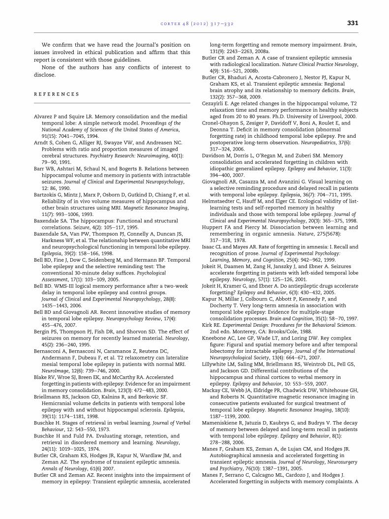
c o r t e x 4 8 ( 2 0 1 2 ) 3 1 7e3 3 2 331
We confirm that we have read the Journal’s position on
issues involved in ethical publication and affirm that this
report is consistent with those guidelines.
None of the authors has any conflicts of interest to
disclose.
r e f e r e n c e s
Alvarez P and Squire LR. Memory consolidation and the medialtemporal lobe: A simple network model. Proceedings of theNational Academy of Sciences of the United States of America,91(15): 7041e7045, 1994.
Arndt S, Cohen G, Alliger RJ, Swayze VW, and Andreasen NC.Problems with ratio and proportion measures of imagedcerebral structures. Psychiatry Research: Neuroimaging, 40(1):79e90, 1991.
Barr WB, Ashtari M, Schaul N, and Bogerts B. Relations betweenhippocampal volume and memory in patients with intractableseizures. Journal of Clinical and Experimental Neuropsychology,12: 86, 1990.
Bartzokis G, Mintz J, Marx P, Osborn D, Gutkind D, Chiang F, et al.Reliability of in vivo volume measures of hippocampus andother brain structures using MRI. Magnetic Resonance Imaging,11(7): 993e1006, 1993.
Baxendale SA. The hippocampus: Functional and structuralcorrelations. Seizure, 4(2): 105e117, 1995.
Baxendale SA, Van PW, Thompson PJ, Connelly A, Duncan JS,Harkness WF, et al. The relationship between quantitative MRIand neuropsychological functioning in temporal lobe epilepsy.Epilepsia, 39(2): 158e166, 1998.
Bell BD, Fine J, Dow C, Seidenberg M, and Hermann BP. Temporallobe epilepsy and the selective reminding test: Theconventional 30-minute delay suffices. PsychologicalAssessment, 17(1): 103e109, 2005.
Bell BD. WMS-III logical memory performance after a two-weekdelay in temporal lobe epilepsy and control groups.Journal of Clinical and Experimental Neuropsychology, 28(8):1435e1443, 2006.
Bell BD and Giovagnoli AR. Recent innovative studies of memoryin temporal lobe epilepsy. Neuropsychology Review, 17(4):455e476, 2007.
Bergin PS, Thompson PJ, Fish DR, and Shorvon SD. The effect ofseizures on memory for recently learned material. Neurology,45(2): 236e240, 1995.
Bernasconi A, Bernasconi N, Caramanos Z, Reutens DC,Andermann F, Dubeau F, et al. T2 relaxometry can lateralizemesial temporal lobe epilepsy in patients with normal MRI.NeuroImage, 12(6): 739e746, 2000.
Blake RV, Wroe SJ, Breen EK, and McCarthy RA. Acceleratedforgetting in patients with epilepsy: Evidence for an impairmentin memory consolidation. Brain, 123(3): 472e483, 2000.
Briellmann RS, Jackson GD, Kalnins R, and Berkovic SF.Hemicranial volume deficits in patients with temporal lobeepilepsy with and without hippocampal sclerosis. Epilepsia,39(11): 1174e1181, 1998.
Buschke H. Stages of retrieval in verbal learning. Journal of VerbalBehaviour, 12: 543e550, 1973.
Buschke H and Fuld PA. Evaluating storage, retention, andretrieval in disordered memory and learning. Neurology,24(11): 1019e1025, 1974.
Butler CR, Graham KS, Hodges JR, Kapur N, Wardlaw JM, andZeman AZ. The syndrome of transient epileptic amnesia.Annals of Neurology, 61(6) 2007.
Butler CR and Zeman AZ. Recent insights into the impairment ofmemory in epilepsy: Transient epileptic amnesia, accelerated
long-term forgetting and remote memory impairment. Brain,131(9): 2243e2263, 2008a.
Butler CR and Zeman A. A case of transient epileptic amnesiawith radiological localization. Nature Clinical Practice Neurology,4(9): 516e521, 2008b.
Butler CR, Bhaduri A, Acosta-Cabronero J, Nestor PJ, Kapur N,Graham KS, et al. Transient epileptic amnesia: Regionalbrain atrophy and its relationship to memory deficits. Brain,132(2): 357e368, 2009.
Cezayirli E. Age related changes in the hippocampal volume, T2relaxation time and memory performance in healthy subjectsaged from 20 to 80 years. Ph.D. University of Liverpool, 2000.
Cronel-Ohayon S, Zesiger P, Davidoff V, Boni A, Roulet E, andDeonna T. Deficit in memory consolidation (abnormalforgetting rate) in childhood temporal lobe epilepsy. Pre andpostoperative long-term observation. Neuropediatrics, 37(6):317e324, 2006.
Davidson M, Dorris L, O’Regan M, and Zuberi SM. Memoryconsolidation and accelerated forgetting in children withidiopathic generalized epilepsy. Epilepsy and Behavior, 11(3):394e400, 2007.
Giovagnoli AR, Casazza M, and Avanzini G. Visual learning ona selective reminding procedure and delayed recall in patientswith temporal lobe epilepsy. Epilepsia, 36(7): 704e711, 1995.
Helmstaedter C, Hauff M, and Elger CE. Ecological validity of list-learning tests and self-reported memory in healthyindividuals and those with temporal lobe epilepsy. Journal ofClinical and Experimental Neuropsychology, 20(3): 365e375, 1998.
Huppert FA and Piercy M. Dissociation between learning andremembering in organic amnesia. Nature, 275(5678):317e318, 1978.
Isaac CL and Mayes AR. Rate of forgetting in amnesia: I. Recall andrecognition of prose. Journal of Experimental Psychology:Learning, Memory, and Cognition, 25(4): 942e962, 1999.
Jokeit H, Daamen M, Zang H, Janszky J, and Ebner A. Seizuresaccelerate forgetting in patients with left-sided temporal lobeepilepsy. Neurology, 57(1): 125e126, 2001.
Jokeit H, Kramer G, and Ebner A. Do antiepileptic drugs accelerateforgetting? Epilepsy and Behavior, 6(3): 430e432, 2005.
Kapur N, Millar J, Colbourn C, Abbott P, Kennedy P, andDocherty T. Very long-term amnesia in association withtemporal lobe epilepsy: Evidence for multiple-stageconsolidation processes. Brain and Cognition, 35(1): 58e70, 1997.
Kirk RE. Experimental Design: Procedures for the Behavioral Sciences.2nd edn. Monterey, CA: Brooks/Cole, 1988.
Kneebone AC, Lee GP, Wade LT, and Loring DW. Rey complexfigure: Figural and spatial memory before and after temporallobectomy for intractable epilepsy. Journal of the InternationalNeuropsychological Society, 13(4): 664e671, 2007.
Lillywhite LM, Saling MM, Briellmann RS, Weintrob DL, Pell GS,and Jackson GD. Differential contributions of thehippocampus and rhinal cortices to verbal memory inepilepsy. Epilepsy and Behavior, 10: 553e559, 2007.
Mackay CE, Webb JA, Eldridge PR, Chadwick DW, Whitehouse GH,and Roberts N. Quantitative magnetic resonance imaging inconsecutive patients evaluated for surgical treatment oftemporal lobe epilepsy. Magnetic Resonance Imaging, 18(10):1187e1199, 2000.
Mameniskiene R, Jatuzis D, Kaubrys G, and Budrys V. The decayof memory between delayed and long-term recall in patientswith temporal lobe epilepsy. Epilepsy and Behavior, 8(1):278e288, 2006.
Manes F, Graham KS, Zeman A, de Lujan CM, and Hodges JR.Autobiographical amnesia and accelerated forgetting intransient epileptic amnesia. Journal of Neurology, Neurosurgeryand Psychiatry, 76(10): 1387e1391, 2005.
Manes F, Serrano C, Calcagno ML, Cardozo J, and Hodges J.Accelerated forgetting in subjects with memory complaints. A

c o r t e x 4 8 ( 2 0 1 2 ) 3 1 7e3 3 2332
new form of mild cognitive impairment? Journal of Neurology,255(7): 1067e1070, 2008.
Martin RC, Loring DW, Meador KJ, and Lee GP. Impaired long-termretention despite normal verbal learning in patients withtemporal lobe dysfunction. Neuropsychology, 5(1): 3e12, 1991.
Mathalon DH, Sullivan EV, Rawles JM, and Pfefferbaum A.Corrections for head-size in brain-imaging measurements.Psychiatry Research: Neuroimaging, 50(2): 121e139, 1993.
Mayes AR, Isaac CL, Holdstock JS, Cariga P, Gummer A, andRoberts N. Long-term amnesia: A review anddetailed illustrative case study. Cortex, 39(4e5):567e603, 2003.
McClelland JL, McNaughton BL, and O’Reilly RC. Why there arecomplementary learning systems in the hippocampus andneocortex: Insights from the successes and failures ofconnectionist models of learning and memory. PsychologicalReview, 102(3): 419e457, 1995.
Milner B. Interhemispheric differences in the localization ofpsychological processes in man. British Medical Bulletin, 27(3):272e277, 1971.
Mueller SG, Laxer KD, Casdollar N, Buckley S, Paul C, andWeiner MW. Voxel-based optimized morphometry (VBM) ofgray and white matter in temporal lobe epilepsy (TLE) with
and without mesial temporal sclerosis. Epilepsia, 47(5):900e907, 2006.
Nelson HE and Willison J. National Adult Reading Test. Slough,England: NFER-Nelson, 1982.
O’Connor M, Sieggreen MA, Ahern G, Schomer D, and MesulamM.Accelerated forgetting in association with temporal lobeepilepsy and paraneoplastic encephalitis. Brain and Cognition,35(1): 71e84, 1997.
Osterrieth PA. Le test de copie d’une figure complex: Contributiona l’etude de la perception et de la memoire. Archives dePsychologie, 30286e30356, 1944.
Snaith RP and Zigmond AS. The Hospital Anxiety and DepressionScale Manual. Windsor, England: NFER-Nelson, 1986.
Squire LR. Memory and forgetting: Long-term and gradualchanges in memory storage. International Review ofNeurobiology, 37243e37269, 1994.
Walhovd KB, Fjell AM, Reinvang I, Lundervold A, Fischl B,Quinn BT, et al. Size does matter in the long run: Hippocampaland cortical volume predict recall across weeks. Neurology,63(7): 1193e1197, 2004.
Wechsler D. Manual for the Wechsler Adult Intelligence Scale e ThirdEdition (WAIS-III). San Antonio, TX: The PsychologicalCorporation, 1997.


![Accelerated long-term forgetting in healthy older adults ......ination III (ACE-III) scores of >88, the upper threshold for detecting mild cognitive impairment [15]. Forty-six returned](https://img.pdfslide.net/doc/110x75/60ab12660f87b73d784ac2eb/accelerated-long-term-forgetting-in-healthy-older-adults-ination-iii-ace-iii.jpg)