Embed Size (px)
Citation preview
Long-Term Alteration in Maternal Blood Pressure andRenal Function After Pregnancy in Normal and
Growth-Restricted RatsLinda A. Gallo, Kate M. Denton, Karen M. Moritz, Marianne Tare, Helena C. Parkington,
Meagan Davies, Melanie Tran, Andrew J. Jefferies, Mary E. Wlodek
Abstract—Intrauterine growth restriction is associated with increased risk of adult cardiorenal diseases. Small birth weightfemales are more likely to experience complications during their own pregnancy, including pregnancy-inducedhypertension, preeclampsia, and gestational diabetes. We determined whether the physiological demand of pregnancypredisposes growth-restricted females to cardiovascular and renal dysfunction later in life. Late gestation bilateraluterine vessel ligation was performed in Wistar-Kyoto rats. At 4 months, restricted and control female offspring weremated with normal males and delivered naturally (ex-pregnant). Regardless of maternal birth weight, at 13 months,ex-pregnant females developed elevated mean arterial pressure (indwelling tail-artery catheter; �6 mm Hg), reducedeffective renal blood flow (14C-PAH clearance; �23%), and increased renal vascular resistance (�27%) compared withage-matched virgins. Glomerular filtration rate (3H-inulin clearance) was not different across groups. This adversecardiorenal phenotype in ex-pregnant females was associated with elevated systemic (�57%) and altered intrarenalcomponents of the renin-angiotensin system. After pregnancy at 13 months, coronary flow (Langendorff preparation)was halved in restricted females compared with controls, and together with reduced NO excretion, this may increasesusceptibility to additional lifestyle challenges. Our results have implications for aging females who have been pregnant,suggesting long-term cardiovascular and renal alterations, with additional consequences for females who were small atbirth. (Hypertension. 2012;60:00-00.) ● Online Data Supplement
Key Words: pregnancy � growth restriction � maternal health � blood pressure � kidney function� Langendorff heart preparation
Uteroplacental insufficiency is the most common cause ofintrauterine growth restriction and affects 7% to 10% of
pregnancies in the Western world.1 Human studies worldwideand animal models have shown that suboptimal conditions inutero alter the development of key organ systems, includingreductions in nephron and cardiomyocyte number.2–5 We andothers have used a rat model to induce uteroplacental insuf-ficiency, whereby uterine vessels are bilaterally ligated dur-ing late gestation resulting in offspring born lighter than shamcontrols.2,3,6,7 Although growth-restricted males go on todevelop elevated blood pressure in adulthood, females appearsomewhat protected up to �18 months, despite both sexeshaving similar nephron deficits.2,3,8,9 This sexual dimorphicresponse to disease development is commonly cited in theprogramming field, with females often presenting with lesssevere cardiovascular disease outcomes.10
Disease risk associated with programmed changes in early lifemay be modulated by exposures after birth and throughout life.
It has been suggested that a number of lifestyle factors, includinghigh-salt/fat diets and aging, can increase or unmask diseaseoutcomes in susceptible offspring.11 Low nephron endowmentin growth-restricted females, for example, may be adequatelycompensated for until a postnatal stressor or “second hit” revealsa clinically relevant phenotype.12
Pregnancy is associated with profound physiological demandsthat could constitute a second hit.13 In early pregnancy, reduc-tions in peripheral vascular tone contribute to a small decrease inmaternal blood pressure, but by late pregnancy, maternal bloodvolume expands by �50% in humans and 30% in rats. Thislow-resistance, hypervolemic state allows for increased utero-placental blood flow and permits greater blood flow to thematernal kidneys, with glomerular filtration rate (GFR) reachingpeak levels at midgestation.14 Associations have been notedbetween a woman’s own birth weight and her future pregnancy-related health, including pregnancy-induced hypertension, pre-
Received March 19, 2012; first decision March 26, 2012; revision accepted April 16, 2012.From the Department of Physiology (L.A.G., M.Tr., A.J.J., M.E.W.), University of Melbourne, Parkville, Victoria, Australia; Department of Physiology
(K.M.D., M.Ta., H.C.P., M.D.), Monash University, Clayton, Victoria, Australia; School of Biomedical Sciences (K.M.M.), University of Queensland,St Lucia, Queensland, Australia.
The online-only Data Supplement is available with this article at http://hyper.ahajournals.org/lookup/suppl/doi:10.1161/HYPERTENSIONAHA.112.195578/-/DC1.
Correspondence to Mary E. Wlodek, Department of Physiology, University of Melbourne, Parkville, Victoria 3010, Australia. [email protected]
© 2012 American Heart Association, Inc.
Hypertension is available at http://hyper.ahajournals.org DOI: 10.1161/HYPERTENSIONAHA.112.195578
1
by guest on May 18, 2018
http://hyper.ahajournals.org/D
ownloaded from
by guest on M
ay 18, 2018http://hyper.ahajournals.org/
Dow
nloaded from
by guest on May 18, 2018
http://hyper.ahajournals.org/D
ownloaded from
by guest on M
ay 18, 2018http://hyper.ahajournals.org/
Dow
nloaded from
by guest on May 18, 2018
http://hyper.ahajournals.org/D
ownloaded from
by guest on M
ay 18, 2018http://hyper.ahajournals.org/
Dow
nloaded from
eclampsia, and gestational diabetes.15–17 This, then, predisposesto adverse outcomes for her offspring, including intrauterinegrowth restriction and prematurity.18
Human studies have also shown that pregnancy-inducedhypertension and/or preeclampsia are associated with increasedrisk for chronic hypertension and subsequent kidney diseaselater in life.19,20 Maternal birth weights are rarely reported infollow-up pregnancy studies, and, thus, the contribution of aprevious developmental insult compared with a spontaneouscomplication in the mother’s own pregnancy is unknown. Wehave demonstrated recently in a setting of low nephron endow-ment that 4-month–old growth-restricted female rats developedimpaired glucose tolerance, altered sodium handling, and glo-merular hypertrophy but remained normotensive during latepregnancy.21 Increased renal and uterine arterial stiffness, evi-dent in growth-restricted virgin rats, was overcome duringpregnancy, highlighting a profound degree of adaptation tomatch control rats and enhance fetal survival.22 These rapid,protective arterial adaptations may be transient, because thelong-term consequences, postpregnancy, are unknown. We,therefore, consider pregnancy as a challenge that may accelerateand, hence, be a potential predictor of future ill health. Thelong-term maternal cardiovascular and renal outcomes associ-ated with the aforementioned compensatory changes were afocus of the current study. We hypothesized that growth-restricted females that had been pregnant would develop hyper-tension and renal and cardiac dysfunction later in life comparedwith previously pregnant, normal birth weight mothers andgrowth-restricted virgin female rats.
MethodsAnimal ProceduresAll of the experiments were approved by the University of Mel-bourne Animal Ethics Committee before commencement. WistarKyoto rats housed in an environmentally controlled room had accessto food and tap water ad libitum. Rats were mated and uteroplacentalinsufficiency (offspring termed “restricted”) or sham (offspringtermed “control”) surgery performed at E18.2 Rats delivered at term(E22) and F1 control and restricted females were allocated to eithervirgin or ex-pregnant groups (1 per litter per group; n�12 pergroup). Those allocated to the ex-pregnant group were mated at 17 to23 weeks of age with a normal male and gave birth naturally. Pupswere weaned at day 35 and allocated to other studies. Ex-pregnantmothers and virgins were studied to 13 months, and a vaginal smearwas performed postmortem to determine the stage of estrus cycle.Please see the online-only Data Supplement for body and organweight measurements, tail-cuff blood pressure, 24-hour urinaryexcretion, tail-artery catheter, renal function and Langendorff heartexperimental methods, and plasma and intrarenal renin-angiotensinsystem (RAS) analyses.
Plasma and Urine Nitrate/Nitrite AnalysesNitrate/nitrite (NOx) was measured (Cayman colorimetric assay kit,Ann Arbor, MI) in plasma samples after a 24-hour fast and in24-hour urine samples corrected for food intake at 12 months. NO2generated from NO3 by the nitrate reductase enzyme was quantifiedby the Greiss reaction.23
Statistical AnalysesValues are expressed as mean�SEM with n representing the numberof female offspring from different mothers per group. Data wereanalyzed using 2-way ANOVA (with repeated measures for systolicblood pressure over time). If significant interactions were observed,
individual group means were compared with the Student unpaired t test.Postischemia effects on isoprenaline responsiveness to maximum dose(2.0 �g/mL�1) were analyzed using the Student paired t test.
Results
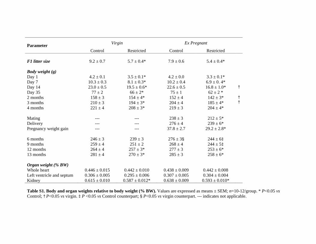
Body and Organ WeightsUteroplacental insufficiency reduced total (male and female)F1 litter size (5–6 restricted pups versus 8–9 control pups) atpostnatal day 1 (P�0.05; please see Table S1 in the online-only Data Supplement). Restricted females were 32% to 38%lighter compared with controls at postnatal day 1 (P�0.05;Table S1). Regardless of pregnancy group allocation, re-stricted females remained lighter at all ages, except at 6 and9 months, where ex-pregnant females only, and not virgins,were smaller than their control counterparts (P�0.05; TableS1). During F1 pregnancy, Restricted females gained 23%less weight than controls (P�0.05), and by 6 months, theywere of similar weight to virgin counterparts, whereas con-trols remained 12% heavier (P�0.05; Table S1). There wereno differences in F2 total litter size (9.3�0.3 versus 8.4�0.5)or day 1 body weight (4.58�0.05 versus 4.45�0.08 g)between control and restricted rats. Kidney weight (correctedfor body weight) was reduced in restricted females comparedwith controls (P�0.05), but there were no differences inrelative heart weights (Table S1).
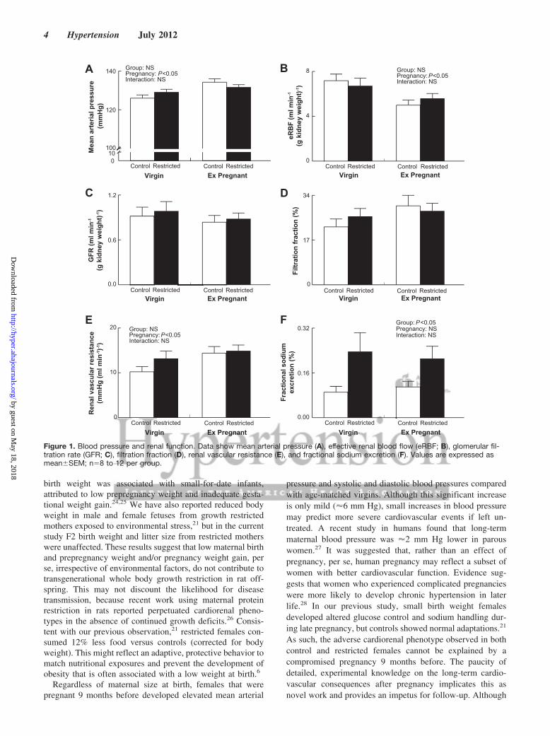
Cardiovascular and Renal FunctionSystolic blood pressure, measured by tail cuff, was notdifferent between control and restricted groups before mating(4 months) or after pregnancy (Table). There were nodifferences in systolic blood pressure between virgin andex-pregnant groups or across time (Table). At 13 months,mean arterial pressure, measured using an indwelling cathe-ter, was elevated in ex-pregnant females compared withvirgins, regardless of maternal birth weight (�6 mm Hg;P�0.05; Figure 1A).
Restricted females consumed 12% less food than controls(P�0.05), with no differences in water intake (Table). Therewere no differences in 24-hour urine output between groups(Table). Urinary sodium and chloride excretion were reducedin ex-pregnant females compared with virgins (P�0.05;Table). In restricted ex-pregnant females, total protein excre-tion was reduced (�42%) compared with control and virgincounterparts, and NOx excretion, corrected for food intake,was reduced (�40%) compared with virgins (P�0.05; Ta-ble). Plasma NOx concentrations, measured after a 24-hourfast were not different between groups (data not shown).
At 13 months, effective renal blood flow (corrected forkidney weight) was reduced by 23% in ex-pregnant femalesversus virgins (P�0.05; Figure 1B), but there was no differ-ence in GFR (Figure 1C). Filtration fraction tended to begreater in ex-pregnant females, but this did not reach statis-tical significance (�22%; P�0.14; Figure 1D). Renal vascu-lar resistance was 27% greater in ex-pregnant females com-pared with virgins (P�0.05; Figure 1E). The sole effect offetal exposure to uteroplacental insufficiency on renal func-tion, independent of previous pregnancy effects, was a�2-fold increase in fractional sodium excretion (P�0.05;
2 Hypertension July 2012
by guest on May 18, 2018
http://hyper.ahajournals.org/D
ownloaded from
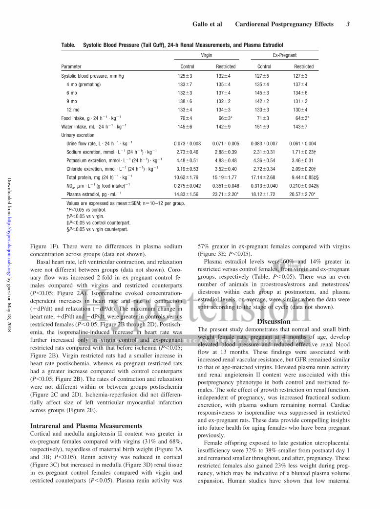
Figure 1F). There were no differences in plasma sodiumconcentration across groups (data not shown).
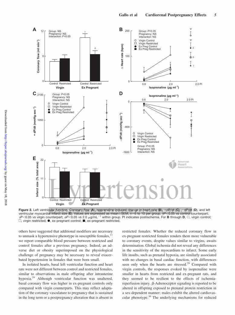
Basal heart rate, left ventricular contraction, and relaxationwere not different between groups (data not shown). Coro-nary flow was increased 2-fold in ex-pregnant control fe-males compared with virgins and restricted counterparts(P�0.05; Figure 2A). Isoprenaline evoked concentration-dependent increases in heart rate and rate of contraction(�dP/dt) and relaxation (�dP/dt). The maximum change inheart rate, �dP/dt and �dP/dt, were greater in controls versusrestricted females (P�0.05; Figure 2B through 2D). Postisch-emia, the isoprenaline-induced increase in heart rate wasfurther increased only in virgin control and ex-pregnantrestricted rats compared with that before ischemia (P�0.05;Figure 2B). Virgin restricted rats had a smaller increase inheart rate postischemia, whereas ex-pregnant restricted ratshad a greater increase compared with control counterparts(P�0.05; Figure 2B). The rates of contraction and relaxationwere not different within or between groups postischemia(Figure 2C and 2D). Ischemia-reperfusion did not differen-tially affect size of left ventricular myocardial infarctionacross groups (Figure 2E).
Intrarenal and Plasma MeasurementsCortical and medulla angiotensin II content was greater inex-pregnant females compared with virgins (31% and 68%,respectively), regardless of maternal birth weight (Figure 3Aand 3B; P�0.05). Renin activity was reduced in cortical(Figure 3C) but increased in medulla (Figure 3D) renal tissuein ex-pregnant control females compared with virgin andrestricted counterparts (P�0.05). Plasma renin activity was
57% greater in ex-pregnant females compared with virgins(Figure 3E; P�0.05).
Plasma estradiol levels were 60% and 14% greater inrestricted versus control females, from virgin and ex-pregnantgroups, respectively (Table; P�0.05). There was an evennumber of animals in proestrous/estrous and metestrous/diestrous within each group at postmortem, and plasmaestradiol levels, on average, were similar when the data weresplit according to the stage of cycle (data not shown).
DiscussionThe present study demonstrates that normal and small birthweight female rats, pregnant at 4 months of age, developelevated blood pressure and reduced effective renal bloodflow at 13 months. These findings were associated withincreased renal vascular resistance, but GFR remained similarto that of age-matched virgins. Elevated plasma renin activityand renal angiotensin II content were associated with thispostpregnancy phenotype in both control and restricted fe-males. The sole effect of growth restriction on renal function,independent of pregnancy, was increased fractional sodiumexcretion, with plasma sodium remaining normal. Cardiacresponsiveness to isoprenaline was suppressed in restrictedand ex-pregnant rats. These data provide compelling insightsinto future health for aging females who have been pregnantpreviously.
Female offspring exposed to late gestation uteroplacentalinsufficiency were 32% to 38% smaller from postnatal day 1and remained smaller throughout, and after, pregnancy. Theserestricted females also gained 23% less weight during preg-nancy, which may be indicative of a blunted plasma volumeexpansion. Human studies have shown that low maternal
Table. Systolic Blood Pressure (Tail Cuff), 24-h Renal Measurements, and Plasma Estradiol
Parameter
Virgin Ex-Pregnant
Control Restricted Control Restricted
Systolic blood pressure, mm Hg 125�3 132�4 127�5 127�3
4 mo (premating) 133�7 135�4 135�4 137�4
6 mo 132�3 137�4 145�3 134�6
9 mo 138�6 132�2 142�2 131�3
12 mo 133�4 134�3 130�3 130�4
Food intake, g · 24 h�1 · kg�1 76�4 66�3* 71�3 64�3*
Water intake, mL · 24 h�1 · kg�1 145�6 142�9 151�9 143�7
Urinary excretion
Urine flow rate, L · 24 h�1 · kg�1 0.073�0.008 0.071�0.005 0.083�0.007 0.061�0.004
Sodium excretion, mmol · L�1 (24 h�1) · kg�1 2.73�0.46 2.88�0.39 2.31�0.31 1.71�0.23†
Potassium excretion, mmol · L�1 (24 h�1) · kg�1 4.48�0.51 4.83�0.48 4.36�0.54 3.46�0.31
Chloride excretion, mmol · L�1 (24 h�1) · kg�1 3.19�0.53 3.52�0.40 2.72�0.34 2.09�0.20†
Total protein, mg (24 h)�1 · kg�1 10.62�1.79 15.19�1.77 17.14�2.68 9.44�0.85‡§
NOx, �m · L�1 (g food intake)�1 0.275�0.042 0.351�0.048 0.313�0.040 0.210�0.042§
Plasma estradiol, pg · mL�1 14.83�1.56 23.71�2.20* 18.12�1.72 20.57�2.70*
Values are expressed as mean�SEM; n�10–12 per group.*P�0.05 vs control.†P�0.05 vs virgin.‡P�0.05 vs control counterpart.§P�0.05 vs virgin counterpart.
Gallo et al Cardiorenal Postpregnancy Effects 3
by guest on May 18, 2018
http://hyper.ahajournals.org/D
ownloaded from
birth weight was associated with small-for-date infants,attributed to low prepregnancy weight and inadequate gesta-tional weight gain.24,25 We have also reported reduced bodyweight in male and female fetuses from growth restrictedmothers exposed to environmental stress,21 but in the currentstudy F2 birth weight and litter size from restricted motherswere unaffected. These results suggest that low maternal birthand prepregnancy weight and/or pregnancy weight gain, perse, irrespective of environmental factors, do not contribute totransgenerational whole body growth restriction in rat off-spring. This may not discount the likelihood for diseasetransmission, because recent work using maternal proteinrestriction in rats reported perpetuated cardiorenal pheno-types in the absence of continued growth deficits.26 Consis-tent with our previous observation,21 restricted females con-sumed 12% less food versus controls (corrected for bodyweight). This might reflect an adaptive, protective behavior tomatch nutritional exposures and prevent the development ofobesity that is often associated with a low weight at birth.6
Regardless of maternal size at birth, females that werepregnant 9 months before developed elevated mean arterial
pressure and systolic and diastolic blood pressures comparedwith age-matched virgins. Although this significant increaseis only mild (�6 mm Hg), small increases in blood pressuremay predict more severe cardiovascular events if left un-treated. A recent study in humans found that long-termmaternal blood pressure was �2 mm Hg lower in parouswomen.27 It was suggested that, rather than an effect ofpregnancy, per se, human pregnancy may reflect a subset ofwomen with better cardiovascular function. Evidence sug-gests that women who experienced complicated pregnancieswere more likely to develop chronic hypertension in laterlife.28 In our previous study, small birth weight femalesdeveloped altered glucose control and sodium handling dur-ing late pregnancy, but controls showed normal adaptations.21
As such, the adverse cardiorenal phenotype observed in bothcontrol and restricted females cannot be explained by acompromised pregnancy 9 months before. The paucity ofdetailed, experimental knowledge on the long-term cardio-vascular consequences after pregnancy implicates this asnovel work and provides an impetus for follow-up. Although
010
100
120
140
Ex PregnantVirgin
Mea
n ar
teria
l pre
ssur
e (m
mH
g)
Control Restricted Control Restricted
Group: NSPregnancy: P<0.05Interaction: NS
A
0
4
8
Ex PregnantVirginControl Restricted Control Restricted
Group: NSPregnancy:P<0.05Interaction: NS
B
0.0
0.6
1.2
Ex PregnantVirgin
GFR
(ml m
in-1
(g k
idne
y w
eigh
t)-1)
Control Restricted Control Restricted
C
0
17
34
Ex PregnantVirginFi
ltrat
ion
frac
tion
(%)
Control Restricted Control Restricted
D
0
10
20
Ex PregnantVirginControl Restricted Control Restricted
Group: NSPregnancy:P<0.05Interaction: NS
E
0.00
0.16
0.32
Ex PregnantVirgin
Frac
tiona
l sod
ium
excr
etio
n (%
)
Control Restricted Control Restricted
Group:P<0.05Pregnancy: NSInteraction: NS
F
eRB
F (m
l min
-1
(g k
idne
y w
eigh
t)-1)
Ren
al v
ascu
lar r
esis
tanc
e(m
mH
g (m
l min
-1)-1
)
Figure 1. Blood pressure and renal function. Data show mean arterial pressure (A), effective renal blood flow (eRBF; B), glomerular fil-tration rate (GFR; C), filtration fraction (D), renal vascular resistance (E), and fractional sodium excretion (F). Values are expressed asmean�SEM; n�8 to 12 per group.
4 Hypertension July 2012
by guest on May 18, 2018
http://hyper.ahajournals.org/D
ownloaded from
others have suggested that additional modifiers are necessaryto unmask a hypertensive phenotype in susceptible females,11
we report comparable blood pressure between restricted andcontrol females after a previous pregnancy. Indeed, an ad-verse diet or obesity superimposed on the physiologicalchallenge of pregnancy may be necessary to reveal exacer-bated hypertension in females that were born small.
In isolated hearts, basal left ventricular function and heartrate were not different between control and restricted females,similar to observations in male offspring after intrauterinehypoxia.29 Although ventricular function was unaltered,basal coronary flow was higher in ex-pregnant controls onlycompared with virgin counterparts. This may reflect adapta-tion of the coronary vasculature to pregnancy that is sustainedin the long term or a postpregnancy alteration that is absent in
restricted females. Whether the reduced coronary flow inex-pregnant restricted females renders them more vulnerableto coronary events, despite values similar to virgins, awaitsdetermination. Global ischemia did not reveal any differencesin the sensitivity of the myocardium to infarct. Some earlylife insults, such as prenatal hypoxia, are similarly associatedwith no changes in basal cardiac function, with differencesseen only when the hearts are stressed.29 Compared withvirgin controls, the responses evoked by isoprenaline weresmaller in hearts from restricted and ex-pregnant rats, andthey seemed to be resilient to the effects of ischemia-reperfusion injury. �-Adrenoceptor signaling is reported to bealtered in offspring exposed to prenatal protein restriction ina sex-dependent manner; males exhibit the altered cardiovas-cular phenotype.30 The underlying mechanisms for reduced
0
6
12
Ex PregnantVirgin
Cor
onar
y flo
w (m
l min
-1)
Control Restricted Control Restricted
δ
γGroup: NSPregnancy: NSInteraction:P<0.05
A
0.6 2.0 2.0 PIIsoprenaline (µg ml -1)
0
100
200
∆ H
eart
rate
(bpm
)
Virgin ControlVirgin RestrictedEx Preg ControlEx Preg Restricted δ
δ
γ
Ψ
Ψ
Group: P<0.05Pregnancy: NSInteraction: NS
B
0.6 2.0 2.0 PI0
1050
2100
+ dP
/dt (
mm
Hg
sec-1
)
C Isoprenaline (µg ml -1)
-1600
-800
0
- dP/
dt (m
mH
g se
c-1)
D
0
11
22
Ex PregnantVirgin
Infa
rct s
ize
(% to
tal a
rea)
Control Restricted Control Restricted
EIsoprenaline (µg ml -1)
Virgin ControlVirgin RestrictedEx Preg ControlEx Preg Restricted
Group: P<0.05Pregnancy: NSInteraction: NS 0.6 2.0 2.0 PI
Virgin ControlVirgin RestrictedEx Preg ControlEx Preg RestrictedGroup: P<0.05Pregnancy: NSInteraction: NS
Figure 2. Left ventricular function. Coronary flow (A), isoprenaline-induced change in heart rate (B), �dP/dt (C), �dP/dt (D), and leftventricular myocardial infarct size (E). Values are expressed as mean�SEM; n�6 to 10 per group. �P�0.05 vs control counterpart;�P�0.05 vs virgin counterpart; �P�0.05 vs 2.0 �g/mL�1 within group. PI indicates postischemia. For B through D, Œ, virgin control;e, virgin restricted; �, ex-pregnant control; �, ex-pregnant restricted.
Gallo et al Cardiorenal Postpregnancy Effects 5
by guest on May 18, 2018
http://hyper.ahajournals.org/D
ownloaded from

responsiveness to isoprenaline in the hearts of restricted andex-pregnant females await elucidation.
Independent of pregnancy effects, the sole effect of growthrestriction on renal function was increased fractional sodiumexcretion in both virgin and ex-pregnant groups. This isconsistent with observations in low birth weight infants,31 butanimal studies often show no difference.7 Underlying mech-anisms for this alteration in tubular sodium handling areunclear but suggest sodium loss because of tubular necrosis.Importantly, there were no differences in GFR, a gold-standard measure of renal function, between control andrestricted females. During pregnancy at 4 months of age, werecently reported reduced nephron endowment with glomer-ular hypertrophy in growth-restricted females.21 Despite this,total protein excretion was reduced in restricted ex-pregnantfemales compared with both control and virgin counterparts.This is an interesting finding given that glomerular hypertro-phy can often predict future glomerular damage and protein-uria.32 As we suggested previously, increased individualglomerular size during pregnancy is likely to reflect “perfectadaptation” for optimal renal function.21 The current findings
further support this as an acute, protective response duringpregnancy that does not progress into the “vicious cycle” ofeventual renal damage and systemic hypertension.
Independent of maternal birth weight, ex-pregnant femaleshad reduced (�28%) urinary sodium and chloride (Na�/Cl�)excretion compared with virgins but no differences in potas-sium excretion at 12 months of age. Although this mightsuggest sustained retention of Na�/Cl� after pregnancy,plasma concentrations were unaffected, and systolic bloodpressure was normal at this age. There were no differences infood or water intake or urine flow rate that could explainreduced Na�/Cl� excretion after pregnancy, such that thephysiological relevance remains unknown. At 13 months,ex-pregnant females had reduced effective renal blood flow(�23%) and concomitant increase in renal vascular resis-tance. However, GFR was maintained at the expense ofelevated (�22%) filtration fraction. This suggests that theincreased renal resistance and reduced blood flow afterpregnancy are predominantly postglomerular effects. Frac-tional excretion of sodium was not different between virginand ex-pregnant groups, but this does not rule out alterations
0
7
14
Ex PregnantVirgin
Cor
tical
Ang
II co
nten
t (n
g g
-1)
Control Restricted Control Restricted
Group: NSPregnancy:P<0.05Interaction: NS
A
0
45
90
Ex PregnantVirgin
Cor
tical
reni
n ac
tivity
(n
g h-
1 mg-
1 )
Control Restricted Control Restricted
Group:P<0.05Pregnancy: NSInteraction: P<0.05
δ
γ
B
0
1
2
Ex PregnantVirgin
Med
ulla
Ang
II co
nten
t (n
g g-
1 )
Control Restricted Control Restricted
Group: NSPregnancy:P<0.05Interaction: NS
C
0.00
0.12
0.24
Ex PregnantVirgin
Med
ulla
reni
n ac
tivity
(n
g h
-1 m
g-1
)Control Restricted Control Restricted
Group: NSPregnancy: NSInteraction:P<0.05
δ
γD
0
10
20
Ex PregnantVirgin
Plas
ma
reni
n ac
tivity
(n
g m
l-1 h
r-1)
Control Restricted Control Restricted
Group: NSPregnancy:P<0.05Interaction: NS
E
Figure 3. Intrarenal and plasma renin-angiotensin system (RAS). Angiotensin II content in cortex and medulla (A and B) and renin activ-ity in cortex and medulla (C and D) and plasma renin activity (E). Values are expressed as mean�SEM; n�10 to 12 per group. �P�0.05vs control counterpart; �P�0.05 vs virgin counterpart.
6 Hypertension July 2012
by guest on May 18, 2018
http://hyper.ahajournals.org/D
ownloaded from
in total urinary excretions that were not measured at this laterage. Alterations in renal hemodynamics and systemic bloodpressure at 13 months are contrary to work by Baylis andRenneke,33 showing no change at 11 months after 5 consec-utive gestations in rats. Despite a similar age of study (�1year), differences between studies may be attributed to a9-month follow-up in the current study compared with ashorter, 2-month follow-up after the final gestation in theformer.
Because systemic blood pressure and renal vascular resis-tance are elevated in NO-deficient states,34 we measuredurinary and plasma NOx levels. Urinary NOx excretion wasreduced in ex-pregnant restricted females compared withvirgin counterparts that may explain, at least in part, thepostpregnancy blood pressure elevation and altered renalhemodynamics in this group. Lack of reduction in urinaryNOx excretion in ex-pregnant controls, together with normalplasma NOx, suggests that other mechanisms may be in-volved. Although unlikely to alter our interpretation, it isworth noting that fasted plasma and 24-hour excretions weredetermined at 12 months of age, whereas renal hemodynamicand systemic blood pressure alterations after pregnancy wereobserved at 13 months.
Plasma renin activity is often used as a marker of thesystemic RAS, and high levels have been linked with adversecardiac and renal outcomes.35,36 Intrarenal components of theRAS also play a critical role in blood pressure and sodiumregulation.37 The role of the RAS during pregnancy has beenextensively studied, whereas postpregnancy investigationshave been limited to the immediate postpartum period ofcomplicated pregnancies.38 Currently, we demonstrate thatelevated blood pressure and reduced effective renal bloodflow after pregnancy were associated with a 57% increase inplasma renin activity. Cortical and medulla angiotensin IIlevels were also elevated in ex-pregnant females, regardlessof maternal birth weight. Interestingly, renin activity wasreduced in cortical tissue but elevated in medulla (�250-foldlower concentration than cortex) of ex-pregnant controls. Thereduction in cortical renin levels may be a compensatorymeasure for elevated systemic renin, albeit not present inrestricted females.
Alterations in plasma estradiol levels were not associatedwith postpregnancy cardiorenal effects. Levels were in-creased in virgin and ex-pregnant restricted offspring. Thismight suggest a compensatory response to reduced estradiolsensitivity, preventing the development of hypertension andrenal dysfunction in restricted females. Others have shownpreviously that RAS blockade protects against ovariectomy-induced hypertension in postpubertal growth-restricted ratoffspring.8 The cardiorenal protective effects of estrogeninvolve the angiotensin-converting enzyme/angiotensin-converting enzyme 2 and angiotensin II type 1/angiotensin IItype 2 receptor pathways.39 Ojeda et al8 showed elevatedrenal angiotensin-converting enzyme 2 mRNA expression ingrowth-restricted female rats, and ovariectomy resulted indecreased expression in growth-restricted but not controloffspring. These data, together with the present findings,highlight the complexity of pathways involved in RAS
regulation between growth-restricted and control offspring,providing an impetus for future investigation.
PerspectivesThe current results have implications for all aging femaleswho have been pregnant, suggesting consequences for car-diovascular and renal health. Our data indicate that a normal,healthy pregnancy can induce long-term alterations in cardio-vascular and renal function later in life that are absent innonparous females. Further investigation into components ofthe RAS may help tailor therapeutic strategies to minimizethe contribution of a previous pregnancy to later life hyper-tension and renal dysfunction in our aging population. Ourfindings indicate that low maternal birth weight was notassociated with overt cardiorenal alterations after pregnancyversus normal birth weight controls. However, reduced cor-onary flow and renal NOx excretion and alterations inintrarenal renin activity may predispose restricted females toincreased disease risk, particularly in settings of additionallifestyle challenges.
AcknowledgmentsWe thank Kerryn Westcott, Rebecca Flower, and Drs Lucinda Hilliard andMichelle Kett for their assistance with animal experiments.
Sources of FundingThis study was supported by March of Dimes and National HeartFoundation grants to M.E.W. and K.M.M. K.M.D. and K.M.M. aresupported by National Health and Medical Research Council Fel-lowships. L.A.G. is supported by a National Heart Foundation ofAustralia Biomedical Scholarship.
DisclosuresNone.
References1. Henriksen T, Clausen T. The fetal origins hypothesis: placental insuffi-
ciency and inheritance versus maternal malnutrition in well-nourishedpopulations. Acta Obstet Gynecol Scand. 2002;81:112–114.
2. Wlodek ME, Mibus A, Tan A, Siebel AL, Owens JA, Moritz KM.Normal lactational environment restores nephron endowment andprevents hypertension after placental restriction in the rat. J Am SocNephrol. 2007;18:1688–1696.
3. Wlodek ME, Westcott K, Siebel AL, Owens JA, Moritz KM. Growthrestriction before or after birth reduces nephron number and increasesblood pressure in male rats. Kidney Int. 2008;74:187–195.
4. Hughson M, Farris AB, Douglas-Denton R, Hoy WE, Bertram JF. Glo-merular number and size in autopsy kidneys: the relationship to birthweight. Kidney Int. 2003;63:2113–2122.
5. Black MJ, Siebel AL, Gezmish O, Moritz KM, Wlodek ME. Normallactational environment restores cardiomyocyte number after uteropla-cental insufficiency: implications for the preterm neonate. Am J PhysiolReg Integ Comp. In press.
6. Simmons RA, Templeton LJ, Gertz SJ. Intrauterine growth retardationleads to the development of type 2 diabetes in the rat. Diabetes. 2001;50:2279–2286.
7. Schreuder MF, Van Wijk JA, Fodor M, Delemarre-van de Waal HA.Influence of intrauterine growth restriction on renal function in the adultrat. J Physiol Biochem. 2007;63:213–219.
8. Ojeda NB, Grigore D, Robertson EB, Alexander BT. Estrogen protectsagainst increased blood pressure in postpubertal female growth restrictedoffspring. Hypertens. 2007;50:679–685.
9. Moritz KM, Mazzuca MQ, Siebel AL, Mibus A, Arena D, Tare M,Owens JA, Wlodek ME. Uteroplacental insufficiency causes a nephrondeficit, modest renal insufficiency but no hypertension with ageing infemale rats. J Physiol. 2009;587:2635–2646.
Gallo et al Cardiorenal Postpregnancy Effects 7
by guest on May 18, 2018
http://hyper.ahajournals.org/D
ownloaded from
10. Grigore D, Ojeda NB, Alexander BT. Sex differences in the fetal pro-gramming of hypertension. Gend Med. 2008;5:S121–S132.
11. Moritz KM, Bertram JF. Barker and Brenner: a basis for hypertension?Curr Hypertens Rev. 2006;2:179–185.
12. Nenov VD, Taal MW, Sakharova OV, Brenner BM. Multi-hit nature ofchronic renal disease. Curr Opin Nephrol Hypertens. 2000;9:85–97.
13. Gallo LA, Tran M, Master JS, Mortiz KM, Wlodek ME. Maternal adap-tations and inheritance in the transgenerational programming of adultdisease. Cell Tissue Res. In press.
14. Thornburg KL, Jacobson SL, Giraud GD, Morton MJ. Hemodynamicchanges in pregnancy. Semin Perinatol. 2000;24:11–14.
15. Dempsey JC, Williams MA, Luthy DA, Emanuel I, Shy K. Weight atbirth and subsequent risk of preeclampsia as an adult. Am J ObstetGynecol. 2003;189:494–500.
16. Klebanoff MA, Secher NJ, Mednick BR, Schulsinger C. Maternal size atbirth and the development of hypertension during pregnancy: a test of theBarker hypothesis. Arch Intern Med. 1999;159:1607–1612.
17. Seghieri G, Anichini R, De Bellis A, Alviggi L, Franconi F, Breschi MC.Relationship between gestational diabetes mellitus and low maternal birthweight. Diabetes Care. 2002;25:1761–1765.
18. Simon DM, Vyas S, Prachand NG, David RJ, Collins JW Jr. Relation ofmaternal low birth weight to infant growth retardation and prematurity.Matern Child Health J. 2006;10:321–327.
19. Nisell H, Lintu H, Lunell NO, Mollerstrom G, Pettersson E. Bloodpressure and renal function seven years after pregnancy complicated byhypertension. Br J Obstet Gynaecol. 1995;102:876–881.
20. Vikse BE, Irgens LM, Leivestad T, Skjaerven R, Iversen BM. Pre-eclampsia and the risk of end-stage renal disease. N Engl J Med. 2008;359:800–809.
21. Gallo LA, Tran M, Moritz KM, Mazzuca MQ, Parry LJ, Westcott KT,Jefferies AJ, Cullen-McEwen LA, Wlodek ME. Cardio-renal and met-abolic adaptations during pregnancy in female rats born small: impli-cations for maternal health and second generation fetal growth. J Physiol.2012;590:617–630.
22. Mazzuca MQ, Tare M, Dragomir NM, Parkington HC, Wlodek ME. Lategestation uteroplacental insufficiency programs regional vascular dys-function in non pregnant and pregnant growth restricted female offspring.J Dev Orig Health Dis. 2009;1:S51.
23. Green LC, Wagner DA, Glogowski J, Skipper PL, Wishnok JS, Tan-nenbaum SR. Analysis of nitrate, nitrite, and [15N]nitrate in biologicalfluids. Anal Biochem. 1982;126:131–138.
24. Velez MP, Santos IS, Matijasevich A, Gigante D, Goncalves H, BarrosFC, Victora CG. Maternal low birth weight and adverse perinatal out-comes: the 1982 Pelotas birth cohort study, Brazil. Rev Panam SaludPublica. 2009;26:112–119.
25. Nohr EA, Vaeth M, Baker JL, Sørensen TIA, Olsen J, Rasmussen KM.Combined associations of prepregnancy body mass index and gestationalweight gain with the outcome of pregnancy. Am J Clin Nutr. 2008;87:1750–1759.
26. Harrison M, Langley-Evans SC. Intergenerational programming ofimpaired nephrogenesis and hypertension in rats following maternalprotein restriction during pregnancy. Br J Nutr. 2009;101:1020–1030.
27. Gunderson EP, Chiang V, Lewis CE, Catov J, Queensberry CP Jr, SidneyS, Wei GS, Ness R. Long-term blood pressure changes measured frombefore to after pregnancy relative to nonparous women. Obstet Gynecol.2008;112:1294–1302.
28. Garovic VD, Bailey KR, Boerwinkle E, Hunt SC, Weder AB, Curb D,Mosley JHJ, Wiste HJ, Turner ST. Hypertension in pregnancy as a riskfactor for cardiovascular disease later in life. J Hypertens. 2010;28:826–833.
29. Li G, Xiao Y, Estrella JL, Ducsay CA, Gilbert RD, Zhang L. Effect offetal hypoxia on heart susceptibility to ischemia and reperfusion injury inthe adult rat. J Soc Gynecol Invest. 2003;10:265–274.
30. Elmes MJ, Haase A, Gardner DS, Langley-Evans SC. Sex differences insensitivity to �-adrenergic agonist isoproterenol in the isolated adult ratheart following prenatal protein restriction. Br J Nutr. 2009;101:725–734.
31. Robinson D, Weiner CP, Nakamura KT, Robillard JE. Effect of intra-uterine growth retardation on renal function on day one of life. Am JPerinatol. 1990;7:343–346.
32. Abdi R, Dong VM, Rubel JR, Kittur D, Marshall F, Racusen LC. Cor-relation between glomerular size and long-term renal function in patientswith substantial loss of renal mass. J Urol. 2003;170:42–44.
33. Baylis C, Rennke HG. Renal hemodynamics and glomerular morphologyin repetitively pregnant aging rats. Kidney Int. 1985;28:140–145.
34. Cadnapaphornchai MA, Ohara M, Morris KG Jr, Knotek M, Rogachev B,LAdtkow T, Carter EP, Schrier RW. Chronic NOS inhibition reversessystemic vasodilation and glomerular hyperfiltration in pregnancy. Am JPhysiol. 2001;280:592–598.
35. Sim JJ, Shi J, Calara F, Rasgon S, Jacobsen S, Kalantar-Zadeh K.Association of plasma renin activity and aldosterone-renin ratio withprevalence of chronic kidney disease: the Kaiser Permanente SouthernCalifornia cohort. J Hypertens. 2011;29:2226–2235.
36. Masson S, Solomon S, Angelici L, Latini R, Anand IS, Prescott M,Maggioni AP, Tognoni G, Cohn JN. Elevated plasma renin activitypredicts adverse outcome in chronic heart failure, independently of phar-macologic therapy: data from the Valsartan Heart Failure Trial (Val-HeFT). J Card Fail. 2010;16:964–970.
37. Kobori H, Nangaku M, Navar LG, Nishiyama A. The intrarenal renin-angiotensin system: from physiology to the pathobiology of hypertensionand kidney disease. Pharmacol Rev. 2007;59:251–287.
38. Hladunewich MA, Kingdom J, Odutayo A, Burns K, Lai V, O’Brien T,Gandhi S, Zimpelmann J, Kiss A, Miller J, Cherney D. Postpartumassessment of the renin angiotensin system in women with previoussevere, early-onset preeclampsia. J Clin Endocrinol Metab. 2011;96:3517–3524.
39. Brosnihan KB, Hodgin JB, Smithies O, Maeda N, Gallagher P. Tissue-specific regulation of ACE/ACE2 and AT1/AT2 receptor gene expressionby oestrogen in apolipoprotein E/oestrogen receptor-� knock-out mice.Exp Physiol. 2008;93:658–664.
Novelty and Significance
What Is New?● Pregnancy results in a long-term increase in blood pressure and reduced
renal blood flow that might be mediated by changes in RAS activity.Maternal low birth weight does not exacerbate this phenotype but isassociated with alterations in renal excretion, intrarenal RAS, andcoronary flow.
What Is Relevant?● Women who have been pregnant may be at increased risk of developing
hypertension later in life compared with never-pregnant women.
Summary
Independent of maternal birth weight, pregnancy is associatedwith increased blood pressure and reduced renal blood flow laterin life. Mechanisms may be pregnancy specific, and future studiesshould focus on tailoring hypertension therapies that consider awoman’s parity.
8 Hypertension July 2012
by guest on May 18, 2018
http://hyper.ahajournals.org/D
ownloaded from
Meagan Davies, Melanie Tran, Andrew J. Jefferies and Mary E. WlodekLinda A. Gallo, Kate M. Denton, Karen M. Moritz, Marianne Tare, Helena C. Parkington,
Normal and Growth-Restricted RatsLong-Term Alteration in Maternal Blood Pressure and Renal Function After Pregnancy in
Print ISSN: 0194-911X. Online ISSN: 1524-4563 Copyright © 2012 American Heart Association, Inc. All rights reserved.
is published by the American Heart Association, 7272 Greenville Avenue, Dallas, TX 75231Hypertension published online May 14, 2012;Hypertension.
http://hyper.ahajournals.org/content/early/2012/05/14/HYPERTENSIONAHA.112.195578World Wide Web at:
The online version of this article, along with updated information and services, is located on the
http://hyper.ahajournals.org/content/suppl/2012/05/14/HYPERTENSIONAHA.112.195578.DC1Data Supplement (unedited) at:
http://hyper.ahajournals.org//subscriptions/
is online at: Hypertension Information about subscribing to Subscriptions:
http://www.lww.com/reprints Information about reprints can be found online at: Reprints:
document. Permissions and Rights Question and Answer this process is available in the
click Request Permissions in the middle column of the Web page under Services. Further information aboutOffice. Once the online version of the published article for which permission is being requested is located,
can be obtained via RightsLink, a service of the Copyright Clearance Center, not the EditorialHypertensionin Requests for permissions to reproduce figures, tables, or portions of articles originally publishedPermissions:
by guest on May 18, 2018
http://hyper.ahajournals.org/D
ownloaded from
ONLINE SUPPLEMENT
LONG-TERM ALTERATION IN MATERNAL BLOOD PRESSURE AND RENAL FUNCTION AFTER PREGNANCY
IN NORMAL AND GROWTH RESTRICTED RATS
Linda A. Gallo1, Kate M. Denton2, Karen M. Moritz3, Marianne Tare2, Helena C. Parkington2, Meagan Davies2, Melanie Tran1, Andrew J. Jefferies1 and Mary E. Wlodek1
1Department of Physiology, The University of Melbourne, Parkville VIC Australia; 2Department of Physiology, Monash University, Clayton VIC Australia; 3School of Biomedical Sciences, University of Queensland, St Lucia QLD Australia. Short title: Cardiorenal post-pregnancy effects Corresponding Author: A/Prof Mary Wlodek Department of Physiology, The University of Melbourne, Parkville, Victoria 3010, Australia Telephone: +61 3 8344 8801 Fax: +61 3 8344 5818 email: [email protected]
Expanded Methods Animal Procedures Body weights from Control and Restricted females were measured at postnatal days 1, 7, 14 and 35 and months 2, 3, 4, 6, 9, 12 and 13, as well as at mating and delivery in ex-pregnant groups. Systolic blood pressure was measured in the morning by tail-cuff prior to mating at 4 months and at 6, 9 and 12 months (1;2). At 12 months, animals were placed in metabolic cages for 24 hr to obtain measurements of food and water intake and urine production, after training (2;3). Electrolyte (Rapidchem 744 Analyzer, Bayer HealthCare LLC, Norwood, MA, USA) and total protein (Cobas Integra 400, Roche Diagnostics, Burgess Hill, West Sussex, UK) concentrations were determined and excretions calculated.
Blood pressure was measured at 13 months using an indwelling tail-artery catheter in conscious rats (2). Rats were anaesthetised and 3H-inulin (5.58 µCi.mL-1) and 14C-PAH (1.7 µCi.mL-1) infused intravenously for estimation of GFR and effective renal blood flow (eRBF), respectively (4). The heart and kidneys were excised and weighed using a three-decimal place precision balance. The Isolated Heart (Langendorff) Preparation The heart was mounted via the aorta, on a Langendorff apparatus (ADInstruments, BellaVista, NSW, Australia). A latex balloon, filled with 0.9% saline was inserted into the left ventricle and diastolic pressure was set at 5 mmHg. The balloon was connected to a blood pressure transducer (MLT0699, ADInstruments) and bridge amplifier. Ventricular pressure, heart rate and coronary flow were acquired using a PowerLab 16/30 and displayed and analysed using Labchart 7.0 (ADInstruments). The heart was perfused at a constant pressure of 60 mmHg at 36°C. The hearts were allowed to equilibrate for 20 min. Physiological saline solution (PSS) contained (in mM): 118 NaCl, 4.7 KCl, 25 NaHCO3, 11 glucose, 1.2 KH2PO4, 1.2 MgSO4, 1.2 CaCl2, pre-warmed and gassed with 95% O2 and 5% CO2. Isoprenaline (in 200 µl warmed PSS, with 200 µl PSS chase, 2x10 sec) was applied through a fine tube into the inflow. Isoprenaline was delivered in increasing concentrations (0.6 & 2 µg/ml) with full recovery from each concentration before the next concentration was administered. Ischemia/reperfusion On recovery from the last dose of isoprenaline the flow of PSS into the heart was stopped for 20 min. Then reperfusion was resumed for 1 hr, with a single dose of isoprenaline (2 µg/ml) administered at 30 min following commencement of reperfusion. At the end of the experiment, the heart was blotted and weighed. The heart was then dissected into atria and left ventricle with septum, and each component was weighed. The left ventricle was cut into 2 mm slices and alternate slices were placed in 2,3,5-triphenyltetrahydrozolium (TTZ 10 mg/ml) and incubated at 37°C for 15 min (5). The slices were stored in 4% paraformaldehyde and photographed within 24 hr and infarct area was determined. Plasma Intrarenal RAS Analyses Blood samples (0.5ml) were collected into chilled tubes containing BAL-EDTA or heparin at completion of the renal function experiment and the left kidney was divided into cortex and inner medulla. Plasma renin activity and tissue levels of renin and AngII were determined via radioimmunoassay (Prosearch International, Malvern, Victoria, Australia) (6) and plasma estradiol levels were determined (Cayman enzyme immunoassay kit).
References
(1) Wlodek ME, Mibus A, Tan A, Siebel AL, Owens JA, Moritz KM. Normal lactational environment restores nephron endowment and prevents hypertension after placental restriction in the rat. J Am Soc Nephrol. 2007; 18:1688-1696.
(2) Moritz KM, Mazzuca MQ, Siebel AL, Mibus A, Arena D, Tare M, Owens JA, Wlodek ME. Uteroplacental insufficiency causes a nephron deficit, modest renal insufficiency but no hypertension with ageing in female rats. J Physiol. 2009; 587:2635-2646.
(3) Gallo LA, Tran M, Moritz KM, Mazzuca MQ, Parry LJ, Westcott KT, Jefferies AJ, Cullen-McEwen LA, Wlodek ME. Cardio-renal and metabolic adaptations during pregnancy in female rats born small: implications for maternal health and second generation fetal growth. J Physiol. 2012; 590:617-630.
(4) Zimanyi MA, Denton KM, Forbes JM, Thallas-Bonke V, Thomas MC, Poon F, Black MJ. A developmental nephron deficit in rats is associated with increased susceptibility to a secondary renal injury due to advanced glycation end-products. Diabetologia. 2006; 49:801-810.
(5) Li G, Xiao Y, Estrella JL, Ducsay CA, Gilbert RD, Zhang L. Effect of fetal hypoxia on heart susceptibility to ischemia and reperfusion injury in the adult rat. J Soc Gynecol Invest. 2003; 10:265-274.
(6) Singh RR, Moritz KM, Wintour EM, Jefferies AJ, Iqbal J, Bertram JF, Denton KM. Fetal uninephrectomy in male sheep alters the systemic and renal responses to angiotensin II infusion and AT1R blockade. Am J Physiol. 2011; 301:F319-F326.
Parameter Virgin Ex Pregnant
Control Restricted Control Restricted F1 litter size Body weight (g)
9.2 ± 0.7
5.7 ± 0.4*
7.9 ± 0.6
5.4 ± 0.4*
Day 1 4.2 ± 0.1 3.5 ± 0.1* 4.2 ± 0.0 3.3 ± 0.1* Day 7 10.3 ± 0.3 8.1 ± 0.3* 10.2 ± 0.4 6.9 ± 0. 4* Day 14 23.0 ± 0.5 19.5 ± 0.6* 22.6 ± 0.5 16.8 ± 1.0* †Day 35 2 months 3 months
77 ± 2 158 ± 3 210 ± 3
66 ± 2* 154 ± 4* 194 ± 3*
75 ± 1 152 ± 4 204 ± 4
62 ± 2 * 142 ± 3* 185 ± 4*
† †
4 months 221 ± 4 208 ± 3* 219 ± 3 204 ± 4* Mating Delivery Pregnancy weight gain 6 months 9 months 12 months 13 months
--- --- ---
246 ± 3 259 ± 4 264 ± 4 281 ± 4
--- --- ---
239 ± 3 251 ± 2 257 ± 3* 270 ± 3*
238 ± 3 276 ± 4
37.8 ± 2.7
276 ± 3§ 268 ± 4 277 ± 3 285 ± 3
212 ± 5* 239 ± 6*
29.2 ± 2.8*
244 ± 6‡ 244 ± 5‡ 253 ± 6* 258 ± 6*
Organ weight (% BW) Whole heart Left ventricle and septum
0.446 ± 0.015 0.306 ± 0.005
0.442 ± 0.010 0.295 ± 0.006
0.438 ± 0.009 0.307 ± 0.005
0.442 ± 0.008 0.304 ± 0.004
Kidney 0.615 ± 0.010 0.587 ± 0.012* 0.638 ± 0.009 0.593 ± 0.010* Table S1. Body and organ weights relative to body weight (% BW). Values are expressed as means ± SEM; n=10-12/group. * P<0.05 vs Control; † P<0.05 vs virgin. ‡ P <0.05 vs Control counterpart; § P<0.05 vs virgin counterpart. --- indicates not applicable.