Embed Size (px)
Citation preview

REVIEW Open Access
Long-term cognitive impairment afteracute respiratory distress syndrome: areview of clinical impact andpathophysiological mechanismsCina Sasannejad1, E. Wesley Ely2 and Shouri Lahiri3,4,5*
Abstract
Acute respiratory distress syndrome (ARDS) survivors experience a high prevalence of cognitive impairment withconcomitantly impaired functional status and quality of life, often persisting months after hospital discharge. In thisreview, we explore the pathophysiological mechanisms underlying cognitive impairment following ARDS, theinterrelations between mechanisms and risk factors, and interventions that may mitigate the risk of cognitiveimpairment. Risk factors for cognitive decline following ARDS include pre-existing cognitive impairment, neurologicalinjury, delirium, mechanical ventilation, prolonged exposure to sedating medications, sepsis, systemic inflammation,and environmental factors in the intensive care unit, which can co-occur synergistically in various combinations.Detection and characterization of pre-existing cognitive impairment imparts challenges in clinical management andlongitudinal outcome study enrollment. Patients with brain injury who experience ARDS constitute a distinctpopulation with a particular combination of risk factors and pathophysiological mechanisms: considerations raised bybrain injury include neurogenic pulmonary edema, differences in sympathetic activation and cholinergic transmission,effects of positive end-expiratory pressure on cerebral microcirculation and intracranial pressure, and sensitivity tovasopressor use and volume status. The blood-brain barrier represents a physiological interface at which multiplemechanisms of cognitive impairment interact, as acute blood-brain barrier weakening from mechanical ventilation andsystemic inflammation can compound existing chronic blood-brain barrier dysfunction from Alzheimer’s-typepathophysiology, rendering the brain vulnerable to both amyloid-beta accumulation and cytokine-mediatedhippocampal damage. Although some contributory elements, such as the presenting brain injury or pre-existing cognitive impairment, may be irreversible, interventions such as minimizing mechanical ventilationtidal volume, minimizing duration of exposure to sedating medications, maintaining hemodynamic stability,optimizing fluid balance, and implementing bundles to enhance patient care help dramatically to reduceduration of delirium and may help prevent acquisition of long-term cognitive impairment.
Keywords: ARDS, Cognitive impairment, Outcomes, ICU delirium, Blood-brain barrier, Inflammation, Mechanicalventilation, Pathophysiological mechanisms
© The Author(s). 2019 Open Access This article is distributed under the terms of the Creative Commons Attribution 4.0International License (http://creativecommons.org/licenses/by/4.0/), which permits unrestricted use, distribution, andreproduction in any medium, provided you give appropriate credit to the original author(s) and the source, provide a link tothe Creative Commons license, and indicate if changes were made. The Creative Commons Public Domain Dedication waiver(http://creativecommons.org/publicdomain/zero/1.0/) applies to the data made available in this article, unless otherwise stated.
* Correspondence: [email protected] of Neurocritical Care, Department of Neurology, Cedars-SinaiMedical Center, 127 S. San Vicente Blvd, AHSP Building, Suite A6600, A8103,Los Angeles, CA 90048, USA4Division of Neurocritical Care, Department of Neurosurgery, Cedars-SinaiMedical Center, 127 S. San Vicente Blvd, AHSP Building, Suite A6600, A8103,Los Angeles, CA 90048, USAFull list of author information is available at the end of the article
Sasannejad et al. Critical Care (2019) 23:352 https://doi.org/10.1186/s13054-019-2626-z
IntroductionCognitive decline following acute respiratory distress syn-drome (ARDS) complicates recovery from critical illness,particularly among elderly patients with pre-existing cogni-tive impairment. Although the exact pathophysiologicalmechanisms are unknown, it is widely believed that neuro-logical injury due to acute systemic inflammatory dysregu-lation or impairments in cerebrovascular hemodynamicscontribute to cognitive decline after ARDS. In this article,we review epidemiology, risk factors, putative pathophysio-logical mechanisms, and possible therapeutic approaches tominimize cognitive decline after ARDS.
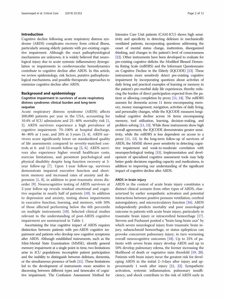
Background and epidemiologyCognitive impairment in survivors of acute respiratorydistress syndrome: clinical burden and long-termsequelaeAcute respiratory distress syndrome (ARDS) affects200,000 patients per year in the USA, accounting for10.4% of ICU admissions and 25–40% mortality risk [1,2]. ARDS survivors experience a high prevalence ofcognitive impairment: 70–100% at hospital discharge,46–80% at 1 year, and 20% at 5 years [3, 4]. ARDS sur-vivors score significantly lower on standardized qualityof life assessments compared to severity-matched con-trols at 6- and 12-month follow-up [5, 6]. ARDS survi-vors also experience higher overall healthcare costs,exercise limitations, and persistent psychological andphysical disability despite lung function recovery at 5-year follow-up [7]. Upon 1-year follow-up, survivorsdemonstrate impaired executive function and short-term memory and increased rates of anxiety and de-pression [1, 8], in addition to post-traumatic stress dis-order [9]. Neurocognitive testing of ARDS survivors at2-year follow-up reveals residual emotional and cogni-tive sequelae in nearly half of patients [10]. In additionto depression and anxiety, testing shows impairmentsin executive function, learning, and memory, with 50%of those affected performing below the 6th percentileon multiple instruments [10]. Selected clinical studiesrelevant to the understanding of post-ARDS cognitiveimpairment are summarized in Table 1.Ascertaining the true cognitive impact of ARDS requires
distinction between patients with pre-ARDS cognitive im-pairment and patients who develop new cognitive symptomsafter ARDS. Although established instruments, such as theMini-Mental State Examination (MMSE), identify generalmemory impairment at a single point in time, two limitationsarise in ICU populations: incomplete patient participationand the inability to distinguish between delirium, dementia,or the simultaneous presence of both [11]. These limitationsled to the development of instruments more sensitive indiscerning between different types and timescales of cogni-tive impairment. The Confusion Assessment Method for
Intensive Care Unit patients (CAM-ICU) shows high sensi-tivity and specificity in detecting delirium in mechanicallyventilated patients, incorporating questions addressing theonset of mental status change, inattention, disorganizedthinking, and changes in the patient’s level of consciousness[12]. Other instruments have been developed to evaluate forpre-existing cognitive deficits: the Modified Blessed Demen-tia Rating Scale (mBDRS) and the Informant Questionnaireon Cognitive Decline in the Elderly (IQCODE) [13]. Theseinstruments more sensitively detect pre-existing cognitiveimpairment by incorporating questions about activities ofdaily living and practical examples of learning or memory inthe patient’s pre-morbid daily life experiences, thereby redu-cing the burden of direct participation expected from the pa-tient or allowing completion by proxy [11, 14]. The mBDRSassesses for dementia across 11 items encompassing mem-ory, money management, navigation, activities of daily living,and personality changes, while the IQCODE measures longi-tudinal cognitive decline across 16 items encompassingmemory, tool utilization, learning, decision-making, andproblem solving [11, 13]. While these instruments show highoverall agreement, the IQCODE demonstrates greater sensi-tivity, while the mBDRS is less dependent on access to aproxy [11, 13]. In the long-term follow-up of survivors ofARDS, the MMSE shows poor sensitivity in detecting cogni-tive impairment and weak-to-moderate correlation withneuropsychological testing [15]. Implementation and devel-opment of specialized cognitive assessment tools may helpbetter guide decisions regarding capacity and medications, inaddition to improving our understanding of the significantimpact of cognitive decline after ARDS.
ARDS in brain injuryARDS in the context of acute brain injury constitutes adistinct clinical scenario from other types of ARDS, char-acterized by earlier sympathetic activation and potentialinteractions between positive pressure ventilation, cerebralautoregulatory, and microcirculatory function [16]. ARDSindependently predicts mortality and poor neurologicaloutcome in patients with acute brain injury, particularly intraumatic brain injury or intracerebral hemorrhage [17].Stevens and Puybasset posited a “brain-lung-brain axis” bywhich severe neurological injury from traumatic brain in-jury, subarachnoid hemorrhage, or status epilepticus canprovoke concurrent pulmonary injury, in turn worseningoverall neurocognitive outcomes [18]. Up to 25% of pa-tients with severe brain injury develop ARDS and up to50% develop pulmonary edema, the former increasing thelikelihood of death or vegetative state threefold [19, 20].Patients with brain injury incur the greatest risk for devel-oping ARDS in the initial 2–3 days after injury and ap-proximately 1 week after injury [21, 22]. Sympatheticactivation, systemic inflammation, pulmonary insuffi-ciency, and shock contribute to the risk of ARDS early in
Sasannejad et al. Critical Care (2019) 23:352 Page 2 of 12
hospitalization [21, 22]. The second peak of ARDS risk re-flects ventilator-associated pneumonia and sepsis [21, 22]:comatose patients experience an increased frequency ofpneumonia between hospital days 4–7 and are at risk foraspiration of oropharyngeal secretions during and after in-tubation [23]. An observational study of patients with
severe traumatic brain injury identified cerebral midlineshift exceeding 5mm and prior drug abuse as risk factorsfor ARDS [19]. Furthermore, patients with concurrentARDS and brain injury who experienced worse long-termcognitive outcome tended to demonstrate lower systemicblood pressure, higher intracranial pressure, and lower
Table 1 Selected clinical studies investigating post-ARDS cognitive impairment
Author(s) Year Methodology Results/conclusions
Davidson et al. 1999 Prospective cohort (n = 146) Patients who survived ARDS experience significantly reduced quality of life followingdischarge compared to critically ill patients without ARDS
Hopkins et al. 1999 Prospective cohort (n = 62) Survivors of ARDS demonstrate cognitive impairments in memory, attention,concentration, and processing speed: 100% at discharge and 78% at 1 year afterdischarge
Contant et al. 2001 Observational (n = 161) ARDS following severe head injury results in severe intracranial hypertension. Targetingintracranial pressure rather than cerebral blood flow improves outcomes
Georgiadiset al.
2001 Prospective interventional (n = 20) In patients with acute stroke receiving mechanical ventilation, changes in cerebralperfusion pressure are mediated by mean arterial pressure rather than by positive end-expiratory pressure. Positive end-expiratory pressure does not increase intracranial pres-sure as long as hemodynamic stability is maintained
Holland et al. 2003 Prospective cohort (n = 137) In patients with traumatic brain injury, ARDS independently predicts mortality and isassociated with worse long-term neurological outcome
Ely et al. 2004 Prospective cohort (n = 275) Delirium independently predicts higher mortality and longer hospital stay amongpatients treated with mechanical ventilation
Mascia et al. 2005 Prospective interventional (n = 12) Positive end-expiratory pressure does not affect intracranial pressure when inducing al-veolar recruitment, but does lead to significant increases in PaCO2 and intracranial pres-sure when inducing alveolar hyperinflation
Muench et al. 2005 Prospective interventional (n = 10) In hemodynamically unstable patients with severe subarachnoid hemorrhage, increasesin positive end-expiratory pressure disturb cerebrovascular autoregulation, resulting insignificant decreases in mean arterial pressure and regional cerebral blood flow
Mascia et al. 2007 Observational (n = 82) High-tidal-volume mechanical ventilation is associated with the development of ARDSafter severe brain injury
Fong et al. 2009 Secondary analysis of prospectivecohort (n = 408)
Delirium accelerates cognitive decline in patients with probable or possible Alzheimer’sdisease
Taccone et al. 2009 Observational (n = 21) Septic shock impairs cerebral autoregulation in patients with septic shock, particularlywith concurrent hypercapnia
Janz et al. 2010 Retrospective cohort (n = 7 fromdatabase of 379)
Brain autopsy of patients with ICU delirium shows hypoxic ischemic damage in thehippocampus, suggesting a link between ICU delirium and long-term cognitiveimpairment
van denBoogard et al.
2011 Exploratory observational (n =100)
The underlying mechanism of delirium may differ in patients with systemic inflammationversus patients without systemic inflammation and is mediated by different cytokines foreach mechanism
Mikkelsen et al. 2012 Prospective cohort (n = 102) Survivors of ARDS 1 year following discharge demonstrate a confluence of cognitiveimpairment, psychiatric sequelae, and diminished quality of life. Hypoxemia andconservative fluid management are associated with these long-term impairments
Elmer et al. 2013 Retrospective cohort (n = 697) High-tidal-volume mechanical ventilation in patients with intracerebral hemorrhage isassociated with the development of ARDS and increased mortality
Pandharipandeet al.
2013 Prospective cohort (n = 821) At 12-month follow-up after discharge, 1/4 of patients who had been critically ill demon-strate cognitive impairment similar in severity to that seen in mild Alzheimer’s disease,and 1/3 similar in severity to that seen in traumatic brain injury
Needham et al. 2014 Prospective cohort (n = 203) At 6- and 12-month follow-up, ARDS survivors demonstrated impairments in 6-min walkdistance and physical function outcomes. Minimizing the duration of intensive care andcorticosteroid use may reflect modifiable risk factors
Girard et al. 2018 Prospective cohort (n = 1040enrolled, n = 586 follow-up)
Patients with ARDS, septic shock, or both experience multiple subtypes of deliriumassociated with long-term cognitive impairment at 3- and 12-month follow-up, includinghypoxic, septic, unclassified, and sedative-associated delirium. The durations of these de-lirium subtypes predict worse cognitive function at 12-month follow-up, particularlysedative-associated delirium
Sasannejad et al. Critical Care (2019) 23:352 Page 3 of 12
cerebral perfusion pressure [19]. Among patients with in-tracerebral hemorrhage, 27% develop ARDS, with high-tidal-volume mechanical ventilation constituting thegreatest risk factor, followed by positive fluid balance andblood transfusion [24]. Despite this, existing prognosticmodels for acute brain injury largely do not include ARDSas a variable that may independently limit the extent ofneurocognitive recovery.
Risk factors for cognitive declineRisk factors for long-term cognitive impairment com-prise a combination of irreversible clinical factors, po-tentially modifiable clinical complications of providerinterventions, and pathophysiological events that mayoccur in the natural history of ARDS in brain injury. Fig-ure 1 illustrates the confluence of these factors and howthey may culminate in an adverse long-term cognitiveoutcome. The heterogeneity of ARDS etiology and sever-ity can expose patients to varying balances of these fac-tors: for instance, ARDS of a pulmonary etiology mayexpose patients to more severe hypoxemia, whereasARDS related to sepsis may expose patients to more se-vere inflammatory activation, while overall disease sever-ity can impact length of stay.
Pre-existing cognitive impairment and the interface ofdelirium and dementiaPre-existing cognitive impairment is a risk factor forcognitive decline after critical illness [25, 26], thoughdata are limited by under-recognition of pre-existingcognitive impairment [14] or exclusion of patients withpre-existing cognitive impairment from longitudinalfollow-up studies [1, 6]. Alzheimer’s disease, character-ized by cerebral accumulation and deposition of theamyloid-β peptide, is the most common type of cogni-tive impairment [27, 28]. Although one third of elderlypatients admitted to the intensive care unit have pre-existing cognitive impairment, this history is often un-known to their medical teams [14], in turn precludingcomparisons of pre-morbid versus longitudinal cognitiveperformance. Among patients confirmed not to havepre-existing dementia, one study found hospitalizationitself significantly associated with a greater likelihood ofdeveloping dementia, along a temporal pattern of abrupt,rather than gradual, cognitive decline [29].Delirium during critical illness predicts long-term
cognitive decline after ARDS [1, 4, 26], and longerdurations of delirium in critically ill patients predictmore severe cognitive impairment at 1-year follow-up[30]. The closely intertwined relationship between de-lirium and pre-existing cognitive impairment raises aphysiological question: does long-term cognitive im-pairment following ARDS reflect a reduction in thethreshold for developing delirium due to underlying
Alzheimer’s pathology, or does delirium contribute in-dependently to this end? It is known that pre-existingcognitive impairment is a key underlying risk factorfor delirium, which affects as many as 70–87% of crit-ically ill patients [31, 32]. Particularly among the eld-erly, ICU delirium can persist during hospitalizationfollowing transfer from the ICU in 40–50% of pa-tients, commonly with incomplete resolution by dis-charge [33]. A meta-analysis of long-term sequelae ofdelirium in elderly patients found increased risk ofdeveloping dementia within 3–5 years of discharge,with an odds ratio of 12.52 (95% CI 1.86–84.21) [34].Patients with Alzheimer’s disease, when hospitalized,are three times as likely as adults without dementiato experience delirium; cognitive deficits can persistup to 5 years after discharge [25]. Clinically, patientswith Alzheimer’s disease who experience delirium suf-fer accelerated cognitive decline beyond the naturalcourse of dementia alone, with twofold increases inthe slope of decline [35]. Recent pathology-basedstudies corroborate delirium-associated acceleration ofcognitive decline independent of pre-existing demen-tia pathology [36].
Delirium subtypes and pathwaysStudies of the pathophysiology of short- and long-termcognitive impairment reflect similarities and differencesbetween delirium and dementia pathways, in which pre-existing pathophysiology diminishes the reserve withwhich to face acute insults. Shared mechanistic featuresof dementia and delirium include synaptic disconnectionresulting from the loss of presynaptic terminals, dimin-ished cholinergic activity leading to impaired arousaland inattention, and microglial activation perpetuatingsystemic inflammation [37]. Girard et al. classify thephenotypes of delirium in the intensive care unit intofive subtypes: sedative-associated, hypoxic, septic/inflam-matory, metabolic, and unclassified [30]. Among these,the duration of metabolic delirium is not associated withlong-term adverse cognitive outcomes [30]. Pathophysio-logically, MacLullich et al. classify the etiologies of delir-ium into two categories: direct brain insults—primaryinsults such as hemorrhage, hypoxia, hypoperfusion, ordrugs—versus aberrant stress responses—the dysfunc-tion of ordinarily adaptive responses to acute systemicstressors such as infection or trauma [38]. Differences inserum markers between patients with inflammatory ver-sus non-inflammatory delirium suggest activation ofmultiple possible pathways: a systemic inflammationpathway associated with IL-8 elevation among patientswith inflammation, and an alternative pathway charac-terized by elevation of amyloid-β and IL-10 in patientswithout inflammation, suggesting activation of existingpathways of underlying cognitive impairment [39].
Sasannejad et al. Critical Care (2019) 23:352 Page 4 of 12

Individual pathophysiological mechanisms may be asso-ciated with multiple delirium subtypes in which a singlephenotype dominates, and these relationships remain anactive area of investigation.
ARDS and hypoxemia: short-term and long-term effectsProfound hypoxemia is one of the cardinal features ofARDS. While, in the short-term, this can predispose pa-tients to hypoxic delirium phenotypes [30], lower PaO2
levels are associated with long-term cognitive impair-ment at 12-month follow-up, particularly in the domainsof executive function and psychomotor tasks [1, 40].
Inflammation and septic deliriumThe production of cytokines TNF-α, IL-6, IL-1α, and IL-1β can produce a constellation of behaviors termed“sickness behavior,” comprising impaired concentration,malaise, diminished motivation, psychomotor retard-ation, and depression [41], corresponding with septic de-lirium. In one study, this state was found to beassociated with recruitment of additional brain struc-tures in order to maintain the same level of cognitiveperformance during systemic inflammation [42]. Existingbrain injury enhances vulnerability to inflammation: inmouse models of neurodegenerative disease, lipopolysac-charide in brain-injured versus control mice induces
Fig. 1 Confluence of clinical risk factors and pathophysiological events culminating in cognitive impairment following ARDS and brain injury.Combinations of irreversible clinical risk factors, pathophysiological events, and modifiable clinical risk factors, each occurring to varying extents,produce an aggregate sum of risk for long-term cognitive impairment. Cognitive outcomes reflect a continuum up to a threshold beyond whicha patient is likely to experience an adverse outcome, defined as long-term cognitive impairment. The aggregate sum of these factors can bringthe patient’s risk for long-term impairment closer toward this threshold (the upward trajectory indicated by the red arrow); however, minimizationof modifiable clinical factors can bring the aggregate sum further away from the threshold, promoting a less adverse cognitive outcome (thedownward trajectory indicated by the green arrow)
Sasannejad et al. Critical Care (2019) 23:352 Page 5 of 12
compounded cognitive deficits on maze-learning tasksgreater than exaggerated sickness behavior alone [43].
Mechanical ventilation and inflammatory and sedative-associated deliriumMechanical ventilation is independently associated withpersistent cognitive impairment, diminished quality oflife, and depression [44]. One third of mechanicallyventilated patients perform abnormally on neurocogni-tive testing at 6 months, comprising deficits in visuo-construction, visual memory, psychomotor speed, andverbal fluency [44]. Accelerated Alzheimer’s disease-typepathophysiology can follow short-term high-tidal-volume mechanical ventilation in mice, including im-paired β-amyloid clearance, increased inflammation me-diated by TNF-α and IL-6, and altered blood-brainbarrier permeability [45]. Prolonged courses of mechan-ical ventilation in ARDS also expose patients to sedating
medications and anesthesia. Among delirium subtypes,sedative-associated delirium is the most common and,with prolonged duration, associated with the greatest de-gree of long-term cognitive impairment at 12 monthsfollowing discharge [30]. Among sedative types, benzodi-azepines impart the greatest risk for delirium, while dex-medetomidine has been associated with lower risk fordelirium [46]; however, data and practice choices arelimited, as most intensive care unit patients are treatedwith multiple sedatives [30]. The effect of paralytic ex-posure on long-term cognitive outcomes is unknown[3]. Continuous exposure to sedative medications overseveral days results in impaired sedative clearance, inturn exacerbating delirium [30]. In addition to inducingacute-on-chronic cholinergic transmission dysfunctionin elderly patients, transient axonal damage may repre-sent another mechanism by which sedatives can contrib-ute to long-term cognitive decline [47].
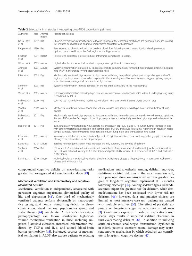
Table 2 Selected animal studies investigating post-ARDS cognitive impairment
Author(s) Year Animalmodel
Results/conclusions
De la Torreet al.
1992 Rat Chronic cerebrovascular insufficiency following ligation of the common carotid and left subclavian arteries in agedrats induces behavioral and cognitive impairments consistent with dementia
Pappas et al. 1996 Rat Rats exposed to chronic reduction of cerebral blood flow following carotid artery ligation develop memorydysfunction and cell loss in the CA1 region of the hippocampus
Feldmanet al.
1997 Rabbit Positive end-expiratory pressure reduces intracranial compliance in rabbits
Wilson et al. 2003 Mouse High-tidal-volume mechanical ventilation upregulates cytokines in mouse lungs
Altmeieret al.
2005 Mouse Systemic inflammation simulated by lipopolysaccharide in mechanically ventilated mice induces cytokine-mediatedlung injury in mechanically ventilated wild-type mice
Fries et al. 2005 Pig Mechanically ventilated pigs exposed to hypoxemia with lung injury develop histopathologic changes in the CA1region of the hippocampus not when exposed to the same degree of hypoxemia alone, suggesting lung injury asa mechanism of damage independent from hypoxemia
Semmleret al.
2005 Rat Systemic inflammation induces apoptosis in the rat brain, particularly in the hippocampus
Wilson et al. 2005 Mouse Pulmonary inflammation following high-tidal-volume mechanical ventilation in mice without underlying lung injuryis mediated by TNF-α
Bickenbachet al.
2009 Pig Low- versus high-tidal-volume mechanical ventilation improves cerebral tissue oxygenation in pigs
Wolthuiset al.
2009 Mouse Mechanical ventilation even at lower tidal volumes causes lung injury in wild-type mice without history of lungdisease
Bickenbachet al.
2011 Pig Mechanically ventilated pigs exposed to hypoxemia with lung injury demonstrate trends toward elevated cytokinesIL-6 and TNF-α in the CA1 region of the hippocampus versus mechanically ventilated pigs exposed to hypoxemiaalone
Heuer et al. 2011 Pig In mechanically ventilated pigs, ARDS results in elevations in TNF-α, IL-6, and IL-1β, which further increase in pigswith acute intracranial hypertension. The combination of ARDS and acute intracranial hypertension results in hippo-campal damage. Acute intracranial hypertension induces lung injury and extravascular lung water
Imamuraet al.
2011 Mouse In a mouse model of septic encephalopathy, an IL-1β cytokine-mediated process disrupts the synaptic processingof long-term potentiation in the hippocampus
Davis et al. 2015 Mouse Baseline neurodegeneration in mice increases the risk, duration, and severity of delirium
Shohamiet al.
2016 Rat TNF-α and IL-6 are detected in the contused hemisphere of rats soon after closed head injury, but not in healthyrats. TNF-α is detected as early as 1 h after injury and peaks at 4 h, whereas IL-6 is detected at 3–5 h and peaks at 8h after injury
Lahiri et al. 2019 Mouse High-tidal-volume mechanical ventilation simulates Alzheimer’s disease pathophysiology in transgenic Alzheimer’sdisease and wild-type mice
Sasannejad et al. Critical Care (2019) 23:352 Page 6 of 12

Putative biological mechanismsInflammation and hypoxemiaAnimal studies of ARDS reflect a wide range of ap-proaches, including murine and porcine models, chem-ical lung injury, ventilator-induced lung injury followingmechanical ventilation, and combinations of lung injurywith systemic inflammatory states, such as sepsis [48].Selected animal studies relevant to the understanding ofthe mechanisms of post-ARDS cognitive impairment aresummarized in Table 2. While ARDS models typicallyinvoke a high-tidal-volume strategy, it is important tonote that mechanical ventilation itself, even at low tidalvolumes, can still cause lung injury [49].Various mechanisms of ARDS-mediated neurological
damage have been theorized, including hypoxemia andcytokine-mediated damage [50]. A porcine model ofARDS identified cytokine-mediated brain damage fromlung injury, rather than hypoxemia, as the major patho-physiological contributor to hippocampal damage, spe-cifically in CA1 and CA2 [50]. A subsequent study onpigs randomized to mechanical ventilation-inducedARDS versus hypoxia-only groups found greater cogni-tive impairment and trends toward increased hippocam-pal inflammation and systemic IL-6 and TNF-αexpression among ARDS subjects, further corroboratingthe distinct pathophysiological roles of mechanical venti-lation and concomitant cytokine release [51]. Amongmechanical ventilation groups, low-tidal-volume ventila-tion is associated with improved brain tissue oxygen-ation and reduced cytokine release compared to high-tidal-volume groups [52].Animal models also demonstrate that ARDS associated
with acute brain injury differs in physiology, time course,and treatment from traditional ARDS: it occurs later andfeatures sympathetic nervous system activation, which,in turn, can trigger neurogenic pulmonary edema sec-ondary to increased alpha-adrenergic activity [53, 54].ARDS is frequently triggered by sepsis, a proinflamma-
tory condition characterized by elevated peripheral cyto-kines and cerebral hypoperfusion, culminating inmultifactorial encephalopathy and end-organ damage[55]. Peripheral cytokine elevation alters blood-brainbarrier metabolism by activating endothelial cells, whilesimultaneously impairing systemic and cerebral bloodflow, altering glucose metabolism in the brain, and ex-posing patients to deleterious environmental factors inthe course of treatment [55]. Cerebral autoregulationdisruption in early septic shock further compounds sys-temic hypotension, as endotoxin-triggered inducible ni-tric oxide synthase production excessively vasodilatesblood vessels, precluding appropriate modulation of vas-cular resistance [56]. Patients with concurrent sepsis andARDS incur further risk for impaired cerebral autoregu-lation, as sepsis renders the cerebral autoregulatory
mechanism more sensitive to PaCO2: in one study, 50%of septic patients with low PaCO2 lost autoregulation,rising to 100% with normal or high PaCO2 [56].
Blood-brain barrier damage and amyloid-β clearanceThe blood-brain barrier is a key neurobiological struc-ture underlying cognitive function. Under normalphysiological conditions, the blood-brain barrier trans-ports amyloid-β protein from within neurons to theextracellular space, where it can be cleared by the glym-phatic system [57–59]. Accumulated amyloid-β worsensblood-brain barrier dysfunction by increasing permeabil-ity, impairing transporter function, and modulatingendothelial cell expression patterns, in turn perpetuatingimpairment in the clearance of amyloid-β and inflamma-tory cytokines [60]. Amyloid-β toxicity induces apoptosisin blood-brain barrier endothelial cells and downregu-lates tight junction proteins Zo-1, occludin, and claudin[61]. Hippocampal blood-brain barrier breakdown hasbeen identified early in the disease course in transgenicAlzheimer’s disease mouse models, with breakdown pre-ceding the detection of amyloid-β deposits, cerebralamyloid angiopathy, or behavioral changes [58]. Animalstudies demonstrate a relationship between amyloid-βdeposits and hippocampal damage, as intrahippocampalinjection of amyloid-β in rats specifically induced apop-tosis in the CA1 region of the hippocampus [62].Amyloid-β impairs the memory-consolidation process oflong-term potentiation in the rat hippocampus in atime- and concentration-dependent fashion [63], mech-anistically comprising impairments in intracellular cal-cium homeostasis and NMDA receptor function [27].Pathophysiological similarities between Alzheimer’s
disease and the acute sequelae of high-tidal-volumemechanical ventilation in mice suggest a link betweenthe mechanisms underlying cognitive damage in ARDS,Alzheimer’s disease, and delirium through a combinationof inflammation and amyloid-β accumulation [45].Mechanical ventilation increases cerebral TNF-α inde-pendently of serum TNF-α, suggesting that cerebralamyloid-β deposition in this setting may reflect a directresponse to mechanical ventilation or pulmonary injuryrather than sequelae of systemic inflammation [45].Neuroimaging comparisons of ARDS survivors within
a year of hospital discharge, versus healthy matched con-trol patients, demonstrate accelerated cerebral and hip-pocampal atrophy [64]. This is consistent with autopsyfindings from deceased intensive care unit patients whohad experienced delirium, in which hippocampal hyp-oxic ischemic lesions were not only the most commonlyidentified abnormality, but were also found only in pa-tients who had experienced ARDS [65]. Magneticresonance imaging studies have identified hippocampalblood-brain barrier dysfunction preceding structural or
Sasannejad et al. Critical Care (2019) 23:352 Page 7 of 12
functional phenotypes [66]. Post-mortem studies reflectconsistency with these results, with evidence of micro-vascular damage and leukocyte infiltration at the sites ofblood-brain barrier damage [66].
Acute and chronic blood-brain barrier insults:linking risk factors and mechanismsAcute weakening, diminished reserve, cytokinecirculation, and apoptosisARDS and concomitant systemic inflammation reflect amultifactorial array of simultaneous insults, comprisingcytokine release, metabolic dysregulation, impaired cere-bral perfusion, medication side-effects, and environmen-tal stimuli including physical restraints and noise [55].Peripheral cytokine release generates positive feedback,inciting further cytokine production through vagal toneincrease, blood-brain barrier endothelial activation, andhumoral system activation [55]. These processes collect-ively culminate in microglial activation, releasing add-itional cytokines, nitric oxide, and reactive oxidativespecies within the central nervous system [55]. Micro-glial activation occurs not only in systemic inflammation,but also in aging, as older rodents injected with periph-eral E. coli lipopolysaccharide produced cytokines, pri-marily IL-1β, selectively in the hippocampus [67, 68]. Astudy investigating anatomic patterns of apoptosis in amouse model of systemic inflammation identified thehippocampus as the most vulnerable region to Bax-mediated apoptotic cascade activation following nitricoxide synthase production downstream of microglial ac-tivation [69].Anatomical studies of humans and rodents confirm
the selective vulnerability of the CA1 hippocampal layerto ischemic injury in a mechanism thought to reflectglutamate excitotoxicity [70]. Mouse models of septicencephalopathy confirm the role of IL-1β in damaginglearning and memory centers of the hippocampus, ashippocampal neurons expressing IL-1β demonstratedelectrophysiological evidence of inhibition of long-termpotentiation [71], thereby acting in a similar mechanismas amyloid-β-mediated impairment of learning andmemory [72]. Existing blood-brain barrier damage sec-ondary to amyloid-β accumulation is associated with di-minished cerebral blood flow and impaired blood flowregulation in the elderly, secondary to impaired neuro-vascular coupling [73]. This, in turn, can render patientswith Alzheimer’s disease susceptible to worsening cogni-tive dysfunction in the setting of systemic hypoperfusionand hypoxemia, which are common in ARDS [73].The mechanistic relationships of brain and lung injur-
ies reflect synergy rather than direct causality, as braininjury itself triggers cytokine production, includingTNF-α and IL-6 [74]. Indeed, lungs harvested from rab-bits that had undergone brain herniation were less
resilient in tolerating high-pressure mechanical ventila-tion compared to sham craniostomy animals, suggestingpotentiation of ARDS by systemic inflammation, in turndue to brain injury [75]. Baseline blood-brain barrierweakening, from chronic amyloid-β accumulation, ren-ders patients with mild cognitive impairment and Alz-heimer’s disease susceptible to increased hippocampalexposure to cytokines. Systemic inflammation, fromARDS and sepsis, in turn, perpetuates cytokine-mediatedhippocampal damage by imparting acute-on-chronicblood-brain-barrier damage while simultaneously in-creasing systemic cytokine circulation.Organ crosstalk in the setting of injurious mechanical
ventilation is not limited to brain-lung interactions. Ani-mal studies demonstrate lung-kidney and lung-gut inter-actions yielding insight into the pathogenesis of multi-organ dysfunction syndrome, as rabbits exposed to high-tidal-volume mechanical ventilation developed epithelialcell apoptosis in the kidney and small intestine [76]. Inaddition to cytokine-mediated damage and apoptosis,additional mechanisms of acute kidney injury associatedwith high-tidal-volume mechanical ventilation includerenal blood flow redistribution, hypoperfusion from sys-temic hemodynamic changes, and metabolic disruptionfrom blood gas changes [77]. Human studies corroboratethis relationship, with a threefold increase in the risk ofacute kidney injury among critically ill patients exposedto invasive mechanical ventilation (OR 3.58, 95% CI1.85–6.92) [78].
Positive pressure ventilation and amyloid-β accumulationDespite benefits of positive end-expiratory pressure(PEEP) in improving oxygenation and alveolar recruit-ment, its application also introduces physiological risk:increased intrathoracic pressure from PEEP can impairboth cerebral venous outflow and systemic venous re-turn, resulting in simultaneously increased intracranialpressure and reduced cerebral perfusion pressure, re-spectively [16]. Animal studies have found increasedintracranial pressure and decreased mean arterial pres-sure with increases in PEEP, in addition to reducedintracranial compliance [79]. In another study on amouse model of stroke, increased PEEP reduced cerebralperfusion pressure by reducing mean arterial pressure,but had minimal effect on intracranial pressure whenmean arterial pressure remained stable, reinforcing theimportance of hemodynamic stability [79]. Human stud-ies confirm the effects of PEEP in hemodynamically un-stable patients, as PEEP up to 20 cm H2O was shown inone study to significantly decrease mean arterial pres-sure and cerebral blood flow [80]. Consequences ofPEEP on cerebral microcirculation therefore may com-pound effects from other potential mechanisms of cogni-tive impairment in critically ill patients with both ARDS
Sasannejad et al. Critical Care (2019) 23:352 Page 8 of 12
and brain injury, exacerbating both the cerebral hypo-perfusion of sepsis and the impairment in amyloid-βclearance following impaired cerebral outflow in vulner-able patients. In another study in acutely brain-injuredpatients, PEEP leading to alveolar hyperinflation in-creased dead space and PaCO2, in turn resulting in arter-ial vasodilation and concomitantly increased intracranialpressure, with further worsening in the setting of dimin-ished intracranial compliance [81]. The effects of PEEPon cerebrovascular hemodynamics, neurological injury,and cognitive outcomes remain largely unknown andrepresent an active area of research.
TherapiesVentilationVentilation of neurologically injured patients entails abalance between preventing intracranial pressure ele-vation secondary to hypercarbia while minimizing ex-posure to an injurious factor. The association betweenhigh-tidal-volume ventilation and ARDS in patientswith intracerebral hemorrhage highlights the import-ance of incorporating tidal volume minimization inventilation setting strategies [24]. Animal studies com-paring low- versus high-tidal-volume ventilation inporcine ARDS models find improved oxygenation andlower lactate levels in brain tissue when using low-tidal-volume ventilation, which is supported by clin-ical outcomes in human patients [52, 82]. Patientsventilated at lower tidal volumes (≤ 8 mL/kg) follow-ing cardiac arrest had a significantly higher chance ofbeing classified as cerebral performance category 1 atfollow-up [82]. Avoidance of benzodiazepines and se-lection of dexmedetomidine as a sedative agent mayreduce the risk of delirium in patients with ARDS re-gardless of neurological injury status [46].Given the systemic inflammatory effects of ARDS, in
addition to murine studies implicating TNF-α in mediat-ing neutrophil recruitment in the early phases of high-tidal-volume-associated stretch lung injury, immunomo-dulating therapies such as monoclonal antibodies againstTNF-α may represent a future area of pharmacotherapy[83]. As the receptor for advanced glycation end prod-ucts (RAGE) has been found to mediate amyloid-β influxand microglial activation at the blood-brain barrier, thismay also represent a future pharmacological target inhumans, with data from mouse models of Alzheimer’sdisease showing effective control of amyloid-β accumu-lation and amyloid-β-mediated cellular stress usingRAGE inhibitors [84].
Fluid managementFluid management reflects another vital juncture of con-sideration, particularly among neurologically injured pa-tients with concurrent sepsis. The Fluid and Catheter
Treatment Trial found that despite similar 60-day mor-tality, patients with ARDS treated with conservativerather than liberal fluid management (central venouspressure < 4 versus 10–14, respectively) experienceshorter intensive care unit stays and fewer days requiringa ventilator [85]. However, follow-up of this trial foundan association between conservative fluid managementand worse cognitive function [1]. The choice of whetherto target treatment toward cerebral perfusion pressureor intracranial pressure goals in the management ofbrain injury invites further consideration of fluid man-agement strategy in this patient population. A studycomparing treatment strategies in patients with severetraumatic brain injury found a significantly elevated riskof developing ARDS among patients receiving cerebralperfusion pressure-targeted treatment compared tothose receiving intracranial pressure-targeted treatment,with an odds ratio of 5.1 [19]. Patients with brain injur-ies appear uniquely susceptible to deleterious effects ofvasopressors: when vasopressors were used in order tomeet cerebral perfusion pressure goals, the risk of devel-oping ARDS further rose, with odds ratios of 5.7 withepinephrine (95% CI 1.0375–34.3323) and 10.8 withdopamine (95% CI 1.4–488.3) [19]. The widespread useof high PEEP in ARDS raises questions of whether inter-actions between PEEP and intracranial pressure or cere-bral perfusion pressure require changes in practice whentreating neurologically injured patients who do go on todevelop ARDS. Studies investigating the relationship be-tween PEEP and cerebral microcirculation identifyhemodynamic stability as an important factor mitigatingagainst adverse effects of PEEP [79, 80]. Specifically, des-pite adverse microcirculatory effects of PEEP, Muenchet al. found that it was only in the scenario of existinghemodynamic instability that PEEP adversely decreasedcerebral perfusion pressure [80], and Georgiadis et al.similarly found that PEEP did not affect intracranialpressure as long as mean arterial pressure remainedstable [79]. Therefore, while fluid minimization may bebeneficial in a general population of critically ill patients[85], aggressive application of a conservative fluid man-agement approach in neurologically injured patients—es-pecially if vasopressors become required to maintainhemodynamic stability—may contribute to not only therisk of ARDS, but also that of cerebral hypoperfusionafter initiating treatment with high-PEEP ventilation. Fu-ture studies are needed to determine an optimal, per-haps individualized PEEP threshold, that maximizespulmonary and neurological function.
Other interventionsThe interdisciplinary, collaborative environment of theintensive care unit allows for coordinated patient careoptimization approaches to mitigate contributors to
Sasannejad et al. Critical Care (2019) 23:352 Page 9 of 12
patient discomfort and long-term adverse effects. Theseinterventions not only are relevant for neurologically in-jured cohorts of patients with ARDS, but may also im-prove outcomes in the general population of intensivecare unit patients. A recent example of such an ap-proach is the ABCDEF bundle, comprising the followingelements: assess, prevent, and manage pain; both spon-taneous awakening trials and spontaneous breathing tri-als; choice of analgesia and sedation; delirium: assess,prevent, and manage; early mobility and exercise; andfamily engagement and empowerment [86]. Implementa-tion of the ABCDEF bundle for 15,000 patients as partof the ICU Liberation Collaborative found that patientstreated with this bundle experienced significant differ-ences in outcomes compared to those who did not, in-cluding higher likelihoods of discharge home andsurvival within the first 7 days of hospitalization, andlower likelihoods of coma, delirium, physical restraintuse, ventilator-dependence, and readmission [86]. Im-provements in each of these outcomes showed a signifi-cant dose-response corresponding with compliance withthe bundle, in which 10% increases in bundle compli-ance produced 15% improvement in survival and dayswithout coma and delirium [86]. Minimizing corticoster-oid use and length of stay may also reduce the risk ofadverse long-term cognitive and physical outcomesamong ARDS survivors [6].
ConclusionReducing the practical burden of cognitive recovery fol-lowing critical illness depends crucially on understand-ing the links between brain injury and lung injury. Newdeficits in learning and memory, and new developmentof psychiatric illness, inherently limit the maximum ex-tent of recovery by restricting the extent to which pa-tients can meaningfully participate in rehabilitation. Theinfluence of pre-existing cognitive impairment on sus-ceptibility and recovery highlights the importance of ac-curate detection and definition of cognitive impairment.Evidence for distinct patterns of inflammatory damage,the predilection of cytokines for the hippocampus, andactivation of systemic inflammatory pathways in high-tidal-volume mechanical ventilation collectively supportminimizing tidal volume as much as possible to avoidARDS, in turn helping prevent further endothelial andmicroglial activation of the inflammatory cascade. Theability to control and monitor parameters such as PEEPand fluid balance in the intensive care unit setting pro-vides an opportunity to optimize treatment on an indi-vidual basis to minimize the risk of long-term cognitiveimpairment.As new data continue to reveal increasing layers of
complexity with which these parameters influence thebrain-lung axis and one another, further studies will
hope to elucidate management strategies that entail notjust minimization or maximization of individual vari-ables, but a balanced and nuanced approach to achieveboth physical and cognitive recovery from ARDS. Mind-ful implementation of multidisciplinary approaches toproviding more humanistic patient care may improvelong-term cognitive outcomes.
AbbreviationsARDS: Acute respiratory distress syndrome; CAM-ICU: Confusion AssessmentMethod for Intensive Care Unit Patients; PEEP: Positive end-expiratory pres-sure; RAGE: Receptor for advanced glycation end products; ABCDEF: Assess,prevent, and manage pain; both spontaneous awakening trials andspontaneous breathing trials; choice of analgesia and sedation; delirium:assess, prevent, and manage; early mobility and exercise; and familyengagement and empowerment
AcknowledgementsNot applicable
Authors’ contributionsCS, EWE, and SL drafted the manuscript. All authors read and approved thefinal manuscript.
FundingNIH/NIA R03AG064106, American Academy of Neurology Institute (SL).
Availability of data and materialsNot applicable
Ethics approval and consent to participateNot applicable
Consent for publicationNot applicable
Competing interestsEWE: Pfizer and Orion honoraria for CME events. NIH and VA Funding. CSand SL declare that they have no competing interests.
Author details1Division of Neurocritical Care and Emergency Neurology, Department ofNeurology, Yale School of Medicine, New Haven, CT, USA. 2Critical Illness,Brain Dysfunction, Survivorship (CIBS) Center, Department of Pulmonary andCritical Care Medicine, Veteran’s Affairs Tennessee Valley Geriatric ResearchEducation and Clinical Center (GRECC), Vanderbilt University School ofMedicine, Nashville, TN, USA. 3Division of Neurocritical Care, Department ofNeurology, Cedars-Sinai Medical Center, 127 S. San Vicente Blvd, AHSPBuilding, Suite A6600, A8103, Los Angeles, CA 90048, USA. 4Division ofNeurocritical Care, Department of Neurosurgery, Cedars-Sinai Medical Center,127 S. San Vicente Blvd, AHSP Building, Suite A6600, A8103, Los Angeles, CA90048, USA. 5Division of Neurocritical Care, Department of BiomedicalSciences, Cedars-Sinai Medical Center, 127 S. San Vicente Blvd, AHSPBuilding, Suite A6600, A8103, Los Angeles, CA 90048, USA.
Received: 18 July 2019 Accepted: 27 September 2019
References1. Mikkelsen ME, Christie JD, Lanken PN, Biester RC, Taylor Thompson B,
Bellamy SL, et al. The adult respiratory distress syndrome cognitiveoutcomes study: long-term neuropsychological function in survivors ofacute lung injury. Am Thoracic Soc. 2012;185(12):1307–15.
2. Bellani G, Laffey JG, Pham T, Fan E, Brochard L, Esteban A, et al.Epidemiology, patterns of care, and mortality for patients with acuterespiratory distress syndrome in intensive care units in 50 countries. JAMA.2016;315(8):788–13.
3. Herridge MS, Moss M, Hough CL, Hopkins RO, Rice TW, Bienvenu OJ, et al.Recovery and outcomes after the acute respiratory distress syndrome
Sasannejad et al. Critical Care (2019) 23:352 Page 10 of 12
(ARDS) in patients and their family caregivers. Intensive Care Med. 2016;42(5):725–38.
4. Wilcox ME, Brummel NE, Archer K, Ely EW, Jackson JC, Hopkins RO.Cognitive dysfunction in ICU patients: risk factors, predictors, andrehabilitation interventions. Crit Care Med. 2013;41:S81–98.
5. Davidson TA, Caldwell ES, Curtis JR, Hudson LD, Steinberg KP. Reducedquality of life in survivors of acute respiratory distress syndrome comparedwith critically ill control patients. JAMA. 1999;281(4):354–60.
6. Needham DM, Wozniak AW, Hough CL, Morris PE, Dinglas VD, Jackson JC, et al.Risk factors for physical impairment after acute lung injury in a national,multicenter study. Am J Respir Crit Care Med. 2014;189(10):1214–24.
7. Herridge MS, Tansey CM, Matté A, Tomlinson G, Diaz-Granados N, Cooper A,et al. Functional disability 5 years after acute respiratory distress syndrome.N Engl J Med. 2011;364(14):1293–304.
8. Mikkelsen ME, Shull WH, Biester RC, Taichman DB, Lynch S, Demissie E, et al.Cognitive, mood and quality of life impairments in a select population ofARDS survivors. Respirology. 2009;14(1):76–82.
9. Kapfhammer HP, Rothenhäusler HB, Krauseneck T, Stoll C, Schelling G. Posttraumaticstress disorder and health-related quality of life in long-term survivors of acuterespiratory distress syndrome. Am J Psychiatr. 2004;161(1):45–52.
10. Hopkins RO, Weaver LK, Collingridge D, Parkinson RB, Chan KJ, Orme JF.Two-year cognitive, emotional, and quality-of-life outcomes in acuterespiratory distress syndrome. Am J Respir Crit Care Med. 2005;171(4):340–7.
11. Lee HB, DeLoatch CJ, Cho S, Rosenberg P, Mears SC, Sieber FE. Detectionand management of pre-existing cognitive impairment and associatedbehavioral symptoms in the intensive care unit. Crit Care Clin. 2008;24(4):723–36- viii.
12. Ely EW, Inouye SK, Bernard GR, Gordon SM, Francis J, May L, et al. Deliriumin mechanically ventilated patients. JAMA. 2001;286(21):2703–10.
13. Pisani MA, Inouye SK, McNicoll L, Redlich CA. Screening for preexistingcognitive impairment in older intensive care unit patients: use of proxyassessment. J Am Geriatr Soc. 2003;51(5):689–93.
14. Pisani MA, Redlich C, McNicoll L, Ely EW, Inouye SK. Underrecognition ofpreexisting cognitive impairment by physicians in older ICU patients. CHESTJ. 2003;124(6):2267–74.
15. Pfoh ER, Chan KS, Dinglas VD, Girard TD, Jackson JC, Morris PE, et al.Cognitive screening among acute respiratory failure survivors: a cross-sectional evaluation of the Mini-Mental State Examination. Crit Care. 2015;19:220.
16. Oddo M, Citerio G. ARDS in the brain-injured patient: what’s different?Intensive Care Med. 2016;42(5):790–3.
17. Holland MC, Mackersie RC, Morabito D, Campbell AR, Kivett VA, Patel R,et al. The development of acute lung injury is associated with worseneurologic outcome in patients with severe traumatic brain injury. JTrauma. 2003;55(1):106–11.
18. Stevens RD, Puybasset L. The brain-lung-brain axis. Intensive Care Med.2011;37:1054–6.
19. Contant CF, Valadka AB, Gopinath SP, Hannay HJ, Robertson CS. Adultrespiratory distress syndrome: a complication of induced hypertension aftersevere head injury. J Neurosurg. 2001;95(4):560–8.
20. Mascia L, Andrews PJ. Acute lung injury in head trauma patients. IntensiveCare Med. 1998;24(10):1115–6.
21. Mascia L, Zavala E, Bosma K, Pasero D, Decaroli D, Andrews P, et al. Hightidal volume is associated with the development of acute lung injury aftersevere brain injury: an international observational study. Crit Care Med.2007;35(8):1815–20.
22. Piek J, Chesnut RM, Marshall LF, van Berkum-Clark M, Klauber MR, Blunt BA,et al. Extracranial complications of severe head injury. J Neurosurg. 1992;77(6):901–7.
23. Bronchard R, Albaladejo P, Brezac G, Geffroy A, Seince P-F, Morris W, et al.Early onset pneumonia: risk factors and consequences in head traumapatients. Anesthesiology. 2004;100:234–9.
24. Elmer J, Hou P, Wilcox SR, Chang Y, Schreiber H, Okechukwu I, et al. Acuterespiratory distress syndrome after spontaneous intracerebral hemorrhage*.Crit Care Med. 2013;41(8):1992–2001.
25. Gross AL, Jones RN, Habtemariam DA, Fong TG, Tommet D, Quach L, et al.Delirium and long-term cognitive trajectory among persons with dementia.Arch Intern Med. 2012;172(17):1324–8.
26. Pandharipande PP, Girard TD, Jackson JC, Morandi A, Thompson JL, Pun BT,et al. Long-term cognitive impairment after critical illness. N Engl J Med.2013;369(14):1306–16.
27. Danysz W, Parsons CG. Alzheimer’s disease, β-amyloid, glutamate, NMDAreceptors and memantine - searching for the connections. Br J Pharmacol.2012;167(2):324–52.
28. Takahashi RH, Nagao T, Gouras GK. Plaque formation and the intraneuronalaccumulation of β-amyloid in Alzheimer’s disease. Pathol Int. 2017;67(4):185–93.
29. Ehlenbach WJ, Hough CL, Crane PK, Haneuse SJPA, Carson SS, Curtis JR,et al. Association between acute care and critical illness hospitalization andcognitive function in older adults. JAMA. 2010;303(8):763–70.
30. Girard TD, Thompson JL, Pandharipande PP, Brummel NE, Jackson JC, PatelMB, et al. Clinical phenotypes of delirium during critical illness and severityof subsequent long-term cognitive impairment: a prospective cohort study.Lancet Respir Med. 2018;6(3):213–22.
31. McNicoll L, Pisani MA, Zhang Y, Ely EW, Siegel MD, Inouye SK. Delirium inthe intensive care unit: occurrence and clinical course in older patients. JAm Geriatr Soc. 2003;51(5):591–8.
32. Pisani MA, Murphy TE, Van Ness PH, Araujo KLB, Inouye SK. Characteristicsassociated with delirium in older patients in a medical intensive care unit.Arch Intern Med. 2007;167(15):1629–34.
33. Ely EW, Shintani A, Truman B, Speroff T, Gordon SM, Harrell FE, et al.Delirium as a predictor of mortality in mechanically ventilated patients inthe intensive care unit. JAMA. 2004;291(14):1753–62.
34. Witlox J, Eurelings LSM, de Jonghe JFM, Kalisvaart KJ, Eikelenboom P, vanGool WA. Delirium in elderly patients and the risk of postdischargemortality, institutionalization, and dementia: a meta-analysis. JAMA. 2010;304(4):443–51.
35. Fong TG, Jones RN, Shi P, Marcantonio ER, Yap L, Rudolph JL, et al. Deliriumaccelerates cognitive decline in Alzheimer disease. Neurology. 2009;72(18):1570–5.
36. Davis DHJ, Muniz-Terrera G, Keage HAD, Stephan BCM, Fleming J, Ince PG,et al. Association of delirium with cognitive decline in late life: aneuropathologic study of 3 population-based cohort studies. JAMAPsychiatry. 2017;74(3):244–51.
37. Davis DHJ, Skelly DT, Murray C, Hennessy E, Bowen J, Norton S, et al.Worsening cognitive impairment and neurodegenerative pathologyprogressively increase risk for delirium. Am J Geriatr Psychiatr. 2015;23(4):403–15.
38. MacLullich AMJ, Ferguson KJ, Miller T, de Rooij SEJA, Cunningham C.Unravelling the pathophysiology of delirium: a focus on the role of aberrantstress responses. J Psychosom Res. 2008;65(3):229–38.
39. van den Boogaard M, Kox M, Quinn KL, van Achterberg T, van der HoevenJG, Schoonhoven L, et al. Biomarkers associated with delirium in critically illpatients and their relation with long-term subjective cognitive dysfunction;indications for different pathways governing delirium in inflamed andnoninflamed patients. Crit Care. 2011;15(6):R297.
40. Hopkins RO, Weaver LK, Pope D, Orme JF, Bigler ED, Larson-LOHR V.Neuropsychological sequelae and impaired health status in survivors ofsevere acute respiratory distress syndrome. Am J Respir Crit Care Med. 1999;160(1):50–6.
41. Dantzer R. Cytokine-induced sickness behavior: mechanisms andimplications. Ann N Y Acad Sci. 2001;933:222–34.
42. Harrison NA, Brydon L, Walker C, Gray MA, Steptoe A, Dolan RJ, et al. Neuralorigins of human sickness in interoceptive responses to inflammation. BiolPsychiatry. 2009;66(5):415–22.
43. Cunningham C, MacLullich AMJ. At the extreme end of thepsychoneuroimmunological spectrum: delirium as a maladaptive sicknessbehaviour response. Brain Behav Immun. 2013;28(C):1–13.
44. Jackson JC, Hart RP, Gordon SM, Shintani A, Truman B, May L, et al. Six-month neuropsychological outcome of medical intensive care unit patients.Crit Care Med. 2003;31(4):1226–34.
45. Lahiri S, Regis GC, Koronyo Y, Fuchs DT, Sheyn J, Kim EH, et al. Acuteneuropathological consequences of short-term mechanical ventilation inwild-type and Alzheimer’s disease mice. Crit Care. 2019;23(1):63.
46. Shah FA, Girard TD, Yende S. Limiting sedation for patients with acuterespiratory distress syndrome - time to wake up. Curr Opin Crit Care. 2017;23(1):45–51.
47. Evered L, Silbert B, Scott DA, Zetterberg H, Blennow K. Association ofchanges in plasma neurofilament light and tau levels with anesthesia andsurgery. JAMA Neurol. 2018;75(5):542–6.
48. Matute-Bello G, Frevert CW, Martin TR. Animal models of acute lung injury.Am J Phys Lung Cell Mol Phys. 2008;295(3):L379–L99.
Sasannejad et al. Critical Care (2019) 23:352 Page 11 of 12
49. Wolthuis EK, Vlaar APJ, Choi G, Roelofs JJTH, Juffermans NP, Schultz MJ.Mechanical ventilation using non-injurious ventilation settings causes lunginjury in the absence of pre-existing lung injury in healthy mice. Crit Care.2009;13(1):R1.
50. Fries M, Bickenbach J, Henzler D, Beckers S, Dembinski R, Sellhaus B, et al. S-100 protein and neurohistopathologic changes in a porcine model of acutelung injury. Anesthesiology. 2005;102(4):761–7.
51. Bickenbach J, Biener I, Czaplik M, Nolte K, Dembinski R, Marx G, et al.Neurological outcome after experimental lung injury. Respir PhysiolNeurobiol. 2011;179(2–3):174–80.
52. Bickenbach J, Zoremba N, Fries M, Dembinski R, Doering R, Ogawa E, et al.Low tidal volume ventilation in a porcine model of acute lung injuryimproves cerebral tissue oxygenation. Anesth Analg. 2009;109(3):847–55.
53. Dai S-S, Wang H, Yang N, An J-H, Li W, Ning Y-L, et al. Plasma glutamate–modulated interaction of A2AR and mGluR5 on BMDCs aggravates traumatic braininjury–induced acute lung injury. J Exp Med. 2013;210(4):839–51.
54. Winklewski PJ, Radkowski M, Demkow U. Cross-talk between theinflammatory response, sympathetic activation and pulmonary infection inthe ischemic stroke. J Neuroinflammation. 2014;11(1):415–8.
55. Sonneville R, Verdonk F, Rauturier C, Klein IF, Wolff M, Annane D, et al.Understanding brain dysfunction in sepsis. Ann Intensive Care. 2013;3(1):15.
56. Taccone FS, Castanares-Zapatero D, Peres-Bota D, Vincent J-L, Berre J, MelotC. Cerebral autoregulation is influenced by carbon dioxide levels in patientswith septic shock. Neurocrit Care. 2009;12(1):35–42.
57. Wang J, Gu BJ, Masters CL, Wang Y-J. A systemic view of Alzheimer disease- insights from amyloid-β metabolism beyond the brain. Nat Rev Neurol.2017;13(10):612–23.
58. Montagne A, Zhao Z, Zlokovic BV. Alzheimer’s disease: a matter of blood-brain barrier dysfunction? J Exp Med. 2017;214(11):3151–69.
59. Iliff JJ, Wang M, Liao Y, Plogg BA, Peng W, Gundersen GA, et al. Aparavascular pathway facilitates CSF flow through the brain parenchymaand the clearance of interstitial solutes, including amyloid beta. Sci TranslMed. 2012;4(147):1–11.
60. Erickson MA, Banks WA. Blood–brain barrier dysfunction as a cause andconsequence of Alzheimer’s disease. J Cerebral Blood Flow Metab. 2013;33(10):1500–13.
61. Song J, Choi S-M, Whitcomb DJ, Kim BC. Adiponectin controls the apoptosis andthe expression of tight junction proteins in brain endothelial cells through AdipoR1under beta amyloid toxicity. Cell Death Dis. 2017;8(10):e3102.
62. Miguel-Hidalgo JJ, Cacabelos R. Beta-amyloid(1-40)-inducedneurodegeneration in the rat hippocampal neurons of the CA1 subfield.Acta Neuropathol. 1998;95(5):455–65.
63. Freir DB, Holscher C, Herron CE. Blockade of long-term potentiation bybeta-amyloid peptides in the CA1 region of the rat hippocampus in vivo. JNeurophysiol. 2001;85(2):708–13.
64. Hopkins RO, Gale SD, Weaver LK. Brain atrophy and cognitive impairment insurvivors of acute respiratory distress syndrome. Brain Inj. 2006;20(3):263–71.
65. Janz DR, Abel TW, Jackson JC, Gunther ML, Heckers S, Ely EW. Brain autopsyfindings in intensive care unit patients previously suffering from delirium: apilot study. J Crit Care. 2010;25(3):538.e7–12.
66. Montagne A, Barnes SR, Sweeney MD, Halliday MR, Sagare AP, Zhao Z, et al.Blood-brain barrier breakdown in the aging human hippocampus. Neuron.2015;85(2):296–302.
67. Barrientos RM, Higgins EA, Biedenkapp JC, Sprunger DB, Wright-Hardesty KJ,Watkins LR, et al. Peripheral infection and aging interact to impairhippocampal memory consolidation. Neurobiol Aging. 2006;27(5):723–32.
68. Chen J, Buchanan JB, Sparkman NL, Godbout JP, Freund GG, Johnson RW.Neuroinflammation and disruption in working memory in aged mice afteracute stimulation of the peripheral innate immune system. Brain BehavImmun. 2008;22(3):301–11.
69. Semmler A, Okulla T, Sastre M, Dumitrescu-Ozimek L, Heneka MT. Systemicinflammation induces apoptosis with variable vulnerability of different brainregions. J Chem Neuroanat. 2005;30(2–3):144–57.
70. Harry GJ, Lefebvre d’Hellencourt C. Dentate gyrus: alterations that occurwith hippocampal injury. NeuroToxicology. 2003;24(3):343–56.
71. Imamura Y, Wang H, Matsumoto N, Muroya T, Shimazaki J, Ogura H, et al.Interleukin-1β causes long-term potentiation deficiency in a mouse modelof septic encephalopathy. Neuroscience. 2011;187:63–9.
72. Shankar GM, Li S, Mehta TH, Garcia-Munoz A, Shepardson NE, Smith I, et al.Amyloid-beta protein dimers isolated directly from Alzheimer’s brains impairsynaptic plasticity and memory. Nat Med. 2008;14(8):837–42.
73. Zlokovic BV. Neurovascular pathways to neurodegeneration in Alzheimer’sdisease and other disorders. Nat Rev Neurosci. 2011;12(12):723–38.
74. Shohami E, Novikov M, Bass R, Yamin A, Gallily R. Closed head injury triggersearly production of TNFα and IL-6 by brain tissue. J Cerebral Blood FlowMetab. 2016;14(4):615–9.
75. López-Aguilar J, Villagrá A, Bernabé F, Murias G, Piacentini E, Real J, et al.Massive brain injury enhances lung damage in an isolated lung model ofventilator-induced lung injury. Crit Care Med. 2005;33(5):1077–83.
76. Imai Y, Parodo J, Kajikawa O, de Perrot M, Fischer S, Edwards V, et al.Injurious mechanical ventilation and end-organ epithelial cell apoptosis andorgan dysfunction in an experimental model of acute respiratory distresssyndrome. JAMA. 2003;289(16):2104–12.
77. Ko GJ, Rabb H, Hassoun HT. Kidney-lung crosstalk in the critically ill patient.Blood Purif. 2009;28(2):75–83.
78. van den Akker JP, Egal M, Groeneveld JA. Invasive mechanical ventilation asa risk factor for acute kidney injury in the critically ill: a systematic reviewand meta-analysis. Crit Care. 2013;17(3):R98.
79. Georgiadis D, Schwarz S, Baumgartner RW, Veltkamp R, Schwab S. Influenceof positive end-expiratory pressure on intracranial pressure and cerebralperfusion pressure in patients with acute stroke. Stroke. 2001;32(9):2088–92.
80. Muench E, Bauhuf C, Roth H, Horn P, Phillips M, Marquetant N, et al. Effectsof positive end-expiratory pressure on regional cerebral blood flow,intracranial pressure, and brain tissue oxygenation*. Crit Care Med. 2005;33(10):2367–72.
81. Mascia L, Grasso S, Fiore T, Bruno F, Berardino M, Ducati A. Cerebro-pulmonary interactions during the application of low levels of positive end-expiratory pressure. Intensive Care Med. 2005;31(3):373–9.
82. Beitler JR, Ghafouri TB, Jinadasa SP, Mueller A, Hsu L, Anderson RJ, et al.Favorable neurocognitive outcome with low tidal volume ventilation aftercardiac arrest. Am J Respir Crit Care Med. 2017;195(9):1198–206.
83. Wilson MR, Choudhury S, Takata M. Pulmonary inflammation induced byhigh-stretch ventilation is mediated by tumor necrosis factor signaling inmice. Am J Phys Lung Cell Mol Phys. 2005;288(4):L599–607.
84. Deane R, Singh I, Sagare AP, Bell RD, Ross NT, LaRue B, et al. A multimodalRAGE-specific inhibitor reduces amyloid beta-mediated brain disorder in amouse model of Alzheimer disease. J Clin Invest. 2012;122(4):1377–92.
85. National Heart L, and Blood Institute Acute Respiratory Distress Syndrome(ARDS) Clinical Trials Network, Wiedemann HP, Wheeler AP, Bernard GR,Thompson BT, Hayden D, et al. Comparison of two fluid-managementstrategies in acute lung injury. N Engl J Med 2006;354(24):2564–2575.
86. Pun BT, Balas MC, Barnes-Daly MA, Thompson JL, Aldrich JM, Barr J, et al.Caring for critically ill patients with the ABCDEF bundle: results of the ICUliberation collaborative in over 15,000 adults. Crit Care Med. 2019;47(1):3–14.
Publisher’s NoteSpringer Nature remains neutral with regard to jurisdictional claims inpublished maps and institutional affiliations.
Sasannejad et al. Critical Care (2019) 23:352 Page 12 of 12


![Chronic Pancreatitis Associated Acute Respiratory Failuremedcraveonline.com/MOJI/MOJI-05-00149.pdf · Chronic Pancreatitis Associated Acute Respiratory ... [1,2]. Acute respiratory](https://img.pdfslide.net/doc/110x75/5ca432de88c993ad338b9ab4/chronic-pancreatitis-associated-acute-respiratory-f-chronic-pancreatitis-associated.jpg)