Embed Size (px)
Citation preview

CLINICAL PERSPECTIVE
i n IT
DOUGLAS S. MOODIE , MD, MS Chairman, Division of Pediatrics, Cleveland Clinic
RICHARD STERBA, M D Department of Pediatric Cardiology, Cleveland Clinic
Long-term outcomes excellent for atrial septal defect repair in adults
ABSTRACT
Congenital atrial septal defect repair is safe and effective in patients of almost any age. Long-term survival among adults is excellent, although children generally appear to fare even better. Our 25-year study of outcomes among adults who underwent suture or patch closure found that survival exceeded 90%. We discuss our observations on use of transesophageal echocardiography, indications for cardiac catheterization, and continuing questions about atrial septal defect in adults.
KEY POINTS
Most patients achieve New York Heart Association functional class I status within the first month and maintain that status indefinitely.
Physicians should consider prophylactic beta-blocker therapy to prevent atrial arrhythmias.
Some experts favor transesophageal echocardiography rather than transthoracic echocardiography for the initial diagnostic evaluation because a transesophageal approach can better detect defects high in the atrial septum.
T'S NEVER TOO LATE to patch a hole in the heart, generally. Most adults with a con-
genital atrial septal defect who undergo surgical repair survive at least 25 years after surgery.
Atrial septal defects are the most common congenital heart abnormalities in adults, accounting for approximately one fourth of all cases.1 They are two to three times more com-mon in women than in men.1
• CHARACTERISTICS OF ATRIAL SEPTAL DEFECTS
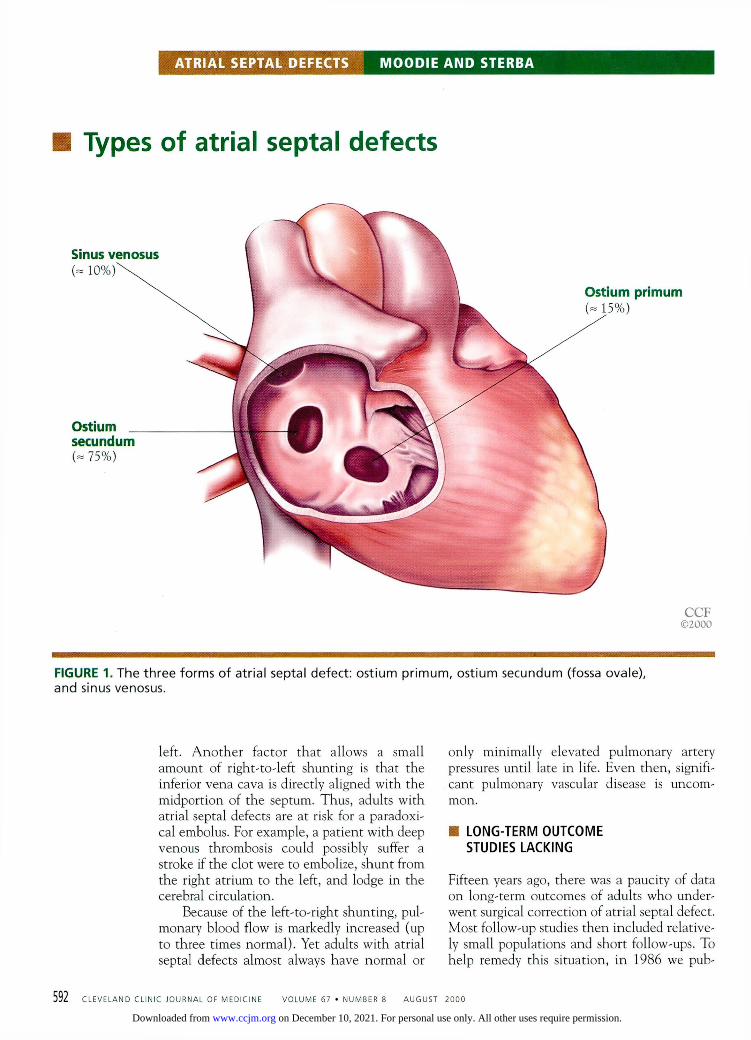
Three types ( F I G U R E 1 )
• Ostium primum defects, located low in the atrial septum, are characterized by a cleft in the anterior leaflet of the mitral valve and occasionally a cleft in the tri-cuspid valve. This type accounts for approximately 15% of cases.1
• Ostium secundum defects (also called fossa ovale defects), are located midway up the atrial septum. This type accounts for approximately 75% of cases.
• Sinus venosus defects are located high in the atrial septum, and are associated with a partial anomalous pulmonary venous return of the right upper pulmonary veins to the right atrium. This type accounts for approximately 10% of cases.
Shunting is mostly f r o m left to r ight In young and middle-aged adults, the left atri-al pressure exceeds the right atrial pressure during most of the cardiac cycle. Therefore, in persons with atrial septal defects, the domi-nant shunt is from left to right. Immediately after atrial systole, however, pressure may be greater in the right atrium than in the left, and thus blood flow can briefly shunt from right to
C L E V E L A N D CLINIC JOURNAL OF MEDICINE VOLUME 67 • NUMBER 8 A U G U S T 2000 5 9 1
on December 10, 2021. For personal use only. All other uses require permission.www.ccjm.orgDownloaded from
ATRIAL SEPTAL DEFECTS MOODIE AND STERBA
• Types of atrial septal defects
p r i m u m
CCF ©2000
FIGURE 1. T h e t h r e e f o r m s o f a t r i a l s e p t a l d e f e c t : o s t i u m p r i m u m , o s t i u m s e c u n d u m (fossa o v a l e ) , a n d s inus venosus .
left. Another factor that allows a small amount of right-to-left shunting is that the inferior vena cava is directly aligned with the midportion of the septum. Thus, adults with atrial septal defects are at risk for a paradoxi-cal embolus. For example, a patient with deep venous thrombosis could possibly suffer a stroke if the clot were to embolize, shunt from the right atrium to the left, and lodge in the cerebral circulation.
Because of the left-to-right shunting, pul-monary blood flow is markedly increased (up to three times normal). Yet adults with atrial septal defects almost always have normal or
only minimally elevated pulmonary artery pressures until late in life. Even then, signifi-cant pulmonary vascular disease is uncom-mon.
• LONG-TERM OUTCOME STUDIES LACKING
Fifteen years ago, there was a paucity of data on long-term outcomes of adults who under-went surgical correction of atrial septal defect. Most follow-up studies then included relative-ly small populations and short follow-ups. To help remedy this situation, in 1986 we pub-
O s t i u m _ s e c u n d u m ( - 75%)
Sinus v e n o s u s (= 10%)
O s t i u m %)
5 9 2 C L E V E L A N D CL INIC JOURNAL OF MEDICINE V O L U M E 67 • NUMBER 8 A U G U S T 2000
on December 10, 2021. For personal use only. All other uses require permission.www.ccjm.orgDownloaded from
lished a report of long-term outcomes in our patients.2
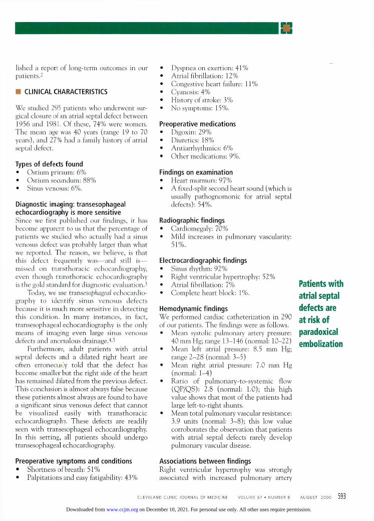
• CLINICAL CHARACTERISTICS
We studied 295 patients who underwent sur-gical closure of an atrial septal defect between 1956 and 1981. Of these, 74% were women. The mean age was 40 years (range 19 to 70 years), and 27% had a family history of atrial septal defect.
Types of defects found • Ostium primum: 6% • Ostium secundum: 88% • Sinus venosus: 6%.
Diagnostic imaging: t ransesophageal echocardiography is more sensitive Since we first published our findings, it has become apparent to us that the percentage of patients we studied who actually had a sinus venosus defect was probably larger than what we reported. The reason, we believe, is that this defect frequently was—and still is— missed on transthoracic echocardiography, even though transthoracic echocardiography is the gold standard for diagnostic evaluation.3
Today, we use trans esophageal echocardio-graphy to identify sinus venosus defects because it is much more sensitive in detecting this condition. In many instances, in fact, transesophageal echocardiography is the only means of imaging even large sinus venosus defects and anomalous drainage.4.5
Furthermore, adult patients with atrial septal defects and a dilated right heart are often erroneously told that the defect has become smaller but the right side of the heart has remained dilated from the previous defect. This conclusion is almost always false because these patients almost always are found to have a significant sinus venosus defect that cannot be visualized easily with transthoracic echocardiography. These defects are readily seen with transesophageal echocardiography. In this setting, all patients should undergo transesophageal echocardiography.
Preoperat ive symptoms and condit ions • Shortness of breath: 51% • Palpitations and easy fatigability: 43%
• Dyspnea on exertion: 41% • Atrial fibrillation: 12% • Congestive heart failure: 11% • Cyanosis: 4% • History of stroke: 3% • No symptoms: 15%.
Preoperat ive medicat ions • Digoxin: 29% • Diuretics: 18% • Antiarrhythmics: 6% • Other medications: 9%.
Findings on examina t ion • Heart murmurs: 97% • A fixed-split second heart sound (which is
usually pathognomonic for atrial septal defects): 54%.
Radiographic f indings • Cardiomegaly: 70% • Mild increases in pulmonary vascularity:
51%.
Electrocardiographic f indings • Sinus rhythm: 92% • Right ventricular hypertrophy: 52% • Atrial fibrillation: 7% • Complete heart block: 1%.
Hemodynamic f indings We performed cardiac catheterization in 290 of our patients. The findings were as follows. • Mean systolic pulmonary artery pressure:
40 mm Hg; range 13—146 (normal: 10-22) • Mean left atrial pressure: 8.5 mm Hg;
range 2-28 (normal: 3-5) • Mean right atrial pressure: 7.0 mm Hg
(normal: 1-4) • Ratio of pulmonary-to-systemic flow
( Q P / Q S ) : 2.8 (normal: 1.0); this high value shows that most of the patients had large left-to-right shunts.
• Mean total pulmonary vascular resistance: 3.9 units (normal: 3—8); this low value corroborates the observation that patients with atrial septal defects rarely develop pulmonary vascular disease.
Associations b e t w e e n f indings Right ventricular hypertrophy was strongly associated with increased pulmonary artery
Patients with atrial septal defects are at risk of paradoxical embolization
593 C L E V E L A N D CL INIC J O U R N A L OF MEDICINE VOLUME 67 • NUMBER 8 A U G U S T 2 0 0 0
on December 10, 2021. For personal use only. All other uses require permission.www.ccjm.orgDownloaded from

ATRIAL SEPTAL DEFECTS MOODIE AND STERBA
Adults with atrial septal defects rarely have notable pulmonary vascular disease
Adult patients improve quickly after surgery for atrial septal defects
100 r -
80
40
20 Before Early surgery after
surgery
Late afler
surgery
FIGURE 2. Percent of pat ients achieving N e w York H e a r t Association funct iona l class I a f te r surgical closure o f atr ial septal defects as adults. Patients improved quickly a n d m a i n -t a i n e d the i r funct ion long - te rm.
pressure, hut not with age. Atrial fibrillation and congestive heart failure were associated with both as well as with each other (P < .001 for all comparisons).
Anatomic f indings on catheter iza t ion Cardiac catheterization also revealed the fol-lowing prevalences of anatomic abnormalities. • Anomalous pulmonary venous return:
13% • Coronary artery disease: 9% • Pulmonary stenosis: 5% • Pulmonary vascular disease: 15% • Normal left ventricular function as
assessed by ventriculography: 96% • Severely impaired left ventricular func-
tion: 0% • Mitral insufficiency: 7% (severe in only
1 % )
• Mitral regurgitation (uncommon in atrial septal defect): only 7%. (Although we did not address the issue of mitral valve pro-lapse in our study, our clinical experience indicates that it occurs in fewer than 5% of patients with atrial septal defect.)
Long-term survival is high among adults who undergo closure of atrial septal defects
:> so
60
o 40
™ 20
10
Years 15 20
FIGURE 3. Survival a t 20 years a m o n g 295 pat ients w h o u n d e r w e n t surgical closure of atrial septal defects as adults.
Indications for cardiac catheter iza t ion . In those years, cardiac catheterization was per-formed because it was the diagnostic proce-dure of choice. We perform it today only if the patient requires coronary angiography, has some anatomic finding on echocardiography that is not completely clear (which is rare), or has significantly elevated pulmonary artery pressures.
For patients with significantly elevated pulmonary artery pressures, we routinely mea-sure pulmonary artery pressure first as the patient breathes room air and again while we give oxygen or other agents or both to deter-mine if there is a decrease in pulmonary vas-cular resistance.
We conducted another study to ascertain the value of coronary angiography in adults with atrial septal defect; that analysis led us to conclude that coronary angiography should be performed in all patients who have at least one risk factor for coronary artery disease. We reviewed the records of 77 consecutive patients (72% women; mean age 53) who underwent selective angiography between 1982 and 1992. Eight (10.4%) of the 77 patients had coronary artery disease (> 60% stenosis in at least one vessel). Three of the 8 patients had a lesion in the left anterior descending vessel, 3 in the right artery, 1 in
5 9 4 C L E V E L A N D CL INIC J O U R N A L OF MEDICINE V O L U M E 67 • NUMBER 8 A U G U S T 2000
on December 10, 2021. For personal use only. All other uses require permission.www.ccjm.orgDownloaded from

the left anterior diagonal vessel, and 1 in the left anterior descending artery. Six of the 8 had abnormal resting electrocardiograms and typical anginal symptoms.
• OUTCOMES
The defects were corrected with a primary suture closure in 66% of our 295 patients, and the remainder received a patch closure. Only 6 patients (2%) died during the initial hospi-talization. Postoperatively, 4 patients (1.4%) had cerebrovascular accidents.
Functional class Before surgery, only 26% of the patients were in New York Heart Association (NYHA) functional class I; at 6 months, the number had increased to 84%, and patients tended to maintain that status long-term ( F I G U R E 2 ) .
Long- term survival Twenty-eight patients (9.5%) died during the late postoperative period in 20 years of follow-up, 8 of noncardiac causes. The actuarial sur-vival rate at 20 years was 92% ( F I G U R E 3). The most common causes of death were arrhyth-mias, major hemorrhage, and myocardial infarction.
Stroke Twelve patients, all older than 35 years, had a stroke at some point after the first postopera-tive month. Only 4 of them had documented atrial fibrillation.
Postper icardiotomy syndrome Atrial septal defect is the most common underlying congenital cardiac defect associat-ed with postpericardiotomy syndrome (a peri-cardial or pleural reaction occurring more than 1 week after opening of the pericardium, characterized by fever, chest pain, and signs of pleural or pericardial inflammation). The rea-son for the higher incidence in atrial septal defect patients is unclear.
The incidence of postpericardiotomy syn-drome was highest during the spring ( F I G U R E 4 ) .
A possible reason for the seasonal variation is that postpericardiotomy syndrome may be due to an immunologic reaction to a virus present more in the spring.
Postpericardiotomy syndrome is most common in the spring
FIGURE 4. Seasonality of t h e incidence o f postper icard io tomy syndrome a m o n g 295 pat ients w h o u n d e r w e n t surgical closure of atr ial septal defects as adults.
Trans-esophageal echo is the best choice to identify sinus venosus defects
Atr ia l a r ry thmia increases w i t h age The electrophysiologic consequences of wait-ing until adulthood to have an atrial septal defect repaired can be serious.6"8 Studies of both adults with atrial septal defect and nor-mal populations show that the incidence of atrial arrhythmia increases with age, probably as a result of pressure and volume loading of the atria. Atrial fibrillation is common in the postoperative period in adults.
Even after successful defect closure, atrial arrhythmias are likely to occur if atrial size, ventricular size, compliance, or function do not normalize.
Atr ia l f ibr i l la t ion Atrial fibrillation developed in 23% of the 295 patients.
During the early (< 1 month) postopera-tive period, the incidence of atrial fibrillation correlated with age and pulmonary artery sys-tolic and diastolic pressures (P < .001). Later, atrial fibrillation correlated with age at surgery, age at follow-up, and atrial fibrillation during the preoperative or early postoperative period (P < .001). There was no correlation between late atrial fibrillation and preopera-tive pulmonary artery pressure or Q P / Q S .
595 CLEVELAND CLINIC JOURNAL OF MEDICINE VOLUME 67 • NUMBER 8 AUGUST 2000
on December 10, 2021. For personal use only. All other uses require permission.www.ccjm.orgDownloaded from

ATRIAL SEPTAL DEFECTS MOODIE AND STERBA
Atrial arrhythmias can occur after repair if atrial size and other factors do not normalize
Treat ing atr ia l f ib r i l l a t ion We studied and successfully performed abla-tion in a small number of patients who had slow atrial flutter (intra-atrial reentry tachy-cardia). In this condition, routine electrocar-diograms typically show an atrial rate between 180 and 240 beats per minute and variable atrioventricular conduction, and intracardiac catheter mapping has shown a right atrial reentrant circuit, which usually travels coun-terclockwise around the anterolateral right atrium, presumably along the site of the previ-ous atriotomy. We followed a strategy of tar-geting the area with the slowest conduction and performing radiofrequency ablation, which has uniformly resulted in short-term success. These findings suggest that the right atriotomy used to close an adult atrial septal defect ought to be modified to prevent reentry around it. Also, using a catheter to close an atrial septal defect should decrease the inci-dence of this unusual tachycardia.
To prevent atrial fibrillation, we give beta-blockers for 3 months after surgery.
• COMPARISON OF ADULT A N D PEDIATRIC OUTCOMES
In a separate analysis, we compared the out-comes of surgical atrial septal defect repair in 287 adults (mean age 41 years) and 153 chil-dren (mean age 10 years). Of this group, 66% of the adults and 72% of the children under-went primary closure.9
Before surgery, chi ldren w e r e heal th ier • Only 61% of the adults were in N Y H A
functional class I, compared with 75% of the children (P < .001).
• Of the adults, 88% had symptoms, primar-ily exertional dyspnea and fatigue, com-pared with 44% of the children (P < . 0 0 1 ) .
• Atrial fibrillation was present in 12% of the adults but none of the children.
Outcomes w e r e similar Mortality rates and improvement in N Y H A functional class were similar for children and adults. However, atrial fibrillation was more common in adults than in children.
Five (1.7%) of the adults died during
surgery, compared with 3 (2%) of the chil-dren. Early atrial fibrillation occurred in 24% of the adults, compared with 5.2% of the children (P < .01).
The cumulative 25-year survival rate among the adults was 92%, compared with 96% in the children. At follow-up, 85% of the adults and 93% of the children achieved N Y H A functional class I. Atrial fibrillation occurred at least 1 month after surgery in 8% of the adults and 6% of the children.
A report from the Mayo Clinic published 20 years ago10 observed that even patients older than 60 years experienced dramatic improvement in symptoms after undergoing atrial septal defect closure. The operative mor-tality rate was slightly higher than that in younger adults, but the overall risk was still low and survival was similar to that of age-matched controls.
• CONTINUING ISSUES
Although the operative mortality rate is low in atrial septal defect patients and long-term outcomes are generally good, a number of questions remain unanswered.
W h y do so f e w atr ia l septa l de fec t pat ients deve lop pu lmonary vascular disease? Patients with atrial septal defects with large shunts generally do not develop pulmonary vascular disease. In contrast, patients with ventricular septal defects or patent ductus arteriosus who have the same degree of left-to-right shunting do have a high incidence of pul-monary hypertension and pulmonary vascular disease by the time they reach adulthood.
Is the answer related to the compliance of the right ventricle? Or is it related to the shear forces in the pulmonary arteries themselves, which are affected more when blood is dumped directly from a ventricular septal defect or a ductus into the pulmonary arteries rather than moving into the low-pressure atri-um and ventricle before it is delivered to the pulmonary arteries?
Does right ventr icular funct ion improve a f ter surgery? Almost all patients with atrial septal defects have normal left ventricular function. In some
5 9 6 C L E V E L A N D CL INIC J O U R N A L OF MEDICINE VOLUME 67 • NUMBER 8 A U G U S T 2 0 0 0
on December 10, 2021. For personal use only. All other uses require permission.www.ccjm.orgDownloaded from

adults who undergo atrial septal defect repair, the right ventricle remains dilated and its func-tion is impaired. Even so, almost all of these patients achieve NYHA functional class I.
Is their right ventricular function any dif-ferent 40 or 50 years after surgery than it is after 20 or 30 years?
Postpericardiotomy syndrome Why is postpericardiotomy syndrome more common in patients with atrial septal defects than in any other group?
W h y is the stroke ra te so high? The incidence of stroke during the late-post-operative and long-term follow-up periods is worrisome. The longer the follow-up, the greater the incidence of stroke.
Does the reason relate to the use of incom-plete suture closures in most patients early in our series? (Because suture repair might not have completely closed the defect, there is a
• REFERENCES
1. Brickner ME, Hillis LD, Lange RA. Congenita l heart disease in adults. First of two parts. N Engl J Med 2000; 342:256-263.
2. Moodie DS, Gill CC, Sterba R, Forsythe S. The unnat-ural history of patients with surgical closure of atrial septal defects in adul thood. In: Gersowy W, editor. Pediatric Cardiology. New York: Springer-Verlag, 1986:1261-1263.
3. Mehta RH, Helmtke F, Nanda NC, Pinheiro L, Samdarshl TE, Shah V K . Uses and l imitations of transthoracic echocardiography in the assessment of atrial septal defect in the adult. A m J Cardiol 1991; 67:288-294.
4. K r o n z o n I, Tunick PA, Freedberg RS, Trehan N, R o s e n z w e i g BP. Schwinger ME. Transesophageal echocardiography is superior to transthoracic echocar-d i o g r a p h y In the diagnosis of sinus venosus atrial sep-tal defect. J A m Coll Cardiol 1991; 17:537-542.
5. Pascoe RD, Oh JIC, Warnes CA, Danielson GK, Tajik AJ, S e w a r d JB. Diagnosis of sinus venosus atrial septal defect w i th transesophageal echocardiography. Circulat ion 1996; 94:1049-1055.
6. Gatzoulis MA, Freeman MA, Siu SC, Webb GD, Harris L. Atrial arrhythmia after surgical closure of atrial septal defects In adults. N Engl J Med 1999; 340:839-846.
7. Heng le in D, Cauchemez B, Bloch G. S imultaneous sur-gical treatment of atrial septal defect and atrial f lut-ter using a simple modif icat ion of the atrial incision. Cardiol Y o u n g 1999; 9:197-199.
8. K o b a y a s h i J , Yamamoto F, Nakano K, et al. Maze pro-cedure for atr ial f ibri l lat ion associated with atrial sep-tal defect. Circulation 1998; 98(Suppl 11): I I-399-II-402.
9. Mandel ik J, Moodie DS, Sterba R, et al. Long-term fol-low-up of children after repair of atrial septal defects. Cleve Cl in J Med 1994; 61:29-33.
chance that right-to-left shunting might have put them at risk for paradoxical embolism.) Or can the stroke rate be attributed to a late devel-opment of atrial fibrillation that was not docu-mented? If so, should we switch to long-term anticoagulation rather than prescribing a course of only 3 to 6 months?
Why are atr ial septal defects so large in adults? We have found the large size of atrial septal defects in adults remarkable. Rarely do we see small ones. In fact, in our early experience, the mean Q P / Q S was nearly 3.
Do some small to medium-sized atrial sep-tal defects in children become larger as they reach adulthood? Brassard et al11 recently reported that as many as 20% of the patients they studied experienced such enlargement over time. They also presented evidence that some atrial septal defects close spontaneously, even in children older than 5 years. gj]
10. Fuster V, Brandenburg RO, McGoon DC, Giul iani ER. Clinical approach and m a n a g e m e n t of congenita l heart disease in the adolescent and adult. Cardiovasc Clin 1980; 10:161-197.
11 Brassard M, Fouron JC, van Doesburg NH, Mercier LA, De Guise P. Outcome of children with atrial septal defect considered too small for surgical closure. A m J Cardiol 1999; 83:1552-1555.
• SUGGESTED READING
H o r v a t h KA, Burke RP, Coll ins JJ Jr, Cohn LH. Surgical treatment of adult atrial septal defect: Early and long-term results. J Am Coll Cardiol 1992; 20 :1156-1159 .
K o n s t a n t i n i d e s S, Geibe l A, O lschewski M , et al . A comparison of surgical and medical therapy for atrial sep-tal defect in adults. N Engl ] Med 1995; 333 :469-473 .
M u r p h y J G , G e r s h BJ, M c G o o n M D , e t a l . Long-term outcome after surgical repair of isolated atrial septal defect. Follow-up at 27 to 32 years. N Engl J Med 1990; 323 :1645-1650 .
P e r l o f f JK. The Clinical Recognition of Congenital Heart Disease. Philadelphia: W B Saunders, 1978:396.
P e r l o f f JK . Surgical closure of atrial septal defect in adults. N Engl J Med 1995; 333 :513-514 .
Stee le PM, Fuster V, C o h e n M , Ri t ter DG, M c G o o n D C . Isolated atrial septal defect with pulmonary vascular obstructive disease long-term follow-up and prediction of outcome after surgical correction. Circulation 1987; 76 :1037-1042 .
ADDRESS: Douglas S. Moodie, MD, Division of Pediatrics, A120, The Cleveland Clinic Foundation, 9500 Euclid Avenue, Cleveland, OH 44195, e-mail [email protected].
597 CLEVELAND CLINIC JOURNAL OF MEDICINE VOLUME 67 • NUMBER 8 AUGUST 2000
on December 10, 2021. For personal use only. All other uses require permission.www.ccjm.orgDownloaded from