Embed Size (px)
Citation preview
- 125 -
Correspondence: Dr. Marco Fricia, Department of Neurosurgery, Cannizzaro University Hospital, via Messina 829, 95126 Catania(CT), Italy, ph. +39-(0)95-7263435, fax +39-(0)95-7263428, e-mail: [email protected] in Neuroscience 2015; 3 (1-4).125-128. ISSN: 2240-5127
doi: 10.14588/PiN.2015.Fricia.125Copyright © 2015 by new Magazine edizioni s.r.l., via dei Mille 69, 38122 Trento, Italy.All rights reserved.www.progressneuroscience.com
INTRODUCTION
The primary objective of cranioplasty is to obtain adurable, stable, and acceptable reconstruction(8).However, the outcome of cranial bone reconstructionis thought to be dependent not only on surgical skills,the quality of adjacent soft tissues, and the size andlocation of the bone defect, but also the choice ofimplant. Regarding the latter, despite much research,there is still no clear consensus regarding the mostappropriate reconstruction materials(9). Although alloplastic materials are biocompatibile,and provide good primary stability, their long-termeffects are seldom investigated, and we cannot ruleout adverse reactions developing after many years. Infact, Gautschi et al.(3) described a case of hyper-sensitivity to PMMA after cranioplasty that led toimplant removal.
Calcium phosphate ceramics and cements are alsohighly biocompatible, and have a very similar structureand chemical composition to that of the mineralphase of bone. Presumably as a result of this likeness,calcium phosphates seem to be remodelled like normalbone through a cell-mediated process. Their ability toact as a synthetic osteoconductive scaffold after im-plantation in natural bone makes them one of the bestimplant options. Hydroxyapatite is one of the forms of calcium phos-phate most commonly used for clinical applications.Although it is known to be non-resorbable over time,long-term follow-up of HA cranioplasty implants isseldom reported in the literature, as the majority ofcomplication arise within the first year after implan-tation.
Short Communication
SUMMARY: Bilateral decompressions in a 16-year-old boy were reconstructed using custom-made porous hydroxyapatite
implants. Long-term follow-up of this biomimetic and osteoconductive material was evaluated at different time point to assess
osteointegration at bone–implant margins. CT scans were performed at 10 days, 58 months and 93 months after cranioplasty.
and used to furnish tissue density measurements along the bone/implant interface. Hounsfield graphs were plotted to assess
tissue density, and therefore osteointegration, at the bone/implant margins at all three time points. Improved osteointegration
over time is evident on both CT scans and Hounsfield graphs. Measured density was always above the bone threshold, even
at implant/bone edges. The interface showed no interruption or evidence of fibrous tissue, and no apparent resorption of the
implant was evident from radiological measurements. Long-term follow-up of porous hydroxyapatite implant confirmed good
osteointegration of the biomimetic prosthesis with the receiving bone. Hydroxyapatite should therefore be considered an ideal
implant material in terms of mid–long term functional recovery.
KEY WORDS: Cranioplasty, Hydroxyapatite, Long-term effect, Osteointegration.
Progress in Neuroscience Vol.3, N. 1-4, 2015
Long-term results of porous hydroxyapatite
bilateral cranioplasty: an 8-year radiological follow-up
M. FRICIA, M. PASSANISI
Department of Neurosurgery, “Cannizzaro” University Hospital, Catania, Italy
AIMS
Long-term radiological follow-up of a patientsuccessfully implanted with porous HA prosthesisalmost 8 years prior to the time of writing wasperformed to assess any signs of resorption and/orosteointegration at the prosthesis/receiving cranialbone interface over time.
MATERIALS AND METHODS
■■ SURGICAL PROCEDURES. The implants were placedaccording to the procedure recommended by themanufacturer, and as described by Staffa et al.(7). ■■ CT SCANS, BONE DENSITY MEASUREMENT AND
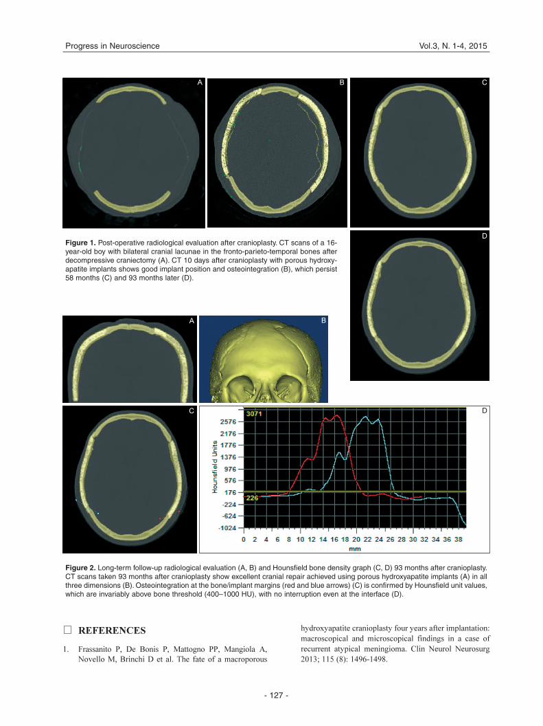
OSTEOINTEGRATION ANALYSIS. Post-operative CTscans were performed at different time points aftercranioplasty to evaluate implant positioning andmargin adhesion. Bone density in the immediatevicinity of the implant was evaluated using MimicsInnovation Suite v17.0 Medical (Materialise,Belgium) and Hounsfield units, which describe thedensity of the CT image at a specific point. TheHounsfield density scale corresponds to differentlevels of beam attenuation, and ranges from -1,000(air) to +3,000 (dense bone), thereby enabling thequantitative differentiation of tissues in the region(i.e., muscle, 35-70 HU; fibrous tissue, 60-90 HU,cartilage, 80-130 HU; bone 150-1800 HU)(4), withcranial bone presenting the highest cancellous bonedensity(6). Density measurements across the bone-implant interface at the various time-points wereperformed twice to provide a mean.■■ CLINICAL CASE. A 16-year-old boy underwent cra-nial bilateral fronto-parieto-temporal decompressionafter severe head injury, due to a car accident, in 2006(Figure 1A). Three months later the skull wassurgically reconstructed using two custom-made HAimplants (CustomBone Service, Fin-Ceramica Faenza
S.p.A). Clinical and radiological follow-up wasperformed from 2007 to 2014.
RESULTS
■■ RADIOLOGIC EVALUATION (POSITIONING, HOUNS-
FIELD VALUES AT BONE-IMPLANT MARGIN). 10 days
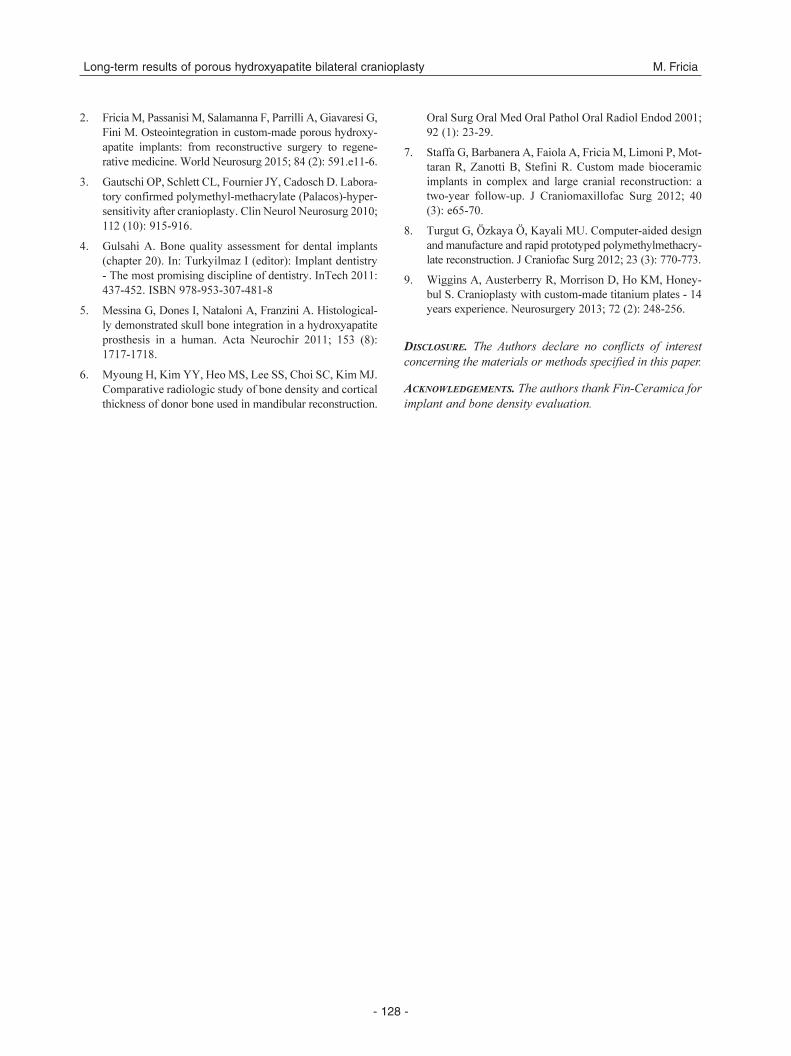
after surgery, a clinical and radiographical assessmentof the patient’s condition and the implant positionwas performed (Figure 1B). Subsequent scans, at 58months (Figure 1C) and 93 months (Figure 1D) aftercranioplasty, confirmed that the implants remainedcorrectly positioned over time. The entire perimeterof the prosthesis remained perfectly adhered to thehost bone, and both internal and external prosthesisprofiles were correctly aligned; this becomes moreevident over time, in particular at 93-month follow-up (Figure 1D and 2 A-B).Hounsfield values of cranial bone ranged from 200 to1,200 HU (Figure 2D), while HU, and thereforedensity, values for hydroxyapatite were far higher (> 3,000 HU, see red and blue arrows in Figure 2C-D). We excluded the formation of fibrous tissue(range 60-90 HU) at the interface since values werealways above bone threshold (Figure 2C-D). Nomacroscopic resorption of the implant was apparenton the scans.
DISCUSSION
8-year radiological follow-up showed the increasingosteointegration of both implants with the receivingcranial bone. Bone healing appeared morehomogeneous over time, with no interruption ordefect. Indeed, HA is the mineral form of calciumapatite that is the main component of bones, and, asdemonstrated by different authors(1,2,5), new boneformation within the prosthesis is a peculiarcharacteristic of porous HA implants. Although boneregeneration and healing times have not beenprecisely defined as yet, it is clear from our resultsthat osteointegration of such implants continues toimprove over time. The osteoconductivity of HAensures physical and mechanical integration with thesurrounding bone, promoting functional recovery inthe mid-to-long term.
CONCLUSIONS
Porous HA appears to be recognized as “self” by theorganism, making it an ideal implant material for alifelong approach to regenerative medicine.
- 126 -
Long-term results of porous hydroxyapatite bilateral cranioplasty M. Fricia
LIST OF ACRONYMS AND ABBREVIATIONS: CT = Computed Tomography; HA = HydroxyApatite; HU = Hounsfield Unit; PMMA= PolyMethyl-MethAcrylate.
REFERENCES
1. Frassanito P, De Bonis P, Mattogno PP, Mangiola A,Novello M, Brinchi D et al. The fate of a macroporous
hydroxyapatite cranioplasty four years after implantation:macroscopical and microscopical findings in a case ofrecurrent atypical meningioma. Clin Neurol Neurosurg2013; 115 (8): 1496-1498.
- 127 -
Progress in Neuroscience Vol.3, N. 1-4, 2015
Figure 1. Post-operative radiological evaluation after cranioplasty. CT scans of a 16-year-old boy with bilateral cranial lacunae in the fronto-parieto-temporal bones afterdecompressive craniectomy (A). CT 10 days after cranioplasty with porous hydroxy-apatite implants shows good implant position and osteointegration (B), which persist58 months (C) and 93 months later (D).
A B C
D
Figure 2. Long-term follow-up radiological evaluation (A, B) and Hounsfield bone density graph (C, D) 93 months after cranioplasty.CT scans taken 93 months after cranioplasty show excellent cranial repair achieved using porous hydroxyapatite implants (A) in allthree dimensions (B). Osteointegration at the bone/implant margins (red and blue arrows) (C) is confirmed by Hounsfield unit values,which are invariably above bone threshold (400–1000 HU), with no interruption even at the interface (D).
A B
C D
2. Fricia M, Passanisi M, Salamanna F, Parrilli A, Giavaresi G,Fini M. Osteointegration in custom-made porous hydroxy-apatite implants: from reconstructive surgery to regene-rative medicine. World Neurosurg 2015; 84 (2): 591.e11-6.
3. Gautschi OP, Schlett CL, Fournier JY, Cadosch D. Labora-tory confirmed polymethyl-methacrylate (Palacos)-hyper-sensitivity after cranioplasty. Clin Neurol Neurosurg 2010;112 (10): 915-916.
4. Gulsahi A. Bone quality assessment for dental implants(chapter 20). In: Turkyilmaz I (editor): Implant dentistry- The most promising discipline of dentistry. InTech 2011:437-452. ISBN 978-953-307-481-8
5. Messina G, Dones I, Nataloni A, Franzini A. Histological-ly demonstrated skull bone integration in a hydroxyapatiteprosthesis in a human. Acta Neurochir 2011; 153 (8):1717-1718.
6. Myoung H, Kim YY, Heo MS, Lee SS, Choi SC, Kim MJ.Comparative radiologic study of bone density and corticalthickness of donor bone used in mandibular reconstruction.
Oral Surg Oral Med Oral Pathol Oral Radiol Endod 2001;92 (1): 23-29.
7. Staffa G, Barbanera A, Faiola A, Fricia M, Limoni P, Mot-taran R, Zanotti B, Stefini R. Custom made bioceramicimplants in complex and large cranial reconstruction: atwo-year follow-up. J Craniomaxillofac Surg 2012; 40(3): e65-70.
8. Turgut G, Özkaya Ö, Kayali MU. Computer-aided designand manufacture and rapid prototyped polymethylmethacry-late reconstruction. J Craniofac Surg 2012; 23 (3): 770-773.
9. Wiggins A, Austerberry R, Morrison D, Ho KM, Honey-bul S. Cranioplasty with custom-made titanium plates - 14years experience. Neurosurgery 2013; 72 (2): 248-256.
DISCLOSURE. The Authors declare no conflicts of interest
concerning the materials or methods specified in this paper.
ACKNOWLEDGEMENTS. The authors thank Fin-Ceramica for
implant and bone density evaluation.
- 128 -
Long-term results of porous hydroxyapatite bilateral cranioplasty M. Fricia