Embed Size (px)
Citation preview
Longitudinal observation of antibody responsesafter SARS-CoV-2 infection at 14 monthsPuya Dehgani-Mobaraki
Associazione Naso SanoAsiya kamber Zaidi ( [email protected] )
Associazione Naso Sano https://orcid.org/0000-0001-6510-6365Nidhi Yadav
All India Institute of Medical Sciences (AIIMS)Alessandro Floridi
Laboratory of Nuclear Lipid BioPathology, Centro Ricerche AnalisiDr. Emanuela Floridi
Laboratory of Nuclear Lipid BioPathology, Centro Ricerche Analisi
Article
Keywords:
Posted Date: June 14th, 2021
DOI: https://doi.org/10.21203/rs.3.rs-618366/v1
License: This work is licensed under a Creative Commons Attribution 4.0 International License. Read Full License
TITLE PAGE
Title : Longitudinal observation of antibody responses after SARS-CoV-2
infection at 14 months
Key words : “SARS-CoV-2” “covid19” “coronavirus” “antibody trend”
Authors:
1. Puya Dehgani-Mobaraki, M.D , M.S(ORL)
-Founder Association “Naso Sano”, Umbria Regional Registry of volunteer activities, Corciano, Italy
-Department of Otorhinolaryngology and Head Neck Surgery, Gubbio-
Gualdo Tadino Hospital, Usl Umbria 1, Italy
-Front Line Covid19 Critical Care Alliance Associate(FLCCC)
Email: [email protected]
ORCID Id: 0000-0001-8339-1868
2. Asiya Kamber Zaidi, M.D, M.S (ORL), F.W.A.M.S [Corresponding Author]
Affiliation: Member, Associazione Naso Sano, Umbria region, Italy.
Email: [email protected]
ORCID Id: 0000-0001-6510-6365
3. Nidhi Yadav, M.Sc (Applied Statistics)
Affiliation: All India Institute of Medical Sciences (AIIMS), New Delhi, India
Email : [email protected]
ORCID Id: 0000-0003-3944-3426
4. Alessandro Floridi , M.Pharm
Affiliation: Laboratory of Nuclear Lipid BioPathology, Centro Ricerche Analisi
Biochimico Specialistiche, Perugia, Italy
Email: [email protected]
5. Emanuela Floridi, M.D
Affiliation: Laboratory of Nuclear Lipid BioPathology, Centro Ricerche Analisi
Biochimico Specialistiche, Perugia, Italy.
Email: [email protected]
Longitudinal observation of antibody responses after SARS-CoV-2
infection at 14 months
Introduction
As the worldwide vaccination implementation programs against severe acute respiratory
syndrome coronavirus 2 (SARS-CoV-2) infection causing Coronavirus disease 2019
(COVID-19) are progressing in full swing, information regarding the kinetics and longevity
of acquired immunity post-natural infection necessitates analysis as well as
documentation. The SARS-CoV-2 shares approximately 79.5% genomic homology with
SARS-CoV-1 with a similar receptor-binding domain (RBD) structure. [1] Therefore, much
understanding of the immunity offered post-SARS-CoV-2 infection is derived from
previous experiences with SARS-CoV-1, where protective antibodies were found to persist
for at least 2 years in addition to real-time emerging data. [2-4] A “robust adaptive
immune response” with positive S-specific neutralizing antibodies (n Abs), memory B cells,
and circulating follicular helper T cells have been demonstrated in recovered patients after
SARS-CoV-2 infection. [5-7]
In this study, we aimed to assess the dynamics of IgG antibody titers against SARS-CoV-
2 in recovered COVID-19 patients over 14 months after mild and moderately-severe
infection. The demographics and clinical profile, that might be associated with the
magnitude and longevity of antibody response was also analyzed. To our knowledge, the
current study provides the longest follow-up reported in the literature till date.
Materials and methods
Patient cohort
A monocentric pilot observational study, that longitudinally analyzed the presence of
antibodies against SARS-CoV-2 was conducted in patients based in Umbria region, Italy
who had tested positive for SARS-CoV-2 in March 2020 by Reverse Transcriptase-
quantitative Polymerase Chain Reaction (RT-qPCR). The RT-qPCR tests were performed
by the Local health regulatory authorities according to the national guidelines and
standard operating protocols. The patients were managed as per the set protocols by
treating the doctor, prescribing home isolation for mild and moderate cases,
hospitalization for cases with increased severity. On recovery, all subjects were informed
about the seroprevalence study and were invited for voluntary participation. After written
informed consent, serological samples were collected and antibody titers were analyzed
using the MAGLUMI® 2019-nCoV lgM/lgG chemiluminescent analytical system (CLIA)
assay and the MAGLUMI® SARS-CoV-2 S-RBD IgG CLIA. (New Industries Biomedical
Engineering Co., Ltd [Snibe], Shenzhen, China). Both these immunoassays; anti-
nucleocapsid (anti NCP) and the anti-Spike-RBD (anti-S-RBD) were granted Emergency
Use Authorization by the US Food and Drug Administration. [8] At the first serum sample
collection, the participants were asked to provide information about their COVID-19
clinical history along with symptoms and treatment undertaken using a standardized
questionnaire. They were then invited for voluntary follow up periodically for sequential
serum sample antibody assessment. The study participants did not receive any
compensation or any other benefit, but were informed individually about their antibody
status.
Patient selection
From May 2020 to January 2021, anti-NCP antibodies developed against of SARS-CoV-2
were analysed using the MAGLUMI® 2019-nCoV lgM/lgG CLIA assay through sequential
serum samples. We treated time as a factor and defined six different time points (TPs);
(T0-T5). The first blood sample was collected in the month of May 2020, 2 months after
the month of infection-March and was defined as T0. Consecutive serological samples
were analysed at different TPs; three months (T1), five months (T2), seven months (T3),
eight months (T4) and ten months (T5) post infection in June, August, October, November
of 2020 and January 2021 respectively. At this point, a more specific immunoassay;
MAGLUMI® SARS-CoV-2 S-RBD IgG CLIA was adopted for future assesments.
From late February 2021, an additional n=12 patients (8 female and 4 male), who met the
eligibility criteria for participation, were enrolled in the study and added to the original
cohort (n=30). These patients (n=12), similar to the original cohort, had a history of testing
positive for SARS-CoV-2 by RT-qPCR in March 2020, updating the sample size to n=42.
Since the legal provisions adopted by the Italian Ministry of Health advised mandatory
vaccination for all Healthcare Workers, irrespective of previous disease status, n=10
patients (4 female and 6 male) were gradually vaccinated from mid-March 2021 and hence
excluded from the original cohort, making the revised final sample size as n=32. The study
design, study findings and the temporal distribution of sequential serological sampling
time points are described in Figure 1. The study group was divided into two groups at
each TP based on disease severity; Mild and Moderately-Severe and the antibody
assessments were done accordingly. [9]
In brief, antibodies against NCP were analyzed at T0-T5 (over 10 months post infection;
March 2020-January 2021) in n=30 patients followed by analysis of antibodies against
Spike-RBD from T6-T8 (over 12-14 months post infection) in n=32 patients.
The blood samples were collected after informed consent by the patients and with the approval of the ethics committee of the Associazione Naso Sano (Document number ANS-
2020/001) at an accredited lab (Laboratory of Nuclear Lipid BioPathology, CRABION,
Perugia, Italy). Data collection and analysis was masked from main principal investigator,
who was also a part of the study sample in order to avoid observer bias. [10,11]
The study was conducted in accordance with the Declaration of Helsinki and national and institutional standards. The STROBE statement checklist can be found in Supplementary
Table S.
Analytical system used
As per the Specifications, anti-SARS-CoV-2 S-RBD IgG assay had a sensitivity 100% with
CI [99.9%-100.0%] at ≥15 days post symptom onset and specificity of 99.6%; CI [98.7%-
100.0%]. High concentration samples were diluted automatically by analyzers and the
recommended dilution was 1:9 with the diluent in kit. The sample, buffer and magnetic
microbeads coated with S-RBD recombinant antigen were mixed thoroughly and
incubated, forming immune-complexes. After precipitation, decanting of supernatant,
and performing a wash cycle, ABEI labeled with anti-human IgG antibody was added, and
incubated to form complexes. Again after precipitation in a magnetic field, decanting of
supernatant, and performing another wash cycle, the Starter 1+2 were added to initiate a
chemiluminescent reaction. The light signal was measured by a photomultiplier as relative
light units (RLUs), which is proportional to the concentration of SARS-CoV-2 S-RBD IgG
presented in the sample. The measurements and interpretation of results were made
according to the manufacturer’s instructions. The analyzer automatically calculates the
concentration in each sample by means of a calibration curve which is generated by a 2-
point calibration master curve procedure. The results were expressed in AU/mL. A result
less than 1.00 AU/mL (<1.00 AU/mL) was considered to be non-reactive while a result
greater than or equal to 1.00 AU/mL (≥1.00 AU/mL) was considered to be reactive. [12]
STATISTICAL ANALYSIS
The descriptive statistics for the main characteristics of the study group were expressed
as Median, [1st -3rd] quartile for continuous variables and as absolute frequency (column
percentage) for the categorical variables. The normal distribution of data was tested by
One sample Kolmogorov-Smirnov test. The p-values resulted from Mann Whitney U test,
Friedman Test, Pearson’s Chi-squared test (for cell frequency n≥5) and Fisher’s exact test (for cell frequency n<5). Statistical significance was defined for p < 0.05. All analyses and
data plotting were performed using SPSS Version 22.
RESULTS
Study group characteristics during infection and at 14 months post infection
N abs against S-RBD of SARS-CoV-2 were analyzed for n=32 at 14 months post infection.
Of these, n=21 (66%) were females and n=11 (34%) were males. The disease severity was
rated as mild and moderately-severe in n=19 (59.3%) and n=13 (40.7%), respectively. It
was noted that n=18 (56.2%) declared one or more comorbidities such as
asthma/seasonal allergies, diabetes, hypertension or cardiovascular diseases. Of these,
n=5 (38.5%) subjects of the moderately-severe category, had a cardiovascular disease
which was a significant finding (p.=0.006). In terms of clinical symptoms experienced, the
subjects in the moderately-severe group (n=13) had significant shortness of breath (n=10,
p=0.029) , fatigue (n=12, p=0.011) and headache (n=8, p=0.046). The baseline clinical,
demographic features and disease characteristics of the study subjects at the time of
infection (March 2020) are reported in Table 1. The main characteristics were expressed
as Median (q2) with First and Third quartiles i.e., (q1-q3) for continuous variables and as
absolute frequency and column percentage for binary variables. The p-values resulted
from Mann Whitney U test, Pearson’s Chi-squared test (for cell frequencies n≥5) and
Fisher’s exact test (for cell frequencies n<5). Table 2 reports the baseline clinical,
demographic features and disease characteristics of subjects followed up at 14 months
with respect to disease severity.
Role of co-morbidities on antibody titers
It was observed that the subjects with one or more comorbidities (n=18) with respect to
disease severity, developed a better antibody titer at 14 months as compared to the group
(n=14) without any comorbidity. The p value was significant. (p=0.033). A study by Huang
et al. also found that diabetes was associated with higher IgG levels. [13] This could also
mean that subjects with a more severe disease due to one or more comorbidities had a
better antibody titer at 14 months. Similar findings were observed by studies by
Gudbjartsson DF et. al, Chirathaworn C et al, Huang M et al, Terpos E et al, and Zhao J et.
al. [14-17] However, larger studies are needed to draw stronger conclusions regarding
these associations.
Role of Loss of smell and taste on antibody titers
It was observed that the subjects who experienced loss of smell and taste during infection,
with respect to disease severity, developed a better antibody titer at 14 months. (p=0.043
and p=0.031 respectively). Although similar p values of significance were observed for
lower antibody titers developed at 14 months in healthcare workers as compared to non-
healthcare workers (p=0.023), a generalized comment is not justified. A significant p value
in such a situation could be due to n=6 subjects in the healthcare workers group and
n=26 for non-healthcare workers resulting in bias.
Serologic status at 14 months post-infection
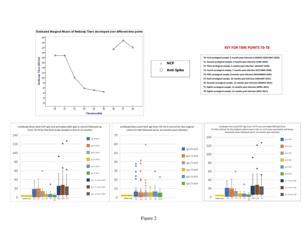
At 14 months post-infection in May 2021, the percentage of anti-SARS-CoV-2 S-RBD IgG
positive subjects were analyzed and 97.14% (34 out of 35) patients were positive for anti-
SARS-CoV-2 RBD IgG at 14 months. This was also observed for the preceding 12 and 13
months (34/35 positive for anti-S-RBD antibodies) but the median neutralization titer
differed at each time point. [Figure 2] In terms of disease severity, it was observed that in
both the groups, the antibody titers developed at different Time periods (TPs) were
discrete and statistically significant ( p value <0.001 for the mild group and the
Moderately- Severe group). The results are described in Table 3 [Table 3] Since the data
repeats over a period of time, multiple comparison test; i.e The Friedman’s Two-way
ANOVA test was applied. [Figure 3] In order to highlight the significance of the TPs with
respect to each other, pairwise multiple comparison tests; i.e Two/More related samples
test was applied.
Role of disease severity on antibody titers
Median neutralization titer (MNT) was calculated for both disease severity groups. It was
observed that the subjects of the moderately-severe group developed a higher median
antibody tire (14.78 AU/ml) at 14 months when compared to the mild group (5.55 AU/ml)
but the dispersion or variation was higher for the Moderately-Severe group, indicating a
larger degree of variability in development of antibodies. The box plot shows the Median
line at the 25th percentile in both the cases indicating that the data is positively skewed
i.e., some values are towards the higher end. The subjects in the mild group show less
variability in terms of developing antibody titers and have a smaller median while subjects
of the Modereately-Severe group exhibit larger variation and also has a higher median.
[Table 4]
DISCUSSION
The SARS-CoV-2 is an enveloped virus with four structural proteins: spike (S) protein,
membrane (M) protein, envelope (E) protein and nucleocapsid (N) protein. Among these
four structural proteins, the S and N proteins are the main immunogens. The S protein is
a major protective antigen that elicits highly potent neutralizing antibodies (nAbs) and
plays an essential role in viral attachment, fusion, entry, and transmission. The S protein
comprises of an N-terminal Sl subunit responsible for virus-receptor binding and a C-
terminal S2 subunit responsible for virus-cell membrane fusion. The S1 sub-unit is further
divided into an N-terminal domain (NTD) and a receptor-binding domain (RBD). The RBD
within Sl interacts directly with host receptors, human angiotensin converting enzyme 2
(hACE2). [18] The immunity against any infectious disease is comprised of two arms:
innate immunity and adaptive or acquired immunity. Each of these arms contain humoral
(B cells) and the cell mediated (T cell) immune elements.
Antibodies are synthesized and secreted by plasma cells that are derived from the B cells
of the immune system and can be used as a correlate of immunity. Antibody tests also
known as serological tests, detect the presence of antibodies against a particular disease-
causing agent in the blood, to evaluate the immune response against it. Antibodies can
come in different varieties known as isotypes or classes which differ in their biological
properties and ability to deal with different antigens and are called Immunoglobulins.
Immunoglobulin (IgM) eliminates pathogens in the early stages of B Cell mediated
immunity and is a marker of active infection, while immunoglobulin G (IgG) provides long
lasting antibody-mediated immunity and is the only antibody capable of crossing the
placenta to give passive immunity to the fetus.
In a recent study by Turner et al it was observed that the SARS-CoV-2 infection induces a
robust antigen-specific, long-lived humoral immune response in humans. The patients
who experienced mild infections (n=77), serum anti-SARS-CoV-2 spike (S) antibodies
declined rapidly in the first 4 months after infection and then more gradually over the
following 7 months, remaining detectable at least 11 months after infection. The S-
binding bone marrow plasma cells (BMPCs) are quiescent, indicating that they are part of
a long-lived compartment. [19]
The neutralizing antibodies (nAbs) are capable of preventing an infectious agent from
infecting a cell by neutralizing or inhibiting its biological effect. The most critical target
for SARS-CoV-2 nAbs is the RBD within the Sl subunit of S protein. Such nAbs can interrupt
the interaction of RBD and its receptor ACE2. Thus, SARS-CoV-2 S-RBD IgG antibody level
in human serum or plasma correlates with protective immune responses in individuals
who have recovered from SARS-CoV-2 infection and could also reflect herd immunity at
a population level aiding in planning of clinical management of patients with past or
ongoing COVID-19 infection. Anti-spike nAbs produced by COVID-19 patients can block
viral infection of human cells in vitro and counter viral replication in vivo. [20-22]
Neutralizing antibody titers (and total Spike antibody titers) have a positive correlation
with COVID-19 disease severity in large cohort studies. [7,11,23]. This was also observed
in our study findings. The subjects in the moderately-severe group developed a higher
Median Neutralisation Titre (14.78 AU/ml , p=0.003) at 14 months when compared to
the Mild group (5.55 AU/ml , p<0.001). [Table 4] This result was statistically significant.
However, the relationship between the neutralizing antibodies, T follicular helper cells
(Tfh cells), and COVID-19 disease severity appears to be complex. A higher neutralizing
antibody titer is associated with severe disease and potentially “extrafollicular B cell responses” [23] whereas the SARS-CoV-2-specific Tfh cells are associated differently.
Moreover, antibodies could act like a useful surrogate marker of CD4+ T cell responses
in many infections, since antibody assays are much easier to perform and more sensitive
in small blood volumes when compared to antigen-specific T cell assays. [24]
A recent study by Abu-Raddad LJ et al in Qatar, assessed the cumulative risk as well as
incidence rate of SARS-CoV-2 reinfection in a nationwide cohort of 43,044 antibody-
positive individuals. This study with a follow period of up to 35 weeks, demonstrated and
confirmed through viral genome sequencing that SARS-CoV-2 reinfection occurs, but
“only rarely” with a cumulative risk of ~2 per 1000 persons and reinfection incidence rate
of <1 per 10,000 person weeks as compared to the complement cohort of 149,923
antibody-negative persons with a much higher cumulative risk of re-infection (~31 per
1000 persons after 46 weeks of follow-up) and estimated incidence rate of infection (~14
per 10,000 person-weeks. ). The estimated efficacy of natural infection against reinfection
was 95%. Moreover, this study showed no evidence of waning protective immunity
against reinfection in this cohort for over 7 months. [25]
An important point that needs to be highlighted in our study is zero cases of re-infection
despite the fact that the Umbria region has been experiencing multiple waves with mutant
strains. [11]
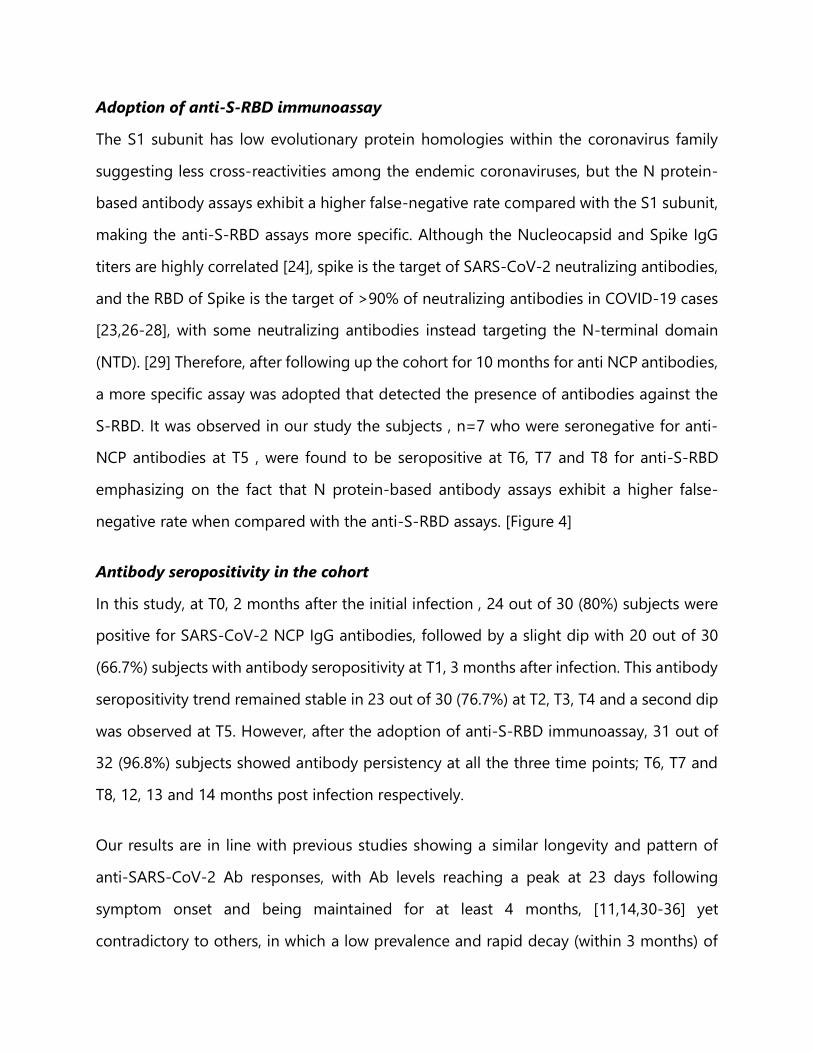
Adoption of anti-S-RBD immunoassay
The S1 subunit has low evolutionary protein homologies within the coronavirus family
suggesting less cross-reactivities among the endemic coronaviruses, but the N protein-
based antibody assays exhibit a higher false-negative rate compared with the S1 subunit,
making the anti-S-RBD assays more specific. Although the Nucleocapsid and Spike IgG
titers are highly correlated [24], spike is the target of SARS-CoV-2 neutralizing antibodies,
and the RBD of Spike is the target of >90% of neutralizing antibodies in COVID-19 cases
[23,26-28], with some neutralizing antibodies instead targeting the N-terminal domain
(NTD). [29] Therefore, after following up the cohort for 10 months for anti NCP antibodies,
a more specific assay was adopted that detected the presence of antibodies against the
S-RBD. It was observed in our study the subjects , n=7 who were seronegative for anti-
NCP antibodies at T5 , were found to be seropositive at T6, T7 and T8 for anti-S-RBD
emphasizing on the fact that N protein-based antibody assays exhibit a higher false-
negative rate when compared with the anti-S-RBD assays. [Figure 4]
Antibody seropositivity in the cohort
In this study, at T0, 2 months after the initial infection , 24 out of 30 (80%) subjects were
positive for SARS-CoV-2 NCP IgG antibodies, followed by a slight dip with 20 out of 30
(66.7%) subjects with antibody seropositivity at T1, 3 months after infection. This antibody
seropositivity trend remained stable in 23 out of 30 (76.7%) at T2, T3, T4 and a second dip
was observed at T5. However, after the adoption of anti-S-RBD immunoassay, 31 out of
32 (96.8%) subjects showed antibody persistency at all the three time points; T6, T7 and
T8, 12, 13 and 14 months post infection respectively.
Our results are in line with previous studies showing a similar longevity and pattern of
anti-SARS-CoV-2 Ab responses, with Ab levels reaching a peak at 23 days following
symptom onset and being maintained for at least 4 months, [11,14,30-36] yet
contradictory to others, in which a low prevalence and rapid decay (within 3 months) of
anti-SARS-CoV-2 Abs in COVID-19 patients with either mild or severe disease were
observed.[37,38]
Average Antibody titers [Anti-NCP IgG and Anti-S-RBD IgG] developed at 14 months
The estimated marginal mean or average of antibody titers (in AU/ml) developed over
different TPs such as T0-T5 and T6-T8 with respect to two different immunological assays
against NCP and Spike protein respectively. It can be observed that the average of Anti-
NCP IgG antibody titers tends to show a decreasing trend across T0-T5, whereas Anti-S-
RBD IgG shows a better average of antibody titers, in terms of magnitude (seropositivity)
across T6-T8.
In Conclusion, our study findings are consistent with recent studies reporting robust
antibody persistency suggesting that induced SARS-CoV-2 immunity through natural
infection, might be very efficacious against re-infection (>90%) and could persist for more
than six months. Our study followed-up patients up to 14 months demonstrating
presence of anti-S-RBD IgG in 96.8% of recovered COVID-19 subjects.
In such a scenario, prioritizing vaccination for “naive” individuals ( with no previous history of COVID-19 infection) and recovered but antibody-negative individuals would be helpful
by saving time, effort and resources that could be used for the vulnerable populations.
This study also provides valuable information for future implementation and vaccine
distribution policies. Further studies need to be conducted to determine antibody
responses in patients infected by mutant strains when compared to the original wild type.
References:
1. Lu R, Zhao X, Li J, et al. Genomic characterisation and epidemiology of 2019
novel coronavirus: implications for virus origins and receptor binding.
Lancet.2020 Feb 22;395(10224):565-574. doi: 10.1016/S0140-6736(20)30251-
8. Epub 2020 Jan 30. PMID: 32007145; PMCID: PMC7159086.
2. Wu LP, Wang NC, Chang YH et al. Duration of antibody responses after severe
acute respiratory syndrome. Emerg Infect Dis. 2007 Oct;13(10):1562-4. doi:
10.3201/eid1310.070576. PMID: 18258008; PMCID: PMC2851497.
3. Hall VJ, Foulkes S, Charlett A, et al. SARS-CoV-2 infection rates of antibody-
positive compared with antibody-negative health-care workers in England: a
large, multicentre, prospective cohort study (SIREN).Lancet. 2021 Apr
17;397(10283):1459-1469. doi: 10.1016/S0140-6736(21)00675-9. Epub 2021
Apr 9. PMID: 33844963; PMCID: PMC8040523.
4. Matusali G, Colavita F, Lapa D, Meschi S, Bordi L, Piselli P, Gagliardini R,
Corpolongo A, Nicastri E, Antinori A, Ippolito G, Capobianchi MR, Castilletti C,
Inmi Covid-Laboratory Team. SARS-CoV-2 Serum Neutralization Assay: A
Traditional Tool for a Brand-New Virus. Viruses. 2021 Apr 10;13(4):655
5. Grifoni, A., Weiskopf, D., Ramirez, S.I., Mateus, J., Dan, J.M., Moderbacher, C.R.,
Rawlings, S.A., Sutherland, A., Premkumar, L., Jadi, R.S., et al. (2020). Targets of
T cell responses to SARS-CoV-2 coronavirus in humans with COVID-19 disease
and unexposed individuals. Cell 181, 1489–1501.e15.
6. Juno, J.A., Tan, H.-X., Lee, W.S., Reynaldi, A., Kelly, H.G., Wragg, K., Esterbauer,
R., Kent, H.E., Batten, C.J., Mordant, F.L., et al. (2020). Humoral and circulating
follicular helper T cell responses in recovered patients with COVID19. Nat. Med.
26, 1428–1434.
7. Robbiani, D.F., Gaebler, C., Muecksch, F., Lorenzi, J.C.C., Wang, Z., Cho, A.,
Agudelo, M., Barnes, C.O., Gazumyan, A., Finkin, S., et al. (2020). Convergent
antibody responses to SARS-CoV-2 in convalescent individuals. Nature 584,
437–442
8. MAGLUMI 2019-nCoV IgM/IgG - Letter of Authorization (fda.gov)
9. World Health Organization. International guidelines for certification and
classification (coding) of COVID-19 as cause of death. Available from:
https://www.who.int/classifications/icd/Guidelines_Cause_of_Death_COVID-
19-20200420-EN.pdf?ua=1. Document Number: WHO/HQ/DDI/DNA/CAT.
Accessed on June 1, 2020.
10. Xydakis MS, Dehgani-Mobaraki P, Holbrook EH, et al. Smell and taste
dysfunction in patients with COVID-19. Lancet Infect Dis. 2020 Sep;20(9):1015-
1016. doi: 10.1016/S1473-3099(20)30293-0. Epub 2020 Apr 15. PMID:
32304629; PMCID: PMC7159875.
11. Dehgani-Mobaraki P, Kamber Zaidi A, Porreca A et. al. Antibody persistency and
trend post-SARS-CoV-2 infection at eight months. Ann Ig. 2021 May. doi:
10.7416/ai.2021.2455. Online ahead of print.
12. https://www.snibe.com/zh_en/en_newsView.aspx?id=647
13. Huang M, Lu QB, Zhao H, et al. Temporal antibody responses to SARS-CoV-2 in
patients of coronavirus disease 2019. Cell Discovery. 2020;6:64. doi:
https://dx.doi.org/10.1038/s41421-020-00209-2. PMID: 32983570.
14. Gudbjartsson DF, Norddahl GL, Melsted P, et al. Humoral Immune Response to
SARS-CoV-2 in Iceland. N Engl J Med. 2020 Sep 1. doi:
https://dx.doi.org/10.1056/NEJMoa2026116. PMID: 32871063.
15. Chirathaworn C, Sripramote M, Chalongviriyalert P, et al. SARS-CoV-2 RNA
shedding in recovered COVID-19 cases and the presence of antibodies against SARS-
CoV-2 in recovered COVID-19 cases and close contacts, Thailand, April-June 2020.
PLoS ONE. 2020;15(10):e0236905. doi:
https://dx.doi.org/10.1371/journal.pone.0236905. PMID: 33119712.
16. Terpos E, Politou M, Sergentanis TN, et al. Anti– SARS-CoV-2 Antibody Responses
in Convalescent Plasma Donors Are Increased in Hospitalized Patients;Subanalyses of
a Phase 2 Clinical Study. Microorganisms. 2020 2020;8(12):1885-. doi:
https://dx.doi.org/10.1080/22221751.2020.17625 15.
17. Zhao J, Yuan Q, Wang H, et al. Antibody Responses to SARS-CoV-2 in Patients With
Novel Coronavirus Disease 2019. Clin Infect Dis. 2020 11 19;71(16):2027-34. doi:
https://dx.doi.org/10.1093/cid/ciaa344. PMID: 32221519
18. Wang Q, Zhang Y, Wu L, Niu S, Song C, Zhang Z et al. Structural and functional
basis of SARS-CoV-2 entry by using human ACE2. Cell. 2020.
https://doi.org/10.1016/j.cell.2020.03.045.
19. Turner, J.S., Kim, W., Kalaidina, E. et al. SARS-CoV-2 infection induces long-lived bone
marrow plasma cells in humans. Nature (2021). https://doi.org/10.1038/s41586-021-
03647-4]
20. Zohar, T. et al. Compromised humoral functional evolution tracks with SARS-
CoV-2 mortality. Cell 183, 1508–1519 (2020). e12.
21. Wu, F. et al. Evaluating the association of clinical characteristics with neutralizing
antibody levels in patients who have recovered from mild COVID-19 in Shanghai. China
JAMA Intern. Med. 180, 1356–1362 (2020).
22. Wang, X. et al. Neutralizing antibody responses to severe acute respiratory syndrome
coronavirus 2 in coronavirus disease 2019 inpatients and convalescent patients. Clin.
Infect. Dis. https://doi.org/10.1093/cid/ciaa721 (2020).
23. Piccoli L, Park YJ, Tortorici MA, et al. Mapping Neutralizing and
Immunodominant Sites on the SARS-CoV-2 Spike Receptor-Binding Domain by
Structure-Guided High-Resolution Serology. Cell. 2020 Nov 12;183(4):1024-
1042.e21. doi: 10.1016/j.cell.2020.09.037. Epub 2020 Sep 16. PMID: 32991844;
PMCID: PMC7494283
24. Sette A, Crotty S. Adaptive immunity to SARS-CoV-2 and COVID-19. Cell. 2021 Feb
18;184(4):861-880. doi: 10.1016/j.cell.2021.01.007. Epub 2021 Jan 12. PMID: 33497610;
PMCID: PMC7803150.
25. Abu-Raddad LJ, Chemaitelly H, Coyle P, et al. SARS-CoV-2 antibody-positivity
protects against reinfection for at least seven months with 95% efficacy.
EClinicalMedicine. 2021 May;35:100861. doi: 10.1016/j.eclinm.2021.100861.
Epub 2021 Apr 28. PMID: 33937733; PMCID: PMC8079668.
26. Brouwer PJM, Caniels TG, van der Straten K, et al. Potent neutralizing antibodies
from COVID-19 patients define multiple targets of vulnerability. Science. 2020
Aug 7;369(6504):643-650. doi: 10.1126/science.abc5902. Epub 2020 Jun 15.
PMID: 32540902; PMCID: PMC7299281.
27. Rogers TF, Zhao F, Huang D, et al. Isolation of potent SARS-CoV-2 neutralizing
antibodies and protection from disease in a small animal model. Science. 2020
Aug 21;369(6506):956-963. doi: 10.1126/science.abc7520. Epub 2020 Jun 15.
PMID: 32540903; PMCID: PMC7299280.
28. Suthar MS, Zimmerman MG, Kauffman RC, et al. Rapid Generation of
Neutralizing Antibody Responses in COVID-19 Patients. Cell Rep Med. 2020 Jun
23;1(3):100040. doi: 10.1016/j.xcrm.2020.100040. Epub 2020 Jun 8. PMID:
32835303; PMCID: PMC7276302.
29. Liu L, Wang P, Nair M.S, et al. Potent neutralizing antibodies directed to multiple
epitopes on SARS-CoV-2 spike. bioRxiv. 2020;
https://doi.org/10.1101/2020.06.17.153486
30. Bolke, E., Matuschek, C., and Fischer, J.C. (2020). Loss of anti-SARS-CoV-2
antibodies in mild covid-19. N. Engl. J. Med. 383, 1694–1695.
31. Iyer, A.S., Jones, F.K., Nodoushani, A., Kelly, M., Becker, M., Slater, D., Mills, R.,
Teng, E., Kamruzzaman, M., Garcia-Beltran, W.F., et al. (2020). Persistence and
decay of human antibody responses to the receptor binding domain of SARS-
CoV-2 spike protein in COVID-19 patients. Sci. Immunol. 5, eabe0367.
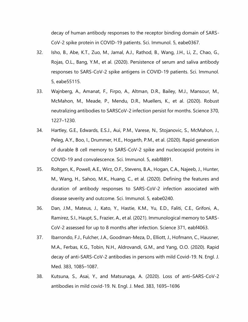
32. Isho, B., Abe, K.T., Zuo, M., Jamal, A.J., Rathod, B., Wang, J.H., Li, Z., Chao, G.,
Rojas, O.L., Bang, Y.M., et al. (2020). Persistence of serum and saliva antibody
responses to SARS-CoV-2 spike antigens in COVID-19 patients. Sci. Immunol.
5, eabe55115.
33. Wajnberg, A., Amanat, F., Firpo, A., Altman, D.R., Bailey, M.J., Mansour, M.,
McMahon, M., Meade, P., Mendu, D.R., Muellers, K., et al. (2020). Robust
neutralizing antibodies to SARSCoV-2 infection persist for months. Science 370,
1227–1230.
34. Hartley, G.E., Edwards, E.S.J., Aui, P.M., Varese, N., Stojanovic, S., McMahon, J.,
Peleg, A.Y., Boo, I., Drummer, H.E., Hogarth, P.M., et al. (2020). Rapid generation
of durable B cell memory to SARS-CoV-2 spike and nucleocapsid proteins in
COVID-19 and convalescence. Sci. Immunol. 5, eabf8891.
35. Roltgen, K., Powell, A.E., Wirz, O.F., Stevens, B.A., Hogan, C.A., Najeeb, J., Hunter,
M., Wang, H., Sahoo, M.K., Huang, C., et al. (2020). Defining the features and
duration of antibody responses to SARS-CoV-2 infection associated with
disease severity and outcome. Sci. Immunol. 5, eabe0240.
36. Dan, J.M., Mateus, J., Kato, Y., Hastie, K.M., Yu, E.D., Faliti, C.E., Grifoni, A.,
Ramirez, S.I., Haupt, S., Frazier, A., et al. (2021). Immunological memory to SARS-
CoV-2 assessed for up to 8 months after infection. Science 371, eabf4063.
37. Ibarrondo, F.J., Fulcher, J.A., Goodman-Meza, D., Elliott, J., Hofmann, C., Hausner,
M.A., Ferbas, K.G., Tobin, N.H., Aldrovandi, G.M., and Yang, O.O. (2020). Rapid
decay of anti-SARS-CoV-2 antibodies in persons with mild Covid-19. N. Engl. J.
Med. 383, 1085–1087.
38. Kutsuna, S., Asai, Y., and Matsunaga, A. (2020). Loss of anti–SARS-CoV-2
antibodies in mild covid-19. N. Engl. J. Med. 383, 1695–1696
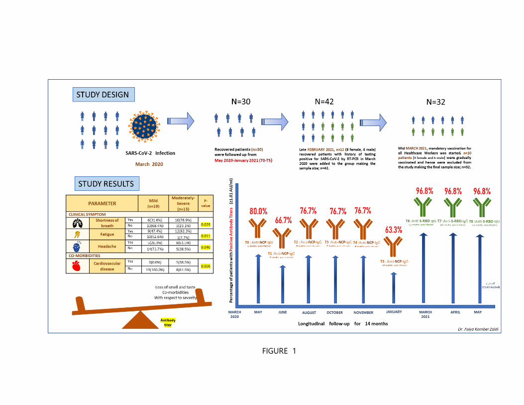
FIGURE 1
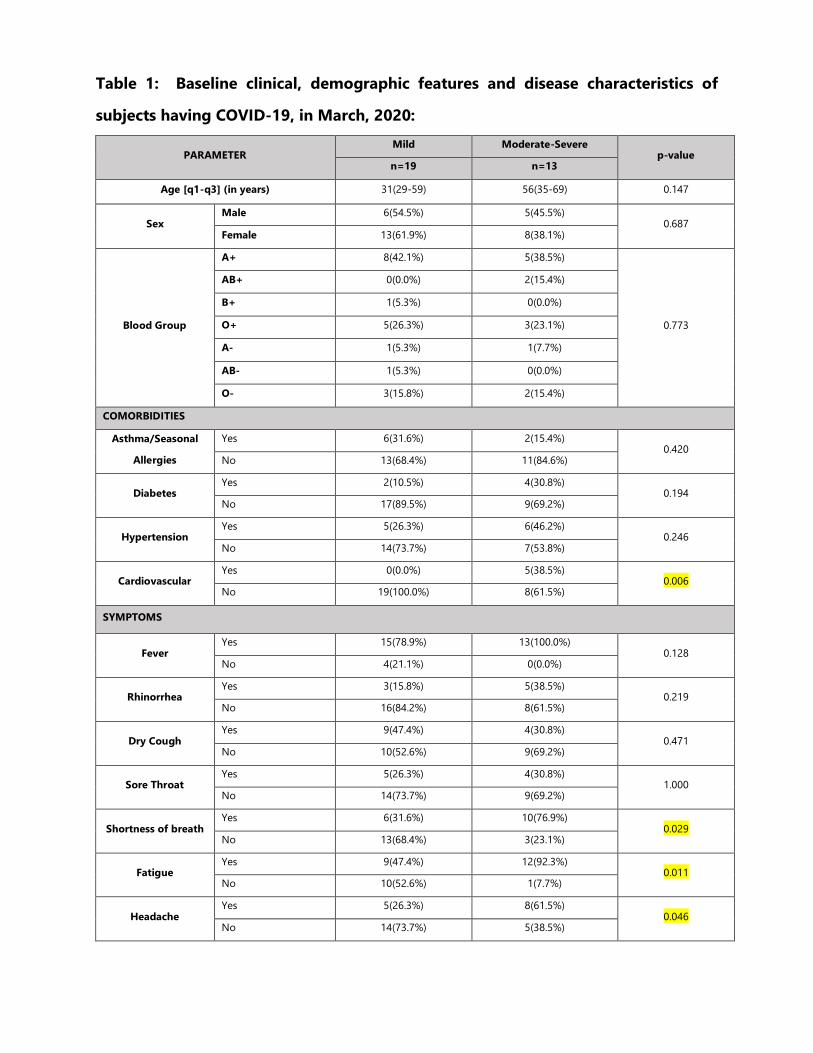
Table 1: Baseline clinical, demographic features and disease characteristics of
subjects having COVID-19, in March, 2020:
PARAMETER Mild Moderate-Severe
p-value n=19 n=13
Age [q1-q3] (in years) 31(29-59) 56(35-69) 0.147
Sex Male 6(54.5%) 5(45.5%)
0.687 Female 13(61.9%) 8(38.1%)
Blood Group
A+ 8(42.1%) 5(38.5%)
0.773
AB+ 0(0.0%) 2(15.4%)
B+ 1(5.3%) 0(0.0%)
O+ 5(26.3%) 3(23.1%)
A- 1(5.3%) 1(7.7%)
AB- 1(5.3%) 0(0.0%)
O- 3(15.8%) 2(15.4%)
COMORBIDITIES
Asthma/Seasonal
Allergies
Yes 6(31.6%) 2(15.4%) 0.420
No 13(68.4%) 11(84.6%)
Diabetes Yes 2(10.5%) 4(30.8%)
0.194 No 17(89.5%) 9(69.2%)
Hypertension Yes 5(26.3%) 6(46.2%)
0.246 No 14(73.7%) 7(53.8%)
Cardiovascular Yes 0(0.0%) 5(38.5%)
0.006 No 19(100.0%) 8(61.5%)
SYMPTOMS
Fever Yes 15(78.9%) 13(100.0%)
0.128 No 4(21.1%) 0(0.0%)
Rhinorrhea Yes 3(15.8%) 5(38.5%)
0.219 No 16(84.2%) 8(61.5%)
Dry Cough Yes 9(47.4%) 4(30.8%)
0.471 No 10(52.6%) 9(69.2%)
Sore Throat Yes 5(26.3%) 4(30.8%)
1.000 No 14(73.7%) 9(69.2%)
Shortness of breath Yes 6(31.6%) 10(76.9%)
0.029 No 13(68.4%) 3(23.1%)
Fatigue Yes 9(47.4%) 12(92.3%)
0.011 No 10(52.6%) 1(7.7%)
Headache Yes 5(26.3%) 8(61.5%)
0.046 No 14(73.7%) 5(38.5%)
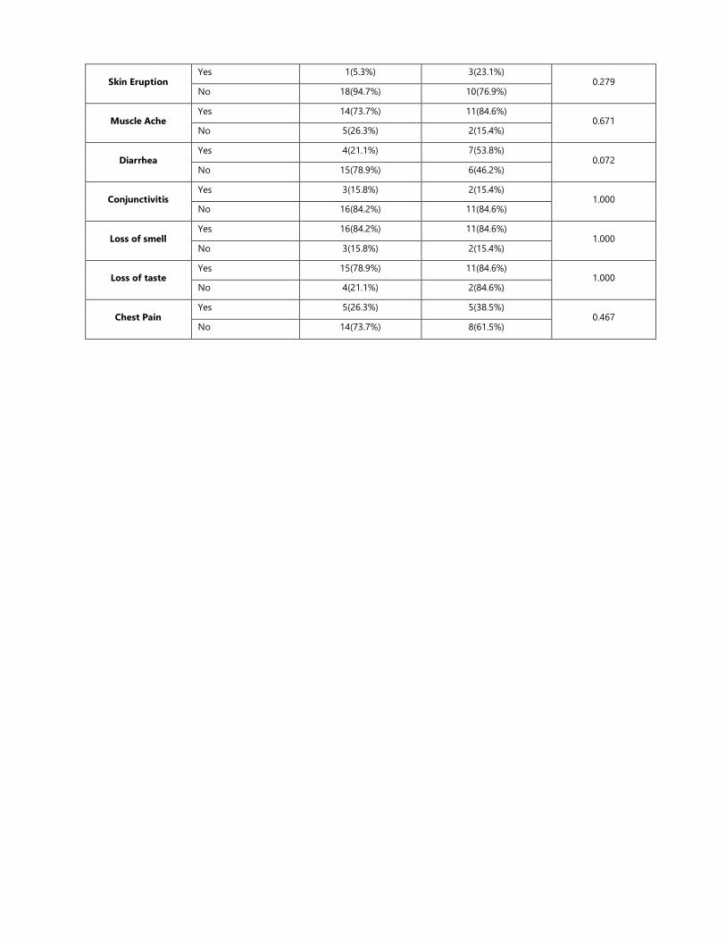
Skin Eruption Yes 1(5.3%) 3(23.1%)
0.279 No 18(94.7%) 10(76.9%)
Muscle Ache Yes 14(73.7%) 11(84.6%)
0.671 No 5(26.3%) 2(15.4%)
Diarrhea Yes 4(21.1%) 7(53.8%)
0.072 No 15(78.9%) 6(46.2%)
Conjunctivitis Yes 3(15.8%) 2(15.4%)
1.000 No 16(84.2%) 11(84.6%)
Loss of smell Yes 16(84.2%) 11(84.6%)
1.000 No 3(15.8%) 2(15.4%)
Loss of taste Yes 15(78.9%) 11(84.6%)
1.000 No 4(21.1%) 2(84.6%)
Chest Pain Yes 5(26.3%) 5(38.5%)
0.467 No 14(73.7%) 8(61.5%)
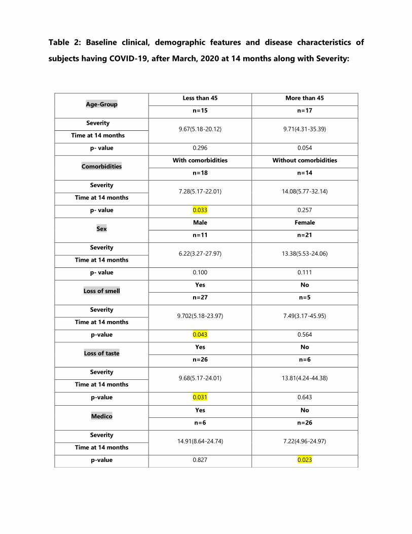
Table 2: Baseline clinical, demographic features and disease characteristics of
subjects having COVID-19, after March, 2020 at 14 months along with Severity:
Age-Group Less than 45 More than 45
n=15 n=17
Severity 9.67(5.18-20.12) 9.71(4.31-35.39)
Time at 14 months
p- value 0.296 0.054
Comorbidities With comorbidities Without comorbidities
n=18 n=14
Severity 7.28(5.17-22.01) 14.08(5.77-32.14)
Time at 14 months
p- value 0.033 0.257
Sex Male Female
n=11 n=21
Severity 6.22(3.27-27.97) 13.38(5.53-24.06)
Time at 14 months
p- value 0.100 0.111
Loss of smell Yes No
n=27 n=5
Severity 9.702(5.18-23.97) 7.49(3.17-45.95)
Time at 14 months
p-value 0.043 0.564
Loss of taste Yes No
n=26 n=6
Severity 9.68(5.17-24.01) 13.81(4.24-44.38)
Time at 14 months
p-value 0.031 0.643
Medico Yes No
n=6 n=26
Severity 14.91(8.64-24.74) 7.22(4.96-24.97)
Time at 14 months
p-value 0.827 0.023
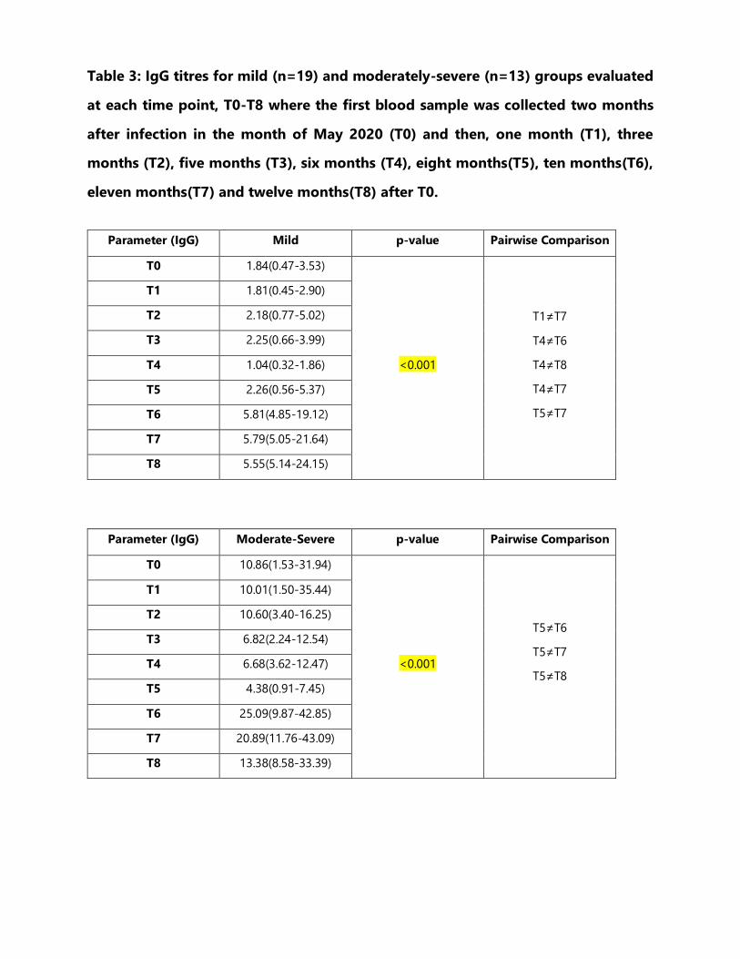
Table 3: IgG titres for mild (n=19) and moderately-severe (n=13) groups evaluated
at each time point, T0-T8 where the first blood sample was collected two months
after infection in the month of May 2020 (T0) and then, one month (T1), three
months (T2), five months (T3), six months (T4), eight months(T5), ten months(T6),
eleven months(T7) and twelve months(T8) after T0.
Parameter (IgG) Mild p-value Pairwise Comparison
T0 1.84(0.47-3.53)
<0.001
T1≠T7
T4≠T6
T4≠T8
T4≠T7
T5≠T7
T1 1.81(0.45-2.90)
T2 2.18(0.77-5.02)
T3 2.25(0.66-3.99)
T4 1.04(0.32-1.86)
T5 2.26(0.56-5.37)
T6 5.81(4.85-19.12)
T7 5.79(5.05-21.64)
T8 5.55(5.14-24.15)
Parameter (IgG) Moderate-Severe p-value Pairwise Comparison
T0 10.86(1.53-31.94)
<0.001
T5≠T6
T5≠T7
T5≠T8
T1 10.01(1.50-35.44)
T2 10.60(3.40-16.25)
T3 6.82(2.24-12.54)
T4 6.68(3.62-12.47)
T5 4.38(0.91-7.45)
T6 25.09(9.87-42.85)
T7 20.89(11.76-43.09)
T8 13.38(8.58-33.39)
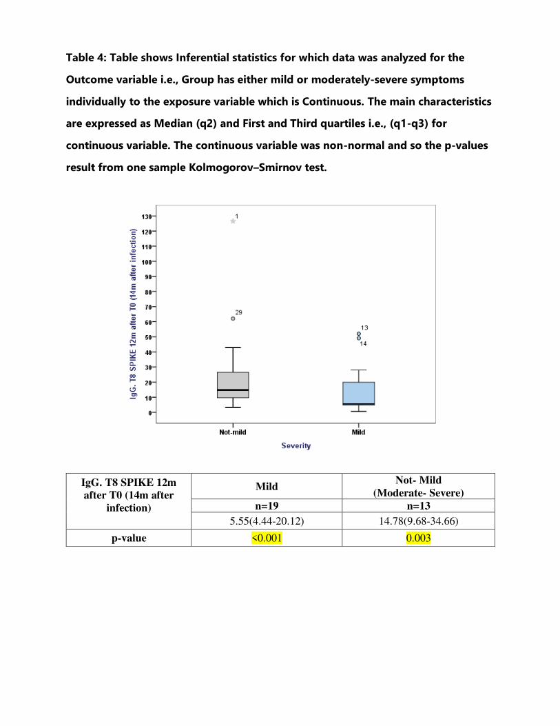
Table 4: Table shows Inferential statistics for which data was analyzed for the
Outcome variable i.e., Group has either mild or moderately-severe symptoms
individually to the exposure variable which is Continuous. The main characteristics
are expressed as Median (q2) and First and Third quartiles i.e., (q1-q3) for
continuous variable. The continuous variable was non-normal and so the p-values
result from one sample Kolmogorov–Smirnov test.
IgG. T8 SPIKE 12m
after T0 (14m after
infection)
Mild Not- Mild
(Moderate- Severe)
n=19 n=13
5.55(4.44-20.12) 14.78(9.68-34.66)
p-value <0.001 0.003
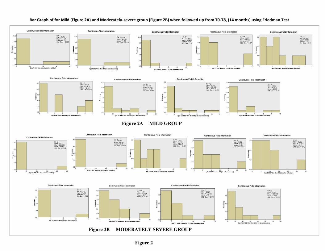
Bar Graph of for Mild (Figure 2A) and Moderately-severe group (Figure 2B) when followed up from T0-T8, (14 months) using Friedman Test
Figure 2B MODERATELY SEVERE GROUP
Figure 2A MILD GROUP
Figure 2
o NCP
□ Anti-Spike
Figure 2
MARCH 2020 MAY JUNE AUG OCT NOV JAN MARCH APRIL MAY
Period of infection
T0: First serological sample, 2 months post infection in MARCH 2020 (MAY 2020)
T1: Second serological sample, 3 months post infection (JUNE 2020)
T2: Third serological sample, 5 months post infection (AUGUST 2020)
T3: Fourth serological sample, 7 months post infection (OCTOBER 2020)
T4: Fifth serological sample, 8 months post infection (NOVEMBER 2020)
T5: Sixth serological sample, 10 months post infection (JANUARY 2021)
T6: Seventh serological sample, 12 months post infection (MARCH 2021)
T7: Eighth serological sample, 13 months post infection (APRIL 2021)
T8: Eighth serological sample, 14 months post infection (MAY 2021)
KEY FOR TIME POINTS T0-T8
MAY JUNE AUG OCT NOV JAN 2021 MARCH 2020
Period of infection Period of infection
MARCH 2020 MAY JUNE AUG OCT NOV JAN MARCH APRIL MAY
2021 2021
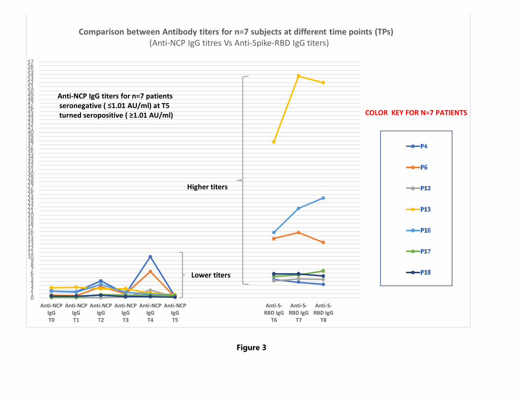
Figure 3
0123456789
101112131415161718192021222324252627282930313233343536373839404142434445464748495051525354555657
Anti-NCP
IgG
T0
Anti-NCP
IgG
T1
Anti-NCP
IgG
T2
Anti-NCP
IgG
T3
Anti-NCP
IgG
T4
Anti-NCP
IgG
T5
Anti-S-
RBD IgG
T6
Anti-S-
RBD IgG
T7
Anti-S-
RBD IgG
T8
Comparison between Antibody titers for n=7 subjects at different time points (TPs)
(Anti-NCP IgG titres Vs Anti-Spike-RBD IgG titers)
Anti-NCP IgG titers for n=7 patients
seronegative ( ≤1.01 AU/ml) at T5turned seropositive ( ≥1.01 AU/ml)
Lower titers
Higher titers
COLOR KEY FOR N=7 PATIENTS


![SARS-CoV-2 (COVID-19) Spike S1 Antibody [SP422] PDF](https://img.pdfslide.net/doc/110x75/61fc5597b12cd0168f6148e6/sars-cov-2-covid-19-spike-s1-antibody-sp422-pdf.jpg)