Embed Size (px)
Citation preview
8/13/2019 Lorenzo Calabro Thesis
http://slidepdf.com/reader/full/lorenzo-calabro-thesis 1/54
Lorenzo Calabro 0
SCIENCE AND ENGINEERING FACULTY, QUT
Improving In vivo Models ofFracture Fixation Associated
Osteomyelitis Research Masters IFT637
LORENZO CALABRO B Phty;MBBS Hons
10/14/2013
Supervisors
Professor Michael Schuetz
Dr Roland Steck
Dr T. Fintan MoriartySubmitted in fulfilment of the requirements for the degree of
Masters of Engineering (Research)
8/13/2019 Lorenzo Calabro Thesis
http://slidepdf.com/reader/full/lorenzo-calabro-thesis 2/54
Lorenzo Calabro 1
AbstractOrthopaedic implant associated infection causes significant morbidity and social cost. It occurs mostfrequently following trauma surgery and is influenced by an array of patient, surgical, implant and injury
factors. Animal research has been integral to our understanding of these factors. Large, well controlledclinical trials of new implant technology are logistically difficult and prohibitively expensive, makingcontinued animal research essential. There is an imperative however, to minimise harm in animal traumaresearch. The central goal of this project was to refine existing animal models in order to reduce thenumber of animals required to generate clinically relevant outcomes. Specifically, the primary aim was tocreate a new, reliable, repeatable, standard, femoral fracture model in rabbits which would have potentialfor wide application in future trauma implant research. The secondary aim of this project was to investigatenovel bacterial inoculation techniques for application in musculoskeletal infection studies.
Method
Two parallel rabbit femur fixation contructs were investigated – the RabbitNail TM, a custom designedretrograde intramedullary (IM) nail, and a locked plate construct. Ex vivo mechanical testing of constructswas performed using 24 thawed cadaveric rabbit femora. A limited in vivo trial was subsequentlyperformed (n=8) where fixation constructs were used to fix a mid diaphyseal osteotomy of right femur of ananaesthetised NZ white rabbit. Animals were euthanized after 7 weeks and fracture healing assessed withnon-destructive mechanical tests and qualitative histology. Left femurs were tested as controls. Inoculationexperiments were performed to evaluate the effect of viscous suspension media on bacterial viability ofStaphylococcus aureus . Four candidate suspension media were tested: Glycerol, CarboxymethylCellulose
(CMC), Gelatine and aqueous ultrasound gel. Serial cultures were performed after two hours suspensionin each medium and compared to bacteria suspended in phosphate buffered saline (PBS).
Results
Ex vivo mechanical testing was performed on 24 femora in 5 groups. Locked plate and RabbitNailconstructs had similar torsional stiffness and yield loads and both were considered adequate for a limited in
vivo trial. The RabbitNail was implanted in 3 rabbits. Two animals sustained an early periprosthetic
fracture and were euthanized. Locked plate fixation of two different sizes was used in 5 rabbits. Threeanimals went on to robust fracture healing with torsional stiffness of healed fractures comparable to theintact contralateral femur, while one healed despite screw breakage and another animal was euthanizedearly following periprosthetic fracture. Further in vivo experiments were suspended due to the highincidence of periprosthetic fracture.
Glycerol and CMC and aqueous ultrasound gel had favourable handling characteristics for amusculoskeletal infection trial. Physical properties of Gelatine precluded its feasible use in fracture
inoculation. Aqueous ultrasound gel consistently hindered bacterial growth. Two hours of suspension in
8/13/2019 Lorenzo Calabro Thesis
http://slidepdf.com/reader/full/lorenzo-calabro-thesis 3/54
Lorenzo Calabro 2
glycerol had little overall effect on bacterial growth, while bacteria suspended in CMC consistently showedlow magnitude, positive growth. Repeat cultures showed greater variability for both, than with PBS.
Conclusions
These results suggest that a femoral fracture model in mature NZ white rabbits is likely to have anunacceptable surgical complication rate. Future animal fracture model research should focus on a different
animal, or different bone. Glycerol and CMC suspension media have potential to improve in vivo musculoskeletal infection trials. Further research including an anatomical study of disseminationcharacteristics at the fracture site and an in vivo trial would be useful.
8/13/2019 Lorenzo Calabro Thesis
http://slidepdf.com/reader/full/lorenzo-calabro-thesis 4/54
Lorenzo Calabro 3
Contents Abstract ........................................................................................................................................................ 1
Method ................................................................................................................................................... 1
Results ................................................................................................................................................... 1
Conclusions ........................................................................................................................................... 2
List of Figures ............................................................................................................................................... 5
List of Tables ................................................................................................................................................ 6
List of Abbreviations ..................................................................................................................................... 6
Statement of Individual Contribution and Original Authorship ....................................................................... 1
Acknowledgments ........................................................................................................................................ 1
Keywords...................................................................................................................................................... 1
1. Introduction ............................................................................................................................................... 2
1.1 Background ......................................................................................................................................... 2
1.2 Research Problem ............................................................................................................................... 3
1.3 Research Questions, Goals, Hypotheses ............................................................................................ 4
2. Literature review ....................................................................................................................................... 4
2.1 Species / model selection .................................................................................................................... 4
2.2 Orthopaedic Trauma in animal models ................................................................................................ 62.3 Existing fracture + infection models ..................................................................................................... 6
2.4 Inoculation methods ............................................................................................................................ 7
2.5 Mechanobiology in fixation constructs ................................................................................................. 8
2.6 Physiologic stress in animals and relevant testing ............................................................................... 8
2.7 Summary ............................................................................................................................................. 8
3. Research Design ...................................................................................................................................... 93.1 Methodology Overview ........................................................................................................................ 9
3.2 Experimental Models ........................................................................................................................... 9
Locked plate ........................................................................................................................................... 9
IM Nail .................................................................................................................................................. 10
3.3 Ex vivo experiments .......................................................................................................................... 10
3.4 In vivo surgery ................................................................................................................................... 11
8/13/2019 Lorenzo Calabro Thesis
http://slidepdf.com/reader/full/lorenzo-calabro-thesis 5/54
Lorenzo Calabro 4
Surgical technique overview ................................................................................................................. 11
LCP surgical technique ........................................................................................................................ 11
IM nail surgical technique ..................................................................................................................... 12
Post-operative care .............................................................................................................................. 12
3.5 Outcome measures ........................................................................................................................... 12
Mechanical testing ............................................................................................................................... 12
Histology .............................................................................................................................................. 13
3.6 In Vitro inoculation media experiments .............................................................................................. 14
Media ................................................................................................................................................... 14
Bacterial strain ..................................................................................................................................... 15
Inoculation Media preparation .............................................................................................................. 15
4. Results ................................................................................................................................................... 18
4.1 Ex vivo (cadaveric) testing ................................................................................................................. 18
Figure 5 Ex vivo test results ........................................................................................................................ 18
4.2 In vivo Surgery .................................................................................................................................. 19
4.3 IM Nail (n=3) ...................................................................................................................................... 20
Nail 1 .................................................................................................................................................... 20
Nail 2 .................................................................................................................................................... 21
Nail 3 .................................................................................................................................................... 23
4.4 Locking plate fixation ......................................................................................................................... 25
2.0mm Plate 1 ...................................................................................................................................... 25
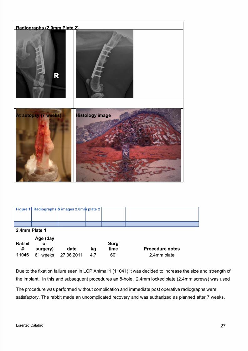
2.0mm Plate 2 ...................................................................................................................................... 26
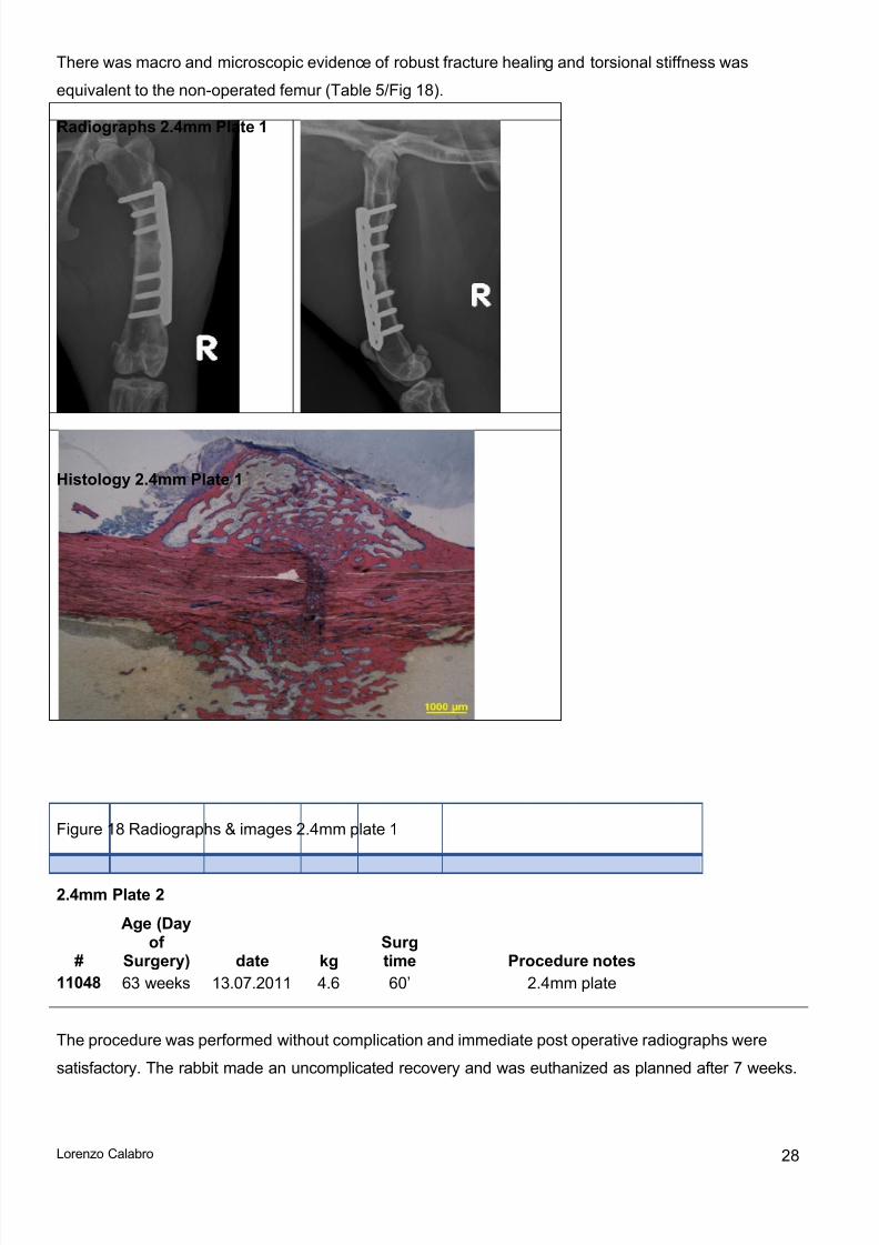
2.4mm Plate 1 ...................................................................................................................................... 27
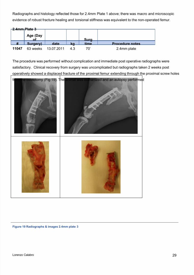
2.4mm Plate 2 ...................................................................................................................................... 282.4mm Plate 3 ...................................................................................................................................... 29
4.5 Inoculation media experiments .......................................................................................................... 30
AquasonicTM gel ................................................................................................................................... 32
Gelatine ............................................................................................................................................... 32
Glycerol ................................................................................................................................................ 33
Carboxymethyl-cellulose (CMC) ........................................................................................................... 33
8/13/2019 Lorenzo Calabro Thesis
http://slidepdf.com/reader/full/lorenzo-calabro-thesis 6/54
Lorenzo Calabro 5
5. Discussion .............................................................................................................................................. 34
IM Nail ..................................................................................................................................................... 34
Locked plates .......................................................................................................................................... 34
Failure mechanisms and potential solutions ............................................................................................ 35
Rabbits as animal models for fracture healing ......................................................................................... 36
In vitro inoculation media experiments ..................................................................................................... 38
6. Conclusions ............................................................................................................................................ 39
7. Reference List ........................................................................................................................................ 40
8. Appendix ................................................................................................................................................ 43
Conferences and Publications ................................................................................................................. 43
Textbook chapter ................................................................................................................................. 43
Australian Orthopaedic Association, Annual Scientific Meeting, Sydney, October 2012. ...................... 44
Australian Orthopaedic Association QLD Branch, Annual Scientific Meeting, Brisbane, June 2012..... 45
List of Figures
Figure 1 Rabbit Nail shown with aiming device for placing interlocking pins ............................................... 10Figure 2 Example radiographs of 2.0mm locked plate construct ................................................................. 11Figure 3 Mechanical testing setup ............................................................................................................. 13Figure 4. Inoculation Methodology. ............................................................................................................ 17Figure 5 Ex vivo test results ........................................................................................................................ 18Figure 6 Ex vivo test results ........................................................................................................................ 18Figure 7 Ex vivo test images ....................................................................................................................... 19Figure 8 Radiographs Nail 1 ....................................................................................................................... 20
Figure 9 Radiographs & Autopsy image Nail 1 ........................................................................................... 21Figure 10 Radiographs Nail 2 ..................................................................................................................... 22Figure 11 Autopsy images Nail 2 ................................................................................................................ 22Figure 12 Failure mechanism diagram Nail 2 .............................................................................................. 23Figure 13 Radiographs Nail 3 ..................................................................................................................... 24Figure 14 Autopsy images Nail 3 ................................................................................................................ 24Figure 15 Failure mechanism diagram Nail 3 .............................................................................................. 24Figure 16 Radiographs & images 2.0mm plate 1 ........................................................................................ 26
Figure 17 Radiographs & images 2.0mm plate 2 ........................................................................................ 27
8/13/2019 Lorenzo Calabro Thesis
http://slidepdf.com/reader/full/lorenzo-calabro-thesis 7/54
Lorenzo Calabro 6
Figure 18 Radiographs & images 2.4mm plate 1 ........................................................................................ 28Figure 19 Radiographs & images 2.4mm plate 3 ........................................................................................ 29Figure 20 Log10 Mean Bacterial growth ....................................................................................................... 31Figure 21. Bacterial growth in Gelatine. Log10 (CFU @ 120'/ CFU @ 5'). .................................................... 32Figure 22. Bacterial growth in Glycerol. Log10 (CFU @ 120'/ CFU @ 5'). .................................................... 33Figure 23. Bacterial growth in Carboxymethyl Cellulose. Log10 (CFU @ 120'/ CFU @ 5'). .......................... 33
List of TablesTable 1 Ex vivo test groups ........................................................................................................................ 10
Table 2. Preliminary empiric evaluation of candidate media....................................................................... 14Table 3 Ex vivo mechanical test results ...................................................................................................... 18Table 4 Summary in vivo results IM nail ..................................................................................................... 20
Table 5 Summary LCP results .................................................................................................................... 25Table 6 Bacterial growth expressed as a proportion. .................................................................................. 31Table 7 Weight range and mean weight NZ White Rabbits. NA= (Not available) ....................................... 37
List of AbbreviationsIM Intra-medullaryPBS Phosphate buffered salineNZ New ZealandCMC Sodium Carboxymethyl CelluloseS. aureus Staphylococcus aureus
AO Arbeitsgemeinschaft fürOsteosynthesfragen / Associationfor the Study of Internal Fixation
ARI AO Research Institute
CH Switzerland
CT Computed tomographyLCP Locking Compression PlatePMMA Polymethyl MethacrylateCSL Compound sodium lactateMTS Material testing systemTSA Tryptic Soy AgarTSB Tryptic Soy BrothOD Optical DensityCFU Colony Forming Units
8/13/2019 Lorenzo Calabro Thesis
http://slidepdf.com/reader/full/lorenzo-calabro-thesis 8/54
Lorenzo Calabro 1
Statement of Individual Contribution and Original Authorship As the principle investigator in this trial I have been involved in procedure design for the locked plate-osteotomy and IM nail-osteotomy models. With the collaboration of Dr T. Fintan Moriarty, Dr Roland Steck,Romano Matthys and other collaborators at the ARI I planned and initiated experimental work. I performedall cadaver surgery and worked in the surgical team at the ARI as the primary surgeon for all in vivo procedures in this study. I planned and oversaw mechanical testing and histologic analysis. I performed allmicrobiologic experiments with the supervision of Dr T Fintan Moriarty. I am the original author of thisthesis.
AcknowledgmentsThis work was undertaken as part of an AO Research Institute Research Fellowship and was funded by AOTrauma. In addition to my research supervisors, I benefited enormously from the assistance and support ofRomano Matthys, Iris Keller, Stephan Zeiter, Daniel Arens, Aswin Beck, Ivan Zderic and Nora Goudsouzian
at the ARI, Davos, Switzerland.
Keywords Animal model, Fracture, Musculoskeletal infection, prosthetic, Orthopaedics, Osteomyelitis, Rabbit

8/13/2019 Lorenzo Calabro Thesis
http://slidepdf.com/reader/full/lorenzo-calabro-thesis 9/54
Statement of Original Authorship
The work contained in this thesis has not been previously submitted to meet
requirements for an award at this or any other higher education institution. To the
best of my knowledge and belief, the thesis contains no material previously published
or written by another person except where due reference is made.
Date: (lttl tI
8/13/2019 Lorenzo Calabro Thesis
http://slidepdf.com/reader/full/lorenzo-calabro-thesis 10/54
Lorenzo Calabro 2
1. Introduction
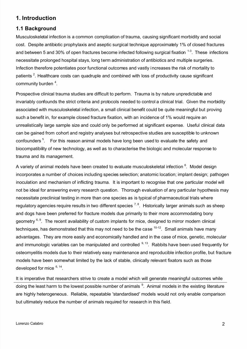
1.1 BackgroundMusculoskeletal infection is a common complication of trauma, causing significant morbidity and socialcost. Despite antibiotic prophylaxis and aseptic surgical technique approximately 1% of closed fracturesand between 5 and 30% of open fractures become infected following surgical fixation 1-3. These infections
necessitate prolonged hospital stays, long term administration of antibiotics and multiple surgeries.Infection therefore potentiates poor functional outcomes and vastly increases the risk of mortality topatients 2. Healthcare costs can quadruple and combined with loss of productivity cause significantcommunity burden 4.
Prospective clinical trauma studies are difficult to perform. Trauma is by nature unpredictable andinvariably confounds the strict criteria and protocols needed to control a clinical trial. Given the morbidityassociated with musculoskeletal infection, a small clinical benefit could be quite meaningful but provingsuch a benefit in, for example closed fracture fixation, with an incidence of 1% would require anunrealistically large sample size and could only be performed at significant expense. Useful clinical datacan be gained from cohort and registry analyses but retrospective studies are susceptible to unknownconfounders 5. For this reason animal models have long been used to evaluate the safety andbiocompatibility of new technology, as well as to characterise the biologic and molecular response totrauma and its management.
A variety of animal models have been created to evaluate musculoskeletal infection 6. Model designincorporates a number of choices including species selection; anatomic location; implant design; pathogen
inoculation and mechanism of inflicting trauma. It is important to recognise that one particular model willnot be ideal for answering every research question. Thorough evaluation of any particular hypothesis maynecessitate preclinical testing in more than one species as is typical of pharmaceutical trials whereregulatory agencies require results in two different species 7, 8. Historically larger animals such as sheepand dogs have been preferred for fracture models due primarily to their more accommodating bonygeometry 6, 9. The recent availability of custom implants for mice, designed to mirror modern clinicaltechniques, has demonstrated that this may not need to be the case 10-12. Small animals have manyadvantages. They are more easily and economically handled and in the case of mice, genetic, molecular
and immunologic variables can be manipulated and controlled 9, 13. Rabbits have been used frequently forosteomyelitis models due to their relatively easy maintenance and reproducible infection profile, but fracturemodels have been somewhat limited by the lack of stable, clinically relevant fixators such as thosedeveloped for mice 6, 14.
It is imperative that researchers strive to create a model which will generate meaningful outcomes whiledoing the least harm to the lowest possible number of animals 9. Animal models in the existing literatureare highly heterogeneous. Reliable, repeatable 'standardised' models would not only enable comparisonbut ultimately reduce the number of animals required for research in this field.
8/13/2019 Lorenzo Calabro Thesis
http://slidepdf.com/reader/full/lorenzo-calabro-thesis 11/54
Lorenzo Calabro 3
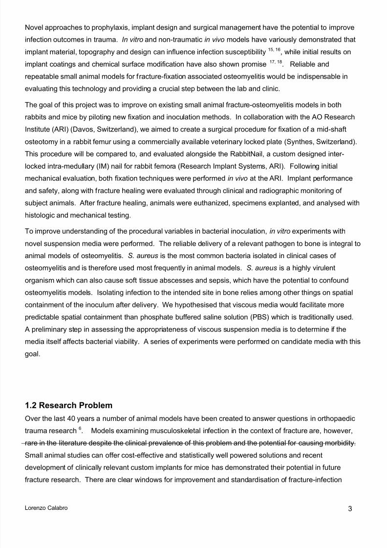
Novel approaches to prophylaxis, implant design and surgical management have the potential to improveinfection outcomes in trauma. In vitro and non-traumatic in vivo models have variously demonstrated thatimplant material, topography and design can influence infection susceptibility 15, 16, while initial results onimplant coatings and chemical surface modification have also shown promise 17, 18. Reliable andrepeatable small animal models for fracture-fixation associated osteomyelitis would be indispensable inevaluating this technology and providing a crucial step between the lab and clinic.
The goal of this project was to improve on existing small animal fracture-osteomyelitis models in bothrabbits and mice by piloting new fixation and inoculation methods. In collaboration with the AO ResearchInstitute (ARI) (Davos, Switzerland), we aimed to create a surgical procedure for fixation of a mid-shaftosteotomy in a rabbit femur using a commercially available veterinary locked plate (Synthes, Switzerland).This procedure will be compared to, and evaluated alongside the RabbitNail, a custom designed inter-locked intra-medullary (IM) nail for rabbit femora (Research Implant Systems, ARI). Following initialmechanical evaluation, both fixation techniques were performed in vivo at the ARI. Implant performance
and safety, along with fracture healing were evaluated through clinical and radiographic monitoring ofsubject animals. After fracture healing, animals were euthanized, specimens explanted, and analysed withhistologic and mechanical testing.
To improve understanding of the procedural variables in bacterial inoculation, in vitro experiments withnovel suspension media were performed. The reliable delivery of a relevant pathogen to bone is integral toanimal models of osteomyelitis. S. aureus is the most common bacteria isolated in clinical cases ofosteomyelitis and is therefore used most frequently in animal models. S. aureus is a highly virulentorganism which can also cause soft tissue abscesses and sepsis, which have the potential to confoundosteomyelitis models. Isolating infection to the intended site in bone relies among other things on spatialcontainment of the inoculum after delivery. We hypothesised that viscous media would facilitate morepredictable spatial containment than phosphate buffered saline solution (PBS) which is traditionally used.
A preliminary step in assessing the appropriateness of viscous suspension media is to determine if themedia itself affects bacterial viability. A series of experiments were performed on candidate media with thisgoal.
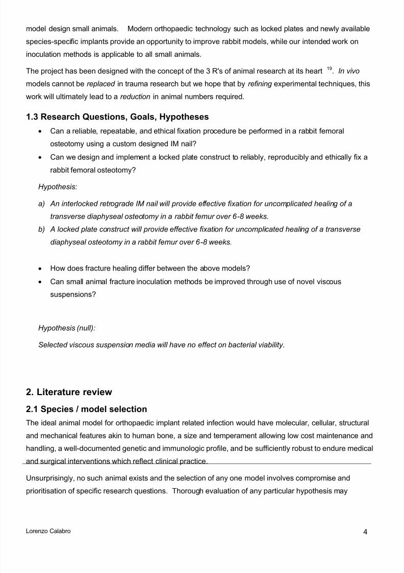
1.2 Research ProblemOver the last 40 years a number of animal models have been created to answer questions in orthopaedictrauma research 6. Models examining musculoskeletal infection in the context of fracture are, however,rare in the literature despite the clinical prevalence of this problem and the potential for causing morbidity.Small animal studies can offer cost-effective and statistically well powered solutions and recentdevelopment of clinically relevant custom implants for mice has demonstrated their potential in future
fracture research. There are clear windows for improvement and standardisation of fracture-infection
8/13/2019 Lorenzo Calabro Thesis
http://slidepdf.com/reader/full/lorenzo-calabro-thesis 12/54
Lorenzo Calabro 4
model design small animals. Modern orthopaedic technology such as locked plates and newly availablespecies-specific implants provide an opportunity to improve rabbit models, while our intended work oninoculation methods is applicable to all small animals.
The project has been designed with the concept of the 3 R's of animal research at its heart 19. In vivo models cannot be replaced in trauma research but we hope that by refining experimental techniques, this
work will ultimately lead to areduction in animal numbers required.
1.3 Research Questions, Goals, Hypotheses Can a reliable, repeatable, and ethical fixation procedure be performed in a rabbit femoral
osteotomy using a custom designed IM nail?
Can we design and implement a locked plate construct to reliably, reproducibly and ethically fix arabbit femoral osteotomy?
Hypothesis:
a) An interlocked retrograde IM nail will provide effective fixation for uncomplicated healing of a
transverse diaphyseal osteotomy in a rabbit femur over 6-8 weeks.
b) A locked plate construct will provide effective fixation for uncomplicated healing of a transverse
diaphyseal osteotomy in a rabbit femur over 6-8 weeks.
How does fracture healing differ between the above models?
Can small animal fracture inoculation methods be improved through use of novel viscoussuspensions?
Hypothesis (null):
Selected viscous suspension media will have no effect on bacterial viability.
2. Literature review
2.1 Species / model selectionThe ideal animal model for orthopaedic implant related infection would have molecular, cellular, structuraland mechanical features akin to human bone, a size and temperament allowing low cost maintenance andhandling, a well-documented genetic and immunologic profile, and be sufficiently robust to endure medicaland surgical interventions which reflect clinical practice.
Unsurprisingly, no such animal exists and the selection of any one model involves compromise andprioritisation of specific research questions. Thorough evaluation of any particular hypothesis may
8/13/2019 Lorenzo Calabro Thesis
http://slidepdf.com/reader/full/lorenzo-calabro-thesis 13/54
Lorenzo Calabro 5
necessitate preclinical testing in more than one species as is typical of pharmaceutical trials where both asmall and large animal model are recommended 8.
In general, larger animals such as non-human primates, sheep, goats and dogs are better able to tolerateinterventions and their bony geometry can accommodate human scale implants 6, 9. This is important, asmechanobiologic variables including fixation stability are known to influence both fracture healing and
infection susceptibility 20, 21
. Smaller animals such as rats, mice and guinea pigs perform less well againstthese criteria but can be housed and fed more easily allowing larger numbers and more powerfulconclusions at an acceptable financial cost 9, 22. Rabbits have been used frequently for osteomyelitismodels due to their relatively easy maintenance, reproducible infection profile, and apparent tolerance toimplantation of readily available implants 6, 14.
Technological advances in small scale fixation devices and the availability of transgenic rodents hasrecently challenged these generalisations and may enable small animal fracture models to better emulateclinical conditions 10, 11. Matthys and Perren developed an internal fixator, analogous to a locked plate for
use in mouse femoral osteotomy models 11. Variations on this implant allow the investigator to choosebetween stable or flexible fixation, and hence choose between a model with primarily intramembranous orendochondral fracture healing 23. An interlocking nail and an external fixator with stable and less stablefixation options have also recently been developed for mouse femora 10, 12.
Interspecies differences in immune response are also important to acknowledge in infection trials.Historically the success of animal osteomyelitis models has been determined by the degree to whichhistological and microbiological outcomes mirror those found in human disease 6. Inoculation and surgery
is typically manipulated to meet this end and it has been noted that some animals such as rats and dogsare more difficult to infect than others such as rabbits 6. Human pathogens may not cause predictabledisease in a particular animal or any disease at all 24. Traditionally immunobiological research has usednon-human primate models in pre clinical testing for their similar genetic profile but it is noted that evenchimpanzees have differences in major histocompatibility complex (MHC) genes and that these differencescan influence disease 24. Transgenic mice with humanised immune systems may provide a novel solution.In this technique human haematopoietic stem cells are engrafted into immunodeficient mice allowingcharacterisation of a human immune response in a pre-clinical model 13. These animals create the
potential for detailed molecular characterisation in preclinical models of human infection 9, 13
. New technology in small animal research provides a means of improving in vivo trauma models. Reliable,ethical and cost-effective models will facilitate future clinical advances.
8/13/2019 Lorenzo Calabro Thesis
http://slidepdf.com/reader/full/lorenzo-calabro-thesis 14/54
Lorenzo Calabro 6
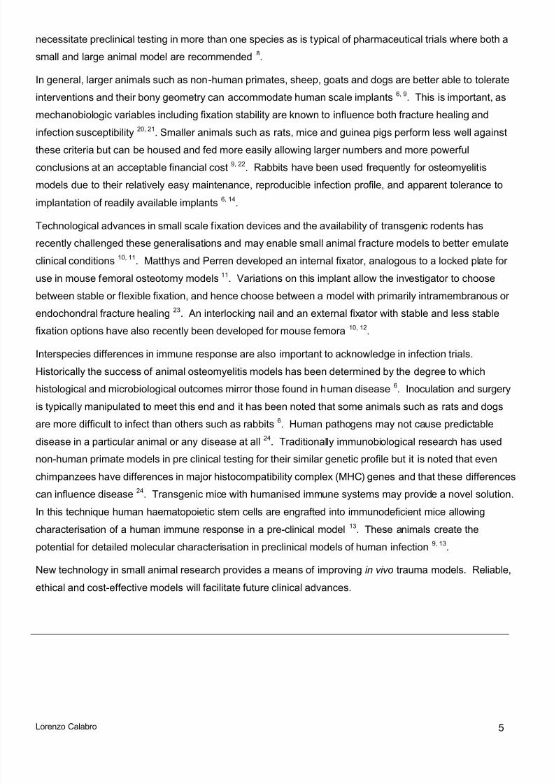
2.2 Orthopaedic Trauma in animal modelsInfection rates associated with fracture fixation are consistently higher than those in elective orthopaedicimplant surgery such as arthroplasty 3, 25. Contributing factors include contaminated and devitalised softtissue, systemic inflammatory response, procedural, implant and host issues 3, 16, 26.
A variety of animal models have been developed to better understand the natural history or to compare
interventions in fracture healing. These models can be categorised according to the nature of the traumainflicted, for example osteotomy versus three point bending versus blunt impact fracture; the subsequentmanagement of this trauma; the animal used; and the anatomic location within the animal.
The first methodological challenge in a trauma model is the need for creation of a repeatable injury. Themost controlled way of achieving this is to create an osteotomy. An obvious drawback, however, is theneed for surgical exposure which in the case of intra-medullary fixation would otherwise be unnecessary.Furthermore, osteotomy does not re-create the typical forces involved in clinical trauma, nor the morecomplex fracture patterns and soft tissue trauma. Despite this, osteotomy healing has been shown togrossly resemble low-energy random fracture healing in sheep 27.
The most common alternative is a three point bending method, with a controlled force delivered by variousmeans. Ashurst et al 28 devised a method of creating a reproducible fracture in the mid shaft of rabbittibiae. In this model the medial cortex was osteotomised, a plate placed in situ and a 3 point bendingdevice subsequently used to fracture the remaining cortex. Jackson et al 29 had previously created a bluntimpaction guillotine device to create a closed fracture over a rat femora which had already been implantedwith an intra-medullary pin. The force was delivered with a pneumatic impulse from a compressed air
cylinder. Bonnarens and Einhorn 30
simplified the Jackson model by creating a similar guillotine devicerelying only on the force of gravity. However, unpublished ex vivo data from trials performed at the ARI,(Davos Switzerland) found the Bonnarens method to be unreliable in creating reproducible, simple rabbitfemoral fractures.
2.3 Existing fracture + infection modelsMany studies, including those above, have characterised the nature of fracture healing with regards toexperimental variables such as pharmaceuticals, soft tissue injury or fixation techniques. Few authorshowever have investigated fracture associated osteomyelitis and the existing literature is highlyheterogeneous.
Rittman and Perren established an early experimental model in sheep to evaluate the impact of fixationstability on healing in an infected fracture 31. Various compression plating techniques were used in an effortto potentiate primary bone healing. All animals were infected with S. aureus , if necessary by repeatinocula. Radiologic, histologic and mechanical characterisation of fracture healing ensued.
Sheep models continue to be used with some success. Stewart et al recently demonstrated the potentialprophylactic utility of an antibiotic implant coating in a mid diaphyseal sheep tibial osteotomy inoculated at
8/13/2019 Lorenzo Calabro Thesis
http://slidepdf.com/reader/full/lorenzo-calabro-thesis 15/54
Lorenzo Calabro 7
the time of surgery with S. aureus 17 . Culture of autopsy specimens obtained from 9 sheep after 3 monthsof fracture healing showed that osteotomies fixed with a Vancomycin-modified plate had a relative risk of0.27 for infection compared with those fixed with a non modified plate.
Other isolated models including a fracture or osteotomy, and infection have since been created in sheep,goats and rats 32-34. Worlock et al developed a rabbit model with relevance to this trial 35. They initially
aimed to reliably cause fracture fixation associated osteomyelitis in rabbits without causing systemic illness36. Using the fracture and plate fixation method of Ashurst in rabbit tibiae, an S. aureus strain isolated froma patient with chronic osteomyelitis was inoculated immediately following wound closure. With an inoculumof 107 bacteria, osteomyelitis developed in 4 of 5 animals without causing sepsis. The same group adaptedthis model for a later study evaluating the impact of fixation stability on infection risk in fractures fixed with astable construct dynamic compression plate (DCP) or an unstable construct (non-locked intramedullary pin)35. Fractures fixed with stable fixation were significantly less likely to develop osteomyelitis than those withunstable fixation when inoculated with 107 bacteria (91% vs 40% (p=<0.01)). The authors however noted a
sub-optimal complication rate in the stable fixation group. Four of 14 animals suffered a peri-implantfracture around the DCP in the early post-operative period and had to be removed from the study.
2.4 Inoculation methodsTypically an anaesthetised animal in a musculoskeletal infection trial will be subjected to a standardisedtrauma and fixation, with perioperative bacterial inoculation. The pathogen used most frequently isStaphylococcus aureus , reflecting its prevalence in clinical osteomyelitis 2. The aim is to reliably create
local osteomyelitis without sepsis 37. This inherently relies on spatial containment of the inoculatedbacteria. The method most commonly used is to place a percutaneous needle at the osteotomy or implantsite under vision during surgery 35, 38. The wound is then closed and bacteria, suspended in PBS solutionare inoculated to the target site via the needle. A particular inoculum dose will not always precipitateinfection in an individual animal and pilot trials are required to determine the dose which will, for example,cause infection in either 50% or 10% of animals 37. The variability in response may be due to immunefunction, bacterial viability and the difficulty in controlling dissemination of bacteria post inoculation. Asolution to the latter issue would improve osteomyelitis models. In one trial, the authors, acknowledging
this issue, used contaminated resorbable collagen gel-foam, rather than PBS to inoculate a fracture incaprine tibiae 32. Detailed analysis of bacterial dissemination using the foam was not published but theauthors did note that remnant foam was identified with evidence of host immune response at the end of thetrial. This perhaps suggests that the extra material is more likely to confound results than control them 32.
A more viscous bacterial suspension is another approach to this problem. Ideally this material would haveno effect of its own on bacterial growth and viability, nor host immune response and would not diffuse overa wide area – particularly during the first hours post contamination. Widely available biocompatiblematerials including glycerol, medical gelatine and hydrogels with existing clinical applications in drugdelivery or burns and reconstructive surgery are candidate media 39, 40. To our knowledge they have not
8/13/2019 Lorenzo Calabro Thesis
http://slidepdf.com/reader/full/lorenzo-calabro-thesis 16/54
Lorenzo Calabro 8
been used previously in this setting and their effect on bacterial viability has not been extensivelydescribed.
2.5 Mechanobiology in fixation constructsIn fixing a fracture with a locked plate, the surgeon faces a number of choices which can impact themechanical performance of the system and therefore fracture healing. Locked plates work in vivo as low-contact internal fixators 41. They are attractive in osteomyelitis models (and infection surgery) as they areassociated with lower susceptibility to infection – a finding thought to stem from reduced periostealcompression compared to conventional plating technique s 38. Locked plate constructs also allow fixation ofboth simple and comminuted fractures, and in an experimental setting can be used to precisely manipulatea fracture gap 42, 43.
Plate and screw configuration also influence the stiffness of the fixation system. Stiffness of the construct
has to be balanced against the fact that micro-movement is almost always necessary for efficient fracturehealing 41. A rigid construct can also focus stresses on small areas of the plate or bone potentiating failure.This is a particular issue in small animal fracture models where weight bearing cannot be prevented in theearly post-operative phase. Factors to be taken into account include the 'working length' of the plate(unsupported length of the centre of the plate bridging the fracture gap, in between the innermost screwson either bone fragment), the distance between the plate and bone, and resistance to likely physiologicloading. Stoffel et al used composite bone models to evaluate the fatigue strength of varying locked plateconstructs under axial and torsional loads 43. They determined that with a 1mm fracture gap, leaving one
hole empty either side of the 'fracture' and hence increasing the working length, decreased the axial andtorsional stiffness but also decreased stress through the plate under these loads. They and other authorsalso showed that increasing the distance between plate and bone decreases construct rigidity 44.
2.6 Physiologic stress in animals and relevant testingRelevant ex vivo mechanical testing should aim to reproduce in vivo forces 45. The physiologic forces onrabbit femora in vivo have not been described. Burstein et al in a seminal study designed criteria againstwhich to evaluate mechanical tests for long bones and reasoned that torsional loading to determine rigidity
and load capacity was the most valid means of assessing strength 45
. Destructive and non-destructivemechanical testing is commonly used to evaluate the integrity of healed fractures in animal models 46-48.
2.7 Summary Animal models are important in evaluating surgical technology prior to clinical trials. There is an ethical andlogistical imperative for reliability and reproducibility in the design of these models. In trauma research,heterogeneity in existing models prevents direct comparison between results obtained using differentexperimental models, or the ability to draw generally valid conclusions from results obtained with a singlemodel. Furthermore, evolving standards in animal research preclude repetition of some early models while
8/13/2019 Lorenzo Calabro Thesis
http://slidepdf.com/reader/full/lorenzo-calabro-thesis 17/54
Lorenzo Calabro 9
recent developments in fracture fixation techniques decrease the relevance of others. This study aimed toaddress this deficit: Specifically, our primary aim was to create reliable and standardised fracture healingmodels in rabbit femora with two different types of fixation (IM nail and locked plate) mirroring modernclinical fixation constructs. Our secondary aim was to improve inoculation techniques in small animalorthopaedic infection models by evaluating viscous suspension media. In vivo fracture/infection models aresparse in the literature and highly variable. Their effectiveness hinges on the ability to reliably inoculate thedesired anatomical area with the desired dose of pathogen. Spatial (anatomical) containment of pathogenis an important factor in inoculation and based on the premise that viscous media would facilitate this, weproceeded to evaluate the effect of candidate media on bacterial viability.
3. Research Design
3.1 Methodology OverviewExperimental work was performed in distinct In vitro, ex vivo and in vivo phases. Methodology and resultsare presented in 3 parts respectively below. All experiments described in this thesis were peformed at the
AO Research Institute (ARI) in Davos, Switzerland. The ARI, Preclinical testing program specialises inorthopaedic research with facilities and experienced personnel for animal care, anesthesia, surgery,biomechanical testing and CT imaging. In vivo experiments were performed in an ISO 9001-2000 certifiedlaboratory and in accordance with the Swiss Animal Protection Law. Animal permission was obtained forthis study from the Office for Food Safety and Animal Health Graubünden (Switzerland) (TVB #12/2010).Skeletally mature, female New Zealand (NZ) white rabbits, sourced from Charles River Laboratories,Sulzfeld (Germany) were used. Two parallel animal models with different fixation constructs for the rabbitfemur were evaluated:
Retrograde interlocked intramedullary nail fixation
Locked plate fixation
Initialex vivo work involved implant construct, procedure design and ex vivo analysis of mechanicalperformance in cadaver rabbit femora. This was followed with an in vivo pilot study to evaluate fracturehealing performance. Microbiologic experiments were performed in the laboratory to assess the viability of
Staphylococcus aureus in candidate viscous media.3.2 Experimental Models
Locked plate
Locking compression plates (LCP)(Synthes, CH) were used in two sizes for both ex vivo and in vivo work.Initially, the 8-hole stainless steel, 56mm x 1.5mm veterinary LCP was used with 6 x 2.0mm locking screws(Synthes, CH). This implant will be referred to as 2.0mm plate in the following as per AO convention.Subsequently a more robust 8-hole stainless steel, 64mm x 2.0mm LCP with 6 x 2.4mm screws was used,
which will be referred to as 2.4mm plate in the following. Plates were fixed with 6 locking screws using a
8/13/2019 Lorenzo Calabro Thesis
http://slidepdf.com/reader/full/lorenzo-calabro-thesis 18/54
Lorenzo Calabro 10
hand powered 4 Nm torque limited screw driver (Synthes, CH) with the central two holes left empty. Whenindicated, a transverse mid diaphyseal osteotomy was then made between the two central holes using a0.66mm gigli wire.
IM Nail
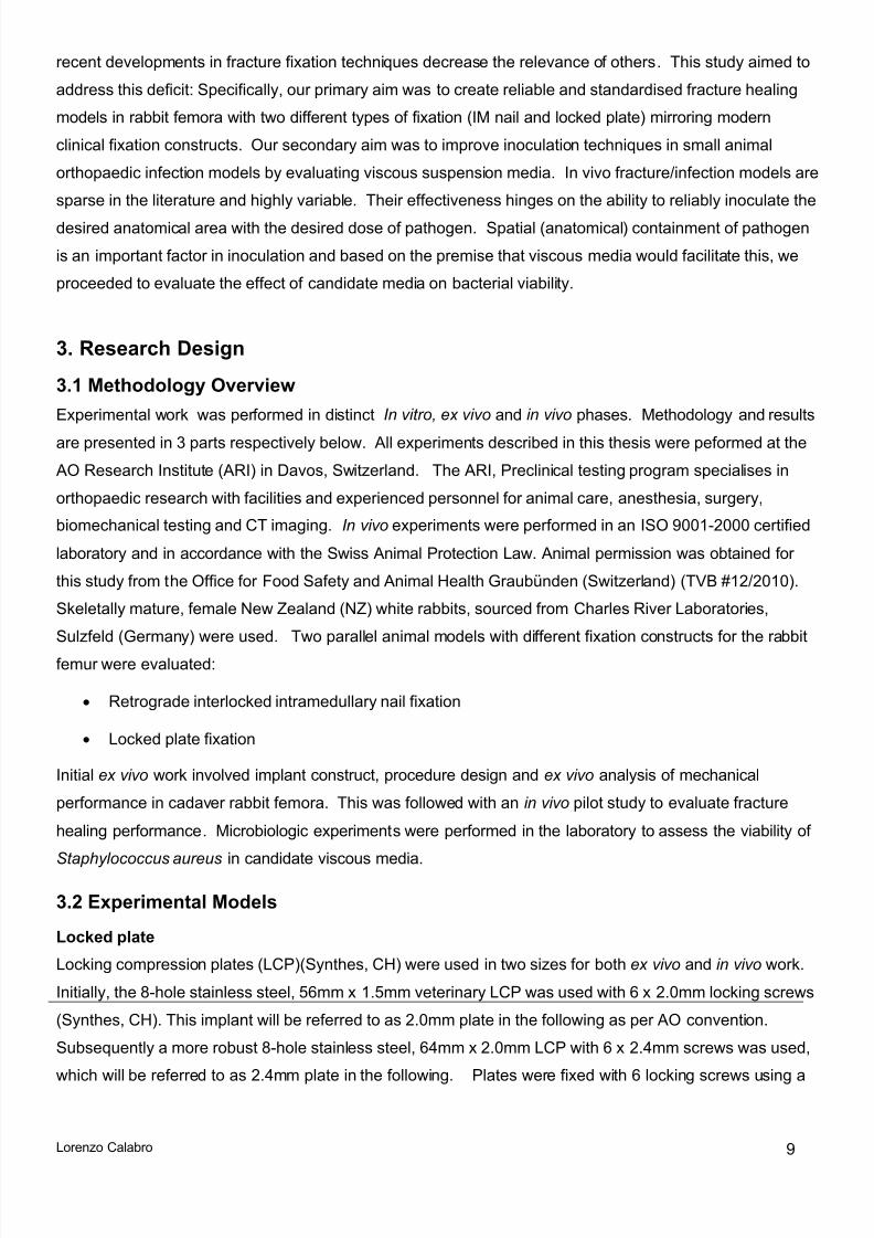
A custom designed, stainless steel interlocked IM nail (Research Implant
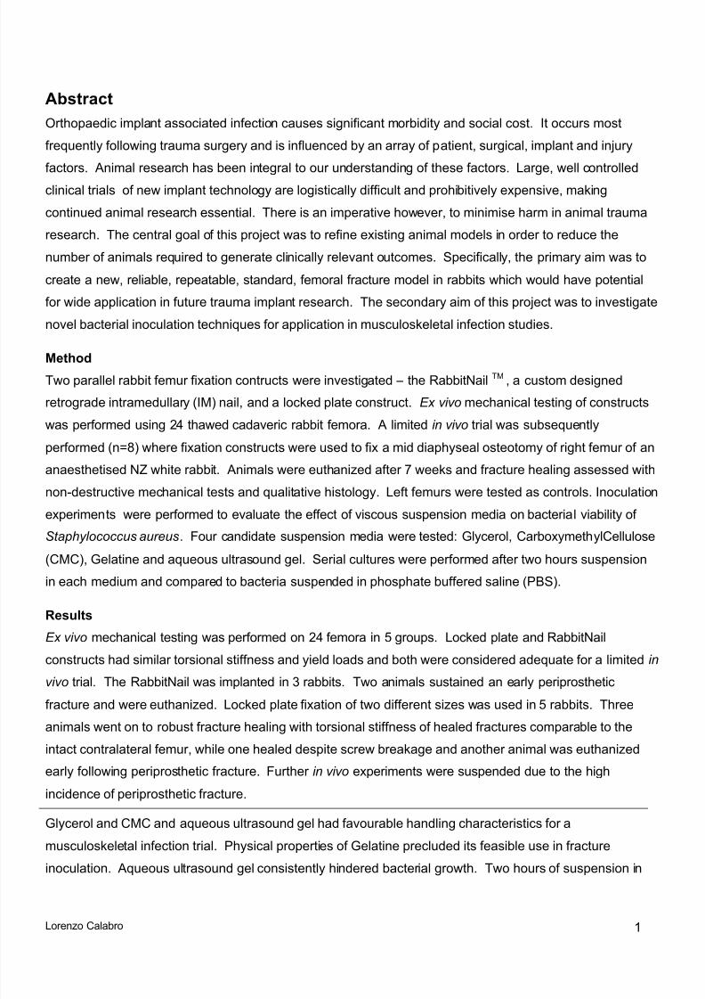
Systems, ARI) was used with four interlocking pins. The nail is 86mm inlength and 2.5mm in diameter and is supplied with custominstrumentation including an aiming jig (Fig 1). The intra-medullary canalwas accessed in retrograde fashion. The canal was opened and reamedto the diaphyseal isthmus using a 2.0mm, and 2.7mm drill bit by hand.The proximal canal was reamed using a 1.8mm Kirschner wire poweredby a micro-surgical drill. The nail was inserted under power till its distalend was clear of the knee joint. The interlocking jig was then applied
and interlocking pins were pre-drilled and inserted from proximal to distal. A saw-guide was clipped to the jig using the most proximal option tostandardise the osteotomy site. A transverse mid-diaphyseal osteotomywas performed using a 0.66mm gigli wire
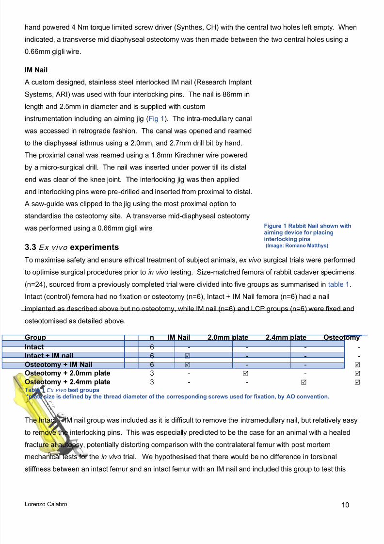
3.3 E x v i v o experimentsTo maximise safety and ensure ethical treatment of subject animals, ex vivo surgical trials were performedto optimise surgical procedures prior to in vivo testing. Size-matched femora of rabbit cadaver specimens
(n=24), sourced from a previously completed trial were divided into five groups as summarised in table 1. Intact (control) femora had no fixation or osteotomy (n=6), Intact + IM Nail femora (n=6) had a nailimplanted as described above but no osteotomy, while IM nail (n=6) and LCP groups (n=6) were fixed andosteotomised as detailed above.
Group n IM Nail 2.0mm plate 2.4mm plate Osteotomy Intact 6 - - - -Intact + IM nail 6 - - -Osteotomy + IM Nail 6 - - Osteotomy + 2.0mm plate 3 - - Osteotomy + 2.4mm plate 3 - - Table 1 Ex vivo test groups*plate size is defined by the thread diameter of the corresponding screws used for fixation, by AO convention.
The Intact + IM nail group was included as it is difficult to remove the intramedullary nail, but relatively easyto remove the interlocking pins. This was especially predicted to be the case for an animal with a healedfracture at autopsy, potentially distorting comparison with the contralateral femur with post mortemmechanical tests for the in vivo trial. We hypothesised that there would be no difference in torsionalstiffness between an intact femur and an intact femur with an IM nail and included this group to test this
Figure 1 Rabbit Nail shown withaiming device for placinginterlocking pins(Image: Romano Matthys)
8/13/2019 Lorenzo Calabro Thesis
http://slidepdf.com/reader/full/lorenzo-calabro-thesis 19/54
Lorenzo Calabro 11
hypothesis and validate our post mortem test protocol. All cadaver femora were then subjected todestructive mechanical testing as detailed in chapter 3.5.
3.4 In v iv o surgery
Surgical technique overview
Rabbits were anaesthetised and monitored by an experienced veterinary anaesthetist. An intramuscular
pre-anaesthetic dose sedative and analgesic (Midazolam, Fentanyl and Medetomidine) was administered,followed by induction with intravenous Propofol. An endotracheal tube was introduced and anaesthesiawas maintained with Isofluorane. Respiratory and haemodynamic status was monitored throughout theprocedure. Following induction, the right hind limb was shaved, then prepared with antiseptic and sterilesurgical drapes. Surgical procedures differed for plate and intramedullary nail models. At the end of thesurgical procedures, after wound closure, a regional nerve block was performed, transdermal opiateanalgesia applied and post-operative monitoring commenced.
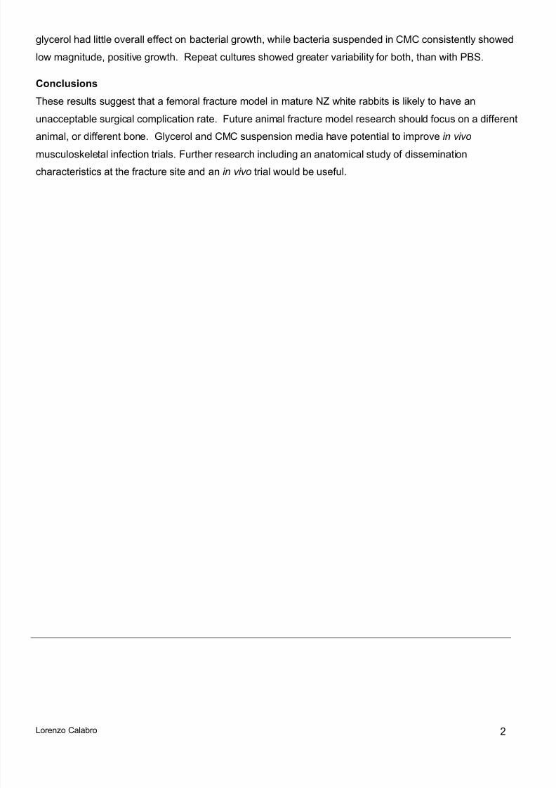
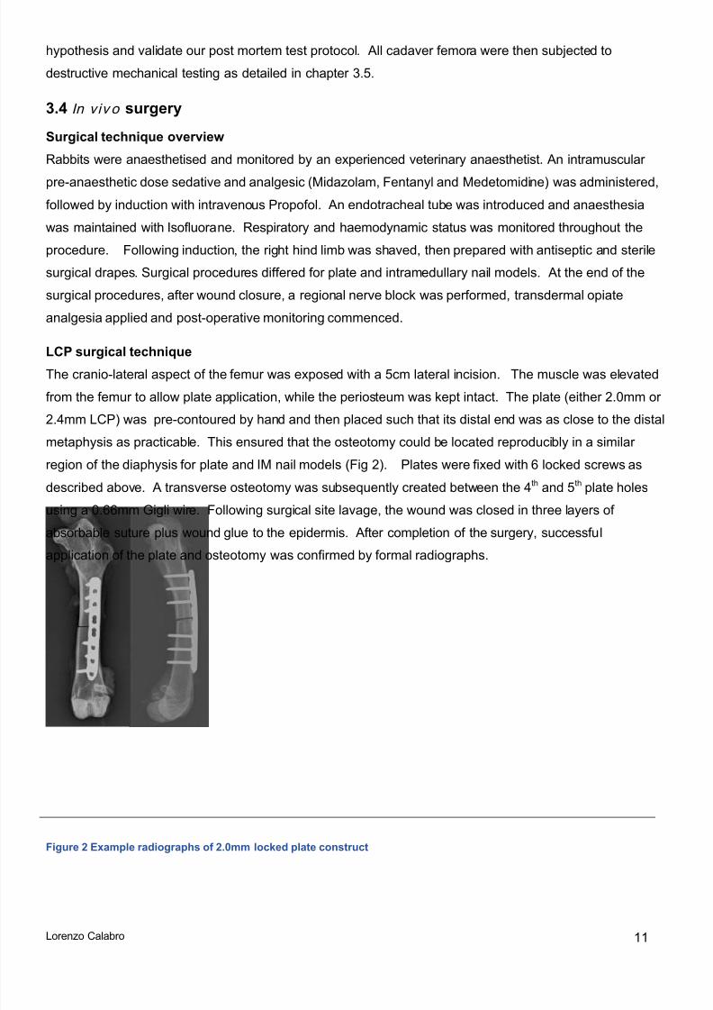
LCP surgical techniqueThe cranio-lateral aspect of the femur was exposed with a 5cm lateral incision. The muscle was elevatedfrom the femur to allow plate application, while the periosteum was kept intact. The plate (either 2.0mm or2.4mm LCP) was pre-contoured by hand and then placed such that its distal end was as close to the distalmetaphysis as practicable. This ensured that the osteotomy could be located reproducibly in a similarregion of the diaphysis for plate and IM nail models (Fig 2). Plates were fixed with 6 locked screws asdescribed above. A transverse osteotomy was subsequently created between the 4 th and 5 th plate holesusing a 0.66mm Gigli wire. Following surgical site lavage, the wound was closed in three layers of
absorbable suture plus wound glue to the epidermis. After completion of the surgery, successfulapplication of the plate and osteotomy was confirmed by formal radiographs.
Figure 2 Example radiographs of 2.0mm locked plate construct
8/13/2019 Lorenzo Calabro Thesis
http://slidepdf.com/reader/full/lorenzo-calabro-thesis 20/54
Lorenzo Calabro 12
IM nail surgical technique
The distal femur was approached through a 2cm lateral para-patellar incision and medial patelladislocation. The intramedullary canal was opened and reamed to the diaphyseal isthmus using a 2.0mm,and 2.7mm drill bit by hand. The proximal canal was reamed using a 1.8mm Kirschner wire powered by amicro-surgical drill. The nail was inserted under power till its distal end was clear of the knee joint. Thecranio-lateral aspect of the femur was exposed with a 4cm lateral incision. Muscle was elevated from thefemur to allow interlocking pin fixation, while the periosteum was kept intact. The aiming device was thenapplied and tapered interlocking pins were inserted from proximal to distal after pre-drilling a 1.0 mm holeusing a microsurgical drill. Although these pins were smooth, they achieved stable fixation due tocompression when the oversized, tapered (1.0/1.1mm taper) pins are inserted into the 1.0mm hole. A saw-guide was then clipped to the aiming device. A transverse mid-diaphyseal osteotomy was performed usinga 0.66mm gigli wire. Following surgical site lavage, the wounds were closed in three layers of absorbablesuture plus wound glue to the epidermis. After completion of the surgery, successful application of the IMnail and osteotomy was confirmed by formal radiographs.
Post-operative care
Animals were maintained in individual cages for the duration of the study. They were monitored at leastthree times a day by an experienced animal caretaker for the first two post-operative weeks, or longer ifnecessary. Daily checks continued until the end of the experiment. Animals were monitored for theirgeneral behavior, appetite and load-bearing on the operated limb. The incision sites, respiratory function,eyes, fur, and faeces were monitored and the results noted on an assessment form and entered into theanimal’s logbook . In the event of undue suffering or fixation failure as determined by a veterinarian,
immediate euthanasia was performed by the veterinarian using intravenous pentobarbital. Otherwiseeuthanasia was performed by a veterinarian at 7 weeks post operatively. Bilateral femora were carefullyexplanted, keeping fracture callus intact and refrigerated at 4 ⁰ C for a maximum of 24hrs before undergoing
non-destructive mechanical testing. Following mechanical testing the right (operated) femur was preparedfor histology.
3.5 Outcome measures
Mechanical testing
Mechanical testing was performed at two distinct points during the study.
a) To evaluate ex vivo fixation constructs prior to commencing in vivo work (destructive and non-destructive testing)
b) To evaluate fracture healing following euthanasia 7 weeks post op (non-destructive testing,allowing subsequent preparation for histology)
8/13/2019 Lorenzo Calabro Thesis
http://slidepdf.com/reader/full/lorenzo-calabro-thesis 21/54
Lorenzo Calabro 13
Specimens were embedded proximally and distally in poly-methyl methacrylate (PMMA, Beracryl, CH)cylinders with a diameter of 30 mm. The free distance between the two embedding sites was 60 mm (Fig3). The probes were aligned so that the axes of the cylinders were co-linear with the diaphyseal axis.During embedding, specimens were kept moist with compound sodium lactate solution (CSL).
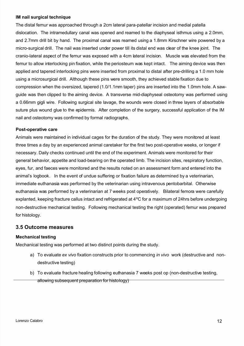
For torsional testing a material testing machine (MTS Bionix 858, MTS systems, Eden Prairie, MN, USA)
was used. The PMMA cylinders were fixed with aluminiumclamps. The clamp on the proximal end of the bone wasmounted on a single universal joint. The single universal jointwas attached to the base plate of the MTS machine. The distalend of the bone was mounted on a double universal joint. Thedouble universal joint was attached to the load cell. The lightestavailable clamps and joints were chosen to avoid undue load onthe femur while setting up. The setup was designed to
compensate for irregularities in axial alignment and minimisebending moments on the specimens.
After mounting the samples on the machine, torque and axialloads were balanced. During the test there was no axialdisplacement. For torsional stiffness testing, a series of fourcycles with internal and external rotation was conducted. First,
internal torsion was applied at a rate of 0.3 degrees/sec up to0.4 Nm (for left femurs). As soon as this threshold wasachieved, external rotation torque was applied at the same rateup to -0.4 Nm (for right femurs the converse was applied. For destructive tests, internal rotation torquewas applied, following the four cycles for stiffness measurement, at a constant angular rate until systemfailure. The following data was recorded with a sampling rate of 64 Hz: index time [sec], axial load [N],axial displacement [mm], torsional angle [deg] and torque [Nm].
Internal and external torsional stiffness was recorded on the fourth cycle. The angle-torque data was
plotted for this cycle and the linear range was selected. The trend line for this range was calculated inExcel and the slope of this line corresponded to torsional stiffness (Nm/deg). For destructive tests theangle and torque at failure was recorded along with the mode of failure. In the in vivo trial, the intact leftfemur was tested as a contralateral control following an identical protocol.
Histology
Immediately following mechanical tests, right (operated) femora were fixed in 70% ethanol for 4 weeks,with fresh ethanol changes weekly. Specimens were subsequently dehydrated through an ascending
series of ethanol (70, 96, 100%, with two changes for each step). They were then transferred to xylene,
Figure 3 Mechanical testing setup
8/13/2019 Lorenzo Calabro Thesis
http://slidepdf.com/reader/full/lorenzo-calabro-thesis 22/54
Lorenzo Calabro 14
and finally infiltrated with and embedded in PMMA. PMMA blocks were trimmed using a saw (BizerbaFK22, Bizerba, Zurich, CH) in order to fit into an annular diamond saw (Leitz 1600 microtome, Leica,Nunningen, CH). Sections of 200 μm thickness were created along the axis of the femur and prepared foranalysis with Giemsa / Eosin stain in order to differentiate fibrous, cartilaginous and bone tissue. Lowpower magnification (25x) images taken through a light microscope (Zeiss Axioscope, Feldbach, CH).
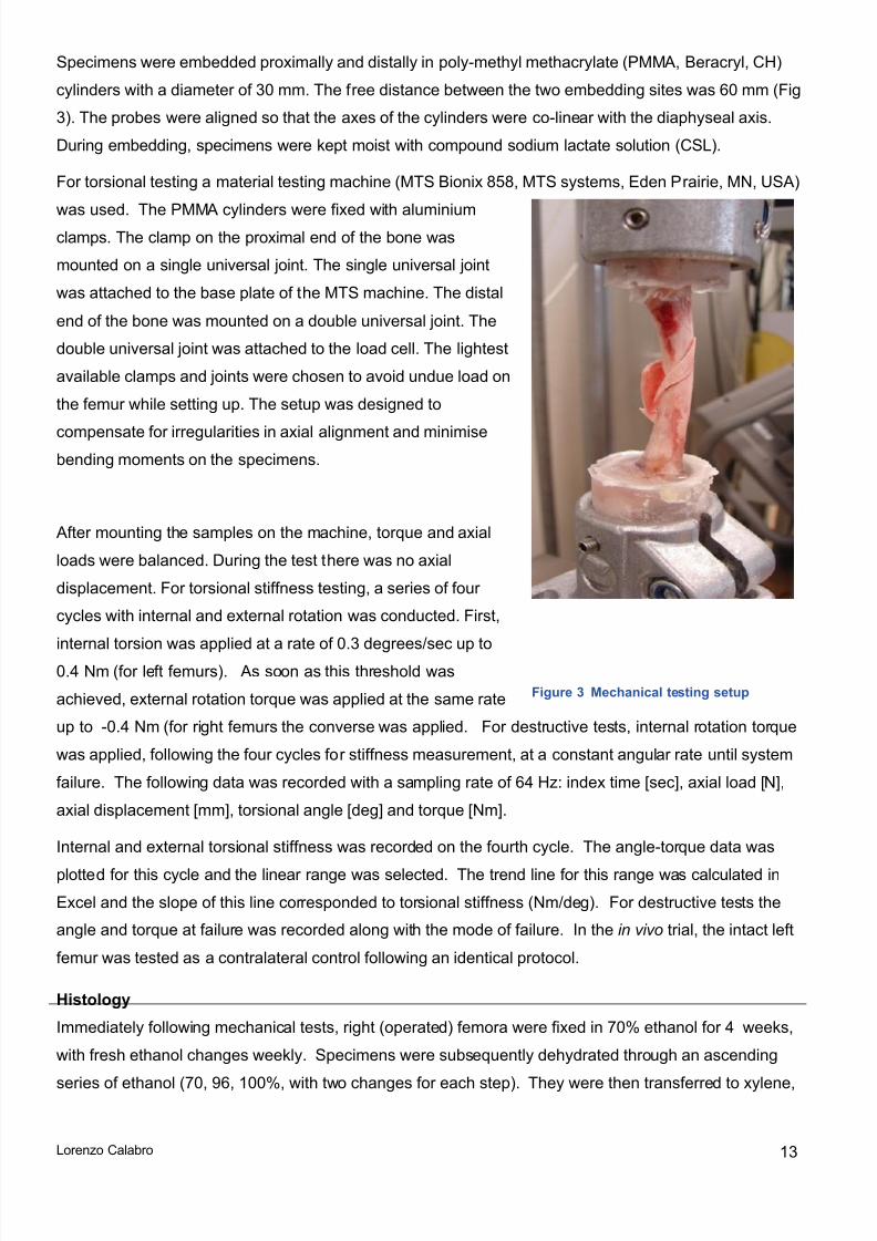
3.6 In Vi tro inoculation media experimentsMedia
A search of the literature, discussion with supervisors and hydrogel chemists at the ARI yielded a widerange of candidate media (Table 2 ). The ideal medium would have the following properties:
Safe to store and handle
Physiologically inert (no unwanted adverse reactions in recipient animal, maintaining bacterialviability without encouraging multiplication)
Readily autoclaveable Viscosity such that likely dissemination through soft tissue planes is minimised while still allowing
mixing, pipette use and reliable injection through a 20G needle.
Cheap and readily attainable
Further literature search and supplier contact allowed empirical evaluation of these candidate mediaagainst certain criteria. Based on this list, the 4 highlighted candidate media were selected forexperimental evaluation.
medium Viscous degradable inert safety Costestimate$AUD/100g
Gelatine ? <10
Aquasonic TM
Ultrasound gel
? ? 12
Glycerol ? 67
PolyEthylene-
Glycol
23
Alginate 67
Carboxymethyl-
Cellulose
56
Hyaluronic acid ? >100
Table 2. Preliminary empiric evaluation of candidate media
8/13/2019 Lorenzo Calabro Thesis
http://slidepdf.com/reader/full/lorenzo-calabro-thesis 23/54
Lorenzo Calabro 15
Bacterial strain
A strain of S aureus, (S. aureus JAR 06.01.31) was used. This strain was isolated from an infected hipprosthesis at the department of medical microbiology, Kantonsspital, Lucerne, Switzerland and haspreviously been used in fracture fixation associated osteomyelitis models 37.
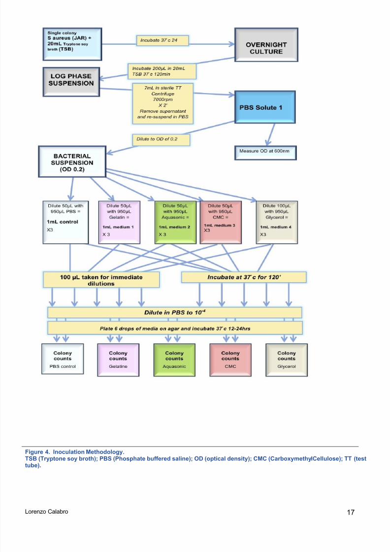
All experiments were undertaken in a laminar flow hood, under sterile conditions, in the PC2 laboratory at
the ARI by the primary author with the supervision of an experienced microbiologist (Dr T F Moriarty). Theprotocols are summarised in Figure 4.
S. aureus JAR was plated on Tryptone soy agar (TSA, Sigma Aldrich, Buchs, CH) and incubated at 37 ⁰ C
overnight. This plate was then refrigerated and single colonies were subsequently taken from this culturefor use in repeated experiments over a maximum period of 3 weeks. On each occasion, a single colonywas inoculated into 20mL of prewarmed Tryptone soy broth (TSB, Sigma Aldrich, Buchs, CH) andincubated at 37 ⁰ C overnight. The following morning (12-15hrs incubation), 200μL of this solution was
subcultured into 20mL of sterile prewarmed TSB and this was incubated for a further 120 minutes to ensurebacteria were in the log phase of growth. Following incubation, 7mL of this solution was centrifuged(7000rpm x 3min) and the resulting bacterial pellet was re-suspended in phosphate buffered solution (PBS,Sigma Aldrich, Buchs, CH). The optical density (OD) of this bacterial suspension at a wavelength of 600nmwas measured using a spectrophotometer, and the suspension was further diluted until the optical densityreached 0.2. An OD of 0.2 was used as this would typically yield a bacterial concentration between 1x10 6 and 1x10 7 CFU/mL, which equated with that used in prior in vivo trials at the ARI.
This stock suspension of log phase S. aureus was used to perform the subsequent dilutions. 50 μL of this
suspension was mixed with 950 μL of each candidate carrier in a 2mL Eppendorf tube. Typically this wouldinclude between 1 and 3 control tubes (950 μL PBS) and 1-3 tubes for each experimental medium tested.
After this index dilution (5x10⁻² PBS2.1), 100 μL was taken from each Eppendorf for serial dilution, while theremaining 900μL of control or experimental suspension in the first Eppendorf was placed in the incubatorfor 120min. After 2 hrs the same procedure was repeated with the incubated suspensions. In each case 6x 20μL drops of the terminally diluted solution (1x10⁻⁴ PBS2.1) was plated out onto a labelled plate withTSA and incubated overnight. The following day bacterial colonies on each plate were counted and theCFU/mL was calculated.
Experiments were repeated a minimum of three times for all materials deemed suitable for testing.
Inoculation Media preparation
Aquason ic TM gel
Sterile AquasonicTM gel (Parker Lab. Fairfield, NJ, USA) is cheap and readily available. It is used widely asa topical application in clinical settings. It proved too viscous in its ‘off -the-shelf’ form to easily pipette or
inject through a needle. Therefore, for the purpose of the experiment it was diluted to 80%(v/v) with PBS.
8/13/2019 Lorenzo Calabro Thesis
http://slidepdf.com/reader/full/lorenzo-calabro-thesis 24/54
Lorenzo Calabro 16
Gelatine
Pharmaceutical gelatine (Gelita GMBH, Eberbach, Germany) with a setting temperature of 25 ⁰ C was used.
Gelatine proved to have sub-optimal handling. It was provided as powder which was diluted to 10%(w/v)with PBS. Gelatine could be easily manipulated and had low viscosity at 28 degrees, but became too stiff
to pipette or inject at 25 degrees meaning that at practical temperatures it did not perform as desired.
Glycerol
One hundred percent glycerol (Sigma Aldrich, Buchs, CH) was used.
CarboxymethylCellulose (CMC)
Research grade Sodium carboxy-methylcellulose (SERVA Feinbiochemica, Heidelberg, New York) with amolecular weight of 100 000 was used for all inoculation experiments with CMC. Two grams was diluted in
50mL PBS (4% (w/v)) . CMC had good handling characteristics at this concentration allowing pipette use,injection and mixing.
8/13/2019 Lorenzo Calabro Thesis
http://slidepdf.com/reader/full/lorenzo-calabro-thesis 25/54
8/13/2019 Lorenzo Calabro Thesis
http://slidepdf.com/reader/full/lorenzo-calabro-thesis 26/54
Lorenzo Calabro 18
4. Results
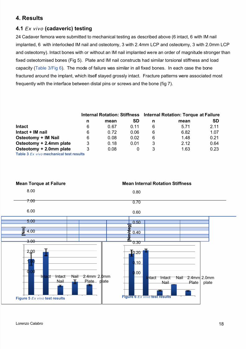
4.1 E x v i v o (cadaveric) testing24 Cadaver femora were submitted to mechanical testing as described above (6 intact, 6 with IM nailimplanted, 6 with interlocked IM nail and osteotomy, 3 with 2.4mm LCP and osteotomy, 3 with 2.0mm LCPand osteotomy). Intact bones with or without an IM nail implanted were an order of magnitude stronger than
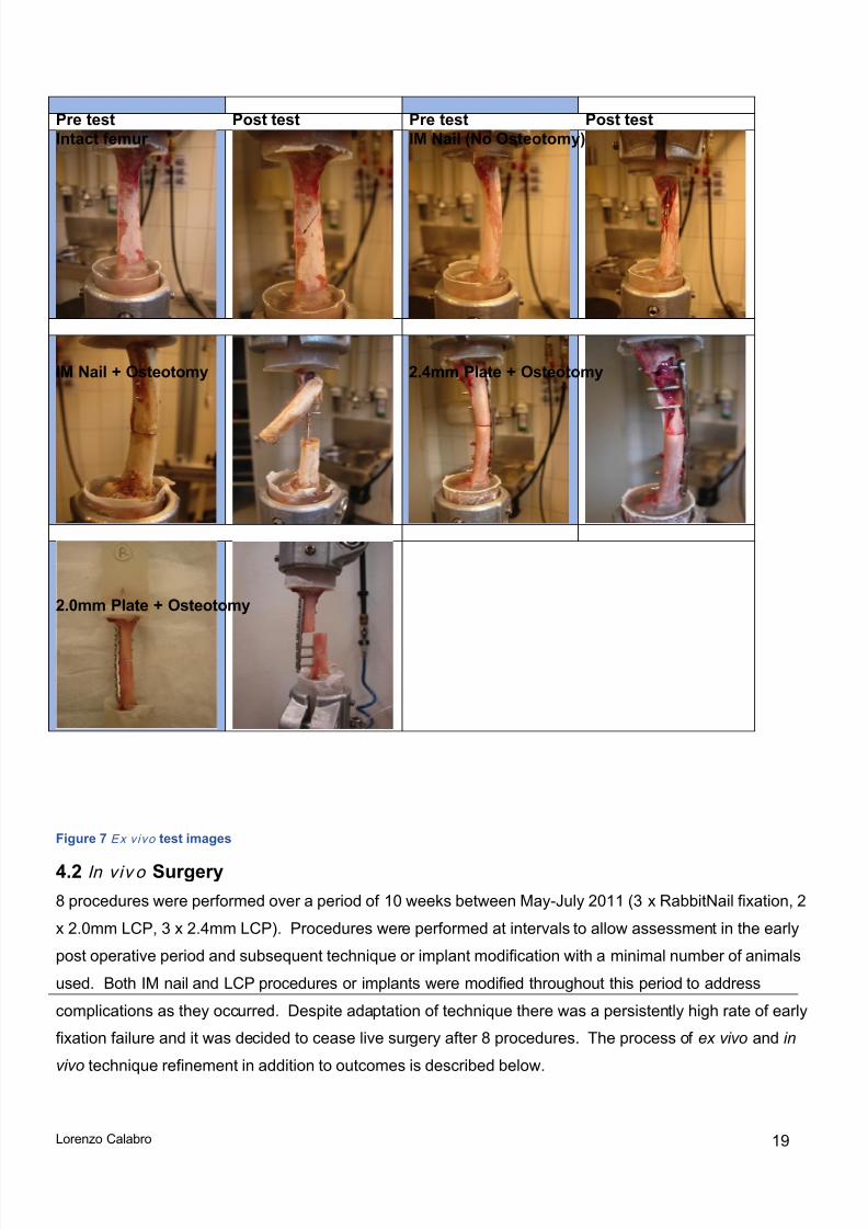
fixed osteotomised bones (Fig 5). Plate and IM nail constructs had similar torsional stiffness and loadcapacity (Table 3/Fig 6). The mode of failure was similar in all fixed bones. In each case the bonefractured around the implant, which itself stayed grossly intact. Fracture patterns were associated mostfrequently with the interface between distal pins or screws and the bone (fig 7).
Internal Rotation: Stiffness Internal Rotation: Torque at Failure
n mean SD n mean SDIntact 6 0.67 0.11 6 5.71 2.11Intact + IM nail 6 0.72 0.06 6 6.82 1.07Osteotomy + IM Nail 6 0.08 0.02 6 1.48 0.21Osteotomy + 2.4mm plate 3 0.18 0.01 3 2.12 0.64Osteotomy + 2.0mm plate 3 0.08 0 3 1.63 0.23 Table 3 Ex vivo mechanical test results
Mean Torque at Failure Mean Internal Rotation Stiffness
Figure 5 E x v i v o test results Figure 6 E x v i v o test results
0.00
1.00
2.00
3.00
4.00
5.00
6.00
7.00
8.00
Intact IntactNail
Nail 2.4mmPlate
2.0mmplate
[ N m
]
0.00
0.10
0.200.30
0.40
0.50
0.60
0.70
0.80
Intact IntactNail
Nail 2.4mmPlate
2.0mmplate
[ N m
/ d e g
]
8/13/2019 Lorenzo Calabro Thesis
http://slidepdf.com/reader/full/lorenzo-calabro-thesis 27/54
Lorenzo Calabro 19
Pre test Post test Pre test Post testIntact femur IM Nail (No Osteotomy)
IM Nail + Osteotomy 2.4mm Plate + Osteotomy
2.0mm Plate + Osteotomy
Figure 7 E x v i v o test images
4.2 In v iv o Surgery8 procedures were performed over a period of 10 weeks between May-July 2011 (3 x RabbitNail fixation, 2x 2.0mm LCP, 3 x 2.4mm LCP). Procedures were performed at intervals to allow assessment in the earlypost operative period and subsequent technique or implant modification with a minimal number of animalsused. Both IM nail and LCP procedures or implants were modified throughout this period to addresscomplications as they occurred. Despite adaptation of technique there was a persistently high rate of earlyfixation failure and it was decided to cease live surgery after 8 procedures. The process of ex vivo and in
vivo technique refinement in addition to outcomes is described below.
8/13/2019 Lorenzo Calabro Thesis
http://slidepdf.com/reader/full/lorenzo-calabro-thesis 28/54
Lorenzo Calabro 20
4.3 IM Nail (n=3)The procedure changed slightly between animal 1 and 2 and the interlocking pins were changed betweenanimals 2 and 3. For animals 2 and 3 the nail was inserted under power using the pneumatic pen drive asopposed to by hand in animal 1. For animal 3, interlocking pins with a ribbed taper and higher break-offstrength were used. Results are detailed below and summarized in Table 4 .
Rabbit Euthanized Stiffness healed femur / Stiffnesscontralateral femur (Nm/deg) Histology
1 7 weeks 0.21/0.68 Hypertrophic callusIncomplete union
2 3 days Not performed due to early fixation failure3 2 weeks Not performed due to early fixation failure Table 4 Summary i n v i v o results IM nail
Nail 1
Rabbit#
Age (dayof
surgery)
date kg Surgtime
Procedure notes pins
11042 54weeks 4.5.2011 5.1 150' Hand insertion nail Original – taperengaged by
hand
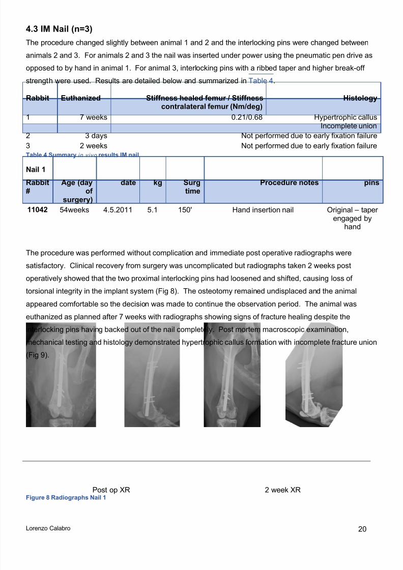
The procedure was performed without complication and immediate post operative radiographs weresatisfactory. Clinical recovery from surgery was uncomplicated but radiographs taken 2 weeks postoperatively showed that the two proximal interlocking pins had loosened and shifted, causing loss oftorsional integrity in the implant system (Fig 8). The osteotomy remained undisplaced and the animalappeared comfortable so the decision was made to continue the observation period. The animal waseuthanized as planned after 7 weeks with radiographs showing signs of fracture healing despite theinterlocking pins having backed out of the nail completely. Post mortem macroscopic examination,mechanical testing and histology demonstrated hypertrophic callus formation with incomplete fracture union(Fig 9).
Post op XR 2 week XRFigure 8 Radiographs Nail 1
8/13/2019 Lorenzo Calabro Thesis
http://slidepdf.com/reader/full/lorenzo-calabro-thesis 29/54
Lorenzo Calabro 21
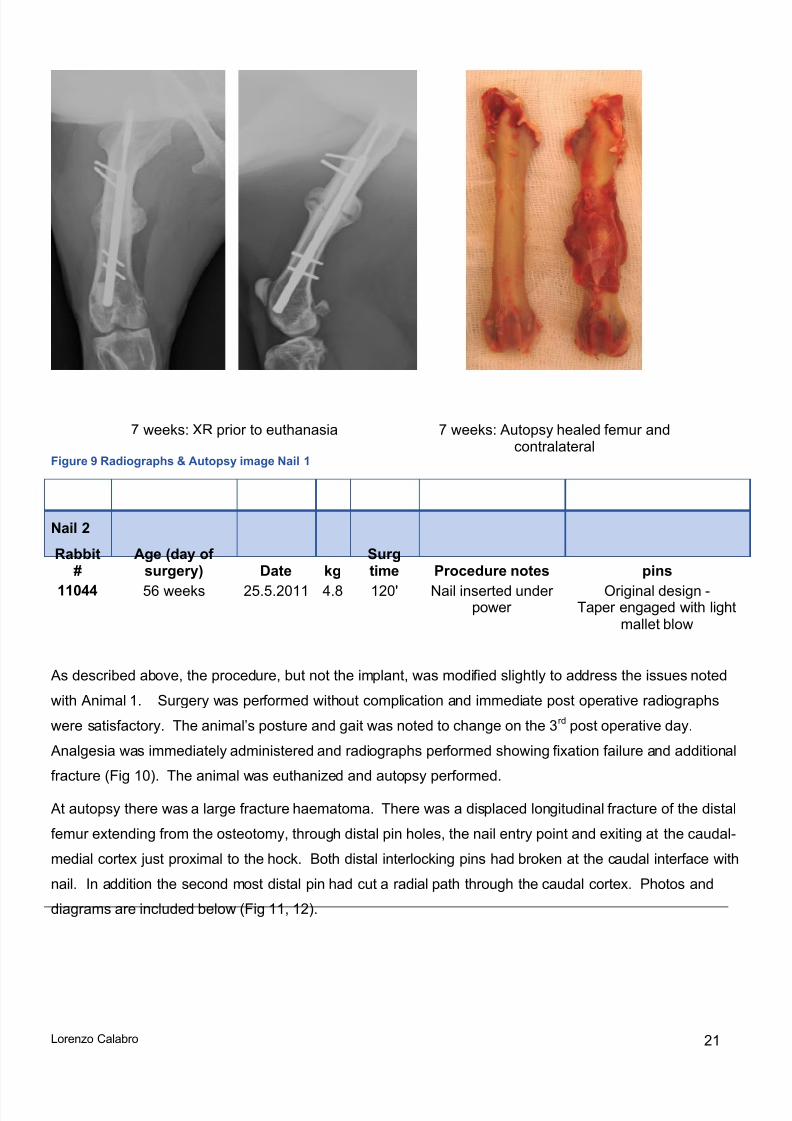
7 weeks: XR prior to euthanasia 7 weeks: Autopsy healed femur andcontralateral
Figure 9 Radiographs & Autopsy image Nail 1
Nail 2
Rabbit#
Age (day ofsurgery) Date kg
Surgtime Procedure notes pins
11044 56 weeks 25.5.2011 4.8 120' Nail inserted underpower
Original design -Taper engaged with light
mallet blow
As described above, the procedure, but not the implant, was modified slightly to address the issues notedwith Animal 1. Surgery was performed without complication and immediate post operative radiographswere satisfactory. The animal ’s posture and gait was noted to change on the 3 rd post operative day.
Analgesia was immediately administered and radiographs performed showing fixation failure and additionalfracture (Fig 10). The animal was euthanized and autopsy performed.
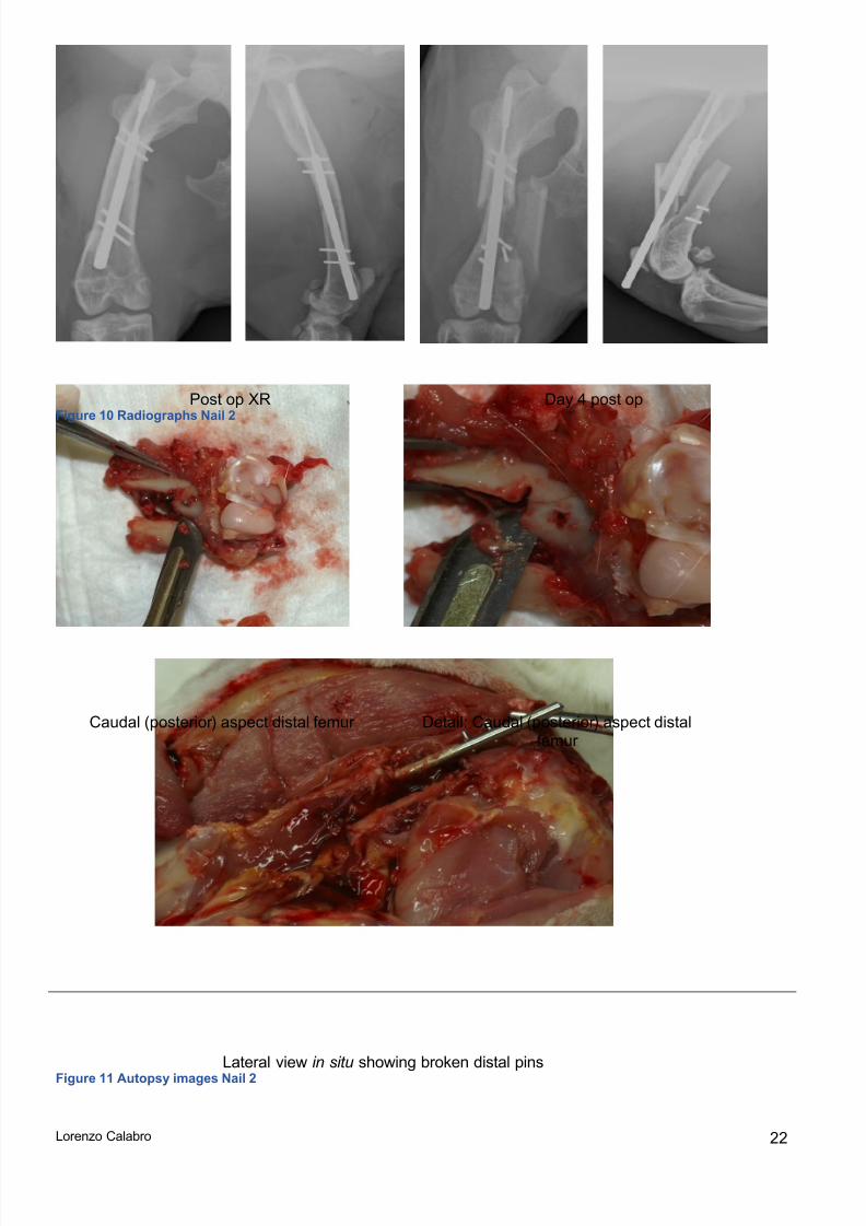
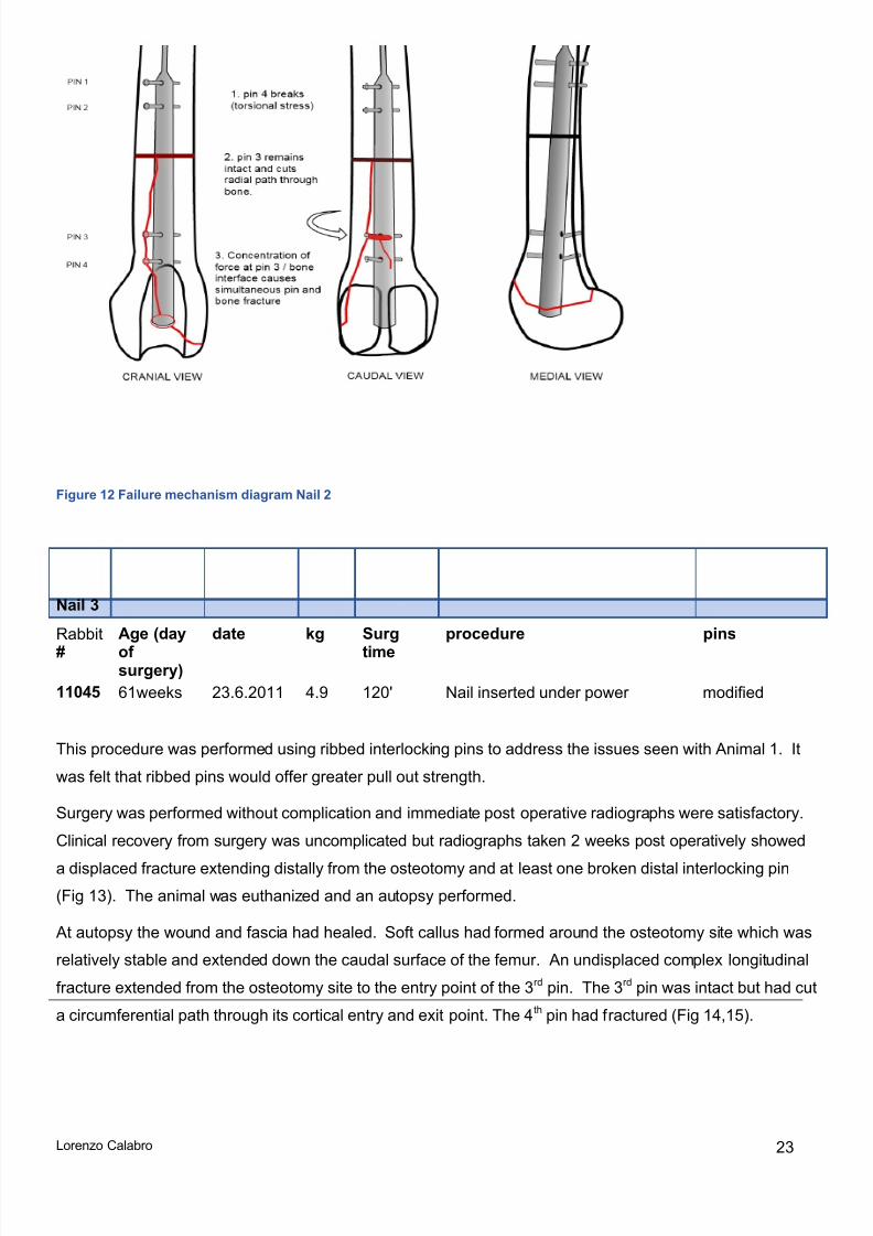
At autopsy there was a large fracture haematoma. There was a displaced longitudinal fracture of the distalfemur extending from the osteotomy, through distal pin holes, the nail entry point and exiting at the caudal-medial cortex just proximal to the hock. Both distal interlocking pins had broken at the caudal interface withnail. In addition the second most distal pin had cut a radial path through the caudal cortex. Photos anddiagrams are included below (Fig 11, 12).
8/13/2019 Lorenzo Calabro Thesis
http://slidepdf.com/reader/full/lorenzo-calabro-thesis 30/54
Lorenzo Calabro 22
Post op XR Day 4 post opFigure 10 Radiographs Nail 2
Caudal (posterior) aspect distal femur Detail: Caudal (posterior) aspect distalfemur
Lateral view in situ showing broken distal pinsFigure 11 Autopsy images Nail 2
8/13/2019 Lorenzo Calabro Thesis
http://slidepdf.com/reader/full/lorenzo-calabro-thesis 31/54
Lorenzo Calabro 23
Figure 12 Failure mechanism diagram Nail 2
Nail 3
Rabbit#
Age (dayofsurgery)
date kg Surgtime
procedure pins
11045 61weeks 23.6.2011 4.9 120' Nail inserted under power modified
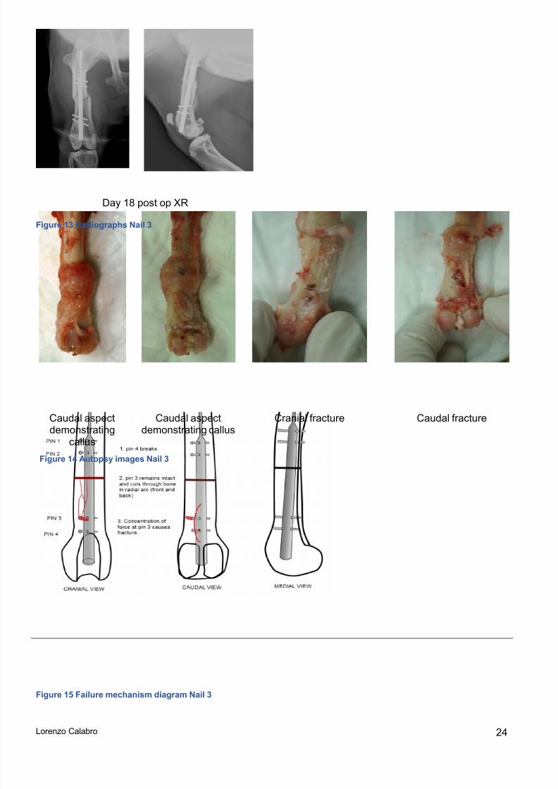
This procedure was performed using ribbed interlocking pins to address the issues seen with Animal 1. Itwas felt that ribbed pins would offer greater pull out strength.
Surgery was performed without complication and immediate post operative radiographs were satisfactory.Clinical recovery from surgery was uncomplicated but radiographs taken 2 weeks post operatively showed
a displaced fracture extending distally from the osteotomy and at least one broken distal interlocking pin(Fig 13). The animal was euthanized and an autopsy performed.
At autopsy the wound and fascia had healed. Soft callus had formed around the osteotomy site which wasrelatively stable and extended down the caudal surface of the femur. An undisplaced complex longitudinalfracture extended from the osteotomy site to the entry point of the 3 rd pin. The 3rd pin was intact but had cuta circumferential path through its cortical entry and exit point. The 4 th pin had fractured (Fig 14,15).
8/13/2019 Lorenzo Calabro Thesis
http://slidepdf.com/reader/full/lorenzo-calabro-thesis 32/54
Lorenzo Calabro 24
Day 18 post op XR
Figure 13 Radiographs Nail 3
Figure 15 Failure mechanism diagram Nail 3
Caudal aspectdemonstrating
callus
Caudal aspectdemonstrating callus
Cranial fracture Caudal fracture
Figure 14 Autopsy images Nail 3
8/13/2019 Lorenzo Calabro Thesis
http://slidepdf.com/reader/full/lorenzo-calabro-thesis 33/54
Lorenzo Calabro 25
4.4 Locking plate fixationFive animals underwent right femoral osteotomy with locked plate fixation. Three healed successfully.Procedures were performed at intervals again to allow technique or procedure modification ascomplications arose. Following failure of two 2.0mm screws at the 2 week mark in the first animal it wasdecided to increase to the 2.4mm LCP with 2.4mm screws. 2 animals had a 2.0mm plate while 3 had a2.4mm plate implanted. The trial was ceased after a peri-prosthetic fracture occurred in a rabbit fixed witha 2.4mm plate. Results are summarised in Table 5 and detailed below.
Rabbit EuthanizedStiffness healed femur / Stiffness contralateral
femur (Nm/deg) Histology
1 7 weeks 0.11/0.54
Hypertrophic callusIncomplete union
Minor malalignment2 7 weeks 0.63/0.65 Robust fracture healing3 7 weeks 0.53/0.63 Robust fracture healing4 7 weeks 0.67/0.61 Robust fracture healing5 2 weeks n/a n/a Table 5 Summary LCP results
2.0mm Plate 1
Rabbit#
Age (day ofsurgery) date kg Surg time Procedure notes
11041 54weeks 04.05.2011 4.9 75’ 2.0 mm plate
The procedure was performed without complication and immediate post operative radiographs weresatisfactory.
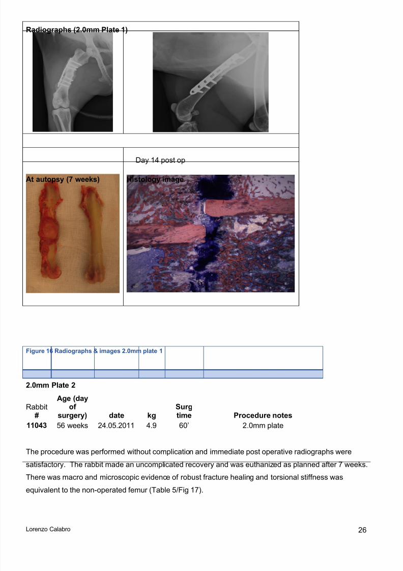
Clinical recovery from surgery was uncomplicated but radiographs taken 2 weeks post operatively showedthat the 2 nd and 3 rd screws (with 1st being most proximal) had broken through the shank at the interface withthe plate (Fig 16) . Very slight rotational malalignment was noted at the osteotomy but the animal showedno signs of discomfort or distress so the decision was made to continue observation. The animal waseuthanized as planned after 7 weeks with radiographs showing signs of fracture healing despite themechanical fixation failure. Post mortem macroscopic examination, mechanical testing and histologydemonstrated hypertrophic callus formation with incomplete fracture union (Table 5/Fig 16).
8/13/2019 Lorenzo Calabro Thesis
http://slidepdf.com/reader/full/lorenzo-calabro-thesis 34/54
Lorenzo Calabro 26
Radiographs (2.0mm Plate 1)
Day 14 post op
At autopsy (7 weeks) Histology image
Figure 16 Radiographs & images 2.0mm plate 1
2.0mm Plate 2
Rabbit#
Age (dayofsurgery) date kg
Surgtime Procedure notes
11043 56 weeks 24.05.2011 4.9 60’ 2.0mm plate
The procedure was performed without complication and immediate post operative radiographs weresatisfactory. The rabbit made an uncomplicated recovery and was euthanized as planned after 7 weeks.There was macro and microscopic evidence of robust fracture healing and torsional stiffness wasequivalent to the non-operated femur (Table 5/Fig 17).
8/13/2019 Lorenzo Calabro Thesis
http://slidepdf.com/reader/full/lorenzo-calabro-thesis 35/54
Lorenzo Calabro 27
Radiographs (2.0mm Plate 2)
At autopsy (7 weeks) Histology image
Figure 17 Radiographs & images 2.0mm plate 2
2.4mm Plate 1
Rabbit#
Age (day
ofsurgery) date kg Surgtime Procedure notes 11046 61 weeks 27.06.2011 4.7 60’ 2.4mm plate
Due to the fixation failure seen in LCP Animal 1 (11041) it was decided to increase the size and strength ofthe implant. In this and subsequent procedures an 8-hole, 2.4mm locked plate (2.4mm screws) was used
The procedure was performed without complication and immediate post operative radiographs weresatisfactory. The rabbit made an uncomplicated recovery and was euthanized as planned after 7 weeks.
8/13/2019 Lorenzo Calabro Thesis
http://slidepdf.com/reader/full/lorenzo-calabro-thesis 36/54
Lorenzo Calabro 28
There was macro and microscopic evidence of robust fracture healing and torsional stiffness wasequivalent to the non-operated femur (Table 5/Fig 18).
Radiographs 2.4mm Plate 1
Histology 2.4mm Plate 1
Figure 18 Radiographs & images 2.4mm plate 1
2.4mm Plate 2
#
Age (Dayof
Surgery) date kg Surgtime Procedure notes
11048 63 weeks 13.07.2011 4.6 60’ 2.4mm plate
The procedure was performed without complication and immediate post operative radiographs weresatisfactory. The rabbit made an uncomplicated recovery and was euthanized as planned after 7 weeks.
8/13/2019 Lorenzo Calabro Thesis
http://slidepdf.com/reader/full/lorenzo-calabro-thesis 37/54
Lorenzo Calabro 29
Radiographs and histology reflected those for 2.4mm Plate 1 above; there was macro and microscopicevidence of robust fracture healing and torsional stiffness was equivalent to the non-operated femur.
2.4mm Plate 3
#
Age (Dayof
Surgery) date kg Surgtime Procedure notes
11047 63 weeks 13.07.2011 4.3 70’ 2.4mm plate
The procedure was performed without complication and immediate post operative radiographs weresatisfactory. Clinical recovery from surgery was uncomplicated but radiographs taken 2 weeks postoperatively showed a displaced fracture of the proximal femur extending through the proximal screw holesfrom the osteotomy (Fig 19). The animal was euthanized and an autopsy performed
Figure 19 Radiographs & images 2.4mm plate 3
8/13/2019 Lorenzo Calabro Thesis
http://slidepdf.com/reader/full/lorenzo-calabro-thesis 38/54
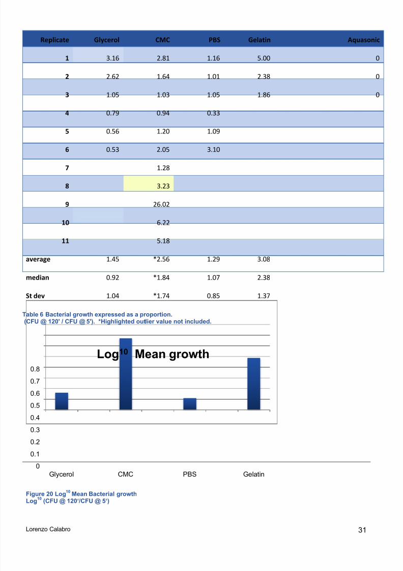
8/13/2019 Lorenzo Calabro Thesis
http://slidepdf.com/reader/full/lorenzo-calabro-thesis 39/54
Lorenzo Calabro 31
Figure 20 Log 10 Mean Bacterial growthLog 10 (CFU @ 120‘/CFU @ 5‘)
0
0.1
0.2
0.3
0.4
0.5
0.6
0.7
0.8
Glycerol CMC PBS Gelatin
Log 10 Mean growth
Replicate Glycerol CMC PBS Gelatin Aquasonic
1 3.16 2.81 1.16 5.00 0
2 2.62 1.64 1.01 2.38 0
3 1.05 1.03 1.05 1.86 0
4 0.79 0.94 0.33
5 0.56 1.20 1.09
6 0.53 2.05 3.10
7 1.28
8 3.23
9 26.02
10 6.22
11 5.18
average 1.45 *2.56 1.29 3.08
median 0.92 *1.84 1.07 2.38
St dev 1.04 *1.74 0.85 1.37
Table 6 Bacterial growth expressed as a proportion.(CFU @ 120' / CFU @ 5'). *Highlighted outlier value not included.
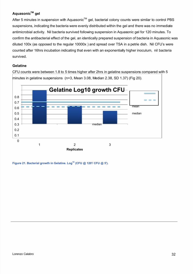
8/13/2019 Lorenzo Calabro Thesis
http://slidepdf.com/reader/full/lorenzo-calabro-thesis 40/54
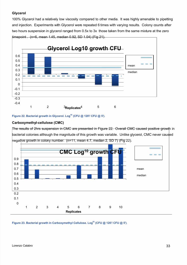
8/13/2019 Lorenzo Calabro Thesis
http://slidepdf.com/reader/full/lorenzo-calabro-thesis 41/54
Lorenzo Calabro 33
Glycerol
100% Glycerol had a relatively low viscosity compared to other media. It was highly amenable to pipettingand injection. Experiments with Glycerol were repeated 6 times with varying results. Colony counts aftertwo hours suspension in glycerol ranged from 0.5x to 3x those taken from the same mixture at the zerotimepoint . (n=6, mean 1.45, median 0.92, SD 1.04) (Fig 21).
Figure 22. Bacterial growth in Glycerol. Log 10 (CFU @ 120'/ CFU @ 5').
Carboxymethyl-cellulose (CMC)
The results of 2hrs suspension in CMC are presented in Figure 22. Overall CMC caused positive growth inbacterial colonies although the magnitude of this growth was variable. Unlike glycerol, CMC never caused
negative growth in colony number. (n=11, mean 4.7, median 2, SD 7) (Fig 22).
Figure 23. Bacterial growth in Carboxymethyl Cellulose. Log 10 (CFU @ 120'/ CFU @ 5').
-0.4-0.3-0.2-0.1
00.10.20.30.40.50.6
1 2 3 4 5 6Replicates
Glycerol Log10 growth CFU
mean
median
00.10.20.30.40.50.60.70.80.9
1 2 3 4 5 6 7 8 9 10Replicates
CMC Log 10 growth CFU
mean
median
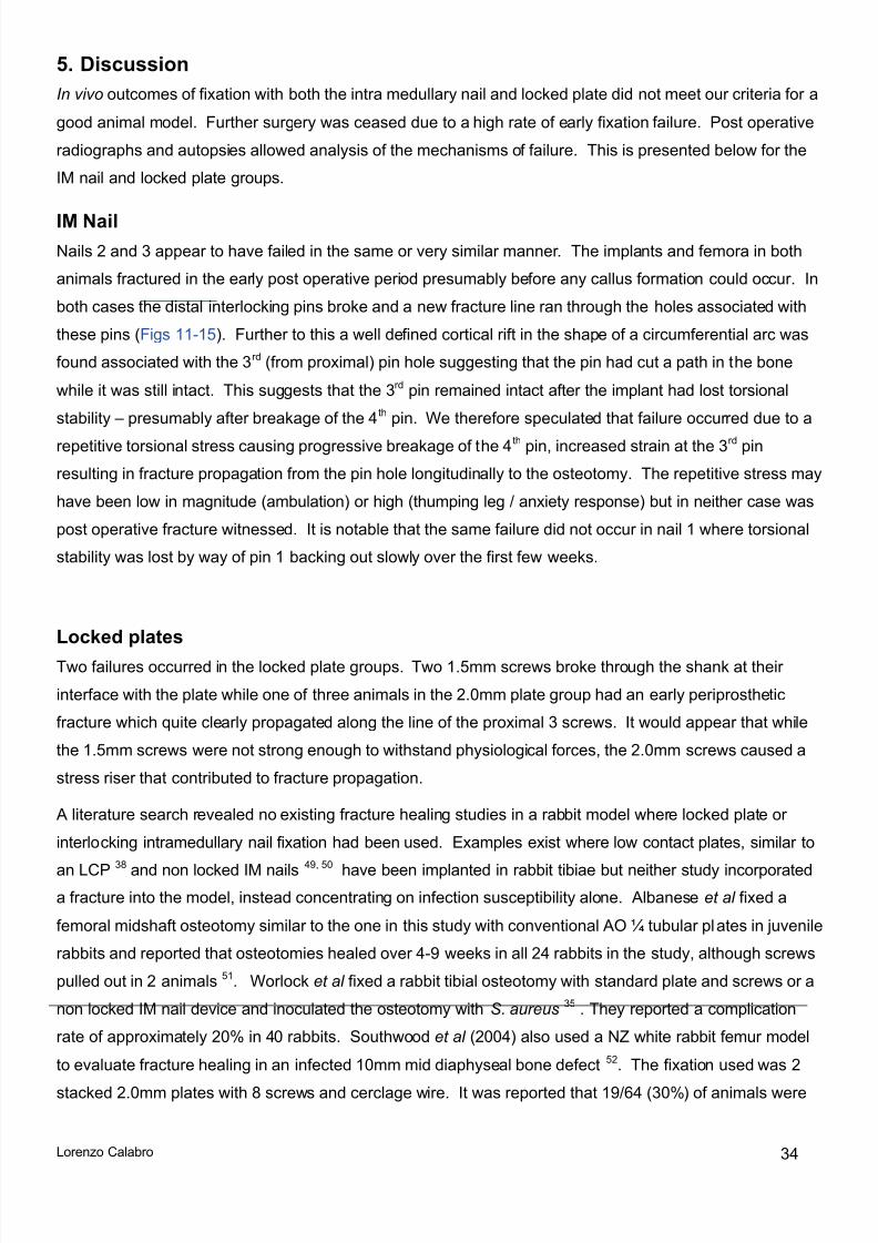
8/13/2019 Lorenzo Calabro Thesis
http://slidepdf.com/reader/full/lorenzo-calabro-thesis 42/54
Lorenzo Calabro 34
5. DiscussionIn vivo outcomes of fixation with both the intra medullary nail and locked plate did not meet our criteria for agood animal model. Further surgery was ceased due to a high rate of early fixation failure. Post operativeradiographs and autopsies allowed analysis of the mechanisms of failure. This is presented below for theIM nail and locked plate groups.
IM NailNails 2 and 3 appear to have failed in the same or very similar manner. The implants and femora in bothanimals fractured in the early post operative period presumably before any callus formation could occur. Inboth cases the distal interlocking pins broke and a new fracture line ran through the holes associated withthese pins (Figs 11-15). Further to this a well defined cortical rift in the shape of a circumferential arc wasfound associated with the 3 rd (from proximal) pin hole suggesting that the pin had cut a path in the bonewhile it was still intact. This suggests that the 3 rd pin remained intact after the implant had lost torsionalstability – presumably after breakage of the 4 th pin. We therefore speculated that failure occurred due to arepetitive torsional stress causing progressive breakage of the 4 th pin, increased strain at the 3 rd pinresulting in fracture propagation from the pin hole longitudinally to the osteotomy. The repetitive stress mayhave been low in magnitude (ambulation) or high (thumping leg / anxiety response) but in neither case waspost operative fracture witnessed. It is notable that the same failure did not occur in nail 1 where torsionalstability was lost by way of pin 1 backing out slowly over the first few weeks.
Locked platesTwo failures occurred in the locked plate groups. Two 1.5mm screws broke through the shank at theirinterface with the plate while one of three animals in the 2.0mm plate group had an early periprostheticfracture which quite clearly propagated along the line of the proximal 3 screws. It would appear that whilethe 1.5mm screws were not strong enough to withstand physiological forces, the 2.0mm screws caused astress riser that contributed to fracture propagation.
A literature search revealed no existing fracture healing studies in a rabbit model where locked plate orinterlocking intramedullary nail fixation had been used. Examples exist where low contact plates, similar to
an LCP 38
and non locked IM nails 49, 50
have been implanted in rabbit tibiae but neither study incorporateda fracture into the model, instead concentrating on infection susceptibility alone. Albanese et al fixed afemoral midshaft osteotomy similar to the one in this study with conventional AO ¼ tubular plates in juvenilerabbits and reported that osteotomies healed over 4-9 weeks in all 24 rabbits in the study, although screwspulled out in 2 animals 51. Worlock et al fixed a rabbit tibial osteotomy with standard plate and screws or anon locked IM nail device and inoculated the osteotomy with S. aureus 35 . They reported a complicationrate of approximately 20% in 40 rabbits. Southwood et al (2004) also used a NZ white rabbit femur modelto evaluate fracture healing in an infected 10mm mid diaphyseal bone defect 52. The fixation used was 2
stacked 2.0mm plates with 8 screws and cerclage wire. It was reported that 19/64 (30%) of animals were
8/13/2019 Lorenzo Calabro Thesis
http://slidepdf.com/reader/full/lorenzo-calabro-thesis 43/54
Lorenzo Calabro 35
euthanized without completing the study although the specific reasons were not described. Fracturemodels using a saw osteotomy have been created in the rabbit tibia. The model described by Darouiche et
al 53 is typical where a Kirschner wire is used for fixation with the fibula left intact to provide further stability.Darouiche reported that 17/44 (39%) of animals were euthanized prior to study completion due tocomplications including wound dehiscence, loss of fixation or failure to thrive.
Failure mechanisms and potential solutionsRepeated failure at the interlocking pin/bone interface suggested that this should be a focus in re-designingthe nails. Potential variables considered were pin diameter, pin spacing, pin number and the method andstrength of pin fixation in bone. Pins in all 3 animals were pre-drilled with a 1mm drill bit and gainedpurchase in the near femoral cortex by means of a taper design with pre-determined break-off torque tostandardise their insertion strength. With a smooth pin design, in nail 1 the proximal interlocking pinsbacked out early in the healing period. This effectively led to a loss of torsional stability of the construct,and therefore torsional loads were no longer transmitted to the distal pin/bone interface when the animal
loaded its femur. The addition of ribs to the pins and an increased break-off torque appeared to havesuccessfully addressed the back-out failure observed in animal 1. But conversely, it appears that themaintained load transfer to the distal nail and interlocking pin section and the repetitive loading due to limbambulation throughout the healing period exceeded the fatigue resistance of the distal interlocking pins andconsequently led to their failure. Increasing the pin diameter or number may have increased the fatigueresistance of the pins and could have prevented pin breakage, which appeared to be the initial point offailure (Figs 12,15), but it was feared that both of these solutions would have increased the likelihood ofcrack formation in the rabbit femora. These bones have been shown to have low fracture toughness inlongitudinal planes 54. This is thought to be due to their abundance of longitudinally oriented vascularcanals 54 . Clustering larger pin holes along a longitudinal axis, as is mandated by the current design of theaiming device, therefore may have weakened the bone and could have led to longitudinal crack formation.
Although increases in pin diameter or number would have increased the overall strength and stiffness ofthe implant construct, earlier ex vivo mechanical tests had shown consistent failure of the bone before anyimplant construct failures. It was therefore not altogether certain that increasing the strength and stiffness ofthe implant would have lead to more reliable results. More importantly, however, these experimentsshowed the limitations of static mechanical tests for the prediction of implant survival in longer term fracturehealing experiments.
A similar conundrum was faced in the locked plate model where an incremental increase in plate and screwsize led to changes in the mechanism of failure. In the 2.0mm plate 1, fixation failed due to implantbreakage. In order to address this, a stronger implant (2.4mm plate) was used for subsequent animals. Butin 2.4mm plate 3 a longitudinal fracture occurred through the screw holes, which were larger than in theoriginally used 2.0mm plate, highlighting the low fracture toughness of rabbit femora in longitudinal planes,as mentioned earlier. It could be argued that the 2.4mm plate model was prematurely abandoned after a
single failure, and that modification of the implants could have reduced the risk of construct failure.
8/13/2019 Lorenzo Calabro Thesis
http://slidepdf.com/reader/full/lorenzo-calabro-thesis 44/54
Lorenzo Calabro 36
Potential modifications may have included tapping the screw holes to reduce the pre-tension and thereforereduced the risk for longitudinal fractures, or the alteration of the working length or screw number to changethe overall construct stiffness. Importantly, any of these potential modifications to implant design wouldhave needed to be mechanically tested ex vivo and the results compared to those achieved with theexisting systems prior to further in vivo work. Similarex vivo testing would be needed to test thehypotheses above regarding pin diameter and placement in the IM nail model. However, considering thefrequency of failure in the in vivo experiments and the nature of the mechanisms of failure in both in vivo and ex vivo trials, it was concluded that the rabbit femur model, due to its low longitudinal fracturetoughness and the high loads applied to this bone during animal locomotion, would have a low safetymargin even with potential modifications.
Rabbits as animal models for fracture healingRabbits cannot be feasibly or ethically immobilised post operatively and have a powerful hind leg with largemuscle bulk in proportion to bone size. As noted above, unpredictable thumping or ambulation forceappears to have overwhelmed the strength of the implant/bone system. Other factors that may havecontributed are rabbit age, weight and bone quality. We used NZ white rabbits with an average weight of4.8 kg and average age of 59 weeks. NZ white rabbits are the species most commonly used inmusculoskeletal infection research 36, 49, 55-63. The rabbits used in this trial were heavier than thosecommonly used in the literature and this may have influenced our results. As noted above Albanese et al reported that 100% of femoral osteotomies healed in their cohort of 24 six week old rabbits 51. Average
animal weights were reviewed in a number of studies that used NZ white rabbits for animal models and arepresented in table 7.
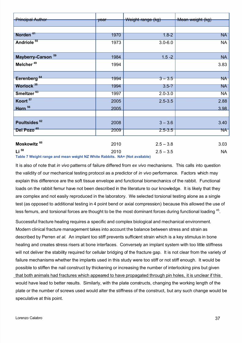
8/13/2019 Lorenzo Calabro Thesis
http://slidepdf.com/reader/full/lorenzo-calabro-thesis 45/54
Lorenzo Calabro 37
Principal Author year Weight range (kg) Mean weight (kg)
Norden 61 1970 1.8-2 NAAndriole 55 1973 3.0-6.0 NA
Mayberry-Carson 59 1984 1.5 -2 NAMelcher 49 1994 3.83
Eerenberg 64 1994 3 – 3.5 NAWorlock 35 1994 3.5-? NASmeltzer 63 1997 2.0-3.0 NAKoort 57 2005 2.5-3.5 2.88Horn 56 2005 3.98
Poultsides 62 2008 3 – 3.6 3.40Del Pozo 65 2009 2.5-3.5 NA
Moskowitz 60 2010 2.5 – 3.8 3.03Li 58 2010 2.5 – 3.5 NA Table 7 Weight range and mean weight NZ White Rabbits. NA= (Not available)
It is also of note that in vivo patterns of failure differed from ex vivo mechanisms. This calls into questionthe validity of our mechanical testing protocol as a predictor of in vivo performance. Factors which may
explain this difference are the soft tissue envelope and functional biomechanics of the rabbit. Functionalloads on the rabbit femur have not been described in the literature to our knowledge. It is likely that theyare complex and not easily reproduced in the laboratory. We selected torsional testing alone as a singletest (as opposed to additional testing in 4 point bend or axial compression) because this allowed the use ofless femurs, and torsional forces are thought to be the most dominant forces during functional loading 45.
Successful fracture healing requires a specific and complex biological and mechanical environment.Modern clinical fracture management takes into account the balance between stress and strain asdescribed by Perren et al . An implant too stiff prevents sufficient strain which is a key stimulus in bonehealing and creates stress risers at bone interfaces. Conversely an implant system with too little stiffnesswill not deliver the stability required for cellular bridging of the fracture gap. It is not clear from the variety offailure mechanisms whether the implants used in this study were too stiff or not stiff enough. It would bepossible to stiffen the nail construct by thickening or increasing the number of interlocking pins but giventhat both animals had fractures which appeared to have propagated through pin holes, it is unclear if thiswould have lead to better results. Similarly, with the plate constructs, changing the working length of theplate or the number of screws used would alter the stiffness of the construct, but any such change would be
speculative at this point.
8/13/2019 Lorenzo Calabro Thesis
http://slidepdf.com/reader/full/lorenzo-calabro-thesis 46/54
Lorenzo Calabro 38
Another reasonable option would be to use a non (or less) weight bearing long bone in the rabbit. Thismay avoid the large potential physiological forces placed through a rabbit femur. The rabbit humerus maybe suitable as they have good soft tissue cover and tend to have a straight intramedullary canal whichcould accommodate an interlocked nail.Subsequent to the completion of this work, rabbit humerus plateand IM nail models have been developed within the same research group with no failures in 16 pilotanimals (8 nails and 8 plates) (Correspondence ARI, August 2013).
In v i t ro inoculation media experimentsTo our knowledge no published data exists in the scientific literature on bacterial viability in the mediatested in our experiments, with the exception of glycerol. Saegeman et al reported on the survival of arange of bacteria including S. epidermidis and S. aureus in 85% glycerol over 24-36 hrs and up to 10 days40. They found that there was a weak antimicrobial effect, more pronounced with gram negative species,and over the longer time frame.
When measured against the criteria listed above: Safety; sterility; handling; cost; impact on bacterialviability; and biocompatibility; none of the candidate media could be said to be ideal. Aquasonic gel andGelatine can be eliminated due to frank antimicrobial effect and poor handling characteristics, respectively.Bacterial viability in Glycerol and CMC was more variable than in PBS. It is not possible from this data todetermine the cause for this variation. It is necessary to examine our data and methods for signs ofrandom or systematic error. Potential sources of random error include heterogeneity in media andimprecision in equipment or processes (light spectrometer, incubator, the dilution process, Eppendorf tubelabelling, timing or colony counts). Random error can be reduced by repetition. We feel that consistency
of results, particularly in PBS control preparations (which were inoculated, diluted and plated in parallel andwith identical technique to experimental preparations) indicates that variation is unlikely to be due totechnique error. Potential sources of systematic error could relate to changes in the source bacteria, due tomutation or contamination in the 3 week period of experimentation. However, the macroscopic appearanceof S. aureus is distinct and remained constant throughout the experiment, indicating this was not a sourceof error. In addition, steps were taken, as detailed above, to ensure that bacteria were in an optimal logphase of growth in each series. Manipulation of inoculation media required a number of steps includingaccurate pipetting and mechanical mixing of visually indistinguishable fluids for each preparation and
dilution. It is possible that a heterogeneous suspension of bacteria in viscous media would cause samplingerror, which then would continue down the chain of dilution distorting results. No trend was identified overtime to suggest that results were influenced by a learning curve. Variation in results is best explained byeither an inherent variation in index colonies, or sources of random error detailed above, or a combinationof both.
In our view both Glycerol and CMC had an acceptable impact on bacterial growth for consideration asinoculation media in a small animal infection model (e.g. in rabbits) where up to 10 7 CFU are inoculated in a1mL preparation. The fact that CMC caused positive growth in all series does not absolutely preclude itsuse in animal infection models. In a study designed to evaluate a treatment for established osteomyelitis

8/13/2019 Lorenzo Calabro Thesis
http://slidepdf.com/reader/full/lorenzo-calabro-thesis 47/54
Lorenzo Calabro 39
where the aim is to create infection in 100% of subject animals (ID100) CMC may be appropriate. Cautionmay be required in an ID50 trial design (evaluating prophylactic interventions for example). In such a trial,where the goal is to prevent infection, a clinically relevant effect may be overwhelmed by a higher thanexpected inoculum. CMC was more viscous than 98% glycerol and for this reason had preferablehandling characteristics although it is difficult to evaluate this objectively.
6. ConclusionsNeither the locked plate or RabbitNail fracture models could be considered to meet criteria for a reliable,reproducible and robust animal model. Given the high incidence of early mechanical failure in both groups,and in the existing literature, future fracture models involving the rabbit femur should be undertaken withcaution and in initially limited numbers. It is not possible to determine the cause of failure from this data butanimal weight and senescence may have influenced our results. The utility of ex vivo mechanical testing offixation constructs for predicting in vivo performance is limited.
The nature of the inoculation media experiments was exploratory rather than definitive. Aquasonic gel andpharmaceutical gelatine with the specifications used can be ruled out as effective inoculation media. CMCand Glycerol performed satisfactorily with regards to handling and bacterial viability and warrant furtherinvestigation. Options to further test the hypothesis that viscous media would improve precision inmusculoskeletal infection models include contrast imaging to determine dissemination and a limited in vivo pilot study.
8/13/2019 Lorenzo Calabro Thesis
http://slidepdf.com/reader/full/lorenzo-calabro-thesis 48/54
Lorenzo Calabro 40
7. Reference List1. Boxma H, Broekhuizen T, Patka P, Oosting H. Randomised controlled trial of single-dose antibiotic
prophylaxis in surgical treatment of closed fractures: the Dutch Trauma Trial. Lancet. 1996; 347 (9009): 1133-7.
2. Darouiche RO. Treatment of infections associated with surgical implants. NEnglJMed. 2004; 350 (14): 1422-9.3. Patzakis MJ, Wilkins J. Factors influencing infection rate in open fracture wounds. ClinOrthopRelat Res. 1989;
(243): 36-40.4. Edwards C, Counsell A, Boulton C, Moran CG. Early infection after hip fracture surgery: risk factors, costs and
outcome. JBone Joint SurgBr. 2008; 90(6): 770-7.5. Espehaug B, Engesaeter LB, Vollset SE, Havelin LI, Langeland N. Antibiotic prophylaxis in total hiparthroplasty. Review of 10,905 primary cemented total hip replacements reported to the Norwegianarthroplasty register, 1987 to 1995. JBone Joint SurgBr. 1997; 79(4): 590-5.
6. An YH, Kang QK, Arciola CR. Animal models of osteomyelitis. IntJArtifOrgans. 2006;29(4): 407-20.7. Aerssens J, Boonen S, Lowet G, Dequeker J. Interspecies differences in bone composition, density, and
quality: potential implications for in vivo bone research. Endocrinology. 1998; 139 (2): 663-70.8. Egermann M, Goldhahn J, Schneider E. Animal models for fracture treatment in osteoporosis. OsteoporosInt.
2005; 16 Suppl 2 : S129-S38.9. Auer JA, Goodship A, Arnoczky S, Pearce S, Price J, Claes L, et al. Refining animal models in fracture
research: seeking consensus in optimising both animal welfare and scientific validity for appropriatebiomedical use. BMC musculoskeletal disorders. 2007; 8: 72.
10. Garcia P, Holstein J, Histing T, Burkhardt M, Culemann U, Pizanis A, et al. A new technique for internal
fixation of femoral fractures in mice: Impact of stability on fracture healing. Journal of Biomechanics. 2008; 41 :1689-96.11. Matthys R, Perren S. Internal fixator for use in the mouse. Injury. 2009; 40 : S103-S9.12. Rontgen V, Blakytny R, Matthys R, Landauer M, Wehner T, Gockelmann M, et al. Fracture healing in mice
under controlled rigid and flexible conditions using an adjustable external fixator. Journal of OrthopaedicResearch. 2010; 28(11): 1456-62.
13. Shultz LD, Ishikawa F, Greiner DL. Humanized mice in translational biomedical research. NatRevImmunol.2007; 7(2): 118-30.
14. Norden CW. Lessons learned from animal models of osteomyelitis. RevInfectDis. 1988; 10(1): 103-10.15. Arens S, Schlegel U, Printzen G, Ziegler WJ, Perren SM, Hansis M. Influence of materials for fixation implants
on local infection. An experimental study of steel versus titanium DCP in rabbits. The Journal of bone and jointsurgery British volume. 1996; 78(4): 647-51.
16. Schlegel U, Perren SM. Surgical aspects of infection involving osteosynthesis implants: implant design and
resistance to local infection. Injury. 2006; 37 Suppl 2 : S67-S73.17. Stewart S, Barr S, Engiles J, Hickok NJ, Shapiro IM, Richardson DW, et al. Vancomycin-modified implantsurface inhibits biofilm formation and supports bone-healing in an infected osteotomy model in sheep: a proof-of-concept study. J Bone Joint Surg Am. 2012; 94(15): 1406-15.
18. Lucke M, Schmidmaier G, Sadoni S, Wildemann B, Schiller R, Stemberger A, et al. A new model of implant-related osteomyelitis in rats. JBiomedMaterResB ApplBiomater. 2003; 67(1): 593-602.
19. Guide for the Care and Use of Laboratory Animals: The National Academies Press; 1996.20. Arens S, Kraft C, Schlegel U, Printzen G, Perren SM, Hansis M. Susceptibility to local infection in biological
internal fixation. Experimental study of open vs minimally invasive plate osteosynthesis in rabbits. ArchOrthopTrauma Surg. 1999; 119 (1-2): 82-5.
21. Claes L, Eckert-Hubner K, Augat P. The effect of mechanical stability on local vascularization and tissuedifferentiation in callus healing. Journal of Orthopaedic Research. 2002; 20 : 1099-105.
22. An YH, Friedman RJ. Animal models of orthopedic implant infection. Journal of investigative surgery : the
official journal of the Academy of Surgical Research. 1998; 11 (2): 139-46.23. Histing T, Garcia P, Matthys R, Leidinger M, Holstein J, Kristen A, et al. An internal locking plate to studyintramembranous bone healing in a mouse femur fracture model. Journal of Orthopaedic Research. 2010;28(3): 397-402.
24. Legrand N, Ploss A, Balling R, Becker PD, Borsotti C, Brezillon N, et al. Humanized mice for modeling humaninfectious disease: challenges, progress, and outlook. Cell HostMicrobe. 2009; 6(1): 5-9.
25. Zimmerli W, Trampuz A, Ochsner PE. Prosthetic-joint infections. NEnglJMed. 2004; 351 (16): 1645-54.26. Bowen TR, Widmaier JC. Host classification predicts infection after open fracture. ClinOrthopRelat Res. 2005;
(433): 205-11.27. Dumont C, Kauer F, Bohr S, Schmidtmann U, Knopp W, Engelhardt T, et al. Long-term effects of saw
osteotomy versus random fracturing on bone healing and remodeling in a sheep tibia model. Journal ofOrthopaedic Research. 2009; 27(5): 680-6.
28. Ashhurst DE, Hogg J, Perren SM. A method for making reproducible experimental fractures of the rabbit tibia.Injury. 1982; 14(3): 236-42.
8/13/2019 Lorenzo Calabro Thesis
http://slidepdf.com/reader/full/lorenzo-calabro-thesis 49/54
Lorenzo Calabro 41
29. Jackson RW, Reed CA, Israel JA, Abou-Keer FK, Garside H. Production of a standard experimental fracture.CanJSurg. 1970; 13(4): 415-20.
30. Bonnarens F, Einhorn TA. Production of a standard closed fracture in laboratory animal bone. Journal oforthopaedic research : official publication of the Orthopaedic Research Society. 1984; 2(1): 97-101.
31. Rittman WW, Perren S. Cortical Bone Healing after Internal Fixation and Fracture. Berlin Heidelberg New YorkSpringer-Verlag; 1974.
32. Curtis MJ, Brown PR, Dick JD, Jinnah RH. Contaminated fractures of the tibia: a comparison of treatmentmodalities in an animal model. Journal of orthopaedic research : official publication of the OrthopaedicResearch Society. 1995; 13(2): 286-95.
33. Lindsey BA, Clovis NB, Smith ES, Salihu S, Hubbard DF. An animal model for open femur fracture andosteomyelitis: Part I. Journal of orthopaedic research : official publication of the Orthopaedic ResearchSociety. 2010; 28(1): 38-42.
34. Hill PF, Clasper JC, Parker SJ, Watkins PE. Early intramedullary nailing in an animal model of a heavilycontaminated fracture of the tibia. Journal of orthopaedic research : official publication of the OrthopaedicResearch Society. 2002; 20(4): 648-53.
35. Worlock P, Slack R, Harvey L, Mawhinney R. The prevention of infection in open fractures: an experimentalstudy of the effect of fracture stability. Injury. 1994; 25(1): 31-8.
36. Worlock P, Slack R, Harvey L, Mawhinney R. An experimental model of post-traumatic osteomyelitis inrabbits. British journal of experimental pathology. 1988; 69(2): 235-44.
37. Moriarty TF, Schlegel U, Perren S, Richards RG. Infection in fracture fixation: can we influence infection ratesthrough implant design? Journal of materials science Materials in medicine. 2010; 21(3): 1031-5.
38. Arens S, Eijer H, Schlegel U, Printzen G, Perren SM, Hansis M. Influence of the Design for Fixation Implants
on Local Infection: Experimental Study of Dynamic Compression Plates versus Point Contact Fixators inRabbits. Journal of orthopaedic trauma. 1999; 13(7).39. Currie LJ, Sharpe JR, Martin R. The use of fibrin glue in skin grafts and tissue-engineered skin replacements:
a review. PlastReconstrSurg. 2001; 108 (6): 1713-26.40. Saegeman VS, De VR, Tebaldi ND, van der Wolf JM, Bergervoet JH, Verhaegen J, et al. Flow cytometric
viability assessment and transmission electron microscopic morphological study of bacteria in glycerol.MicroscMicroanal. 2007;13(1): 18-29.
41. Ito K, Perren S. Biology and biomechanics of bone healing. In: RÅedi T, Buckley R, Moran C, editors. AOPrinciples of Fracture Management. Davos Platz: AO Publishing; 2007. p. 9-32.
42. Gautier E, Sommer C. Guidelines for the clinical application of the LCP. Injury. 2003; 34(Supplement 2): 63-76.
43. Stoffel K, Dieter U, Stachowiak G, G‰chter A, Kuster MS . Biomechanical testing of the LCP - how canstability in locked internal fixators be controlled? Injury. 2003; 34(Supplement 2): 11-9.
44. Ahmad M, Nanda R, Bajwa AS, Candal-Couto J, Green S, Hui AC. Biomechanical testing of the lockingcompression plate: when does the distance between bone and implant significantly reduce construct stability?Injury. 2007; 38(3): 358-64.
45. Burstein AH, Frankel VH. A standard test for laboratory animal bone. Journal of Biomechanics. 1971; 4(2):155-8.
46. Hak DJ, Schulz KS, Khoie B, Hazelwood SJ. The effect of Cox-2 specific inhibition on direct fracture healing inthe rabbit tibia. JOrthopSci. 2011.
47. Meffert RH, Jansen H, Frey SP, Raschke MJ, Langer M. The influence of soft tissue trauma on boneregeneration after acute limb shortening. ClinOrthopRelat Res. 2007; 460 : 202-9.
48. Wildemann B, Bamdad P, Holmer C, Haas NP, Raschke M, Schmidmaier G. Local delivery of growth factorsfrom coated titanium plates increases osteotomy healing in rats. Bone. 2004; 34(5): 862-8.
49. Melcher GA, Claudi B, Schlegel U, Perren SM, Printzen G, Munzinger J. Influence of type of medullary nail onthe development of local infection. An experimental study of solid and slotted nails in rabbits. The Journal ofbone and joint surgery British volume. 1994; 76(6): 955-9.50. Moriarty TF, Debefve L, Boure L, Campoccia D, Schlegel U, Richards RG. Influence of material andmicrotopography on the development of local infection in vivo: experimental investigation in rabbits. TheInternational journal of artificial organs. 2009; 32(9): 663-70.
51. Albanese SA, Spadaro JA, Chase SE, Geel CW. Bone growth after osteotomy and internal fixation in youngrabbits. J Orthop Res. 1996; 14(6): 921-6.
52. Southwood LL, Frisbie DD, Kawcak CE, Ghivizzani SC, Evans CH, McIlwraith CW. Evaluation of Ad-BMP-2for enhancing fracture healing in an infected defect fracture rabbit model. Journal of Orthopaedic Research.2004; 22(1): 66-72.
53. Darouiche RO, Farmer J, Chaput C, Mansouri M, Saleh G, Landon GC. Anti-infective efficacy of antiseptic-coated intramedullary nails. JBone Joint SurgAm. 1998; 80(9): 1336-40.
54. Wang X, Mabrey JD, Agrawal CM. An interspecies comparison of bone fracture properties. BiomedMaterEng.1998; 8(1): 1-9.
8/13/2019 Lorenzo Calabro Thesis
http://slidepdf.com/reader/full/lorenzo-calabro-thesis 50/54
Lorenzo Calabro 42
55. Andriole VT, Nagel DA, Southwick WO. A paradigm for human chronic osteomyelitis. JBone Joint SurgAm.1973; 55(7): 1511-5.
56. Horn J, Schlegel U, Krettek C, Ito K. Infection resistance of unreamed solid, hollow slotted and cannulatedintramedullary nails: an in-vivo experimental comparison. Journal of orthopaedic research : official publicationof the Orthopaedic Research Society. 2005; 23(4): 810-5.
57. Koort JK, Makinen TJ, Suokas E, Veiranto M, Jalava J, Knuuti J, et al. Efficacy of ciprofloxacin-releasingbioabsorbable osteoconductive bone defect filler for treatment of experimental osteomyelitis due toStaphylococcus aureus. Antimicrob Agents Chemother. 2005; 49(4): 1502-8.
58. Li BC, Zhang JJ, Xu C, Zhang LC, Kang JY, Zhao H. Treatment of rabbit femoral defect by firearm with BMP-4
gene combined with TGF-beta1. JTrauma. 2009; 66(2): 450-6.59. Mayberry-Carson KJ, Tober-Meyer B, Smith JK, Lambe DW, Jr., Costerton JW. Bacterial adherence andglycocalyx formation in osteomyelitis experimentally induced with Staphylococcus aureus. InfectImmun. 1984;43(3): 825-33.
60. Moskowitz JS, Blaisse MR, Samuel RE, Hsu HP, Harris MB, Martin SD, et al. The effectiveness of thecontrolled release of gentamicin from polyelectrolyte multilayers in the treatment of Staphylococcus aureusinfection in a rabbit bone model. Biomaterials. 2010; 31(23): 6019-30.
61. Norden CW. Experimental osteomyelitis. I. A description of the model. JInfectDis. 1970; 122 (5): 410-8.62. Poultsides LA, Papatheodorou LK, Karachalios TS, Khaldi L, Maniatis A, Petinaki E, et al. Novel model for
studying hematogenous infection in an experimental setting of implant-related infection by a community-acquired methicillin-resistant S. aureus strain. Journal of Orthopaedic Research. 2008; 26(10): 1355-62.
63. Smeltzer MS, Thomas JR, Hickmon SG, Skinner RA, Nelson CL, Griffith D, et al. Characterization of a rabbitmodel of staphylococcal osteomyelitis. Journal of Orthopaedic Research. 1997; 15(3): 414-21.
64. Eerenberg JP, Patka P, Haarman HJ, Dwars BJ. A new model for posttraumatic osteomyelitis in rabbits.JInvest Surg. 1994; 7(5): 453-65.65. Del Pozo JL, Rouse MS, Euba G, Kang CI, Mandrekar JN, Steckelberg JM, et al. The electricidal effect is
active in an experimental model of Staphylococcus epidermidis chronic foreign body osteomyelitis. AntimicrobAgents Chemother. 2009; 53(10): 4064-8.
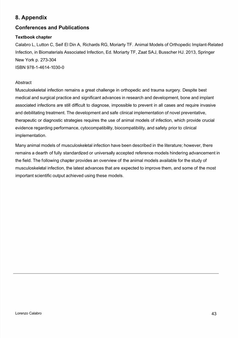
8/13/2019 Lorenzo Calabro Thesis
http://slidepdf.com/reader/full/lorenzo-calabro-thesis 51/54
Lorenzo Calabro 43
8. Appendix
Conferences and Publications
Textbook chapter
Calabro L, Lutton C, Seif El Din A, Richards RG, Moriarty TF. Animal Models of Orthopedic Implant-RelatedInfection, in Biomaterials Associated Infection, Ed. Moriarty TF, Zaat SAJ, Busscher HJ. 2013, Springer
New York p. 273-304ISBN 978-1-4614-1030-0
AbstractMusculoskeletal infection remains a great challenge in orthopedic and trauma surgery. Despite bestmedical and surgical practice and significant advances in research and development, bone and implantassociated infections are still difficult to diagnose, impossible to prevent in all cases and require invasiveand debilitating treatment. The development and safe clinical implementation of novel preventative,
therapeutic or diagnostic strategies requires the use of animal models of infection, which provide crucialevidence regarding performance, cytocompatibility, biocompatibility, and safety prior to clinicalimplementation.
Many animal models of musculoskeletal infection have been described in the literature; however, thereremains a dearth of fully standardized or universally accepted reference models hindering advancement inthe field. The following chapter provides an overview of the animal models available for the study ofmusculoskeletal infection, the latest advances that are expected to improve them, and some of the most
important scientific output achieved using these models.
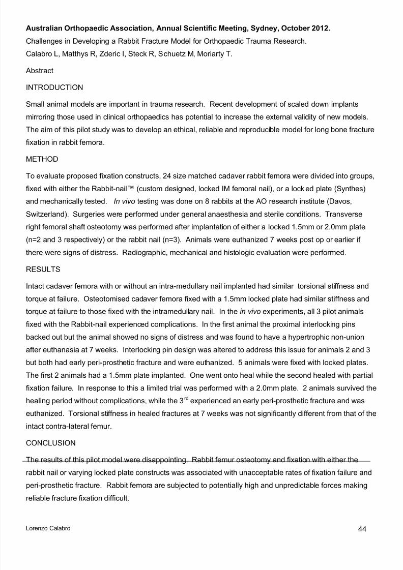
8/13/2019 Lorenzo Calabro Thesis
http://slidepdf.com/reader/full/lorenzo-calabro-thesis 52/54
Lorenzo Calabro 44
Australian Orthopaedic Association, Annual Scientific Meeting, Sydney, October 2012.
Challenges in Developing a Rabbit Fracture Model for Orthopaedic Trauma Research.Calabro L, Matthys R, Zderic I, Steck R, Schuetz M, Moriarty T.
Abstract
INTRODUCTION
Small animal models are important in trauma research. Recent development of scaled down implantsmirroring those used in clinical orthopaedics has potential to increase the external validity of new models.The aim of this pilot study was to develop an ethical, reliable and reproducible model for long bone fracturefixation in rabbit femora.
METHOD
To evaluate proposed fixation constructs, 24 size matched cadaver rabbit femora were divided into groups,fixed with either the Rabbit-nail™ (custom designed, locked IM femoral nail), or a lock ed plate (Synthes)
and mechanically tested. In vivo testing was done on 8 rabbits at the AO research institute (Davos,Switzerland). Surgeries were performed under general anaesthesia and sterile conditions. Transverseright femoral shaft osteotomy was performed after implantation of either a locked 1.5mm or 2.0mm plate(n=2 and 3 respectively) or the rabbit nail (n=3). Animals were euthanized 7 weeks post op or earlier ifthere were signs of distress. Radiographic, mechanical and histologic evaluation were performed.
RESULTS
Intact cadaver femora with or without an intra-medullary nail implanted had similar torsional stiffness and
torque at failure. Osteotomised cadaver femora fixed with a 1.5mm locked plate had similar stiffness andtorque at failure to those fixed with the intramedullary nail. In the in vivo experiments, all 3 pilot animalsfixed with the Rabbit-nail experienced complications. In the first animal the proximal interlocking pinsbacked out but the animal showed no signs of distress and was found to have a hypertrophic non-unionafter euthanasia at 7 weeks. Interlocking pin design was altered to address this issue for animals 2 and 3but both had early peri-prosthetic fracture and were euthanized. 5 animals were fixed with locked plates.The first 2 animals had a 1.5mm plate implanted. One went onto heal while the second healed with partialfixation failure. In response to this a limited trial was performed with a 2.0mm plate. 2 animals survived the
healing period without complications, while the 3rd experienced an early peri-prosthetic fracture and waseuthanized. Torsional stiffness in healed fractures at 7 weeks was not significantly different from that of theintact contra-lateral femur.
CONCLUSION
The results of this pilot model were disappointing. Rabbit femur osteotomy and fixation with either therabbit nail or varying locked plate constructs was associated with unacceptable rates of fixation failure andperi-prosthetic fracture. Rabbit femora are subjected to potentially high and unpredictable forces making
reliable fracture fixation difficult.
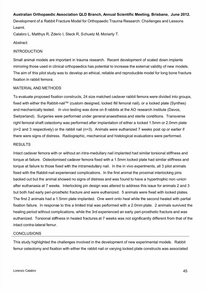
8/13/2019 Lorenzo Calabro Thesis
http://slidepdf.com/reader/full/lorenzo-calabro-thesis 53/54
Lorenzo Calabro 45
Australian Orthopaedic Association QLD Branch, Annual Scientific Meeting, Brisbane, June 2012.
Development of a Rabbit Fracture Model for Orthopaedic Trauma Research: Challenges and LessonsLearnt.Calabro L, Matthys R, Zderic I, Steck R, Schuetz M, Moriarty T.
Abstract
INTRODUCTION
Small animal models are important in trauma research. Recent development of scaled down implantsmirroring those used in clinical orthopaedics has potential to increase the external validity of new models.The aim of this pilot study was to develop an ethical, reliable and reproducible model for long bone fracturefixation in rabbit femora.
MATERIAL AND METHODS
To evaluate proposed fixation constructs, 24 size matched cadaver rabbit femora were divided into groups,
fixed with either the Rabbit-nail™ (custom designed, locked IM femoral nail), or a locked plate (Synthes)
and mechanically tested. In vivo testing was done on 8 rabbits at the AO research institute (Davos,Switzerland). Surgeries were performed under general anaesthesia and sterile conditions. Transverseright femoral shaft osteotomy was performed after implantation of either a locked 1.5mm or 2.0mm plate(n=2 and 3 respectively) or the rabbit nail (n=3). Animals were euthanized 7 weeks post op or earlier ifthere were signs of distress. Radiographic, mechanical and histological evaluations were performed.
RESULTS
Intact cadaver femora with or without an intra-medullary nail implanted had similar torsional stiffness andtorque at failure. Osteotomised cadaver femora fixed with a 1.5mm locked plate had similar stiffness andtorque at failure to those fixed with the intramedullary nail. In the in vivo experiments, all 3 pilot animalsfixed with the Rabbit-nail experienced complications. In the first animal the proximal interlocking pinsbacked out but the animal showed no signs of distress and was found to have a hypertrophic non-unionafter euthanasia at 7 weeks. Interlocking pin design was altered to address this issue for animals 2 and 3but both had early peri-prosthetic fracture and were euthanized. 5 animals were fixed with locked plates.The first 2 animals had a 1.5mm plate implanted. One went onto heal while the second healed with partial
fixation failure. In response to this a limited trial was performed with a 2.0mm plate. 2 animals survived thehealing period without complications, while the 3rd experienced an early peri-prosthetic fracture and waseuthanized. Torsional stiffness in healed fractures at 7 weeks was not significantly different from that of theintact contra-lateral femur.
CONCLUSIONS
This study highlighted the challenges involved in the development of new experimental models. Rabbitfemur osteotomy and fixation with either the rabbit nail or varying locked plate constructs was associated

8/13/2019 Lorenzo Calabro Thesis
http://slidepdf.com/reader/full/lorenzo-calabro-thesis 54/54