Embed Size (px)
Citation preview
Lothian assessment for screening cognition in aphasia (LASCA): A
new non verbal assessment of cognition.
B010018
Msc. Human Cognitive Neuropsychology
The University of Edinburgh
2011
2
Contents
List of Tables……………………………………………………………….…………...ii
List of Figures…………………………………………………………………………..iii
Abstract………………………………………………………………….……….… ….iv
Acknowledgements……………………………………………………………..……….v
1. Introduction…………………………………………………………………………...7
2. Methodology………………………………………………………………………...19
3. Results……………………………………………………………………………….26
4. Discussion…………………………………………………………………………...34
References……………………………………………………………………………...39
Appendix A- Letter of recruitment ……………...……………………………….…….47
Appendix B- Information sheet for participants ……………………………...…….…49
Appendix C- Reasons for excluding yourself……………………………………….....56
Appendix D- Consent form ………...………………………………………………….57
Appendix E- LASCA record form……………………………………………………..58
Appendix F- Table 1…………… ……………………………………………………..63
Appendix G- Figures 7 and 8……………………...……………………..…………….64
Appendix H- Table 4……….………………………………………….……………….65
ii
List of Tables
Table 1 Individual data for the LASCA and ACE-R overall total 63 performance score, premorbid intelligence (STW score), education and age.
Table 2 Descriptive statistics for each of the subtests from the 27 LASCA and the ACE-R..
Table 3 Lower limit normal (cut off scores) for total ACE-R 30 according to age (50-59, 60-69, 70-75, showing control mean minus 2 standard deviations, Moishi et al., 2006).
Table 4 Spot the word scaled scores by age group (adapted from 65 Saxton et al., 2001).
Table 5 Correlation analysis depicting the association between 31 the visual functioning subtests of the LASCA and ACE-R..
Table 6 Correlation analysis for Brixton and executive functioning 32 subtest from the LASCA.
Table 7 Correlation data depicting the association between total 33 scores on the LASCA and total scores of the ACE-R..
iii
List of Figures
Figure1 Star cancellation task from the LASCA 22
Figure 2. Symbol search task from the LASCA. 23
Figure3 Item from the LASCA memory recognition task 23
Figure4 Item no.6 from the LASCA matrices task 24
Figure 5 Mean total LASCA scores (out of a maximum possible 28 score of 150) for participants grouped according to age.
Figure 6 Mean total ACE-R scores (out of a maximum possible 29 score of 100) for participants grouped according to age.
Figure 7 Graph depicting the total distribution of scores on 64 the ACE-R.
Figure 8 Graph depicting the distribution of total scores from 64 the LASCA.
Figure 9. Scattergram depicting the linear relationship between 33 total scores on the LASCA and total scores on the ACE-R.
iv
Abstract
Cognitive dysfunction occurs in more than half of stroke survivors and can have far-
reaching consequences for functioning in daily life. At present, there are no well
established assessments to evaluate the cognitive functioning of individuals with post-
stroke aphasia. Most assessments currently used in clinical practice have limitations
such as dependence on language, need of specialist knowledge, low sensitivity and or
specificity and lengthy administration time. Therefore a working party of Speech and
Language Therapists, Clinical Neuropsychologists and Occupational Therapists in
Lothian, Scotland devised a set of cognitive test materials named the Lothian
Assessment for Screening of Cognition in Aphasia (LASCA) in an attempt to assess
cognition in aphasic patients post stroke. This study aims to evaluate the efficacy of the
LASCA as a non verbal assessment of cognition by comparing it to the widely used
ACE-R. A battery of tests (Spot the word, LASCA, ACE-R, Brixton spatial anticipation
test and the BUTT non verbal reasoning test) was administered to 35 control
participants ( age range = 56-92 years, mean = 68.77 years SD= 7.492) recruited from
across Lothian . Moderate correlations were found between the visual functioning
subtests between the ACE-R and the LASCA (rho (33) = .351; p < .05) and total
performance scores on ACE-R and the LASCA (rho (33) = .614, p< 0.01). A moderate
correlation was also found to exist between the Brixton test and the LASCA executive
functioning subtest (rho (33) = .389, p <. 05). These findings suggest that there is
adequate convergent validity between the LASCA and the widely used ACE-R thus
concluding that the LASCA may be considered an appropriate non verbal assessment
for screening cognition in post stroke aphasic patients.

v
Acknowledgements
I would like to thank all that contributed to my research project, with special thanks to
my supervisors Dr. Marion Murray from the Clinical Neuropsychology department at
the Astley Ainsley Hospital and to Dr. Sarah MacPherson for all their guidance. I would
also like to extend my thanks to all those who participated in my project.
7
1. Introduction
Stroke is a devastating illness, which has been reported to be a strong predictor of
disability and reduced quality of life (Bamford et al., 1988; Bonita., 1992; Hobson,
Leeds & Meara., 2002). In addition to physical impairments, patients often experience
cognitive impairments which significantly restrict their daily functioning as well as
affecting the quality of life of their carer (Anderson, Linto & Stewart-Wynne., 1995).
The type and severity of the cognitive impairment is determined by the site of
neurological damage and its magnitude (Blake et al., 2002). Cognitive impairments can
include a progressive deterioration of intellectual ability (e.g. dementia,), mnemonic
deficits, aphasia, apraxia or one of the agnosias (Robinson. 1997). The frequency of
cognitive deficits following stroke has been estimated at 53.2% (Tatemichi, Desmond,
Stern & Paik., 1994) and may be confounded by pre-existing cognitive decline or
dementia (Kalaria, & Ballard., 2001).
Post stroke cognitive impairment has been found to be a poor prognostic indicator of
recovery, impacting negatively on physical retraining programmes and re-education
programmes (Ebrahaim., 1990; Jeffery & Good,.1995; Mok et al., 2004; Onsworth &
Shum,. 2008; Saxena., 2006; Zinn et al., 2004). The value of neuropsychological
assessment following stroke is increasingly being recognised in the management of
stroke patients (Wade, 1999) given that it is common for stroke patients to be
misdiagnosed (Godefroy et al., 2011). A large emphasis by clinicians is now being
placed on administering measures that assess functional cognition (the ability to
accomplish everyday activities that rely on cognitive abilities, such as locating keys,
conveying information, or planning activities).
As a foundation for investigating the cognitive status of patients following stroke, a
definition of cognition is perhaps warranted. Neisser, (1967) defined cognition as “ all
the processes by which sensory input is transformed, reduced, elaborated, stored,
recovered and used” (p.4). More recently, Williamson, (2006) defined cognition as
8
“process of acquiring, retaining and applying knowledge” (p.293). However, if
cognition is to be formally described it is perhaps necessary to go beyond these rather
broad definitions and to consider the individual components or domains of cognition.
Cognition may be explained for example as having five primary domains: attention,
memory, executive functions, language and visuospatial skills (Helm- Estabrooks.,
2002).
In 5 studies stroke patients were found to have deficits in at least one cognitive domain
(Hoffman., 2001; Hochstenbach, Mulder & van Limbeek., 1998; Tatemichi et al.,1994;
Ballard et al., 2003, Rasquin et al., 2004). The most common impairments were
reduced mental speed, neglect, attention deficits, and aphasia, apraxia and memory
impairments. More specifically, Tatemichi et al., (1994) used a complete
neuropsychological battery to evaluate 227 patients who had suffered stroke and who
did not have pre-existing impairments, and 249 stroke free controls. They report that
35.2% of the patient group were cognitively impaired three months following their
stroke. The most significant areas of deficit were reported to be visuospatial function,
memory, attention, and executive function. Similarly, Pohjasvaara and colleagues
(2001) found cognitive decline in 1, 2, and 3 or more domains in patients who sustained
stroke three months prior to their assessment. The domains most frequently impaired
were visuospatial function and memory. Another study evaluating the cognitive status
of 286 post stroke individuals by Pohjasvuara et al., (2002) found executive dysfunction
in 40.5% of stroke patients. Madureira, Guerreiro & Ferro (2001) found that a
comparable proportion of their patients were impaired. Therefore to summarize,
cognitive deficits in the domains of executive functions, visuospatial function, attention,
memory and orientation are consistently reported to be found in post-stroke patients.
The literature exploring cognitive deficits in post-stroke patients who have aphasia
however is not so established.
It is estimated that 30% of patients with stroke are aphasic (Berthier, 2005). Aphasia
may be explained as a neurological disorder that affects language functioning (Bullain,
Chriki, & Stern, 2007). Furthermore, aphasias may also coexist with speech disorders
9
(e. g apraxia of speech or dysarthria). Usually an improvement is observed in patients’
level of aphasia within the first year following the stroke event. A review by Ferro et al.
(1999) reported that approximately 40% of profoundly aphasic patients experience
complete or almost complete recovery by one year following their stroke. Although the
Copenhagen Aphasia Study reports that 61% of aphasia patients still experience aphasia
at the one year post stroke, this was typically a milder presentation.
Recent studies suggest that early participation in appropriate aphasia therapy is
beneficial not only to improving neurological problems associated with stroke but also
for improving the communication and coping strategies for the individual which in turn
reduces patient isolation and increases mood (Bhogal, Teasell & Speechley., 2003;
Pulvermüller & Berthier., 2008; Cherney, Patterson., Raymer, Frymark, & Schooling.,
2008). Such studies emphasise early identification and diagnosis of language
difficulties as crucial steps towards taking full advantage of rehabilitation goals.
However, given the current constraints in terms of both funding and time, the provision
of speech and language services is often very minimal in many areas, thus limiting the
number of patients that can be assessed in full by a speech and language therapist. ( Al-
Khawaja, Wade & Collin 1999; O’Neill, Cheadle, Wyatt, McGuffog & Fullerton 1990).
The relation between aspects of cognition and language status of individuals with
aphasia remains questionable. Researchers are not unanimous in their findings.
However despite this controversy, there is a growing understanding that all domains of
cognition may play an important role in the rehabilitation outcome. Some researchers
posit that cognitive and mnestic deficits for example, retrograde and short term
memory, (Murray, Ramage, & Hopper., 2001), attention (Coslett, 2001; Murray., 1999),
and reasoning (Borod, Carper, & Goodglass., 1982) can accompany and might even
have an impact on language functioning of individuals following stroke (Silkes, Mc
Neil & Drton., 2004). It is further anticipated that such deficits more than likely impact
upon aphasia rehabilitation (Crosson., 2000; Sarno., 1998). Hinckley and Nash (2007)
report that individuals with aphasia demonstrate a high degree of variability in cognitive
performances such as attention, memory and executive functioning. Interestingly,

10
Kauhanen et al., (2000) found that non-verbal neuropsychological test performance in
aphasic patients was significantly inferior to that of patients with dominant hemisphere
lesions without aphasia.
A growing literature has demonstrated that aphasic adults have attention impairments (
Kreindler & Fradis., 1968; Erickson et al., 1996). Several of these studies have
integrated non-linguistic stimuli in order to demonstrate that any attentional deficits
observed in such studies will be resultant from a fundamental disruption of attention
rather than as a result of the aphasics linguistic impairments. Aphasiacs have been
found to display difficulties on a range of attentional tasks (e.g sustaining and focusing
attention, Murray, 1999). A number of researchers have examined aphasic adults’
ability to orient their attention. To exemplify this point, Robin and Rizzo (1989)
compared the performances of aphasiac adults to those of adults with right hemispheric
brain damage or no brain damage on orienting tasks in both the visual and auditory
modalities. They found that all of their brain damaged subjects displayed orientation
impairment, more interestingly however, aphasic patients displayed the greatest
difficulty orienting to auditory targets and they did not appear to benefit from cueing.
These findings along with similar findings from other studies (Peach et al., 1994; Petry
et al., 1994) highlight that aphasic adults may have difficulties orienting or directing
their attention even when they are given valid attentional cues. It may be worthy to note
however that is still undetermined which one or more aspects of attention orientation
are impaired in adults with aphasia (Allport., 1990; Posner & Petersen 1990; Robin &
Rizzo., 1989).
Caspari, Parkinson, LaPointe, & Katz (1998) suggest there is a correlation between
working memory capacity, written language comprehension and language function.
Further research also implies that deficits in syntactic comprehension in conduction
aphasia can be accredited to a decrease in short term verbal memory more so than to a
universal deficit in language comprehension (Bartha & Benke, 2003; Bartha, Marine,
Poewe, & Benke, 2004; Caramazza, Basili, Kolerm & Bendt, 1981). This finding of a
correlation between short term verbal memory and a deficit in word processing is
11
further supported by Martin and Ayala (2004). Numerous earlier studies indicate a
correlation between language and problem solving in aphasic patients (Archibald,
Wepman, & Jones., 1967; Barid, Carper & Goodglass., 1982; Basso, De Renzi,
Faglioni, Scotti, & Spinnler., 1973; Hjelmquist., 1989). Baldo et al., (2005) suggest
there is a correlation between performance on problem solving tests and language tests (
Ravens progressive matrices and Wisconsin card sorting test). Kertesz and McCabe
(1975) found that cognitive impairment was more pronounced in patients with severe
deficits in comprehension such as patient with Wernicke’s aphasia.
An interesting study by Fridriksson et al., (2006) found evidence to suggest a clear
relationship between scores on executive functioning tasks and functional
communication ability. It is apparent based on these results that decreased executive
functioning ability may correspond with decreased executive functioning ability in
persons with aphasia. Additionally, Keil & Kaszniak (2002) postulated that
performance on measures of executive function by patients with varying profiles of
aphasia may provide insight into the nature of the interaction between normal language
functioning and executive functioning. It can be said therefore that there is increasing
interest among rehabilitation specialists based on clinical experience and initial studies
that all cognitive domains play an important role in the successful treatment of aphasia
by means of therapy.
While there are many widely used tests of executive functioning ability in clinical
practice today however most of these tools were not developed with the intention of
assessing people with language difficulties (Miyaje, Emerson, & Friedman, 2000).
Accordingly many of these tests place demands on linguistic processing and the results
are therefore confounded when administered in the assessment of executive functioning
in aphasic patients. Similarly, tests that assess attention, concentration and memory in
current clinical practice rely heavily on language, an example of such a test is the
Addenbrooke’s Cognitive Examination Revised (ACE-R, Moishi, Dawson, Mitchell,
Arnold & Hodges., 2006).
12
The Addenbrooke's Cognitive Examination (ACE) was established in the dementia
clinics in Addenbrooke's Hospital, Cambridge University Teaching Hospitals, UK, in
the 1990s by Hodges and colleagues (Mathuranath, Nestor, Berrios, Rakowics &
Hodges., 2000). The test's primary objective was to offer a valuable tool that clinicians
can use for screening dementia. It was found to be sensitive to early Alzheimer's disease
diagnosis, differentiation between Alzheimer's disease and frontotemporal dementia and
also in the differentiation between organic brain disease and psychiatric states (Bier,
Ventura, Doncheles et al., 2004; Moishi et al., 2006; Dudas, Berrios & Hodges., 2005).
In its earlier prototype, the ACE had a few weaknesses, such as the insensitivity of its
naming component. Design changes were implemented in an effort to make the test
easier to administer. Content alterations were also made in order to facilitate cross
cultural usage and translation. Following revisions (ACE-revised or ACE-R), the final
version is currently a very popular cognitive screening tool and is used routinely in
several countries (Moishi et al., 2006). The ACE-R was found to be reliable (alpha
coefficient: 0.8) when administered in normal population (Moishi et al., 2006). The
ACE-R assesses five cognitive domains, namely; attention concentration, memory,
language, visuospatial function and verbal fluency. The language component of the test
assesses naming, comprehension, repetition, reading and writing.
Indeed most studies exploring post stroke aphasia and cognitive dysfunction to date
have had some methodological and diagnostic inadequacies. The interpretation of
aphasia reports may be complicated by variable times of initial assessment of aphasia
(Kertesz & McCabe., 1977; Lendrem & Lincoln.,1985). Most frequently though
patients with severe aphasia have typically been excluded and as aphasia affects
approximately 35 % of those with stroke (Berthier, 2005), this is often a significant
number of people (Hobson, Leeds & Meara., 1999; Starkstein &Robinson., 1988;
Astrom et al., 1993; Robinson & Benson., 1981). Furthermore despite this growing
fundamental belief of many researchers that cognitive status plays an important role in
the precise development of treatment plans and outcomes (Crosson, 2000; Sarno, 1998)
most aphasic patients do not receive adequate cognitive assessment.
13
To establish individual cognitive profiles, a complete neuropsychological assessment is
needed. The value of neuropsychological assessment is increasingly being recognised in
the management of stroke patients (Wade, 1999). Clinical guidelines in both England
(Royal College of Physicians, 2008) and Scotland (Scottish Intercollegiate Guidelines
network, 2004) recommend that stroke survivors are routinely cognitively assessed. The
benefits of such assessments are extensive; they provide valuable prognostic
information which is necessary to plan cognitive remediation programmes, provide
essential advice and recommendations to other members of rehabilitation teams,
patients’ families and also to social services. However the cost of carrying out
neuropsychological assessments can be economically demanding and time consuming
therefore it is essential to target such assessment where it is needed most.
While cognitive function is now routinely assessed in stroke survivors in most
mainstream medical or hospital wards, by a variety of standardised neuropsychology
measures, many of these measures may be too linguistically complex to offer a valid
evaluation (Hinckley & Nash., 2007). Individuals may also have difficulty seeing
assessment materials or struggle to hold a pencil or pen, or to press computer keys. To
compound these difficulties there are very few non-verbal assessments that the clinician
can draw upon. Assessment batteries designed for aphasia are still developing. Of the
few non-verbal assessments that are in existence most are often used to describe more
circumscribed cognitive impairments rather than global cognitive function (Hobson,
Leeds & Meara., 2002). In addition many of these assessments have been drawn from
much larger neuropsychological batteries, or have been modified to compensate for
communication or language difficulties or motor impairments, which raise potential
methodological issues with regard to validity and reliability.
The validity, reliability and standardisation of assessment tools are vital to the
clinician’s ability to arrive at appropriate conclusions from assessments (Murray &
Chapey., 2001; Spreen & Risser., 2003). Another important factor in the accurate
assessment of cognition in aphasiac patients, as with all patient populations is the
timing of the assessment procedure. Clinical time must be used effectively (Johnson,

14
Holcombe Jacobson., 2007). Conducting an assessment for longer than the required
time often results in misuse of clinical resources. Equally, too little information
obtained from a short assessment may result in incorrect diagnosis. It is also of
importance to be concerned with using selected adaptations of various tests to create an
institution- specific protocol that may not be a reliable tool (Davis., 2000).
Indeed, how to assess cognition at the bedside of individuals who have sensory, motor
and language difficulties following stroke warrants additional examination. Other
researchers, primarily with traumatic brain injured (TBI) populations have established
that communication during the testing process is promising by using for example basic
fixed choice vocalisations, or in other cases by using eye or hand movements
(Bigge.,1982; Swiercinsky., 1997). Hobson et al.,(2003) endorsed the use of the
preliminary neuropsychological battery (PNB) devised by Cossa et al.,(1999). It was
originally designed for assessing patients with a TBI who had minimal verbal and
motor abilities. Hobson et al..(1999) administered the PNB to stroke patients without
significant aphasia and a sample of similarly healthy aged matched controls. The
limitations of this study were discussed as firstly the PNB being found to be vulnerable
to the affects of educational attainment, functional ADL and depressive symptoms.
Secondly the PNB was found not to be sensitive enough to detect subtle cognitive
changes due to the near ceiling cut-point threshold of 55 or less which is required to
differentiate between cognitively impaired and non-impaired patients. Probably the
most significant limitation of this study however is that stroke patients with severe
aphasia were excluded from participating in the study. This highlights the need for brief
global non-verbal cognitive measure in this population (Hobson, 2002).
Salter, Jutai, Foley, Hellings & Teasell.,(2006) conducted a recent review of the
screening tests used to assess post stroke victims in literature to date and their findings
suggest that indeed, a routine screening test administered by another healthcare
professional may be a vital tool in the identification of patients cognitive status who
have communication problems following stroke . Following this proposed screening
process, patients can then be referred for a more complete assessment, resulting in the
15
necessary treatment or rehabilitation programme. Screening tests are needed to
highlight problem areas therefore they must be sensitive enough to detect all these
cognitive problems and specific, in order to avoid any misdiagnosis of individuals.
Salter et al.,(2006) claim that although screening tests do not provide detailed
descriptions of specific cognitive deficits or permit a differential diagnosis of cognitive
disorders, they do embody a quick and effective means of determining the existence or
absence of cognitive deficits, principally among patients who may be unable to endure a
lengthy assessment process. Additionally Salter et al.,(2006) suggest that screening
tools may aid in assessing basic abilities and monitor development until a more
comprehensive assessment can be carried out (Crary, Haak & Halinsky., 1989;
Enderby, Wood, Wade, Langton & Hewer., 1987).
Blake et al., (2002) assessed the sensitivity and specifity of a screening battery for
detecting cognitive impairment after stroke. They found that the Mini Mental State
Exam (MMSE, Folstein, Folstein, & McHugh., 2000 ) was not an effective screening
tool for memory problems or overall cognitive defect after stroke. The reason for this
was because the MMSE was not found to be a good measure of memory problems as
there was no clear cut off score to indicate a problem requiring further evaluation. This
was mirrored when only the orientation, attention questions were considered. However,
a more recent study by Bour, Rasquin, Boreas, Limburg and Vverhey (2010) found that
cut off scores of 27/28 showed good sensitivity in screening for at least 2 disturbed
domains. Bour et al., 2010 also emphasize that the validity of the MMSE in screening
for cognitive impairment following stroke is still debated in the literature (Folstein,
Folstein & Hugh., 1975; Feher et al., 1992). The general consensus is that it is only
sensitive when a patient is already severely impaired (Anthony et al., 1982).
The Sheffield Screening Test for Acquired Language Disorders (SST Syder, Body,
Parker & Boddy., 1993) was only reported to be an appropriate screen for language
problems, moreover using the full measure was found to be more accurate that either
the receptive or expressive subscales alone (Blake et al., 2002). This finding
appropriates the SST as a brief screening measure for aphasia but not for screening

16
cognition following stroke. The Raven’s Progressive Matrices (RCPM, Raven, 1965)
was found to be suitable as a screen for perceptual problems and visual inattention
however it was not found to be effective for detecting executive deficits ( Blake et al.,
2002).
The last 8 years or so have seen an obvious convergence in agreement as to what types
of cognitive tasks are important to administer in a brief cognitive assessment in aphasia.
Recognition memory, visual or symbol cancellation and non-verbal reasoning have all
displayed significant differences in performance that are not related with aphasias type
or severity (e.g. Helm- Estabrooks., 2002; Kalbe, Reinhold, Brand, Marchowitsch &
Kessler., 2005). Certainly Kalbe et al. (2005) reported that memory was the most
commonly impaired cognitive domain in aphasic patients following stroke in their
sample of 154 adults. With regards to screening for executive dysfunction, Keil &
Kaznaik, (2002) found that other measures of executive function play a very important
role in treatment planning and tasks such as Trail Making Test should be more routinely
explored in post-stroke aphasiacs patients.
A study by Van Mourik, Vershaeve, Boon, Paquier, et al. (1992) assessed cognition in
17 patients with global aphasia. They administered a battery of non-linguistic tasks that
they referred to as the global aphasic neuropsychological battery (GANBA), and a test
that measures auditory comprehension. The GANBA involved six tasks from varying
published sources and one further test designed by the authors of the study. Overall the
GANBA tasks assessed the domains of memory (Rivermead Behavioiural Memory
Test, Wilson et al., 1985), attention/concentration (cancellation test, Diller et al., 1974)
intelligence (RCPM, Raven et al., 1979), visual recognition (Line Orientation Test,
Benton et al., 1978 and the Facial Recognition Test, Benton & Van Allen., 1968) and
non-verbal auditory recognition (audiotaped familiar sounds presented to the patient
and they have to point to the correct answer). Van Mourik et al., (1992) described that
performance scores on the GANBA were independent of the participants spoken
language comprehension ability. From performance on the GANBA Van Mourik et al
1992 identified two main groups of globally aphasia patients that were discussed with
17
relation to consequences for treatment programmes. The first group were reported to
have relatively intact cognitive functions and as a result could respond to language
orientated aphasia treatment. On the other hand the second group of patients had
differing forms of deficits therefore it was important to address these cognitive
impairments before language therapy was implemented. A third group was also
identified by Van Mourik et al (1992). This group comprised patients who were too
aphasic to complete the GANBA and as a result were excluded from the study.
Implications of this study were only discussed by Van Mourik et al in terms of
implications of cognition for language treatment. However, the presence of the third
group, perhaps advocates that the GANBA is not sensitive enough to assess patients
with more severe aphasias.
There is an obvious gap observed in the both research literature and clinical settings that
report on a brief, reliable screening test that assesses cognition in post stroke aphasic
patients. Few tools exist for speech and language therapists to briefly examine the
neuropsychological deficits in aphasic patients. One exception to this is the RCPM
(Raven., 1995), which can be administered to individuals with aphasia and is easily
obtainable. However, this test primarily targets one aspect of cognition, namely, visual
analogical thinking. To test wider spectrum of cognitive skills such as attention,
concentration, memory and executive functions, typically researchers and clinicians
assemble batteries of non-linguistic tests (Van Mourik et al., 1992, Helm-Estabrooks et
al., 1995). While modifying existing assessments (e.g. by omitting test items which are
language based, or those which require verbal responses) may be a pragmatic option for
the clinician, it is unfortunately one with significant limitations. The main limitation
observed is that individuals may be misdiagnosed as suffering from a cognitive problem
when the difficulties they have experienced on the assessment are the result of their
language impairment. Other limitations include the difficulty and cost of modifying and
assembling such batteries. Another concern is that limited therapist time is spent
carrying out cognitive assessments that are merely not valid.
18
On the basis of the aforementioned considerations, a working party of Speech and
Language Therapists (drawn from acute, post acute and community work settings),
Clinical Neuropsychologists and Occupational Therapists in Lothian, Scotland met
during 2007/2008 to devise a set of cognitive test materials that have minimal language
demands. More specifically, the aim was to develop a cognitive screening assessment
that could be administered to individuals with post stroke aphasia (with severities of
language disturbance ranging from mild to severe). The resulting cognitive test
materials were named the 'LASCA' (Lothian Assessment for Screening Cognition in
Aphasia). The LASCA assesses five cognitive domains, namely: orientation, attention,
memory, visual functioning and executive functioning.
The objective of the current study is to assess the convergent validity of the LASCA
and the ACE-R to detect post stroke cognitive impairment determined by correlation
analysis. This will be accomplished by using individual subtest scores for each of the
cognitive domains assessed from the LASCA (orientation/attention, memory, visual
functioning and executive functioning) and comparing them to their counterparts from
the ACE-R.
19
2. Methodology 2.1. Participants
A total of 35 healthy older adults both male (n= 12) and female (n=23) (age range = 56-
92 years, mean = 68.77 years SD= 7.492) participated in the current study. The
participants years of education (mean = 14.74, SD= 3.7475) were recorded. Participants
were recruited from the University of Edinburgh participant database and from a variety
of local community settings including local older adult activity groups, sheltered
housing, community centres, bingo and tea clubs and local church organisations across
Lothian.
2.2. Eligibility criteria
Individuals were included in the study if they had no history of stroke illness,
neurological problems or language impairment and were over the age of 55 years.
2.3. Justification and sample size
The sample size needed was based on a power calculation for a two tailed correlation
analysis as this was the intended primary means for statistical analysis. As there have
been no previously published studies on the LASCA’s inter-reliability there was no data
available to base the power calculation upon. It was regarded that a small effect size
would convert into merely a small number of point’s variation from the true score
which would not threaten the accuracy of the overall conclusions drawn from the
LASCA in clinical practice. On the other hand, a medium effect size would translate
into a distinction considerably large enough that it could alter interpretation of the
LASCA results, which in turn could influence the wider assessment procedure. For that
reason the power calculation determined the number of participants required to identify
a medium effect size, by means of the conservative values of p-value 0.05 and power
0.8. When these values for a correlation analysis were entered into G*Power 3.010 (
20
Faul et al., 2007, from www.psycho.uni duesseldorf.de /abteilungen/ aap/gpower3/) the
results specified that a minimum of 34 participants would be required.
2.4. Measures
2.4.1. Spot the word
The Spot-the-Word Test is used as an estimate of premorbid intelligence. In this brief
lexical decision test participants are presented with a total of 60 pairs of items, each pair
containing one genuine word and one pseudo word designed to be pronounceable and to
have a plausible orthographic composition. Participants work at their own rate, ticking
the item they believe to be the genuine word. They are instructed to answer each pair,
guessing if necessary. Performance is scored in terms of the number of correct
responses (maximum score of 60).
2.4.2. The Addenbrookes cognitive examination revised (ACE-R)
The ACE-R comprises of 5 subtests, each one representing one cognitive domain:
attention/ orientation (18 points), memory (26 points), verbal fluency (14 points),
language (26 points) and visuospatial function (16 points). The language component of
the test assesses naming, comprehension, repetition, reading and writing. The total
score is 100, higher scores indicate better cognitive functioning. It takes between 12 and
20 minutes ( average 16 minutes) to administer the ACE-R and score in a clinical
setting.
2.4.3. Butt non-verbal reasoning test (BNVR)
The BNVR Test (Butt & Bucks, 2004) is a unique non-linguistic approach for
investigating whether a cognitive deficit in addition to a linguistic deficit exists in
individuals with acquired aphasia. It is short, requires minimal linguistic input, contains
real-life situations and is likely to be suitable for non-English speaking individuals.
21
The BNVR test consists of 11 large coloured photographs of people with problems.
Some problems relate to the individual in the photograph, for example, a headache or a
beard, some relate to a problem in the environment, such as on a ward, or at home, for
instance, spilt coffee or a broken cup. On the same page as each of the large
photographs are four smaller photographs of different objects. One is the solution to the
problem; the three others are distractors, one semantic, one visual and one unrelated.
The distractors are position randomly on one page to avoid the possibility of
perseveration in responding. The problems presented in this test were chosen due to
their familiarity and their likeliness of occurring in an environment where mobility may
be restricted.
There is also a screening test which comprises four line drawings of common items, one
from each of four categories, a dog , an apple, a coat and a pair of scissors. Participants
are required to match each drawing with an identical drawing presented on a 2x2 grid.
This is to confirm that subjects have the necessary perceptual skills.
2.4.4. Brixton spatial anticipation test
Although the Brixton test was developed as part of the Hayling and Brixton tests
(Burgess & Shallice, 1997), the present study focuses on performance on the Brixton
test only. The Brixton test was administered and scored according to the instructions in
the manual (Burgess & Shallice, 1997). Participants were presented with a 56 page
booklet. Each page in the booklet had the same basic design, an assortment of 10 circles
(2 rows of 5 circles), which are numbered 1-10. On each page, one circle was always
coloured blue. The position of the blue circle moved around according to various
patterns which came and went without warning. The participant was required to detect
the pattern and to point to where they thought the blue circle would move to on the next
page. Responses were regarded as correct if the participant followed the current rule
and on trials where the rule changed, a response was considered correct if it followed
the previous rule. The total number of errors across the 55 trials was used as an
outcome measure, therefore higher scores reflect worse performance (maximum
number of errors obtainable = 55).
22
2.4.5. Lothian assessment for screening cognition in aphasia (LASCA)
The LASCA is a new clinical tool designed by a working party of speech and language
therapists, Clinical Neuropsychologists and occupational therapists in Lothian, Scotland
in an attempt to assess cognition in aphasic patient’s post- stroke. The LASCA takes on
average, 20 minutes to complete.
The LASCA assess five cognitive domains, namely:
• Orientation (single subtest scores range from 0 to 14)
-Includes questions such as “What is … the year/your age?”, “Who lives with you?”
• Attention (easy, moderate and difficult subtest, out of 10)
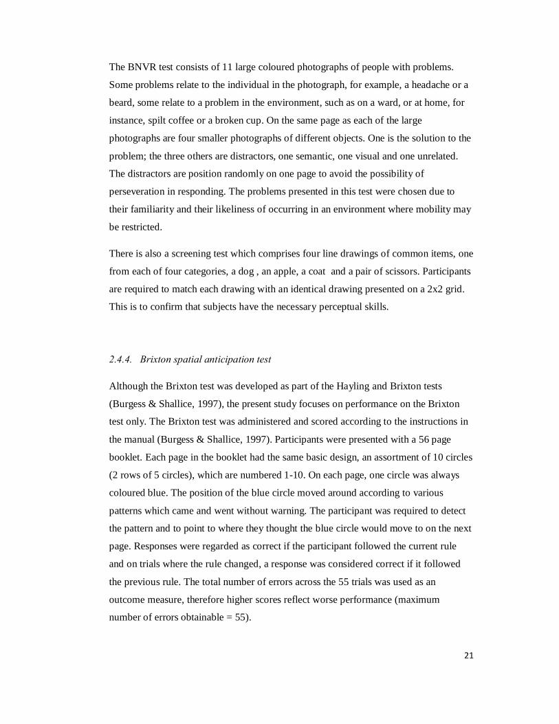
- ‘Cancellation task’ (participant is presented with a page of symbols and asked to
cross off all the little stars on the page)
-
• Figure 1. Star cancellation task from the LASCA.
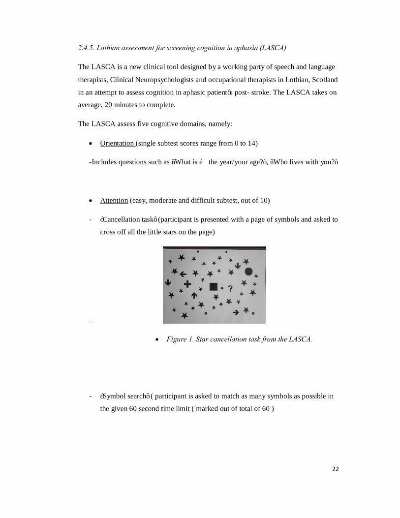
- ‘Symbol search’ ( participant is asked to match as many symbols as possible in
the given 60 second time limit ( marked out of total of 60 )
23
-
• Figure2. Symbol search from the LASCA
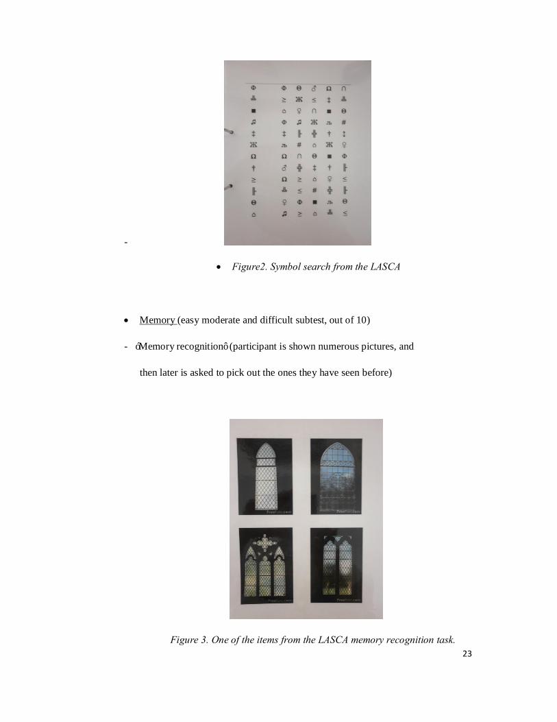
• Memory (easy moderate and difficult subtest, out of 10)
- ‘Memory recognition’ (participant is shown numerous pictures, and
then later is asked to pick out the ones they have seen before)
Figure 3. One of the items from the LASCA memory recognition task.

24
• Visual Functioning (easy, moderate and difficult subtest, out of 10)
- ‘Visual matching task’ (participant is asked to match shapes).
• Executive Function (easy moderate and difficult subtest, out of 10)
- BUTT non-verbal reasoning test- (see above for instructions).
- Matrices- this task assesses non-verbal reasoning. Participants are asked to look
at sequences of shapes. There is always one piece missing. They are required to
select which one from an option of 4 shapes is the missing piece (i.e the one that
fits best).
-
- Figure 4. Item no. 6 from LASCA matrices task.
- Mazes (based on porteus mazes) assesses problem solving and visual
functioning. Participants are asked to trace with their finger/pencil a way out of
the 4 mazes presented. The mazes get more difficult as the levels progress.
25
2.5. Design Procedure
First the characteristics of the study group were described. Second, the score
distributions of the tests were given, searching for possible floor or ceiling effects. Age
effects were investigated on both measures using one way ANOVA. Following this,
correlations between each of the subtests on the ACE-R and LASCA were examined
using Spearman’s correlation coefficients. Relationship between subtests were
considered strong if the coefficient was > 0.6, moderate if the coefficient was between
0.3 and 0.6 and weak if the coefficient was < 0.3. All analyses were performed using
SPSS version 18.
2.6. Research Procedure
Potential participants were invited to participate in the study either by letter, by email or
verbally. Participants who opted into the current study attended a testing session at the
research laboratory, 7 George Square, University of Edinburgh or at, community
centres / sheltered accommodation which lasted on average one hour and 15 minutes.
Each participant received an information sheet (see Appendix B ). Written consent was
obtained from all participants prior to the study commencing (see Appendix D).
Participants were required to complete a battery of tests over one testing session. The
tests included in the battery were the spot the word test, the ACE-R, the BNVR, the
LASCA and the Brixton spatial anticipation test. The order of administration was kept
constant in each assessment.
A standardised set of instructions was read to each participant before the testing session
commenced. Participants had the right to withdraw from the study at any stage up until
the data had been analysed, for whatever reason, without any consequences.
2.7. Ethical Approval
Prior to the study commencing ethical approval was gained from the PPLS University
of Edinburgh Ethics Committee.

26
3. Results
3.1. Descriptive statistics
Overall total performance scores, pre-morbid intelligence scores (STW score) and years
of education for the LASCA and the ACE-R are presented in Table 1 (see Appendix F).
The LASCA was divided into subcomponents namely; attention/orientation, memory,
visual functioning and executive functioning. The ACE-R is already divided into
comparable subtests with the verbal fluency subtest regarded as a measure of executive
functioning. The highest possible score for the LASCA is 150 points. With regards the
individual subtests, the attention/orientation section consists of orientation questions (5
points), the symbol search (20 points) and the star cancellation task (60 points), (total of
85 points). The memory component of the LASCA comprised a retrograde memory
picture task (total of 12 points). The visual functioning subtest of the LASCA
comprised the single matching task (total of 10 points) and the executive functioning
component of the LASCA comprised the mazes (3 mazes with a maximum of 3 points
for each maze, thus a total of 9 points is obtainable if all correct), the BUTT (10 points)
and the matrices (10 points), total of 29 points).
Histograms describing the data (Figures 7 & 8, see Appendix G) were drawn. Looking
at the curve it was possible to conclude that the data representing scores from both
LASCA and ACE-R are not normally distributed within the population sample and
therefore non-parametric analysis was warranted.
3.2 Performance scores on the LASCA and ACE-R
SPSS reported descriptive statistics (Means, range of scores and standard deviations) of
participant’s scores on each of the subtests which are presented in Table 2.
As executive function can be fractionated into different executive abilities, and verbal
fluency from the ACE-R only assesses one domain of executive function, another
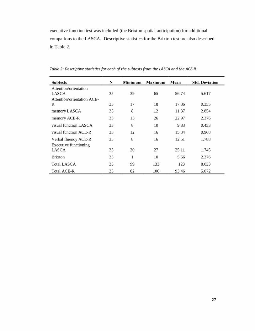
27
executive function test was included (the Brixton spatial anticipation) for additional
comparions to the LASCA. Descriptive statistics for the Brixton test are also described
in Table 2.
Table 2: Descriptive statistics for each of the subtests from the LASCA and the ACE-R.
Subtests N Minimum Maximum Mean Std. Deviation Attention/orientation LASCA 35 39 65 56.74 5.617 Attention/orientation ACE-R 35 17 18 17.86 0.355 memory LASCA 35 8 12 11.37 2.854
memory ACE-R 35 15 26 22.97 2.376
visual function LASCA 35 8 10 9.83 0.453 visual function ACE-R 35 12 16 15.34 0.968 Verbal fluency ACE-R 35 8 16 12.51 1.788 Executive functioning LASCA 35 20 27 25.11 1.745
Brixton 35 1 10 5.66 2.376
Total LASCA 35 99 133 123 8.033 Total ACE-R 35 82 100 93.46 5.072
28
0
20
40
60
80
100
120
140
56 - 59 60-69 70-79 80-92
Mean total score
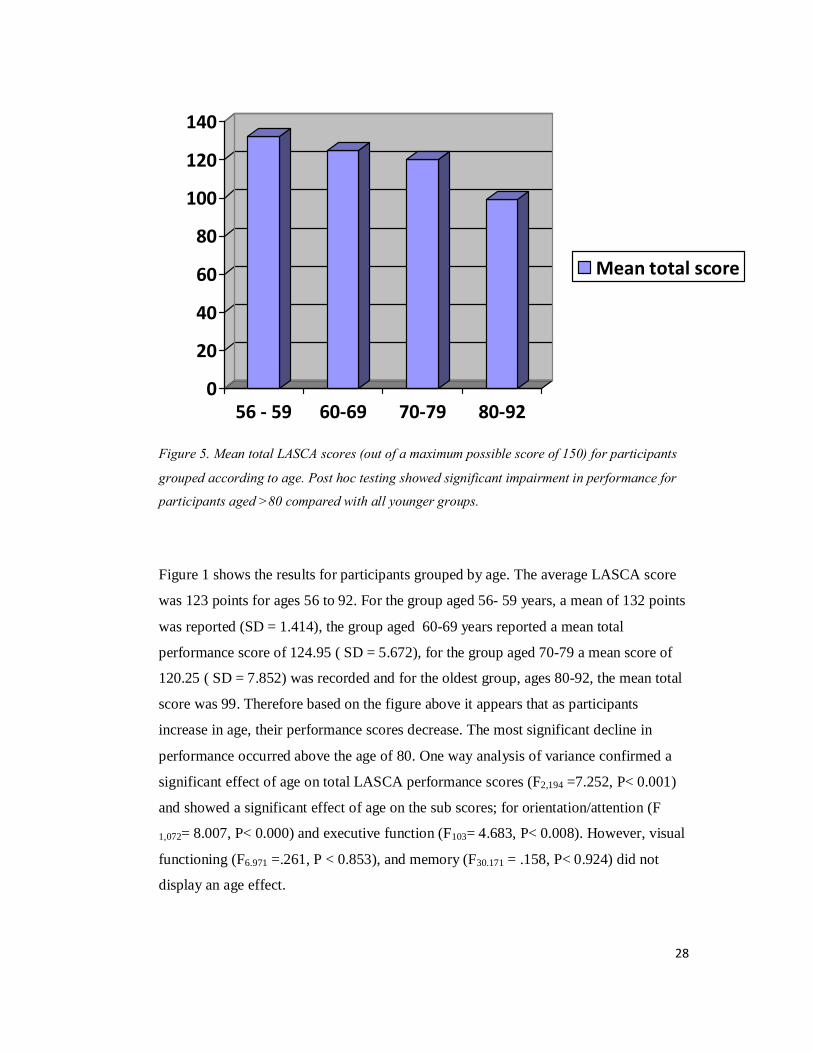
Figure 5. Mean total LASCA scores (out of a maximum possible score of 150) for participants
grouped according to age. Post hoc testing showed significant impairment in performance for
participants aged >80 compared with all younger groups.
Figure 1 shows the results for participants grouped by age. The average LASCA score
was 123 points for ages 56 to 92. For the group aged 56- 59 years, a mean of 132 points
was reported (SD = 1.414), the group aged 60-69 years reported a mean total
performance score of 124.95 ( SD = 5.672), for the group aged 70-79 a mean score of
120.25 ( SD = 7.852) was recorded and for the oldest group, ages 80-92, the mean total
score was 99. Therefore based on the figure above it appears that as participants
increase in age, their performance scores decrease. The most significant decline in
performance occurred above the age of 80. One way analysis of variance confirmed a
significant effect of age on total LASCA performance scores (F2,194 =7.252, P< 0.001)
and showed a significant effect of age on the sub scores; for orientation/attention (F
1,072= 8.007, P< 0.000) and executive function (F103= 4.683, P< 0.008). However, visual
functioning (F6.971 =.261, P < 0.853), and memory (F30.171 = .158, P< 0.924) did not
display an age effect.
29
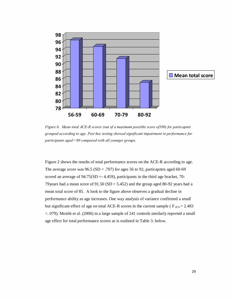
7880828486889092949698
56-59 60-69 70-79 80-92
Mean total score
Figure 6. Mean total ACE-R scores (out of a maximum possible score of100) for particapnts
grouped according to age. Post hoc testing showed significant impairment in performance for
participants aged >80 compared with all younger groups.
Figure 2 shows the results of total performance scores on the ACE-R according to age.
The average score was 96.5 (SD = .707) for ages 56 to 92, particapntrs aged 60-69
scored an average of 94.75(SD =- 4.459), participants in the third age bracket, 70-
79years had a mean score of 91.50 (SD = 5.452) and the group aged 80-92 years had a
mean total score of 85. A look to the figure above observes a gradual decline in
performance ability as age increases. One way analysis of variance confirmed a small
but significant effect of age on total ACE-R scores in the current sample ( F 874 = 2.483
<. 079). Moishi et al. (2006) in a large sample of 241 controls similarly reported a small
age effect for total performance scores as is outlined in Table 3. below.
30
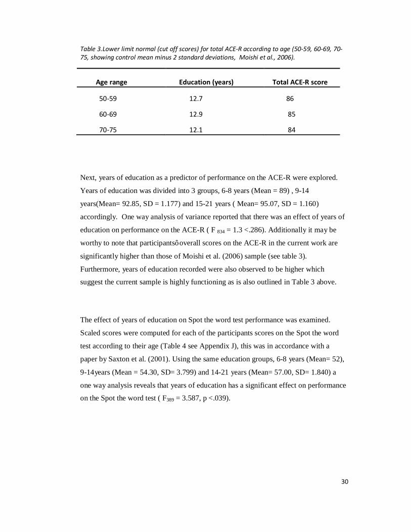
Table 3.Lower limit normal (cut off scores) for total ACE-R according to age (50-59, 60-69, 70-75, showing control mean minus 2 standard deviations, Moishi et al., 2006).
Age range Education (years) Total ACE-R score
50-59 12.7 86
60-69 12.9 85
70-75 12.1 84
Next, years of education as a predictor of performance on the ACE-R were explored.
Years of education was divided into 3 groups, 6-8 years (Mean = 89) , 9-14
years(Mean= 92.85, SD = 1.177) and 15-21 years ( Mean= 95.07, SD = 1.160)
accordingly. One way analysis of variance reported that there was an effect of years of
education on performance on the ACE-R ( F 834 = 1.3 <.286). Additionally it may be
worthy to note that participants’ overall scores on the ACE-R in the current work are
significantly higher than those of Moishi et al. (2006) sample (see table 3).
Furthermore, years of education recorded were also observed to be higher which
suggest the current sample is highly functioning as is also outlined in Table 3 above.
The effect of years of education on Spot the word test performance was examined.
Scaled scores were computed for each of the participants scores on the Spot the word
test according to their age (Table 4 see Appendix J), this was in accordance with a
paper by Saxton et al. (2001). Using the same education groups, 6-8 years (Mean= 52),
9-14years (Mean = 54.30, SD= 3.799) and 14-21 years (Mean= 57.00, SD= 1.840) a
one way analysis reveals that years of education has a significant effect on performance
on the Spot the word test ( F389 = 3.587, p <.039).
31
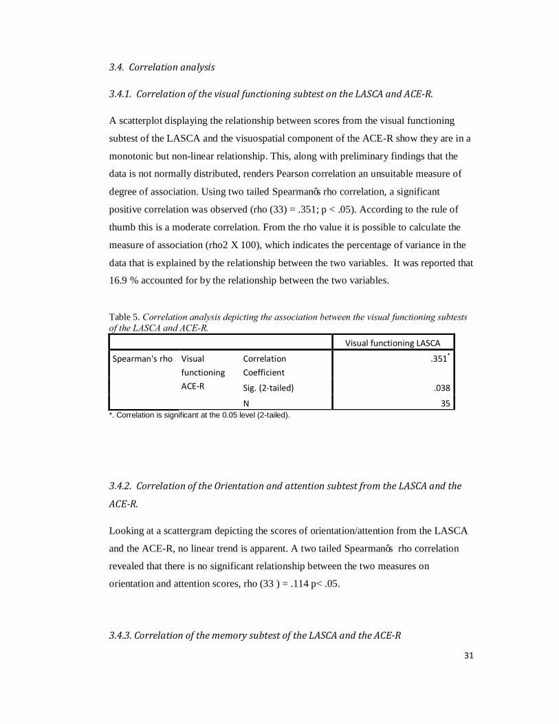
3.4. Correlation analysis
3.4.1. Correlation of the visual functioning subtest on the LASCA and ACE-R.
A scatterplot displaying the relationship between scores from the visual functioning
subtest of the LASCA and the visuospatial component of the ACE-R show they are in a
monotonic but non-linear relationship. This, along with preliminary findings that the
data is not normally distributed, renders Pearson correlation an unsuitable measure of
degree of association. Using two tailed Spearman’s rho correlation, a significant
positive correlation was observed (rho (33) = .351; p < .05). According to the rule of
thumb this is a moderate correlation. From the rho value it is possible to calculate the
measure of association (rho2 X 100), which indicates the percentage of variance in the
data that is explained by the relationship between the two variables. It was reported that
16.9 % accounted for by the relationship between the two variables.
Table 5. Correlation analysis depicting the association between the visual functioning subtests of the LASCA and ACE-R.
Visual functioning LASCA
Spearman's rho Visual functioning ACE-R
Correlation Coefficient
.351*
Sig. (2-tailed) .038
N 35 *. Correlation is significant at the 0.05 level (2-tailed).
3.4.2. Correlation of the Orientation and attention subtest from the LASCA and the
ACE-R.
Looking at a scattergram depicting the scores of orientation/attention from the LASCA
and the ACE-R, no linear trend is apparent. A two tailed Spearman’s rho correlation
revealed that there is no significant relationship between the two measures on
orientation and attention scores, rho (33 ) = .114 p< .05.
3.4.3. Correlation of the memory subtest of the LASCA and the ACE-R
32
A scattergram showing the association between the memory subtest of the LASCA and
the memory subtest of the ACE-R depicts no apparent linear relationship. Following a
two tailed Spearman’s rho correlation to assess the relationship between scores of
memory on LASCA and scores on memory subtest of ACE-R no significant
relationship was found, rho (33) = .320, p<.05.
3.4.4. Correlation of the Executive function component of the LASCA, the ACE-R (
verbal fluency) and the Brixton.
No significant correlation was found to exist between the LASCA and the verbal
fluency subtest from the ACE-R.
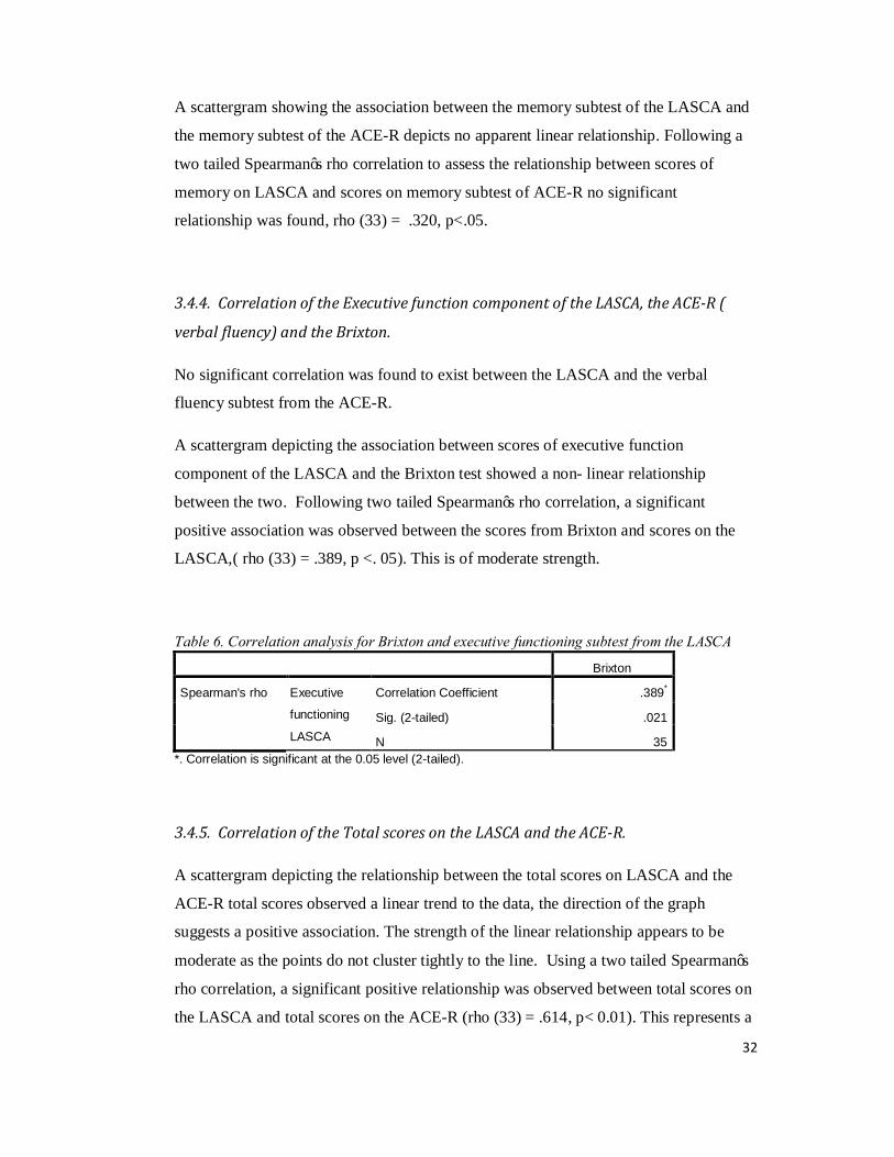
A scattergram depicting the association between scores of executive function
component of the LASCA and the Brixton test showed a non- linear relationship
between the two. Following two tailed Spearman’s rho correlation, a significant
positive association was observed between the scores from Brixton and scores on the
LASCA,( rho (33) = .389, p <. 05). This is of moderate strength.
Table 6. Correlation analysis for Brixton and executive functioning subtest from the LASCA Brixton
Spearman's rho Executive
functioning
LASCA
Correlation Coefficient .389*
Sig. (2-tailed) .021
N 35 *. Correlation is significant at the 0.05 level (2-tailed).
3.4.5. Correlation of the Total scores on the LASCA and the ACE-R.
A scattergram depicting the relationship between the total scores on LASCA and the
ACE-R total scores observed a linear trend to the data, the direction of the graph
suggests a positive association. The strength of the linear relationship appears to be
moderate as the points do not cluster tightly to the line. Using a two tailed Spearman’s
rho correlation, a significant positive relationship was observed between total scores on
the LASCA and total scores on the ACE-R (rho (33) = .614, p< 0.01). This represents a
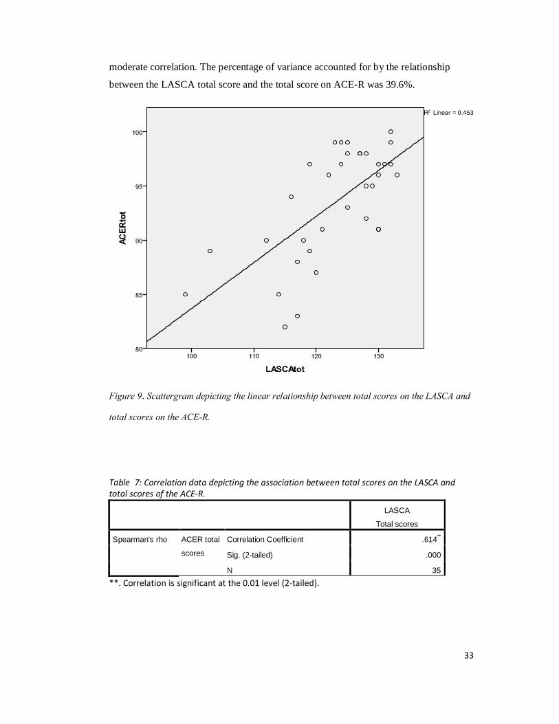
33
moderate correlation. The percentage of variance accounted for by the relationship
between the LASCA total score and the total score on ACE-R was 39.6%.
Figure 9. Scattergram depicting the linear relationship between total scores on the LASCA and
total scores on the ACE-R.
Table 7: Correlation data depicting the association between total scores on the LASCA and total scores of the ACE-R.
LASCA
Total scores
Spearman's rho ACER total
scores
Correlation Coefficient .614**
Sig. (2-tailed) .000
N 35 **. Correlation is significant at the 0.01 level (2-tailed).
34
4. Discussion
The primary aim of the current work was to determine whether the new cognitive
screening measure, the LASCA had convergent validity with the widely used ACE-R.
The data show that there are moderate correlations between the visual functioning
subtests of the LASCA and the ACE-R and the overall total performance scores
between the two measures. The ACE-R score averages 75% of the LASCA score,
enabling the two tests to be easily compared.
While a comparison between the executive functioning components of the LASCA and
the ACE-R (verbal fluency) could not be found, a moderate correlation was observed
between the LASCA’s executive functioning component and the Brixton spatial
anticipation test. On the basis of the aforementioned results it can be stated that overall
the LASCA has adequate similarity to the ACE-R and more specifically it can be said
that it may be appropriate non verbal equivalent of the ACE-R which can be used in the
screening of cognition in post stroke patients with aphasia.
Since cognitive dysfunction frequently occurs in aphasic patients (Baldo et al., 2002;
Murray, 1999; Murray et al., 2001) and can compromise language function and the
rehabilitation of aphasia (Crosson., 2002) the assessment of cognitive abilities post
stroke is of crucial importance. Moreover, the importance of developing such a clinical
tool that is quick, effective and reliable for assessment on post stroke wards is being
increasingly recognized. It is well known that the ACE-R relies heavily on intact verbal
rather than visuospatial skills and it lacks items to assess executive functions and
complex attention (Mickes et al., 2010).The LASCA is of promise as it includes a
broader range of test items and additional assessment of executive functioning
compared with the ACE-R. More importantly, as it is not restricted to administration
solely by qualified Neuropsychologists, it may be administered by Speech and
Language Therapists, Occupational Therapists and other healthcare professionals.
The subtests of the LASCA are screening instruments which can detect presence of
cognitive impairments in several cognitive domains (attention, orientation, visuospatial
35
function, memory and executive function) however they do not provide detailed
information about the severity and nature of the deficit. Therefore if a patient performs
badly on any aspect of the test a further detailed neuropsychological examination by a
Clinical Neuropsychologist is warranted. Nonetheless, the provision of information
about the existence of a possible cognitive deficit by the LASCA is vital for appropriate
language therapy for those who have aphasia following stroke.
It may be worthy to note that the current sample may be regarded as a highly
functioning sample, this was suggested following the comparison of participants ACE-
R total performance scores to the normative data published by Moishi et al., (2006).
Table 6. displays evidence that the scores of the ACE-R were significantly higher for
participants according to their age, compared to the normal cut offs published by
Moishi. It may also be of importance to note that the years of education in the current
population are higher than that of the Moishi et al., (2006) sample and therefore it may
be that higher scores on the ACE-R are indicative of greater years of education.
While there are no standard cut offs as of yet for the LASCA, data from the current
work propose that participants between the ages 56 and 59 scored highest on the
LASCA with a gradual decline observed in performance as age of participants
increased. Performance scores on subtests of the LASCA namely; orientation/attention,
executive function and total performance scores demonstrated a gradual decline with
increase in age. Performance on tasks assessing visual functioning and memory
however, did not show any age effects.
Another interesting finding suggests that as years of education increased so too did
scores on the Spot the word test, which is a measure of pre-morbid intellectual ability.
Studies have shown that performance on the Spot the word test is not impacted by age
or gender though increased education does result in higher scores (Baddeley et al.,
1993; Saxton et al., 2001; Yuspeh & Vanderloeg, 2000).
The current study involved data collection from participants from both the University
volunteer panel and a variety of community settings across Lothian (e.g. community
centre’s and bowling clubs). This resulted in a diverse sample. Interestingly it was
36
observed that in general, participants who were recruited from the University database
of control participants performed better on the assessments than those who had been
recruited from community centres across Lothian. This may account for the ceiling
effects observed on some of the subtest scores on both the LASCA and the ACE-R. To
draw on example, the orientation and attention scores on the ACE-R had a minimum
score of 17 and a maximum of 18 with the majority of participants scoring the
maximum 18 points ( n= 31). A possible reason for the high performance observed in
the former group of participants may be that the University sample are conditioned to
completing clinical tests such as the ACE-R, more explicitly, they regularly take part in
studies that involve completing cognitive tests similar, and sometimes even the same as
the tests administered in the current study. However as the LASCA is a new measure,
the scores for this should not have been affected by conditioning. Perhaps of further
interest, participants recruited from community settings tended to have less years of
education than those from the university database. Indeed any future research involving
the collection of normative data for a new clinical measure should indeed target
populations from varying socioeconomic backgrounds to ensure a diverse sample.
No association was found to exist between participants performance on the memory
subtests of the ACE-R and the LASCA. Perhaps a reason for this is that the memory
section on the LASCA was not sensitive enough. There was just one task assessing
memory in the LASCA- the picture recognition task. The encoded information is
recalled after a short delay of about 4-5 minutes, filled with another task (visual
matching task). Contrastly, the ACE-R memory subtest goes well beyond the one
memory task of the LASCA. The ACE-R memory subtest investigates both short and
long term memory. Not only does it include additional material but the encoded
information is recalled after a long delay (about 15 minutes, filled with other tasks),
making it more sensitive to mild memory impairment. Additionally, in total, 26 out of a
possible 100 points are used to evaluate memory on the ACE-R whereas with the
LASCA only 12 points out of a possible 150 points are used to assess memory.
Similarly no association was found between the verbal fluency subtest from the ACE-R
and the executive functioning subtest of the LASCA. The term “executive function” is
37
used as an umbrella for various complex cognitive processes and sub processes. Very
often efforts to define executive functions result in a list ( e.g. concentration,
suppressing, switching, preparing, setting, sharing and sustaining attention (Stuss,
Shallice, Alexander & Picton, 1995) or more recently, updating, switching, inhibition,
dual tasking ( Miyake et al., 2000) which reflect that executive function is by no means
a unitary concept. Executive function may be fractionated into several different
components which makes assessing it a complex task. The ACE-R may be regarded as
somewhat weak on tests of executive function, with the only component being verbal
fluency (Moishi et al., 2007). Verbal fluency requires both language and executive
skills however according to Bak and Moishi (2007) verbal fluency is not specific (it can
be influenced by many other factors). Thus, given that executive function can be
divided into several different subcomponents and taking into account the weakness of
the ACE-R executive functioning component it was considered appropriate to include
another measure of executive function (Brixton) to investigate if there were correlations
between it, and the LASCA. Additional measures of executive functioning tapping
more executive skills would indeed have been included for further correlational analysis
however due to time limitations in the current study this was not feasible.
As this was the first study to use data from the LASCA for analysis, minor
modifications of the scoring may be beneficial to any future analyses. More
specifically, the mazes section was scored out of 3 however, as this was a timed task,
participants performance varied greatly with regards to the amount of time it took them
to complete each maze. This should be accounted for in future work.
Overall, the LACSA has several advantages over other bedside cognitive tests. The
LASCA measure is short and does not require high level of reading ability or education.
It includes many of the domains assessed by other cognitive screening measures but it is
unique in that it is specifically sensitive to people who have aphasia. The tasks included
in the LASCA are not influenced by motor impairment. The star cancellation task for
example, is intended to be a paper and pencil test but like all the other tasks included in
the LASCA, it can be performed with the patient pointing to the stars to be cancelled by
the administrator. Some of the tasks are timed ( i.e. mazes, symbol search) which plays
a critical role in neuropsychological assessment, where response times are used to
38
differentiate between cognitive impairments associated for example with Alzheimer’s
disease and those associated with vascular dementia ( Erkinjuntti, Roman, Feldman and
Rockwood, 2004).
While research and clinical settings have comparable demands, they generally tend to
have very contrasting resources. Research settings typically have more staff on hand
which results in more time being spent on assessment, while clinical settings require
quick, inexpensive and convenient measures, which can be administered without
professional training. Detailed batteries are too time consuming and need specialist
testers, on the other hand screening tests are often too short. Systematic screening may
improve discharge planning, rehabilitation treatment and long term outcome of persons
with stroke (Edwards et al., 2006). Moreover, tests in current clinical practice
specifically in the field of assessing cognitive function in post stroke patients rely
heavily on language abilities. Therefore stroke patients who have aphasia are either
misdiagnosed or not assessed at all, and this number is significant (30% of people who
have stroke are aphasic, Berthier, 2005). It seems that the LASCA indeed has great
potential to satisfy both clinical and research demands, as reflected by the correlations
found between the LASCA and the ACE-R.
Research is currently being carried out to determine what proportion of patients with
post stroke aphasia can complete the LASCA. Data obtained from the current study will
be used to help contribute in identifying whether performance of controls can be used to
detect ‘abnormal’ cut off values on the different subtests of the LASCA (specifically
scores which lie two standard deviations below the mean control group performance).
In summary, all cognitive screening tests have their strength and limitations. However,
it is suggested that the LASCA the ease of its use and its overall satisfactory similarity
to the ACE-R should encourage its use for aphasia screening in stroke wards. It is an
economical diagnostic instrument which covers a wide range of domains, all of which
are assessed with non verbal tasks that are well suited for aphasic patients.
39
References
Al-Khawaja, I., Wade, D.T. Collin, C.F. (1996) Bedside screening for aphasia : A comparison of two methods. Journal of Neurology 243: 201-204.
Allport, A. (1990), Attention and control: have we been asking the wrong questions? A critical review of twenty five years. In D. E Meyer and S. Kornblum (Eds) Attention and Performance XIV(Cambridge, MA: MIT Press), pp.183-218.
Anderson CS, Linto J, Stewart-Wynne EG. (1995) A population-based assessment of the impact and burden of caregiving for long-term stroke survivors. Stroke. 26(5):843-9.
Anthony, J. C., LeResche, L., Nias, U. et al. (1982) Limits of the ‘MMS’ as a screening tool for Dementia and delirium among hospital patients. Psychol Med 12(2) : 397-408.
Archibald, Z. M., Wepman, J. M., & Jones, L. V (1967). Nonverbal cognitive performance in aphsasic and non-aphasic brain damaged patients, Cortex, 3, 275-294.
Åström M, Adolfsson R, Asplund K (1993) Major depression in stroke patients: A 3- year longitudinal study. Stroke; 24:976–982.
Baldo, J. V., Dronkers, N. F., Wilkins, D., Ludy, C., Raskin, P., & Kim, J. (2005). Is problem solving dependant on language? Brain and Language, 92(3), 240-250.
Ballard C, Stephens S, Kenny RA, Kalaria R, Tovee M, O’Brien J. (2003) Profile of neuropsychological deficits in older stroke survivors without dementia. Dement Geriatr Cogn Disord ; 16: 52–56.
Bartha, L., & Benke, T. (2003). Acute conduction aphasia: An analysis of 20 cases. Brain and Language, 85, 93-108.
Bartha, L., Marien, P., Poewe, W., & Benke, Y. (2004). Linguistic and neuropsychological deficits in crossed conduction aphasia: Report of three cases. Brain and Language, 88, 83-95.
Barod, J.C., Carper, M., & Goodgladd, H. (1982). WAIS performance IQ in aphasia as a function of auditory comprehension and constructional apraxia. Cortex, 18, 199-210.
Basso, A., De Renzi, E., Faglioni, P., Scotti, G., & Spinnler, H. (1973). Neuropsychological evidence for the existence of cerebral areas critical to the performace of intelligence tasks. Brain, 96, 715-728.

40
Berthier, M. L.(2005) Poststroke aphasia: Epidemiology, pathophysiology and treatment. Drugs in Aging; 22, 163-182.
Benton, A. L., and Van Allen, M. W (1968) Impairment in facial recognition patients with cerebral disease Cortex, 4 344-349.
Benton, A. L., Varney, N. R., Hamsher, K. de. S (1978) Visuospatial judgement, a clinical test. Archives of Neurology 35, 364-367.
Bhogal, S. K., Teasell, R., Speechley, M (2003) Intensity of Aphasia Therapy, Impact on Recovery. Stroke; 34: 987-993.
Bier J. C, Ventura M., Donckels,V et al. (2004). Is the Addenbrooke's cognitive examination effective to detect frontotemporal dementia? Journal of Neurology; 251: 428–431.
Biggie, M. L. (1982). Learning theories for teachers (4th edition) : Harper and Row (New York).
Blake, H, M., Treece, K., Lee, E., and Lincoln, N. B. (2002). An evaluation of screening measures for cognitive impairment after stroke. Age and Ageing. Vol 31(6)pp 451-456.
Bonita, R. (1992) Epidemiology of stroke. Lancet 339: 342-4.
Bour, A., Rasquin, S., Boreas, A., Limburg, M., Verhey, F. (2010). How predictive is the MMSE for cognitive performance after stroke? J Neurol.; 257(4): 630–637.
Caramazza, A., Basili, A. G., Koler, J. J., & Berndt, R. S. (1981). An investigation of repetition and language processing in a case of conduction aphasia. Brain and Language, 14, 235-271.
Caspari, I., Parkinson, S. R., LaPointe, L. L., & Katz, R. C. (1998) Working memory and aphasia. Brain and cognition, 37, 205-223.
Cherney, L. R., Patterson, J. P., Raymer, A., Frymark, T., Schooling, T. (2008) Evidence-Based Systematic Review: Effects of Intensity of Treatment and Constraint-Induced Language Therapy for Individuals With Stroke-Induced Aphasia. Journal of Speech, Language, and Hearing Research Vol.51 1282- 1299.
Coslett, H. B. (2001). Language and attention. In R.S., Berndt(Ed). Language and aphasia (pp.257-268). Amsterdam: Elsevier.
Crary M. A, Haak N. J, Malinsky A. E. (1989) Preliminary psychometric evaluation of an acute screening protocol. Aphasiology 3:611–618.
41
Crosson, B. (2000). Systems that support language processes: Attention. In S.E., Nadeau, L., Gonzalez, J., Rothi, & B., Crosson(Eds). Aphasia and language (pp. 372-398). New York: The Guilford Press.
Davis, G.A. ( 2000) Aphasiology Boston: Allyn & Bacon.
Diller, L., Ben-Yishay, Y., Gertsman, L. J., Goodkin, R., Gordon, W., We ( 1985). Studies in cognition and rehabilitation in Heimiplegia ( Rehabilitation Monograp 9 New York Medical Centre Institue of Rehabilitaiton Medicine, New York).
Dudas R. B, Berrios G. E, Hodges J. R. (2005). The Addenbrooke's Cognitive Examination (ACE) in the differential diagnosis of early dementia versus affective disorder. The American Journal of Geriatric Psychiatry; 13: 218–226.
Ebrahim, B. J (1990) Clinical Epidemiology of Stroke: Oxford University Press.
Edwards, D., Hahn, M. G., Baum, C. M., Perlmutter, M. S., Sheedy, C., Dromerick, A. W (2006). Screening Patients with Stroke for Rehabilitation Needs: Validation of the Post-Stroke Rehabilitation Guidelines Neurorehabil Neural Repair vol. 20 no. 1 42-48.
Enderby P. M, Wood V. A, Wade D. T, Langton-Hewer R. (1987) Aphasia after stroke: A detailed study of recovery in the first 3 months. International Rehabilitation Medicine;8:162–165.
Erickson, R. J., Goldfinger, S. D., and LaPointe, L.L., (1996) Auditory vigilance in aphasic individuals: detecting non-linguistic stimuli with full or divided attention. Brain and Cognition, 30, 244-253.
Erkinjuntti, T., Roman, G., Gauthier, S., Feldman, H., Rockwood, K. (2004) Emerging therapies for vascular dementia and vascular cognitive impairment. Stroke Vol 35 1010-17.
Estabrooks, N.H.( 2002) Cognition and aphasia : a discussion and a study Journal of communication disorders 35 171-186.
Feher, E. P., Mahurin, R. L., Doody, R. S., et al. (1992) Establishing the limits of the Mini Mental State Examination of ‘subtests’. Arch Neuol 49 (1): 87-92.
Ferro, J.M., Mariano, G., Madureira, S. (1999) Recovery from aphasia and neglect. Cerebrovascular Disease ; 9 ( Suppl 5) : 6-22.
Folstein, M . F., Folstein, S. E., McHugh, P. R. ( 1975) Mini Mental Statre: A practical methods of grading the cognitive status of patients for clinicians. J. Psychiatric Res 12:189-98.
42
Gaber, T.A-Z.K, Parsons, F., Gautam, V. (2010) Brief report: validation of the language component of the Addenbrookes cognitive Examination – Revised ( ACE-R) as a screening tool for aphasia in stroke patients. Australian Journal on ageing Vol
Godefroy, O., Fickl, A., Roussel, M., Auribault,C., Bugnicourt,J. M., Lamy, C., Canaple, S., and Petitnicolas, G. (2011). Is the Montreal Cognitive Assessment Superior to the Mini-Mental State Examination to Detect Poststroke Cognitive Impairment? : A Study With Neuropsychological Evaluation Stroke 2011, 42:1712-1716
Helm-Estabrooks, N. (2002) Cogntiion and aphasia: A discussion and a study. Journal of Communication Disorders, 35, 171-186.
Hinckley, J., and Nash, C. (2007). Cognitive assessment and aphasia severity. Brain and Language 103; 8-249.
Hobson J. P., Leeds L., Meara R.. J. (2002) The Feasibility of Cognitive Screening of Patients with Ischaemic Stroke Using the Preliminary Neuropsychological Battery Psychology and Health, Vol 18, Number 5, pp. 655-665(11).
Hoffmann M.(2001) Higher cortical function deficits after stroke: An analysis of 1000 patients from a dedicated cognitive stroke registry. Neurorehabil Neural Repair; 15: 113–27.
Hochstenbach JB, Mulder T, van Limbeek J, Donders R, Schoonderwaldt H. (1998) Cognitive decline following stroke: a comprehensive study of cognitive decline following stroke. J Clin Exp Neuropsychol; 20: 503–17.
Johnson, A.F. & Holcomb Jacobson, B. (2007). Medical speech-language pathology: a practitioner's guide. Michigan.
Kalaria, R. N., & Ballard, C. (2001) Stroke and cognition. Current Atherosclerosis Reports Volume 3, Number 4, 334-339.
Kalbe, E., Reinhold, N., Brand, M., Markowitsch, H.J., Kessler, J. ( 2005). A new test battery to assess aphasic disturbances and associated cognitive dysfunctions – german normative data on the aphasia checklist. Journal of clinical and experimental neuropsychology, 27, 779-794.
Kauhanen, M. L., Korpelainena, J. T., Hiltunenb, P., Määttäa, R., Mononena, H., Brusinc, E., Sotaniemia, K. A., Myllyläa, V. V (2000). Aphasia, Depression, and Non-Verbal Cognitive Impairment in Ischaemic Stroke. Cerebrovasc Dis 2000;10:455–461.
Kertesz A, McCabe P (1977) Recovery patterns and prognosis in aphasia. Brain;100:1– 18.
43
Kotila M, Waltimo O, Niemi M-L, LaaksonenR, Lempinen M: The profile of recovery from stroke and factors influencing outcome. Stroke1984;15:1039–1044.
Kreindler, A., and Fradis, A. (1968) Performances in Aphasia: A Neurodynamical Diagnostic and Psychological Study (Paris: Gauthier-Villars).
Lendrem W, Lincoln N.B. (1985). Spontaneous recovery of language in patients with aphasia between 4 and 34 weeks after stroke. J Neurol Neurosurg Psychiatry;48:743–748.
Madureira, S., Guerreiro, M., Ferro, J. M., (2001) Dementia and cognitive impairment three months after stroke .European Journal of Neurology Vol 8, Issue 6, pages 621–627,
Martin, N., and Ayala, J. (2004). Measurement of auditory – verbal short term STM span in aphasia: Effects of item, task and lexical impairment. Brain and Language, 89, 464-483.
Mathuranath P. S, Nestor P. J, Berrios G. E, Rakowics W, Hodges J. R.(2000). A brief cognitive test battery to differentiate Alzheimer's disease and frontotemporal dementia. Neurology; 55: 1613–1620.
Mickes, L., Jacobson, M., Peavy, G,. Wixted, J. T., Goldstein, J. L., & Corey- Bloom, J. (2010). A comparison of two brief screening measures of cognitive impairment in Huntingtons disease. Movement Disorders. VOl 00, No.00 pp 000-000.
Milman,L. H (2008). Initial Validity and Reliability of the SCCAN: Using Tailored Testing to Assess Adult Cognition and Communication; Journal of Speech, Language, and Hearing Research Vol. 51 49–69 .
Miyake A, Friedman NP, Emerson MJ, Witzki AH, Howerter A, Wager TD.(2000) The , tasks: A latent variable analysis. Cognitive Psychology ;41:49–100.
Moishi, E., Dawson, K. Mitchell J., Arnold, R., Hodges J.R. (2006). The Addenbrooke’s congtiive examination revised ( ACE-R) : a brief cognitive test battery for dementia screening International journal of geriatric psychiatry. 21; 1078-1085.
Mioshi, E., Kipps,C. M., Dawson, K., Mitchell, J., Graham, A., and Hodges, J. R (2007). Activities of daily living in frontotemporal dementia and Alzheimer disease Neurology June 12, 2007 68:2077-2084
Murray, L.L (1999) Attention and aphasia: theory, research and clinical implications. Aphasiology, 13, 91-111.
44
Murray, L. L., Ramage, A. E., & Hopper, T. (2001) Memory impairments in adults with neurogeneric communication disorders. Seminars in Speech and Language, 22, 127-136.
Neisser, U. (1967). Cognitive psychology. Meredith Publishing Company.
Onsworth, T., Fleming, J., Shum, D., Kuipers, P.,& Strong (2008). Comparison of individual, group and combined intervention formats in a randomised controlled trial for facilitating goal attainment and improving psychosocial function following acquired brian injury. Journal of Rehabilitation Medicne 40 (2), 81- 88.
O’Neill, P.A., Cheadle, B., Wyatt, R., McGuffog, J., Fullerton, K. J.(1990) The value of Frenchay Aphasia Screening Test in screening for dysaphasia: Better than the clincican? Clincal Rehabilitiaiton 4: 123-128.
Parikh, R. M., Robinson, R. G., Lipsey J. R., Starkstein S.E., Fedoroff J. P., Price T. R (1990) The impact of poststroke depression on recovery in activities of daily living over a 2-year follow-up. Arch Neurol; 47:785–789.
Peach, R. K., Rubin, S. S. and Newhopp, M (1994). A topographic event- related potential analysis of the attention deficit for auditory processing aphasia. Clinical Aphasiology, 22, 81-96.
Petry, M. C., Crosson, B., Gonzalez-Rothi, L. J., Bauer, R . M., and Schauler, C. A. (1994) Selective attention and aphasia in adults: preliminary findings. Neuropsychologia, 32, 1397-1408.
Pohjasvaara T, Vataja R, Leppävuori A, Kaste M, Erkinjuntti T, (2001) Depression is an independent predictor of poor long-term functional outcome post-stroke. European Journal Of Neurology: The Official Journal Of The European Federation Of Neurological Societies [Eur J Neurol], Vol. 8 (4), pp. 315-9
Pohjasvaara T, Leskela M, Vataja R, Kalska H, Ylikoski R, Hietanen M, Leppavuori A, Kaste M, Erkinjuntti T, (2002) Post-stroke depression, executive dysfunction and functional outcome. European Journal Of Neurology: The Official Journal Of The European Federation Of Neurological Societies Vol. 9 (3), pp. 269-75.
Pulvermüller, F., Berthier, M. L (2008) Aphasia therapy on a neuroscience Aphasiology. June; 22(6): 563–599.
Rasquin S. M. C, Lodder J, Ponds R. W. H. M., Winkens I., Jolles, J., Verhey F. R. J. (1990) Cognitive functioning after stroke: A one-year follow-up study. Dement Geriatr Cogn Disord;18: 138–44.
Raven, J. C. (1965) Guide to using the coloured progressive matrices London: H.K Lewis.
45
Robin, D. A., and Rizzo, M. (1989) The effect of focal cerebral lesions on intramodal and cross-modal orienting of attention. Clinical Apshasiology, 18, 61-74.
Robinson, R. G., Benson, D. F. (1981). Depression in aphasic patients: Frequency, severity and clinical- pathological correlations; Brain and Language, 14 pg282- 291.
Salter, K., Jutai, J. W., Teasell, R., Foley, N.C., Langton Hewer R. (1987).The Frenchay Aphasia Screening Test: A short, simple test for aphasia appropriate non specialists. International Journal of Rehabilitation Medicne ; 8, 166-170.
Sarno, M. T. (1998). Recovery and rehabilitation in aphasia (3rd ed). New York : Academic Press.
Saxena, S. K., Ng, T. P., Yong, D., Fong, N. P., Gerald, K. (2006) Total direct cost, length of hospital stay, institutional discharges and their determinants from rehabilitation settings in stroke patients. Acta Neurologica Scandinavica Vol 114, Issue 5, pp 307–314.
Saxton, J. A., Ratcliff, G., Dodge, H., Pandav, R., Baddeley, A., Ganguli, M. (2001) Speed and Capacity of Language Processing Test: Normative Data From an Older American Community-Dwelling Sample Applied Neuropsychology; Vol 8 (4) pg 193-203.
Silkes, J. P., McNeil, M. R., & Drton, M ( 2004). Simulation of aphasic performance in non-brain damaged adults. Journal of Speech Langauge and Hearing Research, 47, 610-623.
Spreen, O., Risser, A. H., (2003) Assessment of aphasia Oxford University Press.
Starkstein S. E., Robinson R. G. (1998) Aphasia and Depression. Aphasiology; 2:1–20.
Starkstein S. E., Robinson R. G., Price, T. R (1988). Comparison of spontaneously recovered versus non recovered patients with post stroke depression Aphasiology 19, 1491-1496.
Stuss, D. T., Shallice, T., Alexander,M. P., & Picton, T.W. (1995). A multi-disciplinary approach to anterior attentional functions. Annals of the New York Academy of Sciences, 769, 191–209. Swiercinsky, D. P., Price, T. L., Leaf, L. E. (1993). Traumatic head injury: cause, consequence and challenge (revised ed). Washington DC National Head Injury Foundation Inc.
Syder, D., Body, R., Parker, M., Boddy, M. (1993). Sheffield screening test for acquired language disorders. Manual: Nfer-Nelson.
46
Tatemichi, T. K., Desmond, D. W., Stern, Y., Paik, M., Sano, M, Bagiella, E.(1994) Cognitive impairment after stroke: frequency, patterns and relationship to functional abilities. J Neurol Neurosurg Psychiatry; 57: 189–98.
Teasell, R., Bitensky, J., Salter, K., Bayona, N.A. (2005) The role of timing and intensity of rehabilitation therapies. Topics in Stroke Rehabilitation 12: 46-57.
Van Mourik, M,. Verschaene, M., Boon, P., Paquier, P. et al. (1992). Cognition in global aphasia: indicators for therapy. Aphasiology, 6(5), 491-499.
Vukovic,M., Vuksanovic,J., Vukovic, I. (2008) Comparison of the recovery patterns of language and cognitive functions in patients with post-traumatic language processing deficits and in patients with aphasia following stroke. Journal of communication disorders 41, 531-552.
Wade D.T, Hewer R. L, David R. M, Enderby, P. M (19860 Aphasia after stroke: Natural history and associated deficits. J Neurol Neurosurg Psychiatry;49:11– 16.
Wade, D (1999) National Guidelines for Stroke. London: The royal College of Physicians Clinical effectiveness and evaluation unit.
Williamson, T. (2006). Can cognition be factorized into internal and external components? In R. J. Stainton (Ed.), Contemporary debates in cognitive science. Malden MA: Blackwell Publishing.
Wilson, B., Cockburn, J., & Baddeley, A., (1985). Rivermead Behavioural Memory Test. Bury, St Edmunds: Thames Valley Test Company
Zinn, S., Dudley, T.K., Bosworth, H. B,. Hoenig, H.M., Duncan, P.W., Horner, R. D (2004) The effect of post-stroke cognitive impairment on rehabilitation process and functional outcome. Archives of Physical Medicine and Rehabilitation Vol 85, Issue 7 Pages 1084-1090.

47
Appendix A
Date 08/05/2011
To whom it may concern,
I am writing to ask if you might be in a position to help with a research project that I am conducting.
The project is entitled Lothian assessment for screening cognition in aphasia: A new non verbal assessment of cognition. Essentially, a group of neuropsychologists and speech and language therapists are trying to devise a new paper-and-pencil assessment tool that can be used to assess thinking skills (e.g. memory, concentration, problem solving) in people who have experience language problems (aphasia) following stroke. At the moment there are few assessment tools that can be used for this group of people.
They have already prepared the assessment tool itself. As a next step, as well as administering it to stroke patients with whom they work, it is also necessary to administer the tool to people who have not had a stroke. This is so that the ‘average’ or expected score for people of different ages can be established. It is only by having this information that they can work out whether an individual patient’s score falls below the level that might be expected of them.
With this in mind, I wondered whether some of the older adults from would be interested in taking part. Each person would need to be seen for approximately 1 hour and 15 minutes and in that time would undertake the paper-and-pencil tasks.
48
They will receive 6 pounds per hour for their time. It should be said that most people find these tasks interesting and enjoyable, and they include recognising pictures that have been shown before; matching line drawings; finding certain symbols that are scattered over a piece of paper.
I would be happy to come over to speak to people as a group about what is involved, if this would be helpful. I can also send a bundle of information sheets about the study (I have enclosed one for you to read), which could be distributed ahead of time, and give people a chance to think about whether they would like to take part.
Thank you for taking the time to read this letter
Yours sincerely
Aisling Warren
Msc student/ Researcher University of Edinburgh
[email protected]) 07923546741

49
Appendix B
Information for Participants
We would like to invite you to take part in a research
study. The study is part of a larger project to develop
tests of ‘cognitive’ skills (that is to say thinking skills
such as concentration and memory) in people with
communication difficulties following a stroke. Before you make a decision about taking part we would like you to understand why the research is being undertaken and what it would involve for you. Please ask us if anything is unclear.
Participant Information Sheet
Assessment of Thinking Skills in People with Aphasia
50
Part 1
What is the purpose of the study? There are very few cognitive tests designed specifically for people with aphasia (language difficulties) after a stroke. Most tests require understanding of complex instructions, reading, and writing. We want to develop a test that will measure thinking skills such as concentration and memory but require little in the way of language to perform the test e.g. the person can point to an answer instead of telling us the answer. Finding out about cognitive skills after a stroke is important when planning the most suitable therapy to give an individual. What would be your role? For the test that we develop to be valid we need to see how “healthy” people (that is to say people without a stroke, and no communication difficulties) perform on the tests also. So, you will be asked to complete the very same test that we propose to use in clinical settings with people who have post-stroke aphasia.
51
Seeing how “healthy” people perform is helpful because we can gauge the difficulty level of the tests, and also get an idea of how people find them.
Why have I been invited? We are approaching large numbers of people from a variety of community settings (e.g. lunch clubs, activity groups) to take part in the research – that is why we inviting you to take part. In total, eighty five people will participate in the research.
What will the study involve? The researcher will work through the test with you. The test will look at concentration, memory and problem solving. The test will involve looking at pictures of different objects and shapes and then being asked some questions about them (e.g. “which pictures have you seen before?”; “which object fits best into the pattern you see?”). You will be shown how each section of the test works and will be asked to choose the picture that represents
52
the best answer. The test takes about an hour. A short test of word reading will be administered too. Results will be recorded along with some basic information about your age, education and occupation and health background. Do I have to take part? Participation in the study is entirely voluntary. Your decision will not affect any aspect of any care given to you in the NHS at any time. Unfortunately we are not able to give any form of payment for participating. You will be asked to sign a consent form if you agree to participate. If you decide to take part, you will be asked to sign a consent form and will have an opportunity to ask questions. You can change your mind at any time and ask for your results not to be used in the study.
What are the benefits of taking part? The information we gain will help us understand how difficult the different sections of the test are for the general
53
population, and will allow us to standardise the test. We hope that standardisation of the test will allow us to develop a useful tool to help when planning the most appropriate therapy for people with post-stroke aphasia.
What are the disadvantages of taking part? We do not envisage any significant risks to you from taking part in the research.
Part 2: Additional information
What happens to the information? All information will be kept confidential and stored securely under the protection of the Principal Investigator. All data will be coded so you’re your name will not appear on any test record forms or on any computer database. Data may be looked at by authorised people to check that the study is being carried out correctly, and all will have a duty of confidentiality to uphold.
If the information in Part 1 has interested you and you are thinking about taking part, please read the rest of this sheet before making a decision.
54
The results will allow us to evaluate and where necessary modify the tests. The results will be written up in a report and we hope to make the tests more widely available. All the information will be kept confidential and no names will be included in the final report of the study. The tests should not take more than 1 hour. We will share the results of the research with you should you wish.
What if there is a problem? If you have a concern about any aspect of this study, you can ask to speak to the researchers who will do their best to answer your questions – the Principal Investigator’s details can be found below. If you remain unhappy and wish to complain formally, you can do this using the normal National Health Service complaints mechanisms. The Principal Investigator can give you details of these. In the unlikely event that you are harmed by taking part in this research study, and this is due to someone’s negligence, then you may have grounds for a legal action against NHS Lothian, but you may have to pay your legal costs.
55
Who has reviewed the study? All research in the NHS is looked at by independent group called a Research Ethics Committee, to protect your interests. This study has been given favourable opinion by the ‘Scotland A’ Research Ethics Committee. If you would like further information, please contact the Principal Investigator: Dr David Gillespie Consultant Clinical Neuropsychologist Department of Clinical Psychology Astley Ainslie Hospital Edinburgh EH9 2HL Tel: 0131 537 9146
56
Appendix C
Assessment of Thinking Skills in People with Aphasia Reasons for exclusion from the study You will not be able to participate in the research study if any of the following apply: 1. You have previously had a stroke
2. You have had any other neurological condition (such as Parkinson’s disease, Alzheimer’s disease, head injury)
3. You have ever had treatment for drugs or alcohol misuse
4. You have any language-related problems (such as dyslexia, or difficulty retrieving words). You do not need to give any details of the reason(s) for excluding yourself from the study. Thank you.
57
Yes No
I have been given information about the project.
I have had a chance to ask questions about the project.
My questions have been answered.
I understand what is involved.
I understand my name will not be used.
I agree to take part in the tests involved.
I understand I can stop the tests at any time
Consent Form
Designing a test of thinking and memory skills Principal Investigator: Dr David Gillespie, Clinical Neuropsychologist
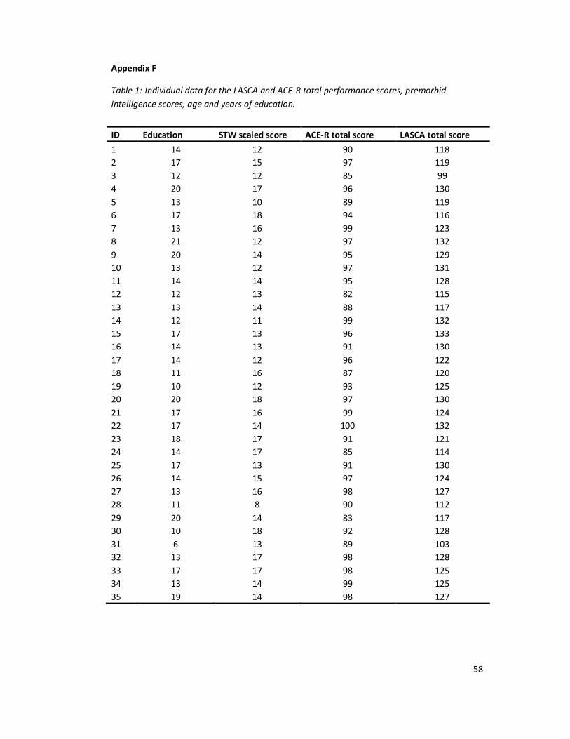
58
Appendix F
Table 1: Individual data for the LASCA and ACE-R total performance scores, premorbid intelligence scores, age and years of education.
ID Education STW scaled score ACE-R total score LASCA total score 1 14 12 90 118 2 17 15 97 119 3 12 12 85 99 4 20 17 96 130 5 13 10 89 119 6 17 18 94 116 7 13 16 99 123 8 21 12 97 132 9 20 14 95 129 10 13 12 97 131 11 14 14 95 128 12 12 13 82 115 13 13 14 88 117 14 12 11 99 132 15 17 13 96 133 16 14 13 91 130 17 14 12 96 122 18 11 16 87 120 19 10 12 93 125 20 20 18 97 130 21 17 16 99 124 22 17 14 100 132 23 18 17 91 121 24 14 17 85 114 25 17 13 91 130 26 14 15 97 124 27 13 16 98 127 28 11 8 90 112 29 20 14 83 117 30 10 18 92 128 31 6 13 89 103 32 13 17 98 128 33 17 17 98 125 34 13 14 99 125 35 19 14 98 127

59
Appendix G
Figure 7. Graph depicting the total distribution of scores on the ACE-R.
Figure 8. Graph depicting the distribution of total scores from the LASCA
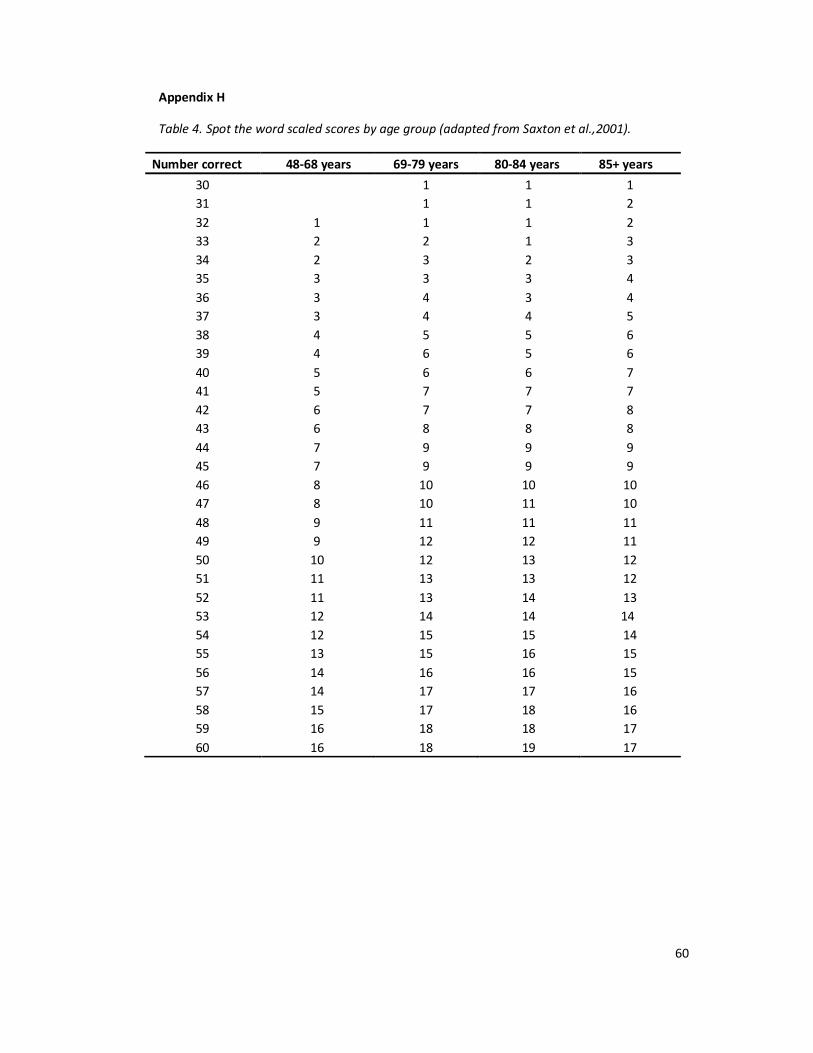
60
Appendix H
Table 4. Spot the word scaled scores by age group (adapted from Saxton et al.,2001).
Number correct 48-68 years 69-79 years 80-84 years 85+ years 30 1 1 1 31 1 1 2 32 1 1 1 2 33 2 2 1 3 34 2 3 2 3 35 3 3 3 4 36 3 4 3 4 37 3 4 4 5 38 4 5 5 6 39 4 6 5 6 40 5 6 6 7 41 5 7 7 7 42 6 7 7 8 43 6 8 8 8 44 7 9 9 9 45 7 9 9 9 46 8 10 10 10 47 8 10 11 10 48 9 11 11 11 49 9 12 12 11 50 10 12 13 12 51 11 13 13 12 52 11 13 14 13 53 12 14 14 14 54 12 15 15 14 55 13 15 16 15 56 14 16 16 15 57 14 17 17 16 58 15 17 18 16 59 16 18 18 17 60 16 18 19 17