Embed Size (px)
Citation preview

5
Low-Anticoagulant Heparins in the Treatment of Metastasis
Narayanam V. Rao1,2, Glenn D. Prestwich2, John R. Hoidal1 and Thomas P. Kennedy3
1Department of Internal Medicine, 2Department of Medicinal Chemistry and Center for Therapeutic Biomaterials,
University of Utah Medical Center, Salt Lake City, Utah, 3Department of Medicine, Georgia Health Sciences University, Augusta, Georgia
USA
1. Introduction
Metastasis is a spread of cancer cells to a distant location from the primary tumor. This process involves a complex series of events similar to those that occur during inflammation. The events of metastasis can be divided into four major steps. First, cancer cells proliferate and transform to acquire motility and ability to invade basement membrane to reach blood vessels. Second, cancer cells penetrate blood vessels due to increased vascular permeability facilitated by inflammatory mediators and enter the circulation. Third, in the circulation cancer cells are shielded by host cells (platelets and neutrophils) to escape surveillance by immune cells and survive shear forces of the bloodstream. Next, integrin-mediated arrest of circulating cancer cells occurs on the endothelial surface before extravasation. Once cancer cells extravasate, heparanase from cancer cells degrades heparan sulfates of the extra cellular matrix. The cleavage of heparan sulfates release growth factors that stimulate cancer cell growth as well as angiogenesis. The Receptor for Advanced Glycation Endproducts (RAGE) belongs to the immunoglobulin superfamily of cell surface molecules. The receptor’s name, RAGE, was coined for its ability to bind its first described ligand, advanced glycation end products (AGEs), which accumulate in physiological (aging) and pathological disorders such as diabetes (Schmidt et al., 1992). RAGE is a pattern recognition receptor for its ligation to structurally unrelated ligands that include Mac-1, HMGB1 and S100 /calgranulins. Similar to immunoglobulin, RAGE contains an extracellular structure with a V-type binding region and two C-type regions. Immediately following the C-type region is a transmembrane region and a short cytoplasmic domain (Fig. 1). The important roles played by RAGE in inflammation, diabetes, Alzhiemer’s disease and cancer have been discussed in detail (Ellerman et al., 2007; Logsdon et al., 2007; Schmidt et al., 2001; Sims et al., 2010; Tang et al., 2010). RAGE is ubiquitously expressed in tissues and inflammatory cells at low levels in homeostasis and its expression is increased in stress conditions. RAGE expression is observed in many tumors, including brain, breast, colon, lung, prostate, pancreatic, ovarian cancers, lymphoma and melanoma (Hsieh et al., 2003; Logsdon et al., 2007) and elevated levels of RAGE have been reported in colon (Sasahira et al., 2005), prostate (Ishiguro et al., 2005) and gastric cancers
www.intechopen.com

Research on Melanoma: A Glimpse into Current Directions and Future Trends
82
(Kuniyasu et al., 2002). In contrast, RAGE levels are down-regulated in lung cancer relative to levels in the normal lung tissue. These observations have led to studies to delineate the pathophysiologic involvement of RAGE in cancer. Notably, AGEs are involved in development and progression of cancers (Abe et al., 2007; Sebekova et al., 2007).
Fig. 1. RAGE domains and ligands involved in cancer. RAGE has ligand-binding V-domain followed by two C-domains of similar to immunoglobulins, a transmembrane region and cytoplasmic tail.
Heparin was discovered in 1916 (McLean, 1916)}, and has been used almost exclusively as an anticoagulant in medicine. However, shortly after its initial discovery, it was reported that heparin had an inhibitory effect on tumor growth in animals (Goerner, 1930). This initial observation led to intensive investigation with regard to heparin’s anticancer and anti-metastatic properties (Casu et al., 2010; Casu et al., 2008; Hettiarachchi et al., 1999; Kragh et al., 2005; Kragh & Loechel, 2005; Lapierre et al., 1996; Lever & Page, 2002; Ono et al., 2002; Ornstein & Zacharski, 1999; Smorenburg et al., 1996; Stevenson et al., 2005; Stevenson et al., 2007; Vlodavsky et al., 2006; Yip et al., 2006; Yoshitomi et al., 2004). The usefulness of heparin as an anticancer drug has been hindered by its anticoagulant effect at therapeutic doses required to inhibit cancer growth and spread. The potential of heparin as an anticancer and anti-inflammatory agent led to discovery of a number of low or nonanticoagulant heparins produced by chemical modifications of the heparin polymer itself (Irimura et al., 1986; Lundin et al., 2000; Vlodavsky et al., 1994), sulfation of other natural polymers (Borgenstrom et al., 2007; Kaeffer et al., 1999; Miao et al., 1999) and sulfated heparin-like polymers produced synthetically (Wellstein et al., 1991; Zugmaier et al., 1992). Studies with heparanoids have been limited to animal models. In human studies, heparin has proven beneficial in cancer treatment but its anticoagulant activity has limited its use. Herein we describe a nonanticoagulant 2-O, 3-O desulfated heparin (ODSH) that retains the
anti-inflammatory activities of heparin, including inhibition of P- and L-selectins,
heparanase, cationic neutrophil proteases, activation of nuclear factor-κB (NF-κB) and
www.intechopen.com
Low-Anticoagulant Heparins in the Treatment of Metastasis
83
ligation of RAGE by HMGB-1, AGEs and S100 calgranulins (Barry et al., 2010; Lapierre et al.,
1996; Rao et al., 2010; Thourani et al., 2000). Also, we describe the first clinical experience
with this modified heparin, which has proven safe and low in anticoagulant activity in 137
humans tested to date. Moreover, we present studies demonstrating that ODSH does not
cause heparin-induced thrombocytopenia (HIT), a thrombotic syndrome occurring in 3% of
individuals receiving heparins (Arepally & Ortel, 2010).
2. Methods
2.1 Production of ODSH
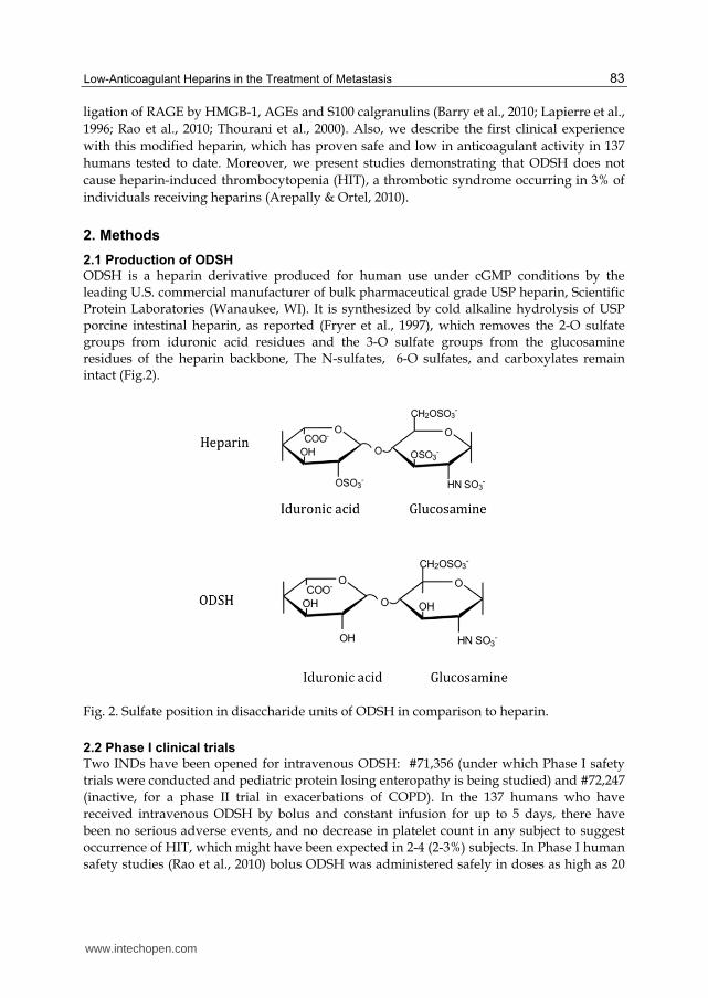
ODSH is a heparin derivative produced for human use under cGMP conditions by the leading U.S. commercial manufacturer of bulk pharmaceutical grade USP heparin, Scientific Protein Laboratories (Wanaukee, WI). It is synthesized by cold alkaline hydrolysis of USP porcine intestinal heparin, as reported (Fryer et al., 1997), which removes the 2-O sulfate groups from iduronic acid residues and the 3-O sulfate groups from the glucosamine residues of the heparin backbone, The N-sulfates, 6-O sulfates, and carboxylates remain intact (Fig.2).
Fig. 2. Sulfate position in disaccharide units of ODSH in comparison to heparin.
2.2 Phase I clinical trials
Two INDs have been opened for intravenous ODSH: #71,356 (under which Phase I safety
trials were conducted and pediatric protein losing enteropathy is being studied) and #72,247
(inactive, for a phase II trial in exacerbations of COPD). In the 137 humans who have
received intravenous ODSH by bolus and constant infusion for up to 5 days, there have
been no serious adverse events, and no decrease in platelet count in any subject to suggest
occurrence of HIT, which might have been expected in 2-4 (2-3%) subjects. In Phase I human
safety studies (Rao et al., 2010) bolus ODSH was administered safely in doses as high as 20
www.intechopen.com
Research on Melanoma: A Glimpse into Current Directions and Future Trends
84
mg/kg, resulting in lower activated partial thromboplastin times (aPTT) than the usual
anticoagulation bolus of unfractionated heparin (80 U/kg, i.e., 0.5 mg/kg at the usual 160
U/mg anti-Xa activity of USP heparin). As part of the Phase I evaluation, ODSH was
infused as a bolus of 8 mg/kg followed by continuous intravenous infusion of 0.5 mg/kg/h,
adjusted to maintain an aPTT between 40 and 45 seconds (0.64 to 1.39 mg/kg/h final
infusion rate).
2.3 Cell culture
U937 monocytes were grown in suspension culture at 37° C in humidified 5% CO2-95% air in RPMI-1640 supplemented with 10% heat inactivated FBS, 2 mM L-glutamine, 1 mM sodium pyruvate, 0.1 mM MEM non-essential amino acids, 100 units/mL penicillin and 100 μg/mL streptomycin. Experiments were performed on cells from passages 1-5. B16F10.1 melanoma cells were grown in Dulbecco’s modified Eagle’s medium, 10% fetal bovine serum, 100 U/mL penicillin, 100 U/mL streptomycin, 2 mM glutamine and 1 mM sodium pyruvate.
2.4 Adhesion assays 2.4.1 Cell surface binding assays
The effect of heparinoids on binding of U937 monocytes to P-selectin or RAGE was studied in high-bind micro plates coated with 8 μg/mL protein A (50 μL/well) in 0.2 M carbonate-bicarbonate buffer (pH 9.4). Plates were washed with phosphate buffered saline (PBS) containing 1% BSA (PBS-BSA), and P-selectin-Fc or RAGE-Fc chimera (50 μL containing 1 μg) was added to each well and incubated for 2 h at room temperature or overnight at 4° C, respectively. Following incubation, wells were washed twice with PBS-BSA. Fifty μL of heparinoids (0 to 1,000 μg/mL) serially diluted in 20 mM HEPES buffer (containing 125 mM NaCl, 2 mM calcium and 2 mM magnesium) were added to each well and incubated at room temperature for 15 min. As a negative control, 50 μL of 10 mM EDTA was added to select wells to prevent cell binding through sequestration of calcium. At the end of the incubation period, 50 μL of U937 cells (105 cells/well, calcein-labeled according to manufacturer’s instructions) were added to each well and plates were incubated an additional 30 min at room temperature. The wells were then washed thrice with PBS, and bound cells were lysed by addition of 100 μL of Tris-Triton X-100 buffer. Fluorescence was measured on a microplate reader using excitation of 494 nm and emission of 517 nm.
2.4.2 Solid phase binding assays
Two types of ELISAs were performed, one to observe the binding between RAGE and its
ligands, including CML-BSA, HMGB-1 and S100b, and a competitive ELISA to study the
ability of ODSH to inhibit/compete for RAGE binding to its ligands.
To confirm RAGE binding to its ligands, polyvinyl 96-well plates were coated with 5 μg/well of specific ligand (CML-BSA, HMGB-1 or S100b calgranulin). Plates were incubated overnight at 4° C and washed thrice with PBS-0.05% Tween-20 (PBST). Next, 50 μL of RAGE from the dilution series ranging from 0.001 to ~ 6 nM was transferred to each respective ligand-coated well and incubated at 37° C for 1 h. Wells were then washed four times with PBST. To detect bound RAGE, 50 μL of anti-RAGE antibody (0.5 μg/mL) was added to each well, the mixture was incubated for 1 h at room temperature, and wells were washed again four times with PBST. HRP-conjugated secondary antibody (50 μL per well) was added,
www.intechopen.com

Low-Anticoagulant Heparins in the Treatment of Metastasis
85
wells were incubated for 1 h at room temperature, and then washed once with PBST. A colorimetric reaction was initiated by addition of 50 μL of TMB and terminated after 15 min by addition of 50 μL of 1 N HCl. Absorbance at 450 nm was read using an automated microplate reader. Binding affinity (KD) was determined from the plot of absorbance values versus concentrations of RAGE. RAGE binding to glycated ECM proteins, collagen-I, collagen -IV, fibronectin and laminin coated plates were used. Plates were incubated with 100 µL of 0.15 M glyoxylic acid + 0.45 M sodium cyanoborohydride mixture at 37 °C for 24 hours. At the end of incubation period, the plates were washed with PBST and used for RAGE binding as described above. For studies of the effect of ODSH or heparin on RAGE binding to its ligands, polyvinyl 96-well plates were coated with specific ligand as described above. Separately, a constant amount of RAGE-Fc chimera (100 μL containing 0.5 μg/mL in PBST containing 0.1% BSA) was incubated with an equal volume of serially diluted ODSH or heparin (0.001 to 1,000 μg/mL in PBST-BSA) overnight at 4° C. The following day, 50 μL of RAGE-heparinoid mix was transferred to each respective ligand-coated well and incubated at 37° C for 2 h. Wells were then washed four times with PBST. Bound RAGE was detected as described above. Absorbance at 450 nm was plotted against concentration of ODSH or heparin. The IC50 values were obtained using non-linear regression analysis.
2.5 Elastase and cathepsin G activity assay
The inhibitory activity of heparin and ODSH against HLE and cathepsin G was monitored
using the specific chromogenic substrates Suc-Ala-Ala-Val-pNA and Suc-Ala-Ala-Pro-Phe-
pNA, respectively, according to methods previously described (Fryer et al., 1997).
2.6 Mouse model of melanoma lung metastasis
Lung metastasis from melanoma was studied using protocols previously reported by the Varki group (Stevenson et al., 2005). Animal use and husbandry followed protocols approved by the IACUC at the University of Utah. Confluent B16F10 melanoma cells (70-80%) were harvested by brief exposure to trypsin, and washed twice with serum-free medium prior to injection. Living cells were counted with Trypan blue staining prior to injection to insure > 95% viability. Female C57BL/6J mice (n = 6 per group) were injected subcutaneously with 100 μL PBS, heparin (30 mg/kg) or ODSH (30 mg/kg). Thirty min later, 5 x 10⁵ B16F10 cells in 200 μL medium were injected intravenously into the lateral tail vein. Mice from each group were injected in alternating order, and cells were resuspended by gently flicking the tube before aspirating the sample for each injection. Twenty-seven days after injection, surviving mice were euthanized. The lungs were removed, perfused intra-tracheally with 10% buffered formalin and photographed. Visible tumor foci were counted independently by two different laboratory personnel blinded with regard to treatment groups, and metastasis quantified in terms of the number of black spots.
3. Results and discussion
3.1 ODSH is a non-anticoagulant heparin that is manufactured at industrial scale and safe from heparin induced thrombocytopenia
The ODSH was manufactured as described in Methods section. Seven serial 1-2 kg batches of material have shown an average molecular mass of 11.7 ± 0.3 kg kDa. ODSH has low affinity for anti-thrombin III (KD = 339 μM or 4 mg/mL vs. 1.56 μM or 22 μg/mL for
www.intechopen.com

Research on Melanoma: A Glimpse into Current Directions and Future Trends
86
heparin) consistent with its low anticoagulant activity. Serial batches of ODSH demonstrated consistently reduced USP (7 ± 0.3 U of anticoagulant activity/mg), anti-Xa (1.9 ± 0.1 U/mg), and anti- IIa (1.2 ± 0.1 U/mg) activities, compared with those of heparin (165-190 U/mg activity for all 3 assays). The current clinical formulation that was used in 137 humans (54 normal and 83 ill patients) is a sterile-filled 20 mL glass vial containing an isotonic 50 mg/mL solution of ODSH in buffered saline. This formulation is cGMP manufactured by Pyramid Laboratories (Costa Mesa, CA). Chemistry and Manufacturing processes are already on file with the FDA in two INDs. A complication of heparin is HIT, which occurs in subjects who produce an activating antibody to the heparin-platelet factor 4 (PF4) complex on the platelet surface, causing thrombocytopenia and thromboembolism (Arepally & Ortel, 2010). In standard serotonin release assays (SRA) performed with human antibodies to the heparin-PF4 complex, heparin triggered release of 14C-serotonin from normal platelets (Fig 3), but ODSH did not trigger HIT at any concentration (Rao et al., 2010).
Fig. 3. ODSH does not trigger platelet activation in the serotonin release assay. Normal platelets were loaded with 14C-serotonin and incubated with serum from 3 patients with heparin-PF4 antibodies. Platelets were activated by heparin as a positive control (≥ 20% serotonin release). Rao et al., Am J Physiol Cell Physiol (2010) Am Physiol Soc, used with permission.
3.2 ODSH is safe to use in humans
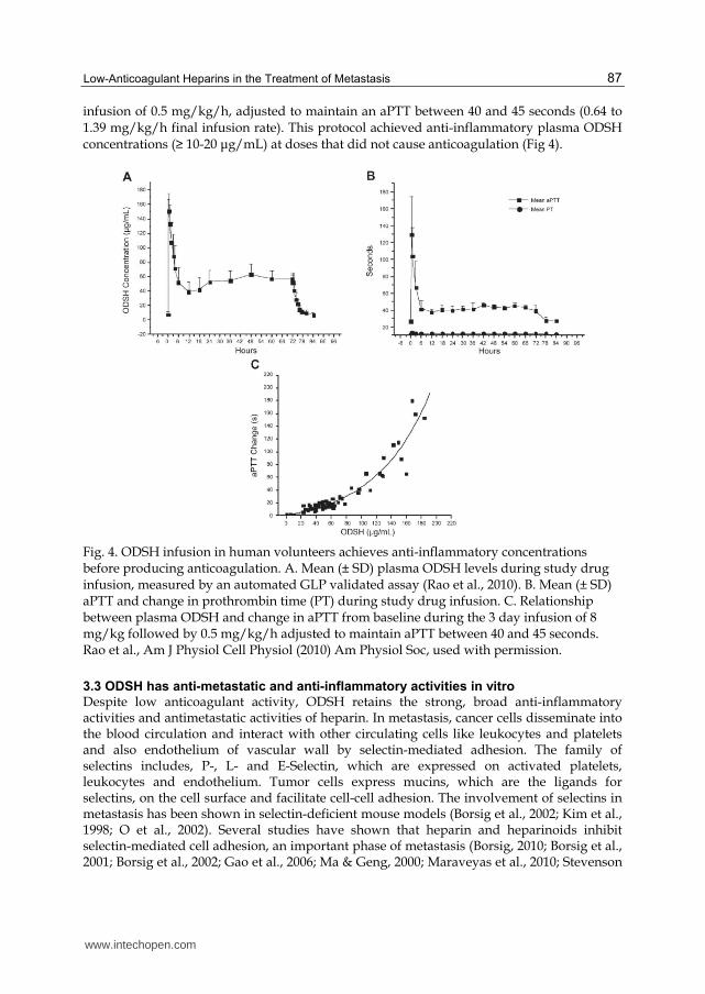
Two INDs have been opened for intravenous ODSH: #71,356 (under which Phase I safety trials were conducted and pediatric protein losing enteropathy is being studied) and #72,247 (inactive, for a phase II trial in exacerbations of COPD). In the 137 humans who have received intravenous ODSH by bolus and constant infusion for up to 5 days, there have been no serious adverse events, and no decrease in platelet count in any subject to suggest occurrence of HIT, which might have been expected in 2-4 (2-3%) subjects (Arepally & Ortel, 2010). In Phase I human safety studies (Rao et al., 2010) bolus ODSH was administered safely in doses as high as 20 mg/kg, resulting in lower activated partial thromboplastin times (aPTT) than the usual anticoagulation bolus of unfractionated heparin (80 U/kg, i.e., 0.5 mg/kg at the usual 160 U/mg anti-Xa activity of USP heparin). As part of the Phase I evaluation, ODSH was infused as a bolus of 8 mg/kg followed by continuous intravenous
www.intechopen.com
Low-Anticoagulant Heparins in the Treatment of Metastasis
87
infusion of 0.5 mg/kg/h, adjusted to maintain an aPTT between 40 and 45 seconds (0.64 to 1.39 mg/kg/h final infusion rate). This protocol achieved anti-inflammatory plasma ODSH concentrations (≥ 10-20 μg/mL) at doses that did not cause anticoagulation (Fig 4).
Fig. 4. ODSH infusion in human volunteers achieves anti-inflammatory concentrations before producing anticoagulation. A. Mean (± SD) plasma ODSH levels during study drug infusion, measured by an automated GLP validated assay (Rao et al., 2010). B. Mean (± SD) aPTT and change in prothrombin time (PT) during study drug infusion. C. Relationship between plasma ODSH and change in aPTT from baseline during the 3 day infusion of 8 mg/kg followed by 0.5 mg/kg/h adjusted to maintain aPTT between 40 and 45 seconds. Rao et al., Am J Physiol Cell Physiol (2010) Am Physiol Soc, used with permission.
3.3 ODSH has anti-metastatic and anti-inflammatory activities in vitro
Despite low anticoagulant activity, ODSH retains the strong, broad anti-inflammatory activities and antimetastatic activities of heparin. In metastasis, cancer cells disseminate into the blood circulation and interact with other circulating cells like leukocytes and platelets and also endothelium of vascular wall by selectin-mediated adhesion. The family of selectins includes, P-, L- and E-Selectin, which are expressed on activated platelets, leukocytes and endothelium. Tumor cells express mucins, which are the ligands for selectins, on the cell surface and facilitate cell-cell adhesion. The involvement of selectins in metastasis has been shown in selectin-deficient mouse models (Borsig et al., 2002; Kim et al., 1998; O et al., 2002). Several studies have shown that heparin and heparinoids inhibit selectin-mediated cell adhesion, an important phase of metastasis (Borsig, 2010; Borsig et al., 2001; Borsig et al., 2002; Gao et al., 2006; Ma & Geng, 2000; Maraveyas et al., 2010; Stevenson
www.intechopen.com

Research on Melanoma: A Glimpse into Current Directions and Future Trends
88
et al., 2005; Stevenson et al., 2007; Varki, 2007; Wahrenbrock et al., 2003; Wei et al., 2004). To investigate the antimetastatic potency of ODSH, we used U937 cells, a human monocytic leukemia cells. U937 cells express P-selectin ligand, P-selectin glycoprotein ligand-1 (PSGL-1), that functions similar to mucins. We tested the ODSH ability to competitively displace fluorescent-labeled U937 cells adherence to P-selectin via PSGL-1. ODSH inhibited P-selectin with an IC50 of 1.1 μg/mL consistent with that of values reported byWang (Wang et al., 2002), who demonstrated that ODSH also inhibits L-selectin-mediated adhesion with an IC50 of 3 μg/mL, versus 0.4 μg/mL for heparin (Wang et al., 2002) (Fig. 5).
Fig. 5. Anti-inflammatory and anti-metastatic profile of ODSH. The 50% inhibitory concentrations (IC50 values) of ODSH to interrupt the receptor ligand pairs, P-selectin/PSGL-1, RAGE/Mac-1, RAGE/CML-BSA, RAGE/S100B and RAGE/HMGB1, interaction are higher than that of heparin. Nonetheless, the IC50 concentrations of ODSH required to inhibit HLE, cathepsin G and heparanase activities, the key enzymes involved in the inflammation and metastasis, are similar to the values for heparin. The IC50 values of ODSH are higher than that of heparin suggesting that ODSH has lower inhibitory potential than heparin, but the values are much lower than achievable plasma concentration (~200 µg/mL) in humans (see Fig 4). Heparanase data is from (Lapierre et al., 1996).
Though RAGE binds to disparate ligands, we will discuss only the ligands AGE, HMGB1
and S100B that have been evaluated for their role in cancer. RAGE and its ligands are co-
expressed in many tumors and the expressed ligands are secreted causing triggering of
cellular signaling pathways. This results in expression of cytokines, growth factors,
transcription factors, adhesion molecules and NF-kB (Sparvero et al., 2009). Therefore we
will focus on the interruption of RAGE-ligand interaction with ODSH.
Several in vitro studies have shown that AGE stimulates growth, proliferation and invasion of cancer cells including melanoma cells (Abe et al., 2004; Yamamoto et al., 1996; Zill et al., 2003; Zill et al., 2001). AGEs are found in extracellular matrix (ECM) proteins (Ling et al., 1998; Mizutari et al., 1997) that can interact with RAGE on tumor cells and enhance RAGE expression causing damage to surrounding tissues. We therefore tested the binding of native and glycated ECM proteins (collagen I, collagen IV, fibronectin and laminin) to RAGE. Results indicate that RAGE binds to native ECM protein with a strong affinity but the affinity is negligibly decreased with glycated ECM proteins. Overall, the KD values of
www.intechopen.com
Low-Anticoagulant Heparins in the Treatment of Metastasis
89
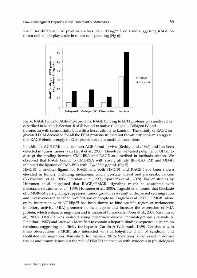
RAGE for different ECM proteins are less than 100 ng/mL or <1nM suggesting RAGE on tumor cells might play a role in tumor cell spreading (Fig 6).
Fig. 6. RAGE binds to AGE-ECM proteins. RAGE binding to ECM proteins was analyzed as described in Methods Section. RAGE bound to native Collagen I, Collagen IV and fibronectin with same affinity but with a lesser affinity to Laminin. The affinity of RAGE for glycated ECM decreased for all the ECM proteins studied but the affinity constants suggest that RAGE binds strongly to ECM proteins even in modified conditions.
In addition, AGE-CML is a common AGE found in vivo (Reddy et al., 1995) and has been detected in tumor tissues (van Heijst et al., 2005). Therefore, we tested potential of ODSH to disrupt the binding between CML-BSA and RAGE as described in methods section. We observed that RAGE bound to CML-BSA with strong affinity (KD 0.43 nM) and ODSH inhibited the ligation of CML-BSA with IC50 of 8.6 µg/mL (Fig 5). HMGB1 is another ligand for RAGE and both HMGB1 and RAGE have been shown
elevated in tumors, including melanoma, colon, prostate, breast and pancreatic cancers
(Brezniceanu et al., 2003; Ellerman et al., 2007; Sparvero et al., 2009). Earlier studies by
Huttunen et al. suggested that RAGE/HMGB1 signaling might be associated with
metastasis (Huttunen et al., 1999; Huttunen et al., 2000). Taguchi et al. found that blockade
of HMGB-RAGE signaling suppressed tumor growth as a result of decreased cell migration
and invasiveness rather than proliferation or apoptosis (Taguchi et al., 2000). HMGB1 alone
or by interaction with NF-kBp65 has been shown to bind specific region of melanocyte
inhibitory activity (MIA) promoter in melanocytes and increase the expression of MIA
protein, which enhances migration and invasion of tumor cells (Poser et al., 2003; Sasahira et
al., 2008). HMGB1 was isolated using heparin-sepharose chromatography (Rauvala &
Pihlaskari, 1987) and later was identified to contain a heparin-binding sequence in its amino
terminus, suggesting its affinity for heparin (Cardin & Weintraub, 1989). Consistent with
these observations, HMGB1 also interacted with carbohydrate chain of syndecan and
facilitated cell migration (Rauvala & Rouhiainen, 2010). Syndecan is expressed in normal
tissues and tumor tissues but the role of HMGB1 interaction with syndecan in physiological
www.intechopen.com

Research on Melanoma: A Glimpse into Current Directions and Future Trends
90
and pathological conditions is not known (Rauvala & Rouhiainen, 2010). Since ODSH is
derived from heparin, we tested whether ODSH can interrupt the RAGE-HMB1 ligation.
Though HMGB1 binds to RAGE with high affinity (KD 0.64nM), ODSH inhibited RAGE-
HMGB1 interaction with an IC50 of 0.23 µg/mL (Figs. 5 and 7).
Fig. 7. ODSH and heparin inhibit binding of HMGB-1 to RAGE. HMGB-1 binding to RAGE-Fc chimera was studied using ELISA techniques as described in Methods. RAGE bound to immobilized HMGB-1 in a saturable fashion (inset) with a KD of 0.64 nM. ODSH (●) and heparin (○) inhibit binding of recombinant HMGB-1 to RAGE with IC50 values of 0.23 and 0.04 μg/mL, respectively. Rao et al., Am J Physiol Cell Physiol (2010) Am Physiol Soc, used with permission.
S100 proteins are calcium dependent proteins of the EF-hand (helix-loop-helix) type that are
differentially located and differentially expressed in a wide variety of cells (Donato, 1999;
Donato, 2001; Donato et al., 2009). Among the 25 S100 protein identified, S100b is unique in
its location on chromosome 21q22.3 and first one of the group found to ligate to RAGE
(Hofmann et al., 1999). S100b is highly expressed in melanoma and considered as possible
biomarker for the prognosis of the disease (Harpio & Einarsson, 2004; Salama et al., 2008).
We studied the binding affinity of S100b to RAGE. RAGE ligated to S100b with a KD of 0.45
nM, a value much lower than the affinity range reported (reviewed in (Leclerc et al., 2009)).
ODSH inhibited the binding of RAGE to S100b with IC50 value of 4.23µg/mL. (Fig 5).
HMGB-1 is released into extracellular milieu as a result of necrosis, apoptosis and secretion. Only HMGB1 released by necrotic cells demonstrates cytokine activity, recruits leukocytes and activates endothelial cell adhesion molecules such as selectins (Dumitriu et al., 2005; Lotze & Tracey, 2005). In cancer, tumor cells undergoing stress are susceptible to death because of factors that include hypoxia, nutrient deprivation and or anticancer treatments (Kepp et al., 2009). These dying cancer cells become a source of HMGB1 (Dong Xda et al., 2007; Scaffidi et al., 2002). Orlova et al (Orlova et al., 2007)) have shown that HMGB1-induced Mac-1 dependent neutrophil recruitment requires the presence of RAGE on neutrophils but not on endothelial cells. Interestingly, three proteins, HMGB1, Mac-1 and RAGE bind to heparin (Diamond et al., 1995; Hanford et al., 2004; Rauvala & Pihlaskari, 1987). We therefore tested Mac-1 dependent binding of U937 cells to immobilized RAGE. ODSH inhibited the adherence of U937 cells to RAGE with IC50 values of 0.11 µg/mL (Fig 5).
www.intechopen.com

Low-Anticoagulant Heparins in the Treatment of Metastasis
91
3.4 ODSH Prevents metastasis in vivo
The vitro studies described above suggested that ODSH, similar to heparin, might interrupt
key receptor-ligand interactions involved in metastasis. We therefore investigated the effects
of ODSH in experimental melanoma lung metastasis. Heparin significantly decreased lung
metastasis at 28 days after melanoma injection, but the same dose of ODSH provided
substantially greater reduction of lung metastasis (Fig 8A). ODSH also significantly
improved 28-day survival. In contrast to 70% mortality in mice treated with PBS, ~ 70% of
ODSH-treated mice survived the experiment (Fig 8B).
Fig. 8. ODSH inhibits melanoma lung metastasis. Female C57BL/6J mice (n = 6/group) were injected SQ with 100 μL PBS, 30 mg/kg heparin, or 30 mg/kg ODSH. Thirty min later, 5 x 10⁵ B16F10 melanoma cells were injected IV into the tail vein. 27 days later surviving mice were euthanized and lungs were removed, fixed, stained and assessed for metastasis by 2 independent observers. A. Heparin (*P < 0.05 vs PBS) or ODSH (**P < 0.01 vs PBS) significantly reduced metastasis. B. SQ heparin showed little effect on lung metastatic outgrowth but ODSH suppressed metastatic colonization microscopically. C. ODSH improved survival (*P < 0.05 vs PBS). Rao et al., Am J Physiol Cell Physiol (2010) Am Physiol Soc, used with permission.
This model is widely used to test the inhibitory capacity of heparin and heparins with no or minimal anticoagulant activity in metastasis (reviewed in (Borsig, 2010). The attenuation of metastasis was attributed to P-selectin and L-selectin as the experimental mice were deficient in selectin but also suggested that there is a selectin-independent mechanism involved. It is not clear whether B16 melanoma cells express mucins on the cells surface to interact with P-selectin, but the expression of RAGE is observed in melanoma cells (Huttunen et al., 2002; Saha et al., 2010). Further, Huttunen et al showed the attenuation of metastasis when B16 cells injected with HMGB1 peptide suggesting the RAGE-mediated tumor cell invasion can be inhibited by competition with RAGE binding ligand such as
B
www.intechopen.com

Research on Melanoma: A Glimpse into Current Directions and Future Trends
92
ODSH and heparin. In summary, because ODSH interacts with the many receptors, ligands and inhibits enzymes involved in the metastasis, the attenuation of metastasis in the experimental model is due to action of ODSH on multiple sites.
4. Conclusion
A number of fully anticoagulant heparins are commercially available, but their use as a chronic treatment for cancer is dose-limited by their anticoagulant activity. Therapeutic anticoagulant doses of heparin or low molecular weight heparin (Hirsh et al., 2001; Koenig et al., 1998) do not provide sufficiently high plasma drug concentrations to reliably inhibit the selectin-, heparanase- or RAGE-mediated processes (Koenig et al., 1998; Lapierre et al., 1996; Rao et al., 2010; Wang et al., 2002) that are now understood to be important for explaining the benefits of heparins in cancer.
Fig. 9. Potential interference of low anticoagulant ODSH in metastasis. ODSH 1) inhibits heparanse, the enzyme secreted by tumor cells during intravsation and extravsation thus prevent spread of the tumor cells, 2) inhibits the tumor cell mucin (P-selectin ligand) binding to P-selectin on endothelial cells and platelets, 3) inhibits tumor cell RAGE interaction with leukocyte Mac-1, 4) inhibits tumor cell RAGE interaction with its ligands, AGE, HMGB1 and S100b, that are either released by tumor cells within cancer micro environment or by the leukocytes thus prevent sustained signaling for tumor progression, 5) inhibits circulating RAGE ligand interaction with endothelial RAGE.
www.intechopen.com

Low-Anticoagulant Heparins in the Treatment of Metastasis
93
Of the available low anticoagulant heparin derivatives (Casu et al., 2008; Fryer et al., 1997; Kragh et al., 2005; Kragh & Loechel, 2005; Lapierre et al., 1996; Ono et al., 2002; Rao et al., 2010; Stevenson et al., 2007; Wang et al., 2002; Yoshitomi et al., 2004), only ODSH has been proven safe from major adverse events in humans (Rao et al., 2010) data on file with FDA) and only ODSH is free from the potential to induce HIT (Rao et al., 2010). The concept that P-selectin is the key molecule in metastasis is based on the attenuation of metastasis in animal models. Because of the complex biology of cancer metastasis, it is not easy to identify the specific mediator(s) involved in development of metastatic disease in patients with cancer. In such situation, a compound that can simultaneously target multiple mediators involved in the diseases might have substantial therapeutic value. We have described that non-anticoagulant ODSH, not only targets P-selectin, but additionally is capable of inhibiting the RAGE interaction with its multiple structurally unrelated ligands that play many biological roles in metastasis (Fig. 9). Thus, the results described herein support the notion that low anticoagulant heparin derivatives such as ODSH might prove useful in prevention of metastasis in human cancer.
5. Acknowledgement
We thank Jeanine Walenga and Margaret Prechel for the help with serotonine release assays,
Brian Argyle for his expert technical assistance and Xiaoyu Xu for help with lung metastasis
animal model. We gratefully acknowledge the expert help of Robert MacArthur and
Bradford Walters with phase I studies. The work has been supported by a sponsored
research project awarded to the University of Utah by ParinGenix (to N. V. Rao).
6. References
Abe, R., Fujita, Y., & Yamagishi, S. (2007) Angiogenesis and metastasis inhibitors for the treatment of malignant melanoma. Mini Rev Med Chem, Vol.7, No.6, pp. 649-661, 1389-5575 (Print) 1389-5575 (Linking)
Abe, R., Shimizu, T., Sugawara, H., Watanabe, H., Nakamura, H., Choei, H., Sasaki, N., Yamagishi, S., Takeuchi, M., & Shimizu, H. (2004) Regulation of human melanoma growth and metastasis by AGE-AGE receptor interactions. J Invest Dermatol, Vol.122, No.2, pp. 461-467, 0022-202X (Print) 0022-202X (Linking)
Arepally, G. M., & Ortel, T. L. (2010) Heparin-induced thrombocytopenia. Annu Rev Med, Vol.61,pp. 77-90, 1545-326X (Electronic) 0066-4219 (Linking)
Barry, W. H., Zhang, X. Q., Halkos, M. E., Vinten-Johansen, J., Saegusa, N., Spitzer, K. W., Matsuoka, N., Sheets, M., Rao, N. V., & Kennedy, T. P. (2010) Nonanticoagulant heparin reduces myocyte Na+ and Ca2+ loading during simulated ischemia and decreases reperfusion injury. Am J Physiol Heart Circ Physiol, Vol.298, No.1, pp. H102-111, 1522-1539 (Electronic) 0363-6135 (Linking)
Borgenstrom, M., Warri, A., Hiilesvuo, K., Kakonen, R., Kakonen, S., Nissinen, L., Pihlavisto, M., Marjamaki, A., Vlodavsky, I., Naggi, A., Torri, G., Casu, B., Veromaa, T., Salmivirta, M., & Elenius, K. (2007) O-sulfated bacterial polysaccharides with low anticoagulant activity inhibit metastasis. Semin Thromb Hemost, Vol.33, No.5, pp. 547-556, 0094-6176 (Print) 0094-6176 (Linking)
www.intechopen.com

Research on Melanoma: A Glimpse into Current Directions and Future Trends
94
Borsig, L. (2010) Antimetastatic activities of heparins and modified heparins. Experimental evidence. Thromb Res, Vol.125 Suppl 2,pp. S66-71, 1879-2472 (Electronic) 0049-3848 (Linking)
Borsig, L., Wong, R., Feramisco, J., Nadeau, D. R., Varki, N. M., & Varki, A. (2001) Heparin and cancer revisited: mechanistic connections involving platelets, P-selectin, carcinoma mucins, and tumor metastasis. Proc Natl Acad Sci U S A, Vol.98, No.6, pp. 3352-3357, 0027-8424 (Print) 0027-8424 (Linking)
Borsig, L., Wong, R., Hynes, R. O., Varki, N. M., & Varki, A. (2002) Synergistic effects of L- and P-selectin in facilitating tumor metastasis can involve non-mucin ligands and implicate leukocytes as enhancers of metastasis. Proc Natl Acad Sci U S A, Vol.99, No.4, pp. 2193-2198, 0027-8424 (Print) 0027-8424 (Linking)
Brezniceanu, M. L., Volp, K., Bosser, S., Solbach, C., Lichter, P., Joos, S., & Zornig, M. (2003) HMGB1 inhibits cell death in yeast and mammalian cells and is abundantly expressed in human breast carcinoma. FASEB J, Vol.17, No.10, pp. 1295-1297, 1530-6860 (Electronic) 0892-6638 (Linking)
Cardin, A. D., & Weintraub, H. J. (1989) Molecular modeling of protein-glycosaminoglycan interactions. Arteriosclerosis, Vol.9, No.1, pp. 21-32, 0276-5047 (Print) 0276-5047 (Linking)
Casu, B., Naggi, A., & Torri, G. (2010) Heparin-derived heparan sulfate mimics to modulate heparan sulfate-protein interaction in inflammation and cancer. Matrix Biol, Vol.29, No.6, pp. 442-452, 1569-1802 (Electronic) 0945-053X (Linking)
Casu, B., Vlodavsky, I., & Sanderson, R. D. (2008) Non-anticoagulant heparins and inhibition of cancer. Pathophysiol Haemost Thromb, Vol.36, No.3-4, pp. 195-203, 1424-8840 (Electronic) 1424-8832 (Linking)
Diamond, M. S., Alon, R., Parkos, C. A., Quinn, M. T., & Springer, T. A. (1995) Heparin is an adhesive ligand for the leukocyte integrin Mac-1 (CD11b/CD1). J Cell Biol, Vol.130, No.6, pp. 1473-1482, 0021-9525 (Print) 0021-9525 (Linking)
Donato, R. (1999) Functional roles of S100 proteins, calcium-binding proteins of the EF-hand type. Biochim Biophys Acta, Vol.1450, No.3, pp. 191-231, 0006-3002 (Print) 0006-3002 (Linking)
Donato, R. (2001) S100: a multigenic family of calcium-modulated proteins of the EF-hand type with intracellular and extracellular functional roles. Int J Biochem Cell Biol, Vol.33, No.7, pp. 637-668, 1357-2725 (Print) 1357-2725 (Linking)
Donato, R., Sorci, G., Riuzzi, F., Arcuri, C., Bianchi, R., Brozzi, F., Tubaro, C., & Giambanco, I. (2009) S100B's double life: intracellular regulator and extracellular signal. Biochim Biophys Acta, Vol.1793, No.6, pp. 1008-1022, 0006-3002 (Print) 0006-3002 (Linking)
Dong Xda, E., Ito, N., Lotze, M. T., Demarco, R. A., Popovic, P., Shand, S. H., Watkins, S., Winikoff, S., Brown, C. K., Bartlett, D. L., & Zeh, H. J., 3rd (2007) High mobility group box I (HMGB1) release from tumor cells after treatment: implications for development of targeted chemoimmunotherapy. J Immunother, Vol.30, No.6, pp. 596-606, 1524-9557 (Print) 1524-9557 (Linking)
Dumitriu, I. E., Baruah, P., Manfredi, A. A., Bianchi, M. E., & Rovere-Querini, P. (2005) HMGB1: guiding immunity from within. Trends Immunol, Vol.26, No.7, pp. 381-387, 1471-4906 (Print) 1471-4906 (Linking)
Ellerman, J. E., Brown, C. K., de Vera, M., Zeh, H. J., Billiar, T., Rubartelli, A., & Lotze, M. T. (2007) Masquerader: high mobility group box-1 and cancer. Clin Cancer Res, Vol.13, No.10, pp. 2836-2848, 1078-0432 (Print) 1078-0432 (Linking)
www.intechopen.com

Low-Anticoagulant Heparins in the Treatment of Metastasis
95
Fryer, A., Huang, Y. C., Rao, G., Jacoby, D., Mancilla, E., Whorton, R., Piantadosi, C. A., Kennedy, T., & Hoidal, J. (1997) Selective O-desulfation produces nonanticoagulant heparin that retains pharmacological activity in the lung. J Pharmacol Exp Ther, Vol.282, No.1, pp. 208-219, 0022-3565 (Print) 0022-3565 (Linking)
Gao, Y., Wei, M., Zheng, S., Ba, X., Hao, S., & Zeng, X. (2006) Chemically modified heparin inhibits the in vitro adhesion of nonsmall cell lung cancer cells to P-selectin. J Cancer Res Clin Oncol, Vol.132, No.4, pp. 257-264, 0171-5216 (Print) 0171-5216 (Linking)
Goerner, A. (1930) The influence of anti-clotting agents on transplantation and growth of tumor tissue. J Lab Clin Med, Vol.16,pp. 369-372,
Hanford, L. E., Enghild, J. J., Valnickova, Z., Petersen, S. V., Schaefer, L. M., Schaefer, T. M., Reinhart, T. A., & Oury, T. D. (2004) Purification and characterization of mouse soluble receptor for advanced glycation end products (sRAGE). J Biol Chem, Vol.279, No.48, pp. 50019-50024, 0021-9258 (Print) 0021-9258 (Linking)
Harpio, R., & Einarsson, R. (2004) S100 proteins as cancer biomarkers with focus on S100B in malignant melanoma. Clin Biochem, Vol.37, No.7, pp. 512-518, 0009-9120 (Print) 0009-9120 (Linking)
Hettiarachchi, R. J., Smorenburg, S. M., Ginsberg, J., Levine, M., Prins, M. H., & Buller, H. R. (1999) Do heparins do more than just treat thrombosis? The influence of heparins on cancer spread. Thromb Haemost, Vol.82, No.2, pp. 947-952, 0340-6245 (Print) 0340-6245 (Linking)
Hirsh, J., Warkentin, T. E., Shaughnessy, S. G., Anand, S. S., Halperin, J. L., Raschke, R., Granger, C., Ohman, E. M., & Dalen, J. E. (2001) Heparin and low-molecular-weight heparin: mechanisms of action, pharmacokinetics, dosing, monitoring, efficacy, and safety. Chest, Vol.119, No.1 Suppl, pp. 64S-94S, 0012-3692 (Print) 0012-3692 (Linking)
Hofmann, M. A., Drury, S., Fu, C., Qu, W., Taguchi, A., Lu, Y., Avila, C., Kambham, N., Bierhaus, A., Nawroth, P., Neurath, M. F., Slattery, T., Beach, D., McClary, J., Nagashima, M., Morser, J., Stern, D., & Schmidt, A. M. (1999) RAGE mediates a novel proinflammatory axis: a central cell surface receptor for S100/calgranulin polypeptides. Cell, Vol.97, No.7, pp. 889-901, 0092-8674 (Print) 0092-8674 (Linking)
Hsieh, H. L., Schafer, B. W., Sasaki, N., & Heizmann, C. W. (2003) Expression analysis of S100 proteins and RAGE in human tumors using tissue microarrays. Biochem Biophys Res Commun, Vol.307, No.2, pp. 375-381, 0006-291X (Print) 0006-291X (Linking)
Huttunen, H. J., Fages, C., Kuja-Panula, J., Ridley, A. J., & Rauvala, H. (2002) Receptor for advanced glycation end products-binding COOH-terminal motif of amphoterin inhibits invasive migration and metastasis. Cancer Res, Vol.62, No.16, pp. 4805-4811, 0008-5472 (Print) 0008-5472 (Linking)
Huttunen, H. J., Fages, C., & Rauvala, H. (1999) Receptor for advanced glycation end products (RAGE)-mediated neurite outgrowth and activation of NF-kappaB require the cytoplasmic domain of the receptor but different downstream signaling pathways. J Biol Chem, Vol.274, No.28, pp. 19919-19924, 0021-9258 (Print) 0021-9258 (Linking)
Huttunen, H. J., Kuja-Panula, J., Sorci, G., Agneletti, A. L., Donato, R., & Rauvala, H. (2000) Coregulation of neurite outgrowth and cell survival by amphoterin and S100
www.intechopen.com

Research on Melanoma: A Glimpse into Current Directions and Future Trends
96
proteins through receptor for advanced glycation end products (RAGE) activation. J Biol Chem, Vol.275, No.51, pp. 40096-40105, 0021-9258 (Print) 0021-9258 (Linking)
Irimura, T., Nakajima, M., & Nicolson, G. L. (1986) Chemically modified heparins as inhibitors of heparan sulfate specific endo-beta-glucuronidase (heparanase) of metastatic melanoma cells. Biochemistry, Vol.25, No.18, pp. 5322-5328, 0006-2960 (Print) 0006-2960 (Linking)
Ishiguro, H., Nakaigawa, N., Miyoshi, Y., Fujinami, K., Kubota, Y., & Uemura, H. (2005) Receptor for advanced glycation end products (RAGE) and its ligand, amphoterin are overexpressed and associated with prostate cancer development. Prostate, Vol.64, No.1, pp. 92-100, 0270-4137 (Print) 0270-4137 (Linking)
Kaeffer, B., Benard, C., Lahaye, M., Blottiere, H. M., & Cherbut, C. (1999) Biological properties of ulvan, a new source of green seaweed sulfated polysaccharides, on cultured normal and cancerous colonic epithelial cells. Planta Med, Vol.65, No.6, pp. 527-531, 0032-0943 (Print) 0032-0943 (Linking)
Kepp, O., Tesniere, A., Schlemmer, F., Michaud, M., Senovilla, L., Zitvogel, L., & Kroemer, G. (2009) Immunogenic cell death modalities and their impact on cancer treatment. Apoptosis, Vol.14, No.4, pp. 364-375, 1573-675X (Electronic) 1360-8185 (Linking)
Kim, Y. J., Borsig, L., Varki, N. M., & Varki, A. (1998) P-selectin deficiency attenuates tumor growth and metastasis. Proc Natl Acad Sci U S A, Vol.95, No.16, pp. 9325-9330, 0027-8424 (Print) 0027-8424 (Linking)
Koenig, A., Norgard-Sumnicht, K., Linhardt, R., & Varki, A. (1998) Differential interactions of heparin and heparan sulfate glycosaminoglycans with the selectins. Implications for the use of unfractionated and low molecular weight heparins as therapeutic agents. J Clin Invest, Vol.101, No.4, pp. 877-889, 0021-9738 (Print) 0021-9738 (Linking)
Kragh, M., Binderup, L., Vig Hjarnaa, P. J., Bramm, E., Johansen, K. B., & Frimundt Petersen, C. (2005) Non-anti-coagulant heparin inhibits metastasis but not primary tumor growth. Oncol Rep, Vol.14, No.1, pp. 99-104, 1021-335X (Print) 1021-335X (Linking)
Kragh, M., & Loechel, F. (2005) Non-anti-coagulant heparins: a promising approach for prevention of tumor metastasis (review). Int J Oncol, Vol.27, No.4, pp. 1159-1167, 1019-6439 (Print) 1019-6439 (Linking)
Kuniyasu, H., Oue, N., Wakikawa, A., Shigeishi, H., Matsutani, N., Kuraoka, K., Ito, R., Yokozaki, H., & Yasui, W. (2002) Expression of receptors for advanced glycation end-products (RAGE) is closely associated with the invasive and metastatic activity of gastric cancer. J Pathol, Vol.196, No.2, pp. 163-170, 0022-3417 (Print) 0022-3417 (Linking)
Lapierre, F., Holme, K., Lam, L., Tressler, R. J., Storm, N., Wee, J., Stack, R. J., Castellot, J., & Tyrrell, D. J. (1996) Chemical modifications of heparin that diminish its anticoagulant but preserve its heparanase-inhibitory, angiostatic, anti-tumor and anti-metastatic properties. Glycobiology, Vol.6, No.3, pp. 355-366, 0959-6658 (Print) 0959-6658 (Linking)
Leclerc, E., Fritz, G., Vetter, S. W., & Heizmann, C. W. (2009) Binding of S100 proteins to RAGE: an update. Biochim Biophys Acta, Vol.1793, No.6, pp. 993-1007, 0006-3002 (Print) 0006-3002 (Linking)
Lever, R., & Page, C. P. (2002) Novel drug development opportunities for heparin. Nat Rev Drug Discov, Vol.1, No.2, pp. 140-148, 1474-1776 (Print) 1474-1776 (Linking)
www.intechopen.com
Low-Anticoagulant Heparins in the Treatment of Metastasis
97
Ling, X., Sakashita, N., Takeya, M., Nagai, R., Horiuchi, S., & Takahashi, K. (1998) Immunohistochemical distribution and subcellular localization of three distinct specific molecular structures of advanced glycation end products in human tissues. Lab Invest, Vol.78, No.12, pp. 1591-1606, 0023-6837 (Print) 0023-6837 (Linking)
Logsdon, C. D., Fuentes, M. K., Huang, E. H., & Arumugam, T. (2007) RAGE and RAGE ligands in cancer. Curr Mol Med, Vol.7, No.8, pp. 777-789, 1566-5240 (Print) 1566-5240 (Linking)
Lotze, M. T., & Tracey, K. J. (2005) High-mobility group box 1 protein (HMGB1): nuclear weapon in the immune arsenal. Nat Rev Immunol, Vol.5, No.4, pp. 331-342, 1474-1733 (Print) 1474-1733 (Linking)
Lundin, L., Larsson, H., Kreuger, J., Kanda, S., Lindahl, U., Salmivirta, M., & Claesson-Welsh, L. (2000) Selectively desulfated heparin inhibits fibroblast growth factor-induced mitogenicity and angiogenesis. J Biol Chem, Vol.275, No.32, pp. 24653-24660, 0021-9258 (Print) 0021-9258 (Linking)
Ma, Y. Q., & Geng, J. G. (2000) Heparan sulfate-like proteoglycans mediate adhesion of human malignant melanoma A375 cells to P-selectin under flow. J Immunol, Vol.165, No.1, pp. 558-565, 0022-1767 (Print) 0022-1767 (Linking)
Maraveyas, A., Johnson, M. J., Xiao, Y. P., & Noble, S. (2010) Malignant melanoma as a target malignancy for the study of the anti-metastatic properties of the heparins. Cancer Metastasis Rev, Vol.29, No.4, pp. 777-784, 1573-7233 (Electronic) 0167-7659 (Linking)
McLean, J. (1916) The thromboplastic action of cephalin. American Journal of Physiology, Vol.41,pp. 250-257,
Miao, H. Q., Elkin, M., Aingorn, E., Ishai-Michaeli, R., Stein, C. A., & Vlodavsky, I. (1999) Inhibition of heparanase activity and tumor metastasis by laminarin sulfate and synthetic phosphorothioate oligodeoxynucleotides. Int J Cancer, Vol.83, No.3, pp. 424-431, 0020-7136 (Print) 0020-7136 (Linking)
Mizutari, K., Ono, T., Ikeda, K., Kayashima, K., & Horiuchi, S. (1997) Photo-enhanced modification of human skin elastin in actinic elastosis by N(epsilon)-(carboxymethyl)lysine, one of the glycoxidation products of the Maillard reaction. J Invest Dermatol, Vol.108, No.5, pp. 797-802, 0022-202X (Print) 0022-202X (Linking)
O, I., Otvos, L., Kieber-Emmons, T., & Blaszczyk-Thurin, M. (2002) Role of SA-Le(a) and E-selectin in metastasis assessed with peptide antagonist. Peptides, Vol.23, No.5, pp. 999-1010, 0196-9781 (Print) 0196-9781 (Linking)
Ono, K., Ishihara, M., Ishikawa, K., Ozeki, Y., Deguchi, H., Sato, M., Hashimoto, H., Saito, Y., Yura, H., Kurita, A., & Maehara, T. (2002) Periodate-treated, non-anticoagulant heparin-carrying polystyrene (NAC-HCPS) affects angiogenesis and inhibits subcutaneous induced tumour growth and metastasis to the lung. Br J Cancer, Vol.86, No.11, pp. 1803-1812, 0007-0920 (Print) 0007-0920 (Linking)
Orlova, V. V., Choi, E. Y., Xie, C., Chavakis, E., Bierhaus, A., Ihanus, E., Ballantyne, C. M., Gahmberg, C. G., Bianchi, M. E., Nawroth, P. P., & Chavakis, T. (2007) A novel pathway of HMGB1-mediated inflammatory cell recruitment that requires Mac-1-integrin. EMBO J, Vol.26, No.4, pp. 1129-1139, 0261-4189 (Print) 0261-4189 (Linking)
Ornstein, D. L., & Zacharski, L. R. (1999) The use of heparin for treating human malignancies. Haemostasis, Vol.29 Suppl S1,pp. 48-60, 0301-0147 (Print) 0301-0147 (Linking)
www.intechopen.com
Research on Melanoma: A Glimpse into Current Directions and Future Trends
98
Poser, I., Golob, M., Buettner, R., & Bosserhoff, A. K. (2003) Upregulation of HMG1 leads to melanoma inhibitory activity expression in malignant melanoma cells and contributes to their malignancy phenotype. Mol Cell Biol, Vol.23, No.8, pp. 2991-2998, 0270-7306 (Print) 0270-7306 (Linking)
Rao, N. V., Argyle, B., Xu, X., Reynolds, P. R., Walenga, J. M., Prechel, M., Prestwich, G. D., MacArthur, R. B., Walters, B. B., Hoidal, J. R., & Kennedy, T. P. (2010) Low anticoagulant heparin targets multiple sites of inflammation, suppresses heparin-induced thrombocytopenia, and inhibits interaction of RAGE with its ligands. Am J Physiol Cell Physiol, Vol.299, No.1, pp. C97-110, 1522-1563 (Electronic) 0363-6143 (Linking)
Rauvala, H., & Pihlaskari, R. (1987) Isolation and some characteristics of an adhesive factor of brain that enhances neurite outgrowth in central neurons. J Biol Chem, Vol.262, No.34, pp. 16625-16635, 0021-9258 (Print) 0021-9258 (Linking)
Rauvala, H., & Rouhiainen, A. (2010) Physiological and pathophysiological outcomes of the interactions of HMGB1 with cell surface receptors. Biochim Biophys Acta, Vol.1799, No.1-2, pp. 164-170, 0006-3002 (Print) 0006-3002 (Linking)
Reddy, S., Bichler, J., Wells-Knecht, K. J., Thorpe, S. R., & Baynes, J. W. (1995) N epsilon-(carboxymethyl)lysine is a dominant advanced glycation end product (AGE) antigen in tissue proteins. Biochemistry, Vol.34, No.34, pp. 10872-10878, 0006-2960 (Print) 0006-2960 (Linking)
Saha, A., Lee, Y. C., Zhang, Z., Chandra, G., Su, S. B., & Mukherjee, A. B. (2010) Lack of an endogenous anti-inflammatory protein in mice enhances colonization of B16F10 melanoma cells in the lungs. J Biol Chem, Vol.285, No.14, pp. 10822-10831, 1083-351X (Electronic) 0021-9258 (Linking)
Salama, I., Malone, P. S., Mihaimeed, F., & Jones, J. L. (2008) A review of the S100 proteins in cancer. Eur J Surg Oncol, Vol.34, No.4, pp. 357-364, 1532-2157 (Electronic) 0748-7983 (Linking)
Sasahira, T., Akama, Y., Fujii, K., & Kuniyasu, H. (2005) Expression of receptor for advanced glycation end products and HMGB1/amphoterin in colorectal adenomas. Virchows Arch, Vol.446, No.4, pp. 411-415, 0945-6317 (Print) 0945-6317 (Linking)
Sasahira, T., Kirita, T., Oue, N., Bhawal, U. K., Yamamoto, K., Fujii, K., Ohmori, H., Luo, Y., Yasui, W., Bosserhoff, A. K., & Kuniyasu, H. (2008) High mobility group box-1-inducible melanoma inhibitory activity is associated with nodal metastasis and lymphangiogenesis in oral squamous cell carcinoma. Cancer Sci, Vol.99, No.9, pp. 1806-1812, 1349-7006 (Electronic) 1347-9032 (Linking)
Scaffidi, P., Misteli, T., & Bianchi, M. E. (2002) Release of chromatin protein HMGB1 by necrotic cells triggers inflammation. Nature, Vol.418, No.6894, pp. 191-195, 0028-0836 (Print) 0028-0836 (Linking)
Schmidt, A. M., Vianna, M., Gerlach, M., Brett, J., Ryan, J., Kao, J., Esposito, C., Hegarty, H., Hurley, W., Clauss, M., & et al. (1992) Isolation and characterization of two binding proteins for advanced glycosylation end products from bovine lung which are present on the endothelial cell surface. J Biol Chem, Vol.267, No.21, pp. 14987-14997, 0021-9258 (Print) 0021-9258 (Linking)
Schmidt, A. M., Yan, S. D., Yan, S. F., & Stern, D. M. (2001) The multiligand receptor RAGE as a progression factor amplifying immune and inflammatory responses. J Clin Invest, Vol.108, No.7, pp. 949-955, 0021-9738 (Print) 0021-9738 (Linking)
www.intechopen.com
Low-Anticoagulant Heparins in the Treatment of Metastasis
99
Sebekova, K., Wagner, Z., Schupp, N., & Boor, P. (2007) Genomic damage and malignancy in end-stage renal failure: do advanced glycation end products contribute? Kidney Blood Press Res, Vol.30, No.1, pp. 56-66, 1420-4096 (Print) 1420-4096 (Linking)
Sims, G. P., Rowe, D. C., Rietdijk, S. T., Herbst, R., & Coyle, A. J. (2010) HMGB1 and RAGE in inflammation and cancer. Annu Rev Immunol, Vol.28,pp. 367-388, 1545-3278 (Electronic) 0732-0582 (Linking)
Smorenburg, S. M., Griffini, P., Tiggelman, A. B., Moorman, A. F., Boers, W., & Van Noorden, J. F. (1996) alpha2-Macroglobulin is mainly produced by cancer cells and not by hepatocytes in rats with colon carcinoma metastases in liver. Hepatology, Vol.23, No.3, pp. 560-570, 0270-9139 (Print) 0270-9139 (Linking)
Sparvero, L. J., Asafu-Adjei, D., Kang, R., Tang, D., Amin, N., Im, J., Rutledge, R., Lin, B., Amoscato, A. A., Zeh, H. J., & Lotze, M. T. (2009) RAGE (Receptor for Advanced Glycation Endproducts), RAGE ligands, and their role in cancer and inflammation. J Transl Med, Vol.7, 17, 1479-5876 (Electronic) 1479-5876 (Linking)
Stevenson, J. L., Choi, S. H., & Varki, A. (2005) Differential metastasis inhibition by clinically relevant levels of heparins--correlation with selectin inhibition, not antithrombotic activity. Clin Cancer Res, Vol.11, No.19 Pt 1, pp. 7003-7011, 1078-0432 (Print) 1078-0432 (Linking)
Stevenson, J. L., Varki, A., & Borsig, L. (2007) Heparin attenuates metastasis mainly due to inhibition of P- and L-selectin, but non-anticoagulant heparins can have additional effects. Thromb Res, Vol.120 Suppl 2,pp. S107-111, 0049-3848 (Print) 0049-3848 (Linking)
Taguchi, A., Blood, D. C., del Toro, G., Canet, A., Lee, D. C., Qu, W., Tanji, N., Lu, Y., Lalla, E., Fu, C., Hofmann, M. A., Kislinger, T., Ingram, M., Lu, A., Tanaka, H., Hori, O., Ogawa, S., Stern, D. M., & Schmidt, A. M. (2000) Blockade of RAGE-amphoterin signalling suppresses tumour growth and metastases. Nature, Vol.405, No.6784, pp. 354-360, 0028-0836 (Print) 0028-0836 (Linking)
Tang, D., Kang, R., Zeh, H. J., 3rd, & Lotze, M. T. (2010) High-mobility group box 1 and cancer. Biochim Biophys Acta, Vol.1799, No.1-2, pp. 131-140, 0006-3002 (Print) 0006-3002 (Linking)
Thourani, V. H., Brar, S. S., Kennedy, T. P., Thornton, L. R., Watts, J. A., Ronson, R. S., Zhao, Z. Q., Sturrock, A. L., Hoidal, J. R., & Vinten-Johansen, J. (2000) Nonanticoagulant heparin inhibits NF-kappaB activation and attenuates myocardial reperfusion injury. Am J Physiol Heart Circ Physiol, Vol.278, No.6, pp. H2084-2093, 0363-6135 (Print) 0363-6135 (Linking)
van Heijst, J. W., Niessen, H. W., Hoekman, K., & Schalkwijk, C. G. (2005) Advanced glycation end products in human cancer tissues: detection of Nepsilon-(carboxymethyl)lysine and argpyrimidine. Ann N Y Acad Sci, Vol.1043,pp. 725-733, 0077-8923 (Print) 0077-8923 (Linking)
Varki, A. (2007) Trousseau's syndrome: multiple definitions and multiple mechanisms. Blood, Vol.110, No.6, pp. 1723-1729, 0006-4971 (Print) 0006-4971 (Linking)
Vlodavsky, I., Abboud-Jarrous, G., Elkin, M., Naggi, A., Casu, B., Sasisekharan, R., & Ilan, N. (2006) The impact of heparanese and heparin on cancer metastasis and angiogenesis. Pathophysiol Haemost Thromb, Vol.35, No.1-2, pp. 116-127, 1424-8832 (Print) 1424-8832 (Linking)
Vlodavsky, I., Mohsen, M., Lider, O., Svahn, C. M., Ekre, H. P., Vigoda, M., Ishai-Michaeli, R., & Peretz, T. (1994) Inhibition of tumor metastasis by heparanase inhibiting
www.intechopen.com

Research on Melanoma: A Glimpse into Current Directions and Future Trends
100
species of heparin. Invasion Metastasis, Vol.14, No.1-6, pp. 290-302, 0251-1789 (Print) 0251-1789 (Linking)
Wahrenbrock, M., Borsig, L., Le, D., Varki, N., & Varki, A. (2003) Selectin-mucin interactions as a probable molecular explanation for the association of Trousseau syndrome with mucinous adenocarcinomas. J Clin Invest, Vol.112, No.6, pp. 853-862, 0021-9738 (Print) 0021-9738 (Linking)
Wang, L., Brown, J. R., Varki, A., & Esko, J. D. (2002) Heparin's anti-inflammatory effects require glucosamine 6-O-sulfation and are mediated by blockade of L- and P-selectins. J Clin Invest, Vol.110, No.1, pp. 127-136, 0021-9738 (Print) 0021-9738 (Linking)
Wei, M., Tai, G., Gao, Y., Li, N., Huang, B., Zhou, Y., Hao, S., & Zeng, X. (2004) Modified heparin inhibits P-selectin-mediated cell adhesion of human colon carcinoma cells to immobilized platelets under dynamic flow conditions. J Biol Chem, Vol.279, No.28, pp. 29202-29210, 0021-9258 (Print) 0021-9258 (Linking)
Wellstein, A., Zugmaier, G., Califano, J. A., 3rd, Kern, F., Paik, S., & Lippman, M. E. (1991) Tumor growth dependent on Kaposi's sarcoma-derived fibroblast growth factor inhibited by pentosan polysulfate. J Natl Cancer Inst, Vol.83, No.10, pp. 716-720, 0027-8874 (Print) 0027-8874 (Linking)
Yamamoto, Y., Yamagishi, S., Hsu, C. C., & Yamamoto, H. (1996) Advanced glycation endproducts-receptor interactions stimulate the growth of human pancreatic cancer cells through the induction of platelet-derived growth factor-B. Biochem Biophys Res Commun, Vol.222, No.3, pp. 700-705, 0006-291X (Print) 0006-291X (Linking)
Yip, G. W., Smollich, M., & Gotte, M. (2006) Therapeutic value of glycosaminoglycans in cancer. Mol Cancer Ther, Vol.5, No.9, pp. 2139-2148, 1535-7163 (Print) 1535-7163 (Linking)
Yoshitomi, Y., Nakanishi, H., Kusano, Y., Munesue, S., Oguri, K., Tatematsu, M., Yamashina, I., & Okayama, M. (2004) Inhibition of experimental lung metastases of Lewis lung carcinoma cells by chemically modified heparin with reduced anticoagulant activity. Cancer Lett, Vol.207, No.2, pp. 165-174, 0304-3835 (Print) 0304-3835 (Linking)
Zill, H., Bek, S., Hofmann, T., Huber, J., Frank, O., Lindenmeier, M., Weigle, B., Erbersdobler, H. F., Scheidler, S., Busch, A. E., & Faist, V. (2003) RAGE-mediated MAPK activation by food-derived AGE and non-AGE products. Biochem Biophys Res Commun, Vol.300, No.2, pp. 311-315, 0006-291X (Print) 0006-291X (Linking)
Zill, H., Gunther, R., Erbersdobler, H. F., Folsch, U. R., & Faist, V. (2001) RAGE expression and AGE-induced MAP kinase activation in Caco-2 cells. Biochem Biophys Res Commun, Vol.288, No.5, pp. 1108-1111, 0006-291X (Print) 0006-291X (Linking)
Zugmaier, G., Lippman, M. E., & Wellstein, A. (1992) Inhibition by pentosan polysulfate (PPS) of heparin-binding growth factors released from tumor cells and blockage by PPS of tumor growth in animals. J Natl Cancer Inst, Vol.84, No.22, pp. 1716-1724, 0027-8874 (Print) 0027-8874 (Linking)
www.intechopen.com

Research on Melanoma - A Glimpse into Current Directions andFuture TrendsEdited by Prof. Mandi Murph
ISBN 978-953-307-293-7Hard cover, 414 pagesPublisher InTechPublished online 12, September, 2011Published in print edition September, 2011
InTech EuropeUniversity Campus STeP Ri Slavka Krautzeka 83/A 51000 Rijeka, Croatia Phone: +385 (51) 770 447 Fax: +385 (51) 686 166www.intechopen.com
InTech ChinaUnit 405, Office Block, Hotel Equatorial Shanghai No.65, Yan An Road (West), Shanghai, 200040, China
Phone: +86-21-62489820 Fax: +86-21-62489821
The book Research on Melanoma: A Glimpse into Current Directions and Future Trends, is divided intosections to represent the most cutting-edge topics in melanoma from around the world. The emergingepigenetics of disease, novel therapeutics under development and the molecular signaling aberrations areexplained in detail. Since there are a number of areas in which unknowns exist surrounding the complexdevelopment of melanoma and its response to therapy, this book illuminates and comprehensively discussessuch aspects. It is relevant for teaching the novice researcher who wants to initiate projects in melanoma andthe more senior researcher seeking to polish their existing knowledge in this area. Many chapters includevisuals and illustrations designed to easily guide the reader through the ideas presented.
How to referenceIn order to correctly reference this scholarly work, feel free to copy and paste the following:
Narayanam V. Rao, Glenn D. Prestwich, John R. Hoidal and Thomas P. Kennedy (2011). Low-AnticoagulantHeparins in the Treatment of Metastasis, Research on Melanoma - A Glimpse into Current Directions andFuture Trends, Prof. Mandi Murph (Ed.), ISBN: 978-953-307-293-7, InTech, Available from:http://www.intechopen.com/books/research-on-melanoma-a-glimpse-into-current-directions-and-future-trends/low-anticoagulant-heparins-in-the-treatment-of-metastasis

© 2011 The Author(s). Licensee IntechOpen. This chapter is distributedunder the terms of the Creative Commons Attribution-NonCommercial-ShareAlike-3.0 License, which permits use, distribution and reproduction fornon-commercial purposes, provided the original is properly cited andderivative works building on this content are distributed under the samelicense.