Embed Size (px)
Citation preview
FULL MANUSCRIPT
Low Doses of Exogenous Interferon-g Attenuated AirwayInflammation Through Enhancing Fas/FasL-Induced CD4 +
T Cell Apoptosis in a Mouse Asthma Model
Yinan Yao, Shan Lu, Hequan Li, Guohua Lu, and Jianying Zhou
To investigate whether low doses of exogenous interferon (IFN)-g attenuate airway inflammation, and theunderlying mechanisms, in asthma. C57BL/6 mice (n = 42), after intraperitoneal ovalbumin (OVA) sensitizationon day 0 and day 12, were challenged with OVA aerosol for 6 consecutive days. Different doses of IFN-g werethen administered intraperitoneally 5 min before each inhalation during OVA challenge. Airway hyperrespon-siveness, airway inflammatory cells, cytokine profiles, and Fas/FasL expression on CD4 + T cells were evaluatedin an asthma model. The effect of various IFN-g doses on Fas/FasL expression and CD4 + T cell apoptosis wereassessed in vitro. We demonstrated that low doses of IFN-g reduced pulmonary infiltration of inflammatory cells,Th2 cytokine production, and goblet cells hyperplasia (P < 0.05), while high doses of endogenous IFN-g hadalmost no effect. We also found that low doses of IFN-g relocated Fas/FasL to the CD4 + T cell surface in theasthma model (P < 0.05) and increased FasL-induced apoptosis in vitro (P < 0.05). Furthermore, treatment withMFL-3, an anti-FasL antibody, partially abolished the anti- inflammatory properties of IFN-g in the airway ratherthan affecting the Th1/Th2 balance. This research has revealed an alternative mechanism in asthma that involveslow doses of IFN-g, which attenuate airway inflammation through enhancing Fas/FasL-induced CD4 + T cellapoptosis.
Introduction
Asthma is a chronic airway disease that is characterizedby airway inflammation, hyperreactivity, and recurrent
episodes of airflow obstruction (Cohn and others, 2004).Although asthma has a multifactorial origin, it has beensuggested that an alteration in cytokines, with overproduc-tion of Th2 cytokines (interleukin [IL]-4, IL-5, and IL-13)combined with a reduction in Th1 cytokines (interferon[IFN]-g and IL-2), is involved in causing asthma (Castro andothers 2000; Lama and others 2011).
IFN-g is a principal Th1 effector cytokine that could act asa central regulator in the pathogenic process of allergic in-flammation. Previous studies have revealed that IFN-g isresponsible for promoting the differentiation of naive T cellstoward the Th1 phenotype and inhibiting Th2 cell recruit-ment (Szabo and others 2003). IFN-g also suppresses Th2cytokine release, reducing the levels of IL-4 and IL-5 pro-duction (Lighvani and others 2001; Afkarian and others2002). However, other compelling evidence showed oppositeresults. At high doses, IFN-g may play a more important roleas a proinflammatory molecule, in the development ofasthma-like reactions (Doukas and Pober 1990; Farrar and
Schreiber 1993; Wild and others 2000; Koch and others 2006).Thus, we speculated that other mechanisms may mediate thedual function of IFN-g in the pathogenic process of allergicinflammation.
Persistent airway inflammation is considered to be a maincontributor to the frequency and severity of asthma exacer-bations. Fas/FasL-induced apoptosis provides a mechanismfor the elimination of antigen-activated T cells and eosino-phils, leading to resolution of the Th2-mediated immuneresponse (Druilhe and others 1998; Jayaraman and others1999; Vignola and others 1999). IFN-g has been shown toinduce mucous cell and eosinophil apoptosis through CD95/Fas-mediated mechanisms (Luttmann and others 2000; Shiand others 2002). Also, previous studies have demonstratedthat IFN-g inhibits allergen-stimulated CD4 + T cell prolifer-ation in asthma by upregulating Fas and FasL surface ex-pression in vitro, thereby triggering the apoptotic pathway(Refaeli and others 2002; De and others 2004). However,there is little evidence regarding the association betweenIFN-g and CD4 + T cell apoptosis in allergic airway infiltratesin vivo.
In this study, we investigated the ability of low IFN-g dosesto induce Fas/FasL-mediated CD4+ T cell apoptosis. We
Department of Respiratory Diseases, The First Affiliated Hospital of College of Medicine, Zhejiang University, Zhejiang, Republic of China.
JOURNAL OF INTERFERON & CYTOKINE RESEARCHVolume 32, Number 11, 2012ª Mary Ann Liebert, Inc.DOI: 10.1089/jir.2012.0016
1
developed a murine asthma model and injected IFN- g intra-peritoneally, then analyzed the following indices: pulmonaryinfiltration of inflammatory cells, local Th2 cytokines produc-tion, goblet cell hyperplasia, airway hyperresponsiveness(AHR), and the expression of Fas/FasL on CD4+ T cells in lungand paratracheal lymph nodes (PLNs). We also assessed thelevel of FasL on CD4+ T cells stimulated in vitro with an aller-gen in the presence or absence of exogenous IFN-g.
Materials and Methods
Animals
Male C57BL/6 mice, 4- to 6-weeks old (weight 20 to 22 g)and free of specific pathogens, were purchased from Na-tional Rodent Laboratory Animal Resources, ShanghaiBranch (Shanghai, China). Experiments were performed ac-cording to protocols approved by the Animal Studies Com-mittee of China.
OVA sensitization and challenge
A murine model of allergic asthma was established asdescribed previously (Li and others 2008). C57BL/6 micewere sensitized using an intraperitoneal (i.p.) injection of25 mg OVA (grade V, Sigma-Aldrich, St. Louis, MO) in0.1 mL alum on days 0 and 12. The experimental group waschallenged for 30 min per day between days 18 and 23 withaerosolized 1% OVA in phosphate-buffered saline (PBS),using an ultrasonic nebulizer. Control mice were subjected tothe same protocol, but received PBS instead of OVA in thechallenge phase.
IFN-g treatment and antibody blockage
All OVA-sensitized and challenged mice (n = 36) wereequally divided into six groups: one OVA group, three IFN-ggroups (10 U/mouse, 100 U/mouse, 1000 U/mouse), oneIFN-g 100 U/mouse + MFL-3 group, and one IFN-g 100 U/mouse + isotype group. The IFN-g group was injected i.p.with murine IFN-g (10 U/mouse, 100 U/mouse, or 1000 U/mouse, per day in 100ml PBS for 6 days, 5 min before eachinhalation; Sigma-Aldrich, St. Louis, MO). Anti-FasL-mAb:MFL-3 (100 mg/mouse, Hamster IgG1, k; BD Bio-sciences, San Jose, CA) or isotype control (ebioscience, SanDiego, CA) was administered i.p. at the second OVA stim-ulation (OVAII).
Airway responsiveness measurements
AHR was measured in vivo 24 h after the last aerosol ex-posure, as previously described18. Briefly, mice were an-esthetized with an i.p. injection of phenobarbital (40 mg/kg),and placed in a whole-body plethysmography chamber. Asmall polyethylene catheter was placed in the jugular veinfor intravenous (i.v.) administrations. Increasing methacho-line doses of 0.5, 1.0, and 2.0 mg/kg were then administeredi.v. at 5-min intervals. Tidal volume, airway flow velocity,and transpulmonary pressure were measured for 2 min aftereach dose by a Transducer-PCLAB Medlab (Nanjing Me-dEase Science and Technology, Nanjing, China) to calculateairway resistance (RL) and dynamic compliance (Cdyn). Re-sults are expressed as the maximal resistance after each doseof methacholine minus the baseline value.
Analysis of bronchoalveolar lavage fluid
After AHR measurements, the right lung was lavaged sixtimes with 1 mL D-Hank’s solution. Total cells in thebronchoalveolar lavage fluid (BALF) were counted within a0.05-mL aliquot. BALF was centrifuged (2500 rpm for10 min at 4�C) and the supernatant was collected for cyto-kine analyses. Cell pellets were resuspended in theD-Hank’s solution, and the total cell number was determinedusing a hemocytometer. Differential counts were performedon May-Grunwald/Giemsa-stained cytospun cells (Sigma-Aldrich).
Cytokine analysis
The concentration of cytokines in the BALF supernatantwas measured using ELISA Kits (R&D Systems, Minneapo-lis, MN) according to the manufacturer’s protocol. Theminimum detectable level of cytokines are as follows: IL-4,less than 2 pg/mL; IL-5, less than 7 pg/mL; IL-13, less than1.5 pg/mL; and IFN-g, less than 2 pg/mL. IL-4, IL-5, IL-13, orIFN-g ELISA kits show no cross-reactivity with any of thecytokines tested at 50 ng/mL.
Flow cytometry of Fas/FasL expression
Cells purified from mouse lungs and PLNs were re-suspended and stained with FITC-conjugated anti-CD4, PE-conjugated anti- Fas/FasL, and the respective isotype controlstains, according to the manufacturer’s instructions (BDPharmingen, San Diego, CA). The fluorescent antibodieswere added (1 mg/100mL) and incubated for 30 min at 4�C.Flow cytometry acquisition was performed using a FACS-Calibur (BD Biosciences, San Jose, CA), and the results wereanalyzed using CellQuest software (BD Biosciences).
Histology and quantitation of epithelial cells
Paraffin-embedded lung sections (4 mm) were stained withhematoxylin–eosin (HE) for general morphology and Alcianblue/periodic acid Schiff (AB/PAS) for detection of gobletcells. The AB/PAS-stained and epithelial areas of lung sec-tions were captured using a light microscope (DP20, Olym-pus, Melville, NY) and quantification was performed asdescribed previously (Wu and others 2001).
Cell culture, treatments, and apoptosis assay
Naive CD4 + T cells were purified from the spleens ofC57BL/6 mice using anti-CD4 + microbeads and a magneticsorter (MACS; Miltenyi Biotec, Auburn, CA). The purity ofthe CD4 + T cells was > 90%. The CD4 + T cells were thensuspended with or without IFN-g (0.1 U/mL, 1 U/mL, or10 U/mL) for 6, 12, 18, or 24 h. The anti-FasL monoclonalantibody (MFL-3, 10 mg/mL; Pharmingen, San Diego, CA,USA) was used in neutralization experiments at a concen-tration of 10 mg/mL. After treatment, CD4 + T cells wereharvested, washed twice with cold PBS, resuspended in100 mL of cold binding buffer plus 1mL FITC-Annexin V and2 mL 7-aminoactinomycin D (7-AAD), and incubated for15 min at room temperature in the dark. Apoptosis was as-sessed by dual-color flow cytometry on a FACScan cyto-fluorometer (BD Bioscience) using CellQuest software (BDBiosciences).
2 YAO ET AL.
Statistical analysis
Results are expressed as the mean – SD. Differences in thedata were analyzed by analysis of variance (followed by theTukey’s honestly significant difference test) or correlationanalysis (Spearman) as appropriate with SPSS 17.0 (SPSS,Chicago, IL). A P-value less than 0.05 denoted the presenceof a statistically significant difference.
Results
Low doses of IFN-g suppress inflammatory cellinfiltration and upregulate Fas/FasL expressionon CD4 + T cells in an asthma model
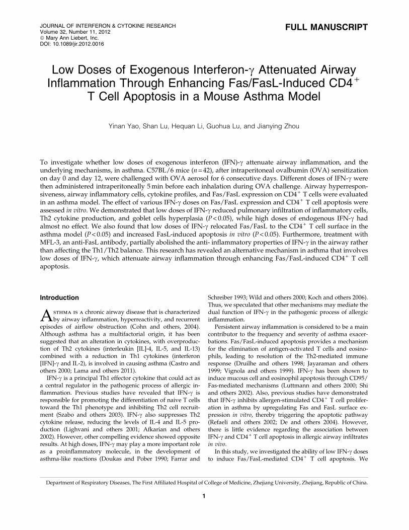
To examine the possible anti-inflammatory effect of IFN-gon allergic airway reactivity, we used a treatment protocol,including i.p. injection of different IFN-g doses of (10 U,100 U, or 1000 U/mouse/day), beginning on the first day ofOVA challenges. After sensitization and challenge withOVA, the total number of inflammatory cells (OVA/OVAversus control: 6.85 – 1.09 versus 0.31 – 0.02 · 105, P < 0.01),eosinophils (OVA/OVA versus control: 1.31 – 0.12 versus0 · 105, P < 0.01), and lymphocytes (OVA/OVA versus con-trol: 0.05 – 0.01 versus 2.85 – 0.43, · 105, P < 0.01) increasedsignificantly in BALF. A low dose of IFN-g noticeablyinhibited the OVA-induced recruitment of eosinophils(100 U/mouse versus OVA/OVA: 0.31 – 0.09 versus1.31 – 0.12, · 105, P < 0.01; 100 U/mouse versus 1000 U/mouse: 0.31 – 0.09 versus 0.55 – 0.07, · 105, P < 0.05) andlymphocyte (100 U/mouse versus OVA/OVA: 1.02 – 0.14versus 2.85 – 0.43, · 105, P < 0.05; 100 U/mouse versus1000 U/mouse: 1.02 – 0.14 versus 1.65 – 0.32, · 105, P < 0.05)into the BALF, whereas the low dose of IFN-g did not affectmacrophages and neutrophils (Fig. 1).
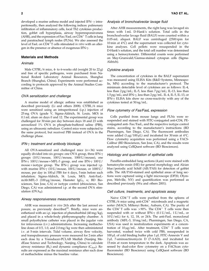
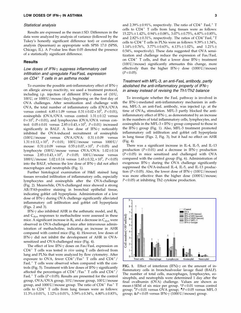
Further histological examination of H&E stained lungtissues revealed infiltration of inflammatory cells, especiallylymphocytes and eosinophils after the OVA-challenge(Fig. 2). Meanwhile, OVA-challenged mice showed a strongAB/PAS-positive staining in bronchial epithelial tissue,indicating goblet cell hyperplasia. Administration of a lowdose of IFN-g during OVA challenge significantly alleviatedinflammatory cell infiltration and goblet cell hyperplasia(Figs. 2 and 3).
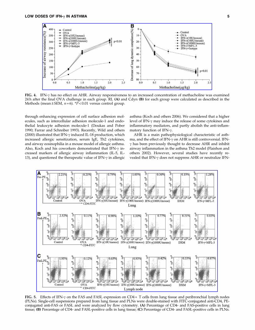
IFN-g also inhibited AHR in the asthmatic model, and RL
and Cdyn responses to methacholine were assessed in thesemice. A significant increase in RL and a decrease in Cdyn wereobserved in OVA-challenged mice after intravenous admin-istration of methacholine, indicating an increase in AHRcompared with control mice (Fig. 4). However, low doses ofIFN-g did not inhibit the development of AHR in OVA-sensitized and OVA-challenged mice (Fig. 4).
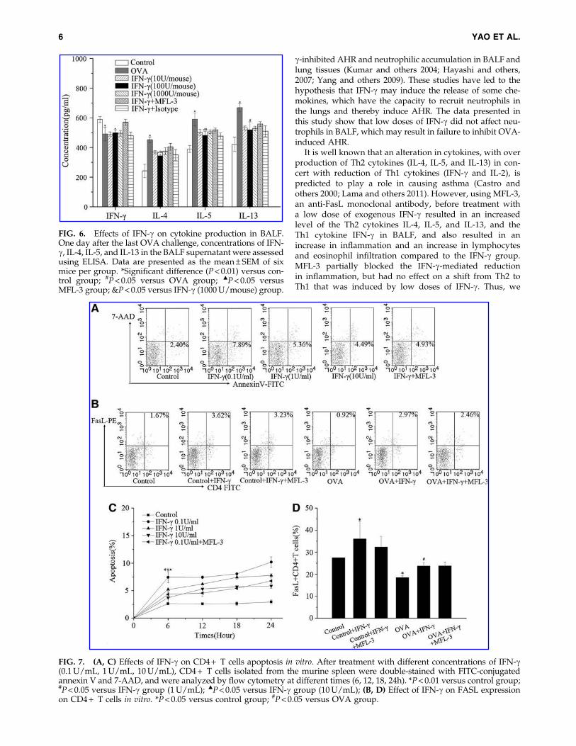
The effect of low IFN-g doses on Fas/FasL expression onCD4 + T cells was tested in vivo using T cells derived fromlung and PLNs that were analyzed by flow cytometry. Afterexposure to OVA, fewer CD4 + /Fas + T cells and CD4 + /FasL + T cells were observed when compared with the con-trols (Fig. 5). Treatment with low doses of IFN-g significantlyaffected the percentages of CD4 + /Fas + T cells and CD4 + /FasL + T cells (P < 0.05). Results are presented for the controlgroup, OVA/OVA group, 10 U/mouse group, 100 U/mousegroup, and 1000 U/mouse group. The ratio of CD4 + Fas + Tcells to CD4 + T cells from lung tissues were as follows:11.3% – 0.01%, 1.12% – 0.01%, 3.59% – 0.34%, 6.80% – 0.83%,
and 2.39% – 0.91%, respectively. The ratio of CD4 + FasL + Tcells to CD4 + T cells from lung tissues were as follows:15.22% – 1.42%, 0.94% – 0.08%, 3.07% – 0.75%, 6.87% – 0.85%,and 2.82% – 0.31%, respectively. The ratios of CD4 + FasL + Tcells to CD4 + T cells in PLNs were as follows: 9.39% – 1.34%,1.14% – 0.76%, 3.77% – 0.63%, 6.13% – 1.02%, and 1.21% –0.54%, respectively). These data suggested that OVA sensi-tization and challenge reduce the expression of Fas/FasLon CD4 + T cells, and that a lower dose IFN-g treatment(100 U/mouse) significantly attenuates this change, moreeffectively than the higher IFN-g dose (1000 U/mouse)(P < 0.05).
Treatment with MFL-3, an anti-FasL antibody, partlyabolished the anti-inflammatory property of IFN-gin airway instead of revising the Th1/Th2 balance
To investigate whether the FasL pathway is involved inthe IFN-g-mediated anti-inflammatory mechanism in asth-ma, MFL-3, an anti-FasL antibody, was injected i.p. at thetime of OVAII stimulation. MFL-3 partly blocked the anti-inflammatory effect of IFN-g, as demonstrated by an increasein the numbers of total inflammatory cells, lymphocytes, andeosinophils in the MFL-3 + IFN-g group compared to those inthe IFN-g group (Fig. 1). Also, MFL-3 treatment promotedinflammatory cell infiltration and goblet cell hyperplasiain lung tissue (Figs. 2, Fig. 3), but it had no effect on AHR(Fig. 4).
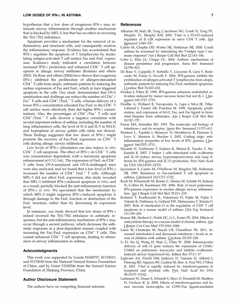
There was a significant increase in IL-4, IL-5, and IL-13production (P < 0.01) and a decrease in IFN-g production(P < 0.05) in mice sensitized and challenged with OVAcompared with the control group (Fig. 6). Administration ofexogenous IFN-g during the OVA challenge significantlysuppressed the OVA-induced IL-4, IL-5, and IL-13 produc-tion (P < 0.05). Also, the lower dose of IFN-g (100 U/mouse)was more effective than the higher dose (1000 U/mouse;P < 0.05) at inhibiting Th2 cytokine production.
FIG. 1. Effect of interferon (IFN)-g on the amount of in-flammatory cells in bronchoalveolar lavage fluid (BALF).The number of total cells, macrophages, lymphocytes, eo-sinophils, and neutrophils were determined 1 day after thefinal ovalbumin (OVA) challenge. Values are shown asmean – SEM of six mice per group. *P < 0.01 versus controlgroup; #P < 0.01 versus OVA group; :P < 0.05 versus MFL-3group; &P < 0.05 versus IFN-g (1000 U/mouse) group.
LOW DOSES OF IFN-c IN ASTHMA 3
MFL-3 treatment promoted Th2 cytokine production (IL-4,IL-5, IL-13) compared to mice that did not receive MFL-3(P < 0.05), and there was also an unexpectedly high level ofsecreted IFN-g in the MFL-3-treated mice (Fig. 6), suggestingthat MFL-3 does not influence the reversion of the Th1/Th2imbalance induced by low doses of IFN-g.
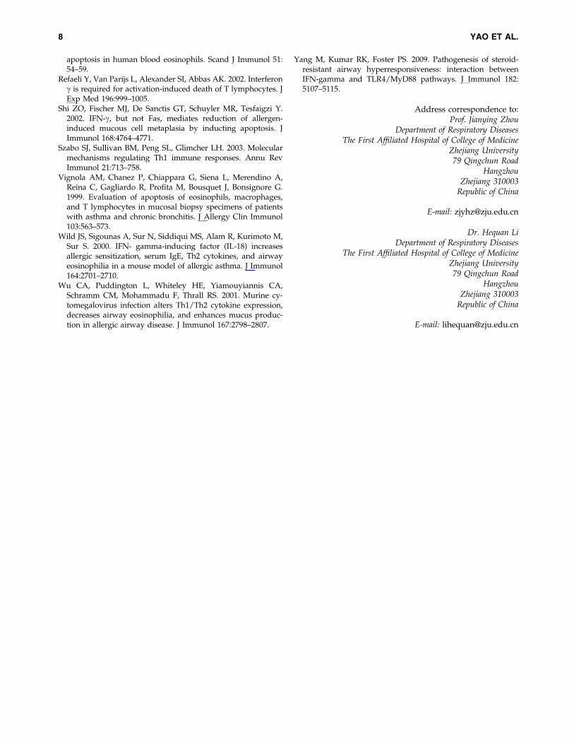
Low doses of IFN-g enhance FasL-inducedapoptosis in vitro
To determine whether low doses of IFN-g enhance FasL-induced apoptosis in vitro, we isolated CD4 + T cells, whichwere preincubated in the presence or absence of lower
(0.1 U/mL) or higher (1 U/mL or 10 U/mL) IFN-g levels. Asshown in Fig. 7, lower levels of IFN-g-induced more CD4 + Tcell apoptosis than did higher levels of IFN-g (P < 0.05) after6, 12, 18, and 24 h. We also assessed the effect of IFN-g onFasL and observed that lower levels of IFN-g upregulatedFasL expression.
MFL-3 partially inhibited IFN-g-induced apoptosis,but did not affect the expression of FasL
The relationship between IFN-g and Fas/FasL were alsoassessed in response to MFL-3 in experimental and controlmice. We observed no significant increase in the levels ofFasL (P > 0.05) in the lung and PLN of experimental mice(Fig. 5). In vitro, the percentage of apoptotic cells, but not thepercentage of FasL + CD4 + T cells, decreased after exposureto MFL-3, compared to the IFN-g group (P < 0.05).
Discussion
Results of our study showed that i.p. administration oflow doses of exogenous IFN-g during an OVA challengesignificantly reduced the numbers of different inflammatorycells in BALF, decreased Th2 cytokine production, and alle-viated goblet cell hyperplasia; these results suggest that IFN-g induced a shift from a Th2-skewed response to a morebalanced Th1/Th2 response. However, high doses of en-dogenous IFN-g had little effect on the airway inflammation.Also, the lower IFN-g dose inhibited inflammatory infiltra-tions more effectively than did the higher dose. Our resultsare similar to those of other previously published studies(Flaishon and others, 2002; Lama and others 2011).
The proinflammatory effect of IFN-g, at a relatively highlevel, has been shown by several researchers. Doukas andFarrar reported that IFN-g, in conjunction with TNF-a, canexpand and amplify the overall inflammatory response
FIG. 3. The percentages of positively stained epithelial ar-eas (AB/PAS). Data are presented as the mean – SEM ofsix mice per group. * Significant difference (P < 0.01) versuscontrol group; #P < 0.01 versus OVA group; :P < 0.01 versusMFL-3 group; &P < 0.05 versus IFN-g (1000 U/mouse) group.
FIG. 2. Lung histology of C56BL/6 mice treated with IFN-g. One day after the final challenge, lung tissues were obtained fromcontrol mice, OVA-sensitized mice and OVA-challenged mice, mice treated with IFN-g (100 U/mouse), mice treated with IFN-g andMFL-3 (100mg/mouse), and IFN-g and Isotype (100mg/mouse). Representative photomicrographs of hematoxylin–eosin (HE)-stained (A and B) and periodic acid Schiff (PAS)-stained (C) lung sections from each group were shown. Arrows in A1–5 showedinflammatory cells; Arrows in C1–5 showed PAS-positive areas. A1–5, magnification · 100; B1–5, magnification · 400; C1–5,magnification · 400.
4 YAO ET AL.
through enhancing expression of cell surface adhesion mol-ecules, such as intercellular adhesion molecule-1 and endo-thelial leukocyte adhesion molecule-1 (Doukas and Pober1990; Farrar and Schreiber 1993). Recently, Wild and others(2000) illustrated that IFN-g induced IL-18 production, whichincreased allergic sensitization, serum IgE, Th2 cytokines,and airway eosinophilia in a mouse model of allergic asthma.Also, Koch and his coworkers demonstrated that IFN-g in-creased markers of allergic airway inflammation (IL-5, IL-13), and questioned the therapeutic value of IFN-g in allergic
asthma (Koch and others 2006). We considered that a higherlevel of IFN-g may induce the release of some cytokines andinflammatory mediators, and partly abolish the anti-inflam-matory function of IFN-g.
AHR is a main pathophysiological characteristic of asth-ma, and the effect of IFN-g on AHR is still controversial. IFN-g has been previously thought to decrease AHR and inhibitairway inflammation in the asthma Th2 model (Flaishon andothers 2002). However, several studies have recently re-vealed that IFN-g does not suppress AHR or neutralize IFN-
FIG. 5. Effects of IFN-g on the FAS and FASL expression on CD4 + T cells from lung tissue and peribronchial lymph nodes(PLNs). Single-cell suspensions prepared from lung tissue and PLNs were double-stained with FITC-conjugated anti-CD4, PE-conjugated anti-FAS or FASL and were analyzed by flow cytometry. (A) Percentage of CD4- and FAS-positive cells in lungtissue; (B) Percentage of CD4- and FASL-positive cells in lung tissue; (C) Percentage of CD4- and FASL-positive cells in PLNs.
FIG. 4. IFN-g has no effect on AHR. Airway responsiveness to an increased concentration of methacholine was examined24 h after the final OVA challenge in each group. RL (A) and Cdyn (B) for each group were calculated as described in theMethods (mean – SEM, n = 6). *P < 0.01 versus control group.
LOW DOSES OF IFN-c IN ASTHMA 5
g-inhibited AHR and neutrophilic accumulation in BALF andlung tissues (Kumar and others 2004; Hayashi and others,2007; Yang and others 2009). These studies have led to thehypothesis that IFN-g may induce the release of some che-mokines, which have the capacity to recruit neutrophils inthe lungs and thereby induce AHR. The data presented inthis study show that low doses of IFN-g did not affect neu-trophils in BALF, which may result in failure to inhibit OVA-induced AHR.
It is well known that an alteration in cytokines, with overproduction of Th2 cytokines (IL-4, IL-5, and IL-13) in con-cert with reduction of Th1 cytokines (IFN-g and IL-2), ispredicted to play a role in causing asthma (Castro andothers 2000; Lama and others 2011). However, using MFL-3,an anti-FasL monoclonal antibody, before treatment witha low dose of exogenous IFN-g resulted in an increasedlevel of the Th2 cytokines IL-4, IL-5, and IL-13, and theTh1 cytokine IFN-g in BALF, and also resulted in anincrease in inflammation and an increase in lymphocytesand eosinophil infiltration compared to the IFN-g group.MFL-3 partially blocked the IFN-g-mediated reductionin inflammation, but had no effect on a shift from Th2 toTh1 that was induced by low doses of IFN-g. Thus, we
FIG. 6. Effects of IFN-g on cytokine production in BALF.One day after the last OVA challenge, concentrations of IFN-g, IL-4, IL-5, and IL-13 in the BALF supernatant were assessedusing ELISA. Data are presented as the mean – SEM of sixmice per group. *Significant difference (P < 0.01) versus con-trol group; #P < 0.05 versus OVA group; :P < 0.05 versusMFL-3 group; &P < 0.05 versus IFN-g (1000 U/mouse) group.
FIG. 7. (A, C) Effects of IFN-g on CD4 + T cells apoptosis in vitro. After treatment with different concentrations of IFN-g(0.1 U/mL, 1 U/mL, 10 U/mL), CD4 + T cells isolated from the murine spleen were double-stained with FITC-conjugatedannexin V and 7-AAD, and were analyzed by flow cytometry at different times (6, 12, 18, 24h). *P < 0.01 versus control group;#P < 0.05 versus IFN-g group (1 U/mL); :P < 0.05 versus IFN-g group (10 U/mL); (B, D) Effect of IFN-g on FASL expressionon CD4 + T cells in vitro. *P < 0.05 versus control group; #P < 0.05 versus OVA group.
6 YAO ET AL.
hypothesize that a low dose of exogenous IFN-g may at-tenuate airway inflammation through another mechanismthat is blocked by MFL-3, but that has no effect on reversingthe Th1/Th2 imbalance.
Apoptosis provides a mechanism for the removal of in-flammatory and structural cells, and consequently resolvesthe inflammatory response. Evidence has accumulated thatIFN-g regulates the apoptosis of T lymphocytes by modu-lating antigen-activated T cell surface Fas and FasL expres-sion. Kodama’s study indicated a correlation betweenincreased IFN-g production and enhanced CD4 + T cell ap-optosis in allergic airway infiltrates (Kodama and others2003). De Rose and others (2004) have shown that exogenousIFN-g inhibited the proliferation of allergen-stimulatedCD4 + T cells from atopic, asthmatic patients by inducing thesurface expression of Fas and FasL, which in turn triggeredapoptosis in the cells. Our study demonstrated that OVAsensitization and challenge can reduce the number of CD4 + /Fas + T cells and CD4 + /FasL + T cells, whereas delivery of alower IFN-g concentration relocated Fas/FasL to the CD4 + Tcell surface more effectively than did higher IFN-g concen-trations. Also, the percentages of CD4 + /Fas + T cells andCD4 + /FasL + T cells showed a negative correlation withseveral important indices of asthma, including the number oflung inflammatory cells, the level of IL-4 and IL-5 in BALF,and hyperplasia of airway goblet cells (data not shown).These findings suggested that low doses of IFN-g mightpromote the recovery of Fas/FasL expression on CD4 + Tcells during allergic airway infiltration.
Low levels of IFN-g stimulation can also induce in vitroCD4 + T cell apoptosis. The effect of IFN-g on CD4 + T cellswas concentration dependent, with a maximum apoptosisenhancement at 0.1 U/mL. The expression of FasL on CD4 +
T cells from OVA-sensitized and OVA-challenged micewere also analyzed, and we found that a low level of IFN-gincreased the number of CD4 + FasL + T cells. AlthoughMFL-3 did not affect FasL expression, this study revealedthat MFL-3 inhibited IFN-g-induced apoptosis in vitro, andas a result, partially blocked the anti-inflammatory functionof IFN-g in vivo. We speculated that the mechanism bywhich MFL-3 might suppress IFN-g-induced apoptosis isthrough damage to the FasL function or destruction of theFasL structure, rather than by decreasing its expressionlevel.
In summary, our study showed that low doses of IFN-gindeed reversed the Th1/Th2 imbalance in asthmatic re-sponses, but the anti-inflammatory mechanism of IFN-g mayoccur through a second pathway, which decreases the asth-matic responses in a dose-dependent manner coupled withincreasing the Fas/FasL expression on CD4 + T cells. Thiscaused enhanced CD4 + T cell apoptosis, leading to attenu-ation of airway inflammation in asthma.
Acknowledgments
This work was supported by Grants 81000757, 81170015,and 81170038 from the National Natural Science Foundationof China, and by Grant Y2100346 from the Natural ScienceFoundation of Zhejiang Province, China.
Author Disclosure Statement
The authors have no competing financial interests.
References
Afkarian M, Sedy JR, Yang J, Jacobson NG, Cereb N, Yang SY,Murphy TL, Murphy KM. 2002. T-bet is a STAT1-inducedregulator of IL-12R expression in naive CD4 + T cells. NatImmunol 3:549–557.
Castro M, Chaplin DD, Walter MJ, Holtzman MJ. 2000. Couldasthma be worsened by stimulating the T-helper type 1 im-mune response? Am J Respir Cell Mol Biol 22:143–146.
Cohn L, Elias JA, Chupp GL. 2004. Asthma: mechanisms ofdisease persistence and progression. Annu Rev Immunol22:789–815.
De Rose V, Cappello P, Sorbello V, Ceccarini B, Gani F, Bosti-cardo M, Fassio S, Novelli F. 2004. IFN-gamma inhibits theproliferation of allergen-activated T lymphocytes from atopic,asthmatic patients by inducing Fas/FasL-mediated apoptosis.J Leukoc Biol 76:423–432.
Doukas J, Pober JS. 1990. IFN-gamma enhances endothelial ac-tivation induced by tumor necrosis factor but not IL-1. J Im-munol 145:1727–1733.
Druilhe A, Wallaert B, Tsicopoulos A, Lapa e Silva JR, Tillie-Leblond I, Tonnel AB, Pretolani M. 1998. Apoptosis, prolif-eration, and expression of bcl-2, Fas, and Fas ligand in bron-chial biopsies from asthmatics. Am J Respir Cell Mol Biol19:747–757.
Farrar MA, Schreiber RD. 1993. The molecular cell biology ofinterferon-g and its receptor. Annu Rev Immunol 11:571–611.
Flaishon L, Topilski I, Shoseyov D, Hershkoviz R, Fireman E,Levo Y, Marmor S, Shachar I. 2002. Cutting edge: anti-inflammatory properties of low levels of IFN- gamma. J Im-munol 168:3707–3711.
Hayashi N, Yoshimoto T, Izuhara K, Matsui K, Tanaka T, Na-kanishi K. 2007. T helper 1 cells stimulated with ovalbuminand IL-18 induce airway hyperresponsiveness and lung fi-brosis by IFN-gamma and IL-13 production. Proc Natl AcadSci USA 104:14765–14770.
Jayaraman S, Castro M, O’Sullivan M, Bragdon MJ, HoltzmanMJ. 1999. Resistance to Fas-mediated T cell apoptosis inasthma. J Immunol 162:1717–1722.
Koch M, Witzenrath M, Reuter C, Herma M, Schutte H, SuttorpN, Collins H, Kaufmann SH. 2006. Role of local pulmonaryIFN-gamma expression in murine allergic airway inflamma-tion. Am J Respir Cell Mol Biol 35:211–219.
Kodama T, Kuribayashi K, Nakamura H, Fujita M, Fujita T,Takeda K, Dakhama A, Gelfand EW, Matsuyama T, Kitada O.2003. Role of interleukin-12 in the regulation of CD4 + T cellapoptosis in a mouse model of asthma. Clin Exp Immunol131:199–205.
Kumar RK, Herbert C, Webb DC, Li L, Foster PS. 2004. Effects ofanticytokine therapy in a mouse model of chronic asthma. AmJ Respir Crit Care Med 170:1043–1048.
Lama M, Chatterjee M, Nayak CR, Chaudhuri TK. 2011. In-creased interleukin-4 and decreased interferon-g levels in se-rum of children with asthma. Cytokine 55:335–338.
Li H, Xie Q, Wang H, Shao C, Chen W. 2008. Intramusculardelivery of mIL-12 gene reduces the expression of CD44/CD49d on pulmonary leucocytes and inhibits ovalbumin-induced airway hyperreactivity. Inflam Res 57:11–17.
Lighvani AA, Frucht DM, Jankovic D, Yamane H, Aliberti J,Hissong BD, Nguyen BV, Gadina M, Sher A, Paul WE, O’SheaJJ. 2001. T-bet is rapidly induced by interferon-gamma inlymphoid and myeloid cells. Proc Natl Acad Sci USA98:15137–15142.
Luttmann W, Dauer E, Schmidt S, Marx O, Hossfeld M, MatthysH, Virchow JC Jr. 2000. Effects of interferon-gamma and tu-mor necrosis factor-alpha on CD95/Fas ligand-mediated
LOW DOSES OF IFN-c IN ASTHMA 7
apoptosis in human blood eosinophils. Scand J Immunol 51:54–59.
Refaeli Y, Van Parijs L, Alexander SI, Abbas AK. 2002. Interferong is required for activation-induced death of T lymphocytes. JExp Med 196:999–1005.
Shi ZO, Fischer MJ, De Sanctis GT, Schuyler MR, Tesfaigzi Y.2002. IFN-g, but not Fas, mediates reduction of allergen-induced mucous cell metaplasia by inducting apoptosis. JImmunol 168:4764–4771.
Szabo SJ, Sullivan BM, Peng SL, Glimcher LH. 2003. Molecularmechanisms regulating Th1 immune responses. Annu RevImmunol 21:713–758.
Vignola AM, Chanez P, Chiappara G, Siena L, Merendino A,Reina C, Gagliardo R, Profita M, Bousquet J, Bonsignore G.1999. Evaluation of apoptosis of eosinophils, macrophages,and T lymphocytes in mucosal biopsy specimens of patientswith asthma and chronic bronchitis. J Allergy Clin Immunol103:563–573.
Wild JS, Sigounas A, Sur N, Siddiqui MS, Alam R, Kurimoto M,Sur S. 2000. IFN- gamma-inducing factor (IL-18) increasesallergic sensitization, serum IgE, Th2 cytokines, and airwayeosinophilia in a mouse model of allergic asthma. J Immunol164:2701–2710.
Wu CA, Puddington L, Whiteley HE, Yiamouyiannis CA,Schramm CM, Mohammadu F, Thrall RS. 2001. Murine cy-tomegalovirus infection alters Th1/Th2 cytokine expression,decreases airway eosinophilia, and enhances mucus produc-tion in allergic airway disease. J Immunol 167:2798–2807.
Yang M, Kumar RK, Foster PS. 2009. Pathogenesis of steroid-resistant airway hyperresponsiveness: interaction betweenIFN-gamma and TLR4/MyD88 pathways. J Immunol 182:5107–5115.
Address correspondence to:Prof. Jianying Zhou
Department of Respiratory DiseasesThe First Affiliated Hospital of College of Medicine
Zhejiang University79 Qingchun Road
HangzhouZhejiang 310003
Republic of China
E-mail: [email protected]
Dr. Hequan LiDepartment of Respiratory Diseases
The First Affiliated Hospital of College of MedicineZhejiang University79 Qingchun Road
HangzhouZhejiang 310003
Republic of China
E-mail: [email protected]
8 YAO ET AL.


![Journal of Inflammation BioMed Central · 2017. 8. 23. · FasL on the endothelium attenuates leukocyte extravasa-tion [5]. FasL over-expression on the endothelium of arteries also](https://img.pdfslide.net/doc/110x75/60b2be4b9c6d3554342c1db0/journal-of-inflammation-biomed-central-2017-8-23-fasl-on-the-endothelium-attenuates.jpg)