Embed Size (px)
Citation preview

LOWER BACK PAINLOWER BACK PAIN
Erestain-Garan

CASECASEAge: 45 years oldCC: Lower back painOccupation: office secretary

CHRONOLOGY OF EVENTSCHRONOLOGY OF EVENTS

NEUROLOGIC EXAMINATION NEUROLOGIC EXAMINATION FINDINGSFINDINGS

NEUROLOGIC EXAMINATION NEUROLOGIC EXAMINATION FINDINGSFINDINGS
Patient in left lateral decubitus with knee flexedNumbness: back of the right calf muscle,
lateral heel, foot and toeWeakness: right plantar flexion of foot and toesDifficulty of walking on toes on the rightAtrophy: Right gastrocnemius and soleus
musclesKnee jerk: (++) both right and leftAnkle jerk: (++) left; (absent) rightBabinski: (-) both right and leftThe rest of the neurological exam is within
normal limit

SALIENT FEATURESSALIENT FEATURESAge: 45 years oldsudden snap and pain in the LEFT LUMBAR
AREAPatient in left lateral decubitus with knee
flexedNumbness: back of the right calf muscle,
lateral heel, foot and toeWeakness: right plantar flexion of foot and
toesDifficulty of walking on toes on the rightAtrophy: Right gastrocnemius and soleus
musclesKnee jerk: (++) both right and leftAnkle jerk: (++) left; (absent) rightBabinski: (-) both right and left

Nature of the ProblemNature of the Problem

Diagnosis of Spinal InjuryDiagnosis of Spinal InjurySpinal Injury is present if:
◦The person complains of severe pain in his or her neck or back.
◦An injury has exerted substantial force on the back or head.
◦The person complains of weakness, numbness or paralysis or lacks control of his or her limbs, bladder or bowel.
◦The neck or back is twisted or positioned oddly.

Spinal Cord InjurySpinal Cord Injury
Possibility of a SCI ◦Pain◦Numbness ◦Difficulty with limb
movements

Nature of the ProblemNature of the ProblemNon-traumatic Spinal Cord Injury
◦Age◦Pain – left lumbar area◦ Resulted from normal physical
strain (lifting a box of papers)

Lumbar SpineLumbar SpineVarious forces that could be
applied on the spine
•nerve pathways narrowing and causing nerve impingement, inflammation, and pain

Key muscles = level of Key muscles = level of injuryinjury C5 - Elbow flexors (biceps, brachialis) C6 - Wrist extensors (extensor carpi radialis longus
and brevis) C7 - Elbow extensors (triceps) C8 - Finger flexors (flexor digitorum profundus) to the
middle finger T1 - Small finger abductors (abductor digiti minimi) L2 - Hip flexors (iliopsoas) L3 - Knee extensors (quadriceps) L4 - Ankle dorsiflexors (tibialis anterior) L5 - Long toe extensors (extensors hallucis longus) S1 - Ankle plantar flexors (gastrocnemius, soleus)

•Numbness in the back of the R calf muscle (S1-S2)•Atrophy of R gastrocnemius and soleus
Numbness in the lateral heel (S1)
Numbness in the foot and toe(L4-L5, S1-S2)

Numbness in the foot and toe(L4-L5, S1-S2)
Weakness of the R plantar flexion of foot and toes
Difficulty walking on toes on the R

What is the difference between a What is the difference between a radicular and myelopathic radicular and myelopathic
manifestations and what is the manifestations and what is the significance of each in relation to significance of each in relation to
the signs and symptoms and clinical the signs and symptoms and clinical management?management?

Radiculopathy◦ Pain and numbness involving the
degeneration or inflammation of the spinal nerve roots
◦ usually without objective signs of neurologic dysfunction
Myelopathy◦ involves degeneration or any
disease of the spinal cord

In Radiculopathy, compression of single root may not cause significant sensory loss (due to overlap of dermatomes in the body)
The main symptom: sharp, burning pain or “shooting pains”.
Follow a dermatomal distribution, accompanied by paresthesias, and loss of muscle power innervated by the root.

Symptoms of myelopathy would usually depend on the cause and severity of the condition
Trauma, herniated disc, OA of the spine, and tumors cause myelopathy
Symptoms: pain, loss of sensation or movement, decreased spinal range of motion, weakness, and deformity

Upon PE, clonus would usually indicate an UMN disorder

SignificanceSignificanceFrom the signs and symptoms
along with the PE results, we can achieve the correct diagnosis of our patient.
The persisting signs and symptoms would help us determine the appropriate mangement.

How does one localize the How does one localize the lesion based on anatomical lesion based on anatomical diagnosis and other diagnosis and other ancillary procedures?ancillary procedures?

Lower Motor Neuron Lower Motor Neuron LesionLesionIn the Case:
◦Patient has the following LMN symptoms: (+) Atrophy of right gastrocnemius
muscle and soleus muscles (+) Areflexia in Right ankle jerk (-) Babinski bilaterally

Peripheral Nerve LesionPeripheral Nerve Lesion
WEAKNESS Distal, symmetrical
OBJECTIVE/SENSORY DEFICITS Distal, symmetical
AUTONOMIC DISTURBANCES May be present
REFLEXES Areflexia

Lumbar Radiculopathy L5-Lumbar Radiculopathy L5-S1S1
◦In the patient:Numbness in the back of the
right calf muscle, lateral heel, foot and toe
Weakness of the right plantar flexion of foot and toes
Difficulty walking on toes on the right
Right ankle jerk absent

Weakness of the right plantar flexion of foot and toes

Numbness in back of the right calf muscle, lateral heel, foot and toe

Both knee jerks are (++)Right ankle jerk absent, left ankle jerk (++)
Numbness in lateral heel, foot and toe

DiagnosisDiagnosis If no improvement in symptoms have occurred in six
weeks or red flags are present, imaging is appropriate.
CT scan used to evaluate the bony anatomy in the lumbar spine, which can show how much space is available for the nerve roots.◦ NEUROFORAMEN- vulnerable point of compression
MRI scan useful for determining where the nerve roots are being compressed ; shows the details of soft-tissue structures, like nerves and discs.

DiagnosisDiagnosisMR neurography
◦modified MRI technique providing better pictures of the spinal nerves and the effect of compression on these nerves.
◦may help in diagnosis and treatment of sciatica/lumbar radiculopathy.

TreatmentTreatmentgoals of treatment :
◦ relieve pain◦prevent or reduce stress on the disc◦maintain normal function
ranges from conservative therapies to surgical interventions
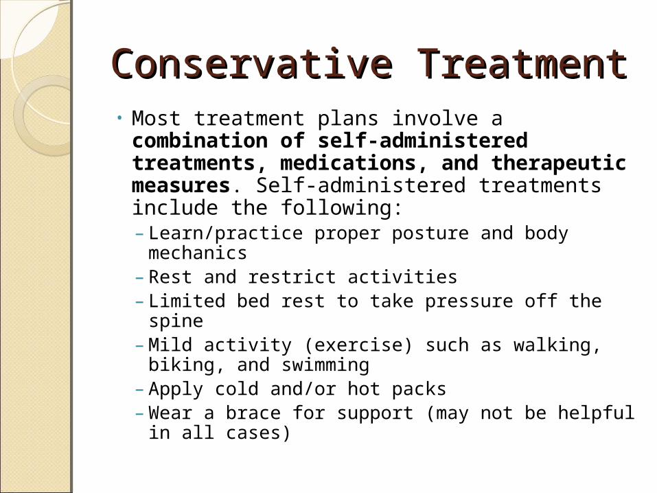
Conservative TreatmentConservative Treatment• Most treatment plans involve a
combination of self-administered treatments, medications, and therapeutic measures. Self-administered treatments include the following: – Learn/practice proper posture and body
mechanics – Rest and restrict activities – Limited bed rest to take pressure off the spine – Mild activity (exercise) such as walking, biking,
and swimming – Apply cold and/or hot packs – Wear a brace for support (may not be helpful in
all cases)

Conservative TreatmentConservative TreatmentTherapeutic treatments for DDD
include the following: ◦Chiropractic treatment to manipulate
the spine ◦Acupuncture to relieve pain ◦Massage therapy to relieve muscle
spasms and tension ◦Physical therapy to improve function
and increase flexibility and strength

MedicationsMedications• Are used to supplement conservative therapy.
– Non-steroidal anti-inflammatory drugs (NSAIDs; e.g., aspirin, ibuprofen, naproxen)
– Pain relievers (e.g., acetaminophen) – Muscle relaxants – Spinal injections (anesthetics or corticosteroids) – Antidepressants – Sleep aids
• Other non-surgical treatments– ultrasound therapy : uses sound waves to warm
the area, increase blood flow, and relieve discomfort
– transcutaneous electrical nerve stimulation (TENS): uses electrical stimulation of the nerve to interrupt pain signals

SurgicalSurgicalPrimary reasons for surgery are
to: ◦relieve pressure on a nerve root or
the spinal cord ◦stabilize an unstable or painful
vertebral segment◦prevent or limit radiculopathy (nerve
damage) ◦reduce deformity or curvature of the
spine (e.g., scoliosis)

SurgicalSurgical• Discectomy and fusion
– involves removing the damaged intervertebral disc and replacing it with a piece of bone or another material
– this replacement fuses with the adjacent vertebrae
• Corpectomy – a section of the vertebrae and discs is removed to create
more space for the remainder of the spine– A bone graft and/or metal plate with screws – attached to
stabilize the spine
• Facetectomy, laminotomy, and spinal laminectomy – procedures that involve removing a portion of the bony
structure of the spine to relieve pressure on the nerve roots
– Foraminotomy and laminoplasty can be used to enlarge areas of the spinal column to make more room for the nerves and spinal cord
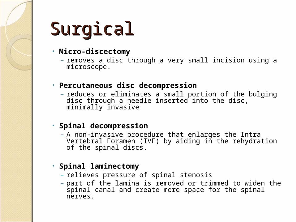
SurgicalSurgical• Micro-discectomy
– removes a disc through a very small incision using a microscope.
• Percutaneous disc decompression– reduces or eliminates a small portion of the bulging disc
through a needle inserted into the disc, minimally invasive
• Spinal decompression– A non-invasive procedure that enlarges the Intra
Vertebral Foramen (IVF) by aiding in the rehydration of the spinal discs.
• Spinal laminectomy– relieves pressure of spinal stenosis – part of the lamina is removed or trimmed to widen the
spinal canal and create more space for the spinal nerves.

Indications for SurgeryIndications for Surgery
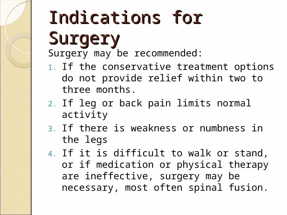
Indications for SurgeryIndications for SurgerySurgery may be recommended:1. If the conservative treatment options do
not provide relief within two to three months.
2. If leg or back pain limits normal activity3. If there is weakness or numbness in the
legs4. If it is difficult to walk or stand, or if
medication or physical therapy are ineffective, surgery may be necessary, most often spinal fusion.

Lumbar surgery ◦ indicated in patients with severe spinal stenosis, in those with
intractable pain, and in patients in whom an appropriate 6- to 12-month nonoperative course of treatment fails.
In elective cases, other conservative modalities should have been tried and observed to fail.
In cases of cervical disk disease with radiculopathy◦ indications for surgical treatment are intractable pain, progressive
motor or sensory deficit, or symptoms refractory in a reasonable period of nonoperative therapy
In cases of cervical disk disease with myelopathy◦ early surgery to decompress the spinal cord is recommended to arrest
progression if the clinical and radiographic changes are well correlated

Thank You!Thank You!
References:◦ http://www.cedars-sinai.edu/515.html◦ http://www.cedars-sinai.edu/889.html◦ http://www.cedars-sinai.edu/5757.html◦ http://www.neurologychannel.com/
degenerative-disc-disease/treatment.shtml◦ http://www.dcmsonline.org/jax-medicine/
1999journals/april99/degenerative.htm◦ http://emedicine.medscape.com/article/
1265453-treatment