Embed Size (px)
Citation preview

29 c h a p t e r
Lower Extremity Venous DiseaseJames Laredo, MD, PhD, and Anton N. Sidawy, MD, MPH
Lower extremity venous disease is extremely com-mon, with varicose veins remaining the most frequently encountered venous condition followed by chronic venous insuffi ciency.1,2 The two conditions often occur together, but each condition may also be present clini-cally without the other. Lower extremity venous disease comprises a clinical spectrum ranging from completely asymptomatic telangiectasias to symptomatic varicose veins to debilitating venous ulcers.1,2 The most frequently encountered symptoms associated with varicose veins include leg swelling, pain, itching, nocturnal cramping, and leg heaviness. Patients with chronic venous insuffi -ciency often present with leg edema, skin hyperpigmen-tation, stasis dermatitis of the skin involving the ankles, fi brosis of the subcutaneous fat (lipodermatosclerosis), and ulceration.1,2 Lower extremity venous ulceration remains a signifi cant worldwide health problem resulting in signifi cant morbidity.1,2
Deep venous thrombosis (DVT) of the lower extremi-ties is another highly prevalent venous condition encoun-tered by health care providers of all disciplines.3 Much has been published regarding venous thromboembolic dis-ease4-8; therefore, the content of this chapter is limited to the intrinsic venous disorders of the lower extremities.
• EPIDEMIOLOGY
Prevalence estimates of lower extremity venous disease vary widely by geographic location, with the highest reported rates observed in Western countries. The variabil-ity in estimates is likely attributable to population differ-ences in risk factor distribution, methods of measurement, variability in diagnosis, and disease defi nition.1,2,9,10 The prevalence of lower extremity varicose veins is estimated
to be as high as 56% in men and 73% in women.9,10 The prevalence of chronic venous insuffi ciency is estimated to be as high as 17% in men and 40% in women.10 Lower extremity venous ulceration has been reported to occur in approximately 0.3% to 1% of the adult population worldwide.9,11
Risk factors associated with the development of lower extremity venous disease are listed in Table 29-1. Family history, female gender, age, and pregnancy have been well established as risk factors for developing varicose veins.9,10 These risk factors have also been shown to contribute to the development of chronic venous insuffi ciency.9,10 Regarding the development of venous ulceration, in addition to the risk factors shown in Table 29-1, history of lower extrem-ity phlebitis, lower extremity trauma, DVT, and congestive heart failure have been shown to be associated with the development of venous ulceration.11
Chronic venous insuffi ciency with venous ulceration is extremely prevalent in the United States and is also the seventh leading cause of chronic debilitating disease.2 Approximately 10% to 35% of the U.S. population has some form of chronic venous insuffi ciency.1,2 More than 500,000 men and women experience chronic venous ulcers.1,2,11 Venous ulcers are associated with signifi cant health care costs and substantial economic effects in terms of days of work lost and diminished quality of life.2,11 The population-based cost in the United States for treatment of chronic venous insuffi ciency and venous ulcers has been estimated to be more than $1 billion a year.2 In addition, more than 6 million days of work are lost each year because of complications associated with chronic venous ulcers.11 Furthermore, patients’ quality of life is signifi cantly impacted by the loss of workdays and debilitating symptoms.2,11
AU: WITH ok as changed here ?Chronic venous insuffi ciency WITH venous ulceration ……….debilitating disease
29_Dieter_Ch29_p001-026.indd 129_Dieter_Ch29_p001-026.indd 1 4/26/10 6:28:48 PM4/26/10 6:28:48 PM

2 • CHAPTER 29
• EMBRYOLOGY
The development of the blood vessels occurs between the third and eight weeks of embryonic gestation.12 The primitive circulation begins to develop toward the end of the third week after appearance of the newly fused heart. This is followed by rapid changes in the fourth week when extensive remodeling occurs and continues through the fi nal month of the embryonic period.12 Development at the cephalad end of the embryo proceeds more rapidly than at the caudal end as the arteries and veins change and interact with the growing thoracoabdominal organs, pari-etes, and extremities.12
Primitive vascular channels appear in the limb during the third gestational week, when a capillary network is initially present during the undifferentiated stage.13 This is followed by large plexiform structures present during the
retiform stage, and fi nally, large channels, arteries, and veins appear during the maturation stage.12,13
The earliest veins that develop are the vitelline veins from the yolk sac, the umbilical veins from the chorion, and the cardinal veins from the body proper.12 Venous developmental changes are more complex than arte-rial changes and involve additions, deletions, intercon-nections, positions, and fl ow changes.12 The vitelline and umbilical veins eventually become the hepatic sinusoids, hepatic veins, portal vein, superior mesenteric vein, and left umbilical vein.12 The paired cardinal veins undergo a series of changes leading to the mature venous drainage of the body.12,13 The left-sided cardinal veins regress, leaving the right-sided veins, which become the superior vena cava (SVC) and inferior vena cava (IVC)12,13 (Figure 29-1). The distal ends of the post cardinal veins persist after regression and eventually become the iliac veins.
Persistence of the left subcardinal vein results in a dou-ble IVC in as many as 2% to 3% of individuals and a single left-sided IVC in 0.2% to 0.5%.13,14 Renal vein anomalies include a retroaortic left renal vein (2%) with or without a normal anterior left renal vein and a circumaortic renal collar (1%).13,14
Persistence of embryonic veins after birth results in venous malformations of the pelvis and lower extremi-ties.13 The marginal vein, often seen in patients with Klippel-Trenaunay syndrome, is a persistent large, lateral, superfi cial embryonic vein that contributes to the devel-opment of chronic venous insuffi ciency.13 Other devel-opmental anomalies include a persistent sciatic vein, valvular agenesis, venous aneurysms, and primary valvular insuffi ciency.13
AU: Do you mean positions and fl ow changes here?
• FIGURE 29-1. Embryology of the venous system. Development of the inferior vena cava and iliac veins.
(Adapted from Valentine RJ, Wind GG: Embryology of the arteries and veins. In Valentine RJ, Wind GG (eds). Anatomic Exposures in Vascular Surgery, 2nd ed. Philadelphia: Lippincott Williams and Wilkins, 2003:1–19.; with permission.)
4 weeks 6 weeks 7 weeks 8 weeks
Caudalextension ofhepatic v’s
Sub-cardinal v. Sub-
supra-anastomosis
Illiac v’s
Renal v’s
IVC
Azygous v.
Supra-cardinal v’s
Intersubcardinalanastomosis
Postcardinal v’s
TABLE 29-1. Risk Factors Associated with the Development of Lower Extremity Venous Disease
Older age
Female gender
Pregnancy
Family history of venous disease
Obesity
Prolonged standing
29_Dieter_Ch29_p001-026.indd 229_Dieter_Ch29_p001-026.indd 2 4/26/10 6:28:49 PM4/26/10 6:28:49 PM

LOWER EXTREMITY VENOUS DISEASE • 3
• ANATOMY
The muscle fascia divides the lower extremity soft tissues into a superfi cial compartment and deep compartment (Figure 29-2). The venous system of the lower extremi-ties include both the deep veins that lie below the mus-cular fascia and drain blood directly into the IVC and the superfi cial veins that lie above the muscular fascia and drain the superfi cial compartment. The saphenous fascia covers the saphenous subcompartment and separates the great saphenous vein and small saphenous vein from other veins in the superfi cial compartment. Connecting the two venous systems are the perforating veins that traverse the muscular fascia and normally drain blood from the superfi -cial system into the deep venous system (see Figure 29-2).15 Also present within the superfi cial compartment is the reticular venous plexus and subpapillary venous plexus. Communicating veins connect veins within the same com-partment.13 The deep, superfi cial, and most perforating veins contain bicuspid valves that maintain unidirectional venous fl ow. The nomenclature of the lower extremities veins has recently been updated (Table 29-2).13,15
The deep veins comprise the venous component of the neurovascular bundle of the lower extremities. The deep veins are adjacent to the similarly named arteries and often occur as paired structures at the popliteal, calf, and ankle levels (Figure 29-3).13,15 The major pelvic veins include the common iliac, internal iliac, and external iliac veins, all of which drain directly into the IVC. The overlying right com-mon iliac artery may compress the left common iliac vein,
resulting in left iliofemoral vein thrombosis (May-Thurner syndrome), which is often caused by a chronic stenosis or occlusion of the vein.13
The superfi cial veins include the subpapillary venous plexus, the reticular venous plexus, and all of the veins in the superfi cial compartment (see Figure 29-2). The great saphenous vein and its tributaries comprise the major superfi cial venous system of the thigh and medial leg (Figure 29-4). The small saphenous vein and its tributaries comprise the major superfi cial venous system of the lateral leg (see Figure 29-4). The great saphenous vein drains into the common femoral vein at the confl uence of the super-fi cial inguinal veins (formerly known as the saphenofem-oral junction). The confl uence of the superfi cial inguinal veins is made up of the great saphenous vein, superfi cial circumfl ex iliac, superfi cial epigastric, and external puden-dal veins.13,15 The small saphenous vein originates from the lateral side of the foot and drains into the popliteal vein. Below the level of the gastrocnemius muscle, the small saphenous vein runs adjacent to the sural nerve, which is prone to injury during surgical stripping procedures.
The perforating veins traverse the muscle fascia con-necting the superfi cial venous system to the deep venous system (Figure 29-5). Competent valves within the perfo-rating veins ensure superfi cial to deep, unidirectional fl ow in the calf and thigh. Perforating vein valvular incompe-tence may result in venous congestion, varicosities, and chronic skin changes, including ulceration.1,2,13,15 The most important perforating veins of the lower extremity are the medial calf perforators.13,15 There are two main groups of
• FIGURE 29-2. Relationship between the fascia and veins of the lower extremity. The fascia covers the muscle and separates the deep from the superfi cial compartment. Super-fi cial veins (a) drain the subpapillary and reticular venous plexuses and are connected to deep veins through perforating veins (b). The saphenous fascia invests the saphenous vein. The saphenous compartment is a subcompartment of the superfi cial compartment.
(From Mozes G, Gloviczki P: New discoveries in anatomy and new terminology of leg veins: clinical implications. Vasc Endovasc Surg, 2004;38:367–374; with permission.)
Epidermis
a
a
b
b
Subpapillaryvenous plexus
Reticularvenous plexus
Saphenousfascia
Deep veins
Dermis
Subcutis
Fascia
Muscle
Dee
pco
mpa
rtm
entS
aphe
nous
com
part
men
t
Sup
erfic
ial
com
part
men
t
DISTAL
PROXIMAL
29_Dieter_Ch29_p001-026.indd 329_Dieter_Ch29_p001-026.indd 3 4/26/10 6:28:49 PM4/26/10 6:28:49 PM

4 • CHAPTER 29
TABLE 29-2. The Most Common “Old” Anatomic Terms Describing Lower Extremity Veins and Their “New” Counterparts15
Old Term New Term
Greater or long saphenous vein Great saphenous vein (GSV)
Smaller or short saphenous vein Small saphenous vein (SSV)
Saphenofemoral junction Confl uence of the superfi cial inguinal veins
Giacomini’s vein Intersaphenous vein
Posterior arch or Leonardo’s vein Posterior accessory great saphenous vein of the leg
Superfi cial femoral fein Femoral vein
Cockett perforators (I, II, III) Posterior tibial perforators (lower, middle, upper)
Boyd’s perforators Paratibial perforators
Sherman’s perforators Paratibial perforators
“24-cm” perforators Paratibial perforators
Hunter’s and Dodd’s perforators Perforators of the femoral canal
May’s or Kuster’s perforators Ankle perforators
• FIGURE 29-3. The deep veins of the lower extremities.
(From Mozes G, Gloviczki P: New discoveries in anatomy and new terminology of leg veins: clinical implications. Vasc Endovasc Surg, 2004;38:367–374; with permission.)
• FIGURE 29-4. The superfi cial veins of the lower extremities. (A) The great saphenous vein and tributaries. Note the course of the vein running medially from the groin to the medial ankle. (B) The small saphenous vein (SSV) and tribu-taries. Note the close proximity of the SSV to the sural nerve. The SSV usually arises from the popliteal fossa and continues down to the lateral ankle.
A B
29_Dieter_Ch29_p001-026.indd 429_Dieter_Ch29_p001-026.indd 4 4/26/10 6:28:50 PM4/26/10 6:28:50 PM

LOWER EXTREMITY VENOUS DISEASE • 5
medial calf perforators, the posterior tibial and the more proximal paratibial perforating veins. The posterior tibial perforating veins (Cockett perforators) connect the pos-terior accessory great saphenous vein with the posterior tibial veins (see Figure 29-5).
• CEAP CLASSIFICATION OF LOWER EXTREMITY VENOUS DISEASE
The CEAP (clinical, etiology, anatomy, pathophysiology) classifi cation of chronic venous disorders was developed and adopted worldwide to facilitate meaningful communi-cation about chronic venous disorders and to serve as a basis for more scientifi c analysis of treatment alternatives.16,17 An international ad hoc committee of the American Venous Forum developed the fi rst CEAP consensus document in 1994.16 This document was later revised in 2004.17
The CEAP classifi cation includes a description of the clinical class (C) based on objective fi ndings—the etiology (E); the anatomical (A) distribution of the affected veins
in the superfi cial, deep, and perforating venous systems; and the underlying pathophysiology (P), which is attribut-able to refl ux, obstruction, or both (Table 29-3).17 Basic CEAP classifi cation designates only the highest clinical (C) classifi cation based on symptoms. Advanced CEAP clini-cal classifi cation includes all symptoms present and fur-ther designates any of 18 involved venous segments with the pathophysiology (P) (Table 29-4).17 A symptomatic patient presenting with varicose veins, leg pain, leg edema, and lipodermatosclerosis and is found to have refl ux of the great saphenous veins above and below the knees bilaterally, with incompetent calf perforating veins, would have a basic CEAP classifi cation of C4bs, Ep, As,p, Pr, and an advanced CEAP classifi cation of C2,3,4bs, Ep, As,p, Pr2,3,18.
• FIGURE 29-5. The superfi cial and perforating veins of the lower extremities.
(From Mozes G, Gloviczki P: New discoveries in anatomy and new terminology of leg veins: clinical implications. Vasc Endovasc Surg, 2004;38:367–374; with permission.)
TABLE 29-3. CEAP Classifi cation of Chronic Venous Disease17
Clinical classifi cation
C0: no visible or palpable sign of venous disease
C1: telangiectasis or reticular veins
C2: varicose veins
C3: edema
C4a: pigmentation of eczema
C4b: lipodermatosclerosis or atrophic blanche
C5: healed venous ulcer
C6: active venous ulcer
S: symptomatic, including ache, pain, tightness, skin irritation, heaviness, muscle cramps, and other complaints attributable to venous dysfunction
A: asymptomatic
Etiologic classifi cation
Ec: congenital
Ep: primary
Es: secondary (postthrombotic)
En: no venous cause identifi ed
Anatomic classifi cation
As; superfi cial veins
Ap: perforator veins
Ad: deep veins
An: no venous location identifi ed
Pathophysiologic classifi cation
Pr: refl ux
Po: obstruction
Pr,o: refl ux and obstruction
Pn: no venous pathophysiology identifi able
CEAP, clinical, etiology, anatomy, pathophysiology.
29_Dieter_Ch29_p001-026.indd 529_Dieter_Ch29_p001-026.indd 5 4/26/10 6:28:53 PM4/26/10 6:28:53 PM

6 • CHAPTER 29
• VENOUS DISEASES OF THE LOWER EXTREMITIES
Varicose Veins
Lower extremity varicose veins are extremely prevalent. Varicose veins are dilated, palpable, tortuous superfi -cial veins (usually >3 mm in diameter) that are found beneath the dermis (subcutaneous space) and drain into communicating veins, incompetent tributaries, and trun-cal (saphenous) veins (see Figure 29-2 and 29-6).1 Dilata-tion of the varicose vein results in valvular incompetence. In addition to varicose veins, patients often present with telangiectasias (usually <1mm in diameter) that arise from the subpapillary venous plexus and are found in the dermis.1,18 Commonly know as spider veins, telangiecta-sias are dilated postcapillary venules demonstrating back-fl ow from incompetent reticular (feeder) veins or directly from incompetent tributary or truncal veins.18 Telangi-ectasias associated with venous insuffi ciency are usually dark red or blue (see Figure 29-6). Larger bluish vessels that are raised above the skin are termed venulectasias.18
Reticular veins are fl at, incompetent, dilated blue and green veins (usually ≤3 mm in diameter) that arise from the reticular venous plexus and are found in the sub-dermal space (see Figure 29-6).1 Reticular veins serve as incompetent feeding veins to telangectasias.18
Primary varicose veins are those that develop in patients without a previous history of an underlying venous con-dition. In contrast, secondary varicose veins develop as a result of a prior DVT, congenital venous malformation, or arteriovenous malformation.19 The majority of patients presenting with varicose veins have primary varicose veins. Approximately 25% to 40% of patients with primary vari-cose veins report symptoms that include aching, itching, heaviness, tiredness, cramping, and swelling.2,10,19,20 In the Edinburgh Vein Study, a cross-sectional population study, 32% of women and 40% of men had lower extremity var-icose veins ,and 19% of all patients were found to have valvular incompetence of the great saphenous vein.9,10,21 In another study of patients with varicose veins, the vast majority (60%–70%) had valvular incompetence of both the saphenofemoral junction and great saphenous vein.22 Furthermore, superfi cial venous insuffi ciency is found in up to 80% of women with varicose veins.23 The likelihood of concomitant superfi cial venous insuffi ciency being pres-ent in patients with varicose veins increases with the pres-ence of symptoms.2,10,19,20 Clearly, a signifi cant percentage of patients presenting with lower extremity varicose veins have underlying venous insuffi ciency. By defi nition, dilated, tortuous varicose veins demonstrate valvular incompetence. However, not every patient with primary varicose veins presents with venous insuffi ciency. Therefore, treatment of patients with primary varicose veins without underlying venous insuffi ciency should be limited to approaches and procedures that are aimed at treating the varicosities. In contrast, treatment of patients with primary varicose veins associated with underlying venous insuffi ciency requires treatment of both the varicosities and the underlying chronic venous insuffi ciency.
Diagnosis. A careful history and physical examination is usually suffi cient to make a diagnosis of primary and secondary varicose veins. The clinical evaluation should focus on the patient’s symptoms and cosmetic concerns, the extent and distribution of the varicose veins (includ-ing reticular veins and telangiectasias), and the severity and location of the stigmata of chronic venous insuffi ciency (edema, hyperpigmentation, stasis dermatitis, lipoderma-tosclerosis, ulceration).19 Patients should be questioned regarding a family history of varicose veins and their his-tory of DVT and superfi cial thrombophlebitis as well as the time of onset of varicose veins and progression of symptoms and past and anticipated future pregnancies.19
Patients with primary varicose veins presenting with secondary skin changes associated with chronic venous insuffi ciency may be indistinguishable from patients with secondary varicose veins. For proper diagnosis, the sequence of varicose vein development and skin changes is impor-tant.19 Patients with primary varicose veins will report the
TABLE 29-4. CEAP Classifi cation of 18 Named Venous Segments Used as Locators for Venous Pathology17
Superfi cial veins
1: Telangiectasias or reticular veins
2: Great saphenous vein above the knee
3: Great saphenous vein below the knee
4: Small saphenous vein
5: Nonsaphenous vein
Deep veins
6: Inferior vena cava
7: Common iliac vein
8: Internal iliac vein
9: External iliac vein
10: Pelvic: gonadal, broad ligament veins, other
11: Common femoral vein
12: Deep femoral vein
13: Femoral vein
14: Popliteal vein
15: Crural: anterior tibial, posterior tibial, peroneal veins (all paired)
16: Muscular: gastrocnemial, soleal veins, other
Perforating veins
17: Thigh
18: Calf
CEAP, clinical, etiology, anatomy, pathophysiology.
29_Dieter_Ch29_p001-026.indd 629_Dieter_Ch29_p001-026.indd 6 4/26/10 6:28:56 PM4/26/10 6:28:56 PM

LOWER EXTREMITY VENOUS DISEASE • 7
appearance of varicose veins occurring many years before the onset of the secondary skin changes of chronic venous insuffi ciency. In contrast, patients with secondary varicose veins may not recall a DVT but do describe having normal legs followed by the development of secondary skin changes and the appearance of varicose veins at a later time.19
Because a signifi cant percent of patients with primary varicose veins have underlying venous insuffi ciency, venous duplex ultrasound examination is essential for proper diagnosis and treatment planning. Patients with second-ary varicose veins also require a venous duplex ultrasound examination for thorough assessment of the superfi cial, deep, and perforating venous systems. Diagnosis of venous insuffi ciency via venous duplex ultrasound examination is discussed in the Chronic Venous Insuffi ciency section.
Complications. The major complications of varicose veins are thrombophlebitis and bleeding in addition to those complications associated with concomitant venous insuffi ciency (frequently found in patients with primary var-icose veins), which include hyperpigmentation, stasis der-matitis, lipodermatosclerosis, and ulceration.1,2,19,21,22,24,25
Varicose Vein Bleeding. Bleeding associated with vari-cose veins and venous insuffi ciency is uncommon and usu-ally occurs from a varix in an area where the overlying skin is thin and prone to breakdown because of long-standing venous insuffi ciency. The true incidence of bleeding is unknown. Fatal hemorrhage from bleeding varicose veins is exceedingly rare, with only a handful of cases reported in the world’s medical literature.25
Superfi cial Thrombophlebitis. Thrombophlebitis or super-fi cial thrombophlebitis is thrombosis of the superfi cial veins, which most often occurs in the veins of the lower extremities but may occur in other areas as well. Thrombophlebitis may occur in both varicose veins and nonvaricose veins, with the saphenous veins and their tributaries being the most com-monly affected veins.26,27 The incidence of thrombophlebitis in patients with varicose veins varies widely in published reports from 4% to 59%.26 Thrombophlebitis presents with pain, erythema, and swelling around a superfi cial vein that becomes fi rm and on palpation feels like a cord. Varicose tributaries of the great saphenous vein and small saphenous vein are most commonly affected.28 When thrombophlebi-tis involves the saphenous trunks, the great saphenous vein is involved in 60% to 80% of cases, and the small saphen-ous vein in 10% to 20% of cases.26 Because of the risk of thrombus propagation into the deep venous system via the saphenofemoral and saphenopopliteal junctions, develop-ment of saphenous thrombophlebitis has a higher risk of DVT and pulmonary embolism.26–28 Propagation into the deep venous system has been reported to occur in 2.6% to 15% of cases.26 In addition, all patients with thrombophle-bitis should undergo venous duplex ultrasound examination to determine the extent of the superfi cial thrombosis and to assess the deep venous system. Furthermore, in patients without a history of varicose veins or an underlying venous condition who develop saphenous thrombophlebitis, throm-bophilia and malignancy should be suspected and the appro-priate workup performed.26
In the majority of patients with thrombophlebitis, the clinical course is benign and self-limiting with resolution
• FIGURE 29-6. (A) Varicose veins of the left lower extremity. Varicose veins are dilated, tortuous, palpable subcutaneous veins. (B) Red and blue telangiectasias and venulectasias. Commonly known as spider veins, telangiectasias are dilated postcapillary venules demonstrating backfl ow from incompetent reticular veins. Venulectasias are raised bluish vessels that often appear with nonraised telangiectasias. (C) Telangiectasias and reticular veins. Also known as feeder veins, reticular veins are fl at, dilated blue and green veins that arise from the reticular venous plexus.
A B C
29_Dieter_Ch29_p001-026.indd 729_Dieter_Ch29_p001-026.indd 7 4/26/10 6:28:56 PM4/26/10 6:28:56 PM

8 • CHAPTER 29
of symptoms in 10 to 14 days. Conservative treatment consists of compression stockings, ambulation, warm com-presses, and nonsteroidal antiinfl ammatory drugs. Failure of conservative treatment or propagation of thrombophle-bitis is an indication for anticoagulation.26 In cases of saphenous thrombophlebitis in which thrombosis involves the saphenofemoral or saphenopopliteal junction with-out extension into the deep vein, surgical treatment with saphenous ligation and anticoagulation are equally good treatment options.26 Saphenous thrombophlebitis propa-gation into the deep venous system requires standard anti-coagulation for DVT.
Treatment. Indications for treatment of varicose veins include patient desire for treatment and symptoms associ-ated with varicose veins, namely pain, edema, leg heavi-ness, and itching. Thrombophlebitis and bleeding are also indications for varicose vein treatment. The majority of patients with primary varicose veins present with superfi -cial venous insuffi ciency. Patients with clinically signifi cant venous insuffi ciency (i.e., hyperpigmentation, stasis der-matitis, lipodermatosclerosis, and ulceration) should also be offered treatment.
Treatment of patients with primary varicose veins with-out underlying venous insuffi ciency should be limited to approaches and procedures that are aimed at treating the varicosities. In contrast, treatment of patients with primary varicose veins associated with underlying venous insuf-fi ciency requires treatment of both the varicosities and the underlying chronic venous insuffi ciency. In addition, treatment of secondary varicose veins also requires treat-ment of the underlying cause of the chronic venous insuffi -ciency (i.e., valvular incompetence or venous obstruction). Treatment of patients with chronic venous insuffi ciency is discussed in the next section.
Compression Therapy. Graduated compression stock-ings improve both the symptoms and venous hemo-dynamics in patients with varicose veins.29,30 Grade II compression (20–30 mm Hg) is recommended and has been shown to confer maximal relief of varicose vein symptoms.29,30 The benefi t of the compression therapy depends on the compliance of the patient.30 Compliance is variable and diffi cult to assess.24 In addition, compres-sion stockings not only provide symptom relief but may also prevent progression of the venous disease.29,30 In the Bonn Vein Study, a cross-sectional population study of 3072 randomly recruited residents from Bonn, Germany, 23% received venous disease treatment and 15% wore graduated compression stockings (range, 1% with C0 to 82% with C5/6 CEAP clinical classifi cation). Compliance was high: compression stockings were worn 5 or more days per week by 73% of patients and 8 or more hours a day by 83% of patients. On average, 71% of the par-ticipants said that the disease for which the compression stockings were prescribed had improved as a result of the compression therapy.31,32
Sclerotherapy. Sclerotherapy involves the injection of a liquid or foam sclerosing agent for the targeted elimination of telangiectasias, reticular veins, varicose veins, and perfo-rating veins.33 Sclerotherapy is considered to be the treat-ment of choice for small-caliber varicose veins, reticular veins, and telangectasias.34 The various available sclerosing agents produce an endothelial injury of the vein, result-ing in transformation into a fi brous cord, a process known as sclerosis.33 This fi brous cord is eventually absorbed over time (Figure 29-7).
The available sclerosing agents include three differ-ent classes: hyperosmotic, chemical, and detergent.34,35 Although each class offers individual advantages, the
• FIGURE 29-7. Sclerotherapy of telangiectasias using 0.2% sodium tetradecyl sulfate before (left) and 8 weeks after treatment (right).
A B
Ahe
29_Dieter_Ch29_p001-026.indd 829_Dieter_Ch29_p001-026.indd 8 4/26/10 6:28:58 PM4/26/10 6:28:58 PM

LOWER EXTREMITY VENOUS DISEASE • 9
detergent sclerosing agents such as sodium tetradecyl sul-fate and polidocanol have long been recognized as the preferred class of agents because of their low side effect profi le and proven effi cacy.35 Polidocanol is the best-known sclerosing agent worldwide and is manufactured by sev-eral countries outside the United States. It has not been approved by the Food and Drug Administration (FDA), and its use in the United States is illegal. Sodium tetrade-cyl sulfate is the only FDA-approved detergent sclerosing agent available in the United States. Hypertonic saline and glycerol are hyperosmotic agents with FDA approval that are used off label for sclerotherapy. Table 29-5 list the most commonly used sclerotherapy agents and typical concen-trations required for treatment of different vein diameters.
Successful sclerotherapy requires that any underlying venous insuffi ciency be treated initially to obtain maximal results. Sclerotherapy is often performed after treatment of superfi cial venous insuffi ciency and for recurrent varicose veins after initial treatment with surgical saphenectomy and phlebectomy. Untreated refl ux can be the obvious cause for recurrent varicosities, persistent symptoms, and poor patient satisfaction.36
Sclerotherapy using both liquid and foam type scleros-ing agents has been show to be effi cacious in the treatment of varicose veins.33,34 Foam sclerotherapy is usually used for treatment of larger diameter varicose veins (≥6 mm). Foam displacement of blood within the vein lumen, allowing a longer duration within the vein, is the major advantage over liquid sclerotherapy. In addition, the foam bubbles provide increased surface area and contact with the vein endothe-lium. This action promotes sclerosis of the treated vessel
with a lower concentration of sclerosing solution.36 The Tes-sari method of producing foam sclerosant has become the most popular method of preparation (Figure 29-8). Foam sclerosant is produced by taking a syringe of detergent scle-rosant (polidocanol or sodium tetradecyl sulfate) and mix-ing it with a syringe of air (1:3 ratio of liquid:air) using a three-way stopcock. After 10 to 20 passages, a high-quality foam is produced.
After sclerotherapy, compression therapy is applied to the treated limb using graduated compression stockings or compression bandaging. Complications associated with sclerotherapy are infrequent and include allergic reaction, skin necrosis, thrombophlebitis, pigmentation, nerve injury, and telangiectatic matting.33
Surgery. Traditional surgery for varicose veins addresses both the varicosities and underlying superfi cial venous insuf-fi ciency (most often involving the great saphenous vein) found in the majority of patients. The standard operation performed consists of two procedures, ligation and strip-ping of the great saphenous vein (Figure 29-9), followed by stab phlebectomy of the varicose veins. This approach to varicose veins is based on the traditional pathophysi-ologic description of varicose vein disease, which is con-sidered to develop from the junctions between the deep venous system and the saphenous veins.37 According to this theory, the appearance of refl ux at the terminal valve of the saphenous vein is the key point in the progression of saphenous insuffi ciency, leading to the appearance of varicose veins in the tributaries throughout the saphenous vein from proximal to distal.38 Unfortunately, this theory
• FIGURE 29-8. Tessari method of producing foam sclerosant. A detergent sclerosant (0.5 mL) in one 3-mLsyringe and room air (1.5 mL) in another 3-mL syringe and a three-way stopcock were used to make foam sclerosant after 20 quick passages of solution. This technique produces a high-quality foam very quickly.
TABLE 29-5. Most Frequently Used Sclerotherapy Agents and Typical Concentrations Used for Treatment
Vein Diameter (mm) Sclerosant Concentration
<1 Hypertonic saline 11.7%Sodium tetradecyl sulfate
0.1 to 0.3%Polidocanol 0.2% to 0.5%Glycerin 72% or lidocaine
with epinephrine
1–3 Hypertonic saline 23.4%Sodium tetradecyl sulfate
0.5% to 0.75%Polidocanol 0.5 to 1%
4 –6 Hypertonic saline 23.4%Sodium tetradecyl sulfate
0.75% to 1.5%Polidocanol 2 to 3%
>6 Hypertonic saline 23.4%Sodium tetradecyl sulfate
1.5% to 3%Polidocanol 3 to 5%
AU: OR correct here for Glycerin
72%?
29_Dieter_Ch29_p001-026.indd 929_Dieter_Ch29_p001-026.indd 9 4/26/10 6:29:00 PM4/26/10 6:29:00 PM

10 • CHAPTER 29
does not explain the presence of varicose veins alone without saphenous refl ux. In patients without superfi cial venous insuffi ciency, only stab phlebectomy is performed (Figure 29-10).
The traditional surgical treatment of patients with var-icose veins and great saphenous refl ux is associated with a 20% rate of varicose vein recurrence at 5 years, and up to 24% of patients require additional treatment.24,39–41 Small saphenous varicose vein recurrence rates have been reported to be as high as 50% at 5 years.41 In addition, reported complications include hematoma, infection, pain, scarring, and saphenous nerve injury in 4% to 7% of patients.24,39–41 Less frequent complications include DVT, femoral artery injury, and lymphatic complica-tions.41 Other disadvantages of combined varicose vein and saphenous vein surgery include the need for general or spinal anesthesia, prolonged recovery time, and signifi -cant postoperative pain.24,39–41
Phlebectomy is the surgical removal of varicose veins through small skin incisions.24,40 The underlying varicosi-ties are removed using fi ne mosquito hemostats or hooks, and the veins are either avulsed or ligated. The procedure is performed with tumescent anesthesia or under general anesthesia in an outpatient setting. Transilluminated pow-ered phlebectomy (Trivex) is an alterative to traditional phlebectomy.24,40 The technique involves an irrigated transilluminator positioned deep to the varicosities and a powered suction resector, each introduced through a skin incision. On activation, the vein is suctioned into the resector under direct vision, morcellated, and removed by suction. Trivex is as effective as traditional phlebectomy in removing varicosities with no signifi cant difference in pain and morbidity.24,40 One main advantage of this tech-nique is the reduced number of incisions required for complete phlebectomy. This technique has not gained widespread acceptance.
• FIGURE 29-9. Traditional ligation and stripping of the great saphenous vein (GSV). (A) After ligation of the saphenofemoral junction, one stripper is passed from the knee to the groin and another from the ankle to the knee. (B) The vein is stripped, removing the specimens from the lower extremity. GSV stripping is usually performed only on the above-knee segment because of the close proximity of the great saphenous nerve to the below-knee GSV. The below-knee nerve is prone to injury during stripping.
A
B
12
• FIGURE 29-10. Phlebectomy of varicose veins. Appearance before (left) and 2 weeks after phlebectomy (right). Phlebectomy was performed using a #64 Beaver blade.
29_Dieter_Ch29_p001-026.indd 1029_Dieter_Ch29_p001-026.indd 10 4/26/10 6:29:01 PM4/26/10 6:29:01 PM

LOWER EXTREMITY VENOUS DISEASE • 11
CHIVA. A more recent pathophysiologic description of var-icose vein disease is based on the premise that varicose veins are the consequence of a pathological venovenous shunt that creates recirculation of blood between the deep and the superfi cial system (Figure 29-11).42,43 In this pathophysi-ologic theory, varicose veins originate in venovenous shunts with an escape point of refl ux, which in turn propagates retrograde fl ow from one venous network into another.43 Four venous networks have been classifi ed that depend on their relationships to the fascial planes of the lower extrem-ity (see Figure 29-11).42,43 The primary venous network, referred to as R1, comprises all the veins located inside the deep fascia and belongs to the deep venous system.
The secondary venous network, or R2, comprises the veins contained between the deep and superfi cial fascia, mainly the greater and lesser saphenous veins, their major branches, and the Giacomini vein. The tertiary venous network, or R3 (see Figure 29-11), corresponds to the veins located outside the superfi cial fascia, mostly tributaries of the saphenous vein. Finally, the quaternary venous network, or R4 (see Fig-ure 29-11), comprises the veins located superfi cially to the superfi cial fascia as the tertiary network but that connect veins from the secondary network. These R4 veins may be longitudinal if they connect a saphenous vein, or R2, to itself at different levels, or may be transverse if they communicate two different veins from the secondary network (e.g., the great saphenous with the small saphenous vein).43
In 1988, Franceschi described the Conservative Hemo-dynamic cure of Venous Insuffi ciency, known by the French acronym CHIVA.44 The minimally invasive CHIVA strat-egy redistributes the superfi cial venous refl ux to the deep veins using a system of shunts that correct the hemody-namic abnormalities by breaking the pressure column and suppressing venovenous shunting.44 This hemodynamic surgical approach is based on four strategic principles: frag-mentation of the venous pressure column, interruption of the venovenous shunt, preservation of reentry perforat-ing veins, and suppression of the tertiary and quaternary venous networks that remain undrained.43 Varicose veins and symptoms disappear while runoff from the superfi cial tissue is preserved via the superfi cial vein network.42–44
The CHIVA strategy requires specially trained ultra-sound technologists, very precise venous duplex ultra-sound mapping, and hemodynamic study. The complex nature of the CHIVA approach and diffi cult execution combined with variable results with high recurrence rates may explain why this approach has not gained widespread acceptance.37
Ambulatory Selective Varices Ablation under Local Anesthesi. Another pathophysiologic description of varicose vein disease is based on the premise that vari-cose disease originates in the reticular venous plexus (see Figure 29-2), resulting in the development of subcuta-neous varicose veins.45 These subcutaneous veins are the fi rst to dilate, creating a refl uxing venous network. When this refl uxing network becomes large enough, it could create an aspiratory effect in the interfascial saphenous vein, leading to decompensation of the vein wall, moving in an ascending manner to reach the saphenofemoral or saphenopopliteal junction.37 The great saphenous vein and the small saphenous veins would be the last veins to expe-rience decompensation as varicose disease progresses.37,45 This may explain the presence of varicose veins without saphenous refl ux.
This pathophysiologic theory has two consequences. If there were no saphenous refl ux, early treatment of varicose veins would be useful to prevent refl ux from spreading to the saphenous veins.37,45 If there were saphenous refl ux, and up until a certain stage of the disease, the fi rst-line therapy should include ablation of the superfi cial varicose
• FIGURE 29-11. The conservative hemodynamic cure of venous insuffi ciency (CHIVA). The hemodynamic theory behind the CHIVA cure is based on the premise that varicose veins are the consequence of a pathological venovenous shunt that creates recirculation of blood between the deep and the super-fi cial system.43 The CHIVA strategy redistributes the superfi cial venous refl ux to the deep veins using a system of shunts that correct the hemodynamic abnormalities by breaking the pressure column and suppressing venovenous shunting.
Sup
erfic
ial f
emor
al v
ein
R1
R2
R2
R3
R3
R1
R4 longitudinal
R4 transversal
PerforatorveinS
uper
ficia
lfa
scia
Min
or s
aphe
nous
vein
VenousnetworkP
oplit
eal v
ein
Superficialfascia
Major saphenous
vein
Major saphenousvein
Superficialfascia
29_Dieter_Ch29_p001-026.indd 1129_Dieter_Ch29_p001-026.indd 11 4/26/10 6:29:03 PM4/26/10 6:29:03 PM

12 • CHAPTER 29
reservoir rather than elimination of saphenous refl ux, which is potentially reversible. Saphenous stripping would only be indicated in cases in which saphenous refl ux is irreversible.37,45
The ASVAL (Ambulatory Selective Varices Abla-tion under Local anesthesia) method involves the selec-tive management of superfi cial venous refl ux, depending on the clinical and hemodynamic context found in each case.45 Only the superfi cial varicose reservoir is treated by phlebectomy; the refl uxing saphenous vein is preserved in the ASVAL method.37
The saphenous vein–sparing ASVAL method results in signifi cant improvement in saphenous refl ux in 90% of cases and improvement in varicose vein symptoms, with a 16% varicose vein recurrence rate at 3 years.37 In cases in which the saphenous vein required stripping, a recurrence rate of 6.3% at 2 years was observed.37
Endovenous Ablation of the Saphenous Vein. Endovenous ablation of the saphenous vein using laser energy or radiofrequency energy is marketed to practitioners and patients as a treatment for varicose veins. Endovenous ablation of the saphenous vein does not directly treat the varicose veins. Instead, the varicose veins are indirectly treated by eliminating the underlying saphenous refl ux. Analogous to saphenous vein ligation and stripping, this procedure treats the superfi cial venous insuffi ciency by ablating the great saphenous vein or small saphenous vein using an endoluminal technique.24,39–41 The saphenous vein is not removed. The ablated saphenous vein under-goes coagulative necrosis, shrinkage, and fi brosis. At 6 to 9 months the vein is no longer visible on venous duplex ultrasound examination.24,39,41 Often combined with phle-bectomy of the varicosities, the procedure is most com-monly performed using tumescent anesthesia in an offi ce setting, eliminating the need for general anesthesia and an operating room setting.24,39–41
VNUS Medical Technologies Inc. (San Jose, CA) received FDA approval in 2000 to market its radiofrequency abla-tion (RFA) endovenous catheter system. With RFA, the catheter applies high-frequency alternating current by direct contact with the vein endothelium, leading to loss of vessel wall architecture, disintegration, and carboniza-tion of the vessel wall.39 Tissue destruction is precise with minimal thrombus formation.39
Diomed Inc. (Andover, MA) received FDA approval in 2002 to market its endovenous laser therapy (EVLT) cath-eter system. With EVLT, the laser catheter delivers laser energy directly into the vein lumen, resulting in collagen contraction and destruction of the vein endothelium. Simi-lar to RFA, the treated vein wall thickens and contracts, and the end result is fi brosis of the vein.39 Since Diomed’s initial FDA approval, several other competing manufactur-ers have marketed similar laser ablation systems.
Compared with ligation and stripping, no or mini-mal scars are created, and the risk of wound infection is minimal, but RFA and EVLT may be associated with
AU: radio fre-quency abla-tion ok with
lowercase…it is not a trade
name?
local cutaneous side effects.24,39–41 Skin burns have been reported in fewer than 1% of the RFA and EVLT pro-cedures and can be easily avoided by applying suffi cient tumescent fl uid.41 The risk of DVT and saphenous nerve injury is less than 1%.24,39–41 Small, short-term comparative studies suggest that EVLT and RFA are equally effective compared with vein stripping.24,39–41 Long-term studies are likely to show a clinical benefi t for these new pro-cedures compared with ligation and stripping, especially in the treatment of the small saphenous vein.41 A recent meta-analysis reported that the success rates (measured in terms of freedom from recanalization of the treated great saphenous vein) of RFA and EVLT are about 84% and 95% after 3 years, respectively.41
Endovenous Ablation Technique. The technique of both RFA and EVLT is similar. The patient is positioned supine for treatment of the great saphenous vein, and venous duplex ultrasonography is used to image the vein from the saphenofemoral junction to the level of the medial knee. The great saphenous vein is then punctured under ultrasound guidance, and a guidewire is inserted into the vein and advanced into the saphenofemoral junction (Figure 29-12A). The treatment catheter is then inserted into the great saphenous vein, and the tip is position 2 cm distal to the saphenofemoral junction (Figure 29-12B). Tumescent anesthetic is then infi ltrated into the perivenous tissues (Figure 29-12C). The tumescent anesthetic com-presses the vein around the treatment catheter and acts as a heat sink to protect the perivenous tissues from the laser or radiofrequency energy delivered to the vein. The vein is then ablated as the treatment catheter is withdrawn (see Figure 12D). The treated limb is then wrapped with an ace wrap, and the patient is instructed to wear a thigh-length (20–30 mm Hg) graduated compression stocking for 2 weeks.
Effect of Saphenous Ablation on Varicose Veins. Similar to the traditional surgical approach to varicose veins and saphen-ous refl ux, in which ligation and stripping of the saphen-ous vein is combined with phlebectomy, RFA and EVLT are often combined with phlebectomy of the varicosities. However, recent data suggest that phlebectomy may not be required in most patients with symptomatic varicose veins and saphenous refl ux who undergo endovenous saphen-ous ablation46–48 (Figure 29-13). In one study, 44 limbs in 45 patients underwent RFA of the great saphenous vein for symptomatic varicose veins and saphenous refl ux.46 The majority of patients (88.7%) experienced a decrease in the size of their varicose veins and improvement in symptoms. Thirteen percent of limbs had immediate resolution of var-icose veins, and an additional 28.4% had resolution of vari-cose veins within 6 months. Forty-one percent of patients required no additional treatment, and 59% of patients had sclerotherapy of their residual varicose veins. No patients required phlebectomy.46 In another study, 184 limbs in 146 patients underwent RFA of the great saphenous vein
29_Dieter_Ch29_p001-026.indd 1229_Dieter_Ch29_p001-026.indd 12 4/26/10 6:29:04 PM4/26/10 6:29:04 PM

LOWER EXTREMITY VENOUS DISEASE • 13
patients (n = 123, 88.5%) reported signifi cant improvement of symptoms and varicose veins and required no further therapy. Subsequent sclerotherapy of residual symptomatic varicose veins was required in only 16 patients (11.5%). No patients required phlebectomy.48
In addition to being an excellent treatment option for saphenous refl ux in patients with symptomatic varicose
for symptomatic varicose veins and saphenous refl ux.47 The majority (65%) of patients had complete resolution of varicose veins and symptoms; only 25% of patients required phlebectomy, and 10% had sclerotherapy.47
In our experience with EVLT, 157 procedures were per-formed in 154 limbs in 139 patients for symptomatic vari-cose veins and saphenous refl ux.48 The majority of treated
• FIGURE 29-12. Technique of endovenous ablation of the great saphenous vein (GSV). (A) Ultrasound-guided puncture of the GSV and placement of the guidewire in the longitudinal view (left) and transverse view (right). (B) Positioning of the treatment catheter at the saphenofemoral junction. The catheter tip is at the junction (left) and is positioned 2 cm distal to the junction (right). (C) Infi ltration of tumescent anesthetic into the perivenous tissues. Appearance before (left) and after infi ltration (right). (D) The GSV is ablated as the treatment catheter is withdrawn.
A
B
29_Dieter_Ch29_p001-026.indd 1329_Dieter_Ch29_p001-026.indd 13 4/26/10 6:29:04 PM4/26/10 6:29:04 PM

14 • CHAPTER 29
veins, the endovenous ablation procedure is also an excel-lent treatment option for chronic venous insuffi ciency of the superfi cial venous system.
Chronic Venous Insuffi ciency
Venous insuffi ciency is a condition of the veins in which the one-way valves present within the lumen of the vessel no longer function properly, leading to valvular dysfunc-tion and incompetence (Figure 29-14). Patients presenting with venous insuffi ciency may have either primary chronic venous insuffi ciency in which the cause of the valvular incompetence is unknown (as is the case for the majority of patients) or secondary chronic venous insuffi ciency in which the cause of the valvular dysfunction is most often attrib-utable to an episode of DVT.1,2,49 Although refl ux alone is responsible for primary chronic venous insuffi ciency, sec-ondary chronic venous insuffi ciency most often results from a combination of deep venous obstruction and refl ux.
The underlying pathophysiology in all patients with chronic venous insuffi ciency is elevated venous pressure (venous hypertension).1,2,13,49 In the majority of cases, venous hypertension is caused by refl ux through incom-petent valves, but other causes include venous outfl ow obstruction and failure of the calf muscle pump because of obesity or leg immobility. Refl ux may occur in the superfi cial, deep, or perforator venous systems. It may occur in one, two, or all three venous systems. A review of 1153 cases of ulcerated legs with refl ux found superfi cial refl ux alone in 45%, deep refl ux alone in 12%, and refl ux in both in 43%.1,50
Valvular Dysfunction. Valvular incompetence was originally believed to originate at the saphenofemoral or saphenopopliteal junctions. The retrograde development theory of refl ux requires incompetence of valves above the saphenofemoral junction, which in turn causes dilata-tion and valvular incompetence sequentially in the great
C
D
• FIGURE 29-12. (continued)
29_Dieter_Ch29_p001-026.indd 1429_Dieter_Ch29_p001-026.indd 14 4/26/10 6:29:06 PM4/26/10 6:29:06 PM

LOWER EXTREMITY VENOUS DISEASE • 15
saphenous vein and its tributaries.1,2,38 This theory has been found to be inaccurate in a number of patients in whom saphenous refl ux exists without saphenofemoral junction or saphenopopliteal junction incompetence.51 The theory, which suggests that valvular incompetence is a conse-quence of intrinsic structural and biochemical abnormali-ties of the vein wall, hypothesizes that varicose veins and
valvular incompetence develop because of underlying connective tissue defects and altered venous tone.1,2 Vari-cose and incompetent veins demonstrate diverse histo-logic abnormalities, including irregular thickening of the intima, fi brosis between the intima and adventitia, atro-phy and disruption of elastic fi bers, thickening of indi-vidual collagen fi bers, and disorganization of the muscular
• FIGURE 29-13. Endovenous laser ablation treatment of varicose veins and super-fi cial venous insuffi ciency. (A) The right great saphenous vein was treated in this patient with symptomatic varicose veins and saphenous refl ux. Appearance before (left) and 2 weeks after treatment (right). (B) The bilateral small saphenous veins were each treated 2 weeks apart in this patient with symptomatic varicose veins and saphenous refl ux. Appearance before (left) and 3 months after treatment (right). Both patients experienced complete resolution of symptoms and varicose veins. No further treatment was required.
A
B
29_Dieter_Ch29_p001-026.indd 1529_Dieter_Ch29_p001-026.indd 15 4/26/10 6:29:09 PM4/26/10 6:29:09 PM

16 • CHAPTER 29
layers that are heterogeneously distributed throughout the great saphenous vein and its tributaries.1,2 In addition, histologic changes suggest that varicose and incompetent veins have reduced contractility and compliance. Varicose saphenous veins show an increased collagen and reduced elastin content. Saphenous smooth muscle content, as well as total protein content, is reduced, and effective contraction may be further compromised by fragmenta-tion of the muscle layers.1,2 All of these biochemical and physiologic changes ultimately result in the development of valvular incompetence.
Consistent with this hypothesis of valvular incompe-tence arising from vein wall abnormalities are clinical stud-ies demonstrating a higher likelihood of refl ux in patients with varicose veins compared with patients without vari-cose veins. Valvular incompetence was found to be present in 15% of asymptomatic patients without varicose veins.51 The presence of refl ux increased to 77% in asymptomatic patients with prominent, nonvaricose veins and to 87% in asymptomatic patients with varicose veins. These fi ndings support involvement of a local or multifocal process in the development of valvular incompetence.51 Valvular incom-petence ultimately leads to venous hypertension.
Tissue Injury Caused by Venous Hypertension. The clinical fi ndings associated with chronic venous insuf-fi ciency and venous hypertension include hemosiderin pigmentation, stasis dermatitis, lipodermatosclerosis, and ulceration (Figure 29-15). The incidence of venous ulcer-ation has been shown to be directly related to the ambu-latory venous pressure.52 In a study of 153 limbs with superfi cial venous insuffi ciency and 83 limbs with deep venous insuffi ciency, no ulceration occurred in limbs with
ambulatory venous pressures below 30 mm Hg. In con-trast, there was a 100% incidence of ulceration in patients with ambulatory venous pressures above90 mm Hg.52 In the groups studied, an increased incidence of ulceration was associated with an increase in ambulatory venous pressure irrespective of whether the venous problem was the result of superfi cial or deep venous insuffi ciency.52 It is clear that elevated venous pressure results in tissue injury and venous ulceration.
The pathophysiologic mechanism for the develop-ment of tissue injury and eventual ulceration is unknown; however, the prevailing theory suggests that prolonged exposure to venous hypertension causes extravasation of macromolecules and red blood cells, which in turn leads to microvascular endothelial cell activation, leukocyte diape-desis, extracellular matrix alterations, and intense collagen deposition.1,2 The changes in the dermal microcirculation and interstitium are partially mediated by increased levels of transforming growth factor β1,, which causes increased extracellular matrix and collagen production and altered tissue remodeling by affecting matrix metalloproteinases and tissue inhibitors of matrix metalloproteinase produc-tion.2 The exact cause of ulcer formation is unknown, but the presence of mast cells suggests that they may play an important regulatory role.2
Diagnosis. The diagnosis of chronic venous insuffi ciency can often be made after a careful history and physical exami-nation. In addition, the clinical evaluation should also deter-mine the nature and the severity of the underlying venous problem and its impact on the patient’s quality of life.2 The physical examination should note the presence of varicose veins, telangiectasias, edema, and the stigmata of chronic venous insuffi ciency (pigmentation, dermatitis, lipoderma-tosclerosis, ulceration, healed ulceration). Measurement of the ankle-brachial index by Doppler ultrasonography is essential in excluding arterial insuffi ciency, which may be present in up to 20% of patients with chronic venous insuffi ciency.1,2,49 Further evaluation requires a venous duplex ultrasound examination. The venous duplex ultra-sound examination helps defi ne the cause of the problem as congenital, primary, or secondary; the anatomic location of the problem in the superfi cial, perforator, or deep sys-tems; and the pathophysiologic mechanism as pure refl ux, refl ux with obstruction, or obstruction alone.2
Venous Duplex Ultrasound Examination. Venous duplex ultrasound examination allows precise anatomic evaluation and assessment of refl ux and obstruction of the deep, superfi cial, and perforator venous systems. Venous duplex ultrasonography also allows accurate determination of the etiologic, anatomic, and pathophysiologic elements of the CEAP classifi cation.
Retrograde fl ow in the lower extremity veins occurs physiologically just before valve closure and pathologically as a result of valve absence or incompetence.53 The dura-tion of retrograde fl ow is also known as the refl ux time, or valve closure time.53 Evaluation of valvular incompetence
• FIGURE 29-14. Valvular incompetence found in patients with venous insuffi ciency. Normally functioning vein valves allow one-way blood fl ow and prevent back-fl ow (left and middle). Incompetent vein valves no longer promote one-way blood fl ow but do allow backfl ow of blood (right).
29_Dieter_Ch29_p001-026.indd 1629_Dieter_Ch29_p001-026.indd 16 4/26/10 6:29:14 PM4/26/10 6:29:14 PM

LOWER EXTREMITY VENOUS DISEASE • 17
in the lower extremities requires that the venous duplex ultrasound examination be performed with the patient in the standing position (Figure 29-16). A rapid infl a-tion pneumatic cuff is placed distal to the venous seg-ment under investigation. Augmentation of venous fl ow
is observed with infl ation, and retrograde fl ow duration is measure with cuff defl ation. Normal valve closure time in the deep calf veins and superfi cial veins is typi-cally 0.5 seconds or less and is 1.0 second or less in the femoropopliteal veins. Venous refl ux is defi ned as a refl ux
• FIGURE 29-15. Skin changes associated with chronic venous insuffi ciency. (A) Hemosiderin pigmentation. (B) Stasis dermatitis. (C) Lipodermatosclerosis. (D) Ulceration.
A B
C D
29_Dieter_Ch29_p001-026.indd 1729_Dieter_Ch29_p001-026.indd 17 4/26/10 6:29:14 PM4/26/10 6:29:14 PM

18 • CHAPTER 29
time or valve closure time of more 0.5 seconds in the deep calf veins and superfi cial veins and more 1.0 second in the femoropopliteal veins.53
Assessment of perforator vein refl ux requires measure-ment of the diameter of the perforator vein and the dura-tion of outward fl ow with calf compression.54 A perforator vein with a diameter larger than 3 mm and outward fl ow duration longer than 0.5 seconds is considered to be incom-petent54 (Figure 29-17). The location of the perforator vein should be noted and distance in centimeters from the fl oor should be measured and recorded.
Limitations of venous duplex ultrasound examina-tion are that the detection and assignment of refl ux and obstruction to the various venous segments may be both operator and equipment dependent, making detection of
subtle degrees of postthrombotic partial obstruction and early valve refl ux diffi cult to discern.2
Venography. Venography as a means for assessing the lower extremity veins for refl ux and obstruction has been replaced by venous duplex ultrasound examination. How-ever, venography will always have its place in the evaluation and assessment of complex venous conditions. Ascending venography provides an overall anatomic map of the lower extremity veins and pathways of venous return. Descend-ing venography is valuable for investigating the venous valves, distinguishing primary valve disease from secondary disease, and estimating the severity of refl ux. Venography is also required for planning deep venous reconstructive surgical procedures.
• FIGURE 29-16. Assessment of retrograde fl ow and valve closure time using the distal cuff method. (A) A rapid infl ation cuff is placed distal to the vein under investi-gation. Augmentation of fl ow is observed with infl ation, and retrograde fl ow is observed with defl ation. (B) The duration of retrograde fl ow (refl ux time or valve closure time) is measured. The valve closure time in this example is greater than 3 seconds. (C) Valve closure time of less than 1 second in a popliteal vein without refl ux (left) and a prolonged valve closure time of 1.2 seconds in a popliteal vein with refl ux (right).
A
Inflation Deflation
B
C
29_Dieter_Ch29_p001-026.indd 1829_Dieter_Ch29_p001-026.indd 18 4/26/10 6:29:20 PM4/26/10 6:29:20 PM

LOWER EXTREMITY VENOUS DISEASE • 19
Compliance with compression therapy is essential to heal ulcers and minimize recurrence.
Patients with venous insuffi ciency experience the same benefi ts from graduated compression stockings as patients with varicose vein disease. Compression stockings have been shown to improve symptoms and venous hemody-namics in patients with venous insuffi ciency. 29,49, 58,59 The benefi ts of graduated compression stockings in venous ulcer healing were demonstrated in a study of 113 patients with venous ulcers.29 Healing occurred in 93% of patients treated with 30 to 40 mm Hg, below-knee elastic compres-sion stockings. Compliance with therapy was crucial. Ulcer healing occurred in 97% of compliant patients compared with 55% of noncompliant patients. The mean time to achieve healing was 5 months. Ulcer recurrence was 29% at 5 years in compliant patients and 100% at 3 years in noncompliant patients.29 The guidelines for the appropri-ate use of graduated compression stocking in patients with chronic venous disorders are shown in Table 29-6.49
Other forms of compression therapy available include multilayer compression wrappings, medicated compres-sion therapy such as Unna’s boot, and intermittent pneu-matic compression devices.29,49,58,59 Multilayer compression wrappings provide sustained, graduated compression to the affected lower extremity. Most wrappings are applied for 1 week at a time or less. Compression wrappings are useful when stockings cannot be put on by the patient because of arthritis of the hands or obesity. Unna’s boot is a medicated compression boot that is used for treating venous ulcers of the ankle and calf. It is a rolled gauze bandage impregnated with calamine, zinc oxide, glycerin, sorbitol, gelatin, and magnesium aluminum silicate that is applied from the base of the toes to the knee.49,58,59 A Kerlix gauze is then applied followed by an elastic bandage (ACE wrap). Unna’s boots are changed weekly and have been shown to be benefi cial in the treatment of venous ulcers. A number of pneumatic compression devices designed for the treatment of lym-phedema are also useful in patients with chronic venous insuffi ciency. These devices are especially useful in patient with limited ambulation.49 All compression devices apply external compression to the lower extremities. Patient compliance is critical for successful therapy.
Surgical and Endovenous Treatment. After careful assessment of the patient with chronic venous insuffi ciency, venous duplex ultrasound examination usually identi-fi es the source of the underlying valvular incompetence. The superfi cial, deep, or perforator venous system may be the source of valvular dysfunction, ultimately resulting in venous hypertension. One, two, or all three venous systems may be involved. In addition to compression therapy, addi-tional surgical or endovenous treatment may shorten the time to healing and prevent venous ulcer recurrence.
The site(s) of valvular incompetence usually dictate the treatment options available to the patient. Table 29-7 lists the surgical and endovenous treatment options available according to the venous system involved. Ablative surgical
AU: refs 58 and 29 were the same, so deleted 58 and renum-bered from here on.
Treatment. Venous ulceration remains the most common indication for treatment of chronic venous insuffi ciency.49 Leg edema, pigmentation, stasis dermatitis, and symptom-atic varicose vein disease with concomitant venous insuf-fi ciency are additional indications for the treatment of patients with venous insuffi ciency. The goal of treatment is aimed at lowering the venous pressure to allow optimal healing of the underlying tissue injury. Prevention of ulcer recurrence and improvement of patient quality of life are additional therapeutic goals.
Venous ulcer treatment requires two components, treat-ment of venous hypertension and aggressive local wound care. The following discussion addresses the available treat-ments aimed at reducing venous pressure. For a compre-hensive discussion of local wound care of venous ulcers, readers are referred to several excellent reviews.11,55–57
Compression Therapy. The benefi t of compression therapy in the treatment of patients with chronic venous insuffi ciency is well documented. Compression therapy is essential for wound healing and is often suffi cient to heal the majority of venous ulcers. Compression can be accomplished with a variety of techniques and devices. Healing, however, may be prolonged, and ulcer recurrence remains a major problem. Patient education is important, and patients must understand that they have a chronic dis-ease that can only be managed and not necessarily cured.
• FIGURE 29-17. Assessment of perforating vein refl ux. A perforator vein with a diameter larger than 3 mm and outward fl ow duration longer than 0.5 seconds is considered to be incompetent. This perforator vein arising from the posterior tibial vein has a diameter of 6 mm and demonstrates outward fl ow.
AU: Spell out PT in this
fi gure.
29_Dieter_Ch29_p001-026.indd 1929_Dieter_Ch29_p001-026.indd 19 4/26/10 6:29:23 PM4/26/10 6:29:23 PM

20 • CHAPTER 29
and endovenous procedures are all that is needed for treat-ment of superfi cial and perforator valvular incompetence, but treatment of the deep venous insuffi ciency requires more complex reconstructive surgical procedures.
Superfi cial Venous System. Great saphenous vein refl ux has long been treated with ligation and stripping.24,39–41 Because of the close proximity of the sural nerve to the small saphenous vein (see Figure 29-4), ligation alone has traditionally been the preferred treatment of small saphen-ous vein refl ux. A recent study of small saphenous vein sur-gery demonstrated no difference in nerve injury incidence when the small saphenous vein was ligated and stripped compared with ligation alone (28% numbness in both groups).60 Recurrent refl ux at 1 year, however, was signifi -cantly higher in the ligation only group (32% versus 13%) compared with ligation and stripping.60
Our own experience with EVLT of the small saphen-ous vein in 95 limbs in 82 patients demonstrated a 98.8% small saphenous ablation rate at 6 months with signifi cant improvement in symptoms.61 Complications were mini-mal, and postprocedure numbness was seen in only 1.2% of patients.61
TABLE 29-6. Indications for Graduated Compression Stockings49
Indication Compression Pressure (mm Hg) Duration
10–20 20–30 30–40 ≥40
Chronic Venous Disorders
C0s First choice If necessary Symptomatic
C1s First choice If necessary Symptomatic
C2 First choice If necessary Symptomatic
C3 If possible First choice If necessary Symptomatic
C4 First choice If necessary Routine
C5 If possible First choice If necessary Always in deep CVD
C6 First choice or bandages Until healing
After procedures If possible First choice If necessary Variable
Venous Thromboembolism
Prevention First choice At risk
Therapy First choice or bandages If necessary 4 wks
Postthrombotic Syndrome
Prevention First choice If necessary >1 y after DVT
Therapy If possible First choice If necessary Indefi nite
CVD, ; DVT, deep venous thrombosis.
Adapted from Partsch H: Evidence-based compression therapy. VASA. 2003;32(suppl 3).
TABLE 29-7. Chronic Venous Insuffi ciency Treatment Optionsa
Superfi cial venous incompetence
Ligation of the saphenous veins
Ligation and stripping of the saphenous veins
Ultrasound-guided foam sclerotherapy
Endovenous ablation
Perforator vein incompetence
Linton procedure
Subfascial endoscopic perforator surgery
Ultrasound-guided foam sclerotherapy
Endovenous ablation
Deep venous incompetence
Valvular reconstructive procedures
Axillary vein valve transfer
a The surgical and endovenous treatment options listed are based on the location of valvular incompetence.
AU: Please spell out
CVD?.
AU: What page num-
bers?
29_Dieter_Ch29_p001-026.indd 2029_Dieter_Ch29_p001-026.indd 20 4/26/10 6:29:24 PM4/26/10 6:29:24 PM

LOWER EXTREMITY VENOUS DISEASE • 21
The Linton procedure is no longer performed because of the wound healing problems and ulcer recurrence associ-ated with the longitudinal incision required for exposure of the subfacial perforator veins.54 SEPS is a laparoscopic, closed approach to the treatment of incompetent perfo-rator veins that uses micro instruments, carbon dioxide insuffl ation of the subfacial space, and visualization of the operative fi eld on a video screen. Incompetent perforator veins are then clipped and divided.62 The North American SEPS registry reported an 88% ulcer healing rate at 1 year and a recurrence rate of 13% at 2 years.63 When combined with great saphenous vein stripping, ulcer recurrence was 13% at 5 years.63
Ultrasound-guided foam sclerotherapy of incompetent perforator veins achieves ablation of the refl uxing perfora-tor vein without the need for general anesthesia or an oper-ating room setting. Initial reports appear promising with one study reporting excellent ulcer healing rates of 83% at 6 months in 113 patients.64 Newer endovenous ablative procedure using radiofrequency and laser energy to ablate incompetent perforator veins have recently become avail-able. Similar to RFA and EVLT of the saphenous veins, the perforator vein is accessed percutaneously under ultra-sound guidance and ablated with a special radiofrequency ablative stylet or a laser fi ber. Both endovenous ablation procedures appear to be promising.65,66
Deep Venous System. Deep venous reconstructive proce-dures are routinely performed in only a few specialized centers. In patients with incompetent deep venous valves amenable to surgical repair (i.e., the majority of primary valvular incompetence patients) versus valves that have been destroyed because of DVT (secondary valvular incom-petence), valvuloplasty has been shown to be a clinically valuable procedure associated with venous ulcer healing rates in the range of 65% to 80% at 5 years and longer.67,68
In patients with secondary deep vein incompetence, axillary vein transfer is the main surgical option for the treatment for deep venous valves that have been destroyed by DVT. In this procedure, a competent valve-bearing segment of axillary vein is taken and sutured to the deep venous system (common femoral vein or popliteal vein) as an interposition graft. Ulcer healing rates have been reported to be 40% to 65% at 5 years or longer.69,70
Chronic Deep Venous Obstruction. Chronic deep venous obstruction of the lower extremities is most commonly caused by absent or incomplete vein recanalization after an episode of DVT.49 The majority of patients (70%–80%) with iliac vein thrombosis have incomplete recanalization even with appropriate anticoagulation.71,72 In addition, deep vein obstruction has been shown to occur with refl ux in 55% of symptomatic patients and is the principle cause of symptoms in one-third of all patients presenting with the postthrombotic syndrome.72
May-Thurner syndrome, or iliac vein compression syn-drome (nonthrombotic iliac vein occlusion), was found to be the cause in 40% of patients treated for iliocaval obstruction.73
• FIGURE 29-18. Treatment of superfi cial venous insuf-fi ciency in a patient with below-knee, great saphenous vein refl ux and a venous ulcer. (A) A 54-year-old man with a right medial ankle venous ulcer for 6 months. Before laser ablation of the below-knee great saphenous vein. (B) Complete healing had occurred within 2 weeks after the procedure.
A
B
Endovenous ablation of the saphenous veins using EVLT or RFA as a treatment of superfi cial venous insuf-fi ciency has essentially replaced the traditional surgical approach (Figure 29-18). Short term studies have shown that EVLT and RFA are equally effective compared with vein stripping.24,39–41
Ultrasound-guided foam sclerotherapy is also an excel-lent method to obliterate incompetent segments of the great and small saphenous veins, which is frequently fol-lowed by accelerated healing of venous ulcers.49 Ultra-sound-guided foam sclerotherapy in 1411 limbs showed occlusion in 88% of great saphenous veins and 82% of small saphenous veins after a mean follow-up of 11 months. Smaller series showed 69% complete sclerosis in 99 limbs after 24 months of follow-up, 44% occlusion in 211 limbs after 5 years of follow-up, and 88% occlusion in 143 limbs after 6 weeks of follow-up.39,41
Perforator Venous System. Surgical and endovenous pro-cedures available for treatment of perforator vein refl ux include the traditional Linton procedure (open subfascial perforator vein interruption), subfascial endoscopic perfo-rator surgery (SEPS), ultrasound-guided foam sclerother-apy, and endovenous radiofrequency or laser ablation.49,54
29_Dieter_Ch29_p001-026.indd 2129_Dieter_Ch29_p001-026.indd 21 4/26/10 6:29:24 PM4/26/10 6:29:24 PM
22 • CHAPTER 29
The occlusion typically occurs in the left common iliac vein at the point where the right common iliac artery crosses over the proximal left common iliac vein. A web or intraluminal band is sometimes present.49,73 Other less common causes of chronic iliocaval obstruction include benign or malignant tumors, retroperitoneal fi brosis, iatrogenic injury, irradiation, cysts, and aneurysms.49
Chronic deep venous obstruction of the lower extremi-ties is described as a blockage of the outfl ow of blood from the lower limbs.74 In contrast to the arterial system, periph-eral resistance in the venous system is low, so a venous stenosis would likely become hemodynamically signifi -cant at a lower degree of stenosis compared with an artery with the same degree of stenosis.74 Critical venous outfl ow obstruction has not been defi ned; therefore, the diagnosis is made by morphologic investigation. Morphologic obstruc-tion greater than 50% as measured by intravascular ultra-sonography has arbitrarily been chosen for stenting.75,76
Venoplasty and Iliocaval Stenting. Venoplasty and iliocaval stenting has become the fi rst-line therapy in the treatment of patients with chronic deep venous obstruc-tion because of its minimally invasive nature. Procedural
success approaches 100% in patients with postthrombotic iliocaval obstruction and in patients with May-Thurner syndrome (Figure 29-19).77 In one report, revasculariza-tion of the iliac vein was performed in 38 limbs with iliocaval obstruction.78 In 28 of 38 limbs, stenting was extended into the common femoral vein. Large-caliber (14 or 16 mm for the iliac vein), fl exible, self-expanding stents were used. Primary, primary assisted, and second-ary patency rates at 24 months were 49%, 62%, and 76%, respectively. There was signifi cant symptomatic improvement in the stented group. Morbidity was mini-mal. In another report by the same investigators, stents were placed in 455 limbs with chronic, nonmalignant obstruction (stenosis or occlusion). At 3 years, primary patency, primary assisted, and secondary patency rates were 75%, 92% and 93%, respectively. Nonthrombotic limbs had better primary patency than thrombotic limbs (89% versus 65%).79
Surgical Bypass Procedures. Before the advent of venoplasty and iliocaval stenting, patients with iliocaval obstruction were treated with surgical bypass reconstruc-tion. Iliac vein obstruction was usually managed by Palma
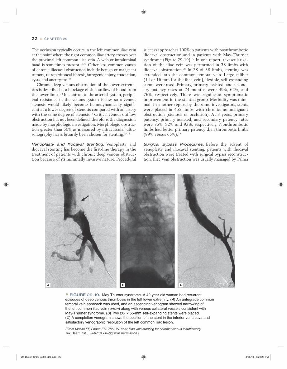
• FIGURE 29-19. May-Thurner syndrome. A 42-year-old woman had recurrent episodes of deep venous thrombosis in the left lower extremity. (A) An antegrade common femoral vein approach was used, and an ascending venogram showed narrowing of the left common iliac vein (arrow) along with venous collateral vessels consistent with May-Thurner syndrome. (B) Two 20- × 55-mm self-expanding stents were placed. (C) A completion venogram shows the position of the stent in the inferior vena cava and satisfactory venographic resolution of the left common iliac lesion.
(From Mussa FF, Peden EK, Zhou W, et al: Iliac vein stenting for chronic venous insuffi ciency. Tex Heart Inst J. 2007;34:60–66; with permission.)
A B C
29_Dieter_Ch29_p001-026.indd 2229_Dieter_Ch29_p001-026.indd 22 4/26/10 6:29:25 PM4/26/10 6:29:25 PM

LOWER EXTREMITY VENOUS DISEASE • 23
femorofemoral bypass or a unilateral bypass between the femoral vein distal to the obstruction and the contralat-eral iliac vein or IVC.80 These major surgical procedures often required lifelong anticoagulation and a temporary or continuous adjunctive arteriovenous fi stula to keep the bypass patent. Furthermore, indications for surgical inter-vention were symptoms associated with severe postthrom-botic syndrome, namely debilitating, refractory venous ulceration. Although reports on the crossover bypass tech-nique claim durable symptomatic relief, most studies lack
consistent follow-up with venography.81,82 At the present time, the only two indications for surgical bypass are fail-ure of endovascular intervention and the need for venous reconstruction after resection for malignancy.
Analysis of 412 Palma operations published in nine series revealed clinical improvement in 63% to 89% of patients. Patency rates ranged between 70% and 85%.49 Experience with femorocaval or iliocaval bypass is limited; however 2-year primary and secondary patency rates of 37% and 54%, respectively, have been reported.49
REFERENCES
1. Bergan JJ, Schmid-Schönbein GW, Coleridge Smith PD, et al: Chronic venous disease. N Engl J Med. 2006;355:488–498.
2. Meissner MH, Gloviczki P, Bergan J, et al: Primary chronic venous disorders. J Vasc Surg. 2007;46(suppl S):54S–67S.
3. Kyrle PA, Eichinger S: Deep vein thrombosis. Lancet. 2005; 365:1163–1174.
4. Fowkes FJI, Price JF, Fowkes FGR: Incidence of diagnosed deep vein thrombosis in the general population: systematic review. Eur J Vasc Endovasc Surg 2003;25:1–5.
5. Heit JA, Silverstein MD, Mohr DN, et al: The epidemiology of venous thromboembolism in the community. Thromb Haemost. 2001;86:452–463.
6. Stein PD, Patel KC, Kalra NK, et al: Deep venous thrombosis in a general hospital. Chest. 2002;122:960–962.
7. Heit JA, O’Fallon WM, Petterson TM, et al: Relative impact of risk factors for deep vein thrombosis and pulmonary embolism: a population-based study. Arch Intern Med. 2002;162:1245–1248.
8. Cushman M, Tsai AW, White RH, et al: Deep vein thrombosis and pulmonary embolism in two cohorts: the longitudinal investigation of thromboembolism etiology. Am J Med. 2004;117:19–25.
9. Robertson L, Evans C, Fowkes FG: Epidemiology of chronic venous disease. Phlebology. 2008;23103–111.
10. Beebe-Dimmer JL, Pfeifer JR, Engle JS, Schottenfeld D: The epidemiology of chronic venous insuffi ciency and varicose veins. Ann Epidemiol. 2005;15:175–184.
11. Etufugh CN, Phillips TJ: Venous ulcers. Clin Dermatol. 2007;25:121–130.
12. Valentine RJ, Wind GG: Embryology of the arteries and veins. In Valentine RJ, Wind GG (eds). Anatomic Exposures in Vascular Surgery, 2nd ed. Philadelphia: Lippincott Williams and Wilkins, 2003:1–19.
13. Meissner MH, Moneta G, Burnand K, et al: The hemo-dynamics and diagnosis of venous disease. J Vasc Surg. 2007;46(suppl S):4S–24S.
14. Valentine RJ, Wind GG: Anatomic variation of the blood vessels. In Valentine RJ, Wind GG (eds). Anatomic Exposures in Vascular Surgery, 2nd ed. Philadelphia: Lippincott Williams and Wilkins, 2003:525–559.
15. Mozes G, Gloviczki P: New discoveries in anatomy and new terminology of leg veins: clinical implications. Vasc Endovasc Surg, 2004;38:367–374.
16. Porter JM, Moneta GL: International Consensus Committee on Chronic Venous Disease. Reporting standards in venous disease: an update. J Vasc Surg. 1995;21:635–645.
17. Eklof B, Rutherford RB, Bergan JJ, et al: Revision of the CEAP classifi cation for chronic venous disorders: consensus statement. J Vasc Surg. 2004;40:1248–1252.
18. Parsi K: Dermatological manifestations of venous insuffi ciency. In Bergan J, Cheng VL (eds). Foam Sclerotherapy: A Textbook. London: Royal Society of Medicine Press Ltd, 2008:13–30.
19. Rutherford RB: Varicose veins. In Cronenwett JL, Rutherford RB (eds). Decision Making in Vascular Surgery. Philadelphia: WB Saunders, 2001:290–293.
20. Langer RD, Ho E, Denenberg JO, et al: Relationships between symptoms and venous disease: the San Diego population study. Arch Intern Med. 2005;165:1420–1424.
21. Evans CJ, Allan PL, Lee AJ, et al: Prevalence of venous refl ux in the general population on duplex scanning: the Edinburgh vein study. J Vasc Surg. 1998;28:767–776.
22. Labropoulos N, Leon M, Nicolaides AN, et al: Superfi cial venous insuffi ciency: correlation of anatomic extent of refl ux with clinical symptoms and signs. J Vasc Surg. 1994;20:953–958.
23. Engelhorn CA, Engelhorn ALV, Cassou MF, Salles-Cunha SX: Patterns of saphenous refl ux in women with primary varicose veins. J Vasc Surg. 2005;41:645–651.
24. Beale RJ, Gough MJ: Treatment options of primary varicose veins—a review. Eur J Vasc Endovasc Surg. 2005;30:83–95.
25. Racette S, Sauvageau A: Unusual sudden death: two case reports of hemorrhage by rupture of varicose veins. Am J Forensic Med Pathol. 2005;26:294–296.
26. Leon L, Giannoukas AD, Dodd D, et al: Clinical signifi cance of superfi cial vein thrombosis. Eur J Vasc Endovasc Surg. 2005;29:10–17.
27. Decousus H, Epinat M, Guillot K et al: Superfi cial vein thrombosis: risk factors, diagnosis and treatment. Curr Opin Pulm Med. 2003;9:393–397.
28. Lofgren, EP Lofgren KA: The surgical treatment of superfi cial thrombophlebitis. Surgery. 1981;90:49–54.
29. Partsch H, Flour M, Smith PC; International Compression Club: Indications for compression therapy in venous and lymphatic disease consensus based on experimental data and scientifi c evidence. Under the auspices of the IUP. Int Angiol. 2008;27:193–219.
29_Dieter_Ch29_p001-026.indd 2329_Dieter_Ch29_p001-026.indd 23 4/26/10 6:29:26 PM4/26/10 6:29:26 PM

24 • CHAPTER 29
30. Labropoulos N, Leon M, Volteas N, Nicolaides AN: Acute and long term effect of elastic stockings in patients with varicose veins. Int Angiol. 1994;13:119–123.
31. Pannier F, Hoffman B, Stang A, et al: Prevalence and acceptance of therapy with medical compression stockings in the adult population—results from the Bonn Vein Study. Phlebologie. 2007:36;245–249.
32. Blattler W, Zimmett SE: Compression therapy in venous disease. Phlebology. 2008;23:205–208.
33. Rabe E, Pannier F, Gerlach H, et al: Sclerotherapy of varicose veins—guidelines of the German Society of Phlebology. In Bergan J, Cheng VL (eds). Foam Sclerotherapy: A Textbook. London: Royal Society of Medicine Press Ltd, 2008:31–41.
34. Drake LA, Dinehart SM, Goltz RW, et al: Guidelines of care for sclerotherapy treatment of varicose and telangectatic leg veins. J Am Acad Dermatol. 1996;34:523–528.
35. Kabnick L: Legal guidelines for use of polidocanol and sodium tetradecyl sulfate for sclerotherapy. In Bergan J, Cheng VL (eds). Foam Sclerotherapy: A Textbook. London: Royal Society of Medicine Press Ltd, 2008:43–52.
36. Dietzek CL: Sclerotherapy: introduction to solutions and techniques. Perspect Vasc Surg Endovasc Ther. 2007;19:317–324.
37. Pittaluga P, Chastanet S: Lesser importance of the saphenous vein in varicose vein treatment. In Bergan J, Cheng VL (eds). Foam Sclerotherapy: A Textbook. London: Royal Society of Medicine Press Ltd, 2008:163–176.
38. Ludbrook J, Beale G: Femoral venous valves in relation to varicose veins. Lancet. 1962;1:79–81.
39. Stirling M, Shortell CK: Endovascular treatment of varicose veins. Semin Vasc Surg. 2006;19:109–115.
40. Subramonia S, Lees TA: The treatment of varicose veins. Ann R Coll Surg Engl. 2007;89:96–100.
41. Nijsten T, Van den Bos RR, Goldman MP, et al: Minimally invasive techniques in the treatment of saphenous varicose veins. J Am Acad Dermatol. 2009;60:110–119.
42. Juan J, Escribano JM, Criado E, Fontcuberta J: Hemodynamic surgery for varicose veins: surgical strategy. Phlebology. 2005;20:2–13.
43. Criado E, Juan J, Fontcuberta J, Escribano JM: Hemodynamic surgery for varicose veins: rationale, and anatomic and hemodynamic basis. Phlebology. 2003;18:158–166.
44. Escribano JM, Juan J, Bofi ll R, et al: Durability of refl ux elimination by a minimal invasive CHIVA procedure on patients with varicose veins. A 3 year prospective case study. Eur J Vasc Endovasc Surg. 2003;25:159–163.
45. Pittaluga P, Rea B, Barbe R, Guex JJ: ASVAL method: principles and preliminary results. In Becquemin JP, Alimi YS, Watelet J (eds). Updates and Controversies in Vascular Surgery. Torino, Italy: Minerva Medica, 2005:182–189.
46. Monahan DL: Can phlebectomy be deferred in the treatment of varicose veins? J Vasc Surg. 2005;42:1145–1149.
47. Welch HJ: Endovenous ablation of the great saphenous vein may avert phlebectomy for branch varicose veins. J Vasc Surg. 2006;44:601–605.
48. Allis J, Simonian SJ, Lee BB, et al: Treatment of superfi cial venous insuffi ciency and symptomatic varicose veins with
endovenous laser ablation does not require concomitant phlebectomy. 9th International Congress of Phlebology, Bologna, Italy, April 4–5, 2008.
49. Meissner MH, Eklof B, Coleridge Smith PD, et al: Secondary chronic venous disorders. J Vasc Surg. 2007;46(suppl):68S–83S.
50. Tassiopoulos AK, Golts E, Oh DS, Labropoulos N: Current concepts in chronic venous ulceration. Eur J Vasc Endovasc Surg. 2000;20:227–232.
51. Labropoulos N, Giannoukas AD, Delis K, et al: Where does venous refl ux start? J Vasc Surg. 1997;26:736–742.
52. Nicolaides AN, Hussein MK, Szendro G, et al: The relation of venous ulceration with ambulatory venous pressure measurements. J Vasc Surg. 1993;17:441–419.
53. Labropoulos N, Tiongson J, Pryor L, et al: Defi nition of refl ux in lower extremity veins. J Vasc Surg. 2003;38:733–798.
54. O’Donnell TF: The present status of surgery of the superfi cial venous system in the management of venous ulcer and evidence for the role of perforator interruption. J Vasc Surg. 2008;48:1044–1052.
55. Rajendran S, Rigby AJ, Anand SC: Venous leg ulcer treatment and practice—part 2: wound management. J Wound Care. 2007;16:68–70.
56. Reichenberg J, Davis M: Venous ulcers. Semin Cutan Med Surg. 2005;24:216–226.
57. Patel NP, Labropoulos N, Pappas PJ: Current manage-ment of venous ulceration. Plast Reconstr Surg. 2006;117(7 suppl):254S–260S.
58. Partsch H, Clark M, Mosti G, et al: Classifi cation of compression bandages: practical aspects. Dermatol Surg. 2008;34:600–609.
59. Partsch H: Compression therapy of venous ulcers. Curr Probl Dermatol. 1997;27:130–140.
60. O’Hare JL, Vandenbroeck CP, Whitman B, et al: A prospective evaluation of the outcome after small saphenous varicose vein surgery with one-year follow up. J Vasc Surg. 2008;48:669–674.
61. Kwock J, Lee BB, Neville RF, Laredo J: Treatment of symptomatic varicose veins and lesser saphenous vein refl ux with endovenous laser ablation does not require concomitant phlebectomy. Society for Military Vascular Surgery and Chesapeake Vascular Society 36th Annual Meeting, Bethesda, MD, Dec 4–6, 2008.
62. O’Donnell TF: Surgical treatment of incompetent perforating veins. In Bergan JJ, Kistner RL (eds). Atlas of Venous Surgery. Philadelphia: WB Saunders,1992.
63. Gloviczki P, Bergan JJ, Rhodes JM, et al: Mid-term results of endoscopic perforator vein interruption for chronic venous insuffi ciency: lessons learned from the North American Subfascial Endoscopic Perforator Surgery Registry. J Vasc Surg. 1999;29:489–502.
64. Cabrera J, Redondo P, Becerra A, et al: Ultrasound-guided injection of polidocanol microfoam in the management of venous leg ulcers. Arch Dermatol. 2004;140:667–673.
65. Proebstle TM, Herdemann S: Early results and feasibility of incompetent perforator vein ablation by endovenous laser treatment. Dermatol Surg. 2007;33:162–168.
AU: Note renumbering. Refs 29 and 58 were the same.
29_Dieter_Ch29_p001-026.indd 2429_Dieter_Ch29_p001-026.indd 24 4/26/10 6:29:26 PM4/26/10 6:29:26 PM

LOWER EXTREMITY VENOUS DISEASE • 25
66. Roth SM: Endovenous radiofrequency ablation of superfi cial and perforator veins. Surg Clin North Am 2007;87:1267–1284.
68. Eriksson I: Reconstructive venous surgery. Acta Chir Scand. 1988;544(suppl):69–74.
68. Masuda EM, Kistner RL: Long-term results of venous valve reconstruction: a 4 to 21year follow up. J Vasc Surg. 1994;19:391–403.
69. Raju S, Neglen P, Doolittle J, Meydrech EF: Axillary vein transfer in trabeculated post-thrombotic veins. J Vasc Surg. 1999;29:1050–1062.
70. Raju S, Fredericks RK, Neglen PN, Bass JD: Durability of venous valve reconstruction techniques for “primary” and post-thrombotic refl ux. J Vasc Surg. 1996;23:357–366.
71. Akesson H, Brudin L, Dahlstrom JA, et al: Venous function assessed during a 5-year period after acute iliofemoral venous thrombosis treated with anticoagulation. Eur J Vasc Surg. 1990;4:43–48.
72. Johnson BF, Manzo RA, Bergelin RO, Strandness DE: Relationship between changes in the deep venous system and the development of the postthrombotic syndrome after an acute episode of lower limb deep vein thrombosis: a one- to six- year follow-up. J Vasc Surg. 1995;21:307–313.
73. Negus D, Fletcher EW, Cockett FB, Thomas ML: Compression and band formation at the mouth of the left common iliac vein. Br J Surg. 1968;55:369–674.
74. Neglen P: Chronic deep venous obstruction: defi nition, preva-lence, diagnosis, management. Phlebology. 2008;23:149–157.
75. Hartung O, Otero A, Boufi M, et al: Mid-term results of endovascular treatment for symptomatic chronic nonmalignant iliocaval venous occlusive disease. J Vasc Surg. 2005;42:1138–1144.
76. Neglen P, Raju S: Proximal lower extremity chronic venous outfl ow obstruction: recognition and treatment. Semin Vasc Surg. 2002;15:57–64.
77. Mussa FF, Peden EK, Zhou W, et al: Iliac vein stenting for chronic venous insuffi ciency. Tex Heart Inst J. 2007;34:60–66.
78. Raju S, McAllister S, Neglen P: Recanalization of totally occluded iliac and adjacent venous segments. J Vasc Surg. 2002;36:903–911.
79. Neglen P, Raju S: Intravascular ultrasound scan evaluation of the obstructed vein. J Vasc Surg. 2002;35:694–700.
80. Gruss JD, Hiemer W: Bypass procedures for venous ob-struction. In Raju S, Villavicencio JL (eds). Surgical Man-agement of Venous Disease. Baltimore: Williams & Wilkins, 1997.
81. Halliday P, Harris J, May J: Femoro-femoral crossover grafts (Palma operation): a long-term follow up study. In Bergan JJ, Yao JST (eds). Surgery of the Veins. Orlando: Grune & Stratton, 1985.
82. Gruss JD: Venous bypass for chronic venous insuffi ciency. In Bergan JJ, Yao JST (eds). Venous Disorders. Philadelphia: WB Saunders, 1991.
29_Dieter_Ch29_p001-026.indd 2529_Dieter_Ch29_p001-026.indd 25 4/26/10 6:29:26 PM4/26/10 6:29:26 PM

29_Dieter_Ch29_p001-026.indd 2629_Dieter_Ch29_p001-026.indd 26 4/26/10 6:29:26 PM4/26/10 6:29:26 PM