Embed Size (px)
Citation preview

LOWER LIMB
Anterior Compartment of the Thighs
& Femoral Triangle
Dr. Kumar K.V02.11.2009
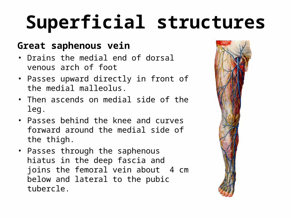
Superficial structuresGreat saphenous vein• Drains the medial end of dorsal venous
arch of foot
• Passes upward directly in front of the medial malleolus.
• Then ascends on medial side of the leg.
• Passes behind the knee and curves forward around the medial side of the thigh.
• Passes through the saphenous hiatus in the deep fascia and joins the femoral vein about 4 cm below and lateral to the pubic tubercle.
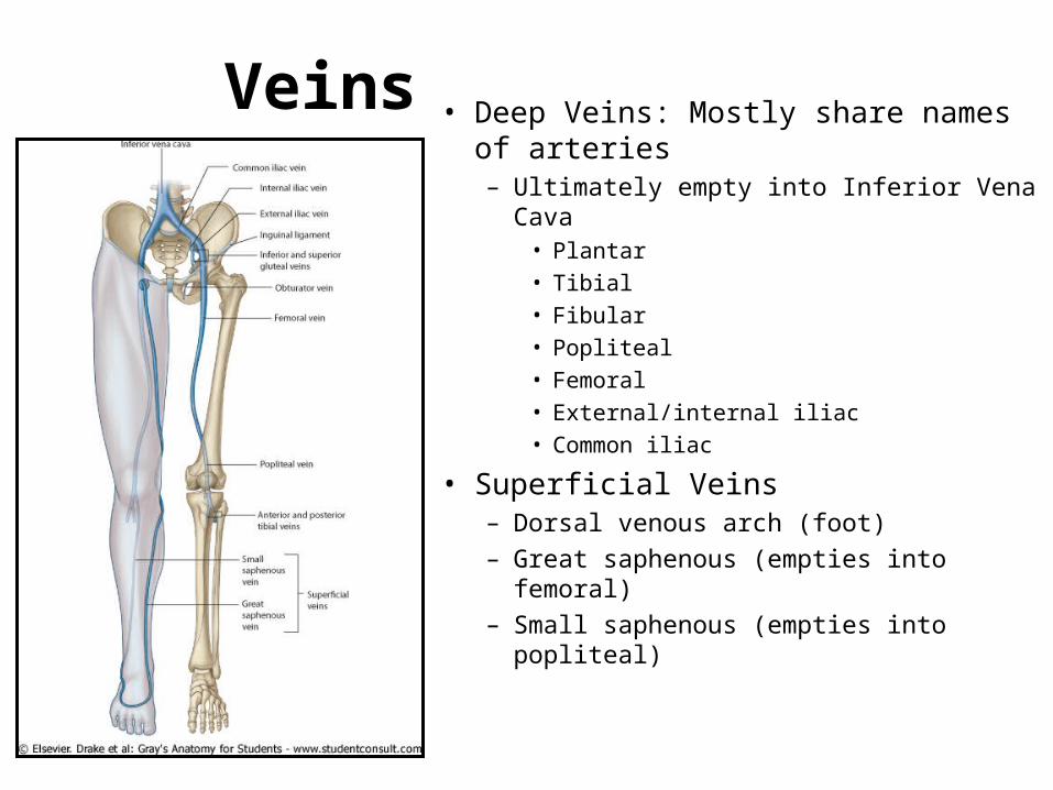
Veins • Deep Veins: Mostly share names of arteries– Ultimately empty into Inferior Vena Cava
• Plantar• Tibial• Fibular• Popliteal• Femoral• External/internal iliac• Common iliac
• Superficial Veins – Dorsal venous arch (foot)– Great saphenous (empties into femoral)– Small saphenous (empties into popliteal)
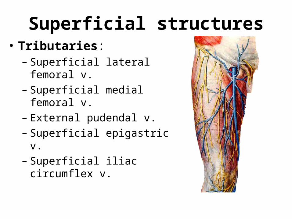
Superficial structures• Tributaries:
– Superficial lateral femoral v. – Superficial medial femoral v. – External pudendal v. – Superficial epigastric v. – Superficial iliac circumflex v.
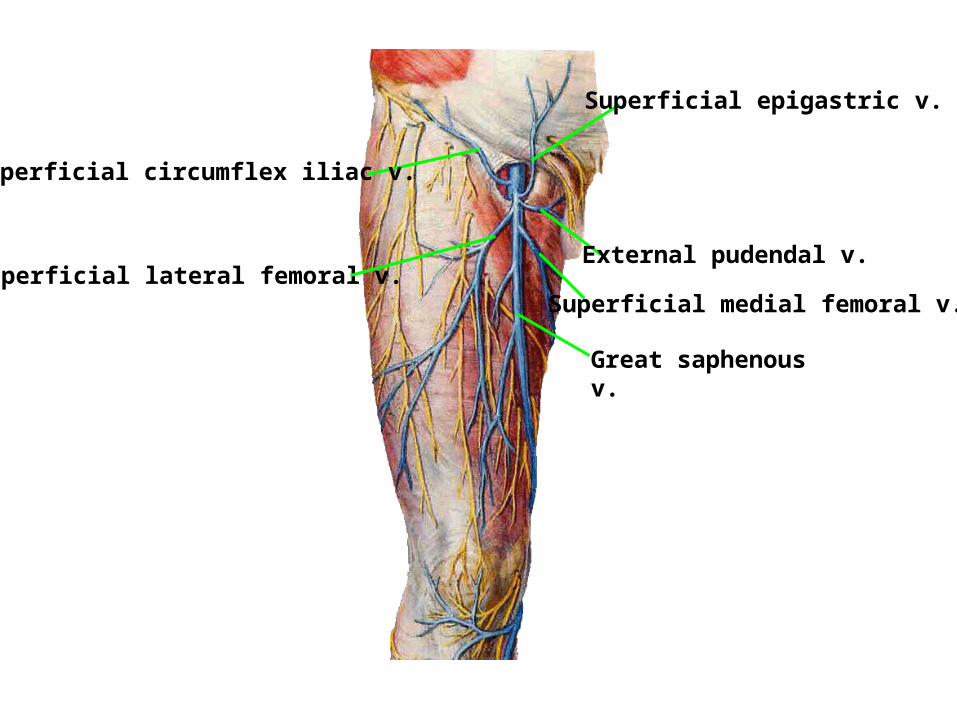
Superficial circumflex iliac v.
Superficial lateral femoral v.
Superficial epigastric v.
External pudendal v.
Superficial medial femoral v.
Great saphenous v.
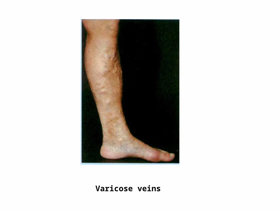
Varicose veins
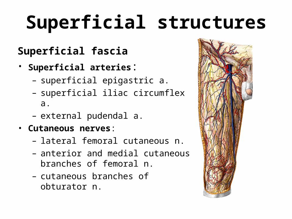
Superficial structures
Superficial fascia
• Superficial arteries: – superficial epigastric a.
– superficial iliac circumflex a.
– external pudendal a.
• Cutaneous nerves:
– lateral femoral cutaneous n.
– anterior and medial cutaneous branches of femoral n.
– cutaneous branches of obturator n.
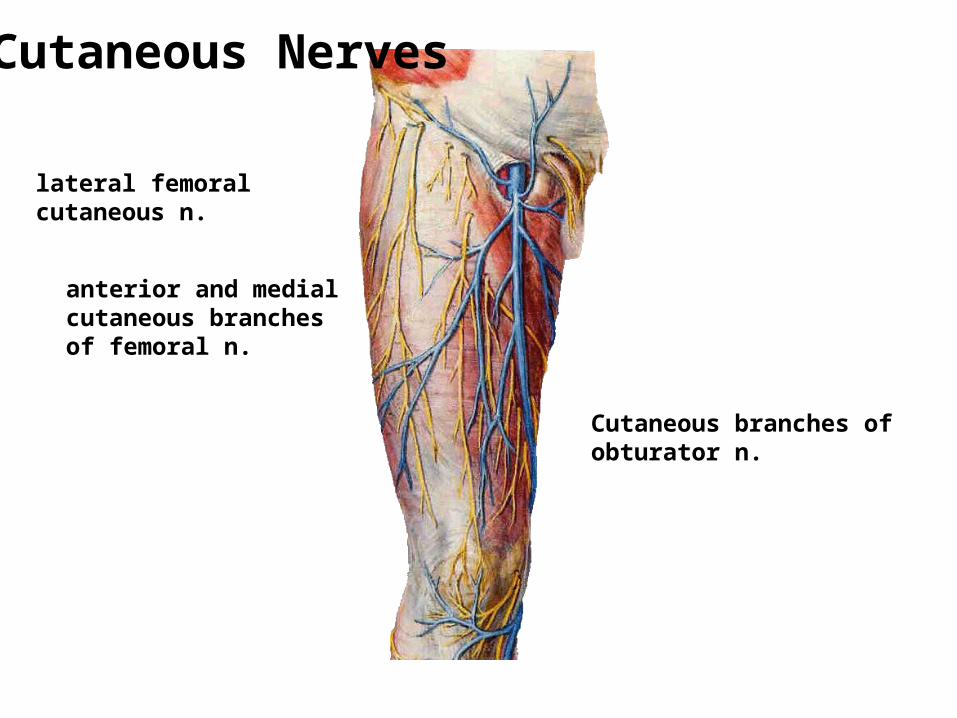
lateral femoral cutaneous n.
anterior and medial cutaneous branches of femoral n.
Cutaneous branches of obturator n.
Cutaneous Nerves
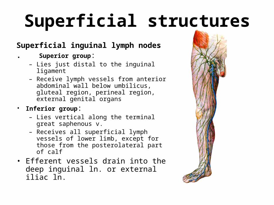
Superficial structuresSuperficial inguinal lymph nodes . Superior group:
– Lies just distal to the inguinal ligament– Receive lymph vessels from anterior
abdominal wall below umbilicus, gluteal region, perineal region, external genital organs
• Inferior group: – Lies vertical along the terminal great
saphenous v.– Receives all superficial lymph vessels of
lower limb, except for those from the posterolateral part of calf
• Efferent vessels drain into the deep inguinal ln. or external iliac ln.
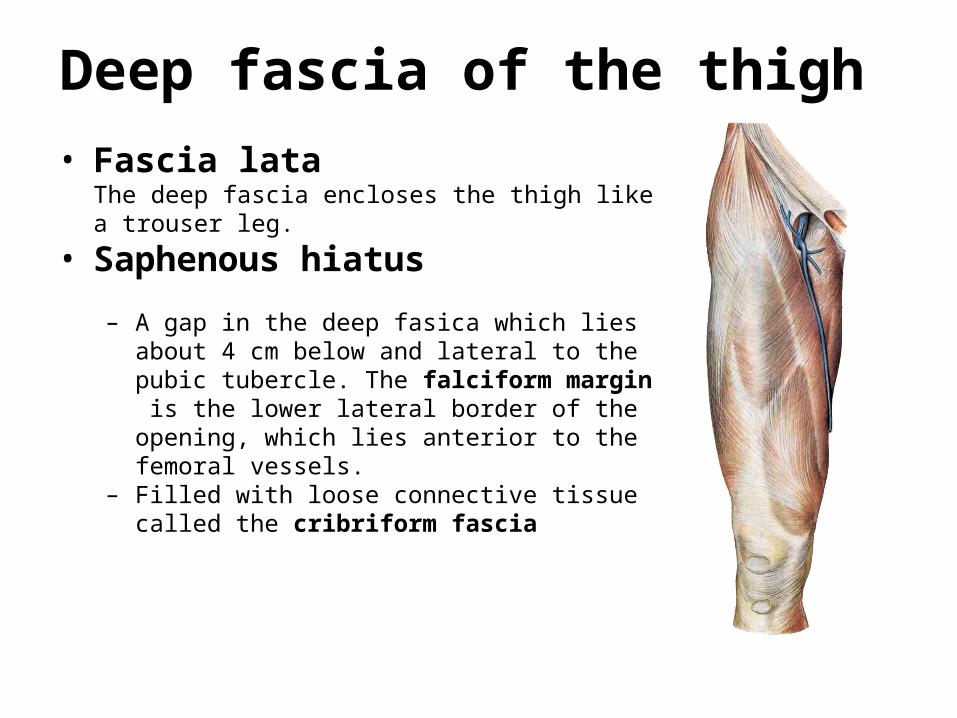
Deep fascia of the thigh
• Fascia lata The deep fascia encloses the thigh like a trouser leg.
• Saphenous hiatus – A gap in the deep fasica which lies about 4 cm
below and lateral to the pubic tubercle. The falciform margin is the lower lateral border of the opening, which lies anterior to the femoral vessels.
– Filled with loose connective tissue called the cribriform fascia
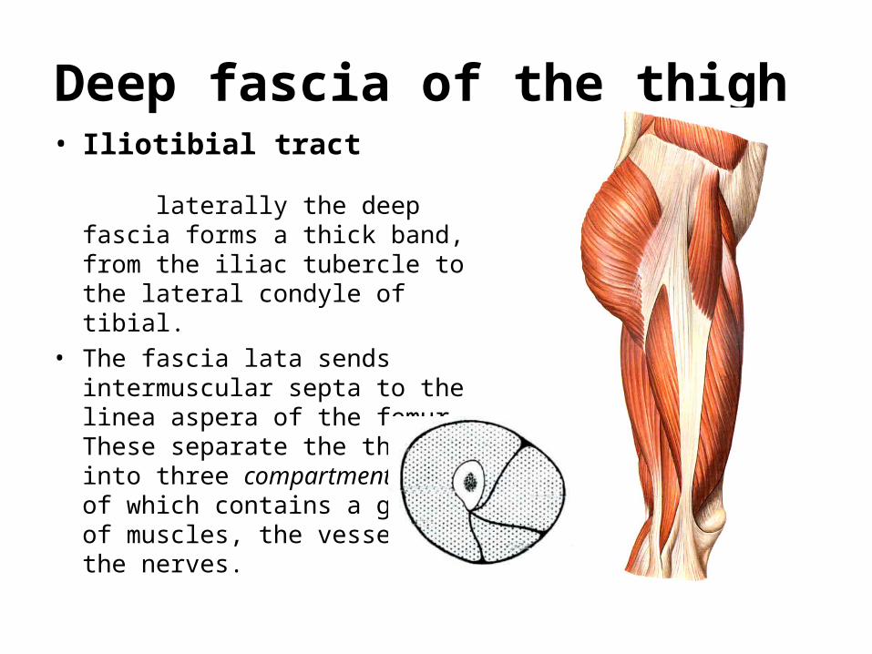
Deep fascia of the thigh • Iliotibial tract
laterally the deep fascia forms a thick band, from the iliac tubercle to the lateral condyle of tibial.
• The fascia lata sends intermuscular septa to the linea aspera of the femur. These separate the thigh into three compartments each of which contains a group of muscles, the vessels and the nerves.
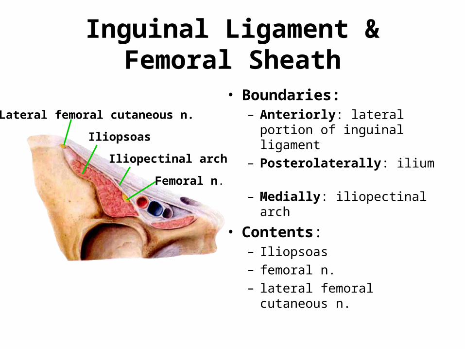
Inguinal Ligament & Femoral Sheath
• Boundaries:– Anteriorly: lateral portion of
inguinal ligament
– Posterolaterally: ilium
– Medially: iliopectinal arch
• Contents: – Iliopsoas
– femoral n.
– lateral femoral cutaneous n.
Iliopectinal arch
Femoral n.
Iliopsoas
Lateral femoral cutaneous n.
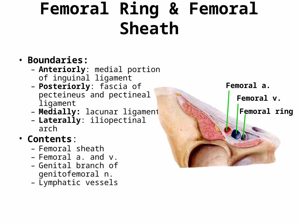
Femoral Ring & Femoral Sheath
• Boundaries:– Anteriorly: medial portion of
inguinal ligament– Posteriorly: fascia of
pecteineus and pectineal ligament
– Medially: lacunar ligament– Laterally: iliopectinal arch
• Contents: – Femoral sheath– Femoral a. and v.– Genital branch of
genitofemoral n. – Lymphatic vessels
Femoral a.
Femoral v.
Femoral ring
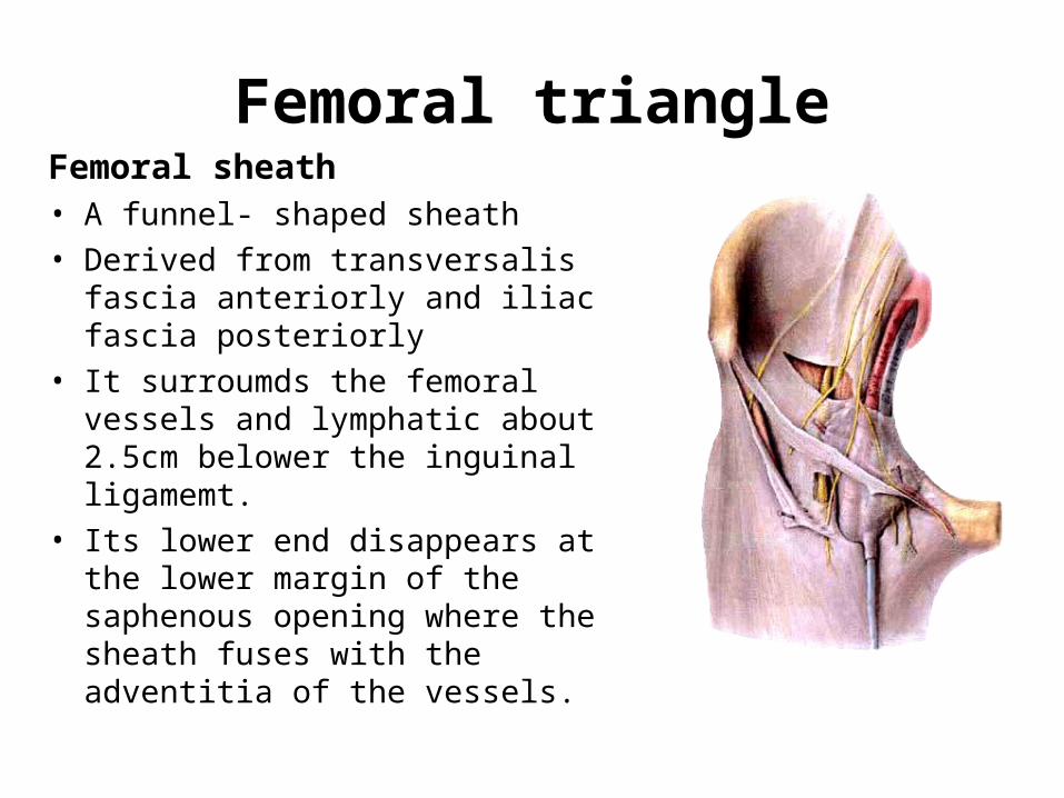
Femoral triangleFemoral sheath• A funnel- shaped sheath • Derived from transversalis fascia
anteriorly and iliac fascia posteriorly
• It surroumds the femoral vessels and lymphatic about 2.5cm belower the inguinal ligamemt.
• Its lower end disappears at the lower margin of the saphenous opening where the sheath fuses with the adventitia of the vessels.
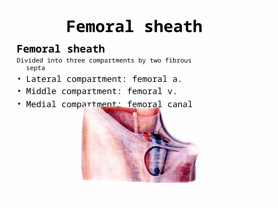
Femoral sheath Femoral sheath Divided into three compartments by two fibrous septa
• Lateral compartment: femoral a. • Middle compartment: femoral v.
• Medial compartment: femoral canal
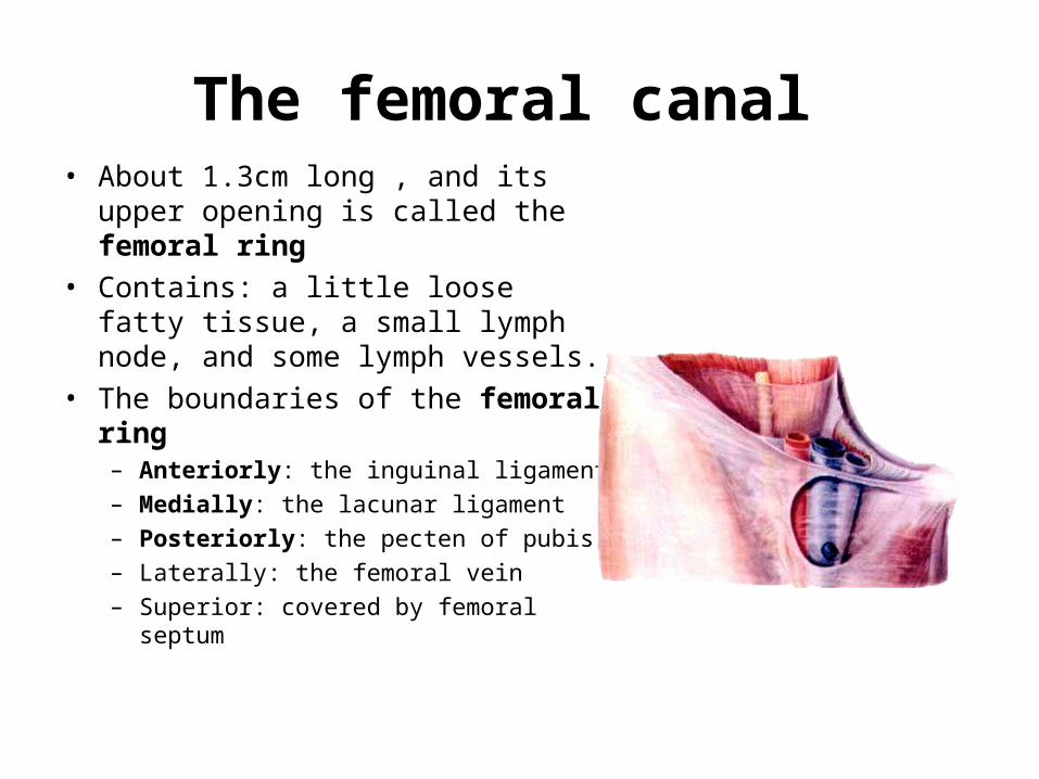
The femoral canal • About 1.3cm long , and its upper
opening is called the femoral ring
• Contains: a little loose fatty tissue, a small lymph node, and some lymph vessels.
• The boundaries of the femoral ring – Anteriorly: the inguinal ligament
– Medially: the lacunar ligament
– Posteriorly: the pecten of pubis
– Laterally: the femoral vein
– Superior: covered by femoral septum
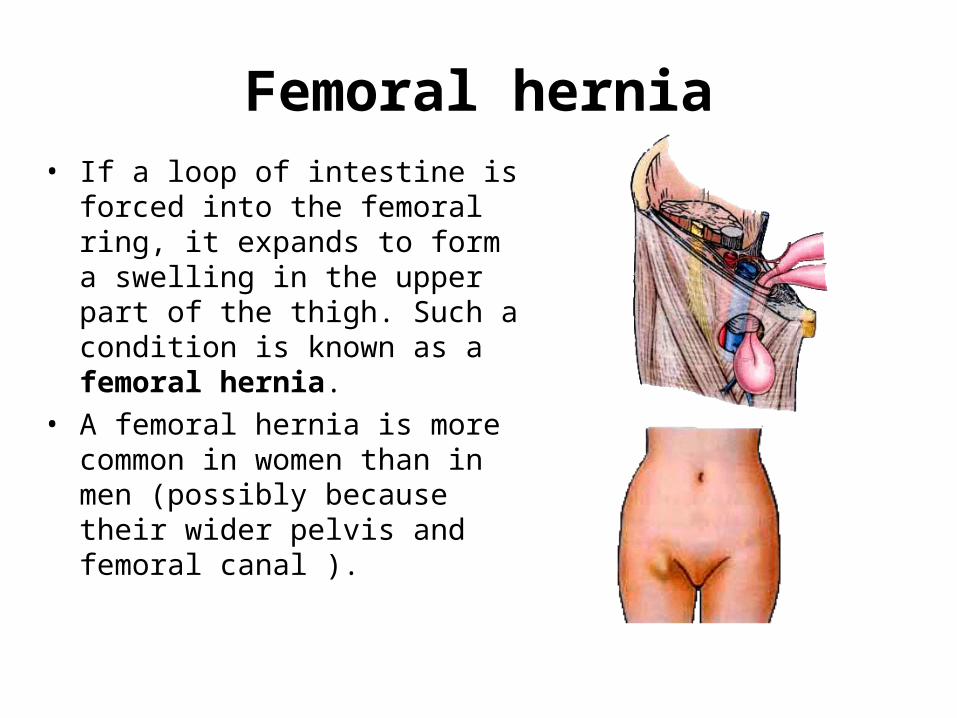
Femoral hernia• If a loop of intestine is forced into
the femoral ring, it expands to form a swelling in the upper part of the thigh. Such a condition is known as a femoral hernia.
• A femoral hernia is more common in women than in men (possibly because their wider pelvis and femoral canal ).
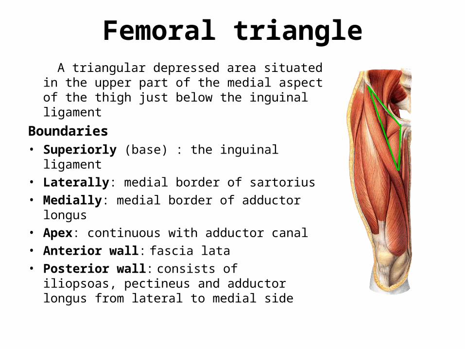
Femoral triangle A triangular depressed area situated in the
upper part of the medial aspect of the thigh just below the inguinal ligament
Boundaries • Superiorly (base) : the inguinal ligament
• Laterally: medial border of sartorius
• Medially: medial border of adductor longus
• Apex: continuous with adductor canal
• Anterior wall: fascia lata
• Posterior wall: consists of iliopsoas, pectineus and adductor longus from lateral to medial side
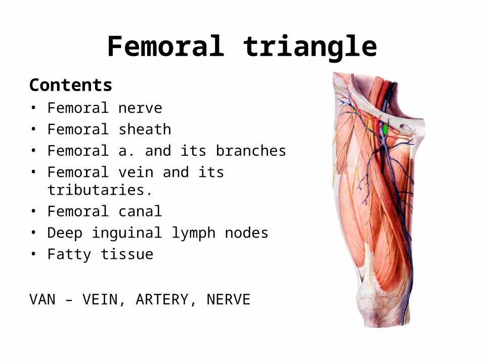
Femoral triangleContents • Femoral nerve
• Femoral sheath
• Femoral a. and its branches
• Femoral vein and its tributaries.
• Femoral canal
• Deep inguinal lymph nodes
• Fatty tissue
VAN – VEIN, ARTERY, NERVE

Contents of the anterior fascial compartments of the thigh
IliopsoasIliacus
Psoas major
Sartorius
Quadriceps femoris
Vastusintermedius
Vastus lateralis
Rectus femoris
Vastus medialis
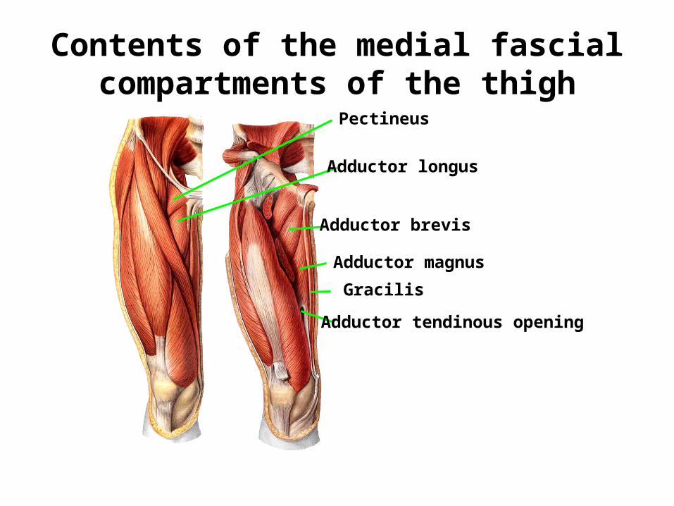
Contents of the medial fascial compartments of the thigh
Pectineus
Adductor longus
Adductor brevis
Adductor magnus
Gracilis
Adductor tendinous opening
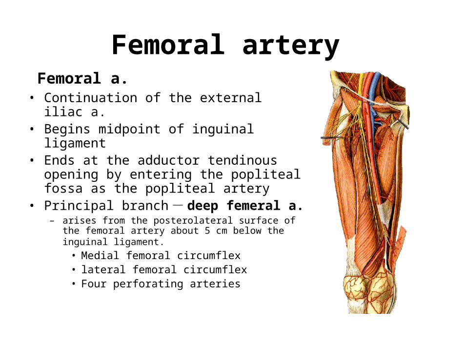
Femoral artery Femoral a. • Continuation of the external iliac a. • Begins midpoint of inguinal ligament• Ends at the adductor tendinous opening
by entering the popliteal fossa as the popliteal artery
• Principal branch - deep femeral a. – arises from the posterolateral surface of the femoral
artery about 5 cm below the inguinal ligament. • Medial femoral circumflex• lateral femoral circumflex• Four perforating arteries
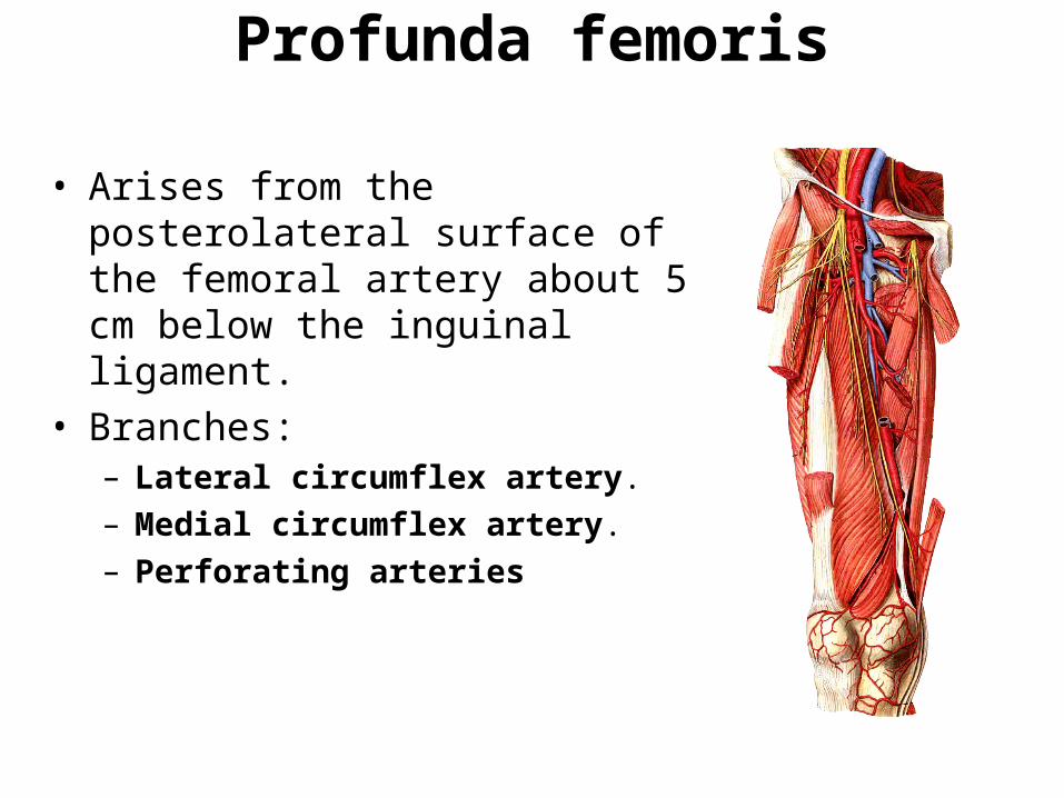
Profunda femoris
• Arises from the posterolateral surface of the femoral artery about 5 cm below the inguinal ligament.
• Branches: – Lateral circumflex artery.
– Medial circumflex artery.
– Perforating arteries
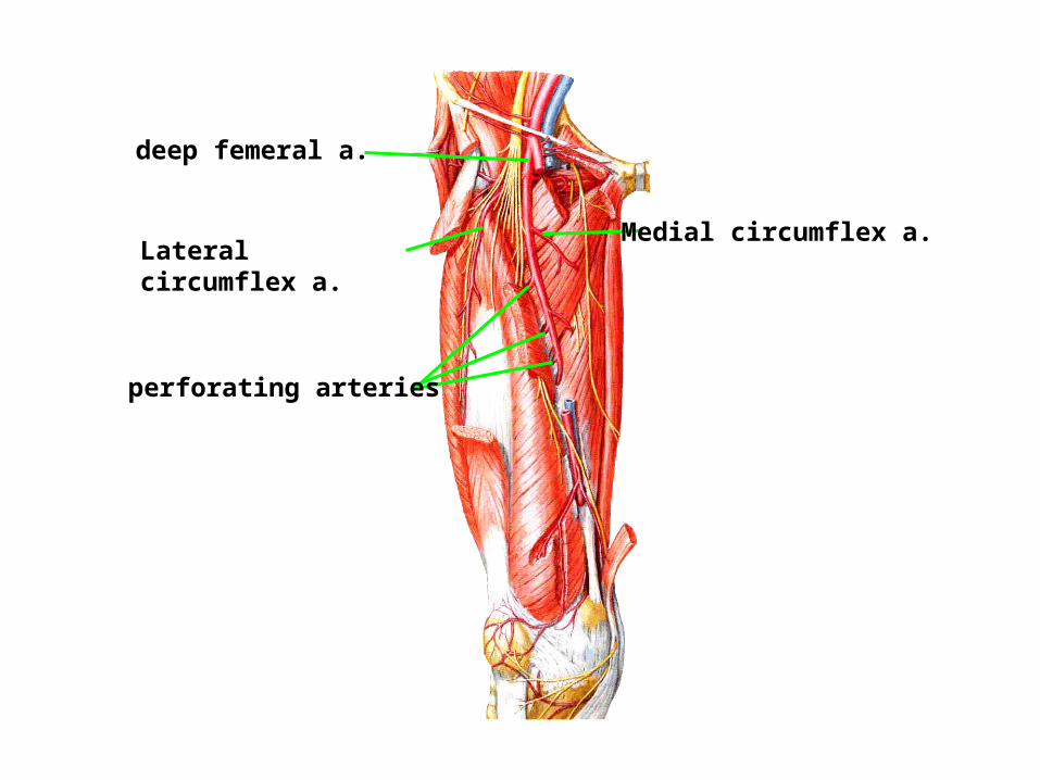
deep femeral a.
Lateral circumflex a. Medial circumflex a.
perforating arteries
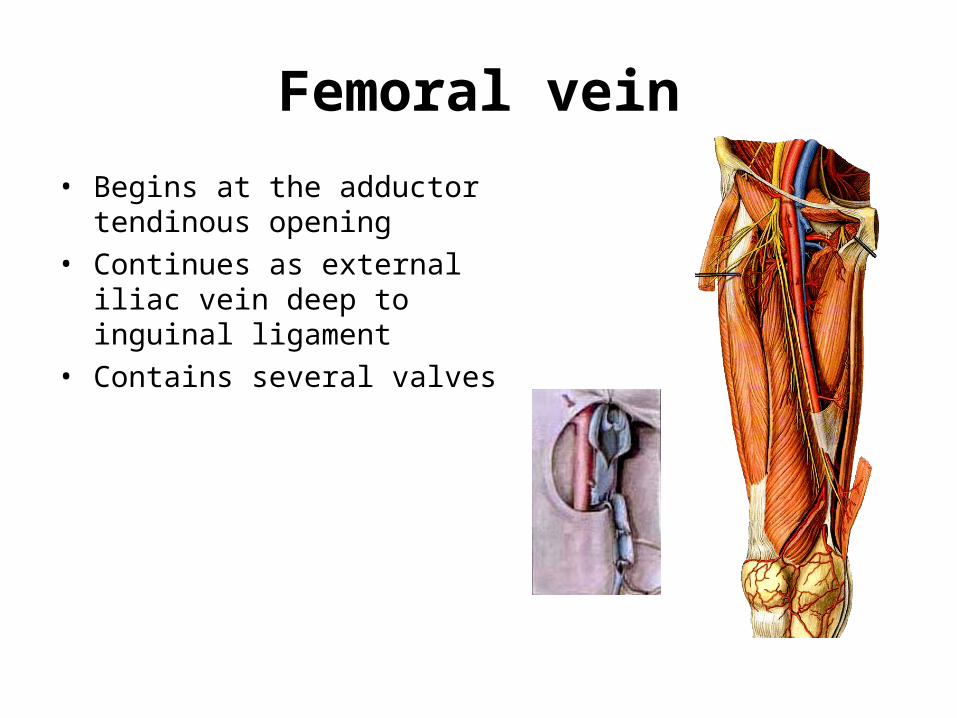
Femoral vein
• Begins at the adductor tendinous opening
• Continues as external iliac vein deep to inguinal ligament
• Contains several valves
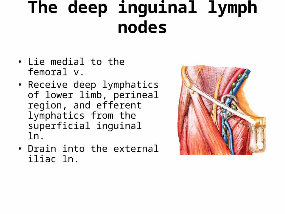
The deep inguinal lymph nodes
• Lie medial to the femoral v.• Receive deep lymphatics of
lower limb, perineal region, and efferent lymphatics from the superficial inguinal ln.
• Drain into the external iliac ln.
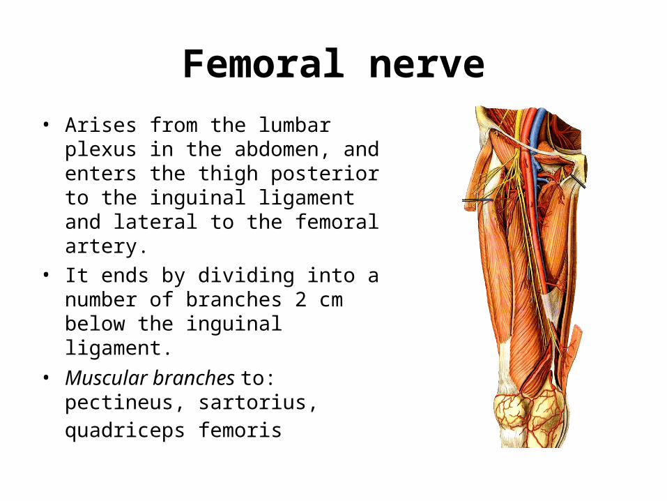
Femoral nerve• Arises from the lumbar plexus in the
abdomen, and enters the thigh posterior to the inguinal ligament and lateral to the femoral artery.
• It ends by dividing into a number of branches 2 cm below the inguinal ligament.
• Muscular branches to: pectineus, sartorius, quadriceps femoris
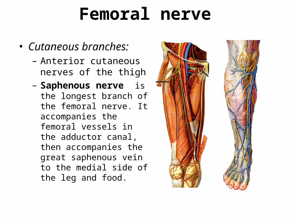
Femoral nerve
• Cutaneous branches: – Anterior cutaneous
nerves of the thigh – Saphenous nerve is the
longest branch of the femoral nerve. It accompanies the femoral vessels in the adductor canal, then accompanies the great saphenous vein to the medial side of the leg and food.
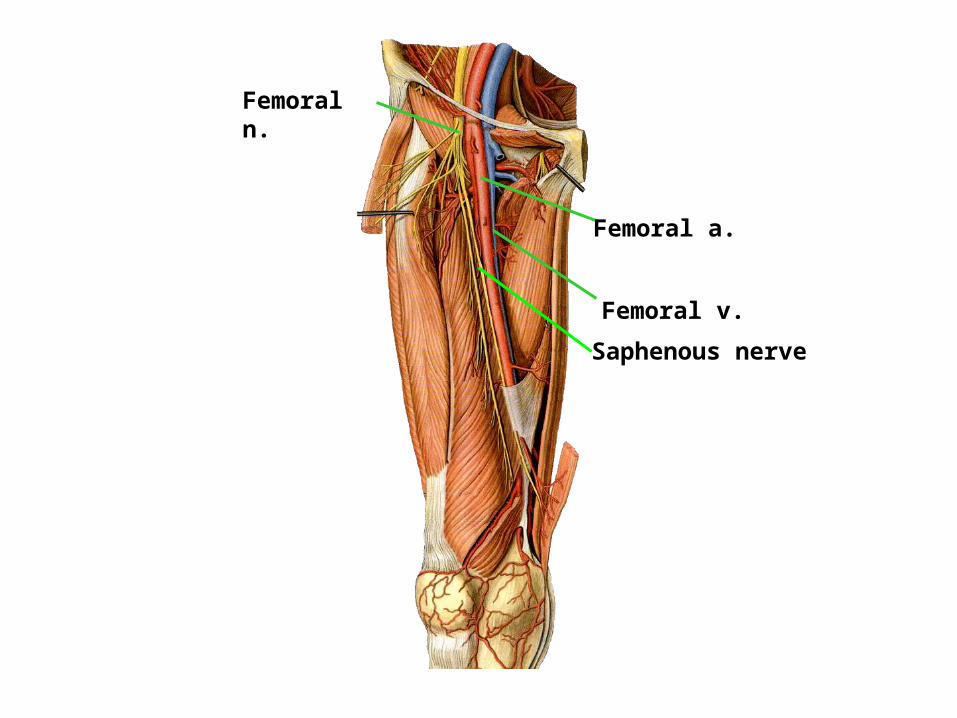
Femoral n.
Femoral a.
Femoral v.
Saphenous nerve
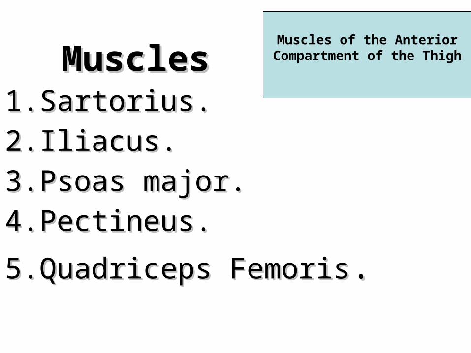
1.1.Sartorius.Sartorius.
2.2.Iliacus.Iliacus.
3.3.Psoas major.Psoas major.
4.4.Pectineus.Pectineus.
5.5.Quadriceps FemorisQuadriceps Femoris..
MusclesMusclesMuscles of the Anterior
Compartment of the Thigh
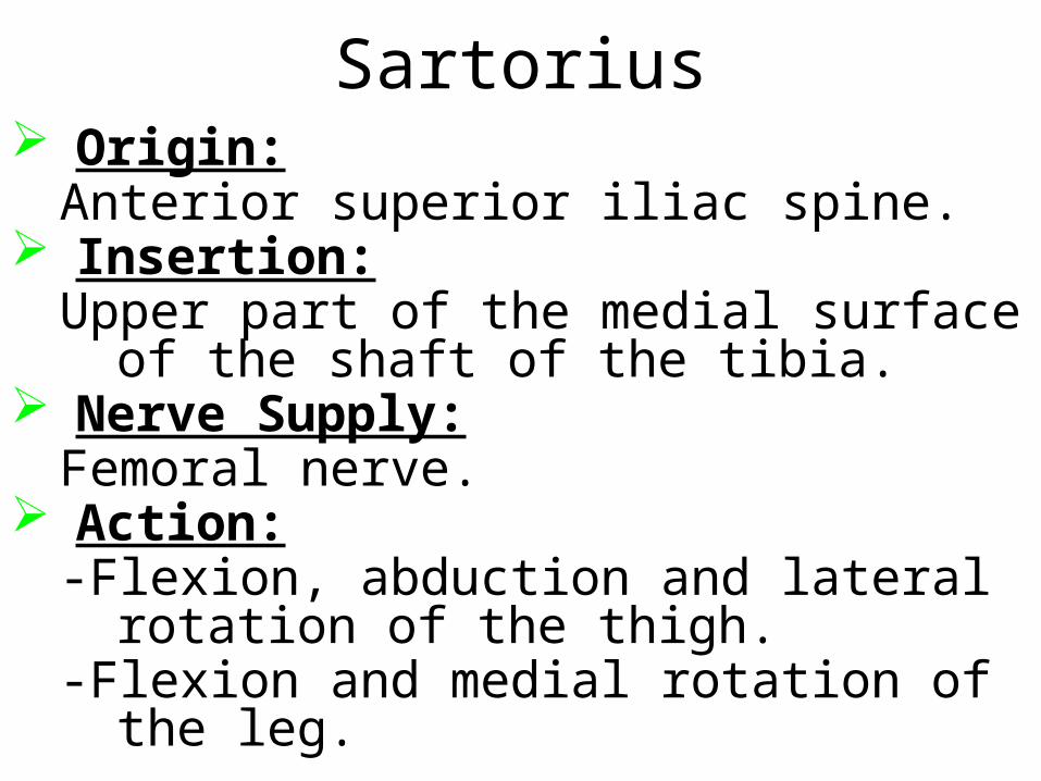
Sartorius Origin:
Anterior superior iliac spine. Insertion:
Upper part of the medial surface of the shaft of the tibia.
Nerve Supply:Femoral nerve.
Action:-Flexion, abduction and lateral rotation of
the thigh.-Flexion and medial rotation of the leg.
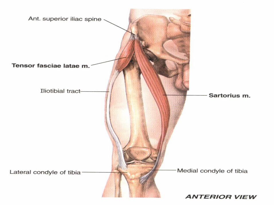
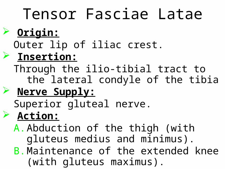
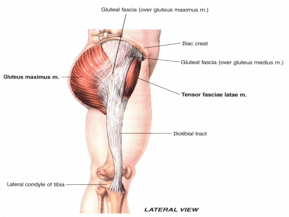
Tensor Fasciae Latae Origin:
Outer lip of iliac crest. Insertion:
Through the ilio-tibial tract to the lateral condyle of the tibia
Nerve Supply:Superior gluteal nerve.
Action:A. Abduction of the thigh (with gluteus medius
and minimus).B. Maintenance of the extended knee (with
gluteus maximus).
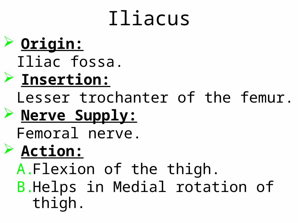
Iliacus Origin:
Iliac fossa. Insertion:
Lesser trochanter of the femur. Nerve Supply:
Femoral nerve. Action:
A.Flexion of the thigh.B.Helps in Medial rotation of thigh.
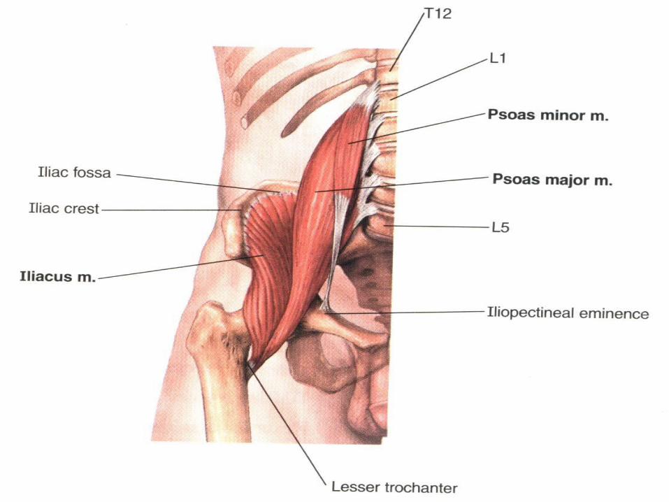
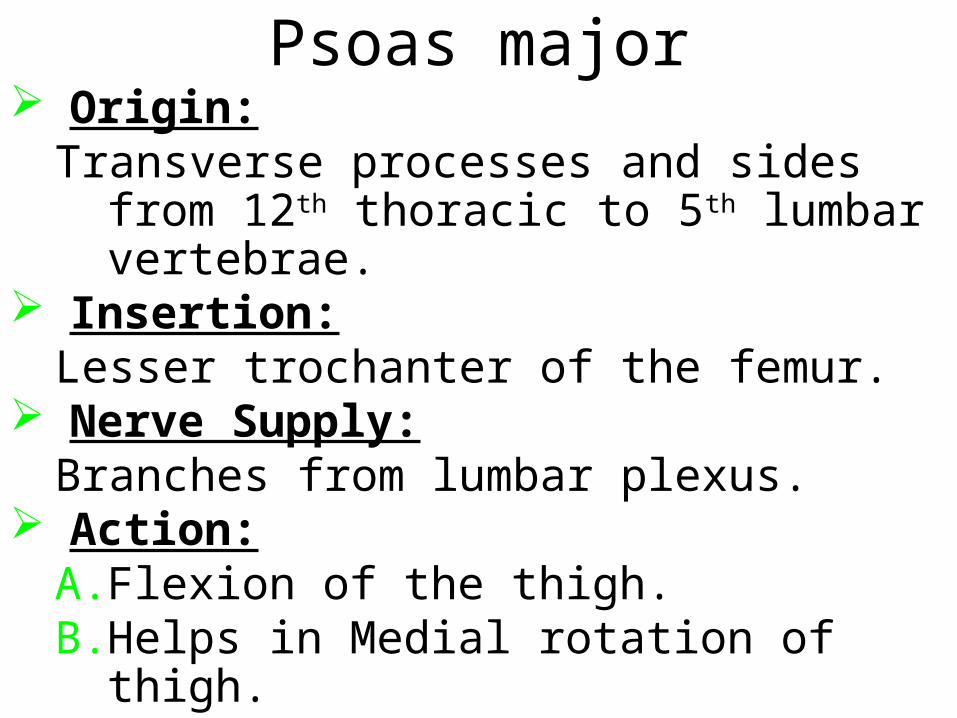
Psoas major Origin:
Transverse processes and sides from 12th thoracic to 5th lumbar vertebrae.
Insertion:Lesser trochanter of the femur.
Nerve Supply:Branches from lumbar plexus.
Action:A. Flexion of the thigh.B. Helps in Medial rotation of thigh.

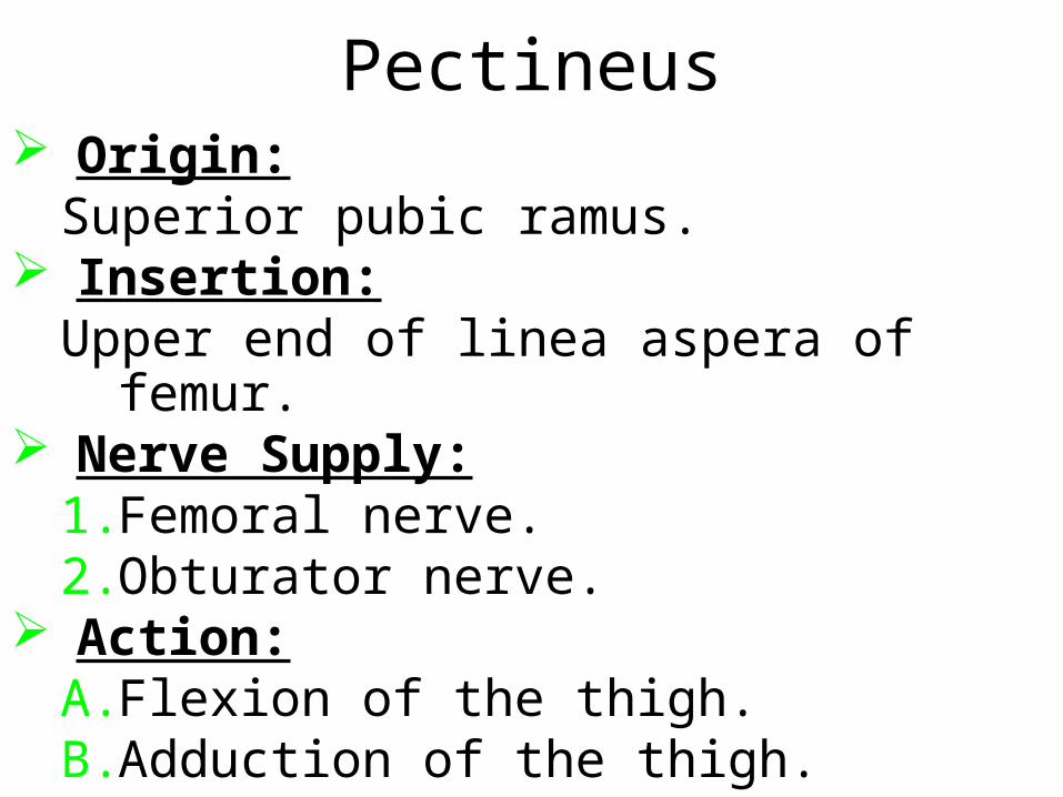
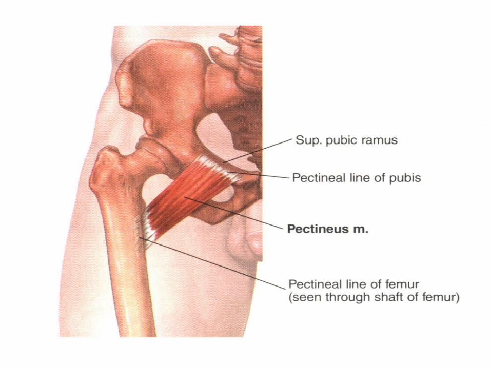
Pectineus Origin:
Superior pubic ramus. Insertion:
Upper end of linea aspera of femur. Nerve Supply:
1. Femoral nerve.2. Obturator nerve.
Action:A. Flexion of the thigh.B. Adduction of the thigh.
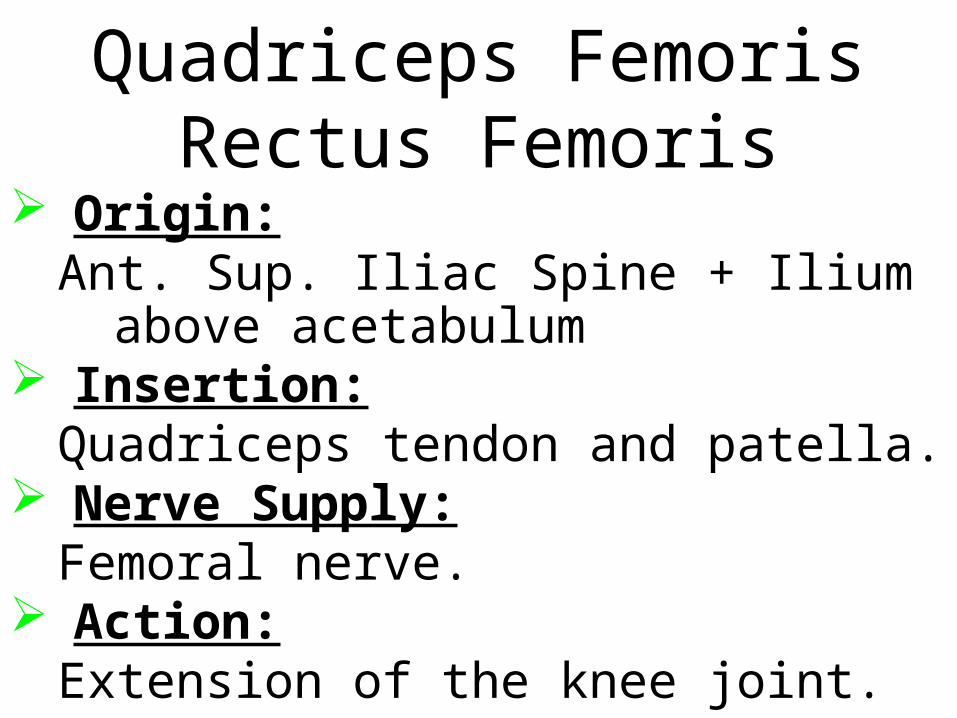
Quadriceps FemorisRectus Femoris
Origin:Ant. Sup. Iliac Spine + Ilium above
acetabulum Insertion:
Quadriceps tendon and patella. Nerve Supply:
Femoral nerve. Action:
Extension of the knee joint.
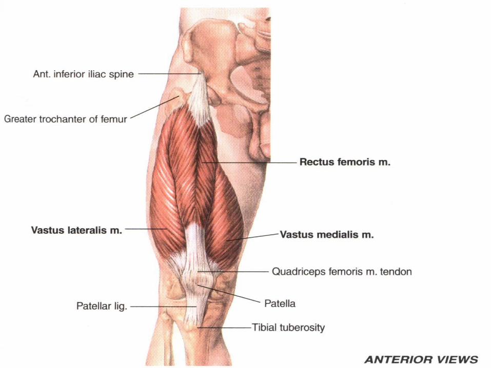
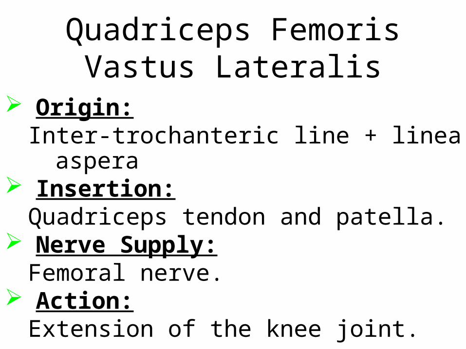
Quadriceps FemorisVastus Lateralis
Origin:Inter-trochanteric line + linea aspera
Insertion:Quadriceps tendon and patella.
Nerve Supply:Femoral nerve.
Action:Extension of the knee joint.
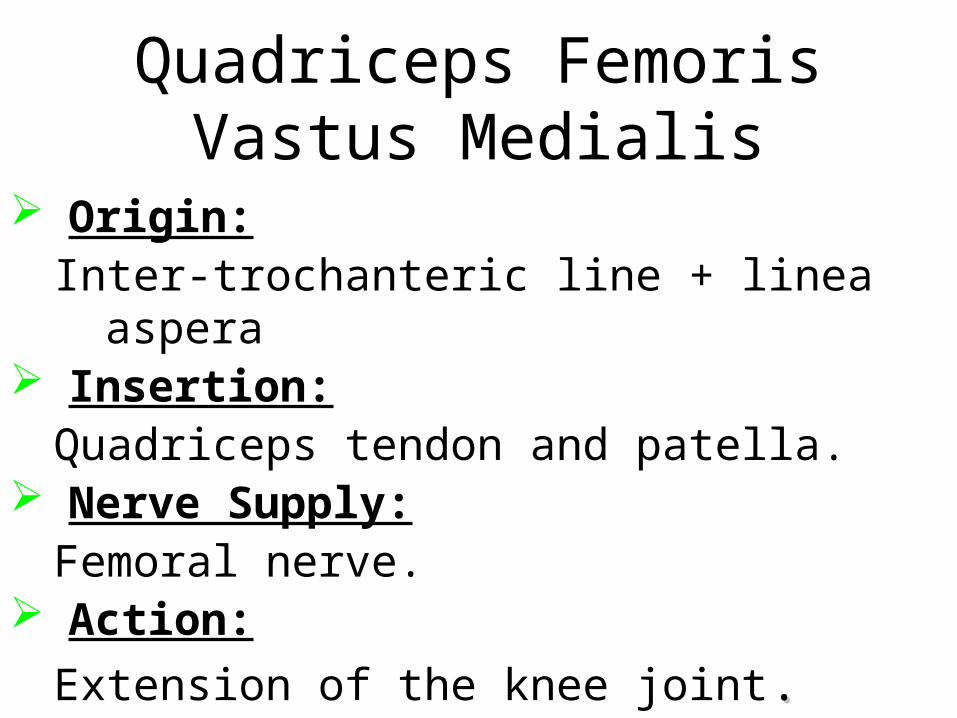
Quadriceps FemorisVastus Medialis
Origin:Inter-trochanteric line + linea aspera
Insertion:Quadriceps tendon and patella.
Nerve Supply:Femoral nerve.
Action:
Extension of the knee joint..
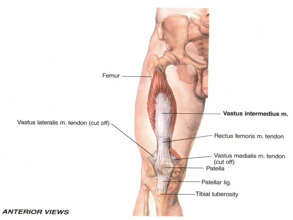
Quadriceps FemorisVastus Intermedius
Origin:Anterior and lateral surfaces of the shaft
of the femur Insertion:
Quadriceps tendon and patella. Nerve Supply:
Femoral nerve. Action:
Extension of the knee joint.
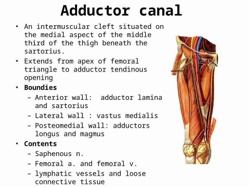
Adductor canal• An intermuscular cleft situated on the medial
aspect of the middle third of the thigh beneath the sartorius.
• Extends from apex of femoral triangle to adductor tendinous opening
• Boundies– Anterior wall: adductor lamina and
sartorius – Lateral wall : vastus medialis– Posteomedial wall: adductors longus and
magmus • Contents
– Saphenous n.– Femoral a. and femoral v.– lymphatic vessels and loose connective
tissue
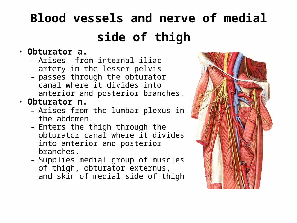
Blood vessels and nerve of medial side of
thigh • Obturator a.
– Arises from internal iliac artery in the lesser pelvis
– passes through the obturator canal where it divides into anterior and posterior branches.
• Obturator n. – Arises from the lumbar plexus in
the abdomen. – Enters the thigh through the
obturator canal where it divides into anterior and posterior branches.
– Supplies medial group of muscles of thigh, obturator externus, and skin of medial side of thigh
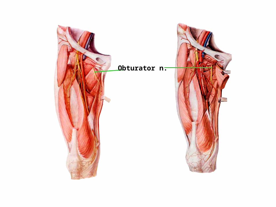
Obturator n.
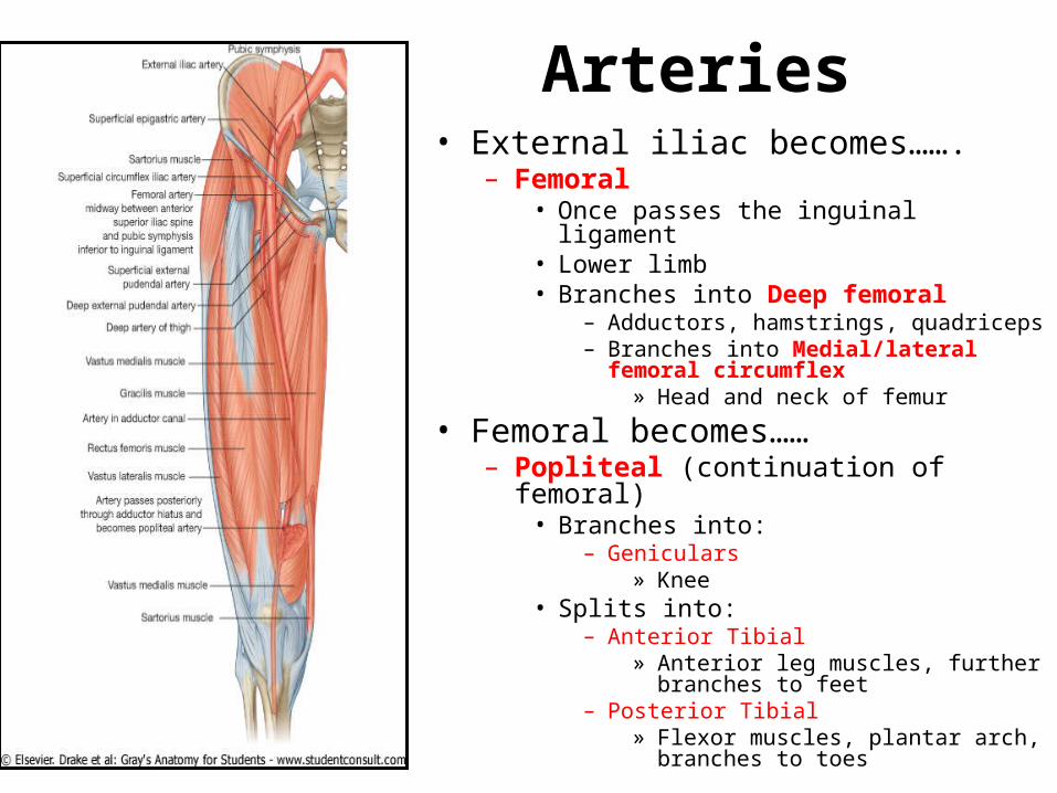
Arteries• External iliac becomes…….
– Femoral • Once passes the inguinal ligament• Lower limb• Branches into Deep femoral
– Adductors, hamstrings, quadriceps– Branches into Medial/lateral femoral
circumflex » Head and neck of femur
• Femoral becomes……– Popliteal (continuation of femoral)
• Branches into:– Geniculars
» Knee• Splits into:
– Anterior Tibial » Anterior leg muscles, further
branches to feet– Posterior Tibial
» Flexor muscles, plantar arch, branches to toes
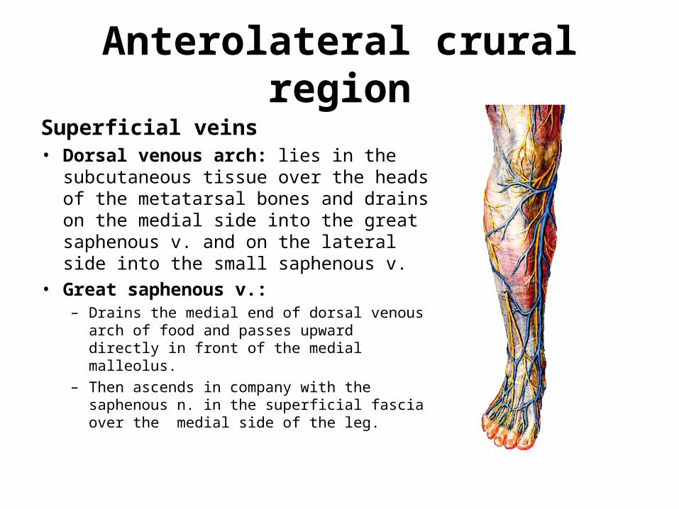
Anterolateral crural region
Superficial veins• Dorsal venous arch: lies in the
subcutaneous tissue over the heads of the metatarsal bones and drains on the medial side into the great saphenous v. and on the lateral side into the small saphenous v.
• Great saphenous v.: – Drains the medial end of dorsal venous arch
of food and passes upward directly in front of the medial malleolus.
– Then ascends in company with the saphenous n. in the superficial fascia over the medial side of the leg.