Embed Size (px)
Citation preview

LOWER QUARTER SCREENING
Rob Ellison, MSPT, FAAOMPT
Rocky Sowards, PT, DPT, OCS

INTRODUCTION
Rob Ellison, MSPT, FAAOMPT
Central States Orthopedics
Director of Therapy
Rocky Sowards, PT, DPT, OCS
University of Oklahoma
Clinic Director OU PT at Tandy

OBJECTIVES
Be able to perform a lower quarter screening examination
Recognize importance and be able to accurately take detailed history
Efficiently perform neurological examination
Link static and dynamic examination to effective treatment strategy
Understand the concept of regional interdependence
Be able to perform manual reset technique for the hip, knee, and ankle

TAKING A DETAILED HISTORY
Initial Observation
Note the unusual (things you can see from across the room)
History and Interview
Goals of Subjective Interview
Interview Format

HISTORY – INITIAL OBSERVATION
Deformity
Gait
Assistive Devices
Patient’s Behavior / Attitude
Quality of Movement Patterns
Handshake
General Appearance
Red Flags
HISTORY – SUBJECTIVE INTERVIEW
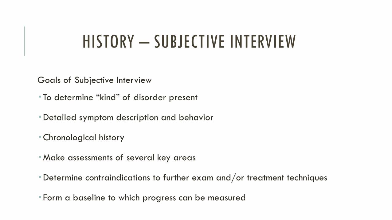
Goals of Subjective Interview
To determine “kind” of disorder present
Detailed symptom description and behavior
Chronological history
Make assessments of several key areas
Determine contraindications to further exam and/or treatment techniques
Form a baseline to which progress can be measured

HISTORY – INTERVIEW FORMAT
Patient description of problem
Onset
Pain description
Aggravates / Alleviates
Interceding episodes
Medical history
Special considerations

NEUROLOGICAL EXAMINATION
Can help determine what part of nervous system involved
Consists of the following:
Myotomes
Dermatomes
DTR’s
Neural Tissue Tension
Neurovasular testing when indicated

NEUROLOGICAL TESTING - MYOTOMES
Lower Extremity
L2: Hip Flexion – Key Muscle: Iliopsoas
L3: Knee Extension – Key Muscle: Quadriceps
L4: Ankle Dorsiflexion – Key Muscle: Tibialis Anterior
L5: Great Toe Extension – Key Muscle: EHL
L5-S1: Ankle Eversion – Key Muscle: Peroneals
S1: Ankle Plantar Flexion – Key Muscle: Gastrocnemius
S2: Knee Flexion – Key Muscle: Hamstrings

NEUROLOGICAL TESTING - DERMATOMES
These are the “most represented” cutaneous innervated areas of individual nerve roots:
L1: Inguinal Region
L2: Middle Anterior Thigh
L3: Medial Aspect of Knee
L4: Medial Lower Leg and Ankle
L5: Web Space between First and Second Toes
S1: Lateral Border of Foot
S2: Popliteal Space
S3-4: Saddle / Perianal Region

NEUROLOGICAL TESTING – DTR’S
DTR’s and Lumbar Reflexes
Patellar Tendon Reflex: L3-4
Medial Hamstring (L5-S1); Lateral Hamstring (S1-S2)
Achilles Tendon Reflex: S1
Babinski Reflex:
Normal Response?
Clonus:
What is acceptable?

LUMBAR ROOT SYNDROMES
L3: Pain Distribution:
Cutaneous Innervation:
Reflex:
Myotome:
L4 Pain Distribution:
Cutaneous Innervation:
Reflex:
Myotome:
L5 Pain Distribution:
Cutaneous Innervation:
Reflex:
Myotome:
S1: Pain Distribution:
Cutaneous Innervation:
Reflex:
Myotome:
S2-3-4: Pain Distribution:
Cutaneous Innervation:
Reflex:
Myotome:
NEURAL TISSUE TENSION / NEURODYNAMICTESTING
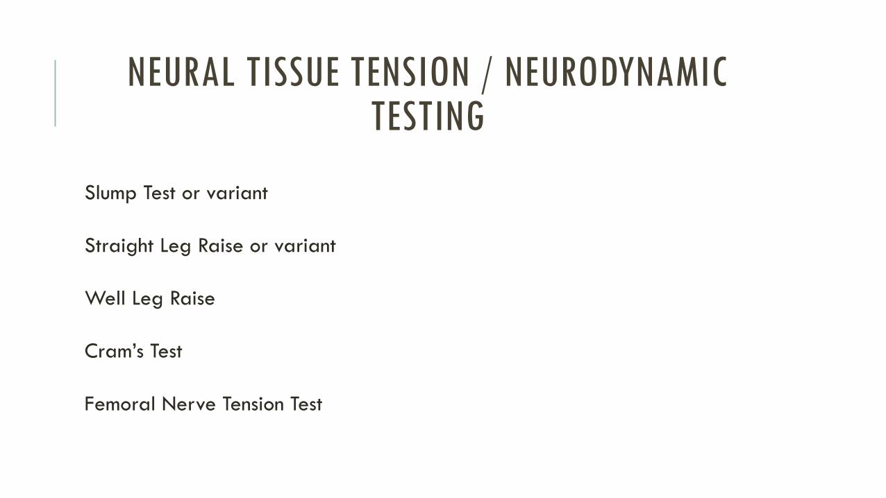
Slump Test or variant
Straight Leg Raise or variant
Well Leg Raise
Cram’s Test
Femoral Nerve Tension Test
CLEARING LUMBAR SPINE
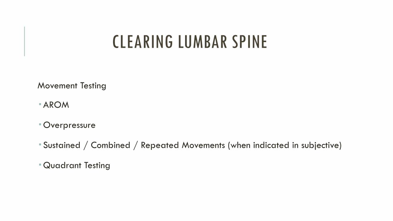
Movement Testing
AROM
Overpressure
Sustained / Combined / Repeated Movements (when indicated in subjective)
Quadrant Testing

STATIC EXAMINATION: POSTURE – LATERAL VIEW
Plumb line

STATIC EXAMINATION: POSTURE – FRONT/BACK
Symmetry
Trunk – rotation, creases
Pelvis – PSIS/ASIS/Iliac crest
Hips – neutral
Knees – neutral, varus, valgus
Feet – parallel, pointed in, pointed out
Achilles – are they straight?
Equal weight bearing?

DYNAMIC EXAMINATION - SFMA
Why do we need a movement assessment?

REGIONAL INTERDEPENDENCE
Clinically relevant relationships exist between separate regions of the body
Impairments in one region of the body are often associated with impairments in other regions of the body
Wainner, Whitman, Cleland, Flynn 2007
PAIN
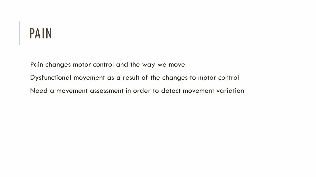
Pain changes motor control and the way we move
Dysfunctional movement as a result of the changes to motor control
Need a movement assessment in order to detect movement variation

COMPENSATION
Practical and high-level performance is possible using compensatory patterns, but you are more likely to be injured secondary to…
Altered mobility
Altered stability
Asymmetry
Cook, Burton, Hoogenboom

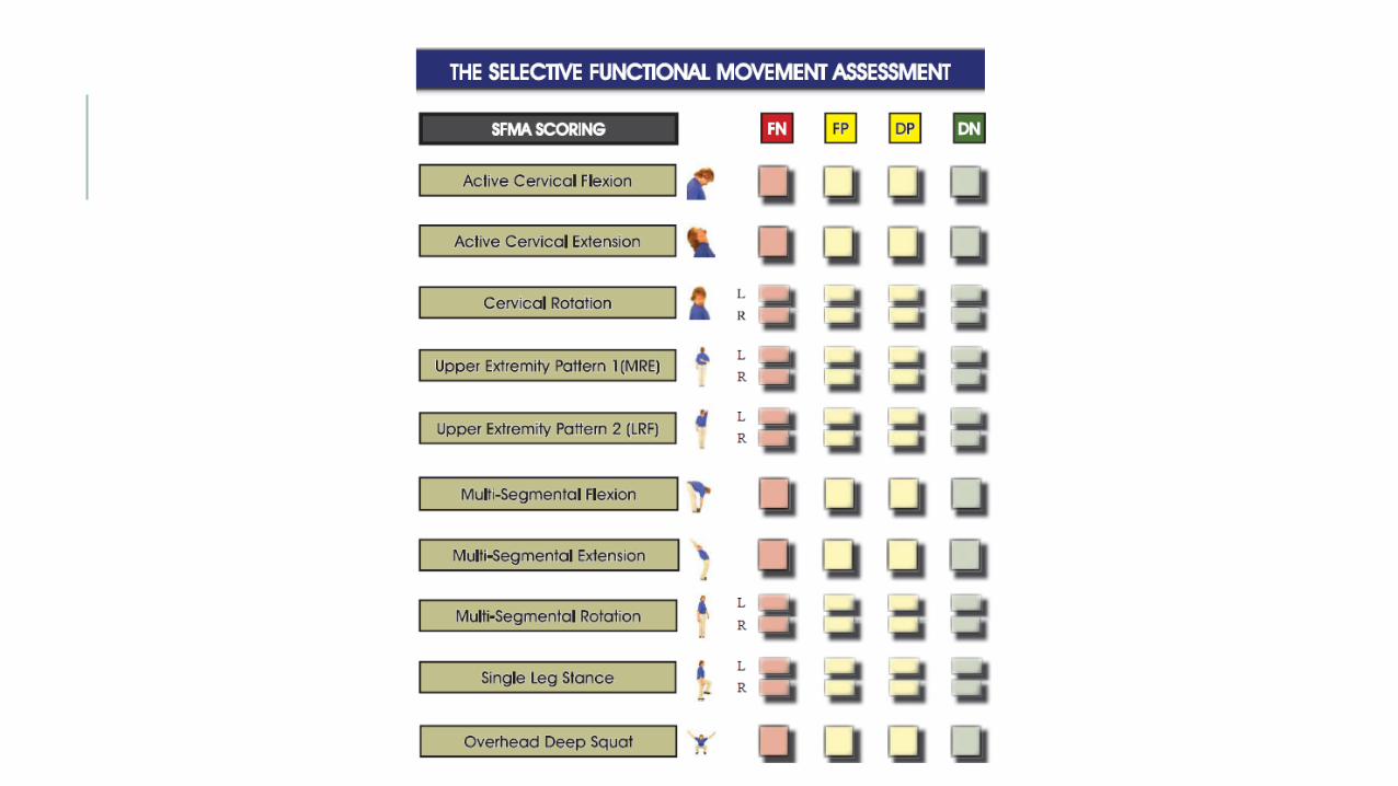
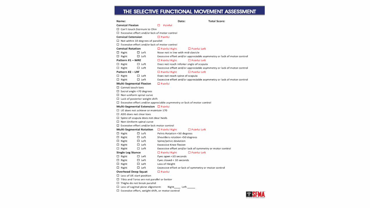
SFMA OVERVIEW
Looks at movement patterns and compares them to a baseline
Systematically breaks down movement to locate the problem
Top Tier Tests
Breakout tests with each top tier

SCORING THE SFMA
Dysfunctional
Functional
Painful
Non-Painful
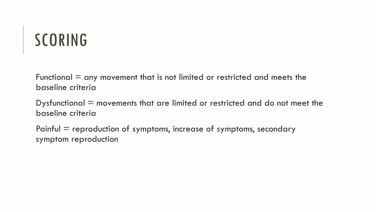
SCORING
Functional = any movement that is not limited or restricted and meets the baseline criteria
Dysfunctional = movements that are limited or restricted and do not meet the baseline criteria
Painful = reproduction of symptoms, increase of symptoms, secondary symptom reproduction

TOP TIER MOVEMENTSCervical Flexion
Cervical Extension
Cervical Rotation
Upper Extremity Pattern 1
Upper Extremity Pattern 2
Multisegmental Flexion
Multisegmental Extension
Multisegmental Rotation
Single Leg Stance
Overhead Deep Squat
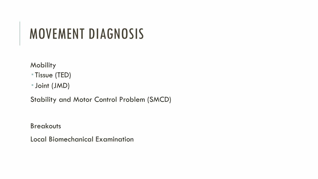
MOVEMENT DIAGNOSIS
Mobility
Tissue (TED)
Joint (JMD)
Stability and Motor Control Problem (SMCD)
Breakouts
Local Biomechanical Examination

BREAKOUT LOGIC
Unilateral vs Bilateral – remove a body part
Loaded vs Unloaded – move to a gravity lessened position
Equally limited with unloaded and loaded = mobility dysfunction
More movement with unloaded = stability and motor control dysfunction
Active vs Passive
If passive movement is within 10 deg of active = mobility dysfunction
If passive movement is much greater = stability and motor control dysfunction
Consistent vs Inconsistent
Consistent = mobility dysfunction
Inconsistnet = stability and motor control dysfunction

JOINT SPECIFIC EVALUATIONHip / Knee / Ankle
Anatomy Review
Relationship to Movement Diagnosis
Patho-Anatomy Quick Assessment Logic
Bone
Joint / Cartilage
Ligament
Muscle / Tendon
Peripheral Nerve
Spinal Nerve

JOINT SPECIFIC EVALUATION - HIP
Anatomy
Clinical Implications
Patho-Anatomy Quick Assessment
Relationship to Movement Diagnosis
JOINT SPECIFIC EVALUATION - KNEE
Anatomy
Clinical
Patho-Anatomy Quick Assessment
Relationship to Movement Diagnosis
JOINT SPECIFIC EVALUATION - ANKLE
Anatomy
Clinical
Patho-Anatomy Quick Assessment
Relationship to Movement Diagnosis

TREATMENT
Reset – typically manual intervention to reset dysfunction
Reinforce – reinforce what has been reset with therapeutic activity, stretching, taping, etc.
Reload – new movement patterns with therapeutic exercise and neuromuscular re-education

RESET
Manual intervention
Joint mobilizations
HVLA
Soft Tissue Mobilization
Myofascial Release
Instrumented assisted soft tissue mobilization
Active soft tissue release
Dry Needling

HIP
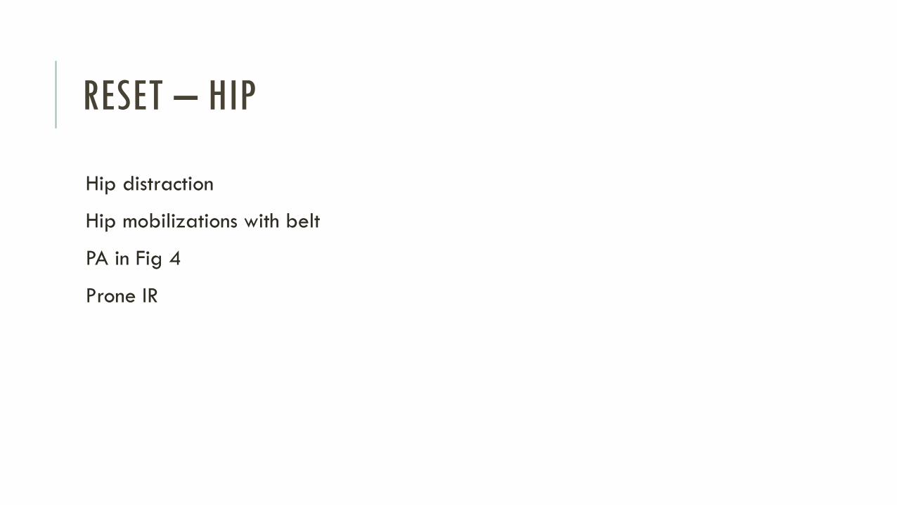
RESET – HIP
Hip distraction
Hip mobilizations with belt
PA in Fig 4
Prone IR

REINFORCE - HIP
Stretching
Positional/postural advise
Taping

RELOAD - HIP
Therapeutic exercise and neuromuscular re-education
½ kneeling
Tall kneeling
Patterns (squat, inline lunge, single leg stance)

KNEE

RESET – KNEE
Flexion Mobs
Extension Mobs
Patellar Glides
Proximal Tib/Fib Mobs
Dry needling

REINFORCE – KNEE
Stretching
Therapeutic Activities
Taping

ANKLE
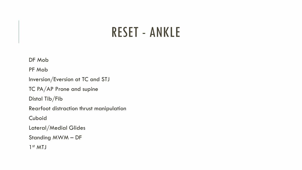
RESET - ANKLE
DF Mob
PF Mob
Inversion/Eversion at TC and STJ
TC PA/AP Prone and supine
Distal Tib/Fib
Rearfoot distraction thrust manipulation
Cuboid
Lateral/Medial Glides
Standing MWM – DF
1st MTJ

REINFORCE – ANKLE
Discussion / Education
Taping

RELOAD – ANKLE
Once mobility is established, treat the joint as if it were a stability – motor
control issue
Functional Strengthening
Return to Sport

NEURAL TENSION

RESET – NEURAL TISSUE TENSION
Techniques
Adverse Neural Tissue Tension
STM to Sciatic, Tibial, Peroneal Nerve
SLR in and out of tension positions
Modified Slump mobilizations

REINFORCE – NEURAL TENSION
Taping
Stretches
Self Mobilizations
Sliders and Tensioners

RELOAD – NEURAL TENSION
Therapeutic Exercise
Neuromuscular Re-education

CONCLUSION

QUESTIONS?