Embed Size (px)
Citation preview

RESEARCH Open Access
LPA rs10455872 polymorphism is associated withcoronary lesions in Brazilian patients submitted tocoronary angiographyPaulo CJL Santos1*, Carolina T Bueno1, Pedro A Lemos2, José E Krieger1 and Alexandre C Pereira1*
Abstract
Background: Polymorphisms in the LPA gene were associated with coronary artery disease (CAD). However, thereare differences in the allelic frequencies, Lp(a) levels, and significant association with CAD according to ethnicgroups. In this scenario, the main aim of this study was to assess the influence of the LPA polymorphisms oncoronary lesions in Brazilian patients.
Methods: 1,394 consecutive patients submitted to coronary angiography to study suggestive CAD and twentycoronary segments were scored. Genotyping for the LPA rs10455872 and rs3798220 polymorphisms wereperformed by high resolution melting analysis.
Results: The frequencies of the rs10455872 G and rs3798220 C variant alleles were 6.4% and 6.2%, respectively. LPArs10455872 G variant allele was associated with higher odds ratio of having coronary lesions in an adjusted model(OR = 2.02, 95% CI = 1.10-3.72, p = 0.02). Scores of coronary lesions (extension, severity, and Gensini scores) weresignificantly different among rs10455872 genotype groups. Coronary lesions was not associated with LPA rs3798220(OR = 1.09, 95% CI = 0.67-1.76, p = 0.73) and scores of coronary lesions were not different among rs3798220genotypes.
Conclusions: We confirmed the association of the LPA rs10455872 with CAD in a large sample of Brazilian patients.For the LPA rs3798220, our finding is consistent with studies which showed the lack of this genetic association.
Keywords: LPA gene, rs10455872, rs3798220, Coronary lesions, Coronary artery disease
IntroductionLipoprotein(a) [Lp(a)] is a plasma lipoprotein synthesizedby the liver that is composed of a low-density lipoprotein(LDL) molecule, a high molecular weight glycoproteinapolipoprotein(a), and a single molecule of apolipoprotein(B). Physiological and pathogenic roles of Lp(a) remainpartially unknown. Studies have suggested that Lp(a) pro-vides a link between the cholesterol transport and the fi-brinolytic system acting as a modulator of the bloodclotting and fibrinolysis systems [1,2].The increased concentration of Lp(a) has been associ-
ated with incidence and severity of cardiovascular dis-ease (CVD), coronary artery disease (CAD), peripheral
artery disease, and stroke [3-7]. A meta-analysis of pub-lished data from 31 prospective studies reported a rela-tive risk for coronary heart disease of 1.60 (95% CI =1.38-1.85) associated with Lp(a) levels [8,9].More than 90% of the variance of Lp(a) concentration is
explained by genetic variation [1]. A genome-wide associ-ation study showed that there is a group of genes stronglyassociated with CAD, such as solute carrier family 22member 3 (SLC22A3), lipoprotein(a)-like 2 (LPAL2), andlipoprotein(a) (LPA), but investigators did not identify thefunctional variants at these loci [10-12].Two polymorphisms in the LPA gene (rs3798220 and
rs10455872) were associated with risk for CAD. How-ever, there are differences in the allelic frequencies, Lp(a) levels, and degree of association with CAD accordingto ethnic groups [13-18]. In this scenario, the main aimof this study was to assess the influence of the LPA
* Correspondence: [email protected]; [email protected] of Genetics and Molecular Cardiology, Heart Institute (InCor),University of Sao Paulo Medical School, Av. Dr. Enéas de Carvalho Aguiar, 44Cerqueira César, São Paulo, SP CEP 05403-000, BrazilFull list of author information is available at the end of the article
© 2014 Santos et al.; licensee BioMed Central Ltd. This is an Open Access article distributed under the terms of the CreativeCommons Attribution License (http://creativecommons.org/licenses/by/4.0), which permits unrestricted use, distribution, andreproduction in any medium, provided the original work is properly credited. The Creative Commons Public DomainDedication waiver (http://creativecommons.org/publicdomain/zero/1.0/) applies to the data made available in this article,unless otherwise stated.
Santos et al. Lipids in Health and Disease 2014, 13:74http://www.lipidworld.com/content/13/1/74

polymorphisms on coronary lesions in Brazilian patientssubmitted to coronary angiography.
Patients and methodsPatients submitted to coronary angiographyOne thousand three hundred and ninety-four consecu-tive patients submitted to coronary angiography tostudy suggestive CAD were selected at the Laboratoryof Hemodynamic, Heart Institute (InCor), Sao Paulo,Brazil. All patients had a clinical diagnosis of anginapectoris and stable angina. No patient enrolled in thisstudy was currently experiencing an acute coronary syn-drome. Patients with heart failure classes III–IV, hepaticdysfunction, familiar hypercholesterolemia, previousheart or kidney transplantation, and in antiviral treat-ment were excluded [19,20]. All patients signed an in-formed consent form and the protocol was approved bythe ethics committee from Hospital das Clínicas fromSão Paulo University (CAPPesq 0398/04).
Demographic data and laboratory testsData regarding general characteristic, weight, height, race/color, main cardiovascular risk factors (hypertension, dia-betes, obesity, dyslipidemia, smoking, and current medicaltreatment) were obtained by interview. Race/color was clas-sified as White, Brown (Pardo in Portuguese; person withadmixture between White and Black), Black or Asiatic [21].Triglycerides, total cholesterol (TC), high-density lipopro-
tein cholesterol, LDL cholesterol, and glucose were evalu-ated by standard techniques in 12-h fasting blood samples.Diabetes mellitus was diagnosed by the presence of fastingglucose ≥ 126 mg/dL or the use of antidiabetic drugs [22].Hyperlipidemia was defined as TC ≥ 240 mg/dL, LDL-C ≥160 mg/dL, and/or use of lipid-lowering drugs [23].
Hemodynamic and angiographic dataBlood pressure was measured in the sitting position withthe use of a standard mercury sphygmomanometer onthe left arm after 5 min rest. The first and fifth phases ofKorotkoff sounds were used for systolic blood pressure(SBP) and diastolic blood pressure (DBP), respectively.The SBP and DBP were calculated from two readingswith a minimal interval of 10 min apart. Hypertensionwas defined as mean SBP ≥140 mm Hg and/or DBP≥90 mm Hg and/or antihypertensive drug use [24].Twenty coronary segments were scored: each vessel
was divided into three segments (proximal, medial, anddistal), except for the secondary branches of the rightcoronary artery (posterior ventricular and posterior de-scending), which were divided into proximal and distalsegments. Stenosis higher than 50% in any coronary seg-ment was graded 1 point and the sum of points for all20 segments constituted the Extension Score. Lesion se-verity was calculated as follows: none and irregularities, 0
points; <50%, 0.3 points; 50–70%, 0.6 points; >70–90%, 0.8points; and >90–100%, 0.95 points. The Severity Scorewas calculated through the sum of points for all 20 coron-ary segments [25].
GenotypingGenomic DNA from subjects was extracted from periph-eral blood following standard salting-out procedure.Additional file 1: Figure S1 shows genotyping detectedby polymerase chain reaction (PCR) followed by highresolution melting (HRM) analysis with the Rotor Gene6000® instrument (Qiagen, Courtaboeuf, France). TheQIAgility® (Qiagen, Courtaboeuf, France), an automatedinstrument, was used according to instructions tooptimize the sample preparation step [26].Amplification of the fragment for the LPA rs10455872
(A > G, intron 25) polymorphism was performed usingthe primer sense 5′- ATGGGCTGGCAACACATAG -3′ and antisense 5′- CACTTTCTCCTCTAACCTGTATAA -3′ (78 pairs base). Amplification of the fragmentfor the LPA rs3798220 (T > C, p.Ile1891Met) poly-morphism was performed using the primer sense 5′-GGCTCCAAGAACAGCCTAGA -3′ and antisense5′- TCCTCAAGGCCTTCATCCTA -3′ (104 pairs base).A 40-cycle PCR was carried out with the following condi-tions: denaturation of the template DNA for first cycle of94°C for 120 s, denaturation of 94ºC for 20 s, annealing of56.7°C for 20 s, and extension of 72°C for 22 s. PCR wasperformed with addition of fluorescent DNA-intercalatingSYTO9® (1.5 μM; Invitrogen, Carlsbad, USA). In the HRMphase, the Rotor Gene 6000® measured the fluorescence ineach 0.1°C temperature increase in the range of 72-81°C.Melting curves were generated by the decrease in fluores-cence with the increase in the temperature; and in ana-lysis, nucleotide changes result in three different curvepatterns (Additional file 1: Figure S1). Samples of thethree observed curves were analyzed using bidirectionalsequencing as a validation procedure (ABI TerminatorSequencing Kit® and ABI 3500XL Sequencer® - AppliedBiosystems, Foster City, CA, USA) [27]. The two methodsgave identical results in all tests. The wild-type, heterozy-gous and mutant homozygous genotypes could be easilydiscernible by HRM analysis. In addition, 4% of the sam-ples were randomly selected and reanalyzed as qualitycontrols and gave identical results.
Statistical analysisCategorical variables are presented as percentage whilecontinuous variables are presented as mean ± standard de-viation. Chi-square test was performed for comparativeanalysis of general characteristics and coronary lesion fre-quency according to LPA polymorphisms. Dominantmodels (AG+GG) for the rs10455872 or (TC +CC) forthe rs3798220 were performed because the frequencies of
Santos et al. Lipids in Health and Disease 2014, 13:74 Page 2 of 7http://www.lipidworld.com/content/13/1/74
the GG and CC homozygous genotypes are low. Student’st-test was performed for comparing the age, body massindex (BMI), biochemical data, blood pressures, and angio-graphic data means according to LPA polymorphisms. Bio-chemical data, blood pressures, and angiographic data wereadjusted for age, gender, and race/color. Logistic regressionunivariate and multivariate analyses were performed toevaluate OR (odds ratio) for coronary lesions. Two adjustedmodels were performed: one using age, gender, and race/color and another with the additional covariates BMI,hyperlipidemia, statin use, and smoking. Linkage disequilib-rium, Hardy-Weinberg equilibrium, and haplotype analyseswere conducted with Haploview 4.0. All statistical analyseswere carried out using the SPSS software (v. 16.0), with thelevel of significance set at p ≤ 0.05.
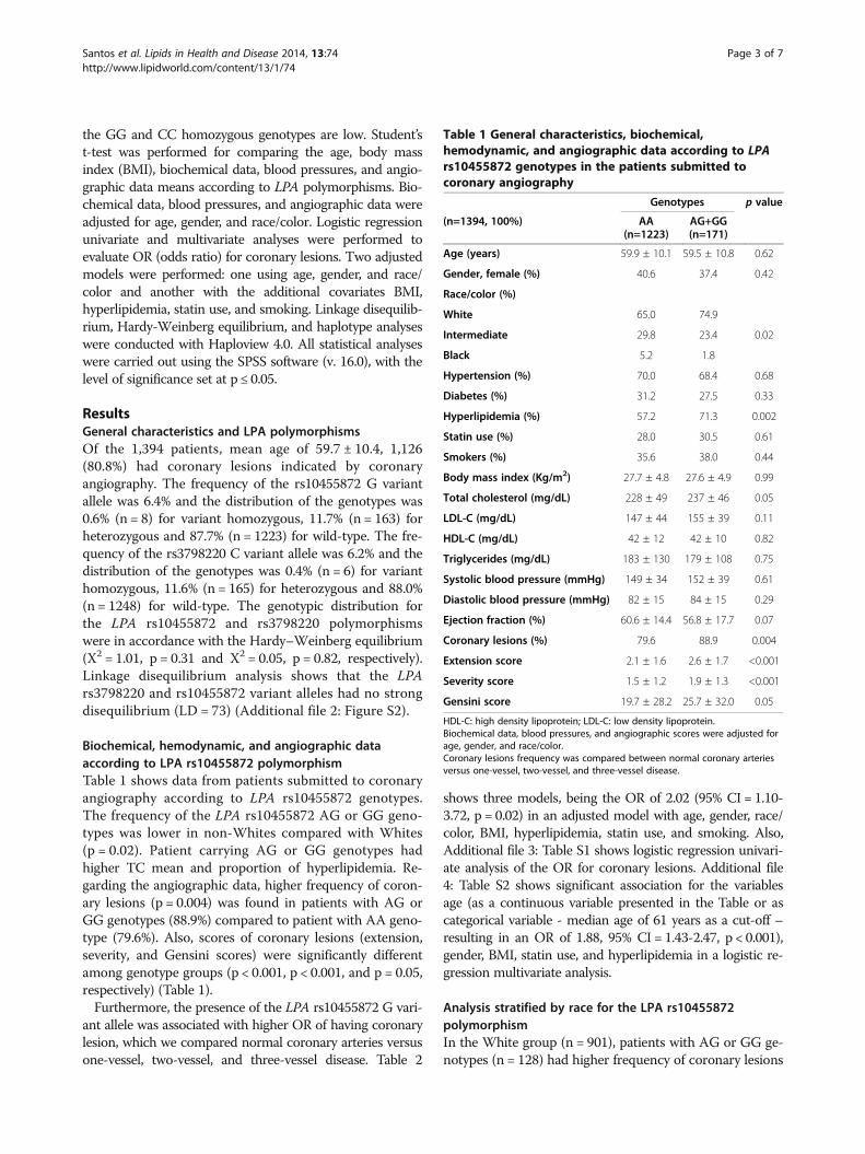
ResultsGeneral characteristics and LPA polymorphismsOf the 1,394 patients, mean age of 59.7 ± 10.4, 1,126(80.8%) had coronary lesions indicated by coronaryangiography. The frequency of the rs10455872 G variantallele was 6.4% and the distribution of the genotypes was0.6% (n = 8) for variant homozygous, 11.7% (n = 163) forheterozygous and 87.7% (n = 1223) for wild-type. The fre-quency of the rs3798220 C variant allele was 6.2% and thedistribution of the genotypes was 0.4% (n = 6) for varianthomozygous, 11.6% (n = 165) for heterozygous and 88.0%(n = 1248) for wild-type. The genotypic distribution forthe LPA rs10455872 and rs3798220 polymorphismswere in accordance with the Hardy–Weinberg equilibrium(X2 = 1.01, p = 0.31 and X2 = 0.05, p = 0.82, respectively).Linkage disequilibrium analysis shows that the LPArs3798220 and rs10455872 variant alleles had no strongdisequilibrium (LD = 73) (Additional file 2: Figure S2).
Biochemical, hemodynamic, and angiographic dataaccording to LPA rs10455872 polymorphismTable 1 shows data from patients submitted to coronaryangiography according to LPA rs10455872 genotypes.The frequency of the LPA rs10455872 AG or GG geno-types was lower in non-Whites compared with Whites(p = 0.02). Patient carrying AG or GG genotypes hadhigher TC mean and proportion of hyperlipidemia. Re-garding the angiographic data, higher frequency of coron-ary lesions (p = 0.004) was found in patients with AG orGG genotypes (88.9%) compared to patient with AA geno-type (79.6%). Also, scores of coronary lesions (extension,severity, and Gensini scores) were significantly differentamong genotype groups (p < 0.001, p < 0.001, and p = 0.05,respectively) (Table 1).Furthermore, the presence of the LPA rs10455872 G vari-
ant allele was associated with higher OR of having coronarylesion, which we compared normal coronary arteries versusone-vessel, two-vessel, and three-vessel disease. Table 2
shows three models, being the OR of 2.02 (95% CI = 1.10-3.72, p = 0.02) in an adjusted model with age, gender, race/color, BMI, hyperlipidemia, statin use, and smoking. Also,Additional file 3: Table S1 shows logistic regression univari-ate analysis of the OR for coronary lesions. Additional file4: Table S2 shows significant association for the variablesage (as a continuous variable presented in the Table or ascategorical variable - median age of 61 years as a cut-off –resulting in an OR of 1.88, 95% CI = 1.43-2.47, p < 0.001),gender, BMI, statin use, and hyperlipidemia in a logistic re-gression multivariate analysis.
Analysis stratified by race for the LPA rs10455872polymorphismIn the White group (n = 901), patients with AG or GG ge-notypes (n = 128) had higher frequency of coronary lesions
Table 1 General characteristics, biochemical,hemodynamic, and angiographic data according to LPArs10455872 genotypes in the patients submitted tocoronary angiography
Genotypes p value
(n=1394, 100%) AA(n=1223)
AG+GG(n=171)
Age (years) 59.9 ± 10.1 59.5 ± 10.8 0.62
Gender, female (%) 40.6 37.4 0.42
Race/color (%)
White 65.0 74.9
Intermediate 29.8 23.4 0.02
Black 5.2 1.8
Hypertension (%) 70.0 68.4 0.68
Diabetes (%) 31.2 27.5 0.33
Hyperlipidemia (%) 57.2 71.3 0.002
Statin use (%) 28.0 30.5 0.61
Smokers (%) 35.6 38.0 0.44
Body mass index (Kg/m2) 27.7 ± 4.8 27.6 ± 4.9 0.99
Total cholesterol (mg/dL) 228 ± 49 237 ± 46 0.05
LDL-C (mg/dL) 147 ± 44 155 ± 39 0.11
HDL-C (mg/dL) 42 ± 12 42 ± 10 0.82
Triglycerides (mg/dL) 183 ± 130 179 ± 108 0.75
Systolic blood pressure (mmHg) 149 ± 34 152 ± 39 0.61
Diastolic blood pressure (mmHg) 82 ± 15 84 ± 15 0.29
Ejection fraction (%) 60.6 ± 14.4 56.8 ± 17.7 0.07
Coronary lesions (%) 79.6 88.9 0.004
Extension score 2.1 ± 1.6 2.6 ± 1.7 <0.001
Severity score 1.5 ± 1.2 1.9 ± 1.3 <0.001
Gensini score 19.7 ± 28.2 25.7 ± 32.0 0.05
HDL-C: high density lipoprotein; LDL-C: low density lipoprotein.Biochemical data, blood pressures, and angiographic scores were adjusted forage, gender, and race/color.Coronary lesions frequency was compared between normal coronary arteriesversus one-vessel, two-vessel, and three-vessel disease.
Santos et al. Lipids in Health and Disease 2014, 13:74 Page 3 of 7http://www.lipidworld.com/content/13/1/74

compared to patient with AA genotype (n = 773) (90.6%and 81.0%, respectively, p = 0.008). The presence of theLPA rs10455872 G variant allele was associated withhigher OR of having coronary lesion in an adjusted model(OR = 2.17, 95% CI = 1.04-4.61, p = 0.03). Also, scores ofcoronary lesions were significantly different among geno-type (extension: 2.7 ± 1.6 and 2.2 ± 1.6; severity: 1.9 ± 1.3and 1.6 ± 1.2; and Gensini scores: 26.3 ± 30.0 and 19.7 ±29.8) (p = 0.001, p = 0.01, and p = 0.04, respectively).In the non-White group (n = 460, formed for Black and
Brown individuals), frequency of coronary lesions was notstatistically different (83.7% for AG or GG genotypes and77.0% for AA genotype, p = 0.30). However, the presence ofthe G variant allele was associated with higher OR (OR =1.95, 95% CI = 1.09-4.39, p = 0.04 – adjusted model) andscores of coronary lesions were different between AG orGG and AA genotypes (extension score: 2.6 ± 1.7 and 2.0 ±1.6; and severity score: 1.9 ± 1.3 and 1.4 ± 1.2) (p = 0.04, andp = 0.03, respectively). Gensini score was marginallydifferent between genotypes (24.7 ± 29.6 and 21.2 ± 27.5;p = 0.06).
Biochemical, hemodynamic, and angiographic dataaccording to LPA rs3798220 polymorphismThe proportion of hyperlipidemia was not differentamong rs3798220 genotypes (p = 0.62). Regarding theangiographic data, the frequency of coronary lesionswas not associated with rs3798220 (p = 0.57) and no sig-nificant OR was observed in an adjusted model (OR =1.09, 95% CI = 0.67-1.76, p = 0.73). Also, scores of cor-onary lesions (extension, severity, and Gensini scores)were not significantly different among genotype groups(p = 0.85, p = 0.56, and p = 0.46, respectively).
DiscussionThe two LPA polymorphisms studied in this study aresome of the most important genetic markers for CAD.In this context, our main finding was that the LPArs10455872 polymorphism is associated with coronarylesions in Brazilian patients submitted to coronoryangiography. On the other hand, no association for the
LPA rs3798220 was observed for any of the testedphenotypes.Studies have reported higher Lp(a) concentration in
sub-Saharan African descent and lower Lp(a) concentra-tion in European descent [13,15,28,29]. Regarding theethnicity, Brazil has one of the most heterogeneouspopulation of the world, composed by a mixture of dif-ferent ethinic groups, mainly European descent, Africandescent and Amerindians. In our data, a stratified ana-lysis by race supported a role for the rs10455872 poly-morphism independent of ethnic group.Corroboring with our study, Anderson et al. found that
the rs10455872 polymorphism strongly predicted preva-lent CAD (per allele OR = 1.43, 95% CI = 1.07-1.91) [30].Helgadottir et al. showed that patients with CAD carryingLPA risk alleles have increased susceptibility to athero-sclerotic manifestations outside of the coronary tree andthey are more likely to be diagnosed earlier with CADthan are CAD cases not carrying this variant [31]. Otherstudies that analyzed the rs10455872 and rs3798220 poly-morphisms together reported an increased risk of coron-ary disease and Lp(a) level that can be explained by theseLPA polymorphisms [11,32]. LPA rs10455872 is an in-tronic polymorphism associated with short KIV-2 repeatregion (kringle IV type 2) which is associated with Lp(a)levels. In the present study, the frequency of thers10455872 G variant allele was 6.4% for overall, but weobserved higher allelic frequency in White compared withnon-White groups. The association of the rs10455872with CAD was significant even in the non-White groupwhich had a small sample size. These data suggest thatrs10455872 is also a strong genetic marker for CAD riskin ethnically mixed populations.For the LPA rs3798220 polymorphism, we did not ob-
serve significant association with coronary lesions. Datafrom other studies also did not support a relationship be-tween this LPA variant and CAD [33,34]. Furthermore,Anderson et al., studying 1,400 participants with coronaryangiography (more than 90% Whites), did not find an as-sociation signal between rs3798220 and CAD (OR = 1.47,95% CI = 0.81-2.67, p = 0.20) [30]. In contrast to our study,the rs3798220 has previously been reported to have an as-sociation with the Lp(a) level and the risk of coronary dis-ease [35-37]. LPA rs3798220 results in an aminoacidsubstitution in the protease domain of LPA, but it can notprovide stronger association than rs10455872 which mightbe representating a more complex group of genetic vari-ants or repeat structures. A possible hypothesis for thelack of this association in the present study could be thelower value of linkage disequilibrium between rs3798220and rs10455872 identified in the Brazilian patients com-pared with some studies with patients predominantly fromEuropean descent [11,35-37]. Another hypothesis may below statistical power, but less likely if the impact of
Table 2 Analysis of the coronary lesions odds ratioassociated with LPA rs10455872 AG or GG genotypes inthe patients submitted to coronary angiography
(n=1394, 100%) OR 95% CI p value
Models
Unadjusted 2.04 1.24-3.36 0.005
Adjusted* 2.05 1.22-3.43 0.006
Adjusted** 2.02 1.10-3.72 0.02
Coronary lesions frequency was compared between normal coronary arteriesversus one-vessel, two-vessel, and three-vessel disease.*Adjusted for age, gender, and race/color.**Adjusted for age, gender, race/color, body mass index, hyperlipidemia, statinuse, and smoking.
Santos et al. Lipids in Health and Disease 2014, 13:74 Page 4 of 7http://www.lipidworld.com/content/13/1/74

rs3798220 was approximately equal to the impact ofrs10455872. However, the exact reason is unclear andother genetic components differently expressed due toethnicity might be important modulators.The exact mechanism by which an increased Lp(a)
level increases the CAD risk is not fully understood.Pathways modulated by Lp(a) may involve the LDL-cholesterol transport system, the inhibition of the ex-pression of tissue factor, the inhibition of conversion ofplasminogen to plasmin, the carriage of pro-inflammatoryoxidized phospholipids, and an atherosclerotic stenoticmechanism [30,38-44]. Some studies reported that the se-verity of coronary artery disease is associated with Lp(a)levels or LDL concentration [45-48]. Regarding to the Lp(a)level as a risk factor in different ethnic groups, Lp(a) hasbeen associated with risk in European populations [1], butnot unequivocally in African Americans [17,18]. However,a recent study identified that the increased risk of CVDwas at least as strong in African Americans as in WhiteAmericans [49]. Another study investigated differentialfrequencies of LPA polymorphisms in non-Hispanicwhites, non-Hispanic blacks, and Mexican Americans[50]. Interestingly, 15 of the 19 polymorphisms testedwere strongly associated with Lp(a) levels in at least onesubpopulation, six in at least two subpopulations, andnone in all three subpopulations. The lack of gene-ralization of associations across ethnicities suggests thatspecific LPA variants may be contributing to the observedLp(a) between-population variance. Authors also com-pared the allele frequencies in HapMap, and observed ex-tremely high correlations (r ≥ 0.99) in allele frequenciesbetween non-Hispanic whites and HapMap CEU (USindividuals of northern and western European ancestry)and between non-Hispanic blacks and both HapMap YRI(Yoruba from West Africa) and ASW (individuals withAfrican ancestry from the Southwest USA) [50].There are some limitations in our study. First, we did
not measure Lp(a) levels and we also did not genotypeKIV-2 repeats to check their association with both theLPA polymorphisms and/or the CAD phenotype. Sec-ond, we did not assess ancestry through genetic markers;instead, we used a self-declared classification which iscommonly applied in Brazil and correlates with geneticancestry determination. In addition, in our stratified ana-lysis by race, we observed significant association of thers10455872 with CAD in the White and non-White pa-tient groups. Third, our plaque burden data are derivedfrom institutional records and represent real-life data, asopposed to core-lab derived hemodinamic data. Thus,and despite the greater external validity of our results,we were not able to determine inter- or intra-observedvariability estimates. In addition, our choosen methodfor establishing atherosclerotic burden in the studied pa-tients has relied upon the Gensini Score, which has been
shown to highly correlate with this end-point. Otherscores could also be used, although they are not as wellfitted for quantifing plaque burden. One example is theSyntax score, an angiographic tool for grading the com-plexity of CAD and designed to better anticipate therisks of percutaneous or surgical revascularization. Fin-naly, it is not possible to completely exclude the inter-action of the covariates as other genetic markers, use ofconcomitant drugs, ethnicity, gender and age on ourfindings [51-54]. Nonetheless, our findings remainedafter multivariate analysis.
ConclusionsIn conclusion, we confirmed the association of the LPArs10455872 with CAD in a large sample of Brazilian pa-tients. For the LPA rs3798220, our finding is consistentwith studies which showed the lack of this geneticassociation.
Additional files
Additional file 1: Figure S1. Graphs of the LPA rs10455872 (A > G,intron 25) genotyping. Nucleotide changes results in different curvepatterns using high resolution melting analysis. A: Graph of normalizedfluorescence by temperature. B: Graph of normalized fluorescence(based on genotype 2) by temperature. 1: wild-type genotype (AA);2: heterozygous genotype (AG); 3: mutant homozygous genotype (GG).
Additional file 2: Figure S2. Linkage disequilibrium and haplotypeanalyses for the LPA rs3798220 and rs10455872 polymorphisms in thepatients submitted to coronary angiography.
Additional file 3: Table S1. Logistic regression univariate analysis ofthe coronary lesions odds ratio in the patients submitted to coronaryangiography.
Additional file 4: Table S2. Logistic regression multivariate analysis ofthe coronary lesions odds ratio in the patients submitted to coronaryangiography.
Competing interestsThe authors declare that they have no competing interests.
Authors' contributionsPCJLS and CTB carried out molecular genetic analyses, performed thestatistical analysis and drafted the manuscript. PCJLS, ACP, PAL, JEKconceived the study, and participated in its design. All authors read andapproved the final manuscript.
AcknowledgmentsPCJL Santos is recipient of fellowship from FAPESP, Proc. 2013-09295-3, andProc. 2013-20614-3, Brazil. We also thank the patients who participated in thestudy. The technical assistance of the Laboratory of Genetics and MolecularCardiology group is gratefully acknowledged.
Author details1Laboratory of Genetics and Molecular Cardiology, Heart Institute (InCor),University of Sao Paulo Medical School, Av. Dr. Enéas de Carvalho Aguiar, 44Cerqueira César, São Paulo, SP CEP 05403-000, Brazil. 2HemodynamicLaboratory, Heart Institute (InCor), University of Sao Paulo Medical School,Sao Paulo, Brazil.
Received: 8 March 2014 Accepted: 22 April 2014Published: 29 April 2014
Santos et al. Lipids in Health and Disease 2014, 13:74 Page 5 of 7http://www.lipidworld.com/content/13/1/74

References1. Kronenberg F, Utermann G: Lipoprotein(a): Resurrected by genetics.
J Intern Med 2013, 273:6–30.2. Koschinsky ML: Novel insights into lp(a) physiology and pathogenicity: More
questions than answers? Cardiovasc Hematol Disord Drug Targets 2006, 6:267–278.3. Erqou S, Kaptoge S, Perry PL, Di Angelantonio E, Thompson A, White IR,
Marcovina SM, Collins R, Thompson SG, Danesh J, Collaboration ERF:Lipoprotein(a) concentration and the risk of coronary heart disease,stroke, and nonvascular mortality. JAMA 2009, 302:412–423.
4. Tsimikas S, Mallat Z, Talmud PJ, Kastelein JJ, Wareham NJ, Sandhu MS, MillerER, Benessiano J, Tedgui A, Witztum JL, Khaw KT, Boekholdt SM: Oxidation-specific biomarkers, lipoprotein(a), and risk of fatal and nonfatalcoronary events. J Am Coll Cardiol 2010, 56:946–955.
5. Danesh J, Collins R, Peto R: Lipoprotein(a) and coronary heart disease.Meta-analysis of prospective studies. Circulation 2000, 102:1082–1085.
6. Klein JH, Hegele RA, Hackam DG, Koschinsky ML, Huff MW, Spence JD:Lipoprotein(a) is associated differentially with carotid stenosis, occlusion,and total plaque area. Arterioscler Thromb Vasc Biol 2008, 28:1851–1856.
7. Ohira T, Schreiner PJ, Morrisett JD, Chambless LE, Rosamond WD, FolsomAR: Lipoprotein(a) and incident ischemic stroke: The atherosclerosis riskin communities (aric) study. Stroke 2006, 37:1407–1412.
8. Bennet A, Di Angelantonio E, Erqou S, Eiriksdottir G, Sigurdsson G,Woodward M, Rumley A, Lowe GD, Danesh J, Gudnason V: Lipoprotein(a)levels and risk of future coronary heart disease: Large-scale prospectivedata. Arch Intern Med 2008, 168:598–608.
9. Nestel PJ, Barnes EH, Tonkin AM, Simes J, Fournier M, White HD, ColquhounDM, Blankenberg S, Sullivan DR: Plasma lipoprotein(a) concentration predictsfuture coronary and cardiovascular events in patients with stable coronaryheart disease. Arterioscler Thromb Vasc Biol 2013, 33:2902–2908.
10. Trégouët DA, König IR, Erdmann J, Munteanu A, Braund PS, Hall AS, GrosshennigA, Linsel-Nitschke P, Perret C, DeSuremain M, Meitinger T, Wright BJ, Preuss M,Balmforth AJ, Ball SG, Meisinger C, Germain C, Evans A, Arveiler D, Luc G,Ruidavets JB, Morrison C, van der Harst P, Schreiber S, Neureuther K, Schäfer A,Bugert P, El Mokhtari NE, Schrezenmeir J, Stark K, et al: Genome-wide haplotypeassociation study identifies the slc22a3-lpal2-lpa gene cluster as a risk locusfor coronary artery disease. Nat Genet 2009, 41:283–285.
11. Clarke R, Peden JF, Hopewell JC, Kyriakou T, Goel A, Heath SC, Parish S, BarleraS, Franzosi MG, Rust S, Bennett D, Silveira A, Malarstig A, Green FR, Lathrop M,Gigante B, Leander K, De Faire U, Seedorf U, Hamsten A, Collins R, Watkins H,Farrall M, Consortium P: Genetic variants associated with lp(a) lipoproteinlevel and coronary disease. N Engl J Med 2009, 361:2518–2528.
12. Hopewell JC, Clarke R, Parish S, Armitage J, Lathrop M, Hager J, Collins R,Group HPSC: Lipoprotein(a) genetic variants associated with coronaryand peripheral vascular disease but not with stroke risk in the heartprotection study. Circ Cardiovasc Genet 2011, 4:68–73.
13. Chretien JP, Coresh J, Berthier-Schaad Y, Kao WH, Fink NE, Klag MJ, Marcovina SM,Giaculli F, Smith MW: Three single-nucleotide polymorphisms in lpaaccount for most of the increase in lipoprotein(a) level elevation in africanamericans compared with european americans. J Med Genet 2006, 43:917–923.
14. Lanktree MB, Anand SS, Yusuf S, Hegele RA, Investigators S: Comprehensiveanalysis of genomic variation in the lpa locus and its relationship toplasma lipoprotein(a) in south asians, chinese, and european caucasians.Circ Cardiovasc Genet 2010, 3:39–46.
15. Deo RC, Wilson JG, Xing C, Lawson K, Kao WH, Reich D, Tandon A,Akylbekova E, Patterson N, Mosley TH, Boerwinkle E, Taylor HA: Single-nucleotide polymorphisms in lpa explain most of the ancestry-specificvariation in lp(a) levels in african americans. PLoS One 2011, 6:e14581.
16. Mooser V, Scheer D, Marcovina SM, Wang J, Guerra R, Cohen J, Hobbs HH:The apo(a) gene is the major determinant of variation in plasma lp(a)levels in african americans. Am J Hum Genet 1997, 61:402–417.
17. Paultre F, Pearson TA, Weil HF, Tuck CH, Myerson M, Rubin J, Francis CK,Marx HF, Philbin EF, Reed RG, Berglund L: High levels of lp(a) with a smallapo(a) isoform are associated with coronary artery disease in africanamerican and white men. Arterioscler Thromb Vasc Biol 2000, 20:2619–2624.
18. Moliterno DJ, Jokinen EV, Miserez AR, Lange RA, Willard JE, Boerwinkle E,Hillis LD, Hobbs HH: No association between plasma lipoprotein(a)concentrations and the presence or absence of coronary atherosclerosisin african-americans. Arterioscler Thromb Vasc Biol 1995, 15:850–855.
19. Maciel SS: Pereira Ada C, Silva GJ, Rodrigues MV, Mill JG, Krieger JE:Association between glutathione s-transferase polymorphisms andtriglycerides and hdl-cholesterol. Atherosclerosis 2009, 206:204–208.
20. Santos PC, Oliveira TG, Lemos PA, Mill JG, Krieger JE, Pereira AC: Mylipp.N342s polymorphism is not associated with lipid profile in thebrazilian population. Lipids Health Dis 2012, 11:83.
21. Santos PC, Alvim Rde O, Ferreira NE, De Sa Cunha R, Krieger JE, Mill JG,Pereira AC: Ethnicity and arterial stiffness in brazil. Am J Hypertens 2011,24:278–284.
22. Executive summary: Standards of medical care in diabetes–2011. DiabetesCare 2011, 34(Suppl 1):S4–S10.
23. Executive summary of the third report of the national cholesteroleducation program (ncep) expert panel on detection, evaluation, andtreatment of high blood cholesterol in adults (adult treatment panel iii).JAMA 2001, 285:2486–2497.
24. The seventh report of the joint national committee on prevention,detection, evaluation, and treatment of high blood pressure. JAMA 2003,2004.
25. Lanz JR, Pereira AC, Martinez E, Krieger JE: Metabolic syndrome andcoronary artery disease: Is there a gender specific effect? Int J Cardiol2006, 107:317–321.
26. Santos PC, Soares RA, Santos DB, Nascimento RM, Coelho GL, Nicolau JC,Mill JG, Krieger JE, Pereira AC: Cyp2c19 and abcb1 gene polymorphismsare differently distributed according to ethnicity in the brazilian generalpopulation. BMC Med Genet 2011, 12:13.
27. Santos PC, Soares RA, Nascimento RM, Machado-Coelho GL, Mill JG, KriegerJE, Pereira AC: Slco1b1 rs4149056 polymorphism associated withstatin-induced myopathy is differently distributed according to ethnicityin the brazilian general population: Amerindians as a high risk ethnicgroup. BMC Med Genet 2011, 12:136.
28. Cobbaert C, Kesteloot H: Serum lipoprotein(a) levels in racially differentpopulations. Am J Epidemiol 1992, 136:441–449.
29. Utermann G: Genetic architecture and evolution of the lipoprotein(a)trait. Curr Opin Lipidol 1999, 10:133–141.
30. Anderson JL, Knight S, May HT, Horne BD, Bair TL, Huntinghouse JA, RolloJS, Muhlestein JB, Carlquist JF: Validation and quantification of geneticdeterminants of lipoprotein-a levels and predictive value forangiographic coronary artery disease. Am J Cardiol 2013, 112:799–804.
31. Helgadottir A, Gretarsdottir S, Thorleifsson G, Holm H, Patel RS, Gudnason T,Jones GT, van Rij AM, Eapen DJ, Baas AF, Tregouet DA, Morange PE,Emmerich J, Lindblad B, Gottsäter A, Kiemeny LA, Lindholt JS, Sakalihasan N,Ferrell RE, Carey DJ, Elmore JR, Tsao PS, Grarup N, Jørgensen T, Witte DR,Hansen T, Pedersen O, Pola R, Gaetani E, Magnadottir HB, et al:Apolipoprotein(a) genetic sequence variants associated with systemicatherosclerosis and coronary atherosclerotic burden but not withvenous thromboembolism. J Am Coll Cardiol 2012, 60:722–729.
32. Ronald J, Rajagopalan R, Cerrato F, Nord AS, Hatsukami T, Kohler T,Marcovina S, Heagerty P, Jarvik GP: Genetic variation in lpal2, lpa, and plgpredicts plasma lipoprotein(a) level and carotid artery disease risk.Stroke 2011, 42:2–9.
33. Li ZG, Li G, Zhou YL, Chen ZJ, Yang JQ, Zhang Y, Sun S, Zhong SL: Lack ofassociation between lipoprotein(a) genetic variants and subsequentcardiovascular events in chinese han patients with coronary arterydisease after percutaneous coronary intervention. Lipids Health Dis 2013,12:127.
34. Lv X, Zhang Y, Rao S, Liu F, Zuo X, Su D, Wang M, Xia M, Guo H, Feng D,Hong C, Li D, Ma W, Ouyang P, Li X, Feng X, Yang Y, Ling W, Qiu J: Lack ofassociation between four snps in the slc22a3-lpal2-lpa gene cluster andcoronary artery disease in a chinese han population: A case controlstudy. Lipids Health Dis 2012, 11:128.
35. Luke MM, Kane JP, Liu DM, Rowland CM, Shiffman D, Cassano J, Catanese JJ,Pullinger CR, Leong DU, Arellano AR, Tong CH, Movsesyan I, Naya-Vigne J,Noordhof C, Feric NT, Malloy MJ, Topol EJ, Koschinsky ML, Devlin JJ, Ellis SG:A polymorphism in the protease-like domain of apolipoprotein(a) isassociated with severe coronary artery disease. Arterioscler Thromb VascBiol 2007, 27:2030–2036.
36. Chasman DI, Shiffman D, Zee RY, Louie JZ, Luke MM, Rowland CM, CataneseJJ, Buring JE, Devlin JJ, Ridker PM: Polymorphism in the apolipoprotein(a)gene, plasma lipoprotein(a), cardiovascular disease, and low-dose aspirintherapy. Atherosclerosis 2009, 203:371–376.
37. Li Y, Luke MM, Shiffman D, Devlin JJ: Genetic variants in the apolipoprotein(a) gene and coronary heart disease. Circ Cardiovasc Genet 2011, 4:565–573.
38. Kathiresan S, Willer CJ, Peloso GM, Demissie S, Musunuru K, Schadt EE,Kaplan L, Bennett D, Li Y, Tanaka T, Voight BF, Bonnycastle LL, Jackson AU,
Santos et al. Lipids in Health and Disease 2014, 13:74 Page 6 of 7http://www.lipidworld.com/content/13/1/74

Crawford G, Surti A, Guiducci C, Burtt NP, Parish S, Clarke R, Zelenika D,Kubalanza KA, Morken MA, Scott LJ, Stringham HM, Galan P, Swift AJ,Kuusisto J, Bergman RN, Sundvall J, Laakso M, et al: Common variants at 30loci contribute to polygenic dyslipidemia. Nat Genet 2009, 41:56–65.
39. Caplice NM, Panetta C, Peterson TE, Kleppe LS, Mueske CS, Kostner GM,Broze GJ, Simari RD: Lipoprotein (a) binds and inactivates tissue factorpathway inhibitor: A novel link between lipoproteins and thrombosis.Blood 2001, 98:2980–2987.
40. Grainger DJ, Kemp PR, Liu AC, Lawn RM, Metcalfe JC: Activation oftransforming growth factor-beta is inhibited in transgenicapolipoprotein(a) mice. Nature 1994, 370:460–462.
41. Tsimikas S, Brilakis ES, Miller ER, McConnell JP, Lennon RJ, Kornman KS,Witztum JL, Berger PB: Oxidized phospholipids, lp(a) lipoprotein, andcoronary artery disease. N Engl J Med 2005, 353:46–57.
42. Koschinsky ML, Marcovina SM: Structure-function relationships inapolipoprotein(a): Insights into lipoprotein(a) assembly andpathogenicity. Curr Opin Lipidol 2004, 15:167–174.
43. Miles LA, Plow EF: Lp(a): An interloper into the fibrinolytic system?Thromb Haemost 1990, 63:331–335.
44. Cushing GL, Gaubatz JW, Nava ML, Burdick BJ, Bocan TM, Guyton JR,Weilbaecher D, DeBakey ME, Lawrie GM, Morrisett JD: Quantitation andlocalization of apolipoproteins [a] and b in coronary artery bypass veingrafts resected at re-operation. Arteriosclerosis 1989, 9:593–603.
45. Malek F, Dvorak J, Svitil J, Skalnikova V, Mates M, Kmonicek P, Formanek P,Aschermann O, Kopriva K, Neuzil P: Correlation of lipoprotein(a)concentration with the extent of coronary artery disease in patients onlipid lowering therapy. Neuro Endocrinol Lett 2012, 33(Suppl 2):55–59.
46. Moon JY, Kwon HM, Kwon SW, Yoon SJ, Kim JS, Lee SJ, Park JK, Rhee JH,Yoon YW, Hong BK, Rim SJ, Kim HS: Lipoprotein(a) and ldl particle size arerelated to the severity of coronary artery disease. Cardiology 2007,108:282–289.
47. Miller NE, Hammett F, Saltissi S, Rao S, van Zeller H, Coltart J, Lewis B:Relation of angiographically defined coronary artery disease to plasmalipoprotein subfractions and apolipoproteins. Br Med J (Clin Res Ed) 1981,282:1741–1744.
48. Brilakis ES, McConnell JP, Lennon RJ, Elesber AA, Meyer JG, Berger PB:Association of lipoprotein-associated phospholipase a2 levels withcoronary artery disease risk factors, angiographic coronary artery disease,and major adverse events at follow-up. Eur Heart J 2005, 26:137–144.
49. Virani SS, Brautbar A, Davis BC, Nambi V, Hoogeveen RC, Sharrett AR, CoreshJ, Mosley TH, Morrisett JD, Catellier DJ, Folsom AR, Boerwinkle E, BallantyneCM: Associations between lipoprotein(a) levels and cardiovascularoutcomes in black and white subjects: The atherosclerosis risk incommunities (aric) study. Circulation 2012, 125:241–249.
50. Dumitrescu L, Glenn K, Brown-Gentry K, Shephard C, Wong M, Rieder MJ,Smith JD, Nickerson DA, Crawford DC: Variation in lpa is associated withlp(a) levels in three populations from the third national health and nutri-tion examination survey. PLoS One 2011, 6:e16604.
51. Ferreira NE, Omae S, Pereira A, Rodrigues MV, Miyakawa AA, Campos LC,Santos PC, Dallan LA, Martinez TL, Santos RD, Mill JG, Krieger JE, Pereira AC:Thioredoxin interacting protein genetic variation is associated withdiabetes and hypertension in the brazilian general population.Atherosclerosis 2012, 221:131–136.
52. Alvim RO, Santos PC, Ferreira NE, Mill JG, Krieger JE, Pereira AC: Thioredoxininteracting protein (txnip) rs7212 polymorphism is associated with arterialstiffness in the brazilian general population. J Hum Hypertens 2012, 26:340–342.
53. De Oliveira AR, Santos PC, Musso MM, De Sá CR, Krieger JE, Mill JG, PereiraAC: Impact of diabetes mellitus on arterial stiffness in a representativesample of an urban brazilian population. Diabetol Metab Syndr 2013, 5:45.
54. Santos PC, Morgan AC, Jannes CE, Turolla L, Krieger JE, Santos RD, PereiraAC: Presence and type of low density lipoprotein receptor (ldlr) mutationinfluences the lipid profile and response to lipid-lowering therapy inbrazilian patients with heterozygous familial hypercholesterolemia.Atherosclerosis 2014, 233:206–210.
doi:10.1186/1476-511X-13-74Cite this article as: Santos et al.: LPA rs10455872 polymorphism isassociated with coronary lesions in Brazilian patients submitted tocoronary angiography. Lipids in Health and Disease 2014 13:74.
Submit your next manuscript to BioMed Centraland take full advantage of:
• Convenient online submission
• Thorough peer review
• No space constraints or color figure charges
• Immediate publication on acceptance
• Inclusion in PubMed, CAS, Scopus and Google Scholar
• Research which is freely available for redistribution
Submit your manuscript at www.biomedcentral.com/submit
Santos et al. Lipids in Health and Disease 2014, 13:74 Page 7 of 7http://www.lipidworld.com/content/13/1/74