Embed Size (px)
Citation preview

Copyright © 2020, The Korean Society of Veterinary Service. All Rights Reserved. 113
한국가축위생학회지 제43권 제3호 (2020)
Korean J Vet Serv, 2020, 43(3), 113-128
ISSN 1225-6552, eISSN 2287-7630
https://doi.org/10.7853/kjvs.2020.43.3.113
< Review Article >
Korean Journal of
Veterinary ServiceAvailable online at http://kjves.org
*Corresponding author: Jae Gyu Yoo, E-mail. [email protected]
ORCID https://orcid.org/0000-0002-8542-9193
최근 10년간 국내 소 질병 원인체에 관한 문헌적 고찰
이한규1ㆍ조아라
1ㆍ오상익
1ㆍ노재희
1ㆍ정영훈
1ㆍ최창용
1ㆍ도윤정
1ㆍ엄재구
2ㆍ손동수
3ㆍ류재규
1*
농촌진흥청 국립축산과학원 가축질병방역과1, 전북대학교 수의과대학 수의전염병학 실험실
2, 수 컨설팅
3
A ten-year retrospective study of bovine infectious disease
agents occurred in Korea from 2010 to 2019
Han Gyu Lee1, Ara Cho
1, Sang-Ik Oh
1, Jae-Hee Roh
1, Yong Hoon Jung
1,
Changyong Choe1, Yoon Jung Do
1, Jae Ku Oem
2, Dong-Soo Son
3, Jae Gyu Yoo
1*
1Division of Animal Disease & Health, National Institute of Animal Science,
Rural Development Administration, Wanju 55365, Korea2Department of Veterinary Infectious Diseases, College of Veterinary Medicine, Jeonbuk National University, Iksan 54596, Korea
3Soo Consulting, Cheonan 31119, Korea
(Received 28 August 2020; revised 18 September 2020; accepted 18 September 2020)
Abstract
For estimating the prevalence of bovine infectious disease agents, the pathogens were classified as fol-
lows: the digestive disease agents, respiratory disease agents, reproductive disease agents, and tick-borne
disease agents. This study covered 81 published papers regarding bovine infectious diseases in Korea
that determined the presence of diverse pathogens or the antibodies elicited by the infectious agents in
cattle from 2010 to 2019. In total, 59,504 cows were involved in the papers reporting the causative
agents in their cases. The disease prevalence for the digestive, respiratory, reproductive, and tick-borne
cases was 9.0%, 13.4%, 10.4%, and 7.8%, respectively. Bovine viral diarrhea virus, Escherichia coli,
Mycobacterium avium subsp. paratuberculosis, and Eimeria spp were more significantly prevalent in the
cows under one-year age than over one-year age. Bovine viral diarrhea virus, Escherichia coli,
Mycobacterium avium subsp. paratuberculosis, and Anaplasma spp. were more significantly prevalent
in Hanwoo than dairy cattle. Coxiella burnetii, Neospora caninum, and Theilieria spp. were more sig-
nificantly prevalent in dairy cattle than Hanwoo. Tick-borne disease agents were more prevalent in cows
grazing than the case in housing. Our analytic data obtained from this study emphasize the need for
more studies on the occurrence of these pathogens according to the breed, age, and the region, to come
up with bovine infectious disease control measures in Korea.
Key words : Diseases, Prevalence, Cattle
서 론
2018년 농림축산식품부 통계에 따르면, 우리나라의
소 사육 농가 수는 102,990농가(한우 96,630, 젖소 6,360)
이며, 사육두수는 3,112,992두로 조사되었으며, 축산
생산액 전체 약 16조원 중에 약 23%에 해당하는 약 3
조 7천억원 규모를 한ㆍ육우 및 젖소 생산액이 차지
할 정도로 경제적 비중이 크다(농림축산식품부, 2019c).
가축 질병에 의한 피해액은 축산물 생산액의 약 20%
로 추정되어 가축질병이 축산 생산성에 미치는 영향
이 크다(한국농촌경제연구원, 2006). 최근 10년동안
우리나라에서는 여러 차례 구제역과 조류인플루엔자
가 발생하였고 2019년 9월에는 아프리카돼지열병이
국내에서 처음 발생하여 축산 분야에 막대한 손실을

114 이한규ㆍ조아라ㆍ오상익ㆍ노재희ㆍ정영훈ㆍ최창용ㆍ도윤정ㆍ엄재구ㆍ손동수ㆍ류재규
Korean J Vet Serv, 2020, Vol. 43, No. 3
야기하고 있다. 또한 요네병(Johne’s disease), 소바이러
스성설사병(Bovine viral diarrhea) 등과 같이 치사율은
낮지만 축산 생산성에 영향을 주는 질병이 지속적으
로 보고되고 있다.
일반적으로 소에서 발생하는 질병은 주로 소화기
계, 호흡기계, 생식기계 질병 등 으로 분류 할 수 있
다. 질병을 일으키는 원인체는 크게 바이러스, 세균,
기생충 등으로 분류되며 그 외 비 감염성 요인이 있는
것으로 알려져 있다. 소 소화기질병의 원인체 중 바이
러스는 Rotavirus, Coronavirus, Bovine viral diarrhea vi-
rus (BVDV), Adenovirus, Kobuvirus, Norovirus, Parvovirus
등이 있으며, 세균으로는 Escherichia coli (E. coli),
Clostridium perfringens, Mycobacterium avium subspices.
paratuberculosis (M. paratuberculosis), Salmonella spp.
등이 있고 기생충으로는 Eimeria spp., Giardia spp.,
Cryptosporidium spp. 및 Entamoeba spp. 등이 보고되
었다(Thorel 등, 1990; Cho와 Yoon, 2014; Koh 등,
2019; Lee 등, 2019b). 소 호흡기 질병 중 바이러스 원
인체는 Bovine parainfluenza type3, Bovine respiratory
syncytial virus, BVDV, Infectious bovine rhinotracheitis
virus (IBRV)가 있으며, 세균은 Actinomyces spp., E.
coli, Mycobacterium bovis (M. bovis), Mycoplasma spp.,
Pasteurella spp., Staphylococcus spp. 등이 알려져 있다
(Baker, 1995; Smith, 2009; Caswell, 2014). 유사산을
일으키는 생식기계 질병 원인체 중 바이러스는 Ainovirus,
Akabane virus, BVDV, Chuzan virus, IBRV 등이 있으
며, 세균은 Brucella spp., Campylobacter spp., Coxiella
burnetii (C. burnetii), 기생충은 Neospora caninum (N.
caninum), Toxoplasma gondii (T. gondii) 등이 있다(Inaba
등, 1975; Baca와 Paretsky, 1983; Levine, 1985; Bicknell
등, 1994; Baker, 1995; Dubey와 Schares, 2011). 진드기
매개 질병으로는 C. burnetii, Anaplasma spp., Rickettsia
spp., Theileria spp. 등이 있다(Baca와 Paretsky, 1983;
de la Fuente 등, 2008).
이와 같이 많은 가축질병이 축산 생산성에 영향을
미칠 수 있음에도 우리나라의 경우 방역실시요령에
따라 구제역, 결핵, 브루셀라와 같은 주요 가축질병은
전국적으로 모니터링을 하고 있는데 반해(농림축산식
품부 2019a; 농림축산식품부 2019b), 그 외 질병에 대
한 모니터링은 일부 대규모 농장에서만 실시하고 있
다(Kim 등, 2012a). 국가가축방역통합시스템(KAHIS)
의 통계자료는 주요 가축전염병의 통계를 제시하고
있으며 그 외 질병의 경우는 병성감성의뢰에 관한 통
계로 실제 농가에서 발생하고 있는 질병 상황을 파악
하기가 쉽지 않다. 국내에 발생하는 가축질병의 관리
계획 수립과 향후 필요한 연구방향 설정을 위해서는
최근 발생하는 국내 가축질병 상황을 전체적으로 정
리 및 분석할 필요가 있다.
문헌적고찰(Systematic analysis) 또는 메타분석(Meta-
analysis)은 의약분야에서 사용되고 있는 고찰 방법 중
에 하나로 좋은 논문들을 종합하여 일반화하고, 논문
의 영향력을 극대화 시키는데 도움을 주는 논문이다
(Maggio 등, 2016). 최근에는 축산 및 수의학분야에서
도 특정 질병 또는 질병 원인체에 대한 분석을 보고
하고 있다(Scharnböck 등, 2018; Paramanandham 등,
2019). 대부분의 문헌적 고찰 연구는 충분한 연구 자
료를 바탕으로 특정 질병 원인체를 한정해서 분석하
지만, 본 연구는 최근 10년간 국내외 학술지에 게재된
다양한 소 질병 원인체 연구결과를 고찰하여 실제적
인 문헌적 고찰 또는 메타분석까지 진행하기에는 연
구자료가 부족하지만, 보고된 개별 연구자들이 조사
한 검사결과를 토대로 국내 가축질병 원인체 특성을
분석하고자 하였다.
방 법
연구대상 및 자료수집방법
본 연구에서는 국내 소 질병 원인체와 관련된 분석
대상 논문은 2010년 1월 1일부터 2019 12월 31일까지
게재된 논문으로 ScienceDirect와 PubMed를 활용하여
국외 논문을 검색하였고, Korea Citation Index과 국내
학술지(한국가축위생학회지, 대한수의학회지, 한국임
상수의학회지) 데이터베이스를 통해 수집하였다. 검색
어는 ‘prevalence, epidemiology, incidence 또는 distri-
bution’과 ‘cow disease, bovine disease, cattle disease, calves
disease, Korean native cattle disease, Hanwoo disease 또
는 dairy cattle disease’와 ‘Korea 또는 Korean’에 용어
조합을 사용하였다.
자료선택방법 및 기준
논문 선정과정에 있어서 2명 이상의 저자가 참여하
였으며, 총 3,495건의 논문 중에 EndNote를 활용해 중
복된 것을 제외한 1,163건의 논문 중에 제목 및 초록
을 바탕으로 역학 및 감염률과 무관한 논문 551건, 소
질병과 무관한 논문을 294건 제외하였다. 또한 EndNote

최근 10년간 국내 소 질병 원인체에 관한 문헌적 고찰 115
Korean J Vet Serv, 2020, Vol. 43, No. 3
Fig. 1. Flow diagram of the selection of eligible studies.
에서 중복 제외되지 않은 일부 논문들을 확인 후 일일
이 제외하여 총 227건의 논문을 1차 선정하였고, 그
중 외국 지역 가축질병발생 논문(28건), 실험 데이터
가 없는 논문(33건), 질병 원인체가 없는 논문(4건), 증
례보고 논문(9건), 유방염 관련 논문(3건), 논문상 수
치가 명확하게 기재 되지 않았거나 활용에 적절하지
않은 논문(72건)을 제외하여 최종 81건을 본 연구에
활용하였다(Fig. 1).
자료 분석방법
연구대상으로 선정된 81편의 논문을 임상 증상별
및 진드기매개질병에 따라 분류하였다(Table 1). 추가
로 품종별, 나이별, 사육 방식별, 계절별 차이를 비교
하였다. 나이의 경우 생후 1세 미만을 자우 및 육성우
로 생후 1세이상을 성우로 설정하였다. BVDV 등 과
같이 각각 분류 기준에 모두 해당하는 경우는 중복 기
재하였으며, 분류기준에 모두 해당하지 않은 일부 질
병 원인체는 별도로 기술 하였다. 통계분석은 SPSS
V.26.0 프로그램을 사용하였다. 분류기준에 따라 각
세부적인 비교 기준(연령, 품종 등)으로 나눈 후 빈도
(%)에 유의성을 검정 산출하였다. 유의성 검정 시
Chi-square test를 우선 실시하였으며, 적합하지 않은
경우 Fisher exact test를 실시하였다. 유의성 판단은
P-value가 0.05보다 적은 경우(P<0.05)로 판단하였다.
결 과
소화기질병 원인체
소 소화기질병 원인체는 총 36편 논문을 활용하였
다. 원인체별 조사 두수는 105두에서 13,109두로 다양
하며 검출률 또한 0.4%에서 22.7%까지 분포를 보였다
(Fig. 2). 소화기질병 원인체의 전체 평균 검출률은
9.0%였으며, 원인체별로 평균 검출률을 보면 Eimeria
spp. (22.7%), Rotavirus (20.5%), E. coli (19.5%), Kobuvirus
(18.1%), Giardia spp. (12.9%), BVDV (10.3%), Parvovirus
(8.5%), Clostridium spp. (7.8%), Cryptosporidium spp.
(7.7%), Coronavirus (7.1%), Torovirus (6.2%), M. para-
tuberculosis (5.0%), Entamoeba spp. (4.8%), Norovirus
(2.2%), Adenovirus (1.0%)로 각각 조사되었다. 소화기

116 이한규ㆍ조아라ㆍ오상익ㆍ노재희ㆍ정영훈ㆍ최창용ㆍ도윤정ㆍ엄재구ㆍ손동수ㆍ류재규
Korean J Vet Serv, 2020, Vol. 43, No. 3
Table 1. Category of bovine disease pathogen
Category Pathogen Reference
Diarrhea
(Digestive
disease)
Adenovirus, BVDV*, Coronavirus,
Kobuvirus, Norovirus, Parvovirus,
Rotavirus, Torovirus, Clostridium spp.,
E. coli, M. paratuberculosis†
,
Salmonella spp., Cryptosporidium
spp., Eimeria spp., Entamoeba spp.,
Giardia spp.
Cho 등, 2013; Choi, 2012; Choi과 Song, 2011; Chu 등, 2013; Han 등, 2018b; Ismail
등, 2010; Jeong 등, 2012; Jeoung 등, 2011; Kang 등, 2014; Kang 등, 2015; Kim 등,
2018; Kim과 Lee, 2013; Kim 등, 2015a; Kim 등, 2012a; Kim 등, 2010b: Kim 등,
2016; Kim 등, 2019; Koh 등, 2019; Lee 등, 2019a; Lee 등, 2019b; Lee 등, 2018b;
Lee 등, 2016a; Lee 등, 2016b; Lee 등, 2017b; Lee 등, 2018c; Lee 등, 2019d; Oem
등, 2010; Park과 Kim, 2019; Park 등, 2013; Park 등, 2010b; Park 등, 2011; Ryu와
Choi, 2019; Son 등, 2013; Song 등, 2014; Song과 Choi, 2010a ; Song과 Choi,
2010b; Youn 등, 2010
Respiratory
disease
Bovine Parainfluenza type3, Bovine
Respiratory Virus, BVDV*, Infectious
bovine rhinotracheitis, Actinomyces
spp., E. coli, Mycobacterium bovis
Mycoplasma spp. Pasteurella spp.
Staphylococcus spp.
Cho 등, 2013; Choi, 2012; Choi과 Song, 2011; Han 등, 2018b; Jeong 등, 2012; Kang
등, 2014; Kim 등, 2015a; Kim 등, 2016; Kim 등, 2019; Koh 등, 2019; Ku 등, 2018;
Lee 등, 2019b; Lee 등, 2017b; Oem 등, 2010; Park과 Kim, 2019b; Park 등, 2013;
Ryu와 Choi, 2019; Seo 등, 2011b; Song 등, 2014; Song과 Choi, 2010a; Song과
Choi, 2010b; Youn 등, 2010
Reproductive
disease
Ainovirus, Akabane virus, BVDV*,
Chuzan virus, Infectious bovine
rhinotracheitis, Brucella spp.
Campylobacter spp., Coxiella burnetii,
Neospora caninum, Toxoplasma gondii
Hwang, 2010; Jun 등, 2018; Jung과 Shim, 2013; Kim 등, 2011a; Kim 등, 2010a;
Kim 등, 2012b; Kim 등, 2014b; Kim 등, 2015b; Kim 과 Kim, 2015; Lee 등, 2018a;
Lee 등, 2011b; Lee 등, 2019c; Lim 등, 2019; Lyoo 등, 2017; Na 등, 2016; Oem 등,
2012; Oh 등, 2016; Ouh 등, 2013a; Ouh 등, 2013b; Park 등, 2010a; Park과 Kim,
2019; Seo 등, 2011b; Seo 등, 2019; Seo 등, 2018a; Seo 등, 2017; Son 등, 2011;
Song 등, 2011; Song 등, 2014; Yang 등, 2013; Yang 등, 2018; Youn 등, 2010
Tick born
disease
Anaplasma spp, Coxiella burnetii,
Rickettsia spp., Theileria spp.
Cho 등, 2016; Choi 등, 2016; Han 등, 2018a; Kim 등, 2014b; Kim 등, 2015b; Kim
등, 2017b; Lee 등, 2019c; Lim 등, 2019; Lyoo 등, 2017; Na 등, 2016; Ouh 등,
2013b; Park 등, 2018; Park 등, 2017b; Seo 등, 2011a; Seo 등, 2011a; Seo 등,
2018a; Seo 등, 2018b; Seo 등, 2017; Seo 등, 2018c; Yu 등, 2010; Yu 등, 2011
*BVDV, Bovine viral diarrhea virus; †
M. paratuberculosis, Mycobacterium avium subspecies paratuberculosis.
Fig. 2. Overall prevalence (%) of
bovine diarrhea antigen (○, ●)
and antibody (▲). prevalence of
agents is more than mean of diar-
rhea agents represent White (○),
and prevalence of agents is less
than mean of diarrhea agents rep-
resent Black (●, ▲).
질병 원인체는 대부분 항원 검출하였으며, M. para-
tuberculosis만 항체 검출하였다.
소 설사병(소화기질병) 원인체의 검출률 조사 보고
36편 중 연령과 관련된 29편의 연구를 활용하여 소의
연령에 따라 비교하였을 때, Table 2에서 보는 바와
같이 BVDV, E. coli, M. paratuberculosis, Clostridium
최근 10년간 국내 소 질병 원인체에 관한 문헌적 고찰 117
Korean J Vet Serv, 2020, Vol. 43, No. 3
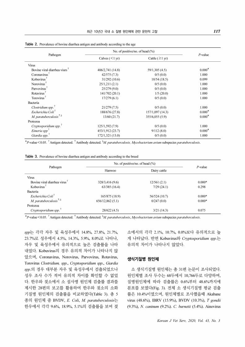
Table 2. Prevalence of bovine diarrhea antigen and antibody according to the age
PathogenNo. of positive/no. of head (%)
P-valueCalves (<1 yr) Cattle (≥1 yr)
Virus
Bovine viral diarrhea viurs†
406/2,741 (14.8) 59/1,305 (4.5) 0.000*
Coronavirus†
42/575 (7.3) 0/5 (0.0) 1.000
Kobuvirus†
31/292 (10.6) 10/54 (18.5) 0.099
Norovirus†
25/1,211 (2.1) 0/5 (0.0) 1.000
Parvovirus†
25/279 (9.0) 0/5 (0.0) 1.000
Rotavirus†
141/702 (20.1) 1/5 (20.0) 1.000
Torovirus†
17/279 (6.1) 0/5 (0.0) 1.000
Bacteria
Clostridium spp.†
21/279 (7.5) 0/5 (0.0) 1.000
Escherichia Coli†
188/676 (27.8) 157/1,097 (14.3) 0.000*
M. paratuberculosis‡,§
13/60 (21.7) 355/6,055 (5.9) 0.000*
Protozoa
Cryptosporidium spp.†
125/1,592 (7.9) 0/5 (0.0) 1.000
Eimeria spp†
453/1,912 (23.7) 9/112 (8.0) 0.000*
Giardia spp.†
172/1,321 (13.0) 0/5 (0.0) 1.000
*P-value<0.05. †
Antigen detected. ‡
Antibody detected. §M. paratuberculosis, Mycobacterium avium subspecies paratuberculosis.
Table 3. Prevalence of bovine diarrhea antigen and antibody according to the breed
PathogenNo. of positive/no. of head (%)
P-valueHanwoo Dairy cattle
Virus
Bovine viral diarrhea virus†
328/3,416 (9.6) 12/561 (2.1) 0.000*
Kobuvirus†
63/385 (16.4) 7/29 (24.1) 0.298
Bacteria
Escherichia Coli†
165/875 (18.9) 56/524 (10.7) 0.000*
M. paratuberculosis‡,§
656/12,862 (5.1) 0/247 (0.0) 0.000*
Protozoa
Cryptosporidium spp.†
28/622 (4.5) 3/21 (14.3) 0.075
*P-value<0.05. †
Antigen detected. ‡
Antibody detected. §M. paratuberculosis, Mycobacterium avium subspecies paratuberculosis.
spp는 각각 자우 및 육성우에서 14.8%, 27.8%, 21.7%,
23.7%로 성우에서 4.5%, 14.3%, 5.9%, 8.0%로 나타나,
자우 및 육성우에서 유의적으로 높은 검출률을 나타
내었다. Kobuvirus의 경우 유의적 차이가 나타나지 않
았으며, Coronavirus, Norovirus, Parvovirus, Rotavirus,
Torovirus Clostridium, spp., Cryptosporidium spp., Giardia
spp.의 경우 대부분 자우 및 육성우에서 검출되었으나
성우 조사 수가 적어 유의적 차이를 확인할 수 없었
다. 한우와 젖소에서 소 설사병 원인체 검출률 결과를
제시한 28편의 보고를 활용하여 한우와 젖소의 소화
기질병 원인체의 검출률을 비교하였다(Table 3). 총 5
종의 원인체 중 BVDV, E. Coli, M. paratuberculosis는
한우에서 각각 9.6%, 18.9%, 5.1%의 검출률을 보여 젖
소에서의 각각 2.1%, 10.7%, 0.0%보다 유의적으로 높
게 나타났다. 반면 Kobuvirus와 Cryptosporidium spp.는
유의적 차이가 나타나지 않았다.
생식기질병 원인체
소 생식기질병 원인체는 총 31편 논문이 조사되었다.
원인체별 조사 두수는 68두에서 10,766두로 다양하며,
질병원인체에 따라 검출률은 0.6%부터 48.6%까지에
분포를 보였다(Fig. 3). 전체 소 생식기질병 평균 검출
률은 10.4%이였으며, 원인체별로 조사했을때 Akabane
virus (48.6%), IBRV (15.9%), BVDV (10.3%), T gondii
(9.3%), N. caninum (9.2%). C. burnetii (5.4%). Ainovirus

118 이한규ㆍ조아라ㆍ오상익ㆍ노재희ㆍ정영훈ㆍ최창용ㆍ도윤정ㆍ엄재구ㆍ손동수ㆍ류재규
Korean J Vet Serv, 2020, Vol. 43, No. 3
Fig. 3. Overall prevalence (%) of
bovine reproductive disease anti-
gen (●) and antibody (△, ▲). pre-
valence of agents is more than
mean of diarrhea agents represent
White (△), and prevalence of
agents is less than mean of diar-
rhea agents represent Black (●,
▲).
Table 4. Prevalence of bovine reproductive antigen and antibody
according to the species
PathogenNo. of positive/no. of head (%)
P-valueHanwoo Dairy cattle
Bovine viral
diarrhea virus†
328/3,416 (9.6) 12/561 (2.1) 0.000*
Coxiella burnetii‡
194/6,846 (2.8) 335/2,438 (13.7) 0.000*
Neospora caninum‡
271/5,111 (5.3) 423/1,868 (22.6) 0.000*
Toxoplasma gondii‡
137/1,125 (12.2) 49/438 (11.2) 0.664
*P-value<0.05. †
Antigen detected. ‡
Antibody detected.
Table 5. Prevalence of Tick-born disease antigen and antibody in
cattle according to the species
No. of positive/no. of head (%)P-value
Hanwoo Dairy cattle
Anaplasma spp.†
30/568 (5.3) 5/433 (1.2) 0.000*
Coxiella burnetii‡
194/6,846 (2.8) 335/2,438 (13.7) 0.000*
Theileria spp.†
85/472 (18.0) 213/550 (38.7) 0.000*
*P-value<0.05. †
Antigen detected. ‡
Antibody detected.
(4.8%), Chuzan virus (4.4%), Brucella spp. (4.2%), Cam-
pylobacter fetus subsp. venerealis (C. venerealis, 0.6%)
로 각각 조사되었다. 생식기질병 원인체는 대부분 항
체조사 보고를 조사하였고, BVDV만 항원조사 보고를
활용하였다. 31편의 논문 중에서 한우와 젖소를 명시
한 23편의 연구 보고를 활용하여 한우와 젖소의 검출
률을 비교하였을 때, BVDV는 한우에서 9.6%, 젖소에
서 2.1%로 나타나, 한우에서 유의적으로 더 많이 발생
하는 것으로 조사되었다. C. burnetii, N. caninum은 젖
소에서 각각 13.7%, 22.6%, 한우에서 2.8%, 5.3%로 나
타나 젖소에서 유의적으로 더 많이 발생하는 것으로
조사되었다. 반면 T. gondii는 유의적 차이가 나타나지
않았다(Table 4).
진드기 매개 질병 원인체
소 진드기 매개 질병 원인체 연구 21편을 조사한
결과, Theileria spp.는 35.5%, C. burnetii는 5.4%,
Anaplasma spp.는 2.5%의 검출률을 나타내었으며,
Rickettsia spp.는 1건의 논문에서 1.6%의 검출률을 나
타내었다(Cho 등, 2016). C. burnetii는 항체 검사했으
며, 나머지는 항원 검사하였다. 소 진드기 매개 질병
원인체에 검출률을 품종(한우, 젖소)에 따라 비교하였
다(Table 5). Anaplasma spp.는 한우(5.3%)에서 젖소
(1.2%)보다 유의적으로 더 많이 발생하는 것으로 확인

최근 10년간 국내 소 질병 원인체에 관한 문헌적 고찰 119
Korean J Vet Serv, 2020, Vol. 43, No. 3
Table 6. Prevalence of Tick-born disease agent in cattle according
to the growth type
No. of positive/no. of head (%)P-value
Housing Gazing
Anaplasma spp. 1/343 (0.3) 14/320 (4.4) 0.000*
Theileria spp. 111/715 (15.0) 187/307 (60.9) 0.000*
*P-value<0.05.
되었다. C. burnetii와 Theileria spp.는 젖소에서 각각
13.7%, 38.7%의 검출률을 보여 한우에서 각각 2.8%,
18.0%보다 유의적으로 높게 발생하는 것으로 조사되
었다. 소 진드기 매개 질병 원인체의 검출률 조사 21
편의 연구 중에서 사육방식을 명시한 6편의 연구를
활용하여 사육방식에 따라 검출률을 비교하였을때, Ana-
plasma spp.와 Theileria spp.는 사사의 경우 각각 0.3%
와 15.05%, 방목의 경우 각각 4.4%와 60.9%로 나타나
방목우에서 유의적으로 높게 발생하는 것으로 조사되
었다(Table 6).
호흡기 질병 원인체
소 호흡기 질병 원인체 연구는 22편의 논문이 조사
되었다. 원인체별 조사 두수는 80두에서 4,612두로 다
양하며, 검출률 또한 5.0%부터 76.3%까지 분포를 보
였다. 호흡기 질병 원인체의 평균 양성율은 13.4%로
Mycoplasma spp. (76.3%), Bovine parainfluenza type3
(52.3%), Bovine respiratory syncytial virus (52.3%),
Pasturella spp. (45.5%), Staphylococcus spp. (19.8%),
E. coli (19.5%), Infectious bovine rhinothracheitis virus
(15.9%)는, Actinomyces spp. (11.9%), BVDV (10.3%)
M. bovis (7.6%), M. hemolytica (5.0%)로 각각 조사되
었다. Actinomyces spp. Mycoplasma spp. Pasturella spp.
Staphylococcus spp.는 항원 검사하였으며, Bovine par-
ainfluenza type3, Bovine respiratory virus, Infectious bo-
vine rhinothracheitis virus, M. bovis는 항체 검사하였
다.
소 기타 질병 원인체 분석 결과
기타 소 질병 원인체는 7종으로 9편의 논문이 조사
되었다. Bluetongue 18.2% (Hwang 등, 2019), Leukemia
virus 9.4% (Jung 등, 2012; Kim 등, 2017a), Besonitia
besnoiti 3.4% (Lee 등, 2017a), Bovine ephemeral fever
virus 2.2% (Yang 등, 2013; Yang 등, 2018), Peaton vi-
rus 1.1% (Jun 등, 2018), Sathuperi virus 4.8% (Jun 등,
2018), Shamonda virus 5.5% (Jun 등, 2018)로 항체검
사 하였으며, Bovine papular stomatitis virus는 6.7%
(Oem 등, 2013)로 항원 검사하였다.
지역별 소 질병 검출률 비교
최초 선정된 81편의 소 질병 원인체 조사 연구보고
중에서 지역이 명시된 연구 64편의 연구를 활용하여
지역을 경기, 강원ㆍ경북, 충북ㆍ충남, 전북ㆍ전남, 경
남, 제주지역으로 구분하였고, 지역별로 30두 이상의
개체를 검사하고, 그 결과 검출률이 20% 이상을 보인
원인체를 나타내었다(Fig. 4). 경기지역에서는 E. coli
(28.9%)가 가장 높게 검출되었으며, 강원ㆍ경북지역은
Pasteurella spp. (39.3%), 충북ㆍ충남지역은 Brucella spp.
(43.2%), 전북ㆍ전남과 경남지역에서는 Akabane virus
(각각 46.6% 및 63.7%)가 제주지역에서는 Theileria
spp. (69.7%)가 가장 높게 나타났다. 이외에도 지역별
로 Actinomyces spp. BVD, C. burnetii Eimeria spp.,
Kobuvirus, M. paratuberculosis, Mycobacterium bovis,
Neospora caninum, Parvovirus, Rotavirus, T. gondii 등
이 높은 검출률을 보였다.
고 찰
최근 10년 동안 우리나라에서는 구제역, 조류인플
루엔자, 아프리카돼지열병 등의 발생으로 축산분야뿐
만 아니라 사회전반에 많은 영향을 미쳤다. 이러한 악
성 가축 전염병에 의한 피해뿐만 아니라 설사, 유사
산, 진드기 매개 등에 의한 가축 질병 역시 농가의 생
산성 저하를 초래하므로 예방과 관리가 필요하다. 영
국의 경우 송아지 설사에 의한 경제적 피해가 1년에
1,100만 파운드(한화 약165억원)에 다다르며(Bennett과
Ijpelaar, 2005), 뉴질랜드에서는 BVD에 의한 젖소 피
해가 44.5백만NZ$로 보고되었다(Houe, 2003). 이처럼
전 세계적으로 축산분야에서 가축질병에 의한 경제적
피해규모는 크다.
일반적으로 특정 질병에 대한 검사는 질병진단과
혈청학적 검사로 나눌 수 있으며, 질병진단은 질병감
염이 의심되는 가축을 대상으로 임상검사, 병리검사
및 원인체 분리를 통한 정밀검사를 의미하며, 혈청학
적 검사는 주로 특정 질병 원인체에 대한 항체 유무를

120 이한규ㆍ조아라ㆍ오상익ㆍ노재희ㆍ정영훈ㆍ최창용ㆍ도윤정ㆍ엄재구ㆍ손동수ㆍ류재규
Korean J Vet Serv, 2020, Vol. 43, No. 3
Fig. 4. Prevalent bovine disease antigen and antibody according to region of Korea. *Gy, Gyeonggi; GW, Gangwon; KB, Kyeongbuk; CB,
Chungbuk; CN, Chungnam; JB, Jeonbuk; JN, Jeonnam; KN, Gyeongnam; JJ, Jeju; Act, Actinomyces spp.; AKA, Akabane virus; Bru, Brucella spp.;
BVD, Bovine viral diarrhea virus; Cox, Coxiella burnetii; E.C, Escherichia coli; Eim, Eimeria spp.; Giar, Giardia spp.; Kobu, Kobuvirus; MAP,
Mycobacterium avium subspices paratuberculosis.; M.B, Mycobacterium bovis; Neo, Neospora caninum; Parvo, Parvovirus; PAS, Pasteurella spp.;
Rota, Rotavirus; STA, staphylococcus spp.; Toxo, Toxoplasma gondii; The, Theileria spp.
토대로 감염 여부나 발생 동향을 파악하기 위한 수단
으로 활용된다. 따라서 본 연구는 최근 10년간 국내외
학술지에 게재된 국내 소 질병에 관해 위의 두가지 방
법을 활용한 질병 원인체와 항체 검출률을 조사하였
다.
한우 및 젖소 농가에서 소화기 질병은 다양한 형태
로 발생할 수 있지만, 설사 유발 원인체에 대한 보고
를 조사한 결과, Eimeria spp. (22.7%), Rotavirus (20.5%),
E. coli (19.5%) 순으로 높은 검출률을 나타냈다. 외국
의 사례를 살펴보면 오스트리아에서는 Rotavirus (21.1%),
E. coli (18.9%) Eimeria spp. (14.4%)가 설사 증상 있는
소에서 검출되었으며, 아일랜드에서는 Rotavirus, Crypto-
sporidum spp. Coccidia, E. coli (원인체별 검출률 데이
터 없음)순으로 많이 검출되었다(Luna, 2008; DAFM,
2017). Eimeria spp.는 소에서 콕시듐증을 유발하는 원
인체로 감염된 개체에서 출혈을 동반한 심한 설사를
유발한다(Lee 등, 2018b). Daugschies과 Najdrowski (2005)
은 소에서 Eimeria spp. 감염 시 생후 3주에서 6개월
사이에 가장 증상이 많이 나타나며, 성우의 경우 Eimeria
spp.감염에 충분히 저항할 만한 면역력을 가지는 것으
로 보고하였다. 이번 조사에서도 Eimeria spp.는 이와
유사하게 자우 및 육성우(1연령 이하)에서 더 많이 검
출되는 것으로 조사되었다. Bangoura와 Bardsley (2020)
는 자우 및 육성우의 경우 최초 감염 시 다량의 병원
체가 분변을 통해 배출되며, 이는 감염 전파의 주요
요인으로 보고하였다. Eimeria spp.의 관리를 위해서는
어린 소에서 초유 섭취를 통한 면역 증진 뿐만 아니라
항콕시듐제와 같은 치료법을 고려해볼 수 있으며, 축
사 소독과 건조를 통해 축사 환경내 Eimeria spp.의 노
출을 줄여나가야 한다.
Rotavirus는 소에 감염 시 장내 흡수를 저하시켜, 많
은 양의 설사를 유발하는 질병이다(Mcnulty, 1983).
Rotavirus는 1∼2주령의 송아지에서 설사를 일으키는
중요한 원인체로 알려져 있는 반면 성우의 Rotavirus
감염에 대한 연구는 매우 제한적으로 되어있다(Cho와
Yoon, 2014; Masuda 등, 2014). 이번 조사에서는 Rotavirus
는 성우(20.0%)와 자우 및 육성우(20.1%)에서 모두 20%
이상의 높은 검출률을 보였다. 성우의 Rotavirus 검출
률과 관련해서는 한 논문만(Koh 등, 2019) 인용되어
표본수가 부족하였지만, Rotavirus감염된 개체에서 분

최근 10년간 국내 소 질병 원인체에 관한 문헌적 고찰 121
Korean J Vet Serv, 2020, Vol. 43, No. 3
변을 통해 전파되는 만큼(Mcnulty, 1983) 성우 감염과
관련된 더 많은 연구가 필요하다.
E. coli는 소에서 시가독소(Shiga-like toxins), 내열성
독소(heat-stable enterotoxin), 이열성독소(heat-labile en-
terotoxin)등을 생산하여 설사를 유발한다(Salvadori 등,
2003). Ferens와 Hovde (2011)은 초유의 모체이행항체
로 E. coli 감염을 방어하나, 점차 항체가 감소하여 감
염가능성이 증가한다고 보고하였고, Mir 등(2015)는
성우의 경우 2년령 때 가장 많이 발생하며 점차 발생
이 감소한다고 보고하였다. 이번 조사에서 자우 및 육
성우(27.8%)에서 유의적으로 높은 E. coli 검출률을 보
이나, 성우(14.3%)에서도 검출되는 만큼 발생을 줄이
기 위해 농장 전반적인 관리가 필요한 질병으로 생각
된다. 또한 젖소보다 한우에서 검출률이 높은 것으로
조사되었고, 인용 논문 중 Kang 등(2014)은 한우(17.6%)
젖소(10.7%)로 품종별 비교를 하였으나, 품종별 차이
가 발생하는 요인에 대해서 더 많은 연구가 필요하다.
또 다른 주요 설사 유발 원인체인 BVDV 감염 시
소에서 임상증상은 무증상부터 급만성까지 다양하며,
호흡기, 소화기 질병뿐 아니라 유사산 및 지속감염우
(Persistently Infection) 출산과 같은 번식장애를 유발한
다(Baker, 1995). 특히 임신초기(40∼120일) BVDV감
염 시 지속감염우가 발생 할 수 있으며, 지속감염우에
의한 지속 전파로 농장 내 심각한 피해를 유발한다
(Barker 등, 1993). Curits 등(1988)은 BVDV가 모든 연
령의 소에서 감염되나 60일령이상 240일령 사이의 송
아지에서 감수성이 높은 것으로 보고하였다. 이번 조
사에서는 BVDV가 자우 및 육성우에서 더 많이 검출
되어 유사한 결과를 나타냈다. 또한 한우에서 젖소보
다 검출률이 높은 것으로 조사되었다. 그러나 품종별
차이를 나타내는 요인에 대한 연구가 미흡하여 추가
연구가 필요하다. 또한 이번 조사에서 BVDV 항체 검
출률은 72.0%로 나타나(데이터 나타내지 않음), 우리
나라 실시하고 있는 BVDV 백신의 효과가 있다고 판
단된다. 다만 BVDV를 전파의 핵심인 지속감염우의
감염실태 파악이 부족하다. 따라서 백신접종과 더불
어 송아지 조기진단으로 농장내 바이러스의 전파를
차단해야 한다.
M. paratuberculosis는 소에서 요네병을 유발하는 원
인체로 감염 시 장염을 유발해 만성 설사, 쇠약, 증체율
감소 등을 유발 한다(McFadden 등, 1987). 이유기의
송아지의 경우 감염된 어미로부터 M. paratuberculosis
균이 전파되는 반면(Park 등, 2017a), 요네병의 증상은
무증상 잠복기를 거쳐 2년령 이상부터 나타나기 시작
한다(Irenge 등, 2009). 또한 송아지와 어린 암소에서
ELISA 검사 시 위음성이 자주 나와 진단에 어려움이
있다(Sweeney, 1996). 이번 조사에서 M. paratuberculosis
는 자우 및 육성우(21.7%)에서 높은 검출률을 보이나,
성우(5.9%)에서도 검출되었다. 따라서 임상증상을 보
이는 성우를 검사 후 즉시 도태해야 하며, 무증상 감
염축 검사 방법에 대한 효과적인 진단방법 개발이 필
요하다.
소화기 질병을 유발하는 원인체에 검출률은 지역별
로 다른 양상을 보였다(Fig. 4). E. coli의 경우 경기, 강
원ㆍ경북 및 경남지역에서 각각 28.9%, 17.9%, 46.2%
의 검출률이 조사되었다. Kang 등(2014)에 따르면 경
기지역은 타 지역에 비해 단위면적당 사육두수가 높
아 밀사 환경으로 E. coli 검출률이 높고, 강원지역은
야생동물에 의한 E. coli의 전파가 있을 수 있다고 보
고하였다. Rotavirus의 경우는 전라지역에서 43.0%
(Koh 등, 2019), 강원ㆍ경북지역에서 27.5% (Lee 등,
2019b), 경남지역은 15.7% (Jeong 등, 2012)로 모든 지
역에서 높은 검출률을 보이고 있다. 그러나 지역별 직
접 비교 연구가 없어 Rotavirus에 관한 연구가 필요하
다. Kobuvirus의 경우 국토 서부지역(경기, 충청, 전라)
에 검출률이 33.0% (61/185), 동부지역(강원, 경북, 경
남)은 4%로(9/227) 서부지역에서 더 많이 검출되는 것
으로 보여진다. 사육환경, 품종 등 지역별 차이에 따
른 검출률 차이에 관한 연구가 필요하다.
한우 및 젖소 농가에서 생식기 질병은 다양한 형태
로 발생할 수 있지만, 유사산 관련 원인체에 대한 보
고를 조사한 결과, Akabane virus (48.6%), IBRV
(15.9%), BVDV (10.3% [항원], 72.0% [항체]), T. gon-
dii (9.3%), N. caninum (9.2%)로 조사되었다. Song 등
(2014)은 전북지역 유사산 질병에 대한 원인체 항체가
조사 결과 BVDV 72.4% IBRV 13.0%, T. gondii 10.4%
N. caninum 1.2%, C. venerealis 0.6%로, 보고하였으나,
외국의 경우 멕시코에서 BVDV 78.8%, IBRV 73%,
Neospora spp. 36.8%, Brucella spp. 14.7%로 보고
(Milián-Suazo 등, 2016)하여 국내 검출류과 차이점을
보이고 있다. Akabane virus는 임신우에서 유사산, 조
산, 선천적 기형 송아지를 출산을 유발하는 질병이다
(Kono 등, 2008). 우리나라에서는 연간 약 54만두에
백신접종을 실시하고 있다(농림축산식품부, 2019c).
Akabane virus와 같이 백신을 하는 질병의 경우 혈청
중앙항체 양성률이 30% 이하 일 경우 주의를 해야한
다(Yang 등, 2013). 비록 과거 10년의 연구결과를 조
사한 것이지만, 이번 조사에서 Akabane viurs의 전체

122 이한규ㆍ조아라ㆍ오상익ㆍ노재희ㆍ정영훈ㆍ최창용ㆍ도윤정ㆍ엄재구ㆍ손동수ㆍ류재규
Korean J Vet Serv, 2020, Vol. 43, No. 3
평균 항체양성률은 48.6%이나, 일부 지역에서 양성률
이 30%이하로 나타나 해당 지역에서는 Akabane viurs
감염에 대한 면밀한 조사가 필요해 보인다.
N. caninum은 소에서 주로 임신 5∼6개월에 유산을
유발하며 2개월령 이하의 송아지에서는 기립 불능 등
신경 증상의 마비증상이 나타나거나 무증상 보균우로
축산농가 지속적으로 경제적인 피해를 줄 수 있다
(Dubey, 2003; Lee 등, 2018a). N. caninum의 품종별 감
염률은 사육기간과 사육방식에 따라 차이가 있었다.
사육 기간의 경우 한우는 보통 24개월이 되면 도축하
는 반면에 젖소는 4∼5년 이상의 산차 동안 사육하여
항체 양성률이 높다고 생각된다(Hwang, 2010). 또한
사육 방식에 있어서 개방된 공간에서 사육되어 N.
caninum의 종숙주이자 감염원(McAllister 등, 1998)인
개와 접촉이 용이한 젖소가 더 높게 나온다고 생각된
다. 또한 이번 연구에서 강원ㆍ경북지역 N. caninum
검출률(27.5%)이 높았는데, Kim 등(2013)에 따르면 국
내 야생너구리의 N. caninum에 항체 양성율이 23%라
고 보고해, 야생동물이 감염률을 증가시켰을 가능성
이 있다 N. caninum에 대한 효과적인 방역대책 추진
을 위해서는 앞으로 소 뿐만 아니라, 종숙주인 개, 감
염경로에 관련되거나 관련될 가능성이 있는 야생동물
등에 대한 추가 연구가 이루어져야 할 것으로 생각된
다.
C. burnetii는 소에서 큐열을 유발하는 원인체로 감
염된 개체에서 대체로 무증상을 나타내지만 암컷에서
만성유방염, 수태율 감소, 유산을 유발한다(To 등,
1998). 이번 조사에서 C. burnetii는 한우보다 젖소에서
더 많이 검출되었다. 이는 N. caninum처럼 젖소에서
긴 사육기간으로 인한 검출률 증가와, 착유기를 공동
이용으로 인한 감염 전파 증가가 원인으로 생각한다.
또한 이번 조사에서는 제주도 지역에서 유일하게 C.
burnetii가 다른 지역보다 높은 항체 양성률(22.9%)을
보였으며, 이는 국내 기후 변화에 따른 진드기 매개
질병 발생률이 증가하는 것과 같은 맥락에서 이해될
수 있다(Ouh 등, 2013b). 많은 연구 결과들이 보고되
었지만, 소 사육 농가의 큐열 발생의 역학적 전파 양
상 규명에 관한 연구들이 부족한 실정이다. 향후에 공
중보건학적인 측면에서 C. burnetti의 농장 내외 전파
요인 규명에 관한 연구가 필수적으로 수행되어야 할
것이다.
소 Anaplasma spp. 감염에 관한 국내 연구는 제한적
이다(Kim 등, 2006; Lee와 Chae, 2010; Doan 등, 2013).
Theileria spp.와 마찬가지로 H. longicornis가 Anaplasam
spp.를 전파하는 가장 큰 매개체로 생각한다. 이번 조
사에서는 Anaplasma spp.가 방목우에서 검출률이 사
사우보다 높게 나타나 Theileria spp.와 같은 양상을 보
인 반면 젖소보다 한우에서 검출률이 더 높게 나와
Theileria spp.와 다른 양상을 보였다. 이는 조사 두수
나 방법에 따라 차이가 발생한 것으로 생각된다.
Anaplasma spp.는 세계적으로 많이 연구되고 있는 인
수공통전염병 원인체이지만, 국내에서는 연구가 미비
한 실정이다(Kim 등, 2017b). 추후에 더 많은 수의 한
우 및 젖소와 다른 종류의 동물에서도 추가적인 조사
가 필요하며, Anaplasma spp. 뿐만 아니라, Eimeria spp.,
Ehrlichia spp.와 같은 다양한 원충성 질병에 대한 연
구가 수행되어야 한다. Theileria spp.는 소의 적혈구
내 기생하는 기생충으로 진드기에 의해 전파되며, 빈
혈, 허약, 고열, 황달 및 유산을 일으킬 수 있는 원인
체 이다(Takahashi 등, 1976; Minami 등, 1980). 이번
조사에서 Theileria spp.의 검출률은 젖소에서 한우보
다 높은 것으로 조사되었다. Suh와 Jang (1982)은 젖소
도입우가 한우보다 Theileria spp. 감염에 감수성이 높
다고 보고하였으며, Jeon (1970)은 젖소가 한우보다
빈혈 소견이 높은 것으로 보고하여 이번 조사를 뒷받
침하고 있다. 이번 조사에서 Theileria spp.는 방목우에
서 사사우보다 검출률이 높은 것으로 나타났다. 이는
Haemaphysalis longicornis (H. longicornis)와 관련되 있
는 것으로 생각한다. H. longicornis는 한국에서 가장
흔한 진드기로 소와 야생동물에게 Theileria spp.를 전
파하는 매개체이다(Kim 등, 2017b). 방목우는 초지에
사육하는 방식으로 사사우에 비해 진드기에 더 많이
노출될 가능성이 높다. Park 등(2017)은 제주지역에
강우, 습도, 온도와 같은 환경이 진드기 번식과 활동
을 증가시켰으며, 강원ㆍ경북지역은 Theileria spp.에
감염된 야생동물에 의해 전파되었을 가능성이 높다고
보고하였다. 이보다 더 나아가 이번 연구에서 Theileria
spp. 감염률이 조사된 지역(제주 69.7%, 강원ㆍ경북
23.1%, 충청 23.2%, 전라 19.2%)에서는 모두 높은 검
출률을 보여 소 타일레이아증이 전국적으로 만연해
있다고 생각한다. 주목할 만한 점은 방목을 하지 않는
사육 소에서도 약 15%의 Theileria spp. 감염이 확인되
었다는 점이다. 이는 농장 내에 진드기를 포함한 감염
을 일으키는 인자 또는 증상을 나타내지 않는 무증상
감염우가 존재한다는 것을 암시한다. 그러므로 농장
내 감염 위험인자 및 무증상 감염우를 색출하는 것이
농장 내 타일레리아증 감염 고리를 끊는데 필수적이
다.

최근 10년간 국내 소 질병 원인체에 관한 문헌적 고찰 123
Korean J Vet Serv, 2020, Vol. 43, No. 3
결 론
본 논문은 문헌을 바탕으로 10년간 국내 소 전체
질병 원인체에 대해 조사한 첫 번째 보고이다. 국내외
학술지에 발표된 총 81건의 연구 보고를 선정하고 분
석하였다. 원인체는 소화기질병, 호흡기질병, 생식기
질병, 진드기매개 질병으로 분류하였고, 원인체별 검
출률과 각 원인체별 비교 기준에 따른 유의성을 파악
하였다.
소화기질병 원인체의 평균 검출률은 9.0%로 Eimeria
spp. (22.7%), Rotavirus (20.5%), E. coli (19.5%) 순으
로 높은 검출률을 보였다. 생식기질병 원인체의 평균
검출률은 10.4%로 Akabane virus (48.6%), IBRV (15.9%)
는 평균보다 높게 검출되었다.
BVDV, E. coli, M. paratuberculosis, Eimeria spp.는
자우 및 육성우에서 높은 검출률을 보였다. 한우에서
는 BVDV, E. coli, M. paratuberculosis, Anaplasma spp.
가 젖소에서는 Coxiella burnetii, Neospora caninum,
Theileria spp.가 높은 검출률을 보였다. 또한 진드기
매개 질병은 방목우에서 사사우보다 높은 검출률을
보였다.
원인체별로는 Theileria spp.의 전국적인 모니터링을
실시하여 국내 감염률을 파악할 필요가 있으며,
Rotaviurs, Kobuvirus와 같은 질병의 지역별 분포 또한
조사할 필요가 있다. 연령에 따라 주로 발생하는 질병
에 차이가 있고, 한우와 젖소에서 유의적으로 많이 발
생하는 질병이 달라 연령별, 품종별 질병예방 우선순
위에 차등을 두어야 할 것으로 생각되며, 지역별로 다
발하는 질병에 대한 관리가 필요할 것으로 생각된다.
감사의 글
본 연구는 농촌진흥청 연구사업(세부과제명: 축산
원 가축질병 위기대응 실무 매뉴얼 적용 및 방역 최적
화 연구, 세부과제번호: PJ011915012020) 지원으로 이
루어졌습니다.
CONFLICT OF INTEREST
No potential conflict of interest relevant to this article
was reported.
ORCID
Han Gyu Lee, https://orcid.org/0000-0002-3531-1971
Ara Cho, https://orcid.org/0000-0001-5309-7721
Sang-Ik Oh, https://orcid.org/0000-0003-0877-9170
Jae-Hee Roh, https://orcid.org/0000-0003-4223-1096
Yong Hoon Jung, https://orcid.org/0000-0002-8094-0304
Changyong Choe, https://orcid.org/0000-0003-4222-3360
Yoon Jung Do, https://orcid.org/0000-0003-3207-3514
Jae Ku Oem, https://orcid.org/0000-0002-4298-0604
Dong-Soo Son, https://orcid.org/0000-0003-4790-6064
Jae Gyu Yoo, https://orcid.org/0000-0002-8542-9193
REFERENCES
농림축산식품부. 2019a. 결핵병 및 브루셀라병 방역실시요령. 농
림축산식품부 고시 제2019-30호.
농림축산식품부. 2019b. 구제역 방역실시요령. 농림축산식품부
고시 제2019-52호.
농림축산식품부. 2019c. 농림축산식품통계연보.
한국농촌경제연구원. 2006. 가축질병의 경제적 영향 분석. pp.
1-7.
Baca OG, Paretsky D. 1983. Q fever and Coxiella burnetii: a
model for host-parasite interactions. Microbiol Rev. 47:
127-149.
Baker JC. 1995. The clinical manifestation of bovine viral diar-
rhea infection. Vet Clin North Am Food Anim Pract 11:
425-445.
Bangoura B, Bardsley KD. 2020. Ruminant Coccidiosis. Vet Clin
North Am Food Anim Pract 36: 187-203.
Barker IK, Dreumel AAV, Palmer N. 1993. Bovine virus
diarrhea. pp. 149-159. In: Jubb KVF, Kennedy PC,
Palmer N(ed.). Pathology of domestic animals. Vol. 2.
4th ed. Academic Press, San Diego.
Bennett R, Ijpelaar J. 2005. Updated Estimated of the Costs
Associated with Thirty Four Endemic Livestock Diseases
In Great Britain: A Note. J. Agric. Econ. 56: 135-144.
Bicknell EJ, Reggiardo C, Noon TH, Bradley GA,
Lozano-Alarcon F. 1994. Abortion diseases of range
cattle. https://cals.arizona.edu/arec/sites/cals.arizona.edu.arec/
files/publications/27%20abortiondiseasecattle94.pdf
Caswell JL. 2014. Failure of respiratory defenses in the patho-
genesis of bacterial pneumonia of cattle. Vet Pathol 51:
393-409.
Cho JC, Jeon WJ, Kim SS, Kim SG. 2016. A survey for tick-
borne pathogens in Korean native cattle from northern
area of Gyeongbuk. Korean J Vet Serv 39: 29-34.
Cho JS, Kim GD, Park HJ, Lim YS, Hong SH, Seo CW, Ryu
HJ, Sin RJ. 2013. Prevalence for persistently infected

124 이한규ㆍ조아라ㆍ오상익ㆍ노재희ㆍ정영훈ㆍ최창용ㆍ도윤정ㆍ엄재구ㆍ손동수ㆍ류재규
Korean J Vet Serv, 2020, Vol. 43, No. 3
cattle with bovine viral diarrhea virus in Korea. Korean
J Vet Serv 36(2): 105-110.
Cho YI, Yoon KJ. 2014. An overview of calf diarrhea – in-
fectious etiology, diagnosis, and intervention. J Vet Sci
15: 1-17.
Choi KS, Song MC. 2011. Epidemiological observation of bovine
viral diarrhea virus in Korean indigenous calves. Virus
Genes 42: 64-70.
Choi KS, Yu DH, Chae JS, Park BK, Yoo JG, Park JH. 2016.
Seasonal changes in hemograms and Theileria orientalis
infection rates among Holstein cattle pastured in the
mountains in the Republic of Korea. Prev Vet Med 127:
77-83.
Choi KS. 2012. Acute BVDV-1b outbreak in Korean Indigenous
Calves. J Vet Clin 29(5): 395-399.
Chu KS, Kim SH, Ha YS, Lee JW. 2013. Seroprevalence of para-
tuberculosis in Korean cattle in western Jeonbuk area,
Korea. Korean J Vet Serv 36: 133-137.
Curits CR, Erb NH, White ME. 1988. Descriptive epidemiology
of calfhood morbidity and mortality in New York
Holstein herds. Prev Vet Med 5: .293-298.
DAFM. 2017. Regional veterinary laboratories reports. http://
agriculture.gov.ie/animalhealthwelfare/laboratoryservieces/
regionalveterinary laboratoryreports/rvlmonthlyreports2017/
Daugschies A, Najdrowski M. 2005. Eimeriosis in cattle: current
understanding. J Vet Med B Infect Dis Vet Public Health
52: 417-427.
de la Fuente J, Estrada-Pena A, Venzal JM, Kocan KM,
Sonenshin DE. 2008. Overview: Tick as vectors of
pathogens that cause disease in humans and Animals.
Front Biosci 13: 6938-6946.
Doan HT, Noh JH, Choe SE, Yoo MS, Kim YH, Reddy KE,
Quyen DV, Nguyen LT, Nguyen TT, Kweon CH, Jung
SC, Chang KY, Kang SW. 2013. Molecular detection
and phylogenetic analysis of Anaplasma bovis from
Haemaphysalis longicornis feeding on grazing cattle in
Korea. Vet Parasitol. 196: 478-481.
Dubey JP, Schares G. 2011. Neosporosis in animals-the last five
years. Vet Parasitol 180: 90-108.
Dubey JP. 2003. Review of Neospora caninum and neosporosis
in animals. Korean J Parasitol 41: 1-16.
Ferens WA, Hovde CJ. 2011. Escherichia coli O157:H7: animal
reservoir and sources of human infection. Foodborne
Pathog Dis 8: 465-487.
Han DG, Ryu JH, Chae JB, Kim DW, Kwon CH, Choi KS.
2018a. First report of Anaplasma phagocytophilum in-
fection in Holstein cattle in the Republic of Korea. Acta
Trop 183: 110-113.
Han DG, Ryu JH, Park JH, Choi KS. 2018b. Identification of a
new bovine viral diarrhea virus subtype in the Republic
of Korea. BMC Vet Res 14: 233.
Houe H. 2003. Economic impact of BVDV infection in dairies.
Biologicals 31: 137-143.
Hwang EK. 2010. Seroprevalence of antibodies to Neospora cani-
num in dairy cattle raised in Kangwon province. Korean
J Vet Res 50: 19-24.
Hwang JM, Kim JG, Yeh JY. 2019. Serological evidence of blue-
tongue virus infection and serotype distribution in dairy
cattle in South Korea. BMC Vet Res 15: 255.
Inaba Y, Kurogi H, Omori T. 1975. Letter: Akabane disease: epi-
zootic abortion, premature birth, stillbirth and congenital
arthrogryposis-hydranencephaly in cattle, sheep and goats
caused by Akabane virus. Aust Vet J 5: 584-585.
Irenge LM, Walravens K, Govaerts M, Godfroid J, Rosseels V,
Huygen K, Gala JL. 2009. Development and validation
of a triplex real-time PCR for rapid detection and specif-
ic identification of M. avium sub sp. paratuberculosis in
faecal samples. Vet Microbiol 136: 166-172.
Ismail HA, Jeon HK, Yu YM, Do CH, Lee YH. 2010. Intestinal
parasite infections in pigs and beef cattle in rural areas
of Chungcheongnam-do, Korea. Korean J Parasitol 48:
347-349.
Jeon Y. 1970. Hematological Survey on Hematooza of Cattle in
Korea. Korea J Vet Res 1970, 10: 81-87.
Jeong MH, Lee MK, Kim HS, Lee SU, Seong MH, Park DY,
Hwang BW, Park HJ, Cho JH. 2012. Detection of etio-
logic agents in diarrhea fecal samples from calves in
Gyeongnam province, Korea. Korean J Vet Serv 35:
339-342.
Jeoung HY, Lim JA, Jeong WS, Oem JK, An DJ. 2011. Three
clusters of bovine kobuvirus isolated in Korea, 2008-
2010. Virus Genes 42: 402-406.
Jun K, Yanaka T, Lee KK, Lee JB. 2018. Seroprevalence of bo-
vine arboviurs belonging to genus Orthobunyavirus in
South Korea. J Vet Med Sci 80: 1619-1623.
Jung K, Shim HS, Baek JJ. 2012 Investigation of bovine leuke-
mia virus infection in dairy farm of northern Gyeonggi
province, Korea. Korean J Vet Serv 35: 333-337.
Jung K, Shim HS. 2013. Seroprevalence of Neospora caninum in
dairy cattle of northern Gyeonggi province in Korea.
Korean J Vet Serv 36: 53-56.
Kang E, Hwang SY, Kwon KH, Kim KY, Kim JH, Park YH.
2014. Prevalence and characteristics of Shiga toxin-pro-
ducing Escherichia coli (STEC) from cattle in Korea be-
tween 2010 and 2011. J Vet Sci 15: 369-379.
Kang WC, Yang HS, Ko JA, Lee DS, Son WG. 2015. Prevalence
of Johne’s disease of Korean native cattle in Jeju Province,
Korea. Korean J Vet Serv 38: 221-225.
Kim CM, Yi YH, Yu DH, Lee MJ, Cho MR, Desai AR Smriti
S, Klein TA, Kim HC, Song JW, Baek LJ, Chong ST,
O’Guinn M, Lee JS, Lee IY, Park JH, Foley J Chae JS.
2006. Tick-borne rickettsial pathogens in ticks and small
mammals in Korea. Appl Environ Microbiol 72: 5766-
5776.
Kim HC, Choe CY, Kim SH, Chae JS, Yu DH, Park JH, Park
BK, Choi KS. 2018. Epidemiological Survey on Eimeria
spp. Associated with Diarrhea in Pre-weaned Native
Korean Calves. The Korean J Parasitol 56: 619-623.
Kim HT, Lee KW. 2013. Sero-prevalence of paratuberculosis
(Johne’s disease) of Korean native cattle in Busan area.
최근 10년간 국내 소 질병 원인체에 관한 문헌적 고찰 125
Korean J Vet Serv, 2020, Vol. 43, No. 3
J Vet Clin 30: 230-235.
Kim HT, Park MS, Lee GH, Lee KW. 2015a. Study on preva-
lence of antigens to bovine viral diarrhea virus (BVDV)
of Cattle in Busan area (2013∼2014). Korean J Vet
Serv 38: 43-49.
Kim HU, Lee EY, Lee KK, Kim SH, Moon BY, So BJ, Kim YH.
2017a. Seroprevalence of the bovine leukemia virus
among Korean native cattle in South Korea. J Prev Vet
Med 41: 52-55.
Kim HY, Byun JW, Jeon AB, Park BS, Jung JA, Park M, Lim
YS, Jung BY. 2012a. Seroprevalence of paratuberculosis
in pure-bred breeding cattle in Korea. J Life Sci 22:
794-798.
Kim JE, Son JW, Yang YM, Jeon HC, Jin KS, Kim KH, Shin
BW, Lee JH. 2011a. Seroprevalence of antibodies to
Neospora caninum in cattle at Seoul slaughtering center.
Korean J Vet Serv 34: 179-185.
Kim JH, Kang MS, Lee BC, Hwang WS, Lee CW, So BJ, Dubey
JP, Kim DY. 2003. Seroprevalence of antibodies to
Neospora caninum in dogs and raccoon dogs in Korea.
Korean J Parasitol 41: 243-245.
Kim JH, Lim JJ, Kim DH, Lee JJ, Kim DG, Jun MH, Kim, SH,
Chang HH, Lee HJ, Min WG, Kim S. 2010a. Biological
characterization of Brucella spp. isolated from cattle in
Gyeoungbuk, Korea. Korean J Vet Res 50: 117-124.
Kim JY, Kang SS, Her M, Lee KC, Sung SR, Gu JH, Kang SI,
Lee HK, Kim YJ, Kim DG, Jung SC. 2012b. Investiga-
tion of occurrence factors on brucellosis-outbreak farm
in Korea. Korean J Vet Serv 35: 263-268.
Kim JY, Sung SR, Pyun JI, Her M, Kang SI, Lee HK, Jung SC.
2014b. Seroprevalence of Q-fever in Korean native cattle.
Korean J Vet Res 54: 147-150.
Kim KH, Kwak KH, Song JH, Cho JG. 2010b. Detection of
Mycobacterium avium ssp paratuberculosis in Korean
cattle by the polymerase chain reaction. J vet Clin 27:
23-28.
Kim NH, Kim HR, Park HS, Kim Ys, Lee JH. 2015b.
Seroprevalence of Coxiella burnetii and Toxoplasam
gondii in cattle in Seoul, Korea. Korean J Vet Serv 38:
233-239.
Kim SH, Kang JH, Lee CJ, Lee YS, Chae JB, Kang SW, Jeong
SH, Yu DH, Jo A, Yoo JG, Choi KS, Kim HC, Park
BK, Chae JS, Park J. 2016. Detection of diarrheagenic
pathogens from feces and incidence of diarrhea in Korean
calves. Korean J Vet Serv 39: 187-192.
Kim SH, Yu DH, Kang SW, Chae JB, Choi KS, Kim HC, Park
BK, Chae JS, Park JH. 2017b. Hematological Changes
Associated with Theileria orientalis Infection in Korean
Indigenous Cattle. Korean J Parasitol 55: 481-489.
Kim YH, Kim D. 2015. Detection of Coxiella burnetii in Cattle.
J Vet Clin 32: 504-507.
Kim YS, Kim YK, Lee SY, Lee KK, Lee KH, Song JC, Oem
JK. 2019. Identification of Korean native cattle persis-
tently infected with BVDV using Ear-notch method.
Korean J Vet Serv 42: 117-120.
Koh BRD, Kim HJ, Oh AR, Jung BR, Park JS, Lee JG, Na HM,
Kim YH. 2019. Prevalence of enteropathogens in the fe-
ces from diarrheic Korean native cattle in Gwangju area,
Korea. Korean J Vet Serv 42: 93-112.
Kono R, Hirata M, Kaji M, Goto Y, Ikeda S, Yanase T, Kato
T, Tanaka S, Tsutsui T, Imada T, Yamakawa M. 2008.
Bovine epizootic encephalomyelitis caused by Akabane
virus in southern Japan. BMC Vet Res 4: 20.
Ku BK, Jeon BY, Kim JM, Jang YB, Lee HY, Choi JY, Jung
SC, Nam HM, Park H, Cho SN. 2018. Investigation of
bovine tuberculosis outbreaks by using a trace-back sys-
tem and molecular typing in Korean Hanwoo beef cattle.
J Vet Sci 19: 45-50.
Lee EY, Kang HW, Kim HY, Kim SH, Moon B, So BJ, Lee KK,
Kim YH. 2019a. Survey of bovine norovirus infection
from diarrheic calves in South Korea, 2015-2017. Korean
J Vet Res 59: 33-36.
Lee JW, Sohn JH, Kim JH, Kim SY, Cho KH. 2018a. Serosurvey
for antibodies aganinst Neospora caninum in Korean in-
digenous cattle in the southern area of Gyeongbuk and
Ulleung-gun. Korean J Vet Serv 41: 185-190.
Lee MJ, Chae JS. 2010. Molecular detection of Ehrlichia chaf-
feensis and Anaplasma bovis in salivary glands from
Haemaphysalis longicornis ticks. Vector Borne Zoonotic
Dis 10: 411-413.
Lee MK, Park JS, Kim MH, Park DY, Kim CH, Kim GH, Cho
JH. 2011b. Seroprevalence of antibodies to Neospora
caninum and Toxoplasma gondii in cattle in northern
area of Gyeongnam. Korean J Vet Serv 34: 245-250.
Lee SH, Eo KY, Jung BY, Dongmi Kwak DM, Kwon OD.
2017a. Seroprevalence and risk factors of Besnoitia bes-
noiti infection in Korean cattle – short communication.
Acta Vet Hung 65: 510-516.
Lee SH, Kim HY, Choi EW, Kim D. 2019b. Causative agents
and epidemiology of diarrhea in Korean native calves. J
Vet Sci 20: e64.
Lee SH, Kim HY, Lee HS, Kim JW, Lee YR, Chae MJ, Oh SI,
Kim JH, Rhee MH, Kwon OD, Goo YK, Kim TH,
Geraldino PJL, Kwak D. 2018b. Eimeria species in cat-
tle with diarrhoea in the Republic of Korea regarding
age, season and nature of diarrhoea. Vet Rec 183: 504.
Lee SH, VanBik D, Kim HY, Cho A, Kim JW, Byun JW, Oem
JK, Oh SI, Kwak D. 2016a. Prevalence and molecular
characterisation of Giardia duodenalis in calves with
diarrhoea. Vet Rec 178: 633.
Lee SH, VanBik D, Kim HY, Lee YR, Kim JW, Chae M, Oh
SI, Goo YK, Kwon OD, Kwak D. 2016b. Multilocus
typing of Cryptosporidium spp. in young calves with di-
arrhea in Korea. Vet Parasitol 229: 81-89.
Lee SW, Kim JH, Kim D. 2017b. Bacterial Pathogens and Their
Antimicrobial Susceptibility in Calves with Summer
Pneumonia. J Vet Clin 34: 161-164.
Lee TH, Rhee SH, Yoon CH. 2019c. Prevalence of antibodies to
Coxiella burnetii in cattle in Sejong. Korean J Vet Serv
42: 177-181.

126 이한규ㆍ조아라ㆍ오상익ㆍ노재희ㆍ정영훈ㆍ최창용ㆍ도윤정ㆍ엄재구ㆍ손동수ㆍ류재규
Korean J Vet Serv, 2020, Vol. 43, No. 3
Lee YJ, Han DG, Ryu JH, Chae JB, Chae JS, Yu DH, Park JH,
Park BK, Kim HC, Choi KS. 2018c. Identification of zo-
onotic Giardia duodenalis in Korean native calves with
normal feces. Parasitol Res 117: 1969-1973.
Lee YJ, Ryu JH, Shin SU, Choi KS. 2019d. Prevalence and mo-
lecular characterization of Cryptosporidium and Giardia
in pre-weaned native calves in the Republic of Korea.
Parasitol Res 118: 3509-3517.
Levine ND. 1985. Toxoplasma gondii. pp. 248-259. Veterinary
protozoology. 5 ed. Iowa State University Press, Ames.
Lim HS, Yang CR, Kim HD, Kim KH, Do JY, Cho JK. 2019.
Seroprevalence of Coxiella burnetii in bulk-tank milk
and cattle in Daegu area, Korea. Korean J Vet Serv 42:
61-65.
Luna CVH. 2008. Infectious agents associated with diarrhea of
calves and characterization of virulence genes in
Escherichia coli isolated from diarrhoeic and healthy ne-
onatal calves in austraia. Ph D. Thesis, University of
Veterinary Medicine Vienna, Vienna, Austria.
Lyoo KS, Kim D, Hyung HG, Lee SJ, Park MY, Hahn TW.
2017. Prevalence of Antibodies Against Coxiella burnetii
in Korean Native Cattle, Dairy Cattle, and Dogs in
South Korea. Vector-Borne Zoonotic Dis 17: 213-216.
Maggio LA, Sewell JL, Artino Jr. 2016. The Literature Review:
A Foundation for High-Quality Medical Education Re-
search. J Grad Med Educ 8: 297-303.
Masuda T, Nagai M, Yamasato H, Tsuchiaka S, Okazaki S,
Katayama Y, Oba M, Nishiura N, Sassa Y, Omatsu T,
Furuya T, Koyama S, Shirai J, Tanguchi K, Fujii Y,
Todaka R, Katayama K, Mizutani T. 2014. Identification
of novel bovine group A rotavirus G15P[14] strain from
epizootic diarrhea of adult cows by de novo sequencing
using a next-generation sequencer. Vet Microbiol 171:
66-73.
McAllister MM, Dubey JP, Lindsay DS, Jolley WR, Wills RA
McGuire AM, Tranas JD. 1998. Dogs are definitive
hosts of Neospora caninum. Int J Parasitol 28: 1473-
1478.
McFadden JJ, Butcher PD, Thompson J, Chiodini R, Hermon-
Taylor J. 1987. The use of DNA probes identifying re-
striction-fragment-length polymorphisms to examine the
Mycobacterium avium complex. Mol Microbiol 1: 283-
291.
Mcnulty MS. 1983. The etiology, pathology and epidemiology of
viral gastroenteritis Rotavirus infections in calves. Ann
Rech Vet. 14: 427-432.
Milián-Suazo F, Hernández-Ortíz R, Hernández-Andrade L, Alvarado-
lslas A, Díaz-Aparicio E, Mejía-Estrada F, Palomares-
Reséndiz EG, Reyes IB, Zemdekas-Martinez H. 2016.
Seroprevalence and risk factors for reproductive diseases
in dairy cattle in Mexico. J Vet Med Anim Health. 8:
89-98.
Minami T, Fujinaga T, Furuya K, Ishihara T. 1980. Clinico-hem-
atologic and serological comparison of Japanese and
Russian strains of Theileria sergenti. Natl Inst Anim
Health Q (Tokyo). 20: 44-52.
Mir AR, Weppelmann TA, Kang MY, Bliss TM, DiLorenzo N,
Lamb GC, Ahn SH, Jeong KW. 2015. Association be-
tween animal age and the prevalence of Shiga toxin-pro-
ducing Escherichia coli in a cohort of beef cattle. Vet
Microbiol 175: 325-331.
Na HM, Bae SY, Koh BRD, Park JS, Seo YJ, Jeong Hj, Park
JY, Park SD, Kim ES, Kim YH. 2016. Prevalence of an-
tibody titers for Coxiella burnetii in cattle in Gwangju
area, Korea. Korean J Vet Serv 39: 125-129.
Oem JK, Chung JY, Roh IS, Kim HR, Bae YC, Lee KH, Jin YH,
Lee OS. 2010. Characterization and phylogenetic analy-
sis of Bovine viral diarrhea virus in brain tissues from
nonambulatory (downer) cattle in Korea. J Vet Diagn
Invest 22: 518-523.
Oem JK, Lee EY, Lee KK, Kim SH, Lee MH, Hyun BH. 2013.
Bovine papular stomatitis virus(BPSV) infections in
Korean native cattle. J Vet Med Sci 75: 675-678.
Oem JK, Lee KH, Kim HR, Bae YC, Chung JY, Lee OS, Roh
IS. 2012. Bovine epizootic encephalomyelitis caused by
Akabane Virus infection in Korea. J Comp Pathol 147:
101-105.
Oh JJ, Lee SH, Lee SJ, KIM YH, Park SC, Rhee MH, Kwon OD,
Kim TH, Kwak DM. 2016. Detection of Antibodies
against Toxoplasma gondii in Cattle Raised in Gyeongbuk
Province, Korea. J Food Prot 79: 821-824.
Ouh IO, Seo MG, Do JC, Kim IK, Cho MH, Kwak DM. 2013a.
Seroprevalence of Coxiella burnetii in bulk-tank milk
and dairy cattle in Gyeongbuk province, Korea. Korean
J Vet Serv 36: 243-248.
Ouh IO, Seo MG, Jang YS, Kim SY, Kwak DM. 2013b.
Seroprevalence of Coxiella burnetii in cattle with re-
productive disorders in eastern Gyeongbuk province,
Korea. Korean J Vet Serv 36: 249-254.
Paramanandham K, Mohankumar A, Puttahonnappa Suresh K,
Susan Jacob S, Roy P. 2019. Prevalence of Anaplasma
species in India and the World in dairy animals: A sys-
tematic review and meta-analysis. Res in Vet Sci 123:
159-170.
Park AR, Hah DS, Jo SS, Kwun YT, Park DY, Lee KC, Heo JH.
2010a. Seroprevalence of antibodies to Neospora cani-
num in Korean indigenous cattle in Gyeongnam central
area. Korean J Vet Serv 33: 151-156.
Park HT, Park HE, Cho YI, Kim EH, Jung MH, Shin SW, Lee
SH, Kim DY, Yoo HS. 2017a. Potential biomarkers as
an indicator of vertical transmission of Johne`s disease
in a Korean native cattle farm. J Vet Sci 18: 343-349.
Park JH, Han DG, Ryu JH, Chae JB, Chae JS, Yu DH, Park BK,
Kim HC, Choi KS. 2018. Molecular detection of
Anaplasma bovis in Holstein cattle in the Republic of
Korea. Acta Vet Scand 60: 15.
Park JH, Han YJ, Han DG, Chae JB, Chae JS, Yu DH, Lee YS,
Park BK, Kim HC, Choi KS. 2017b. Genetic character-
ization of Theileria orientalis from cattle in the Republic
of Korea. Parasitol Res 116: 449-454.

최근 10년간 국내 소 질병 원인체에 관한 문헌적 고찰 127
Korean J Vet Serv, 2020, Vol. 43, No. 3
Park JH, Kim D. 2019. Detection of Respiratory Viral Pathogens
and Mycoplasma spp from Calves with Summer
Pneumonia in Korea. Korean J Vet Serv 36: 185-189.
Park JS, Park JK, Cho EJ, Kim EG, Lee JM, Kim DK, Son SK.
2013. Prevalence of bovine viral diarrhea virus from dai-
ry cattle farms in Gyeongnam southern area, Korea.
Korean J Vet Serv 36: 7-13.
Park SI, Jeong YJ, Kim HJ, Park JG, Kang SY, Woo SK, Kim
CH, Jung CH, Kang MI, Cho KO. 2010b. Genetically
Diverse Group C Rotaviruses Cause Sporadic Infection
in Korean Calves. J Vet Med Sci 73: 479-482.
Park SJ, Kim HK, Song DS, Moon HJ, Park BK. 2011.
Molecular detection and genetic characterization of ko-
buviruses in fecal samples collected from diarrheic cattle
in Korea. Infect Genet Evol 11: 1178-1182.
Ryu JH, Choi KS. 2019. Genetic analysis of bovine viral diarrhea
virus in pre-weaned native Korean calves. Trop Anim
Health Prod 51: 2085-2090.
Salvadori MR, Valadares GF, Leite DDS, Blanco J, Yano T.
2003. Virulence factors of Escherichia coli isolated from
calves with diarrhea in Brazil. Braz. J Microbiol 34:
230-235.
Scharnböck B, Roch F, Richter V, Funke C, Firth CL, Obritzhauser
W, Baumgartner W, Käsbohrer A, Pinior B. 2018. A
meta-analysis of bovine viral diarrhoea virus (BVDV)
prevalences in the global cattle population. Sci Rep 8:
14420.
Seo MG, Do JC, Cho MH, Seo HJ, Kim JK, Kim YH, Park NC,
Kwak DM. 2011a. Prevalence of Theileria sergenti in-
fection in cattle of eastern areas in Gyeongbuk province
by PCR. Korean J Vet Serv 34: 251-258.
Seo MG, Do JC, Ouh IO, Cho MH, Kim JK, Kim YH, Park NC,
Kwak DM. 2011b. Prevalence of infectious agents in
cattle reared in Ulleung island. Korean J Vet Serv 34:
303-311.
Seo MG, Kwon OD, Kwak DM. 2019. Molecular Detection of
Coxiella burnetii in Cattle on Ulleung Island, Korea: A
Population-based Study with Four Years of Follow Up.
Korean J Parasitol 57: 69-73.
Seo MG, Ouh IO, Kim YH, Kim JK, Kwon OD, Kwak DM.
2018a. Seroprevalence of Coxiella burnetii infection in
cattle on ulleung Island, Korea. Korean J Vet Res 58:
147-151.
Seo MG, Ouh IO, Kwon OD, Kwak DM. 2018b. Molecular de-
tection of Anaplasma phagocytophilum-like Anaplasma
spp. and pathogenic A. Phagocytophilum in cattle from
South Korea. Mol Phylogenet Evol 126: 23-30.
Seo MG, Ouh IO, Lee SH, Kim JW, Rhee MH, Kwon OD, Kim
TH, Kwak D. 2017. Prevalence of Coxiella burnetii in
cattle at South Korean national breeding stock farms.
PLoS One 12: e0177478.
Seo MG, Ouh IO, Lee SH, Son UH, Geraldino PJL, Rhee MH,
Kwon OD, Kim TH, Kwak DM. 2018c. Serological
Detection of Antibodies against Anaplasma spp. in
Cattle Reared in the Gyeongsangbuk-do, Korea. The
Korean J Parasitol 56: 287-290.
Smith BP. 2009. The bronchopneumonias (respiratory disease
complex of cattle, sheep, and goats). pp. 602-613. In: St
Louis, Missouri. Large Animal Internal Medicine. 4th
ed. Mosby Elsevier.
Son BG, Seok JM, Jang EH, Ji DH, Shin JS, Hwang BW. 2013.
Prevalence of Johne’s disease from slaughtered cattle in
central area of Gyeongnam province, Korea. Korean J
Vet Serv 36: 31-36.
Son JH, Park BK, Seo SH, Son HY, Cho SW, Ryu SY. 2011.
Estimation of Neospora caninum seroprevalence in dairy
cattle in Gongju and Yeongi and transmission pattern to
newborn calves. J Vet Clin 28: 46-51.
Song ES, Jung SI, Park BK, You MJ, Kim DH, Song KH. 2011.
Latex agglutination test based prevalence of Toxoplasma
gondii in native Korean cattle. Korean J Vet Res 51:
59-61.
Song JM, Shon KR, Koh WS, Lee JW. 2014. Seroprevalence of
abortion and stillbirth inducing disease in Hanwoo, in
Jeonbuk eastern area. Korean J Vet Serv 37: 179-183.
Song MC, Choi KS. 2010a. Genetic characterization of bovine vi-
ral diarrhea virus from korean indigenous calves in
gyeongbuk province. J Vet Clin 27: 220-224.
Song MC, Choi KS. 2010b. Phylogenetic analysis of bovine viral
diarrhea virus from Korean indigenous calves in gyeong-
buk province. J Vet Clin 27: 635-639.
Suh MD, Jang DH 1982. Studies on incidence of tick-borne para-
sites and chemotherapeutic control to theileria sergenti
infections in exotic cattle in Korea. Korean J Vet Publ
Hlth 6: 33-57.
Sweeney RW. 1996. Transmission of paratuberculosis. Vet Clin
North Am Food Anim Pract 12: 305-312.
Takahashi K, Yamashita S, Isayama Y, Shimizu Y. 1976. Serological
response to the indirect fluorescent antibody test of cat-
tle infected with Theileria sergenti. Br Vet J. 132: 112-
117.
Thorel MF, Krichevsky M, Lévy-Frébault VV. 1990. Numerical
taxonomy of mycobactin-dependent mycobacteria, emended
description of Mycobacterium avium, and description of
Mycobacterium avium subsp. avium subsp. nov., Myco-
bacterium avium subsp. paratuberculosis subsp. nov.,
and Mycobacterium avium subsp. silvaticum subsp. nov.
Int J Syst Bacteriol 40: 254-260.
To H, Htwe KK, Kako N, Kim HJ, Yamaguchi T, Fukushi H,
Hirai K. 1998. Prevalence of Coxiella burnetii infection
in dairy cattle with reproductive disorders. J Vet Med
Sci 60: 859-861.
Yang DK, Kim SY, Kim HH, Kang MS, Nah JJ, Choi SS, Seok
KO, Cho JS, Song JY. 2013. The follow up study after
massive outbreak of Akabane and bovine ephemeral fe-
ver viruses in Korea. Korean J Vet Serv 36: 151-155.
Yang DR Yang MS, Rhim H, Han JI, Oem JK, Kim YH, Lee
KK, Lim CW, Kim B. 2018. Analysis of Five Arbo-
viruses and Culicoides Distribution on Cattle Farms in
Jeollabuk-do, Korea. Korean J Parasitol 56: 477-485.

128 이한규ㆍ조아라ㆍ오상익ㆍ노재희ㆍ정영훈ㆍ최창용ㆍ도윤정ㆍ엄재구ㆍ손동수ㆍ류재규
Korean J Vet Serv, 2020, Vol. 43, No. 3
Youn CK, Lim YS, Lyoo YS. 2010. Prevelance of neutralizing
antibody related with viral respiratory disease in cattle.
Korean J Vet Res 50: 205-211.
Yu DH, Li YH, Chae JS, Park JH. 2010. Genetic diversity in the
major surface protein gene of Theileria buffeli in Korean
indigenous cattle. J Vet Clin 27: 501-507.
Yu DH, Li YH, Chae JS, Park JH. 2011. Genetic diversity in the
major surface protein gene of Theileria buffeli in Korean
indigenous cattle. Korean J Vet Res 51: 107-115.


![[명우니닷컴] 스마트폰 질병 및 중독 예방 시스템 논문 요약 발표](https://img.pdfslide.net/doc/110x75/58ecbcad1a28abc3238b45df/-58ecbcad1a28abc3238b45df.jpg)