Embed Size (px)
Citation preview
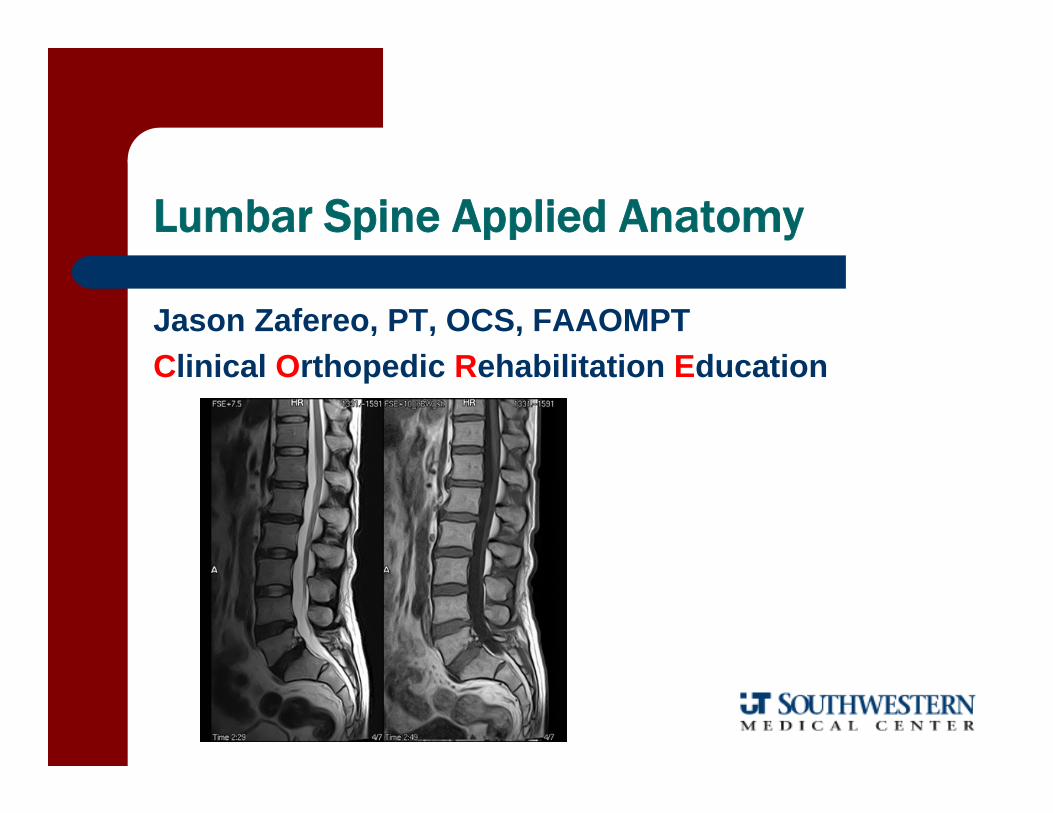
Lumbar Spine Applied Anatomy
Jason Zafereo, PT, OCS, FAAOMPTClinical Orthopedic Rehabilitation Education
Objectives
Discuss concepts relevant to pathophysiology and differential diagnosis for lumbar radiculopathy
Discuss concepts relevant to pathophysiology and differential diagnosis for lumbar disc and joint disorders
Discuss concepts relevant to pathophysiology and differential diagnosis for lumbar instability
RADICULOPATHY
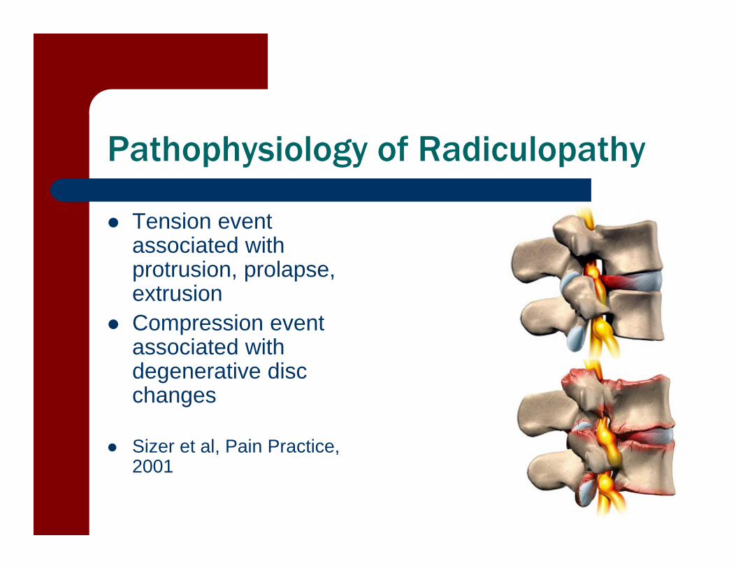
Pathophysiology of Radiculopathy
Tension event associated with protrusion, prolapse, extrusion
Compression event associated with degenerative disc changes
Sizer et al, Pain Practice, 2001
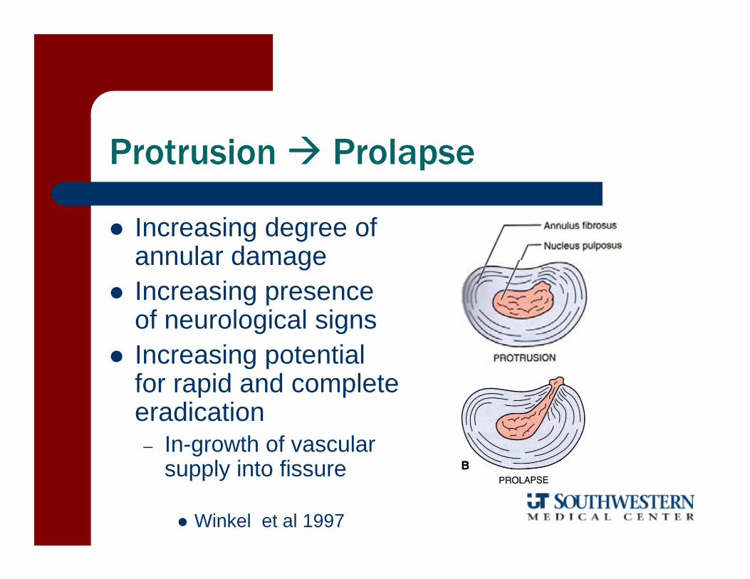
Protrusion Prolapse
Increasing degree of annular damage
Increasing presence of neurological signs
Increasing potential for rapid and complete eradication– In-growth of vascular
supply into fissure
Winkel et al 1997
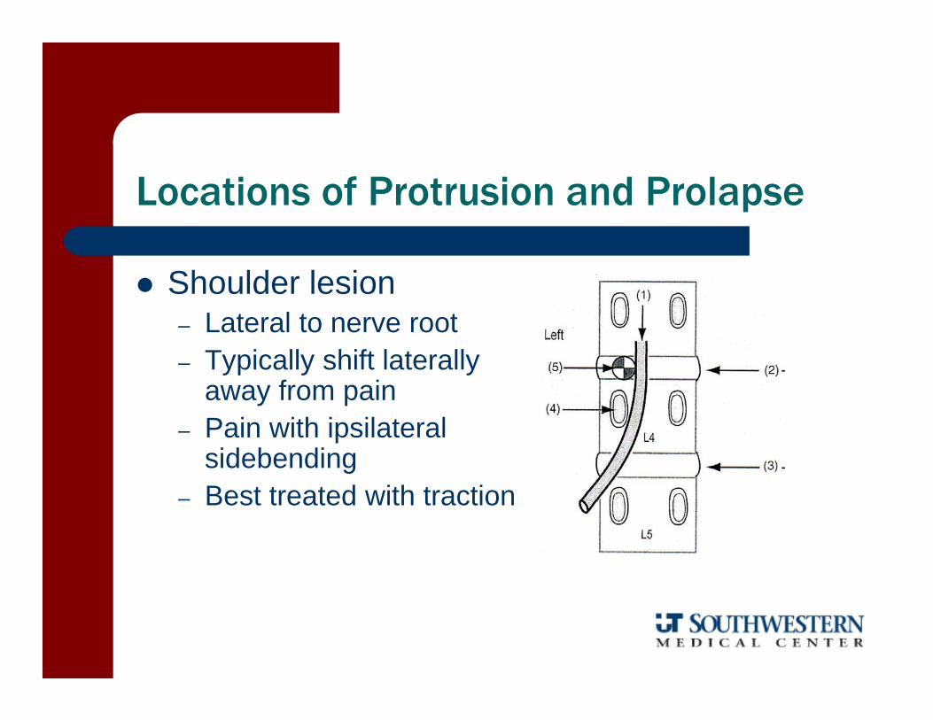
Locations of Protrusion and Prolapse
Shoulder lesion– Lateral to nerve root– Typically shift laterally
away from pain– Pain with ipsilateral
sidebending– Best treated with traction
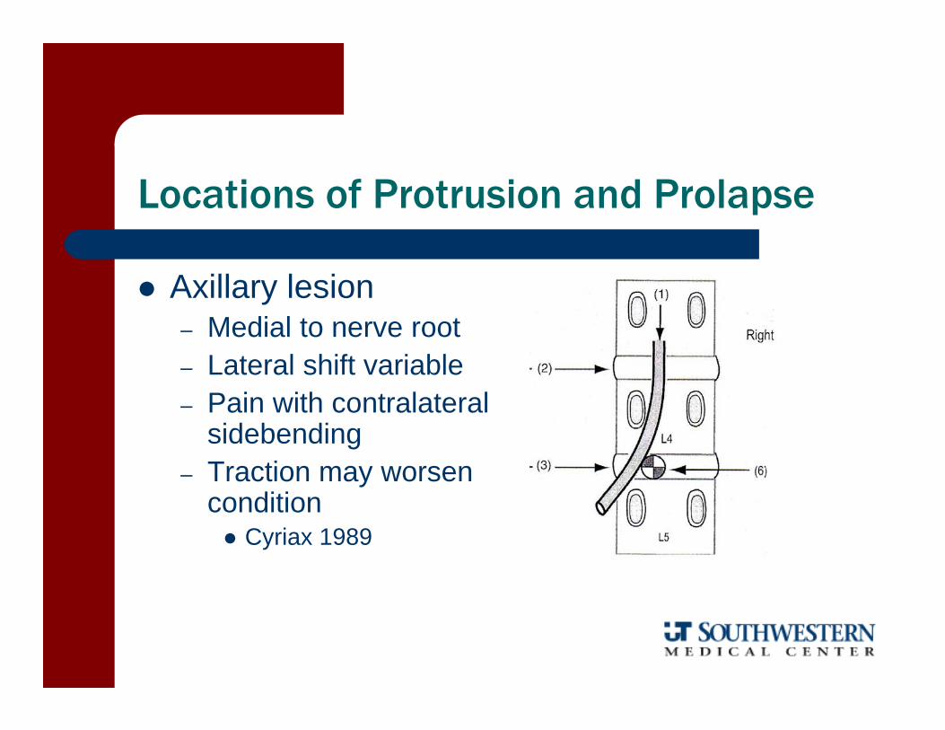
Locations of Protrusion and Prolapse
Axillary lesion– Medial to nerve root– Lateral shift variable– Pain with contralateral
sidebending– Traction may worsen
condition Cyriax 1989

Degenerative Disc Disease
L4-5 and L5-S1– Decreased nuclear hydrostatic
pressure and disc height with aging
– Opposite effects at L3-4 and above
– Leads to lower lumbar accelerated degeneration Sizer et al 2001
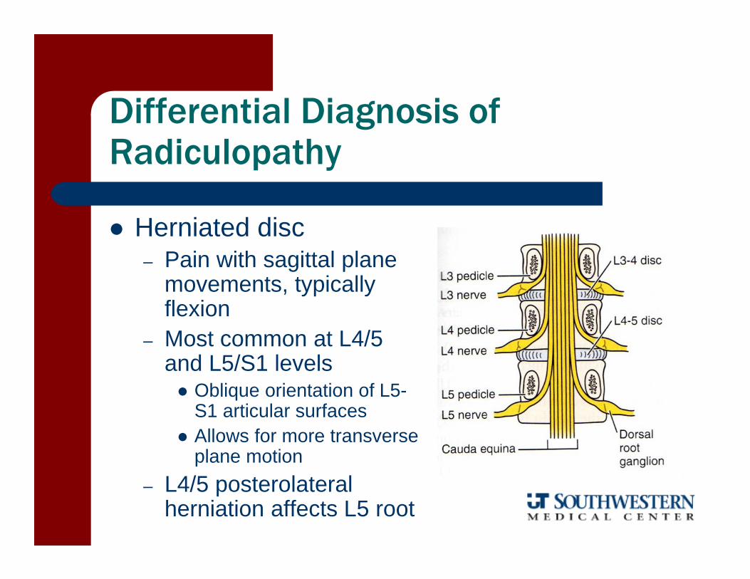
Differential Diagnosis of Radiculopathy
Herniated disc– Pain with sagittal plane
movements, typically flexion
– Most common at L4/5 and L5/S1 levels Oblique orientation of L5-
S1 articular surfaces Allows for more transverse
plane motion– L4/5 posterolateral
herniation affects L5 root

Differential Diagnosis of Radiculopathy
Degenerative disc disease– Pain with foraminal closing– Most common at L4/5 to
L5/S1 levels Site of degenerative changes Long and narrow IVF (L5-S1) Thickest lumbar root (L5)
– Sizer et al 2001
– L5/S1 stenosis affects L5 root
LOCAL LUMBAR PAIN
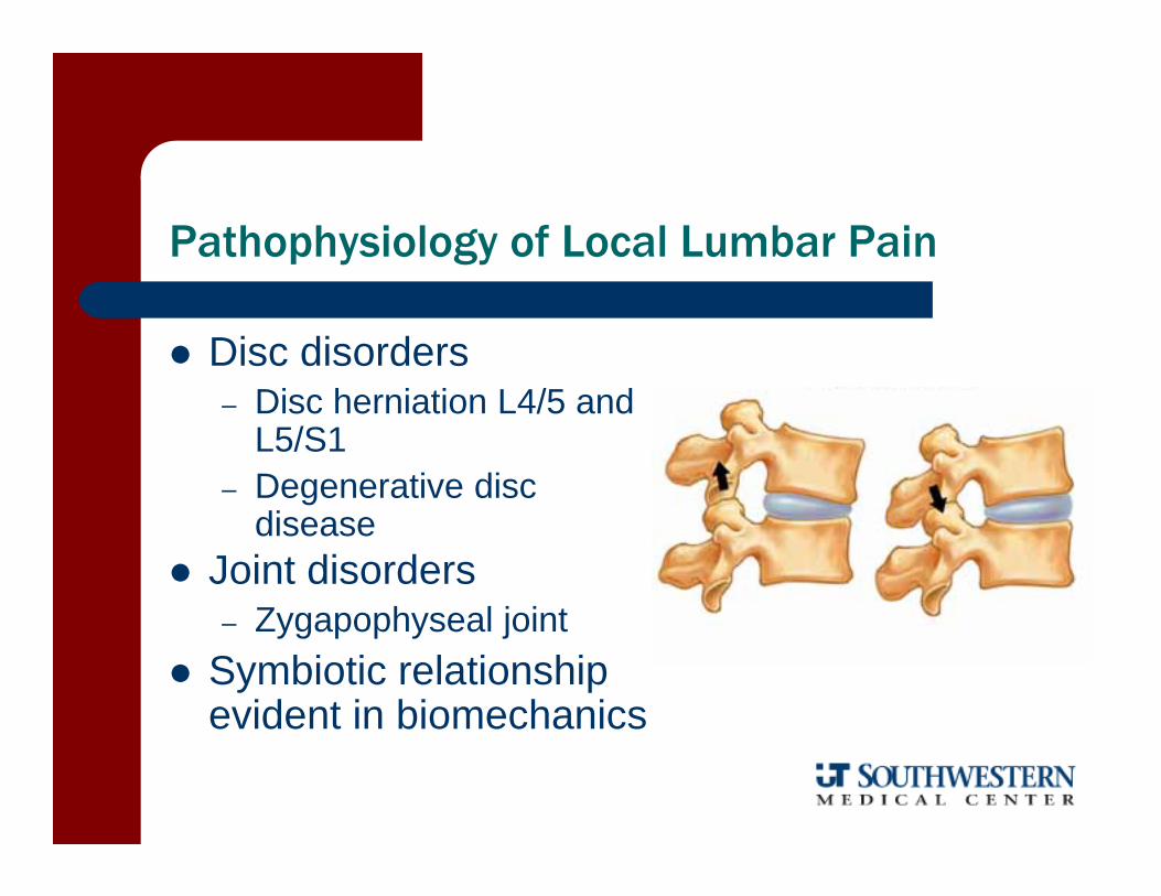
Pathophysiology of Local Lumbar Pain
Disc disorders– Disc herniation L4/5 and
L5/S1– Degenerative disc
disease Joint disorders
– Zygapophyseal joint Symbiotic relationship
evident in biomechanics
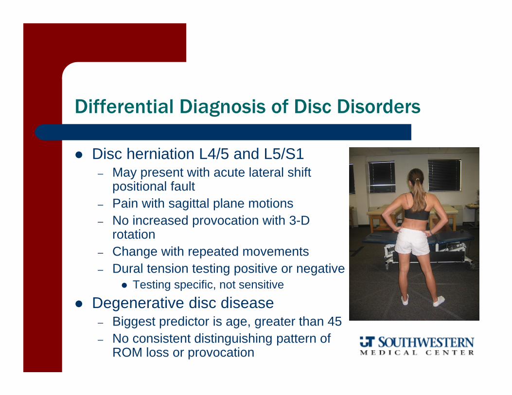
Differential Diagnosis of Disc Disorders
Disc herniation L4/5 and L5/S1– May present with acute lateral shift
positional fault– Pain with sagittal plane motions– No increased provocation with 3-D
rotation – Change with repeated movements– Dural tension testing positive or negative
Testing specific, not sensitive
Degenerative disc disease– Biggest predictor is age, greater than 45– No consistent distinguishing pattern of
ROM loss or provocation
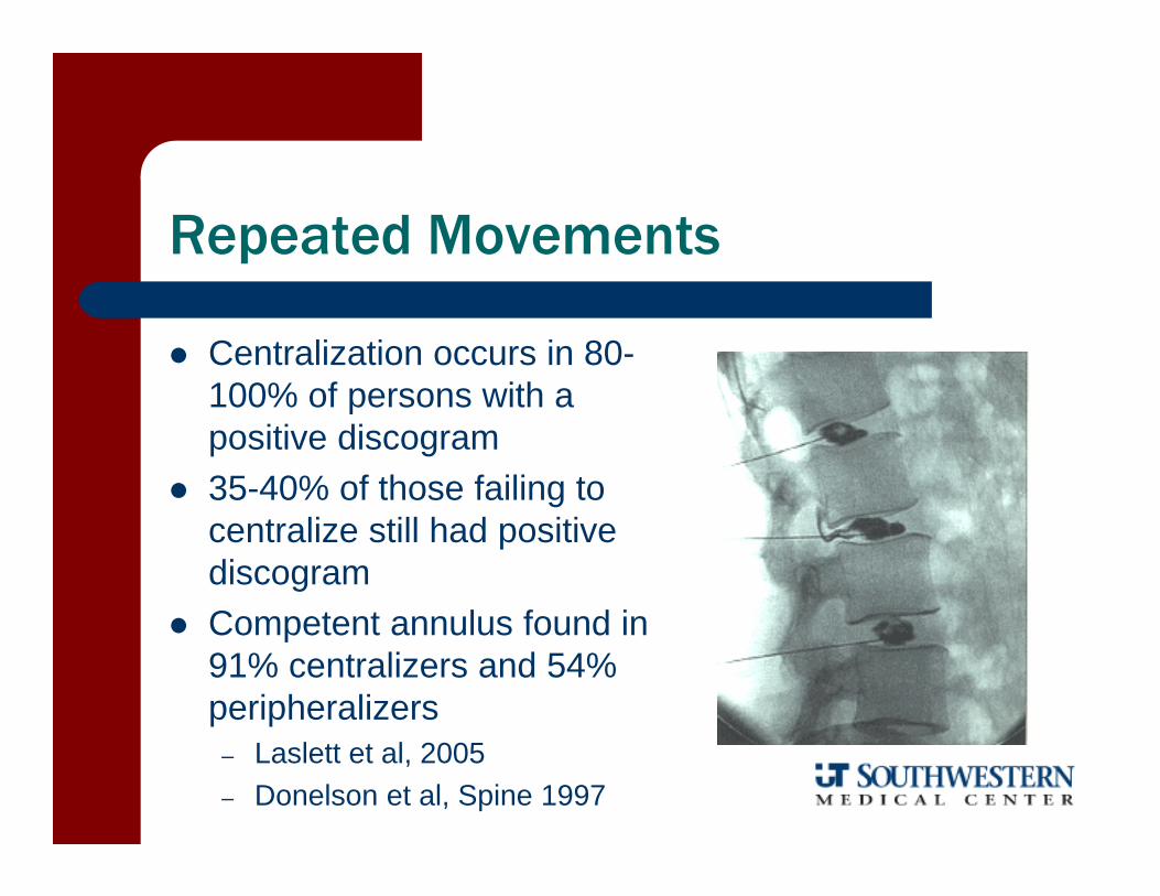
Repeated Movements
Centralization occurs in 80-100% of persons with a positive discogram
35-40% of those failing to centralize still had positive discogram
Competent annulus found in 91% centralizers and 54% peripheralizers
– Laslett et al, 2005– Donelson et al, Spine 1997
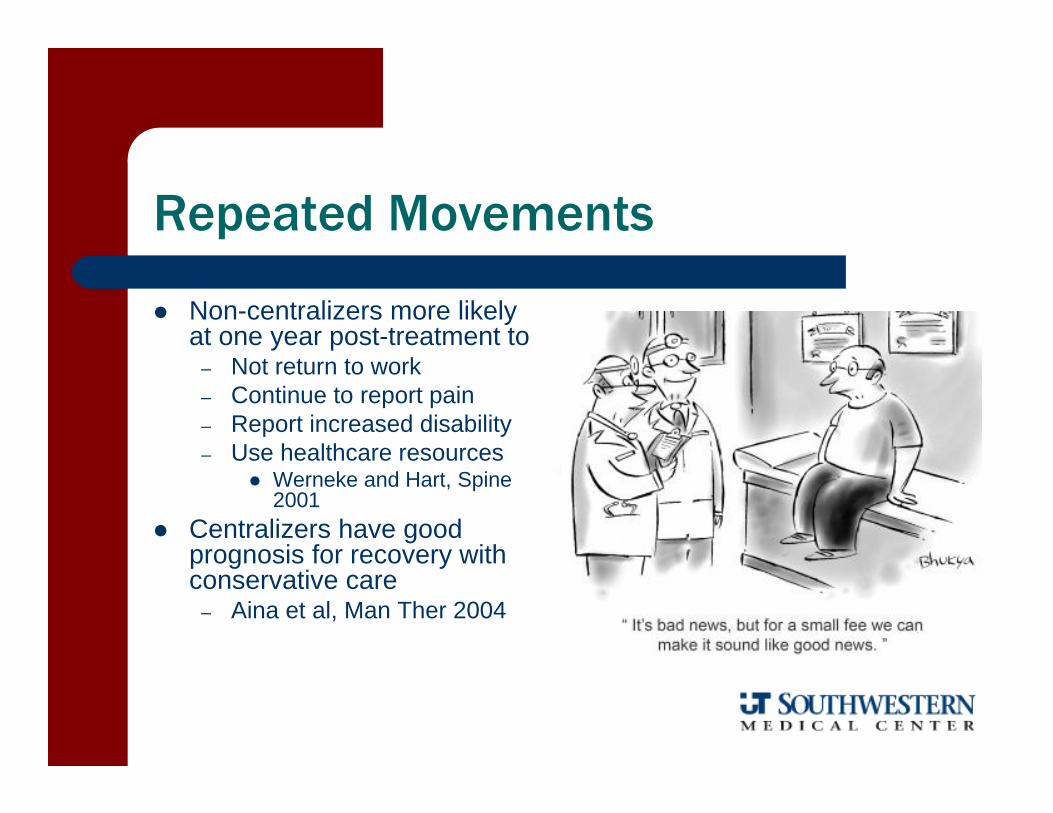
Repeated Movements
Non-centralizers more likely at one year post-treatment to
– Not return to work– Continue to report pain– Report increased disability– Use healthcare resources
Werneke and Hart, Spine 2001
Centralizers have good prognosis for recovery with conservative care
– Aina et al, Man Ther 2004
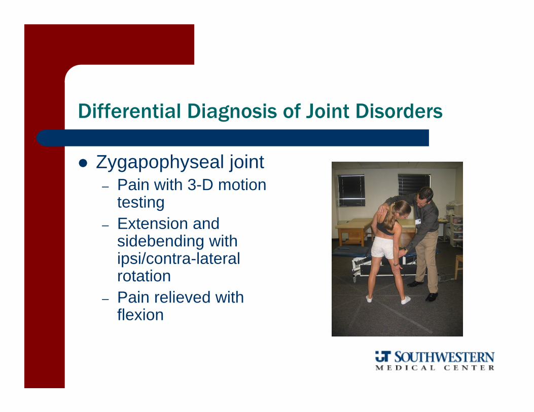
Differential Diagnosis of Joint Disorders
Zygapophyseal joint– Pain with 3-D motion
testing – Extension and
sidebending with ipsi/contra-lateral rotation
– Pain relieved with flexion
INSTABILITY
Pathophysiology of Instability
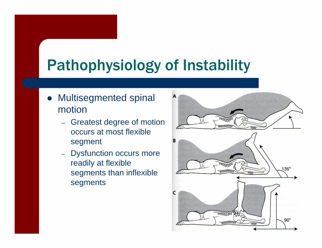
Multisegmented spinal motion
– Greatest degree of motion occurs at most flexible segment
– Dysfunction occurs more readily at flexible segments than inflexible segments
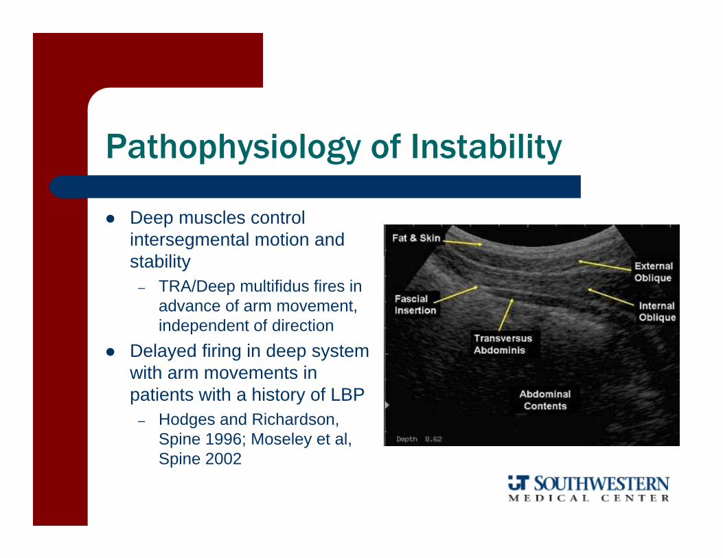
Pathophysiology of Instability
Deep muscles control intersegmental motion and stability
– TRA/Deep multifidus fires in advance of arm movement, independent of direction
Delayed firing in deep system with arm movements in patients with a history of LBP
– Hodges and Richardson, Spine 1996; Moseley et al, Spine 2002
Pathophysiology of Instability
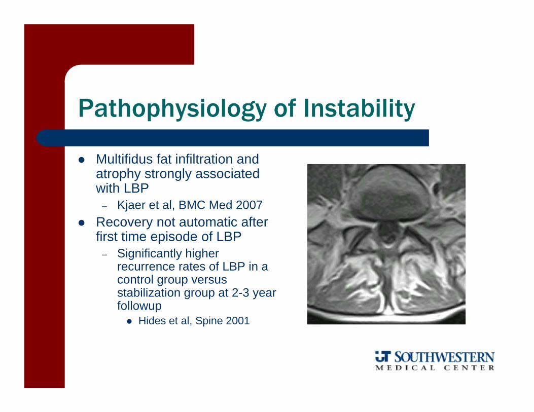
Multifidus fat infiltration and atrophy strongly associated with LBP
– Kjaer et al, BMC Med 2007 Recovery not automatic after
first time episode of LBP– Significantly higher
recurrence rates of LBP in a control group versus stabilization group at 2-3 year followup Hides et al, Spine 2001
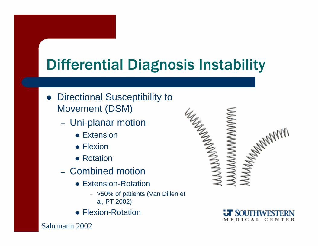
Differential Diagnosis Instability
Directional Susceptibility to Movement (DSM)– Uni-planar motion
Extension Flexion Rotation
– Combined motion Extension-Rotation
– >50% of patients (Van Dillen et al, PT 2002)
Flexion-Rotation
Sahrmann 2002
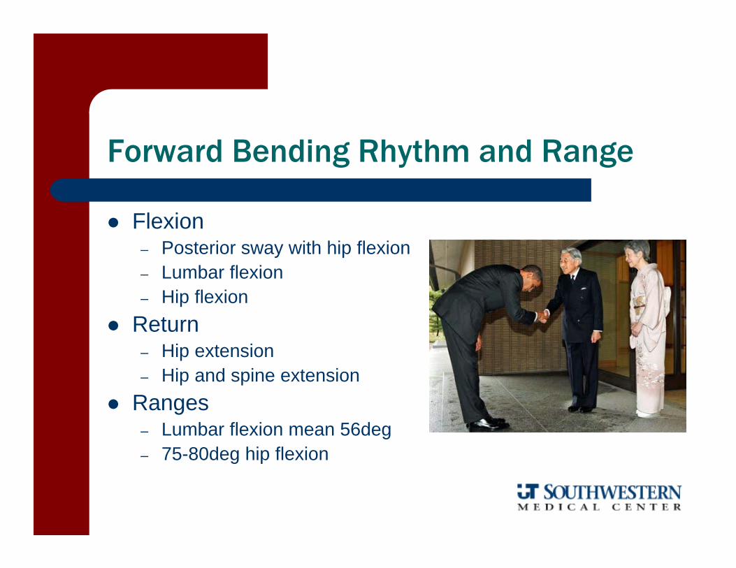
Forward Bending Rhythm and Range
Flexion– Posterior sway with hip flexion– Lumbar flexion– Hip flexion
Return– Hip extension– Hip and spine extension
Ranges– Lumbar flexion mean 56deg – 75-80deg hip flexion
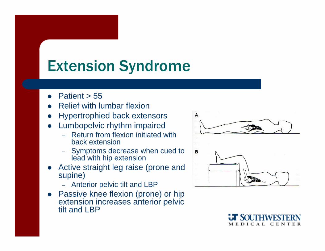
Extension Syndrome
Patient > 55 Relief with lumbar flexion Hypertrophied back extensors Lumbopelvic rhythm impaired
– Return from flexion initiated with back extension
– Symptoms decrease when cued to lead with hip extension
Active straight leg raise (prone and supine)
– Anterior pelvic tilt and LBP Passive knee flexion (prone) or hip
extension increases anterior pelvic tilt and LBP
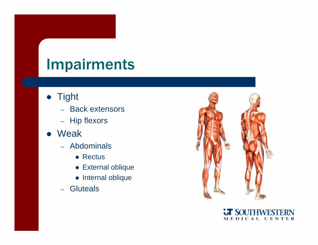
Impairments
Tight– Back extensors– Hip flexors
Weak– Abdominals
Rectus External oblique Internal oblique
– Gluteals
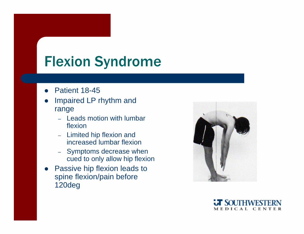
Flexion Syndrome
Patient 18-45 Impaired LP rhythm and
range– Leads motion with lumbar
flexion– Limited hip flexion and
increased lumbar flexion– Symptoms decrease when
cued to only allow hip flexion Passive hip flexion leads to
spine flexion/pain before 120deg
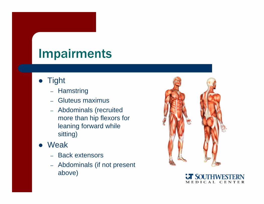
Impairments
Tight– Hamstring– Gluteus maximus– Abdominals (recruited
more than hip flexors for leaning forward while sitting)
Weak– Back extensors– Abdominals (if not present
above)
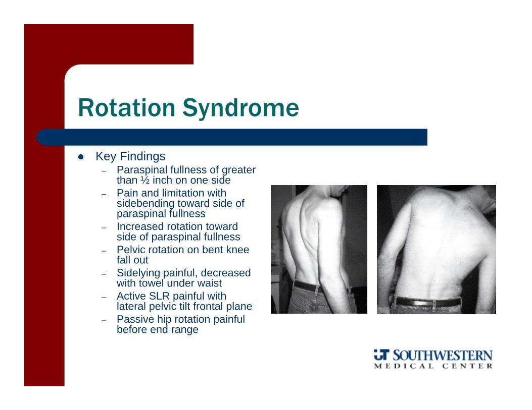
Rotation Syndrome
Key Findings– Paraspinal fullness of greater
than ½ inch on one side– Pain and limitation with
sidebending toward side of paraspinal fullness
– Increased rotation toward side of paraspinal fullness
– Pelvic rotation on bent knee fall out
– Sidelying painful, decreased with towel under waist
– Active SLR painful with lateral pelvic tilt frontal plane
– Passive hip rotation painful before end range
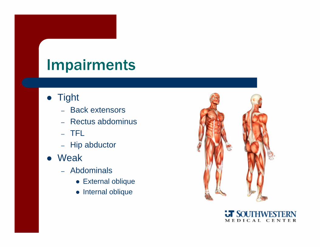
Impairments
Tight– Back extensors– Rectus abdominus– TFL– Hip abductor
Weak– Abdominals
External oblique Internal oblique