Embed Size (px)
Citation preview

Lung
Cancer

Definitions
Epidemiology
Risk factors
Screening
Pathology
Clinical manifestations
Evaluation
Diagnosis
Staging
Treatment
Prognosis

Definitions
The term lung cancer, or bronchogenic carcinoma, refers to
malignancies that originate in the airways or pulmonary parenchyma.
Approximately 95 percent of all lung cancers are classified as either
small cell lung cancer (SCLC) or non-small cell lung cancer (NSCLC).
This distinction is essential for staging, treatment, and prognosis. Other
cell types comprise approximately 5 percent of malignancies arising in
the lung.
Epidemiology
Lung cancer is the leading cause of cancer deaths worldwide in
both men and women. Around 1953, lung cancer became the most
common cause of cancer deaths in men, and in 1985, it became
the leading cause of cancer deaths in women.
The relative incidence of adenocarcinoma has risen dramatically,
and there has been a corresponding decrease in the incidence of
other types of non-small cell lung cancer (NSCLC) and small cell
lung cancer (SCLC).
Non-small cell lung cancer (NSCLC) accounts for the majority
(approximately 85 percent) of lung cancers with the remainder as
mostly small cell lung cancer (SCLC).

Risk Factors
Smoking
Account for approximately 90 percent of all lung cancers.
The risk of developing lung cancer for a current smoker of one pack per day for 40 years is approximately 20 times that of
someone who has never smoked. Factors that increase the risk of developing lung cancer in smokers include the extent of
smoking and exposure to other carcinogenic factors, such as asbestos.
In individuals who do quit smoking, the risk of developing lung cancer falls compared with those who continue to smoke; the
benefit is greatest in those who stop by age 30. Despite stopping smoking, the risk for lung cancer continues to rise with age at a
faster rate than those who never smoked.
Radiation therapy can increase the risk of a second primary lung cancer in patients who have been
treated for other malignancies.
Environmental toxins: secondhand smoke, asbestos, radon, metals (arsenic, chromium, and nickel),
ionizing radiation, and polycyclic aromatic hydrocarbons.

Pulmonary fibrosis: Sevenfold in patients with pulmonary fibrosis. This increased risk appears to be independent of smoking.
HIV infection
Genetic factors – Genetic factors can affect both the risk for and prognosis from lung cancer.
Alcohol
Dietary factors: Various dietary factors (antioxidants, cruciferous vegetables, phytoestrogens) may reduce the risk of lung cancer, but the role of these factors is not well established.
SCREENING
Chest radiography and sputum cytology had NOT been shown to
reduce mortality from lung cancer.
CT scan:
National Lung Screening Trial that compared CT screening with chest
radiograph. This trial demonstrated a 20 percent decrease in lung
cancer mortality in heavy smokers who were screened annually for
three years. For age 55 to 77 who have no symptoms of lung cancer,
have a 30 pack-year smoking history, and if they have quit, have done so within 15 years.

PATHOLOGY
NSCLC and SCLC
Separation of adenocarcinoma and squamous cell carcinoma is important in determining optimal therapy for stage IV disease. Subtype analysis of NSCLC epidermal growth factor receptors (EGFR), anaplastic lymphoma kinase (ALK), and c-ROS oncogene 1 (ROS1) mutations are not only identifiable but their targeted treatment results in responses better than that with standard chemotherapy
EGFR gene: 15 to 30 percent of non-Asian patients and 30 to 60 percent of Asian patients .
The ALK gene mutations present in 2 to 7 percent of patients with NSCLC.
Checkpoint inhibitors promote recognition of cancer cells as foreign cells by the immune system and reverse the tumor-driven inhibition of the immune system that promotes tumor growth. Clinical trials using antibodies to programmed death receptor 1 and programmed death ligand 1 have shown significant survival benefit in advanced NSCLC

Tumor type Changes from 2004 WHO classification 2015 variants (if applicable)
Adenocarcinoma
•Discontinuation of terms bronchioalveolarcarcinoma, mixed subtype adenocarcinoma, clear cell and signet ring adenocarcinoma (these subtypes are descriptive features in the 2015 system), and mucinous cystadenocarcinoma (these tumors are reclassified as colloid adenocarcinomas).•Addition of adenocarcinoma in situ and minimally invasive adenocarcinoma.•Classification of invasive adenocarcinoma according to predominant subtype.•Introduction of the term lepidic for a noninvasive component of an invasive adenocarcinoma.
•Lepidic•Acinar•Papillary•Micropapillary•Solid•Invasive mucinous (mixed invasive mucinous and non-mucinous)•Colloid•Fetal•Enteric•Minimally invasive•Preinvasive (atypical adenomatous hyperplasia, adenocarcinoma in situ [non-mucinous and mucinous])
Adenosquamous cell carcinoma •No significant changes. N/A
Squamous cell carcinoma
•Papillary, clear cell, small cell, and basaloid carcinoma have been replaced.•Clear cell change is now regarded as a cytologic feature.
•Keratinizing•Non-keratinizing•Basaloid
Large cell carcinoma
•The following have been reclassified from "large cell carcinomas" to other subgroups: basaloid carcinoma is a subgroup of squamous cell carcinomas, large cell neuroendocrine carcinoma is a neuroendocrine carcinoma, and lymphoepithelioma-like carcinoma belongs to "other and unclassified carcinomas."•Clear cell and rhabdoid are now descriptive features rather than subtypes.
N/A
Sarcomatoid carcinoma•No significant changes, although molecular testing now recommended.
•Pleomorphic carcinoma•Spindle cell carcinoma•Giant cell carcinoma•Carcinosarcoma•Pulmonary blastoma
Neuroendocrine carcinoma
•Previously, small cell and large cell neuroendocrine tumors were in different categories and are now grouped together.
•Small cell carcinoma•Large cell neuroendocrine carcinoma•Carcinoid (typical and atypical)
Diffuse idiopathic pulmonary neuroendocrine cell hyperplasia
•No significant changes. N/A

CLINICAL MANIFESTATIONS
Symptoms and signs from the tumor itself (both local and systemic
symptoms and signs)
Symptoms and signs from metastasis
Symptoms and signs from paraneoplastic syndromes.
The majority of patients with lung cancer have advanced disease at clinical presentation

Symptom Patients (percent)
Cough 45-74
Weight loss 46-68
Dyspnea 37-58
Chest pain 27-49
Hemoptysis 27-29
Bone pain 20-21
Hoarseness 8-18

Clinical presentation from the tumor itself (both local
and systemic)
Cough: 50 to 75 percent of patients. Most in patients with central tumors (squamous and small)
Hemoptysis: Hemoptysis is reported by 20 to 50 percent of patients.
Chest pain: Chest pain is present in approximately 20 to 40 percent of patients. From mediastinal, pleural, or
chest wall extension. Obstructive pneumonitis or a pulmonary embolus related to a hypercoagulable state
may also cause chest pain.
Dyspnea: 25 to 40 percent of cases. Dyspnea may be due to extrinsic or intraluminal airway obstruction,
obstructive pneumonitis or atelectasis, lymphangitic tumor spread, tumor emboli, pneumothorax, pleural
effusion, or pericardial effusion with tamponade.
Bronchorrhea or cough productive of large volumes of thin, mucoid secretions may be a feature of
mucinous adenocarcinoma and usually indicates advanced disease.

Post-obstructive pneumonia.
Bronchiectasis: Slow-growing neoplasms such as carcinoid tumor or hamartoma are more likely to present with bronchiectasis.
Unilateral paralysis of the diaphragm may be due to damage of the phrenic nerve. Patients may be asymptomatic or report shortness of breath.
Pancoast syndrome can present with pain in the shoulder, Horner syndrome, bony destruction, and atrophy of hand muscles.
Hoarseness of voice from vocal cord paralysis. The differential diagnosis of persistent hoarseness in a smoker includes both laryngeal cancer and lung cancer. In patients with lung cancer, this is due to malignancy involving the recurrent laryngeal nerve along its course under the arch of the aorta and back to the larynx.


Pleural involvement:
Can cause dyspnea and cough, approximately one-fourth of patients who have lung cancer and
pleural metastases are asymptomatic. During the course of their disease, approximately 10 to 15
percent of patients who have lung cancer will have malignant pleural effusions.
Superior vena cava syndrome:
Sensation of fullness in the head and dyspnea. Cough, pain, and dysphagia are less frequent.
Physical findings include dilated neck veins, a prominent venous pattern on the chest, facial
edema, and a plethoric appearance. The chest radiograph typically shows widening of the
mediastinum or a right hilar mass. CT can often identify the cause, level of obstruction, and extent of
collateral venous drainage. The SVC syndrome is more common in patients with SCLC than NSCLC.
Weight loss

Symptoms and signs from metastasis
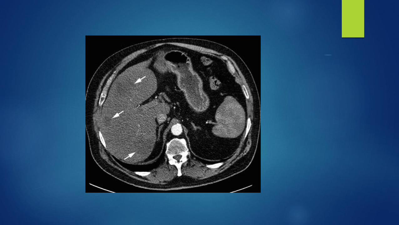
Liver:
Uncommon.
Asymptomatic liver metastases may be detected at presentation by liver enzyme abnormalities, CT,
or positron emission tomography (PET).
Bone
Symptomatic.
Pain in the back, chest, or extremity, and elevated levels of serum alkaline phosphatase are usually
present in patients who have bone metastasis.
The serum calcium may be elevated due to extensive bone disease. Approximately 20 percent of
patients with NSCLC have bone metastases on presentation and more in SCLC.
An osteolytic radiographic appearance is more frequent than an osteoblastic one, and the most
common sites of involvement are the vertebral bodies.

Adrenal
The adrenal glands are a frequent site of metastasis but such metastases are only rarely symptomatic.
Brain
Neurologic manifestations of lung cancer include metastases and paraneoplastic syndromes.
Symptoms from central nervous system metastasis are similar to those with other tumors and include
headache, vomiting, visual field loss, hemiparesis, cranial nerve deficit, and seizures.
In patients with NSCLC, the frequency of brain metastasis is greatest with adenocarcinoma and least
with squamous cell carcinoma.
In patients with SCLC, metastasis to brain is present in approximately 20 to 30 percent of patients at presentation.

Symptoms and signs from paraneoplastic syndromes.
Paraneoplastic phenomena: Paraneoplastic effects of tumor are remote effects that are not related to the direct invasion, obstruction, or metastasis.
Hypercalcemia:
Hypercalcemia in patients with lung cancer may arise from a bony metastasis or less commonly tumor secretion of a parathyroid hormone-related protein (PTHrP), calcitriol or other cytokines, including osteoclast activating factors.
More in squamous cell carcinoma but seen frequently with the other types.
Most patients with hypercalcemia have advanced disease (stage III or IV) and a median survival of a few months.
Symptoms: anorexia, nausea, vomiting, constipation, lethargy, polyuria, polydipsia, and dehydration. Confusion and coma are late manifestations as are renal failure and nephrocalcinosis.
Treatment: hydration, calcitonin and bisphosphonate.
SIADH secretion
SIADH is frequently caused by SCLC. Approximately 10 percent of patients who have SCLC exhibit SIADH.
SCLC accounts for approximately 75 percent of all malignancy-related of SIADH.
The severity of symptoms is related to the degree of hyponatremia and the rapidity of the fall in serum sodium.
Symptoms include anorexia, nausea, and vomiting. Cerebral edema can occur when the onset of hyponatremia is rapid. Symptoms caused by cerebral edema may include irritability, restlessness, personality changes, confusion, coma, seizures, and respiratory arrest.
The treatment of SIADH focuses on treating the malignancy. In the majority of patients with SCLC, the hyponatremia will resolve within weeks of starting chemotherapy.

Neurologic
Lung cancer is the most common cancer associated with paraneoplastic neurologic syndromes,
typically these are associated with SCLC.
Paraneoplastic neurologic syndromes are thought to be immune-mediated, and autoantibodies
have been identified in a number of instances.
Lambert-Eaton myasthenic syndrome (LEMS), cerebellar ataxia, sensory neuropathy, limbic
encephalitis, encephalomyelitis, autonomic neuropathy, retinopathy, and opsomyoclonus.
The most common of these is LEMS, which may be seen in approximately 3 percent of patients with
SCLC.
As many as 70 percent of patients who have SCLC and an associated paraneoplastic neurologic
syndrome have limited stage disease.
Paraneoplastic neurologic syndromes generally do not improve with immunosuppressive treatment.
However, symptoms may stabilize with response of the underlying neoplasm to treatment.


Hematologic manifestations
Anemia: Contribute to fatigue and dyspnea.
Leukocytosis: Thought to be due to overproduction of granulocyte-colony stimulating factor. Leukocytosis in association with lung cancer is associated with a poor prognosis and has also been associated with hypercalcemia.
Thrombocytosis – Thrombocytosis is common and maybe present in as many as 14 percent of patients with lung cancer at presentation. Thrombocytosis at presentation has been identified as an independent predictor of shortened survival.
Eosinophilia: Eosinophilia in tissue or blood is rare but has been reported in patients with large cell carcinoma.

Hypercoagulable disorders
Trousseau syndrome (migratory superficial thrombophlebitis)
Deep venous thrombosis and thromboembolism
Disseminated intravascular coagulopathy
Thrombotic microangiopathy
Nonthrombotic microangiopathy


Hypertrophic osteoarthropathy
Hypertrophic pulmonary osteoarthropathy (HPO) is defined by the presence of clubbing and periosteal proliferation of the tubular bones associated with lung cancer or other lung disease.
Clinically, HPO is characterized by a symmetrical, painful arthropathy that usually involves the ankles, knees, wrists, and elbows. The metacarpal, metatarsal, and phalangeal bones may also be involved.
A radiograph of the long bones (ie, tibia and fibula) shows characteristic periosteal new bone formation in patients with HPO.
The symptoms of HPO may resolve after tumor resection.
Dermatomyositis and polymyositis
These inflammatory myopathies can be the presenting symptom in patients with lung cancer or can develop later in the course of disease. In addition to lung cancer, other frequent primary sites associated with these disorders include the ovary, cervix, pancreas, bladder, and stomach.
Cushing syndrome
Ectopic production of adrenal corticotropin (ACTH) can cause Cushing syndrome. Patients typically present with muscle weakness, weight loss, hypertension, hirsutism, and osteoporosis. Hypokalemic alkalosis and hyperglycemia are usually present.
Cushing syndrome is relatively common in patients with SCLC and with carcinoid tumors of the lung.


GENERAL GOALS AND TIMING OF EVALUATION
Clinical extent and stage of disease
Optimal target site and modality for the first tissue biopsy
Specific histological subtype
Presence of comorbidities, secondary complications, and paraneoplastic
syndromes that influence treatment options and outcome


INITIAL EVALUATION
Clinical:
Every patient with suspected lung cancer should undergo a thorough history and
physical exam. Diagnostic tests should be directed to symptoms.
Examples:
Hip pain may prompt plain radiographs of the hip.
Horner's syndrome (ipsilateral ptosis, anhidrosis, and miosis) may prompt an MRI of the superior sulcus.
Neurologic symptoms may prompt imaging of the brain or spinal cord.
Hypotension with sinus tachycardia and pulsus paradoxus may prompt an echocardiogram to evaluate for
malignant pericardial effusion.

Laboratory
Complete blood count
Electrolytes
Calcium
Alkaline phosphatase
Alanine aminotransferase (ALT) and aspartate aminotransferase (AST), total bilirubin
Creatinine
Albumin and lactate dehydrogenase.
Liver function test abnormalities possibly due to liver metastasis should prompt evaluation of the liver with liver-directed imaging.
Calcium elevation should prompt additional imaging for bone metastasis and/or a work up for a paraneoplastic manifestation of the primary tumor.
Elevation of the alkaline phosphatase could be due to liver or bone metastases and should prompt measurement of gamma glutamyl transpeptidase (GGT). When GGT is normal an evaluation for bone metastasis is indicated; when abnormal, an evaluation for liver metastases is indicated.
Radiographic
The clinical staging of patients with suspected lung cancer starts with radiographic imaging. Determining the highest radiographic stage prior to biopsy facilitates the selection of a modality that optimizes tissue sampling for diagnosis.
CXR
Chest radiograph demonstrating a new or enlarging focal lesion, a
pleural effusion, pleural nodularity, enlarged hilar or paratracheal
nodes, endobronchial lesion, post-obstructive pneumonia or
segmental or lobar atelectasis.
Findings suggestive of cancer or cancer-related complications on
chest radiograph should be further evaluated with CT chest.


CT
Every patient with suspected lung cancer should undergo CT scan
of the chest.
CT findings: large lesion size (e.g., >15 mm), irregular or spiculated
borders, upper lobe location, thick-walled cavitation, presence or
development of a solid component within a ground glass lesion,
and detection of growth by follow-up imaging. The finding of
multiple nodules in a patient with a known or suspected
extrathoracic malignancy strongly suggests pulmonary metastasis.


PET Scan
Although whole-body PET is more accurate than CT in detecting
occult disease, its use has not been shown to improve survival
False positives can occur with benign FDG-avid lesions such as
infections, inflammation, and granulomatous disease.
False negatives typically occur when there are microscopic foci of
metastasis, and in non-enlarged lymph nodes (<10 mm).


Integrated PET/CT
Although randomized trials suggest that the use of integrated PET/CT
reduced futile thoracotomies, and is probably superior to either
modality alone, it has not been shown to improve survival.


Imaging metastatic disease
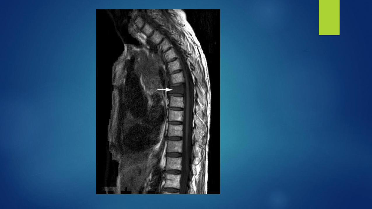
Brain, spine, and nerves:
Patients with clinical stage III or IV disease have an increased risk of
occult intracranial metastasis and may benefit from routine imaging of
the brain with magnetic resonance imaging (MRI) or CT if MRI is not
available.
Gadolinium-enhanced MRI detects spinal bone lesions and brain
lesions and differentiates metastases from other central nervous system
lesions with greater sensitivity than non-enhanced MRI.

Other mets: With the exception of brain metastases, whole body PET
or PET/CT scanning is more accurate than conventional scanning
(abdominal CT, bone scan) for the detection of unsuspected
pleural and extrathoracic metastases.
When PET, PET/CT or MRI is not available, conventional staging with
abdominal CT, bone scintigraphy, and CT scan of the brain should be
performed when indicated.
Sometimes US, Body MRI, Bone scan…etc

Suggestive of metastasisSymptoms elicited in history
Constitutional - weight loss >10 pounds
Musculoskeletal - focal skeletal pain
Neurologic - headaches, syncope, seizures, extremity weakness, recent change in
mental status
Signs found on physical exam
Lymphadenopathy (>1 cm)
Hoarseness, superior vena cava syndrome
Bone tenderness
Hepatomegaly (>13 cm span)
Focal neurologic signs, papilledema
Soft tissue mass
Routine laboratory tests
Hematocrit, <40% in males
Hematocrit, <35% in females
Elevated alkaline phosphatase, GGT, SGOT


Pathologic diagnosis
Diagnosis of lung cancer should not be made without definitive pathology. At a minimum, this involves selecting a biopsy site and obtaining an adequate sample for microscopic examination. Additional consideration needs to be given to obtaining a large enough sample for supplemental immunohistochemical and genetic analysis.
Modality
Cytologic specimens can be obtained from the following sites:
Lung: Sputum, transthoracic needle aspirates, and bronchoscopic washings, brushings, or needle aspirates
Lymph node: Transthoracic, transbronchial, and transesophageal aspirates
Distant metastasis: Pleural fluid, needle aspirates of metastatic tissue (eg, liver)
Core or biopsy tissue can be obtained from the following:
Lung: Endobronchial biopsy (forceps), transbronchial biopsy (forceps or needle), transthoracic (needle) biopsy, surgical biopsy
Lymph node: Bronchoscopic and transthoracic needle core biopsy, surgical biopsy
Distant metastasis: Core needle aspirates of metastatic tissue (eg liver, bone, adrenal)

Immunohistochemical and histochemical stains useful in the differential diagnosis of various carcinomas
Tumor type Immunohistochemical staining
CarcinomaPositive: Pankeratin, AE 1/3, CAM 5.2, OSCAR, EMANegative: CD 45Variable: CK 7, CK 20, S-100, vimentin
Colorectal carcinomaPositive: CK 20, CDX-2Negative: CK 7
Lung carcinoma
Adenocarcinoma Positive: TTF-1, napsin A, CK 7, mucicarmine, PAS-D
Squamous cell carcinomaPositive: p 40, p 63, CK 5/6, desmogleinNegative: CK 7 (usually)
Small-cell carcinomaPositive: TTF-1, high proliferative rate (Ki-67, MIB-1)Variable: Chromogranin, synaptophysin
Neuroendocrine carcinoma Positive: Chromogranin, synaptophysin, epithelial stains
Germ cell tumorPositive: HCG, AFP, Oct4 transcription factor, placental alkaline phosphatase, epithelial stains
Hepatocellular carcinomaPositive: Hep par 1, CEA, AFP, glypican 3Negative: CK 7, CK 20
Renal cell carcinomaPositive: Pan keratin, CAM 5.2, Pax-8, CK 7, vimentin, RCC, CD 10Negative: CK 20, CEA
Prostate carcinomaPositive: PSA, prostatic acid phosphataseNegative: CK 7, 20
Pancreas carcinomaPositive: CA 19-9, CK 7, CDX-2, CK 17Variable: CK 20
Breast carcinomaPositive: ER, PR, Her-2-neu, CK 7, gross cystic fluid protein 15, epithelial stains, GATA 3, mammaglobinNegative: CK 20
Ovarian carcinomaPositive: CK 7, WT-1, Pax-8, ERNegative: CK 20, CDX-2

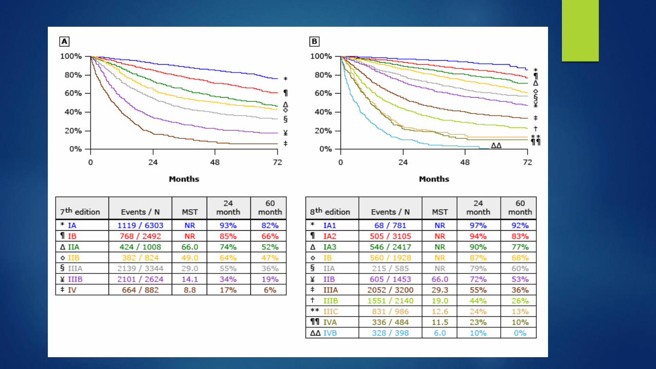
STAGING
The 8th edition for staging non-small cell lung cancer (NSCLC) is in
use. Staging NSCLC determines the appropriate therapy and, when
combined with the patient's unique features, provides valuable
prognostic information.
The TNM system for staging NSCLC
The staging for small cell lung cancer (limited versus extensive)and
TNM)
The clinical-diagnostic stage and the surgical-pathologic stage

T: Primary tumor
Tx Primary tumor cannot be assessed or tumor proven by presence of malignant cells in sputum or bronchial washings but not visualized by imaging or bronchoscopy
T0 No evidence of primary tumorTis Carcinoma in situ
T1 Tumor ≤3 cm in greatest dimension surrounded by lung or visceral pleura without bronchoscopic evidence of invasion more proximal than the lobar bronchus (ie, not in the main bronchus)*
T1a(mi) Minimally invasive adenocarcinoma¶
T1a Tumor ≤1 cm in greatest dimension*
T1b Tumor >1 cm but ≤2 cm in greatest dimension*
T1c Tumor >2 cm but ≤3 cm in greatest dimension*
T2
•Tumor >3 cm but ≤5 cm or tumor with any of the following features:ΔInvolves main bronchus regardless of distance from the carina but without involvement of the carina
•Invades visceral pleura•Associated with atelectasis or obstructive pneumonitis that extends to the hilar region, involving part or all of the lung
T2a Tumor >3 cm but ≤4 cm in greatest dimensionT2b Tumor >4 cm but ≤5 cm in greatest dimension
T3Tumor >5 cm but ≤7 cm in greatest dimension or associated with separate tumor nodule(s) in the same lobe as the primary tumor or directly invades any of the following structures: chest wall (including the parietal pleura and superior sulcus tumors), phrenic nerve, parietal pericardium
T4Tumor >7 cm in greatest dimension or associated with separate tumor nodule(s) in a different ipsilateral lobe than that of the primary tumor or invades any of the following structures: diaphragm, mediastinum, heart, great vessels, trachea, recurrent laryngeal nerve, esophagus, vertebral body, and carina
N: Regional lymph node involvement
Nx Regional lymph nodes cannot be assessed
N0 No regional lymph node metastasis
N1 Metastasis in ipsilateral peribronchial and/or ipsilateral hilar lymph nodes and intrapulmonary nodes, including involvement by direct extension
N2 Metastasis in ipsilateral mediastinal and/or subcarinal lymph node(s)
N3 Metastasis in contralateral mediastinal, contralateral hilar, ipsilateral or contralateral scalene, or supraclavicular lymph node(s)
M: Distant metastasisM0 No distant metastasis
M1 Distant metastasis present
M1a Separate tumor nodule(s) in a contralateral lobe; tumor with pleural or pericardial nodule(s) or malignant pleural or pericardial effusion◊
M1b Single extrathoracic metastasis§
M1c Multiple extrathoracic metastases in one or more organs

Stage groupings
Occult carcinoma TX N0 M0
Stage 0 Tis N0 M0
Stage IA1T1a(mi) N0 M0
T1a N0 M0
Stage IA2 T1b N0 M0
Stage IA3 T1c N0 M0
Stage IB T2a N0 M0
Stage IIA T2b N0 M0
Stage IIB
T1a to c N1 M0
T2a N1 M0
T2b N1 M0
T3 N0 M0
Stage IIIA
T1a to c N2 M0
T2a to b N2 M0
T3 N1 M0
T4 N0 M0
T4 N1 M0
Stage IIIB
T1a to c N3 M0
T2a to b N3 M0
T3 N2 M0
T4 N2 M0
Stage IIICT3 N3 M0
T4 N3 M0
Stage IVAAny T Any N M1a
Any T Any N M1b
Stage IVB Any T Any N M1c

Limited disease – Tumor confined to the ipsilateral hemithorax and
regional nodes able to be included in a single tolerable
radiotherapy port (corresponding to Tumor, Node, Metastasis [TNM]
stages I through IIIB).
Extensive disease – Tumor beyond the boundaries of limited disease
including distant metastases, malignant pericardial, or pleural
effusions, and contralateral supraclavicular and contralateral hilar
involvement.

Treatment
Surgical
Chemotherapy
Radiotherapy
Molecularly targeted therapy
Immunotherapy

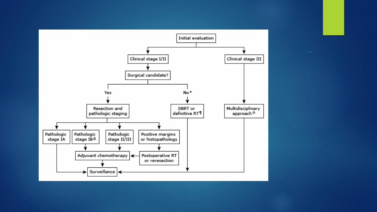
Treatment of NSCLC
Stage I or II NSCLC should be treated with complete surgical resection whenever possible. Postoperative adjuvant chemotherapy improves survival in patients with pathologic stage II disease and may have a role for patients with stage IB disease.
Concurrent chemoradiotherapy is preferred for those with more extensive intrathoracic disease.
Patients with advanced disease are managed palliatively with systemic therapy and/or local palliative modalities.
Patients with stage IV disease are generally treated with systemic therapy or a symptom-based palliative approach. In appropriately selected patients, chemotherapy, molecularly targeted therapy, and/or immunotherapy may prolong survival without sacrificing quality of life.
Patients with stage IV disease based upon the presence of an isolated metastasis (eg, brain, adrenal) may benefit from resection of the metastasis as well as aggressive treatment of the primary tumor

Treatment of SCLC
SCLC is a disseminated disease in most patients at presentation and is very responsive to chemotherapy. Thus systemic chemotherapy is an integral part of the initial treatment.
Patients with limited-stage disease are primarily treated with a combination of chemotherapy and radiation therapy
Surgery is not used except in the rare patient who presents with a solitary pulmonary nodule without distant metastases or regional lymph node involvement
Prophylactic cranial irradiation decreases the incidence of brain metastases and prolongs survival in patients with limited-stage SCLC who respond to their initial treatment.
For patients with extensive-stage SCLC, chemotherapy is used as the initial therapy, with or without immunotherapy. Radiation therapy, including both prophylactic cranial irradiation and thoracic RT, may be beneficial in patients with a complete or partial response to their initial chemotherapy.

Prognosis
Prognosis of NSCLC (Some of them)
Stage of disease: The TNM stage at presentation in patients with NSCLC is the factor that has the greatest impact on prognosis.
Performance status: Poor performance status and weight loss have been associated with shortened survival. Reduced appetite, a precursor of weight loss, also has negative prognostic implications.
Histopathology: The degree of differentiation, lymphatic invasion, occult lymph node metastases and intense tumor lymphocytic infiltration
Molecular characterization:
Activating mutations in EGFR have a significantly better prognosis than those without EGFR mutations
The presence of the ROS1 or EML4-ALK fusion oncogene are highly responsive to inhibitors of ALK.
Expression of tumor PD-L1 predicts response to certain immunotherapies
Recurrence after complete resection

Prognosis of SCLC
The most important prognostic factor in patients with SCLC is the extent of disease (stage) at presentation. For patients with limited-stage disease, median survivals range from 15 to 20 months, and the reported five-year survival rate is 10 to 13 percent. By contrast, for patients with extensive-stage disease, the median survival is 8 to 13 months, and the five-year survival rate is 1 to 2 percent.
Clinical parameters also have prognostic importance in patients with SCLC. Poor performance status and/or weight loss have been associated with shortened survival.

Questions??