Embed Size (px)
Citation preview
•LUNG PATHOLOGY
Dr. Sofia Zilber
PNEUMONIA – any infection of the lung parenchyma.
(although this term is used for many interstitial non infectious lung diseases).
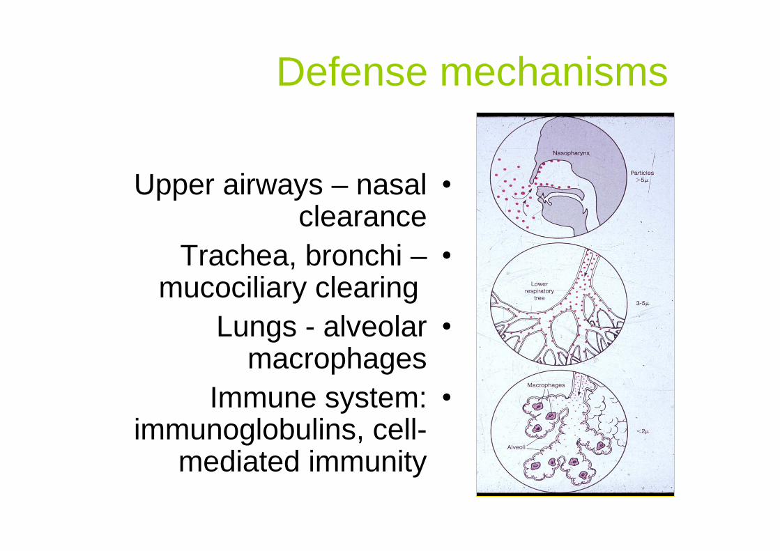
Defense mechanisms
•Upper airways – nasal clearance
•Trachea, bronchi –mucociliary clearing
•Lungs - alveolar macrophages
•Immune system: immunoglobulins, cell-
mediated immunity
�One type of pneumonia predisposes to another (the most common cause of death
in viral influenza epidemics is bacterial pneumonia)
�The portal of entry for most pneumonias is the respiratory tract. Hematogenousspread from other organs can occur.
�Many patients with chronic diseases acquire terminal pneumonia while
hospitalized (nosocomial infection).
Classification of pneumonia
•By specific etiologic agent•By the clinical setting in which the infection
occurs
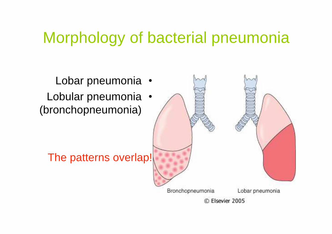
Morphology of bacterial pneumonia
•Lobar pneumonia
•Lobular pneumonia (bronchopneumonia)
The patterns overlap!
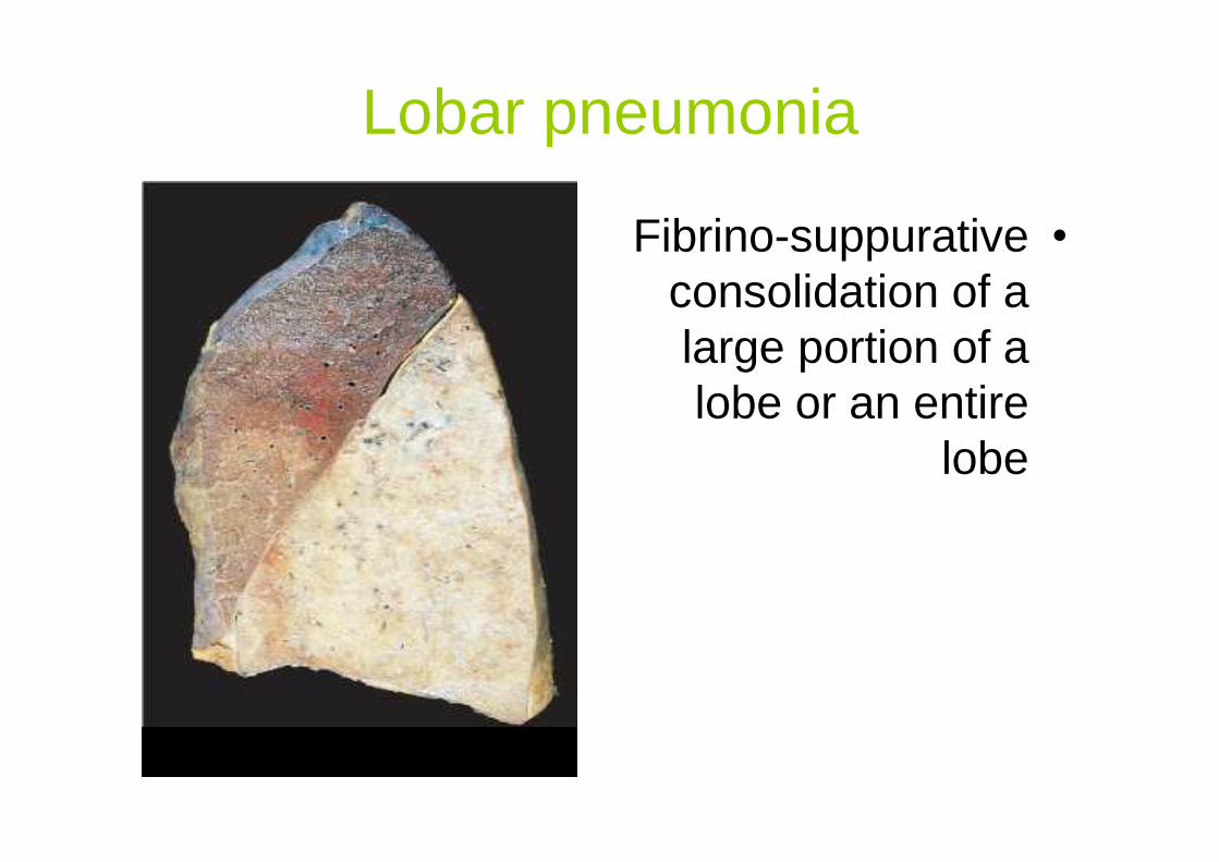
Lobar pneumonia
•Fibrino-suppurativeconsolidation of a large portion of a lobe or an entire
lobe
Lobar pneumonia stages
•Congestion•Red hepatization•Gray hepatization•Resolution
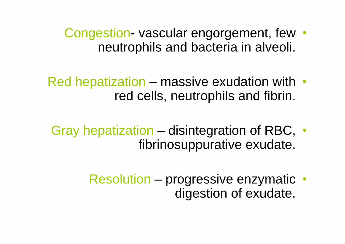
•Congestion- vascular engorgement, few neutrophils and bacteria in alveoli.
•Red hepatization – massive exudation with red cells, neutrophils and fibrin.
•Gray hepatization – disintegration of RBC, fibrinosuppurative exudate.
•Resolution – progressive enzymatic digestion of exudate.
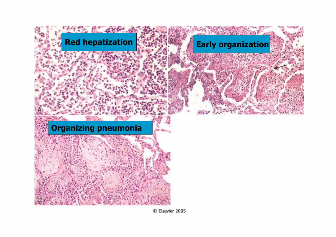
Red hepatization
Organizing pneumonia
Early organization
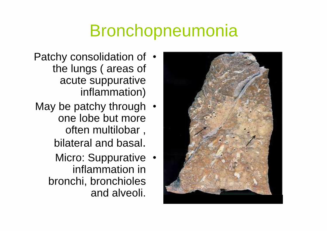
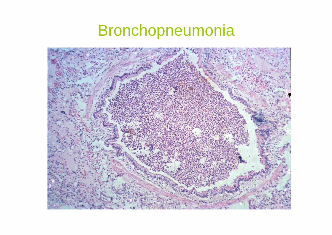
Bronchopneumonia•Patchy consolidation of
the lungs ( areas of acute suppurative
inflammation)•May be patchy through
one lobe but more often multilobar ,
bilateral and basal.•Micro: Suppurative
inflammation in bronchi, bronchioles
and alveoli.
BronchopneumoniaBronchopneumonia
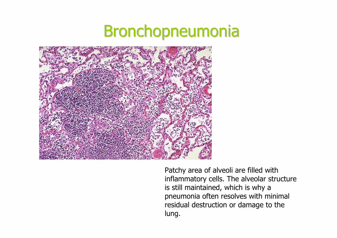
Patchy area of alveoli are filled with inflammatory cells. The alveolar structure is still maintained, which is why a pneumonia often resolves with minimal residual destruction or damage to the lung.
Bronchopneumonia
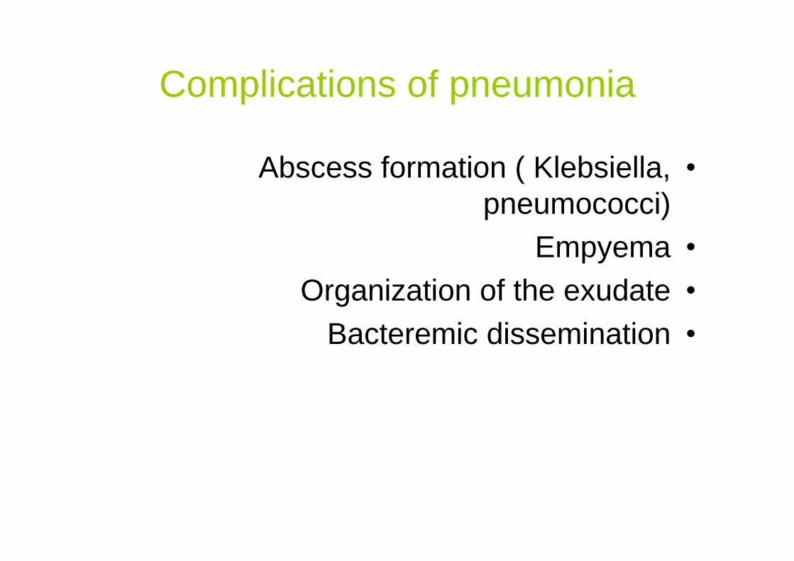
Complications of pneumonia
•Abscess formation ( Klebsiella, pneumococci)
•Empyema•Organization of the exudate•Bacteremic dissemination
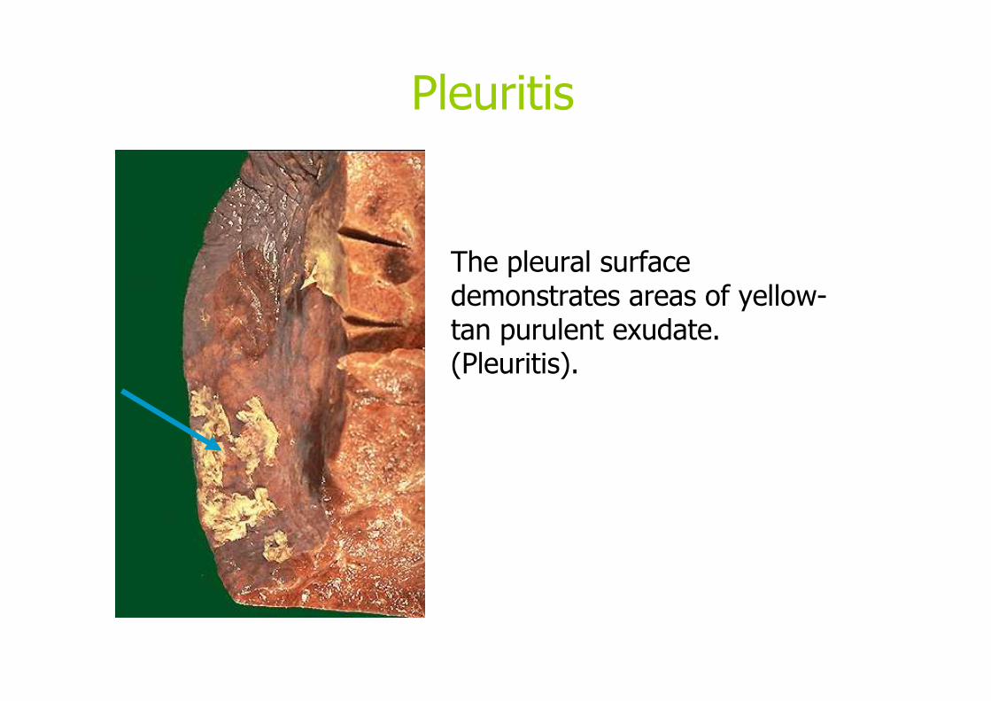
Pleuritis
The pleural surface demonstrates areas of yellow-tan purulent exudate.(Pleuritis).
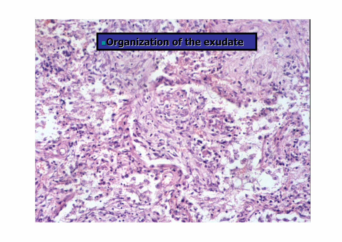
��Organization of the Organization of the exudateexudate
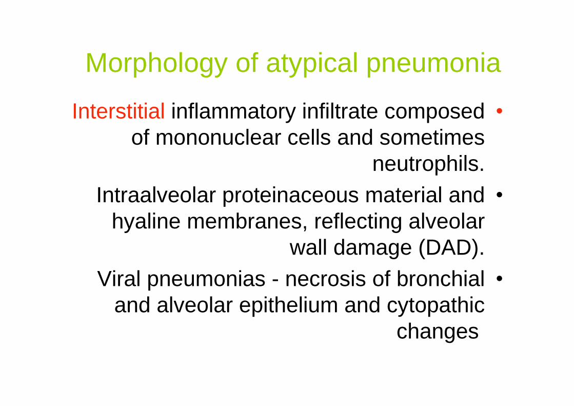
Morphology of atypical pneumonia
•Interstitial inflammatory infiltrate composed of mononuclear cells and sometimes
neutrophils.•Intraalveolar proteinaceous material and
hyaline membranes, reflecting alveolar wall damage (DAD).
•Viral pneumonias - necrosis of bronchial and alveolar epithelium and cytopathic
changes
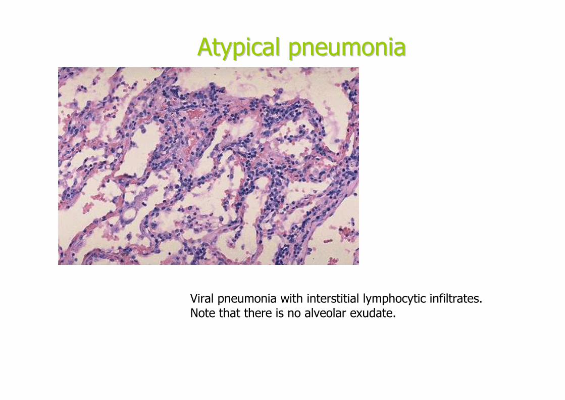
Viral pneumonia with interstitial lymphocytic infiltrates. Note that there is no alveolar exudate.
Atypical pneumoniaAtypical pneumonia
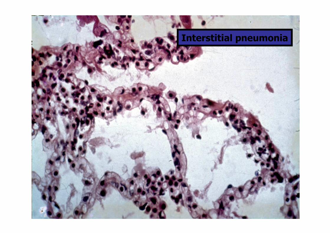
Interstitial pneumonia
Nosocomial (hospital-acquired) pneumonia
•Gram- negative rods (Klebsiella, Enterobacter, Pseudomonas)
•Staphylococcus aureus (usually penicillin-resistant)
•Common in patients with severe underlying disease.
Aspiration pneumonia
•Occurs in markedly debilitated patients.•Partly chemical, partly bacterial, from the
oral flora: anaerobic –Bacteroides, Fusobacterium,
Peptostreptococcus; aerobic– Streptococcus pneumonia,
Staphylococcus aureus Haemophilisinfluenzae and Pseudomonas aeruginosa.
Aspiration pneumonia
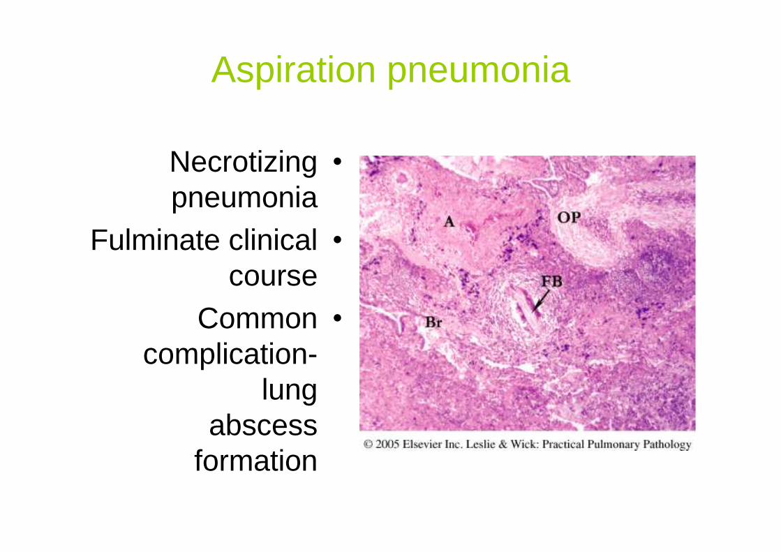
•Necrotizing pneumonia
•Fulminate clinical course
•Common complication-
lung abscess
formation
Lung Abscess
•Local suppurative process with necrosis of lung tissue.
•Causative organisms: aerobic and anaerobic
streptococci, Staphylococcus aureus,
gram-negative organisms and anaerobes from the oral cavity.
Lung Abscess mechanisms of development
•Aspiration of infective materials and gastric contents.
•Antecedent primary bacterial infections.•Septic embolism.•Neoplasia (secondary obstruction)•Lung trauma and spread of infections from
a neighboring organs.•Primary cryptogenic lung abscess
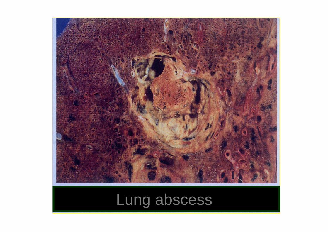
Lung abscess
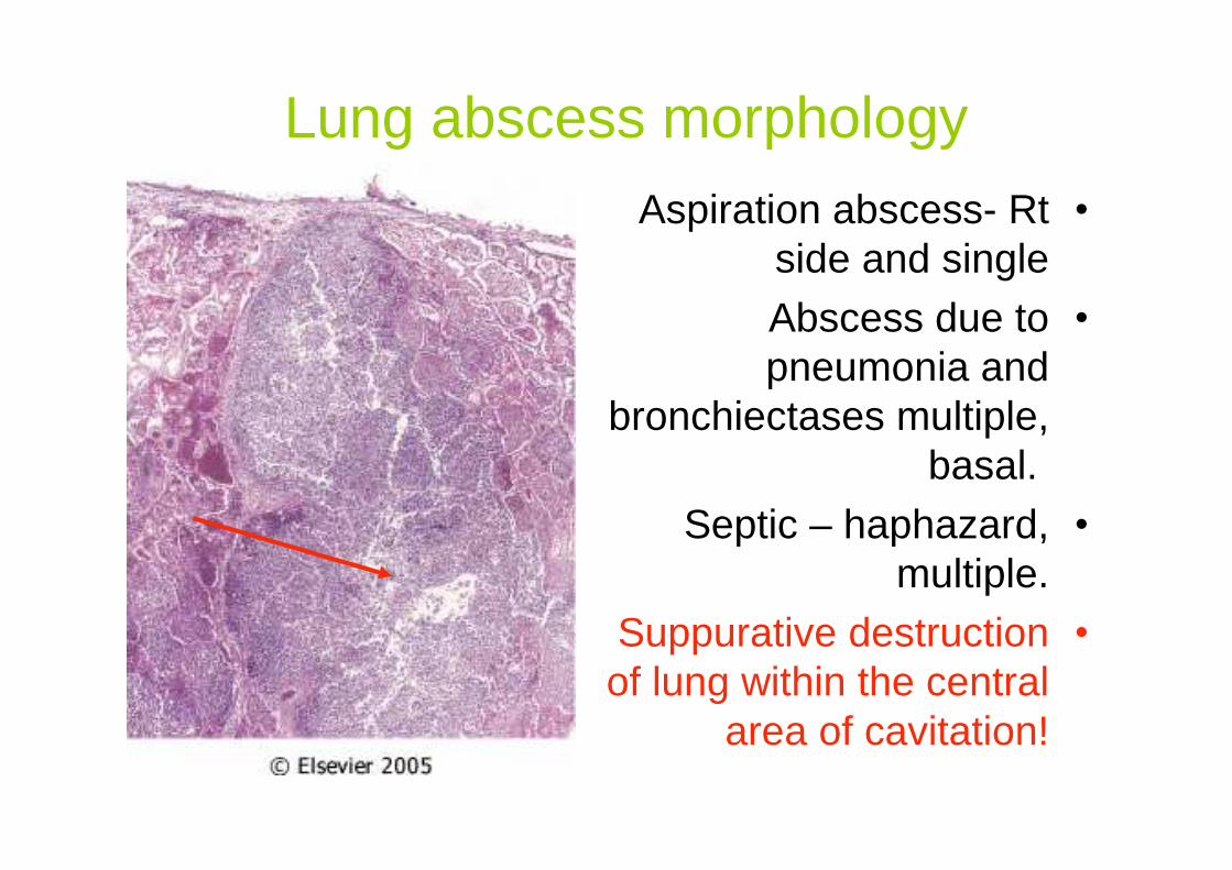
Lung abscess morphology
•Aspiration abscess- Rtside and single
•Abscess due to pneumonia and
bronchiectases multiple, basal.
•Septic – haphazard, multiple.
•Suppurative destruction of lung within the central
area of cavitation!
Chronic Pneumonia•A localized lesion in immunocompetent
patient.•Causative agents:
NocardiaActinomyces
Histoplasma capsulatumCoccidoides immitis
Blastomyces dermatitidisMycobacterium (discussed separately)
•Morphology – granulomatous inflammation
Pneumonia in the immunocompromised host
•CMV•Pneumocystis carinii•Mycobacterium avium intracellulare•Invasive aspergillosis•Invasive candidiasis•“Usual” bacterial, viral and fungal
organisms
Pneumocystis carinii
•Occurs predominantly in immunosuppressed persons.
•Pneumocystis is a FUNGUS!•Clinically – fever, dyspnea and dry cough
progressing to respiratory failure.•Radiologically – bilateral alveolar and
interstitial infiltrates radiating from the hilus.•Diagnosis – BAL and lung biopsy
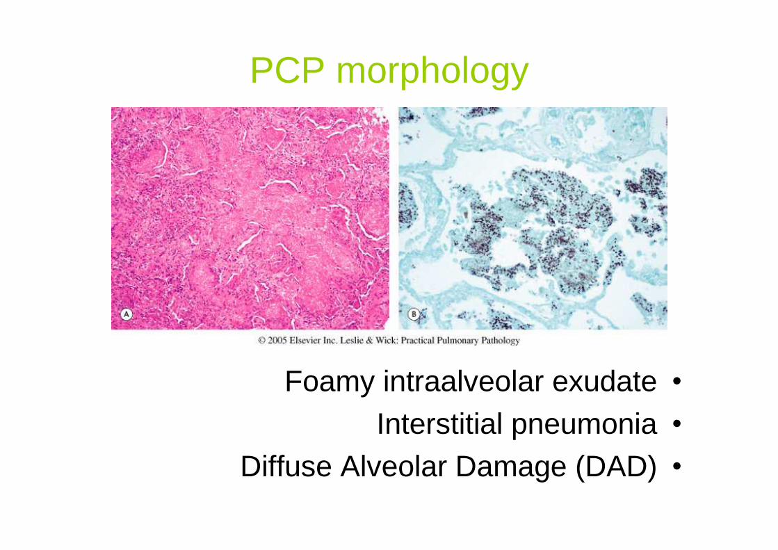
PCP morphology
•Foamy intraalveolar exudate•Interstitial pneumonia•Diffuse Alveolar Damage (DAD)
•GMS (Gomory methenamine silver) stain highlights cysts containing sporosoites and
free throphozoites. Cysts – 4-6µm in diameter with one or two
dots.•Monoclonal antibodies to pneumocystis.•In situ hybridization and PCR
Identification of organisms
CMV (β-group herpesvirus)
•CMV infects and remains latent in WBC and can be reactivated when immune
status is depressed.•The major envelop glycoprotein of CMV
binds to EGFR.•CMV pneumonia clinical features: fever,
dyspnea, nonproductive cough, diffuse chest infiltrates.
CMV morphology
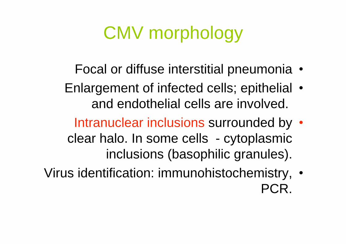
•Focal or diffuse interstitial pneumonia•Enlargement of infected cells; epithelial
and endothelial cells are involved. •Intranuclear inclusions surrounded by
clear halo. In some cells - cytoplasmicinclusions (basophilic granules).
•Virus identification: immunohistochemistry, PCR.
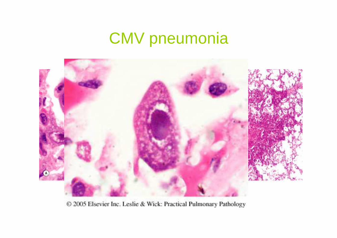
CMV pneumonia
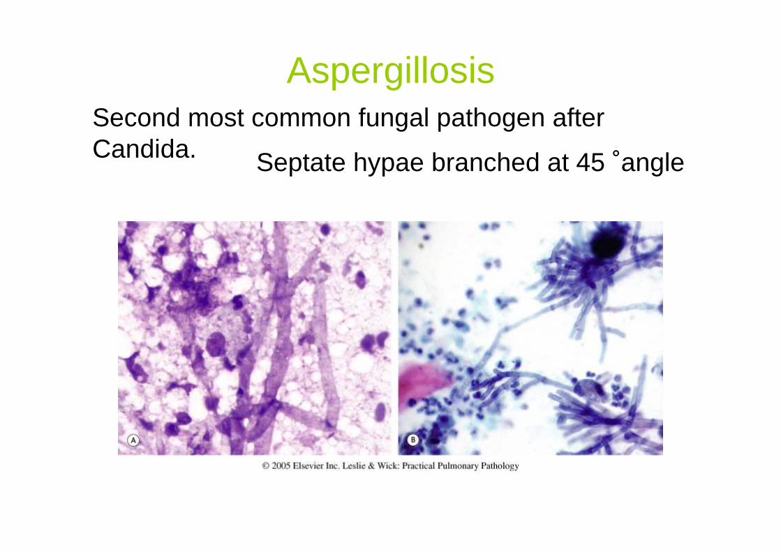
AspergillosisSecond most common fungal pathogen after Candida. Septate hypae branched at 45 ˚angle
Patterns of pulmonary aspergillosis:ColonisationFungus ball
Hypersensitivity reactionAllergic bronchopulmonary aspergillosis
Eosinophilic pneumoniaHypersensitivity pneumonitis
Invasive (in immunosuppressed)Acute invasive aspergillosis
Chronic necrotizing pneumonia
(usually angiocentric, produce infarcts)

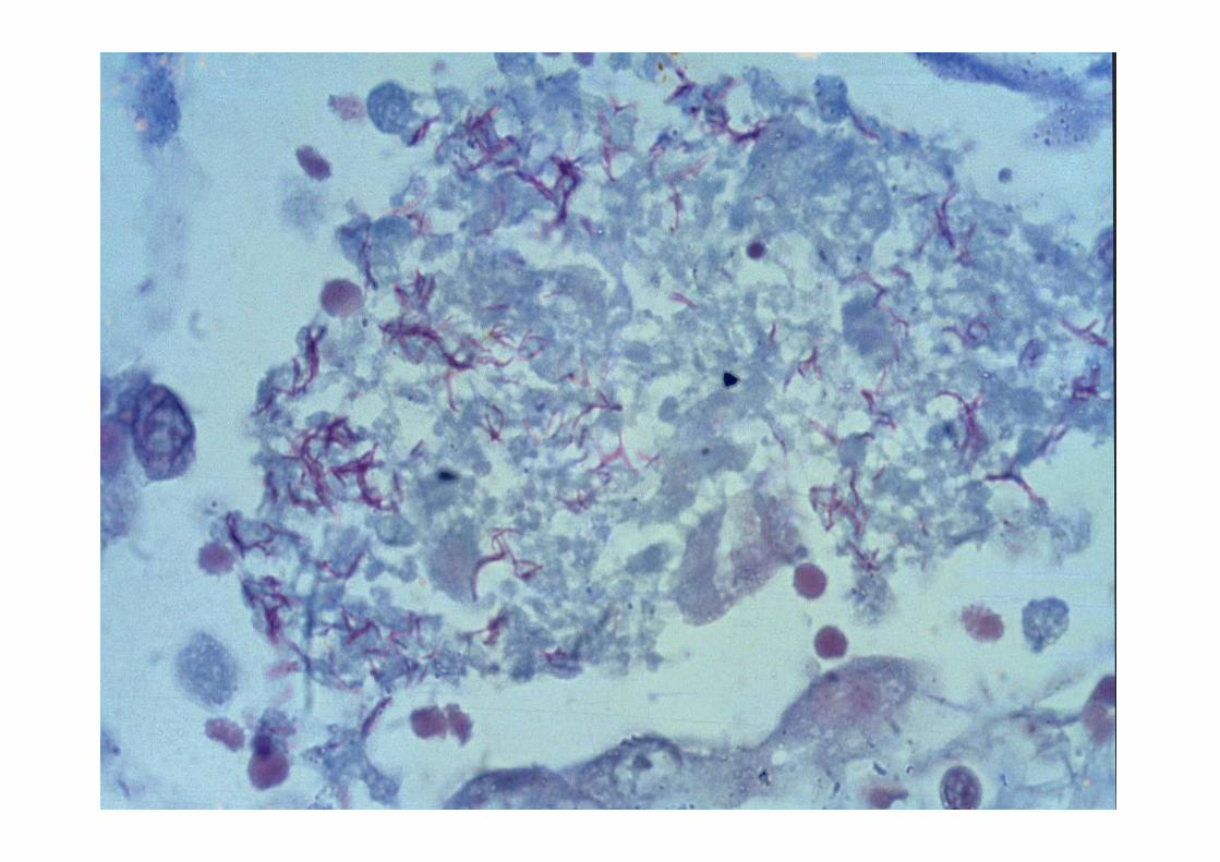
Mycobacteria
•Slender aerobic rods growing in strait or branching chains.
•Mycobacterium have a waxy cell wall composed of mycolic acid which makes
them acid fast - they retain stains on treatment with a mixture acid and alcohol -
Ziehl –Neelsen stain.
a ZiehlFranz (described by two German doctors; a pathologist)NeelsenFriedrich bacteriologist and
Tuberculosis epidemiology
•Leading infectious case of death in the world after HIV.
•HIV makes people susceptible to TB•Other conditions, like diabetes, renal
failure, chronic lung disease, alcoholism and others increase risk of TB.
•The disease affects people from low socio-economic levels
Clinical features of TB
•Primary TB develops in a unsensitizedperson. Latent disease. Rare –
progressive infection.•Secondary TB arises in a previously
sensitized person. May occur: 1) shortly after primary
2) from reactivation of dormant primary lesions (most common)
3) reinfection
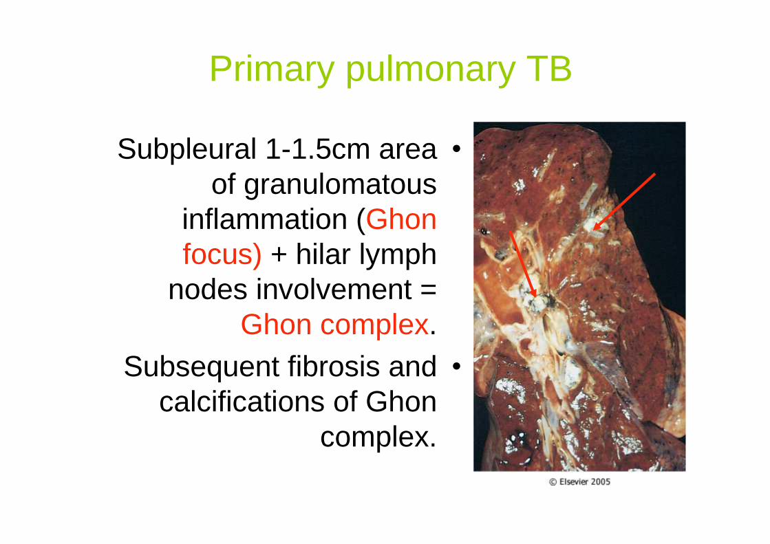
Primary pulmonary TB
•Subpleural 1-1.5cm area of granulomatous
inflammation (Ghonfocus) + hilar lymph
nodes involvement = Ghon complex.
•Subsequent fibrosis and calcifications of Ghon
complex.
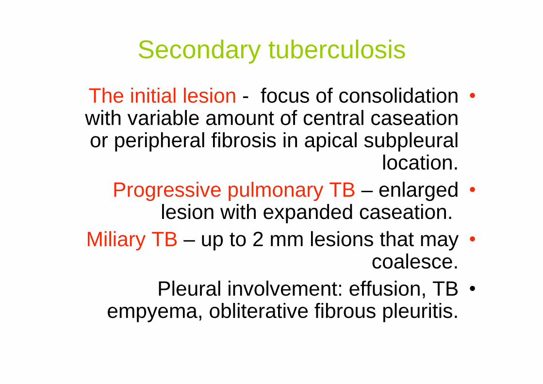
Secondary tuberculosis
•The initial lesion - focus of consolidation with variable amount of central caseationor peripheral fibrosis in apical subpleural
location.•Progressive pulmonary TB – enlarged
lesion with expanded caseation. •Miliary TB – up to 2 mm lesions that may
coalesce.•Pleural involvement: effusion, TB
empyema, obliterative fibrous pleuritis.
Secondary tuberculosis•Endobronchial, endotracheal, laryngeal
TB.•Systemic miliary TB •Isolated-organ TB (meningies, kidneys,
adrenals, bones, fallopian tubes etc. Vertebral TB- Pott’s dis. Paraspinal “cold
abscesses”)•Lymphadenitis ( “scrofula”)•Intestinal TB
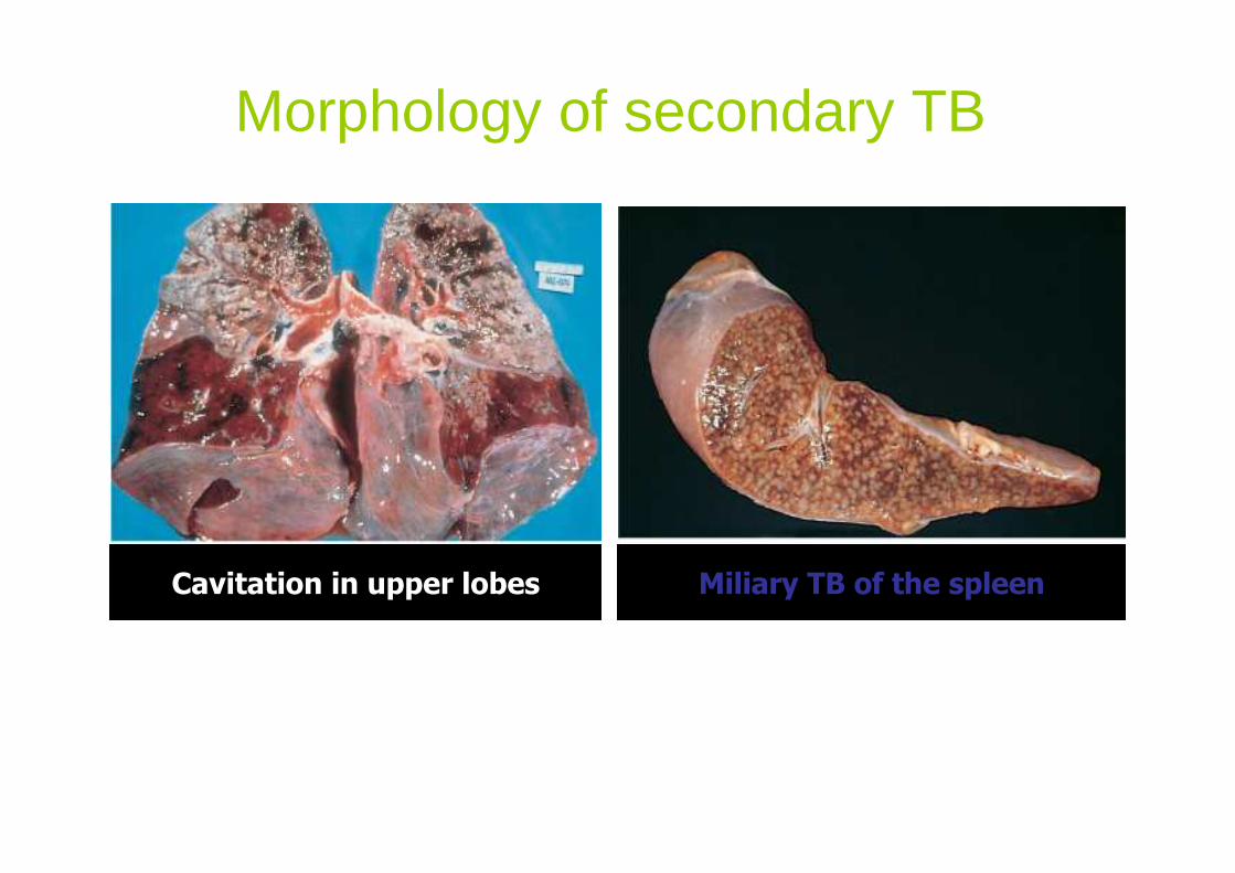
Morphology of secondary TB
Cavitation in upper lobes Miliary TB of the spleen
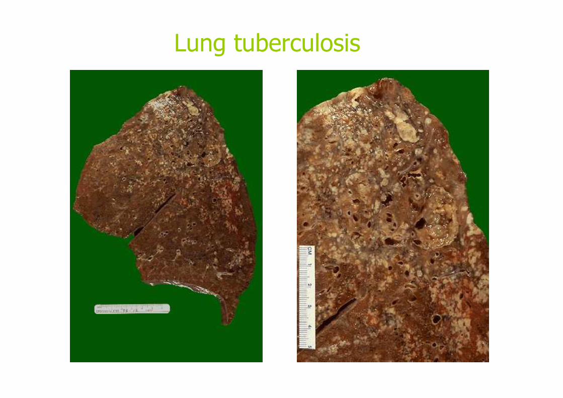
Lung tuberculosis
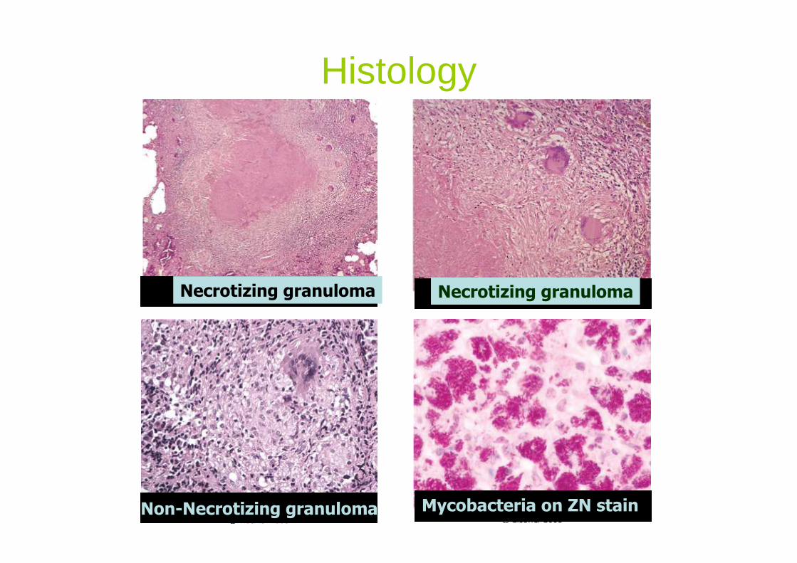
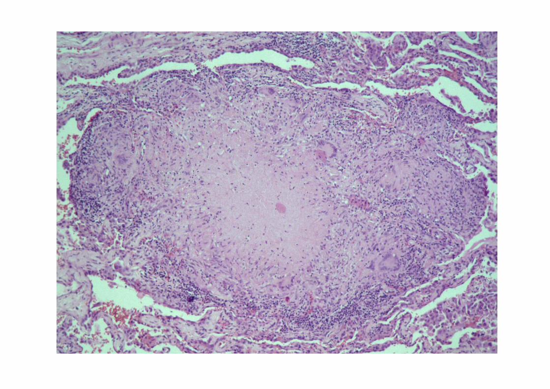
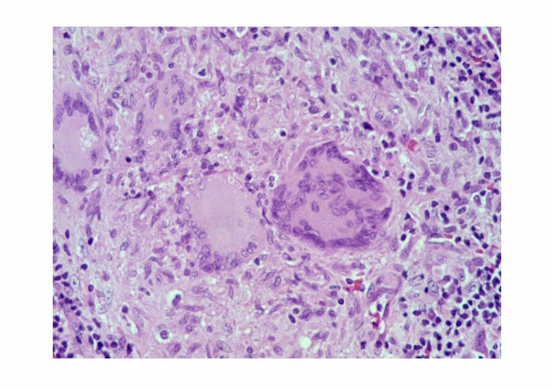
Histology
Non-Necrotizing granuloma
Necrotizing granuloma Necrotizing granuloma
Mycobacteria on ZN stain
OBSTRUCTIVE AND RESTRICTIVE LUNG DISEASES
Obstructive diseases
Characterized by an increase in resistance to airflow due to partial or complete obstruction at any level, from the trachea to the terminal and respiratory bronchioles
Characterized by an increase in resistance to airflow due to partial or
complete obstruction at any level, from the trachea to the terminal and
respiratory bronchioles
CHRONIC OBSTUCTIVE PULMONARY DISEASE
� EMPHYSEMA
� CHRONIC BRONCHITIS
� BRONCHIAL ASTHMA
� BRONCHIECTASES

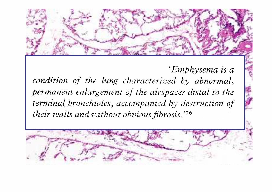
EMPHYSEMA
CENTRIACINAR CENTRIACINAR
(CENTRILOBULAR) EMPHYSEMA (CENTRILOBULAR) EMPHYSEMA
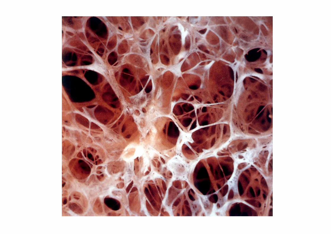
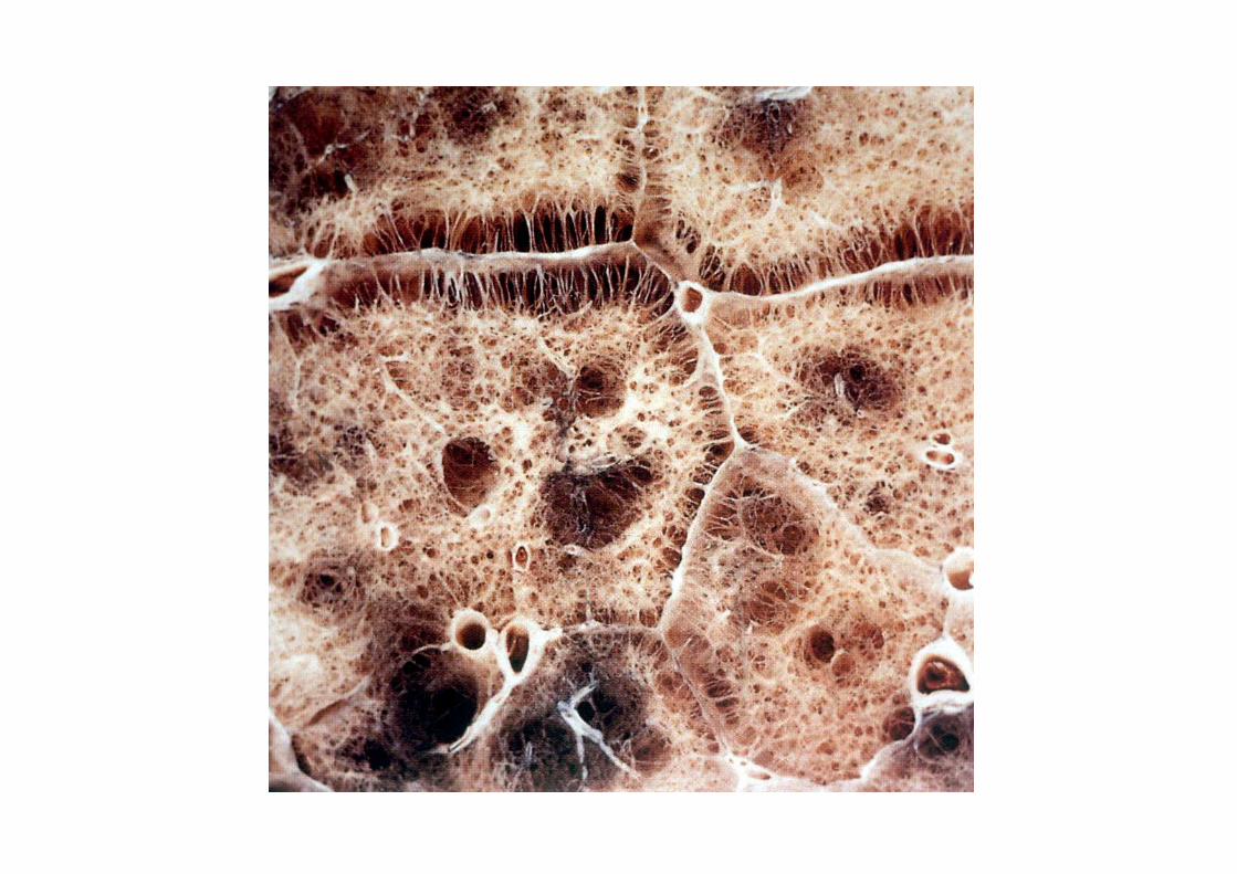
Centriacinar emphysema
•Occurs predominantly in heavy smokers•Central portion of the acini is affected•The lesion more severe in the upper
lobes•Black pigment is present in the walls of
the emphysematous space•Inflammation around bronchi and
bronchioles is common
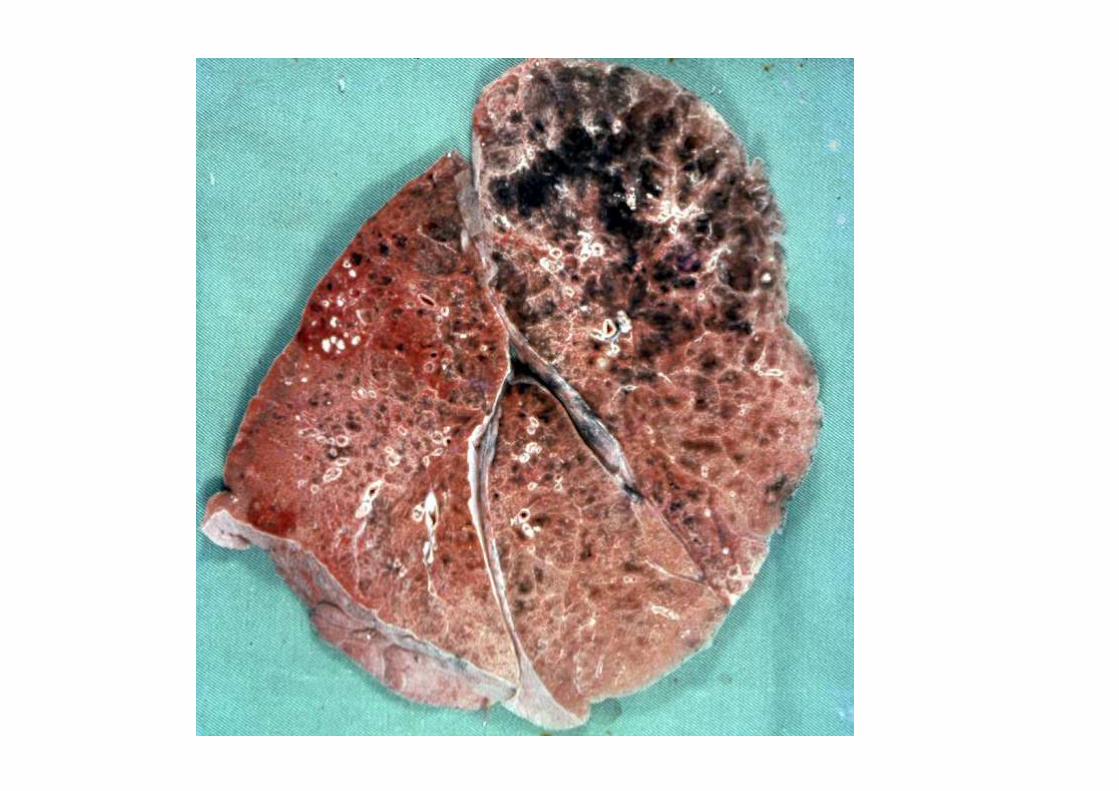
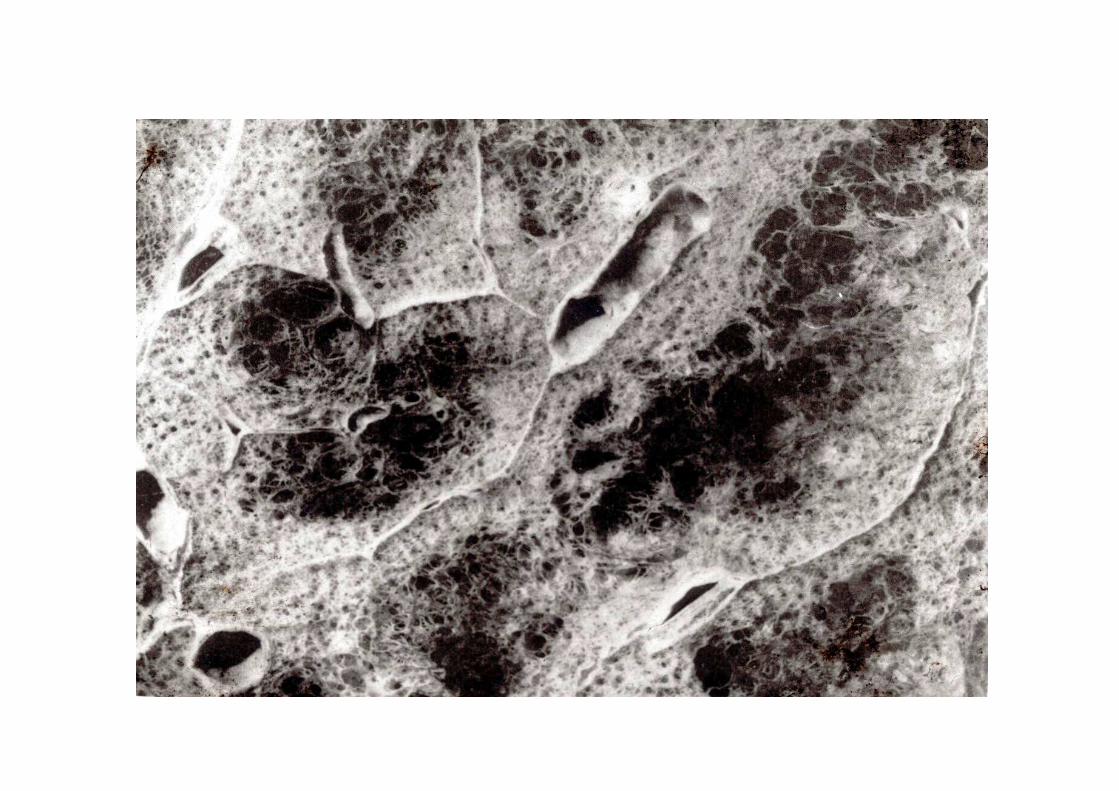
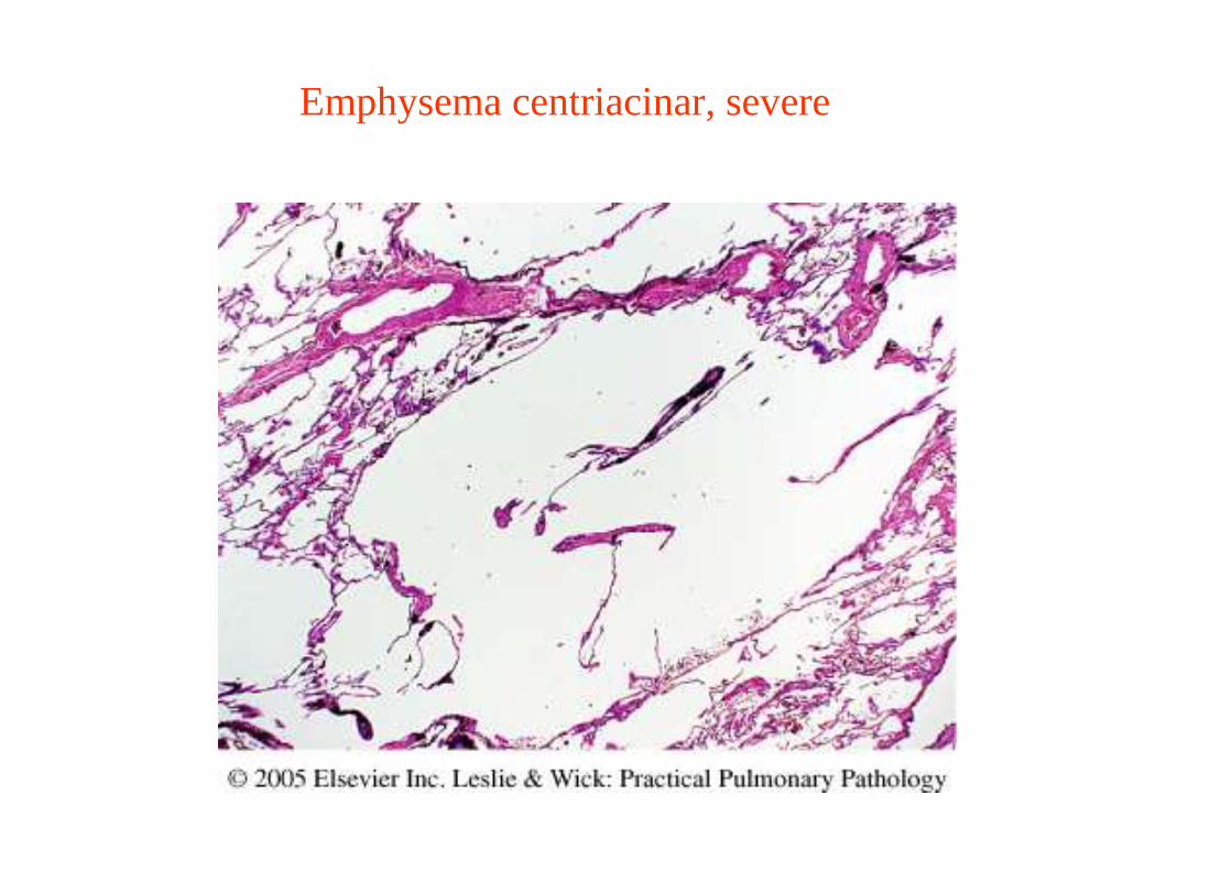
Emphysema centriacinar, severe
PANACINAR (PANLOBULAR) PANACINAR (PANLOBULAR)
EMPHYSEMAEMPHYSEMA
Panacinar emphysema
•Occurs more commonly in the lower zones and in the anterior margins of the lungs
•The acini are uniformly enlarged
•Associated with α1-antitrypsin deficiency
α1-Antitrypsin Deficiency
•Abnormally low serum level of protease inhibitor (Pi)
•Genetics: gene for α1-antitrypsin (chromosome 14) is very polymorphic and
at least 75 forms of protein have been identified.
PiMM is the most common form. PiZZ – only 10% of circulating protein.
Risk of clinical disease.
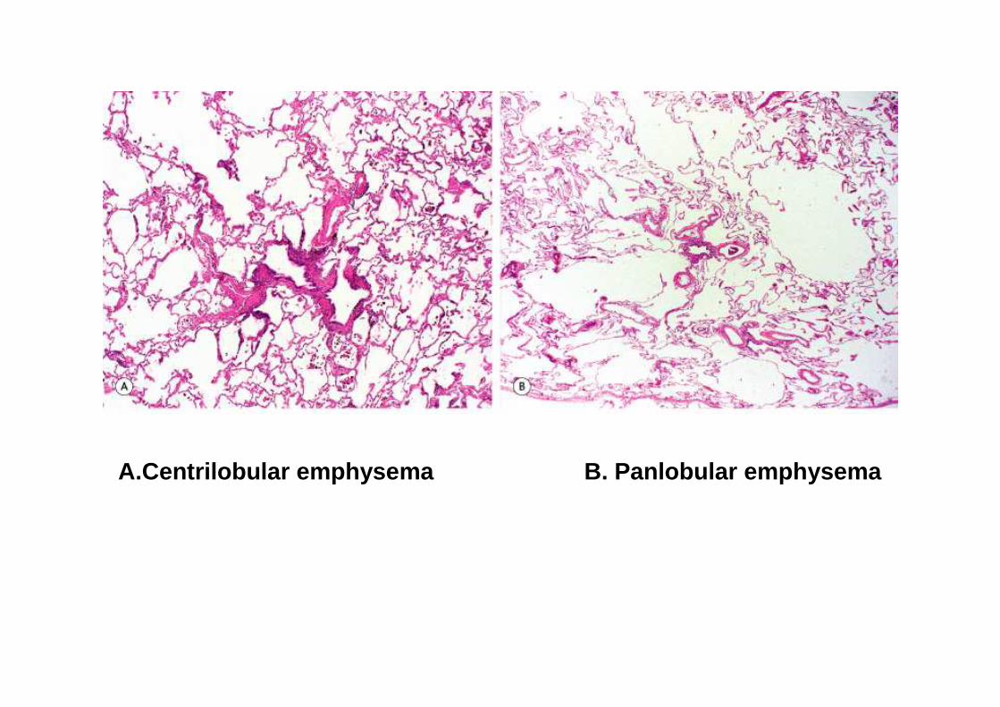
A.Centrilobular emphysema B. Panlobular emphysema
DISTAL ACINAR (PARASEPTAL) DISTAL ACINAR (PARASEPTAL)
EMPHYSEMAEMPHYSEMA
Paraseptal emphysema
•Upper parts of the lungs are more severely affected
•Involves the lung tissue adjacent to the pleura, along the lobular connective
tissue and at the margins of the lobules•Occurs adjacent to areas of fibrosis,
scarring, atelectasis•Spontaneous pneumothorax in young
adults
Other types of emphysema
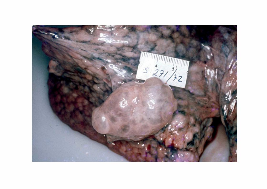
Bullous emphysema
•Any form of emphysema that produces large subpleural spaces more than 1 cm in
diameter•Most often subpleural and occurs near the
apex•Rupture of bullae leads to pneumothorax
CHRONIC BRONCHITIS CHRONIC BRONCHITIS
+ +
BRONCHIOLITISBRONCHIOLITIS
Chronic bronchitis
•Persistent cough and sputum production for at least 3 months in at
least 2 consecutive years–Simple chronic bronchtis–Chronic asthmatic bronchitis (+airways
hyperreactivity)
–Chronic obstructive bronchitis (+airflow obstruction and emphysema)
CHRONIC BRONCHITIS PATHOGENESIS
•Initiating factor – chronic irritation (tobacco smoke, inorganic dusts. 90% of patients are
smokers)•Bacterial and viral infections are triggers of
acute exacerbation.•The earliest feature of chronic bronchitis is
hypersecretion of mucus.•Accompanying alteration in the small airways
– BRONCHIOLITIS - can result in earlier manifestation of chronic obstruction
•Macro: hyperemia and edema of mucous membranes with excessive mucous or
mucopurulent secretion•Micro:
•Chronic inflammation•Enlargement of mucus-secreting glands of
trachea and bronchi•Increased number of goblet cells•Squamous metaplasia and dysplasia of
bronchial epithelium•Narrowing of bronchioles by mucous
plugging, inflammation and fibrosis up to bronchiolitis obliterans
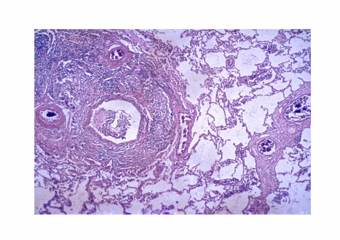

BRONCHIAL ASTHMABRONCHIAL ASTHMA
Asthma is a chronic inflammatory disorder of the airways that causes recurrent episodes of wheezing, breathlessness, chest tightness, and cough.These symptoms are associated with bronchoconstriction and airflow limitation that are at least partially reversible
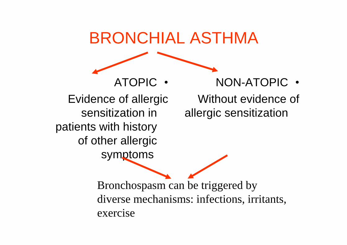
BRONCHIAL ASTHMA
•ATOPICEvidence of allergic
sensitization in patients with history
of other allergic symptoms
•NON-ATOPICWithout evidence of
allergic sensitization
Bronchospasm can be triggered bydiverse mechanisms: infections, irritants, exercise
ATOPIC ASTHMA
•Classic example of type I IgE-mediated hypersensitivity reaction
•Usually begins in childhood•Triggered by environmental allergens
(dusts, pollens, food etc)•Positive family history of asthma is
common•Positive skin tests
NON-ATOPIC ASTHMA
•No evidence of allergic sensitization•Viral respiratory infections are common
triggers•Hyperirritability of the bronchial tree•Virus –induced inflammation of the
respiratory mucosa lowers the threshold of the subepithelial vagal receptors to
irritants
DRUG-INDUCED ASTHMA
•Aspirin-sensitive asthma occurs in individuals with recurrent rhinitis and
nasal polyps•Sensitivity to small doses of aspirin•Aspirin inhibits the cyclooxygenase
pathway of arachidonic acid metabolism without affecting the lipooxygenase
pathway → ↑ leucotrienes →bronchoconstriction
Morphology
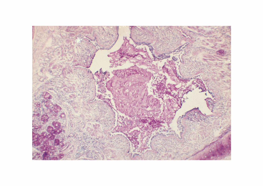
•Mucous plugs with Curschmann spirals and
Charcot-Leyden crystals•Basement membrane thickenning•Inflammation and edema of bronchial
wall•Increase in size of the submucosal
glands•Bronchial smooth muscle hypertrophy
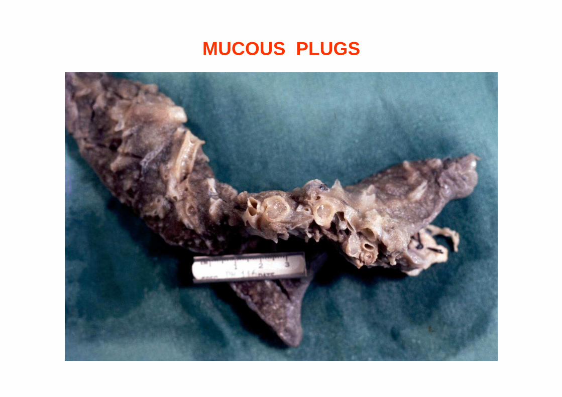
MUCOUS PLUGS
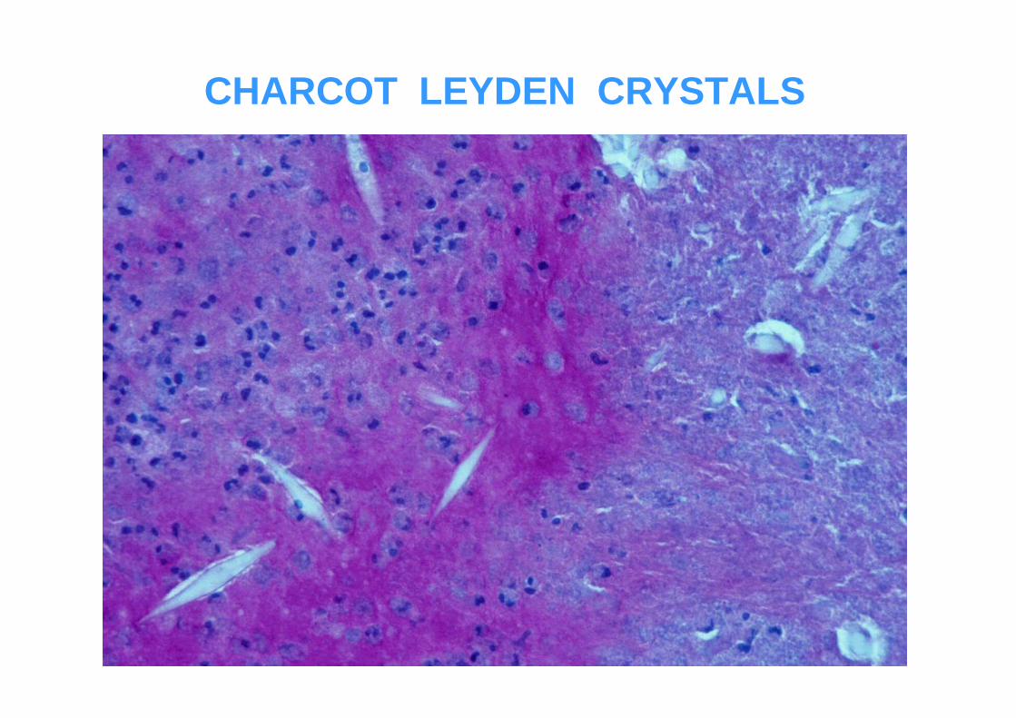
CHARCOT LEYDEN CRYSTALS
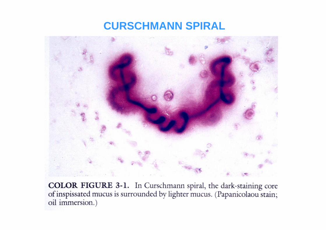
CURSCHMANN SPIRAL
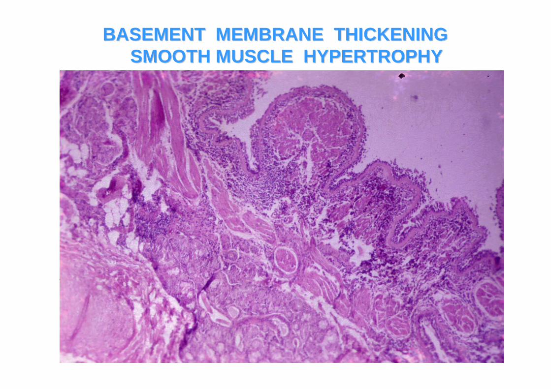
BASEMENT MEMBRANE THICKENINGBASEMENT MEMBRANE THICKENINGSMOOTH MUSCLE HYPERTROPHY SMOOTH MUSCLE HYPERTROPHY
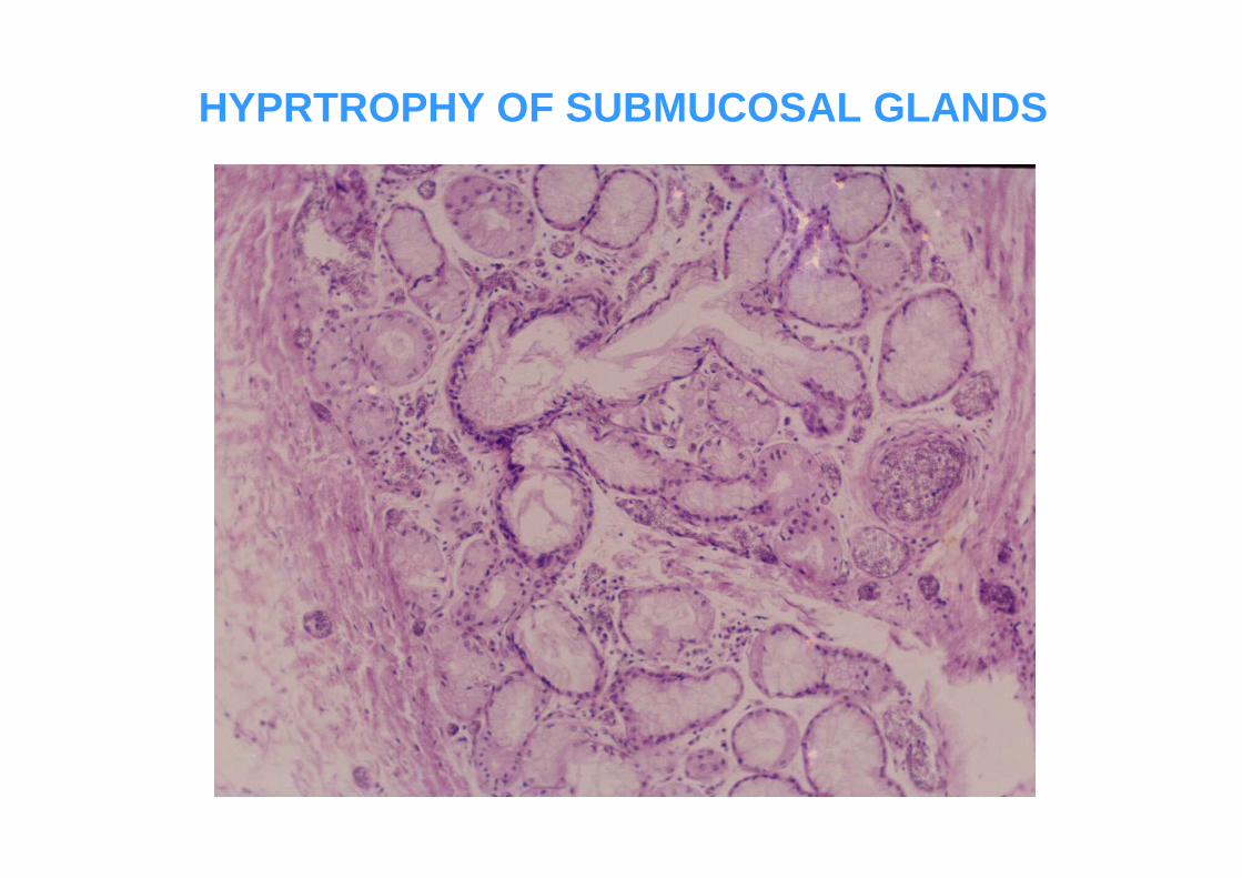
HYPRTROPHY OF SUBMUCOSAL GLANDS

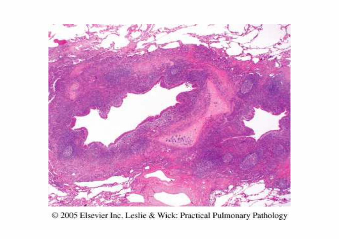
BRONCHIECTASISBRONCHIECTASIS
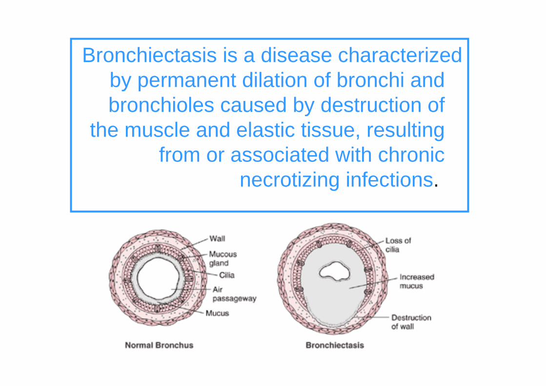
Bronchiectasis is a disease characterized by permanent dilation of bronchi and bronchioles caused by destruction of
the muscle and elastic tissue, resulting from or associated with chronic
necrotizing infections.

Etiology and pathogenesis
•Postinfectious conditions (necrotizing pneumonia)
•Congenital conditions (cystic fibrosis, intralobarsequestartion, ciliary dyskinesia and
Kartagener syndrome, immunodefficiency)
•Bronchial obstruction (tumor, foreign bodies, mucus)
•Others ( Rheumatoid arthritis, SLE, IBD,GVHD)
Morphology
•Bilateral lower lobes involvement•Airways dilatation: cylindrical, fusiform,
saccular•Intense acute and chronic inflammation.
Necrosis. Lung abscess formation
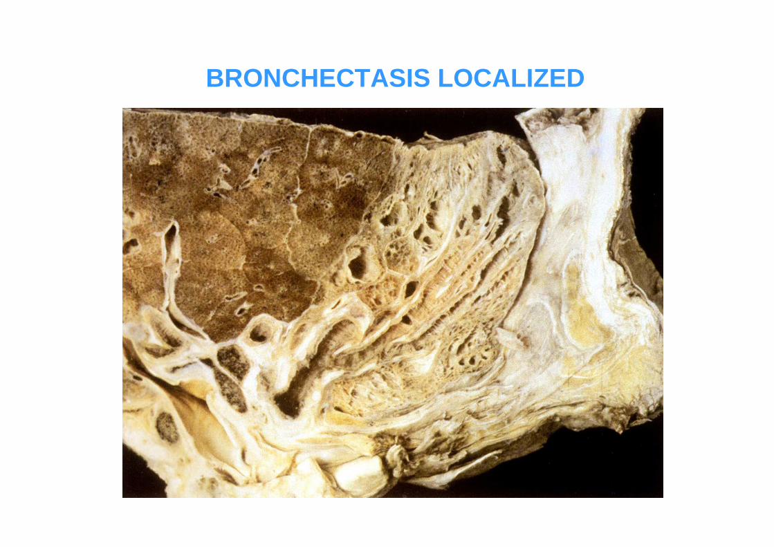
BRONCHECTASIS LOCALIZED
Diffuse interstitial lung diseases
Diffuse Interstitial (Infiltrative, Restrictive) Diseases
•Characterized by reduced expansion of lung parenchyma with decreased
total lung capacity
Diffuse Interstitial (Infiltrative, Restrictive) Diseases
•Heterogeneous group of disorders characterized predominantly by diffuse and usually chronic involvement of the
pulmonary connective tissue, principally the most peripheral and delicate interstitium in the alveolar
walls.

Idiopathic pulmonary fibrosis
Pathogenesis
•“Repeated cycles” of alveolitis•Healing with exuberant fibroblastic
proliferation (mediator TGF-β)
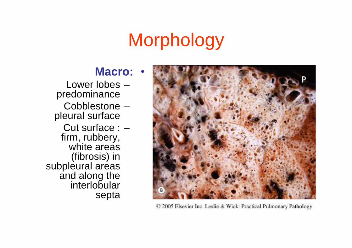
Morphology
•Macro:–Lower lobes
predominance–Cobblestone
pleural surface–Cut surface :
firm, rubbery, white areas (fibrosis) in
subpleural areas and along the
interlobular septa

Microscopically: usual interstitial pneumonia (UIP)
•Patchy interstitial fibrosis•Fibroblastic foci•Honeycomb fibrosis
•Hyperplasia of type II pneumocytes•Mild to moderate inflammation•Smooth muscle hyperplasia•Intimal fibrosis and medial thickening of pulmonary
arteries
Temporal heterogeneity!
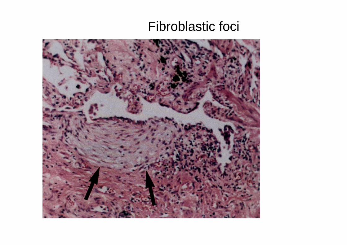
Fibroblastic foci
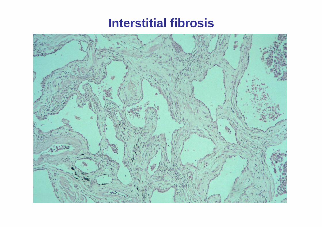
Interstitial fibrosis
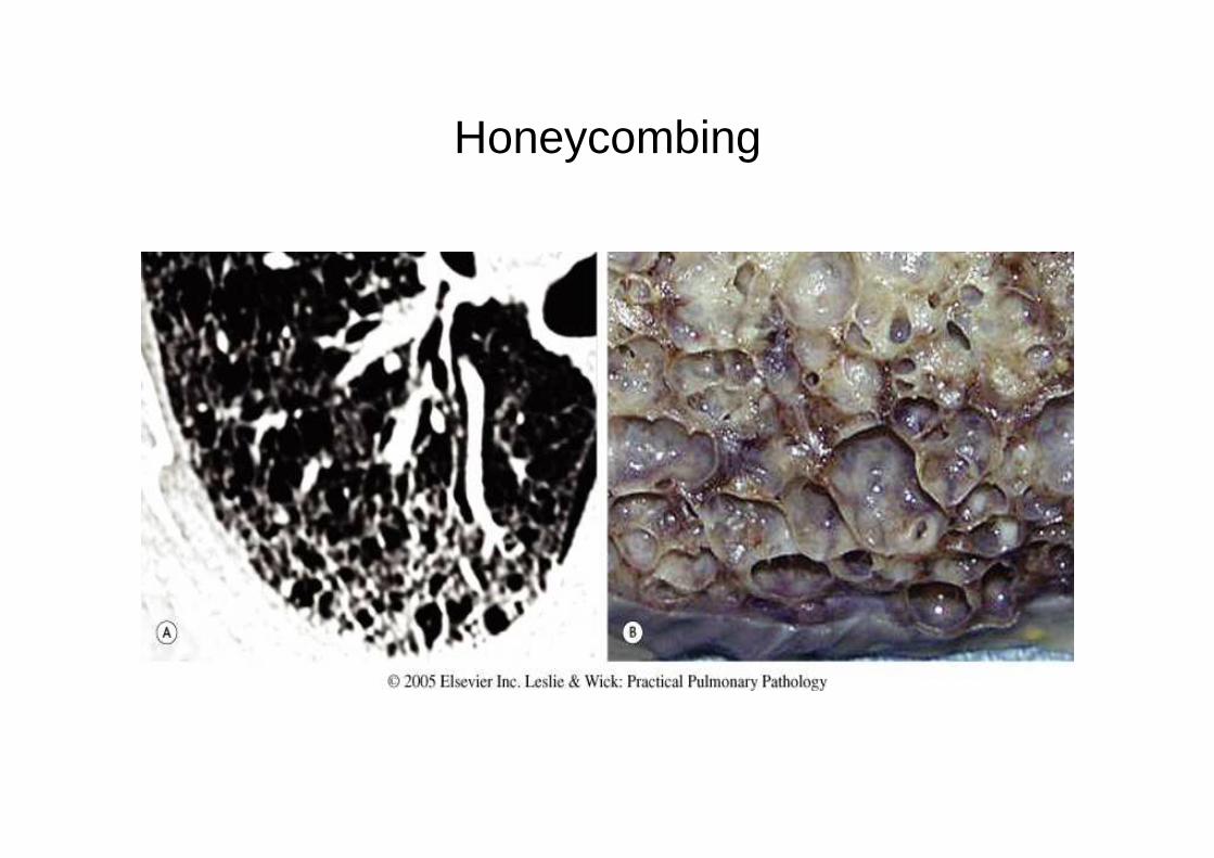
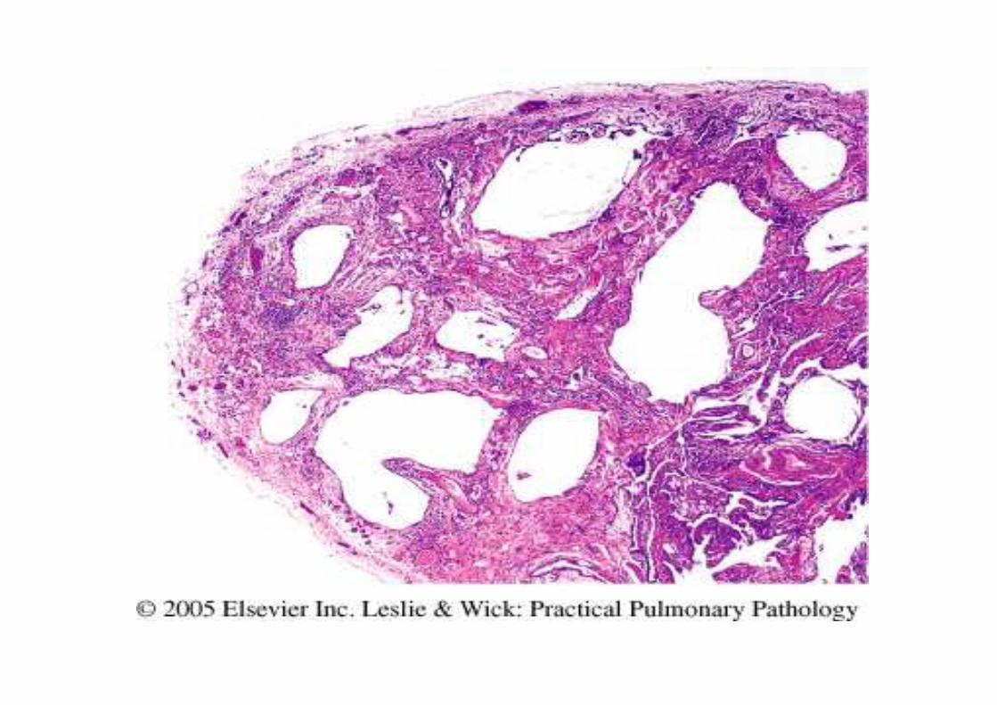
Honeycombing
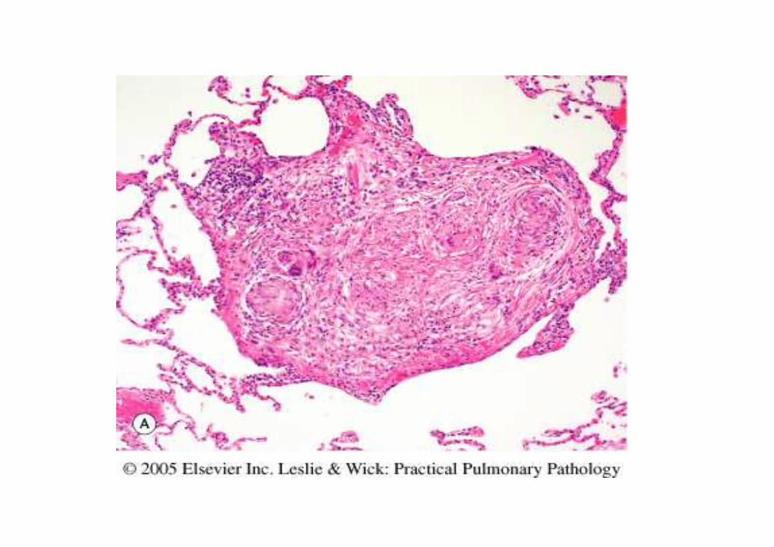
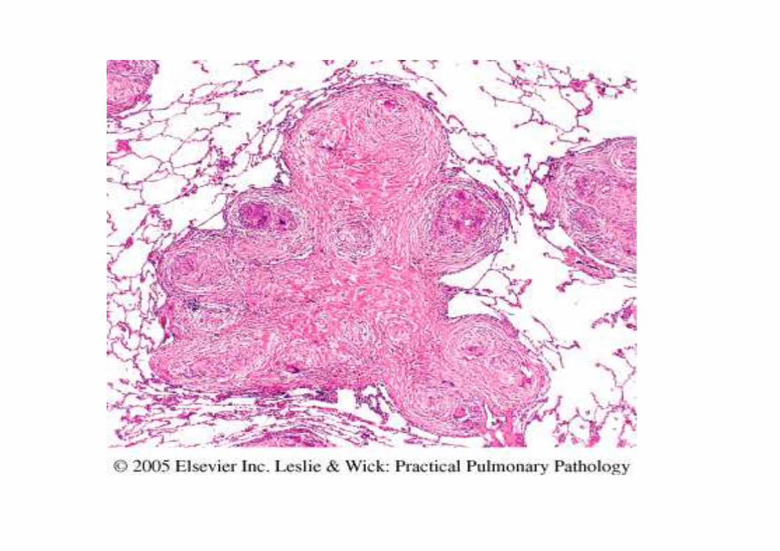
Bronchiolitis obliteransorganizing pneumonia
(BOOP)
Characteristics
•Unknown etiology•Cough and dyspnea•Radiologically: subpleural or peribronchial
patchy areas of airspace consolidation•Spontaneous recovering or treatment with
oral steroids for 6 months
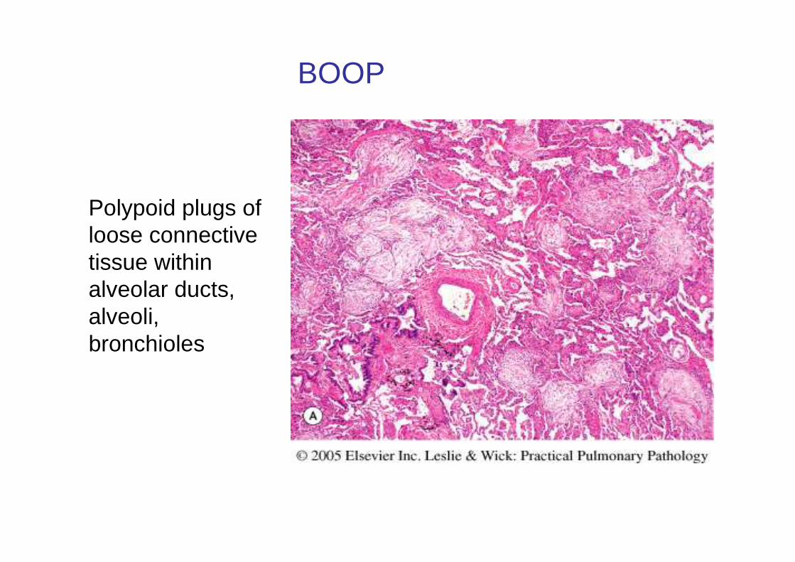
Polypoid plugs of loose connective tissue withinalveolar ducts, alveoli, bronchioles
BOOP
Sarcoidosis
•Systemic disease of unknown cause characterized by noncaseating
granulomas in many tissues•Women>Men•Black>White•Histologic diagnosis by exclusion!•Variable clinical course; 90% of patients
have hilar lymphoadenopathy + lung involvement
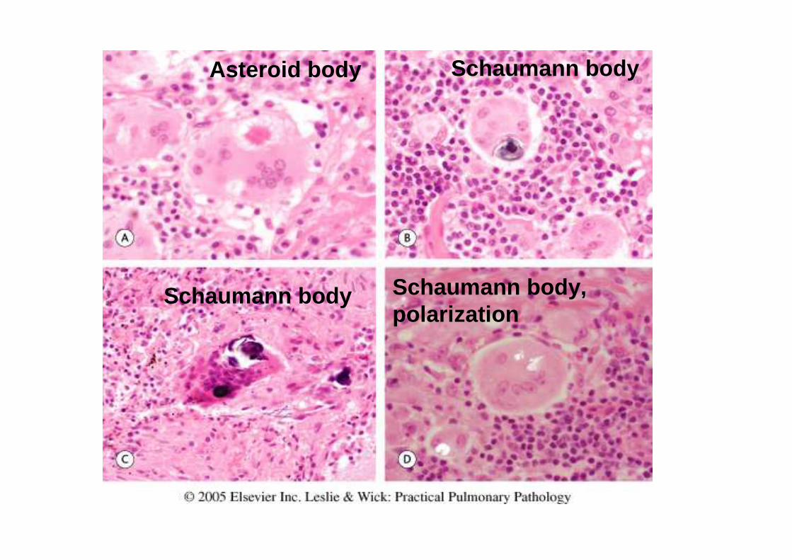
Asteroid body Schaumann body
Schaumann body Schaumann body,polarization

Environmentaland occupational
diseases
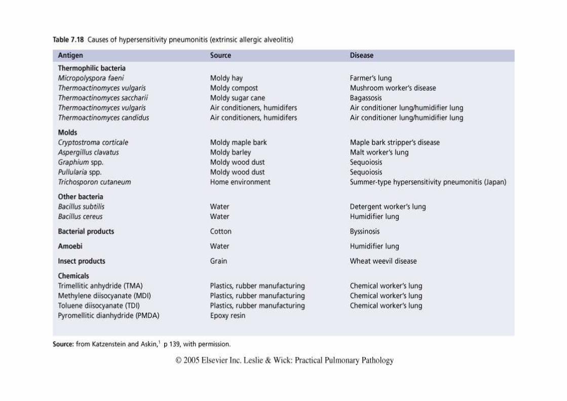
Hypersensitivity pneumonitis(allergic alveolitis)
•Spectrum of immunologically mediated predominantly interstitial lung disorders
•Abnormal sensitivity to antigen, which involves alveoli ( in contrast to asthma)
•Acute attack (4-6 hours after exposure): fever, dyspnea, cough, leukocytosis
•Chronic disease – progressive respiratory failure.
•Removal of the causative agent prevents transition to fibrosis
Drug- Induced pulmonary diseases

Table 15-7. Examples of Drug-Induced Pulmonary Disease
Bronchospasmβ-Antagonists
BronchospasmAspirin
Hypersensitivity pneumonitis
Nitrofurantoin
Pneumonitis and fibrosisAmiodarone
Hypersensitivity pneumonitis
Methotrexate
Pneumonitis and fibrosisBleomycin
Cytotoxic drugs
Pulmonary DiseaseDrug
Acute lung injury
Acute respiratory distress syndrome (ARDS)
Diffuse alveolar damage (DAD)- morphologic equivalent
Acute respiratory distress syndrome
•Rapid onset of severe life threatened respiratory insufficiency, refractory to
oxygen therapy•Complication of numerous conditions•Radiologically: diffuse alveolar infiltration
Causes of ARDS
•Sepsis*
•Diffuse pulmonary infections*•Gastric aspiration*•Mechanical trauma (including head
injuries)*•Inhaled irritants, smoke, oxygen•Narcotics or barbiturate overdose•Hypersensitivity reactions•Others
Pathogenesis
•Damage to the alveolar capillary membrane ( including microvascular
endothelium and alveolar epithelium) →↑ vascular permeability, alveolar flooding,
loss of diffusion capacity and surfactant abnormality (due to damage to
pneumocytes) → hyaline membranes•Neutrophiles play an important role•Coagulation system dysregulation
Morphology•Acute phase
–Congestion, edema
–Inflammation–Fibrin deposition – Hyaline membranes!
•Organizing phase:–Organization of the fibrin exudate–Thickenning of alveolar septa
–Type II cells hyperplasia
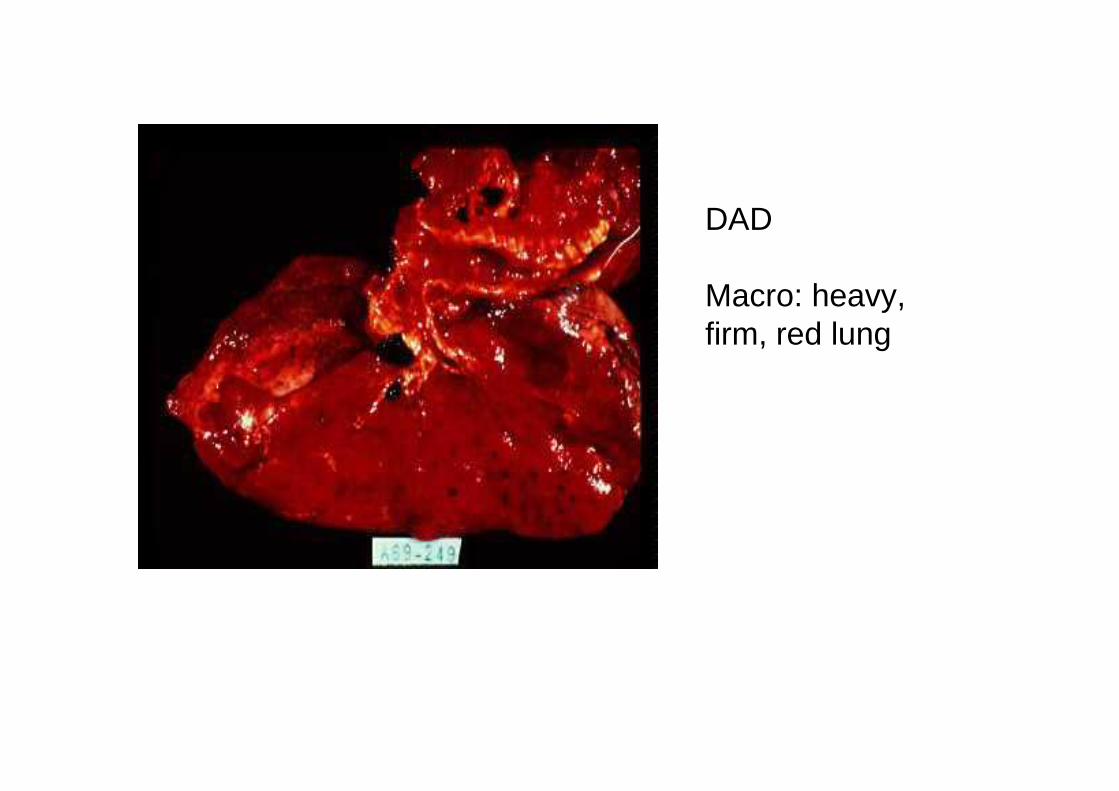
DAD
Macro: heavy, firm, red lung
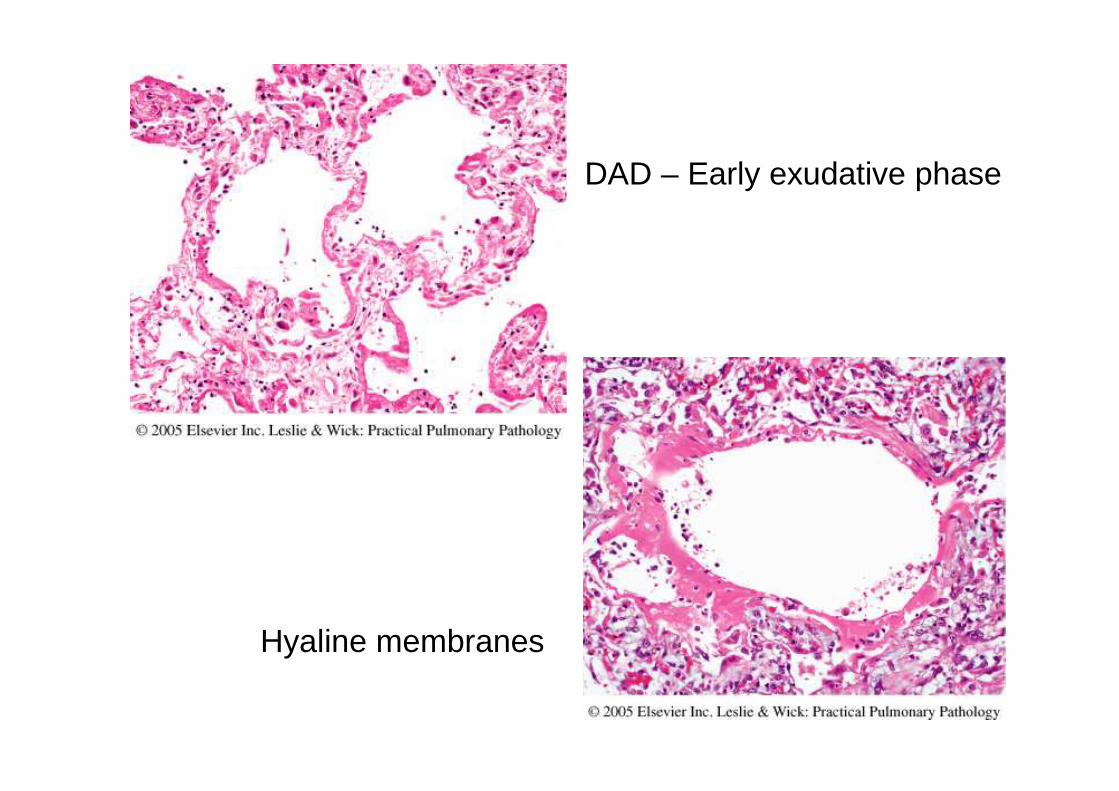
DAD – Early exudative phase
Hyaline membranes
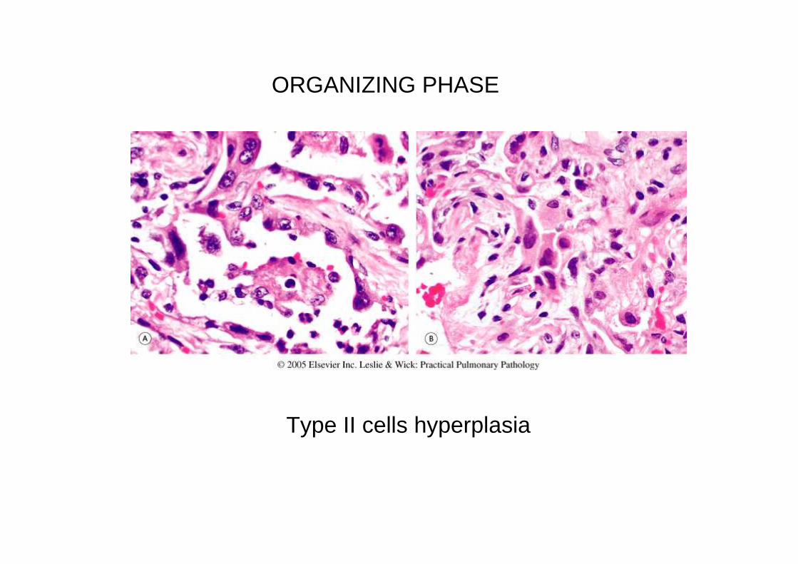
Type II cells hyperplasia
ORGANIZING PHASE
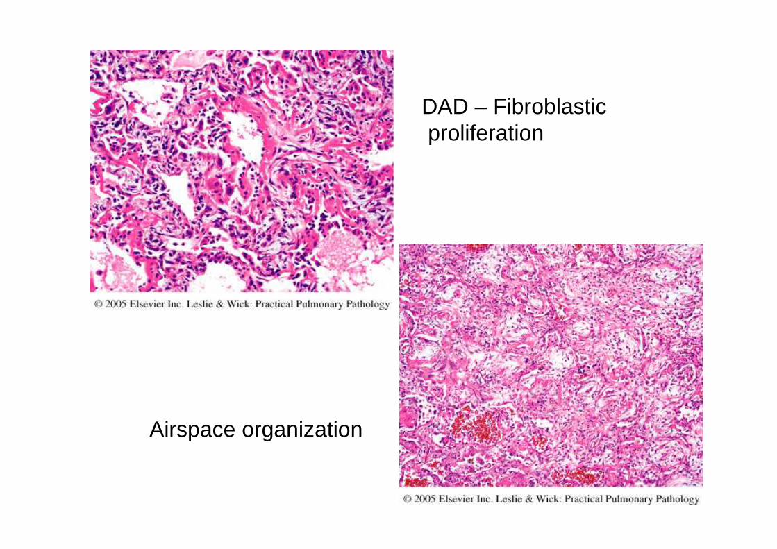
DAD – Fibroblasticproliferation
Airspace organization
LUNG TUMORS
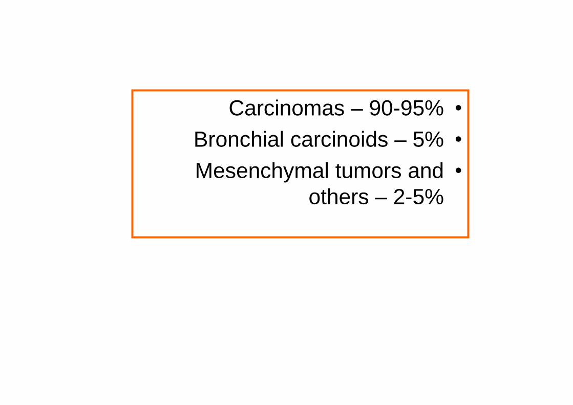
•Carcinomas – 90-95%•Bronchial carcinoids – 5%•Mesenchymal tumors and
others – 2-5%
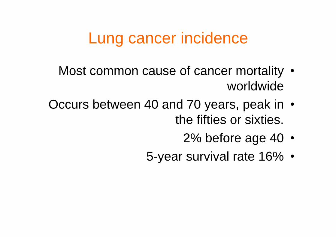
Lung cancer incidence
•Most common cause of cancer mortality worldwide
•Occurs between 40 and 70 years, peak in the fifties or sixties.
•2% before age 40•5-year survival rate 16%

Lung cancer etiology and pathogenesis
Tobacco smoking
�average smokers – 10-fold greater risk
� heavy smokers – 60-fold greater risk
�women have a higher susceptibility to carcinogens than men do
� cessation of smoking for 10 years reduces risk but never to control levels
Industrial hazards
•High-dose ionizing radiation•Uranium•Asbestos ( asbestos + smoking = 50-90
times greater risk )
Air pollutions
•Athmosphericpollutants
•Radon
Classification (WHO)•Squamous cell carcinoma•Adenocarcinoma
Acinar, papillary, bronchioloalveolar, solid, mixed
•Large cell carcinoma •Adenosquamous carcinoma •Pleomorphic, sarcomatoid carcinoma•Small cell carcinoma•Large cell neuroendocrine carcinoma •Carcinoid tumor (typical, atypical)•Carcinomas of salivary gland type•Unclassified carcinoma
Clinically important:
•Small cell carcinoma most often metastatic, high initial response
to chemotherapy
•Non- small cell carcinoma less often metastatic, less responsive
Pathology
•75% - area of lung hilus•Small number – on periphery
(adenocarcinoma)•Distant spread occurs by lymphatic and
hematogenous pathwaysadenocarcinoma metastasize at an early stage
SCC metastasize outside the thorax late
Sites of metastases:
•Adrenal – more than half of the cases
•Liver – 30-50%•Brain – 20%•Bones – 20%
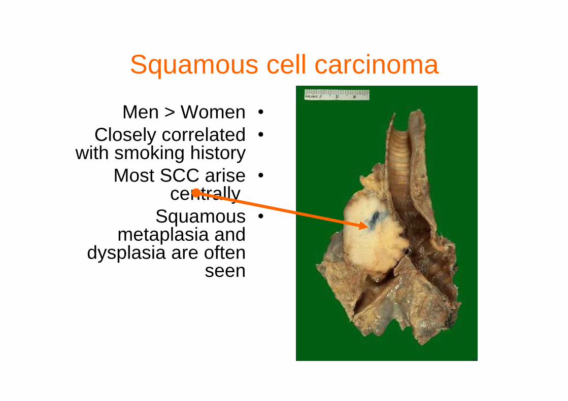
Squamous cell carcinoma
•Men > Women•Closely correlated
with smoking history•Most SCC arise
centrally •Squamous
metaplasia and dysplasia are often
seen
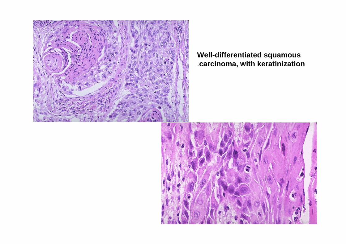
Well-differentiated squamouscarcinoma, with keratinization.
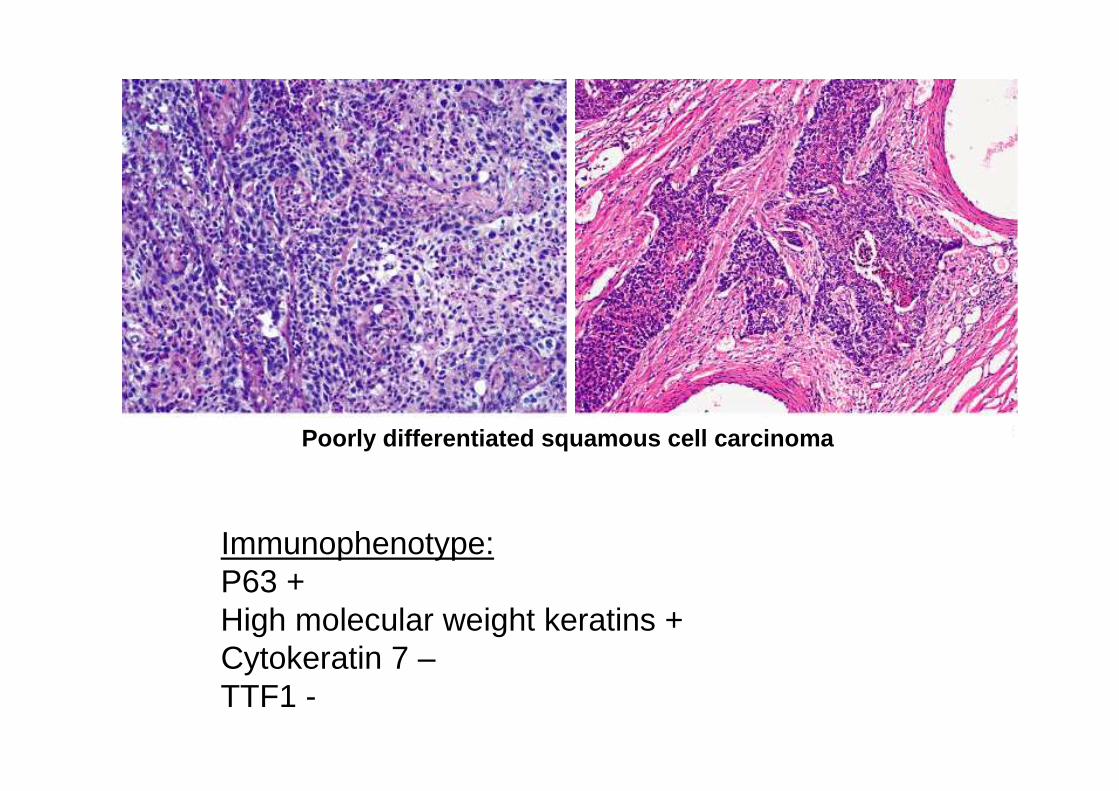
Poorly differentiated squamous cell carcinoma
:ImmunophenotypeP63 +High molecular weight keratins +Cytokeratin 7 –TTF1 -
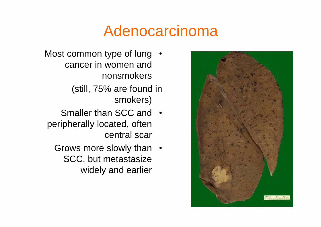
Adenocarcinoma•Most common type of lung
cancer in women and nonsmokers
(still, 75% are found in smokers)
•Smaller than SCC and peripherally located, often
central scar•Grows more slowly than
SCC, but metastasize widely and earlier
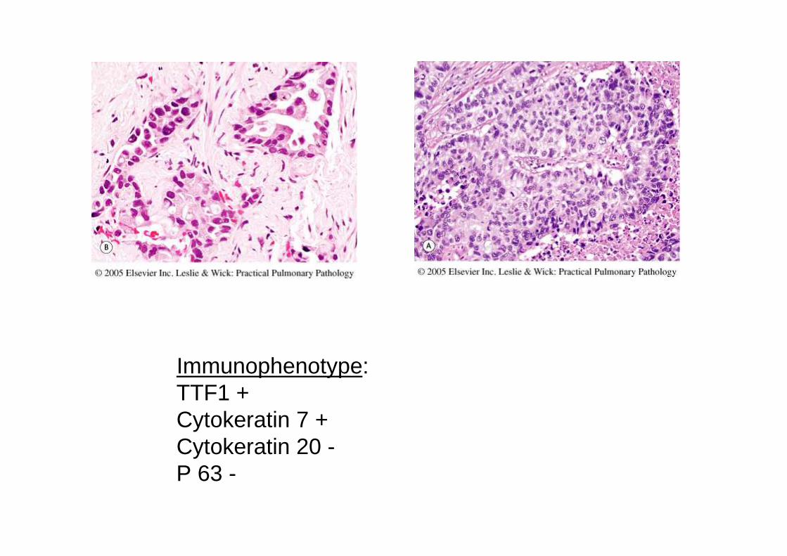
:ImmunophenotypeTTF1 +Cytokeratin 7 +Cytokeratin 20 -P 63 -
Bronchioloalveolar carcinoma
•Peripheral portion of the lung as single nodule or in pneumonia-like consolidation
•Histologically – no evidence of stromal, vascular or pleural invasion!
•Growth along preexisting structures without destruction of alveolar architecture
(“lepidic” growth)
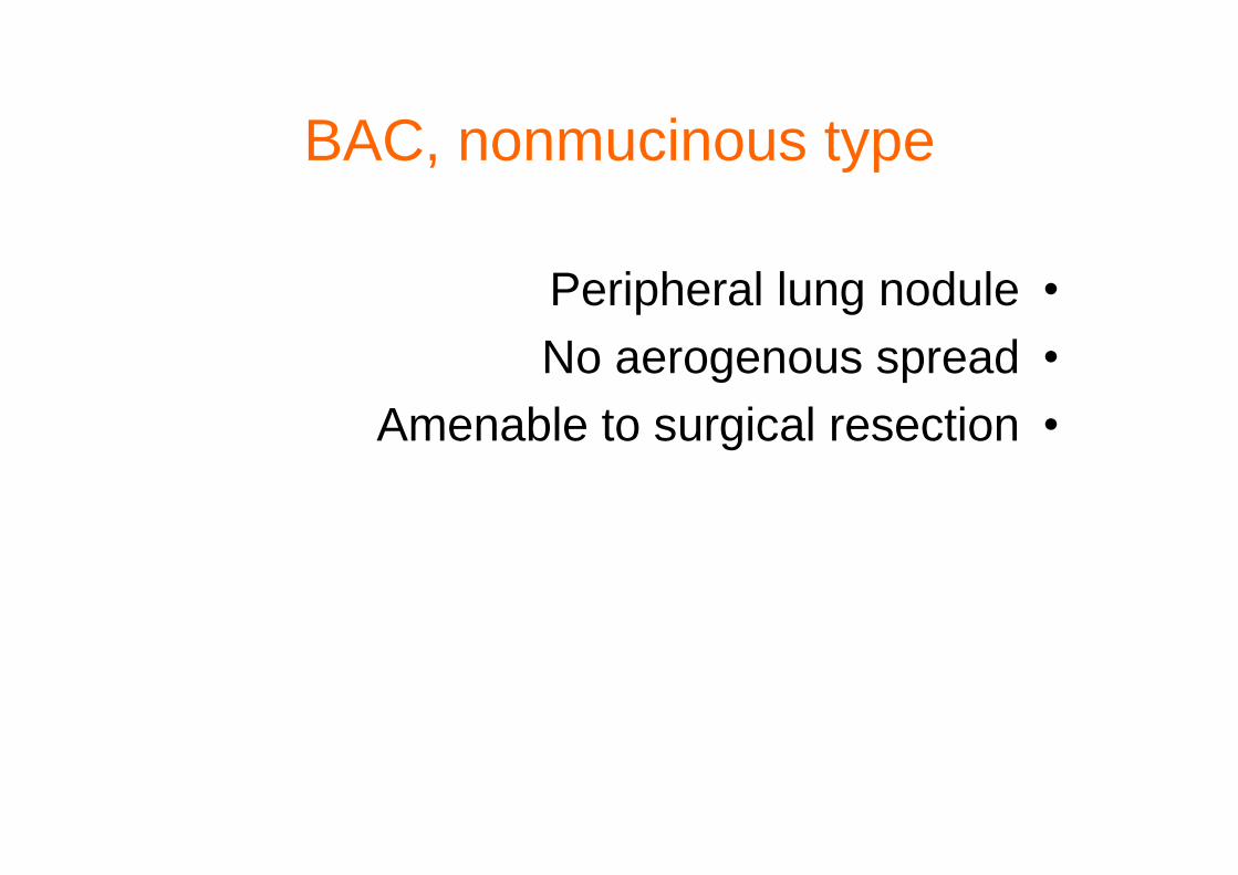
BAC, nonmucinous type
•Peripheral lung nodule•No aerogenous spread•Amenable to surgical resection
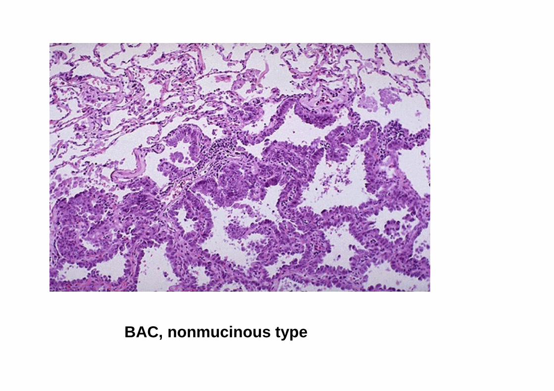
BAC, nonmucinous type
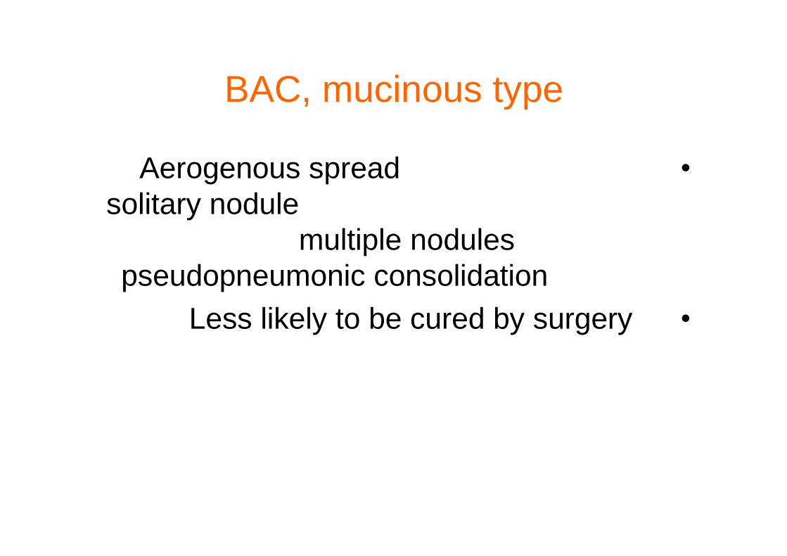
BAC, mucinous type
•Aerogenous spread solitary nodule
multiple nodules pseudopneumonic consolidation
•Less likely to be cured by surgery
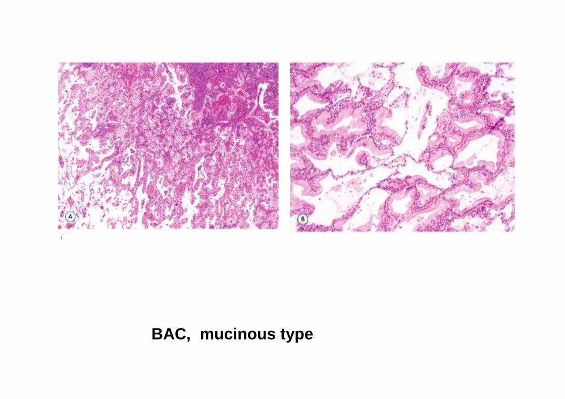
BAC, mucinous type
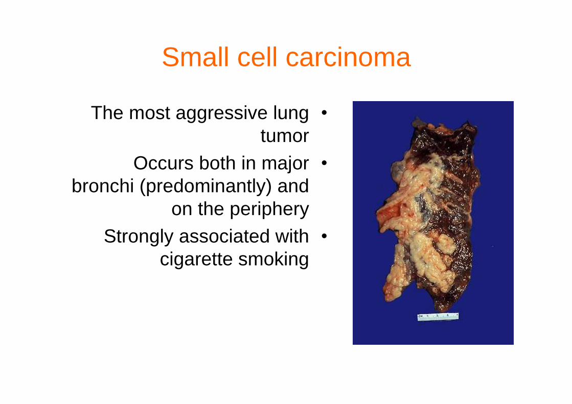
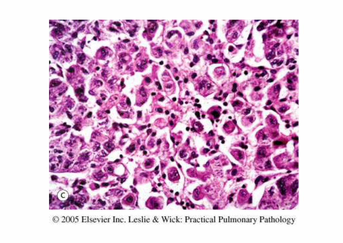
Small cell carcinoma
•The most aggressive lung tumor
•Occurs both in major bronchi (predominantly) and
on the periphery•Strongly associated with
cigarette smoking
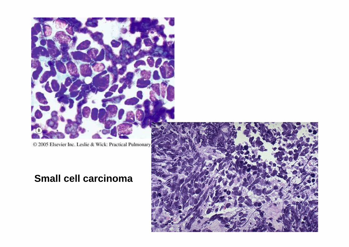
Small cell carcinoma
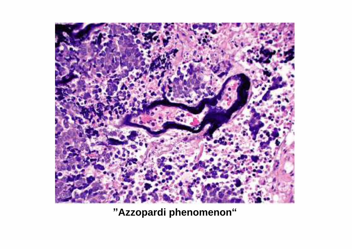
“Azzopardi phenomenon”
Large cell carcinoma
Undifferentiated carcinoma that lacksthe cytological features of small cell carcinoma
and glandular or squamous differentiation.
On EM – minimal glandular or squamousdifferentiation is common
Large cell neuroendocrine carcinoma has the same molecular changes as small cell
carcinoma
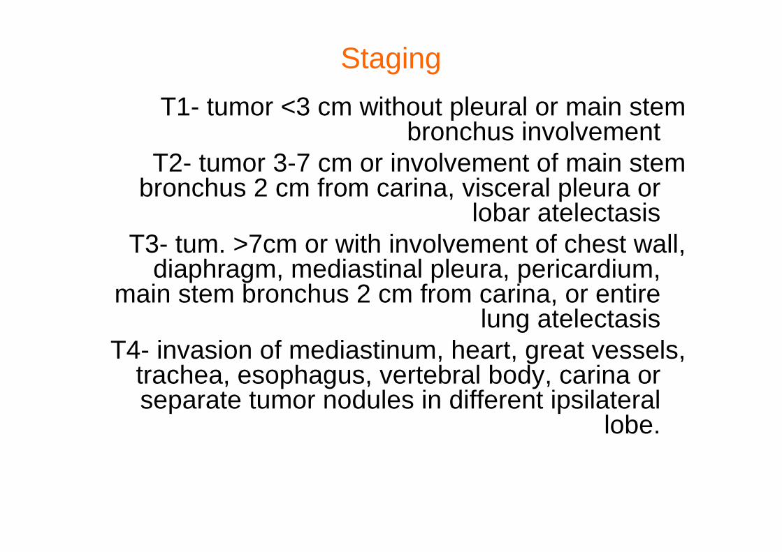
Staging
T1- tumor <3 cm without pleural or main stem bronchus involvement
T2- tumor 3-7 cm or involvement of main stem bronchus 2 cm from carina, visceral pleura or
lobar atelectasisT3- tum. >7cm or with involvement of chest wall,
diaphragm, mediastinal pleura, pericardium, main stem bronchus 2 cm from carina, or entire
lung atelectasisT4- invasion of mediastinum, heart, great vessels,
trachea, esophagus, vertebral body, carina or separate tumor nodules in different ipsilateral
lobe.
Prognosis
•Overall 5-year survival rate is 15%•For localized cases 48% survival rate•Adenocarcinoma and squamous cell ca
tend to remain localized longer and have a slightly better prognosis than
undifferentiated ca.•Small cell ca – resection is ineffective.
Particular sensitivity to radio and chemotherapy. Mean survival ≈ 1 year

Neuroendocrine proliferations and tumors
•Tumorlets•Carcinoids
–typical–atypical
•Small cell carcinoma•Large cell neuroendocrine carcinoma
Tumorlet
•Benign neuroendocrine cell proliferation, resembles carcinoid
•The size is no more than 4 mm•Occur within and around the
bronchovascular sheaths•May occur with or without associated lung
disease
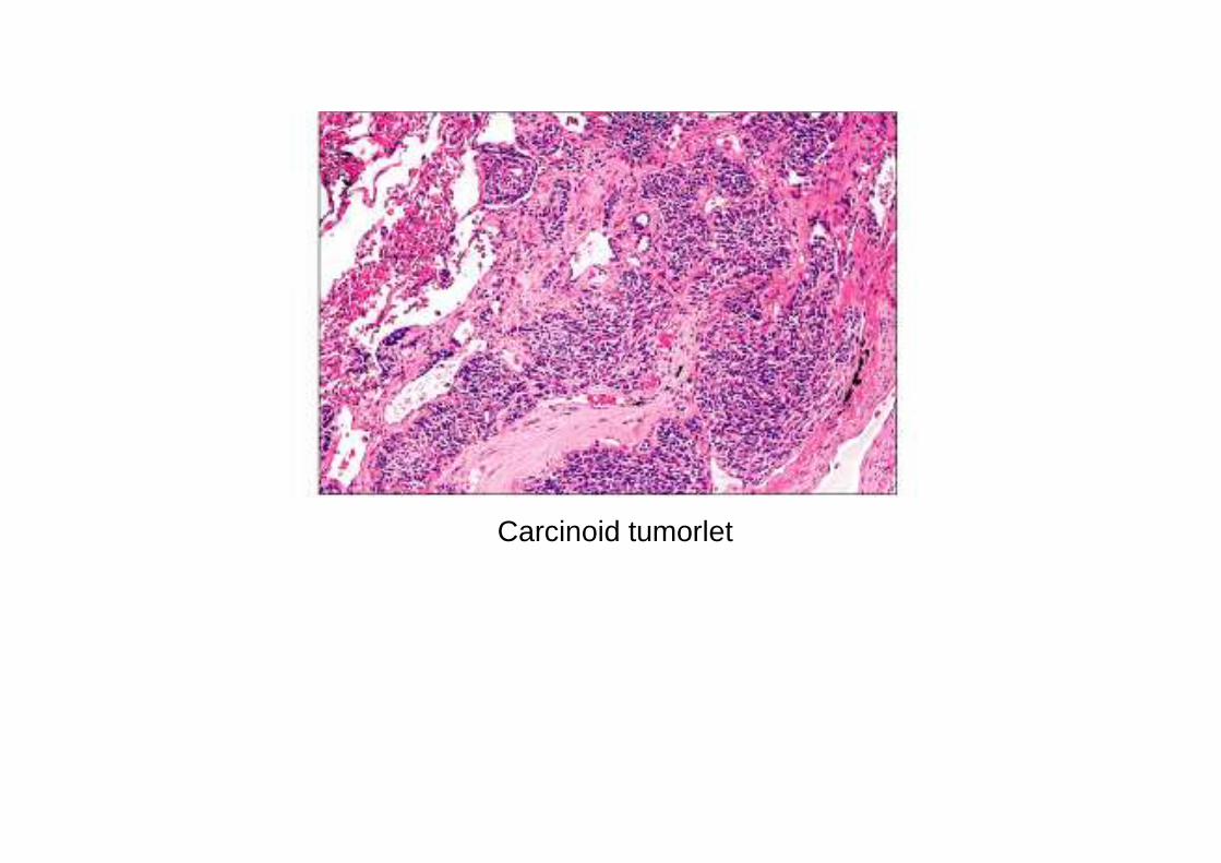
Carcinoid tumorlet
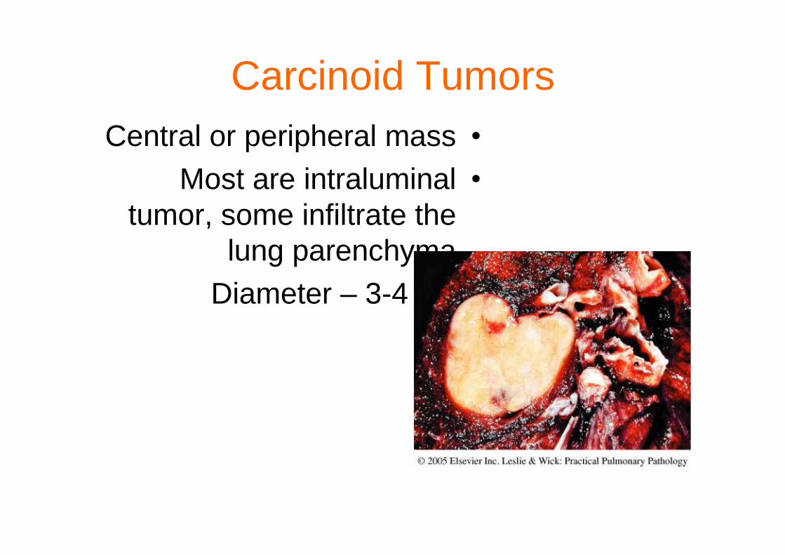
Carcinoid Tumors•Central or peripheral mass•Most are intraluminal
tumor, some infiltrate the lung parenchyma
•Diameter – 3-4 cm
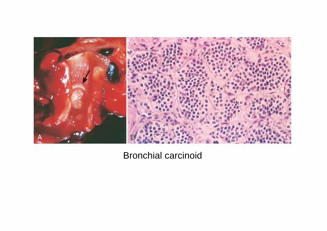
Bronchial carcinoid
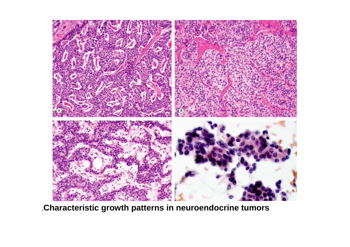
Characteristic growth patterns in neuroendocrine tumors.
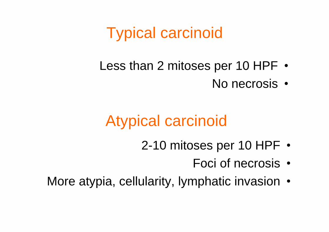
Typical carcinoid
•Less than 2 mitoses per 10 HPF•No necrosis
Atypical carcinoid
•2-10 mitoses per 10 HPF•Foci of necrosis•More atypia, cellularity, lymphatic invasion
Clinical presentationIntraluminal growth → cough, hemophtysis,secondary infection, bronchiectasis,
emphysema, atelectasis.
Secretory activity → carcinoid syndrome (intermittent attacks of diarrhea, flushing and cyanosis)
Most carcinoids have relatively benign course. (Low grade malignancy).
Can occur in patient with MEN 1.
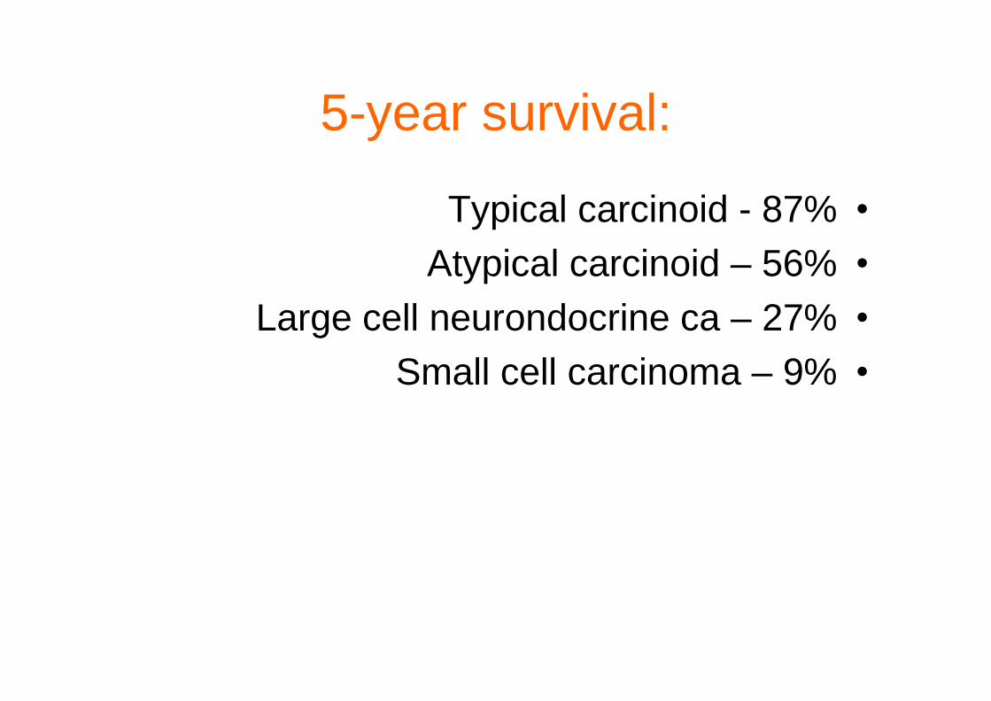
5-year survival:
•Typical carcinoid - 87%•Atypical carcinoid – 56%•Large cell neurondocrine ca – 27%•Small cell carcinoma – 9%
Other lung tumors•, hemangioma: Mesenchymal
hemangiopericytoma, inflammatory myofibroblastic tumor, leiomyoma,
leiomyosarcoma
•Hodgkin and Hodgkin -: nonHematopieticlymphoma, Langerhans cell histiocytosis
•Hamartoma
Lung hamartoma
•Relatively common lesion•Peripheral solid nodule 3-4 cm, well
circumscribed•Microscopically: nodules of mesenchymal
tissue (cartilage, fat ,fibrous tissue) intersected by epithelial clefts
A hamartoma is a lesion in an organ that is composed of tissue elements normally found at that site, but growing in a haphazardmass
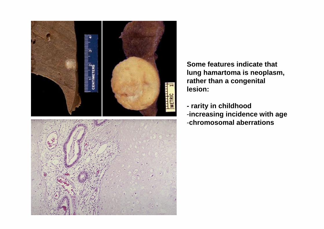
Some features indicate that lung hamartoma is neoplasm, rather than a congenital lesion:
- rarity in childhood-increasing incidence with age-chromosomal aberrations
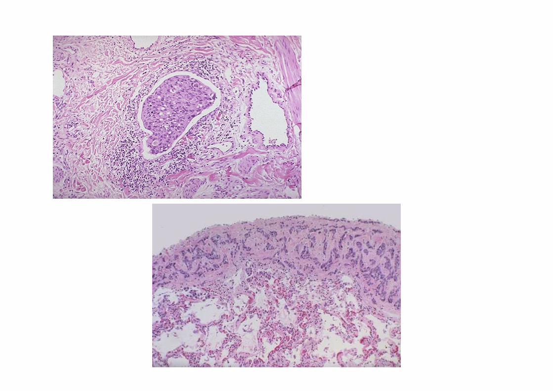
Metastatic tumors
•Multiple discrete nodules (mostly on periphery)•Solitary nodule
•Endobronchial•Pleural
•“Lepidic” growth, similar to BAC•Growth in peribronchiolar and perivascular
tissue spaces
•Diffuse intralymphatic dissemination
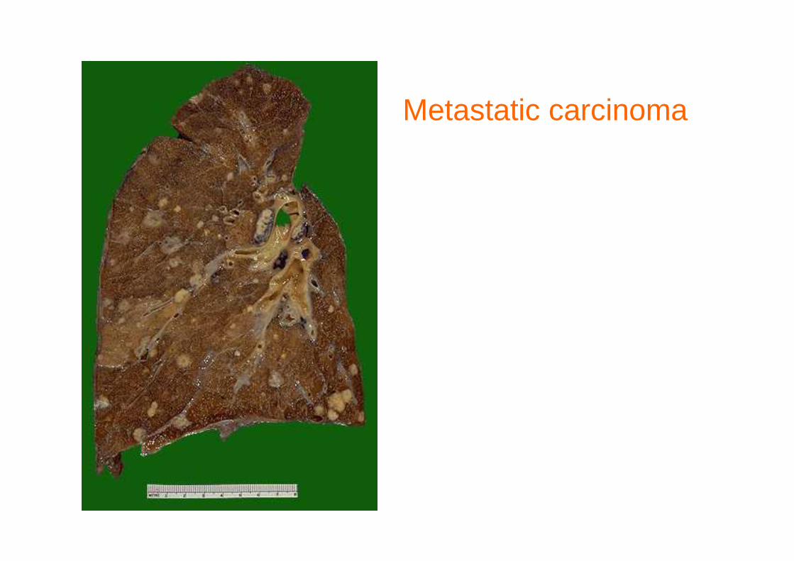
Metastatic carcinoma
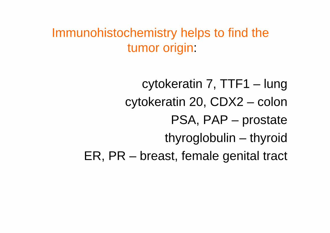
Immunohistochemistry helps to find the tumor origin:
cytokeratin 7, TTF1 – lungcytokeratin 20, CDX2 – colon
PSA, PAP – prostatethyroglobulin – thyroid
ER, PR – breast, female genital tract
PLEURAL TUMORS
Malignant pleural tumors
•Primary•Secondary - more common! (most
common origins - lung and breast)
Malignant mesothelioma
•Asbestos exposure related•Long latent period – 25-40 years•Risk for mesothelioma is not
magnified by smoking (in contrast to carcinoma)
Malignant mesothelioma morphology
•Epithelioid type•Sarcomatous type •Mixed
Diffuse spread in the pleura associated withextensive pleural effusion
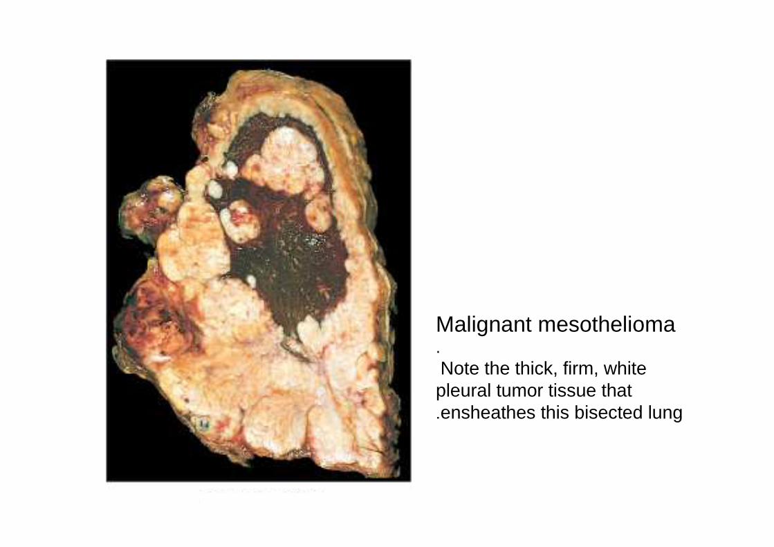
Malignant mesothelioma.Note the thick, firm, white pleural tumor tissue that ensheathes this bisected lung .
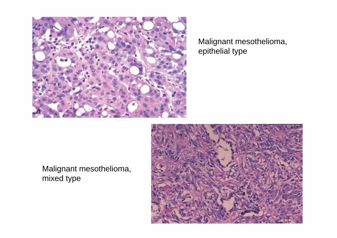
Malignant mesothelioma, epithelial type
Malignant mesothelioma, mixed type
Immunohistochemistry of MM
•Calretinin +•WT 1 +•Thrombomodulin +•Keratin 5/6 +•CEA –•TTF1 –•Different epithelial markers -
Clinical course
•Recurrent pleural effusion•Chest pain, dyspnea•50% die within 12 months•Some improvement with aggressive
therapy (extrapleural pneumonectomy, chemotherapy, radiation)
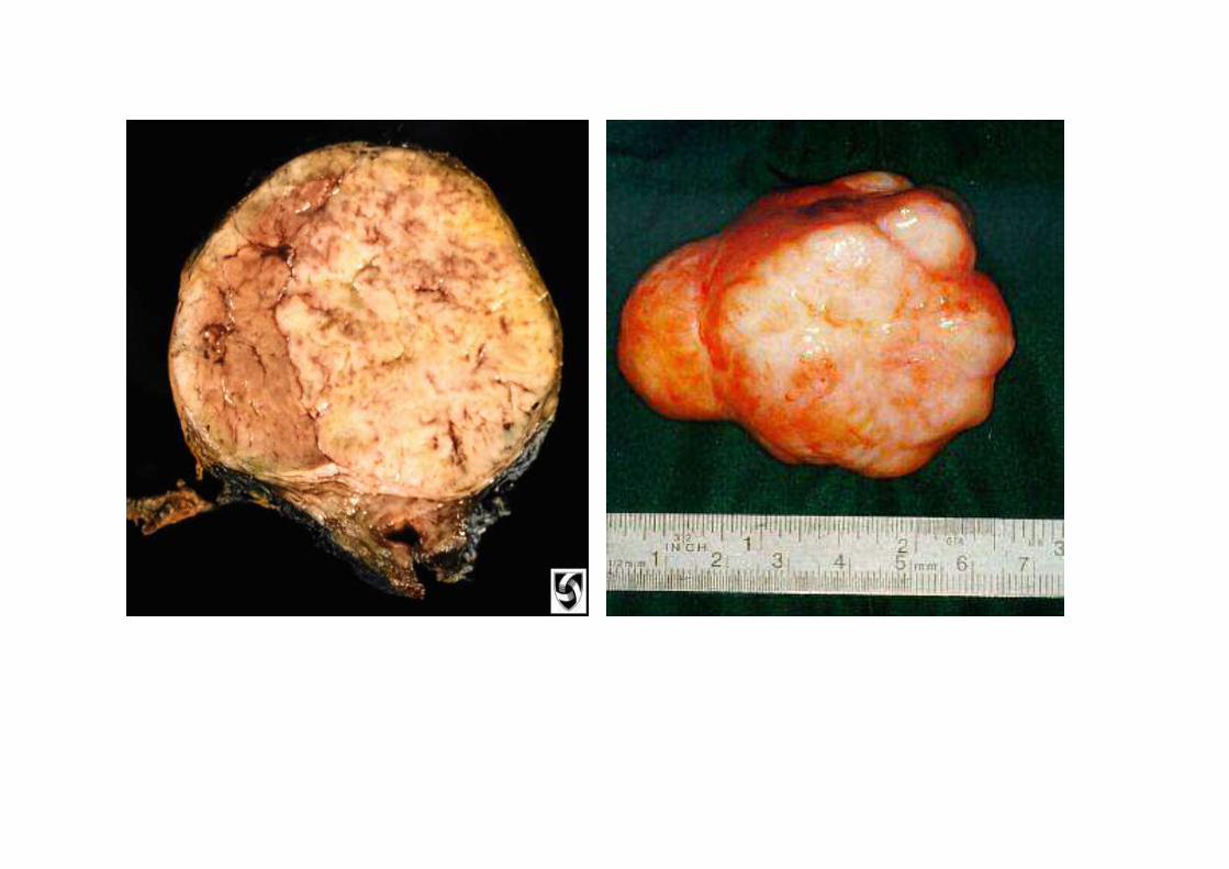
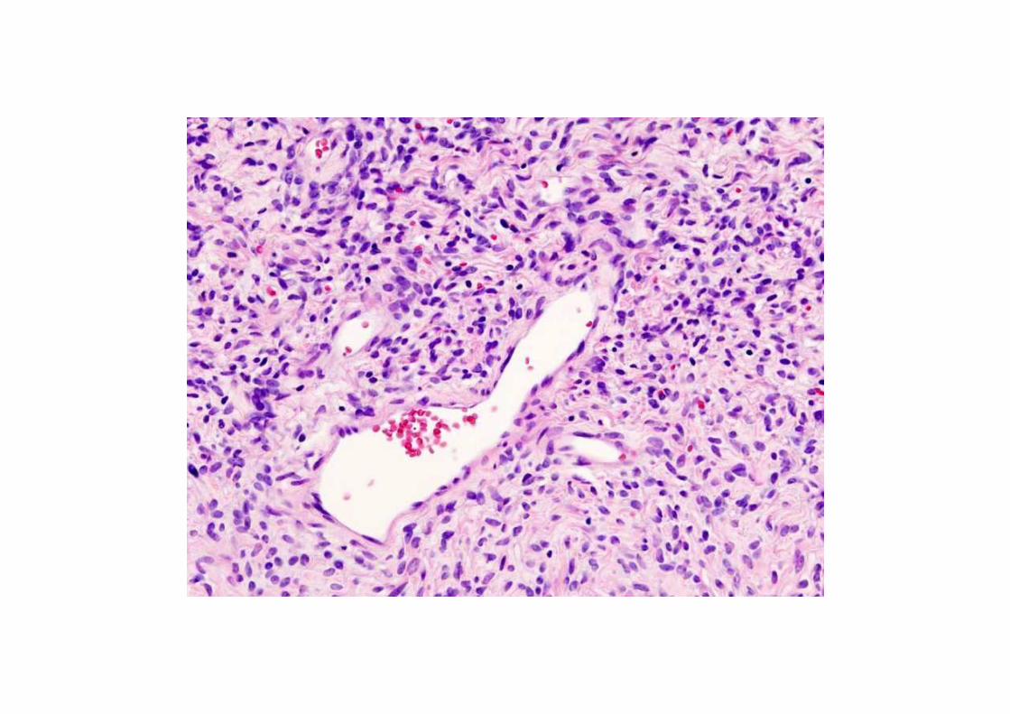
Solitary fibrous tumor
•Soft tissue tumor, occurs in pleura and other sites
•Often is attached to the pleura by pedicle•Do not produce pleural effusion•Microscopically: whorls of reticulin and
collagen fibers and interspersed spindle cells
•Immunohistochemistry: CD 34+
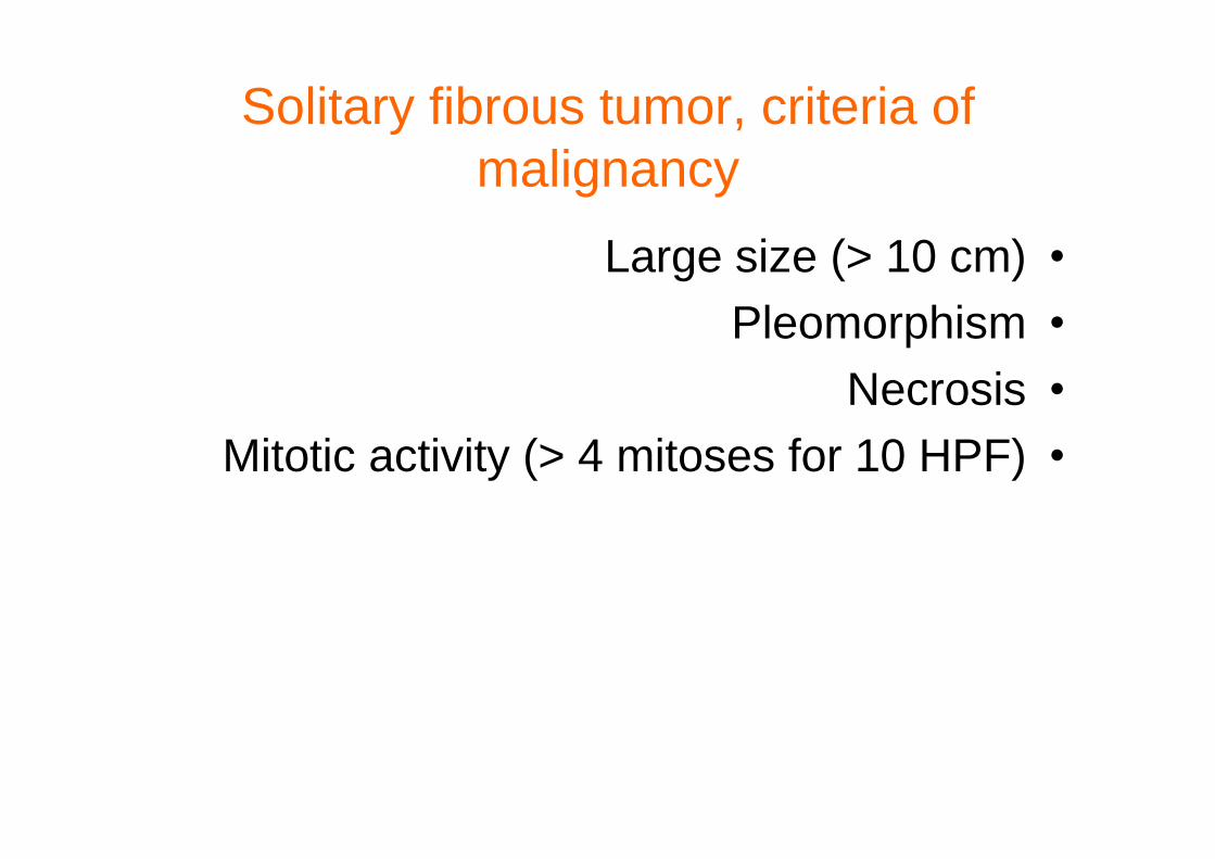
Solitary fibrous tumor, criteria of malignancy
•Large size (> 10 cm)•Pleomorphism•Necrosis•Mitotic activity (> 4 mitoses for 10 HPF)

Dr. Sofia Zilber


![The Involvement of Tumor Suppressor p53 Leukemia Hemopoiesiscancerres.aacrjournals.org/content/canres/54/2/582.full.pdf · [CANCER RESEARCH 54. 582-586, January 15.19q4] The Involvement](https://img.pdfslide.net/doc/110x75/5dd12436d6be591ccb646ba6/the-involvement-of-tumor-suppressor-p53-leukemia-cancer-research-54-582-586-january.jpg)