Embed Size (px)
Citation preview

Lung-RADS™ Pushing the Limits
Maria Daniela Martin, MD Jeffrey P. Kanne, MD
Lynn S. Broderick, MD Ella M. Kazerooni, MD
Cristopher A. Meyer, MD
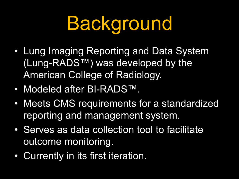
• Lung Imaging Reporting and Data System (Lung-RADS™) was developed by the American College of Radiology.
• Modeled after BI-RADS™. • Meets CMS requirements for a standardized
reporting and management system. • Serves as data collection tool to facilitate
outcome monitoring. • Currently in its first iteration.
Background

Objectives • Identify through clinical cases gray
areas and ambiguous situations that arise while using Lung-RADS.
• Propose solutions for these circumstances, focusing on classification and management of findings.

1. Eligibility
How Would You Manage This Nodule? 80-year-old M will turn 81 years old in 3 months. Has a new 5 mm solid nodule.

Screening Eligibility Organization Current Eligibility Criteria
• American College of Chest Physicians • American Society of Clinical Oncology • American Cancer Society • American Lung Association • American Thoracic Society • CMS + • USPSTF *
• ≥ 55 years old up to 74 years old + 77 years old * 80 years old • Smoked ≥ 30 pack-years • Quit smoking < 15 years ago
• National Comprehensive Cancer Network (NCCN)
• ≥ 55 -74 years old • Smoked ≥ 30 pack-years • Quit smoking < 15 years ago OR • ≥ 50 years old • smoked ≥ 20 pack-years and 1 more risk factor (other than second-hand
smoke) including: radon or occupational exposure, cancer history, history of lung cancer in first-degree relative, disease history (COPD or pulmonary fibrosis)
• American Association for Thoracic Surgery (AATS)
• ≥ 55 -79 years old • Smoked ≥ 30 pack-years OR • Lung cancer survivors starting 5 years after treatment until age 79 OR • ≥ 50 years old, • Smoked ≥ 20 pack-years • 5% risk of developing a lung cancer in the next 5 years (COPD,
environmental/occupational exposure, prior cancer/radiation therapy, genetics, or family history)
At 81 years old, this patient will no longer qualify for lung cancer screening
* U.S. Preventive Services Task Force + Centers for Medicare & Medicaid Services

Teaching Points
• Many organizations advocate CT lung cancer screening. • Eligibility criteria vary depending on the organization. • 80 years is the upper age limit and only for USPSTF. • Those who do not meet CMS criteria will not be covered
by Medicare (up to 77 years old).

What Can You Do For This Patient?
• Can manage similarly but outside of the screening program.
• Management of older patients is controversial: benefits vs. risks (life expectancy, comorbidities, etc.)
• Make clear recommendations: when to follow up and most importantly when to stop monitoring.
1.2 Eligibility - Aging out of screening

If Phased Out of Screening… Should You Use Lung-RADS or Fleischner?
Lung-RADS Fleishner1,2
Published in 2014
Based on newer data from lung cancer screening trials
Published in 2005* and 2013+
Based on older data, pending review
Developed for the management of nodules in the setting of lung cancer screening
Developed for the management of incidentally detected nodules
Adressed how to manage nodules that are new or growing
Does not address how to manage nodules that are new or growing
All nodule types are included Separate guidelines for solid and subsolid nodules
* Guidelines for Management of Small Pulmonary Nodules Detected on CT Scans: A Statement from the Fleischner Society + Recommendations for the Management of Subsolid Pulmonary Nodules Detected at CT: A Statement from the Fleischner Society
1.3 Eligibility – Lung-RADS vs. Fleishner
Lung-RADS Fleishner1,2
Published in 2014
Based on newer data from lung cancer screening trials
Published in 2005* and 2013+
Based on older data, pending review
Developed for the management of nodules in the setting of lung cancer screening
Developed for the management of incidentally detected nodules
Adressed how to manage nodules that are new or growing
Does not address how to manage nodules that are new or growing
All nodule types are included Separate guidelines for solid and subsolid nodules
Teaching Point
For the reasons underlined above, Lung-RADS recommendations are more appropriate to follow in
this case.

Vague Symptoms: Screening vs. Routine CT
• 63 F with mass detected at baseline screen • Retrospective review of the chart after diagnosis revealed
complaints of cervicalgia. • Screening should be reserved for patients without symptoms
attributable to lung cancer. Even vague symptoms suspicious for pathology (weight loss, fatigue) should prompt routine CT.
à small cell carcinoma.
1.4 Eligibility
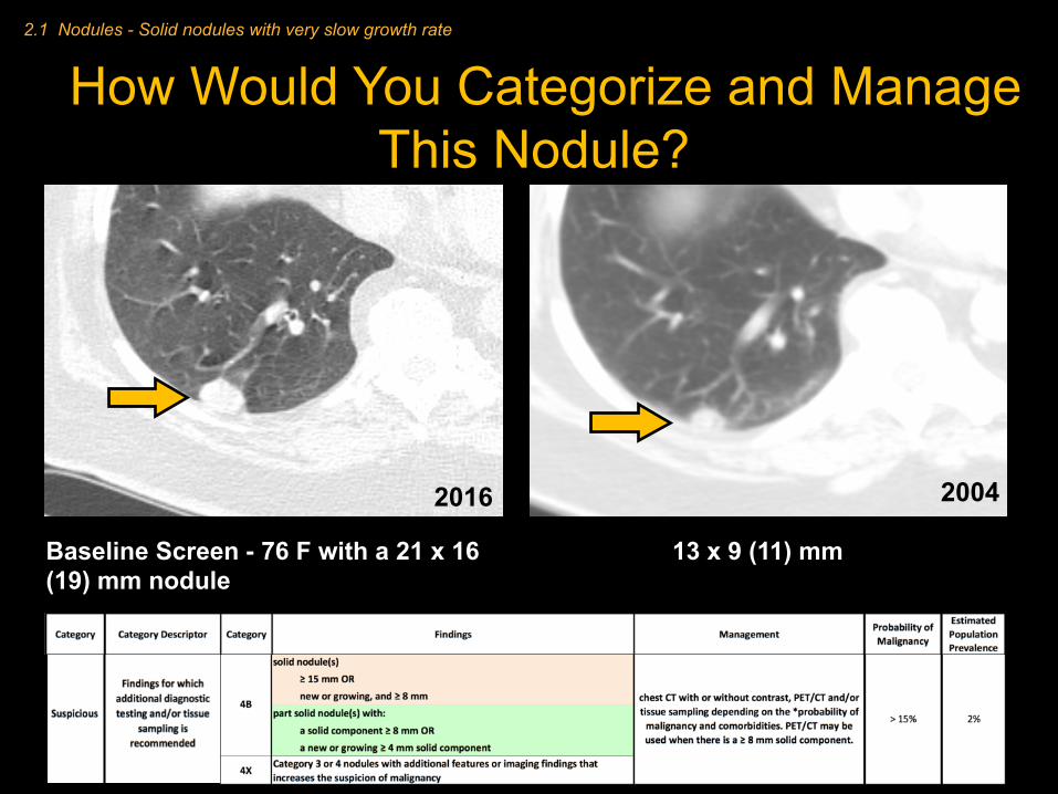
Baseline Screen - 76 F with a 21 x 16 (19) mm nodule
13 x 9 (11) mm 2004CTAP
How Would You Categorize and Manage This Nodule?
2016 2004
2.1 Nodules - Solid nodules with very slow growth rate

Category • Version 1.0 of Lung-RADS does not address categorizing a nodule present on an examination before baseline screen.
• Growth = increase > 1.5 mm. • This nodule, by growth, would fall under Category 4B
• Do you think this nodule is malignant after 12 years?
2016 2004
> 15%.

• Growth of a solid nodule is assessed by volume doubling time (VDT) 3.
• VDT helps determine malignant potential3. • VDT formula based on axial dimensions and time between studies à
many online calculators available. • Mesenchymal tumors and granulomatous lesions can grow very slowly
and lack definitively benign features (reference).
Not Everything That Grows is Cancer
VDT in Days
< 20 < 100 (range of 20 – 400) > 400
Inflammatory/Infectious Malignant Benign
Growing Pattern
Biopsy proven hamartoma. Nodule grew and lacked definitively benign features (e.g. macroscopic fat or benign calcifications). Remote history of testicular cancer prompted the biopsy.
2016 2009
VDT=948days

• This is an excellent example to use the *. • *Footnote 9: 4B management based on probability of
malignancy (patient evaluation, patient preference, and risk of malignancy).
Knowing that VDT= 5.2 years, Would You Manage Nodule Differently?
2016 2004 No activity on PET

Teaching Points
• Lung-RADS provides guidelines and is not absolute. • Use all resources available to propose.
management options appropriate to each case (VDT, prior exams, online assessment tools).
• Possible solutions for future versions of Lung-RADS include: • Address comparison of baseline LDCT to prior
exams. • Categorization of a large nodule as benign at
baseline based on behavior over time (volume doubling time).

6 mm ground-glass
2012 2016
6 mm at Baseline
65 F with baseline screen CT in 2016. A comparison CT from 2012 is available. How would you categorize and manage?
Footnote 4) Growth: an increase in size of > 1.5 mm. Footnote 10) Category 4X – Nodules with additional findings that increase suspicion of lung cancer (spiculation, GGN* that doubles in size in 1 year, enlarged lymph nodes, etc.) * GGN = Ground-glass Nodule
2.2 Nodules – Subsolid nodules growth
Nodule was classified as 4X (new large solid component).

Teaching Points
• X definition on Lung-RADS is flexible and includes: nodule with spiculations, GGN* that doubles in size in 1 year, enlarged lymph nodes, etc.
• Use your judgement and experience to “X” a nodule that you are convinced is cancer.

• Growth of subsolid nodules may manifest as increase in size, attenuation, development of a solid component, or enlarging solid component3 (increase in mass).
• Currently, the footnote definition of nodule growth in Lung-RADS is only related to change in size.
• Category 4 implies growth by development of a solid component, or enlarging solid component.
• Future iterations will likely clarify the concept of growth.
Definition of Growth
This nodule was an adenocarcinoma. Although size did not change, density did. There are also mild spiculations.

Teaching Point
• Remember that growth also includes increase in density or a new or enlarging solid component.

What does slowly growing mean to you?
Case
• Currently, growth is defined as an increase in size of > 1.5 mm. – It does not address solid vs. sub-solid. – “Slowly growing” is not defined.
• VDT varies greatly between nodule type.
Solid Part-solid Pure ground-glass 20 – 400 300 – 450 600–900 (widely variable)
VDT(indays)3,4
2.2 Nodules – Sub-solid nodules growth

2011 2012 2013
• If GGOs persist, they are preinvasive lesions sufficiently often to warrant surveillance2.
• VDT of GGO is much longer than solid and part-solid nodules - on the order of years.
• It has been reported that an increase in length of maximal diameter > 1.72 mm is needed to identify true growth of a GGO5.
77-year-old M, on surveillance for contralateral NSCLC.
Teaching Points
• VDT of GGO is much longer than solid and part-solid nodules.
• Remember to compare GGO to the oldest scans as an interval longer than1 year may be required to detect a change.
• Future versions of LungRads could include a definition of slow (addressing change over periods longer than 1 year).

à 2
à 3
à 4B
à 4A
Ground-glass≥20mm
Baseline
?• We had a poll amongst several
thoracic radiologist who rated this nodule as almost all categories listed below.
• Greatest controversy related to how to measure solid component.
• However, they all agreed that it looks like an adenocarcinoma, which allows use of the X modifier, and recommendations at your discretion.
• Some advocated f/u CT in 3 months to assess for change (most conservative approach, related to overdiagnosis).
• Some were more aggressive and recommended biopsy or resection, which is valid, as it looks like a cancer.
How would you categorize this 23 x 19 mm nodule found at baseline? No priors.
à 3
• Path -> Adenocarcinoma with mucinous component.

Teaching Point
• Regardless of the category, X modifier allows you to target the recommendations based on your experience, taking into account patients’ comorbidities, etc.

2.3 Nodules – GGOs
CANScar or Adenocarcinoma?
Scar All nodules need to be evaluated in multiple planes!
Adenocarcinoma

Category 3 Nodule Becomes Smaller but Denser How Would You Manage?
7mm July 2015
5 mm July 2016
• Lung-RADS does not address nodules that decrease in size.
• Footnote 12 “All Category 3 and 4A nodules that are unchanged on interval CT should be coded as category 2, and returned to screening in 12 months”.
• We suggest that increase in density should be considered as a change.
• Temporary regression of malignant lung nodules can be seen in lung cancers3,6.
• Felt to be related to development of a fibrous component and subsequent collapse of the fibrosis3.
• Continued surveillance is required.
2.4 Nodules - Regression
Teaching Points
• Remember that cancer growth may not be fully exponential, and they may decrease in volume at some point.
• The most accurate assessment in this case would be to measure the mass of the nodule (volume and density).
• Continued surveillance through screening is suggested.

How Would You Categorize This Nodule?
04/2015
Baseline 5 mm
04/2016
5 mm Intrapulmonary lymph node (IPLN)
2.5 Nodules – IPLN

A
C
D
E
F G
• IPLN are well circumscribed, smoothly marginated, in contact or closely related to the fissure, most commonly triangular (A,B,D), polygonal (C), or oval, often with a septal attachment7 (D, E).
• Notice how they can become engorged when edema is present (same patient: D&E).
• Several studies based on lung cancer screening trials have shown that IPLN (perifissural nodules) have no malignant potential7,8.
A
B
C
D
E

Nodules with Features of IPLN
• Management is controversial - contradicts clinical practice for radiologists who do not routinely follow-up when characteristic findings are present.
• Lung-RADS management developed due to potential for confusion with early lung cancers.
• Given their usual small size, most IPLN fall under Category 2 (same management as Category 1).
Footnote 11 Nodules with features of an IPLN should be managed by mean diameter and the 0‐4 numerical category classification.

Teaching Points
• By size, management of IPLN will nearly always be the same (category 1 and 2).
• A multireader study could be conducted evaluating the degree of agreement for recognition of IPLN.
• If high agreement can be achieved, future iterations could allow documenting IPLN in the report and assigning a Category 1, as IPLN are a normal finding.

Airway Lesions: Real or Mucus Plugging?
• Mucus plugging is common in smokers and former smokers and can mimic endobronchial lesions.
• If strictly followed à multiple 3 month LDCTs every time a new mucus plug is identified.
3. Airway Lesions

59 F with new endobronchial lesion compared to CT 4 months earlier performed for other reasons.
Helpful clues to identify secretions include: • Dependent location (layering) • Low/water density • Presence of gas within
secretions
None of these was present in this case…
The patient was brought back the same day and repeat scanning was performed after vigorous coughing, indicating the filling defect was a mucus plug
Teaching Points
• Consider asking the patient to vigorously cough before screening CT
• If in doubt, manage as endobronchial nodules
• If there are multiple CTs showing waxing and waning mucus plugs, use clinical judgement to avoid unnecessary scans

Incidental (Potentially) Significant Findings Other than Lung Cancer
• Rate of incidental findings needing additional evaluation = 10.2% at
baseline and 7.5% overall in the National Lung Cancer Screening Trial9. • “Clinically significant” definition not clear. • Until Lung-RADS includes specific recommendations, practices should
develop or adopt management guidelines. • We encourage use of ACR white papers on management of incidental
findings on CT10,11 and ACR Select TM.
4.1 S modifier - management
Coronary Ca2+ Aortic Ectasia+ Adrenal Nodule

What Could be Included and How Often?
Finding Rationale
Coronary arterial calcifications (CAC) S à Although studies based on lung cancer screening trials showed CAC are predictive of future all-cause mortality and cardiovascular events12, others question if CAC scoring will be clinically useful13
Langerhans cell histiocytosis S à Although uncommon, can be seen in smokers. May lead to a health benefit if patient stops smoking
Any findings that can be interpreted as malignant such as a solid mass including mediastinal, kidney, and liver
S à Follow ACR white papers for further work-up and management11
Thyroid nodules Follow ACR white paper guidelines. Only give S if the nodule warrants further work up and has not already been evaluated10
Benign findings (e.g., mediastinal duplication cyst) Leave in body of the report. S for a finding that does not warrant follow up will lead to confusion and potential harm and cost from unnecessary testing
Emphysema
CONTROVERSIAL. Not routinely encouraged to report as an S à very common finding in smokers and unlikely to change management
• Include as S: – Findings that may lead to a health benefit if a behavior is modified. – Findings that will lead to adverse outcome if not further evaluated or treated.
• There is no value to adding an S modifier for the same finding in subsequent scans.

Returning Back to Screening: What Does Yearly Mean?
• Although not clarified on Lung-RADS, ACR recommends that 12 months should be counted from the day of the scan that prompted the follow up. This refers to the annual date based on their first lung cancer screening CT (in this case, Oct 2016).
• This topic is controversial. Many believe that there will be likely no change in less than a year, so the 12 months should be counted from the last scan (Jan 2016).
• Future Lung-RADS iterations will likely clarify this recommendation.
73-year-old M with indeterminate nodule at baseline, category 4A à 3 month f/u
No change at 3 months
Footnote 12 Category 3 and 4A nodules that are unchanged on interval CT should be coded as category 2, and individuals returned to screening in 12 months
Oct 2015 January 2016
When should the next CT be performed?
5 – Return to screening

Recent Infection: What Lung-RADS Category?
2 months prior, had symptoms of pneumonia.
Spiculated part-solid nodule, smaller than consolidation on chest radiograph.
• Recent infection or other inflammation not addressed by Lung-RADS.
• We categorized it as 3 - probably benign with 6 months follow-up.
• Rationale: • Nodule was smaller but • Not resolved. • Right upper lobe location.
Teaching Points
• Patients with symptoms or recent infection should not be scanned.
• This patient was scanned 2 months after an episode of pneumonia. Future iterations can address a timeframe of no symptoms prior to CT.
• Remember that cancers can become a little smaller before further growth.
6 – Incidental infection

• No clear guidelines for the management of cavitary nodules.
• Lung cancer, pulmonary Langerhans cell histiocytosis nodules, infection, and some metastases can be cavitary.
• Should probably manage similar to solid nodules • Use the X modifier if high suspicion for malignancy.
Cavitary Nodules
C
NSCLC
7 – Cavitary nodules

Patients with Prior Lung Cancer • Previous lung cancer à 3% annual risk of developing new lung cancer. à Peak recurrence incidence 2-3 years after treatment14. • AATS* recommends
– 4 years of CT surveillance after surgery AND – Annual LDCT screening starting 5 years after treatment until age
79.
• USPSTF and CMS do not address previous lung cancer. • Lung-RADS provides a modifier (C). Categories coded as
usual.
*American Association for Thoracic Surgery
8.1 - Prior Lung Cancer

Patients with Non-Lung Malignancy
• Current recommendations do not address patients treated for malignancies with low risks of recurrence (e.g. early stage breast or prostate cancer or low-grade lymphoma).
Should an eligible patient 18-months out from treated stage 1 breast cancer be screened for
lung cancer?
• We believe that screening may be appropriate for patients more likely than not to be alive within the next 5 years.
8.2 - Prior malignancy, not lung

• Lung-RADS is a very helpful guideline that allows consensus in reporting and management in lung cancer screening.
• Several topics remain unclear, but will likely be addressed in future iterations.
• We proposed solutions to several areas of ambiguity based on current literature and our collective experience.
• As we gather data from lung cancer screening and continue to learn about nodules and cancer behavior, Lung-RADS will evolve to best serve our patients.
Thank you!
Conclusions

1. MacMahon H, Austin JH, Gamsu G, Herold CJ, Jett JR, et al. Guidelines for management of small pulmonary nodules detected on CT scans: a statement from the Fleishner Society. Radiology. 2005 Nov;237(2):395-400.
2. Naidich DP, Bankier AA, MacMahon H, Schaefer-Prokop CM, Pistolesi M, et al. Recommendations for the management of subsolid pulmonary nodules detected at CT: a statement from the Fleishner Society. Radiology. 2013;266(1):304-17.
3. Truong M, Ko J, Rossi S, Rossi I, Viswanathan C, et al. Update in the evaluation of the solitary pulmonary nodule. Radiographics. 2014;34:1658-79
4. Kobayashi Y, Mitsudomi T. Management of ground-glass opacities: should all pulmonary lesions with ground-glass opacity be surgically resected? Transl Lung Cancer Res. 2013 October;2(5):354-363
5. Kakinuma R, Ashizawa K, Kuriyama K, et al. Measurement of focal ground-glass opacity diameters on CT images: interobserver agreement in regard to identifying increases in the size of ground-glass opacities. Acad Radiol 2012;19:389-94.
6. Lindell RM, Hartman TE, Swensen SJ, Jett JR, Mid- thun DE, Mandrekar JN. 5-year lung cancer screening experience: growth curves of 18 lung cancers compared to histologic type, CT attenuation, stage, survival, and size. Chest 2009;136(6):1586–1595.
7. Ahn MI, Gleeson TG, Chan IH, Mc Williams AM, Macdonald SL, et al. Perifissural nodules seen at CT screening for lung cancer. Radiology. 2010;254(3):949-56. Chiles C, Duan F, Gladish GW, Ravenel JG, Baginski SG, et al.
8. McWilliams A, Tammemagi MC, Mayo JR, et al. Probability of Cancer in Pulmonary Nodules Detected on First Screening CT. The N Engl J Med. 2013;369(10):910-919.
9. Finterlmann FJ, Bernheim A, Digumartthy SR, Lennes IT, Kalra MK, et al. The 10 Pillars of lung cancer screening: rationale and logistics of a lung cancer screening program. Radiographics. 2015;35(7):1893-908.
10. Hoang JK, Lander JE, Middleton WD, Wu CC, Hammers LW, et al. Managing Incidental Thyroid Nodules Detected on Imaging: White Paper of the ACR Incidental Thyroid Findings Committee. J Am Coll Radiol. 2015 Feb;12(2):143-50.
References

11. Berland LL, Silverman SG, Gore RM, Mayo-Smith WW, Megibow AJ, Yee J, et al. Managing incidental findings on abdominal CT: white paper of the ACR incidental findings committee. J Am Coll Radiol. 2010 Oct;7(10):754-73.
12. Jacobs PC, Gondrie MJA, van der Graaf Y, et al. Coronary artery calcium can predict all-cause mortality and cardiovascular events on low-dose CT screening for lung cancer. Am. J. Roentgenol. 2012; 198:505–511.
13. Bernheim A, Auffermann WF, Stillman AE. The Dubious Value of Coronary Calcium Scoring on Lung Cancer Screening CT. J Am Coll Radiol. 2016 Oct 4. pii: S1546-1440(16)30754-2. doi: 10.1016/j.jacr.2016.08.011. [Epub ahead of print]
14. http://www.aats.org 15. Eberth JM, Sercy E. Implementation of Lung Cancer Screening in the United States: Changing the Trends
Based on a Survey of the Society of Thoracic Radiology Members. J Thorac Imaging. 2015;30(6):W60-2. 16. Hiramatsu M, Inagaki T, Inagaki T, Matsui Y, Satoh Y, et al. Pulmonary ground-glass opacity (GGO) lesions-
large size and a history of lung cancer are risk factors for growth. J Thoracic Oncol. 2008 Nov;3(11):1245-50.
17. Management of ground-glass opacities: should all pulmonary lesions with ground-glass opacity be surgically resected? Transl Lung Cancer Res. 2013 October;2(5):354-363.
18. MacMahon H. Compliance with Fleischner Society guidelines for management of lung nodules: lessons and opportunities. Radiology. 2010 Apr;255(1):14-5.
19. Association of Coronary Artery Calcification and Mortality in the National Lung Screening Trial: A Comparison of Three Scoring Methods. Radiology. 2015 Jul;276(1):82-90.
20. http://www.nccn.org 21. http://www.cdc.gov/cancer/lung/pdf/guidelines.pdf 22. http://www.acr.org/Quality-Safety/Resources/LungRADS
References
Contact: Maria Daniela Martin [email protected]