Embed Size (px)
Citation preview

TUMOURS OF THE LUNG
Primary
- Benign
- Malignant
Secondary
The incidence of lung cancer has been increasing almost logarithmically and is now reaching epidemic levels.
The overall cure rate is very low and the disease has a 90% death rate.

Risk factors:- Cigarrette smoking
- Occupational hazards (asbestos, nickle, chromates)
85% of lung cancer patients are cigarette smokers.
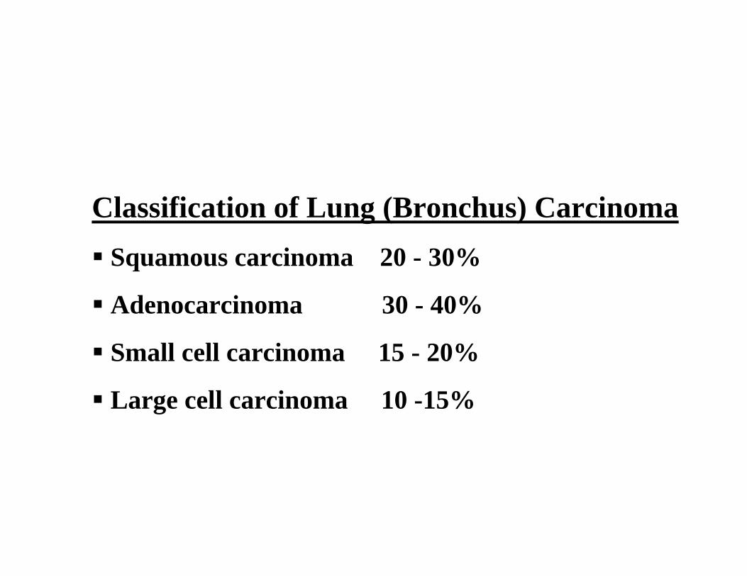
Classification of Lung (Bronchus) CarcinomaSquamous carcinoma 20 - 30%
Adenocarcinoma 30 - 40%
Small cell carcinoma 15 - 20%
Large cell carcinoma 10 -15%

Squamous and adenocarcinoma have about the same 5-year survival probability. It is worse for large cell carcinoma, and oat cell carcinoma has almost no 5-year survival probability with a mean lifespan of months from the time of diagnosis.
Overall 8% of lung cancer cases live past 5 years.

Squamous (epidermoid) carcinomaDerived from reserve cells that differentiate into squamous cells.
Most commonly found centrally in the lung, usually in the major lobar or first segmental bronchus.

Smokers:
Squamous metaplasia of bronchial epithelium
→ Squamous carcinoma in-situ
→ Invasive squamous cell carcinoma


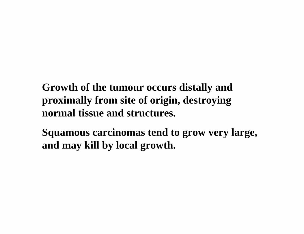
Growth of the tumour occurs distally and proximally from site of origin, destroying normal tissue and structures.
Squamous carcinomas tend to grow very large, and may kill by local growth.

These tumours often cavitate and resemble abscesses on radiographic studies grossly.
The tumour may be surgically resected but cure rate depends on the clinical stage at the time of diagnosis.
Early diagnosis is possible by cytologic examination of a sputum sample.

AdenocarcinomaMost adenocarcinomas are insidious and asymptomatic for a long time.
Adenocarcinomas carcinomas tend to occur more peripherally in the lung.
Adenocarcinoma is the one cell type of primary lung tumour that occurs more often in non-smokers.
Treatment is surgical and involves removal of the entire lobe with associated lymph nodes.
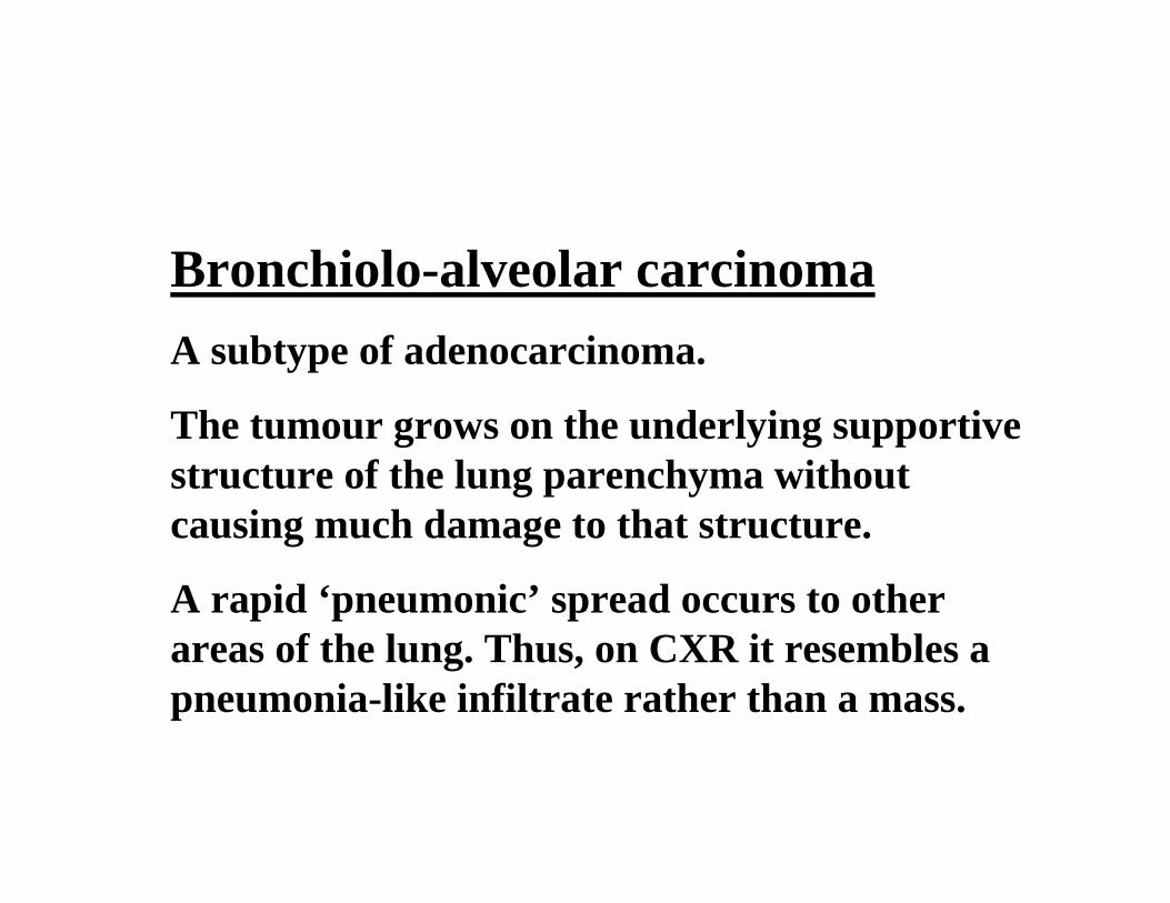
Bronchiolo-alveolar carcinomaA subtype of adenocarcinoma.
The tumour grows on the underlying supportive structure of the lung parenchyma withoutcausing much damage to that structure.
A rapid ‘pneumonic’ spread occurs to other areas of the lung. Thus, on CXR it resembles a pneumonia-like infiltrate rather than a mass.
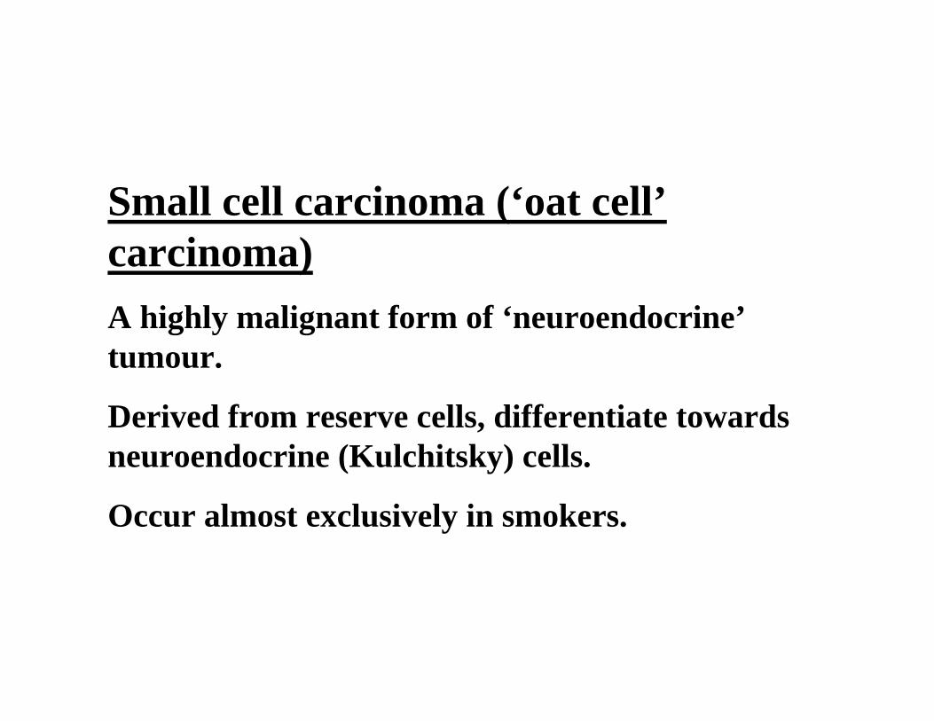
Small cell carcinoma (‘oat cell’carcinoma)A highly malignant form of ‘neuroendocrine’tumour.
Derived from reserve cells, differentiate towards neuroendocrine (Kulchitsky) cells.
Occur almost exclusively in smokers.
Often associated with paraneoplasticsyndromes e.g. hormonal effects
- Ectopic ACTH production
- Inappropriate ADH secretion.

Small cell carcinomas arise centrally andtends to spread diffusely along and into the bronchial wall.
Extensive necrosis within the tumour is common due to its rapid growth outgrowing the blood supply.
Small cell carcinomas are not generally considered resectable cancers since dissemination is likely to have occured by the time they are discovered.
They are responsive to chemotherapy.
Radiotherapy is also useful, but they are rarely curable.
Carcinoid tumoursNeuro-endocrine tumours.
Not related to cigarette smoking.
Polypoid intrabronchial masses, but may infiltrate bronchial wall and surrounding lung tissue.
Slow-growing, low-grade malignant, as opposed to the aggressive behaviour of small cell carcinomas.
Similar in appearance and behaviour to carcinoid tumours arising in other organs.
Neuropeptides demonstrable in tumour cells, but the majority are endocrinologically silent.
Large Cell CarcinomaCarcinomas which are so poorly differentiated that by routine light microscopy they cannot be placed into either the epidermoid or glandular groups.
Ultrastructurally, however, these tumoursfrequently demonstrate features of either epidermoid cells or glandular cells or both.
Arise centrally in lung. Highly aggressive tumours.
Other primary malignant lung tumours are rare.
Commonest: Lymphoma.
Spread of lung carcinomaLocal
Lymphogenic to regional lymph nodes
- hilar, mediastinal, supraclavicular
Haematogenous
- adrenal glands, brain, bones, liver
Local infiltration
Bronchial obstruction ⇒ pneumonia, atelectasis,
bronchiectasis distal to obstruction.
Infiltration of lung parenchyma, pleura, pericardium, chest
wall, vertebrae.
Infiltration of superior vena cava ⇒ SVC syndrome
(swelling of face, fullness of neck veins)
Apical tumours may infiltrate:
- Cervical sympathetic nerves ⇒ Horner’s syndrome (ptosis,
miosis, anhydrosis on same side of lesion)
- Brachial plexus ⇒ Pancoast syndrome (neurological
manifestations e.g. pain in upper extremity)

Secondary (metastatic) tumoursMore common than primary lung tumours.
Carcinomas or sarcomas.
Benign lung tumoursRare.
Commonest: Hamartoma (chondroma)
Malignant mesotheliomaMalignant tumour of pleura, rarely pleura + peritoneum .
Most if not all mesotheliomas are related to asbestos exposure.
Typically encountered in middle-aged men occupationally exposed to asbestos, even for a short time (in RSA: N-W Cape).
Tumour may appear many years after exposure
(20 years+).
Pathologic Expressions of Asbestos-Related Disease• Pleural effusions
• Pleural fibrosis/plaques
• Interstitial fibrosis
• Bronchogenic carcinoma
• Mesothelioma

Pathology of mesothelioma:
Multiple nodules studding the pleura and/or a diffuse thickening of the pleura, often accompanied by a painless effusion.
The tumour extends into lobar septa, thereby encasing the lung and obliterating the pleural space. May invade pericardium, thoracic wall.
Micro: Epithelial and sarcomatous components. If epithelial component predominates, it may be difficult to distinguish from an adenocarcinoma.

The tumour tends to remain localised to the thorax / peritoneum, but haematogenous metastases may rarely occur.
Treatment is ineffective and the prognosis is poor.