Embed Size (px)
Citation preview
Skin Integrity and Wound Care
8FOCUSING ON PATIENT CAREThis chapter will help you develop some of the skills related to skin integrity and woundcare necessary to care for the following patients:
Lori Downs, a patient with diabetes mellitus, is admitted with a chronic ulcer of herleft foot.
Tran Nguyen, diagnosed with breast cancer, has had a modified radical mastectomy.
Arthur Lowes, has an appointment with his surgeon today for a follow-up examina-tion and removal of surgical staples following a colon resection.
CH
AP
TE
R
1. Clean a wound and apply a dry, sterile dressing.
2. Apply a saline-moistened dressing.
3. Apply a hydrocolloid dressing.
4. Perform wound irrigation.
5. Collect a wound culture.
6. Apply Montgomery straps.
7. Provide care to a Penrose drain.
8. Provide care to a T-tube drain.
9. Provide care to a Jackson-Pratt drain.
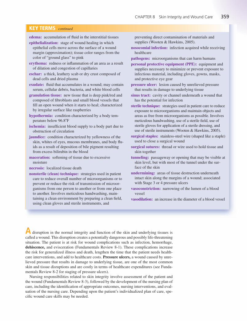
KEY TERMS
approximated wound edges: edges of a wound that are
lightly pulled together; epithelialization of wound mar-
gins; edges touch, wound is closed.
debridement: removal of devitalized tissue and foreign
material from a wound
10. Provide care to a Hemovac drain.
11. Apply negative pressure wound therapy.
12. Remove sutures.
13. Remove surgical staples.
14. Apply an external heating pad.
15. Apply a warm sterile compress to an open wound.
16. Assist with a Sitz bath.
17. Apply cold therapy.
LEARNING OBJECTIVESAfter studying this chapter, you will be able to:
dehiscence: accidental separation of wound edges,
especially a surgical wound
ecchymosis: discoloration of an area resulting from
infiltration of blood into the subcutaneous tissue
358
LWBK545_C08_p358-435.qxd 08/07/2010 1:56 PM Page 358 Aptara
CHAPTER 8 Skin Integrity and Wound Care 359
A disruption in the normal integrity and function of the skin and underlying tissues is
called a wound. This disruption creates a potentially dangerous and possibly life-threatening
situation. The patient is at risk for wound complications such as infection, hemorrhage,
dehiscence, and evisceration (Fundamentals Review 8-1). These complications increase
the risk for generalized illness and death, lengthen the time that the patient needs health-
care interventions, and add to healthcare costs. Pressure ulcers, a wound caused by unre-
lieved pressure that results in damage to underlying tissue, are one of the most common
skin and tissue disruptions and are costly in terms of healthcare expenditures (see Funda-
mentals Review 8-2 for staging of pressure ulcers).
Nursing responsibilities related to skin integrity involve assessment of the patient and
the wound (Fundamentals Review 8-3), followed by the development of the nursing plan of
care, including the identification of appropriate outcomes, nursing interventions, and eval-
uation of the nursing care. Depending upon the patient’s individualized plan of care, spe-
cific wound care skills may be needed.
KEY TERMS continued
preventing direct contamination of materials and
supplies (Wooten & Hawkins, 2005).
nosocomial infection: infection acquired while receiving
healthcare
pathogens: microorganisms that can harm humans
personal protective equipment (PPE): equipment and
supplies necessary to minimize or prevent exposure to
infectious material, including gloves, gowns, masks,
and protective eye gear
pressure ulcer: lesion caused by unrelieved pressure
that results in damage to underlying tissue
sinus tract: cavity or channel underneath a wound that
has the potential for infection
sterile technique: strategies used in patient care to reduce
exposure to microorganisms and maintain objects and
areas as free from microorganisms as possible. Involves
meticulous handwashing, use of a sterile field, use of
sterile gloves for application of a sterile dressing, and
use of sterile instruments (Wooten & Hawkins, 2005).
surgical staples: stainless-steel wire (shaped like a staple)
used to close a surgical wound
surgical sutures: thread or wire used to hold tissue and
skin together
tunneling: passageway or opening that may be visible at
skin level, but with most of the tunnel under the sur-
face of the skin
undermining: areas of tissue destruction underneath
intact skin along the margins of a wound; associated
with Stage 3 or 4 pressure ulcers
vasoconstriction: narrowing of the lumen of a blood
vessel
vasodilation: an increase in the diameter of a blood vessel
edema: accumulation of fluid in the interstitial tissues
epithelialization: stage of wound healing in which
epithelial cells move across the surface of a wound
margin (approximation); tissue color ranges from the
color of “ground glass” to pink
erythema: redness or inflammation of an area as a result
of dilation and congestion of capillaries
eschar: a thick, leathery scab or dry crust composed of
dead cells and dried plasma
exudate: fluid that accumulates in a wound; may contain
serum, cellular debris, bacteria, and white blood cells
granulation tissue: new tissue that is deep pink/red and
composed of fibroblasts and small blood vessels that
fill an open wound when it starts to heal; characterized
by irregular surface like raspberries
hypothermia: condition characterized by a body tem-
perature below 96.8�F
ischemia: insufficient blood supply to a body part due to
obstruction of circulation
jaundice: condition characterized by yellowness of the
skin, whites of eyes, mucous membranes, and body flu-
ids as a result of deposition of bile pigment resulting
from excess bilirubin in the blood
maceration: softening of tissue due to excessive
moisture
necrosis: localized tissue death
nonsterile (clean) technique: strategies used in patient
care to reduce overall number of microorganisms or to
prevent or reduce the risk of transmission of microor-
ganisms from one person to another or from one place
to another. Involves meticulous handwashing, main-
taining a clean environment by preparing a clean field,
using clean gloves and sterile instruments, and
LWBK545_C08_p358-435.qxd 08/07/2010 1:56 PM Page 359 Aptara
360 UNIT II Promoting Healthy Physiologic Responses
One of the most common causes of nosocomial infections is carelessness in practicing
asepsis when providing wound care. It is extremely important to use appropriate aseptic
technique and follow Standard Precautions and, if needed, Transmission-Based Precau-
tions in providing wound care. Chronic wounds and pressure ulcers may be treated using
clean technique. (Refer to Chapter 4, Asepsis and Infection Control for a discussion of
infection control precautions, sterile technique and clean technique).
Nurses must also be skilled in assessing for pain and employing strategies to minimize
the pain experience of the patient because some patients may experience both physiologic
and/or psychological pain related to dressing changes and wound care.
Additionally, ongoing assessment for possible skin or wound complications will be
required. There are many wound care products/dressings available, each with distinctive
actions, as well as indications, contraindications, advantages, and disadvantages. It is very
important for the nurse to be aware of the products available in a particular facility and be
familiar with the indications for, and correct use of, each type of dressing and wound care
product. Fundamentals Review 8-4 outlines the purposes and uses for several wound
dressing/product categories. In addition, it is often appropriate and necessary to consult
with the wound care specialist, often a wound certified nurse specialist, to plan and coordi-
nate the most effective care for the patient.
This chapter will cover skills to assist the nurse in providing care related to skin integrity
and wounds. In addition to the Fundamentals Review boxes in this chapter, refer to those
found in Chapter 4 (Asepsis and Infection Control) for a quick review of critical knowl-
edge to assist you in understanding the skills related to skin integrity and wound care.
Fundamentals Review 8-1
WOUND HEALING AND COMPLICATIONS
• Wounds heal by primary, secondary, or tertiary
intention.
• Wounds healing by primary intention form a clean,
straight line with little loss of tissue. The wound edges
are well approximated with sutures. These wounds
usually heal rapidly with minimal scarring.
• Wounds healing by secondary intention are large
wounds with considerable tissue loss. The edges are
not approximated. Healing occurs by formation of
granulation tissue. These wounds have a longer heal-
ing time, a greater chance of infection, and larger
scars.
• Wounds healing by primary intention that become
infected heal by secondary intention. These wounds
generate a greater inflammatory reaction and more
granulation tissue. They have large scars and are less
likely to shrink to a flat line as they heal.
• Wounds healing by delayed primary intention or ter-
tiary intention are left open for several days to allow
edema or infection to resolve or exudates to drain.
They are then closed.
• Wound complications include infection, hemorrhage,
dehiscence, and evisceration. These problems increase
the risk for generalized illness, lengthen the time during
which the patient needs healthcare interventions, and
increase the cost of healthcare, and can result in death.
• Multiple psychological effects can occur as a result of
trauma to the integumentary system. Actual and poten-
tial emotional stressors are common in patients with
wounds. Pain is part of almost every wound. In addi-
tion, anxiety and fear play a large role in a patient’s
recovery from a wound. Many patients must deal with
changes in body image, body structure, and function
related to a wound.
LWBK545_C08_p358-435.qxd 08/07/2010 1:56 PM Page 360 Aptara
CHAPTER 8 Skin Integrity and Wound Care 361
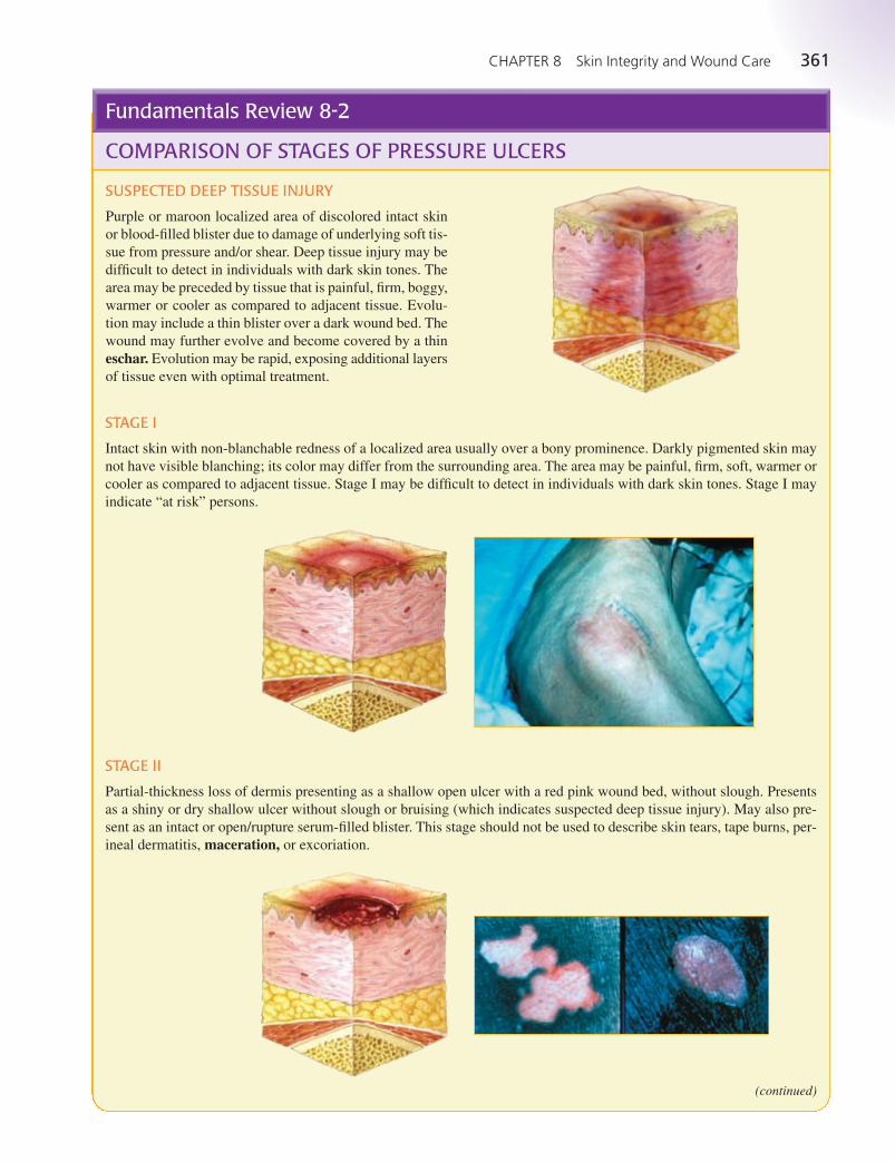
Fundamentals Review 8-2
COMPARISON OF STAGES OF PRESSURE ULCERS
SUSPECTED DEEP TISSUE INJURY
Purple or maroon localized area of discolored intact skin
or blood-filled blister due to damage of underlying soft tis-
sue from pressure and/or shear. Deep tissue injury may be
difficult to detect in individuals with dark skin tones. The
area may be preceded by tissue that is painful, firm, boggy,
warmer or cooler as compared to adjacent tissue. Evolu-
tion may include a thin blister over a dark wound bed. The
wound may further evolve and become covered by a thin
eschar. Evolution may be rapid, exposing additional layers
of tissue even with optimal treatment.
STAGE I
Intact skin with non-blanchable redness of a localized area usually over a bony prominence. Darkly pigmented skin may
not have visible blanching; its color may differ from the surrounding area. The area may be painful, firm, soft, warmer or
cooler as compared to adjacent tissue. Stage I may be difficult to detect in individuals with dark skin tones. Stage I may
indicate “at risk” persons.
STAGE II
Partial-thickness loss of dermis presenting as a shallow open ulcer with a red pink wound bed, without slough. Presents
as a shiny or dry shallow ulcer without slough or bruising (which indicates suspected deep tissue injury). May also pre-
sent as an intact or open/rupture serum-filled blister. This stage should not be used to describe skin tears, tape burns, per-
ineal dermatitis, maceration, or excoriation.
(continued)
LWBK545_C08_p358-435.qxd 08/07/2010 1:56 PM Page 361 Aptara
362 UNIT II Promoting Healthy Physiologic Responses
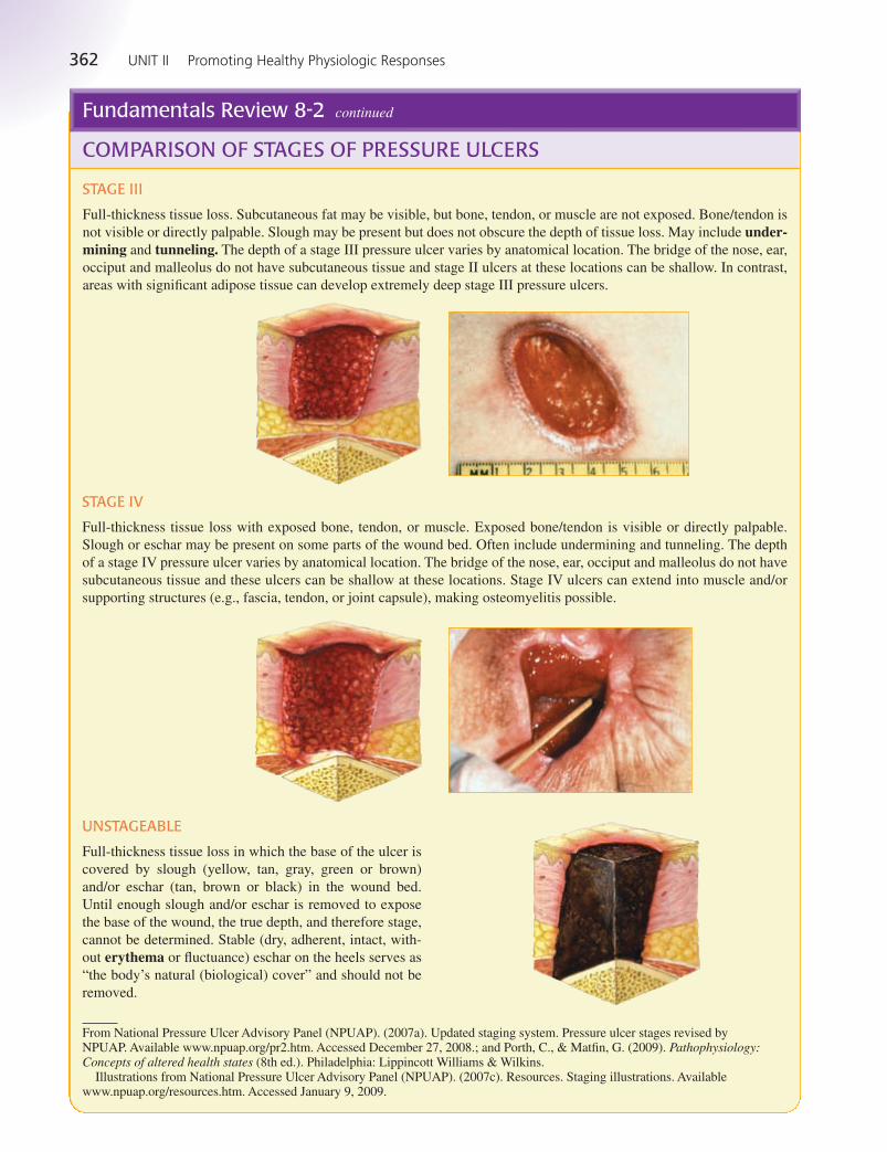
Fundamentals Review 8-2 continued
COMPARISON OF STAGES OF PRESSURE ULCERS
STAGE III
Full-thickness tissue loss. Subcutaneous fat may be visible, but bone, tendon, or muscle are not exposed. Bone/tendon is
not visible or directly palpable. Slough may be present but does not obscure the depth of tissue loss. May include under-mining and tunneling. The depth of a stage III pressure ulcer varies by anatomical location. The bridge of the nose, ear,
occiput and malleolus do not have subcutaneous tissue and stage II ulcers at these locations can be shallow. In contrast,
areas with significant adipose tissue can develop extremely deep stage III pressure ulcers.
STAGE IV
Full-thickness tissue loss with exposed bone, tendon, or muscle. Exposed bone/tendon is visible or directly palpable.
Slough or eschar may be present on some parts of the wound bed. Often include undermining and tunneling. The depth
of a stage IV pressure ulcer varies by anatomical location. The bridge of the nose, ear, occiput and malleolus do not have
subcutaneous tissue and these ulcers can be shallow at these locations. Stage IV ulcers can extend into muscle and/or
supporting structures (e.g., fascia, tendon, or joint capsule), making osteomyelitis possible.
UNSTAGEABLE
Full-thickness tissue loss in which the base of the ulcer is
covered by slough (yellow, tan, gray, green or brown)
and/or eschar (tan, brown or black) in the wound bed.
Until enough slough and/or eschar is removed to expose
the base of the wound, the true depth, and therefore stage,
cannot be determined. Stable (dry, adherent, intact, with-
out erythema or fluctuance) eschar on the heels serves as
“the body’s natural (biological) cover” and should not be
removed.
From National Pressure Ulcer Advisory Panel (NPUAP). (2007a). Updated staging system. Pressure ulcer stages revised byNPUAP. Available www.npuap.org/pr2.htm. Accessed December 27, 2008.; and Porth, C., & Matfin, G. (2009). Pathophysiology:Concepts of altered health states (8th ed.). Philadelphia: Lippincott Williams & Wilkins.
Illustrations from National Pressure Ulcer Advisory Panel (NPUAP). (2007c). Resources. Staging illustrations. Availablewww.npuap.org/resources.htm. Accessed January 9, 2009.
LWBK545_C08_p358-435.qxd 08/07/2010 1:56 PM Page 362 Aptara
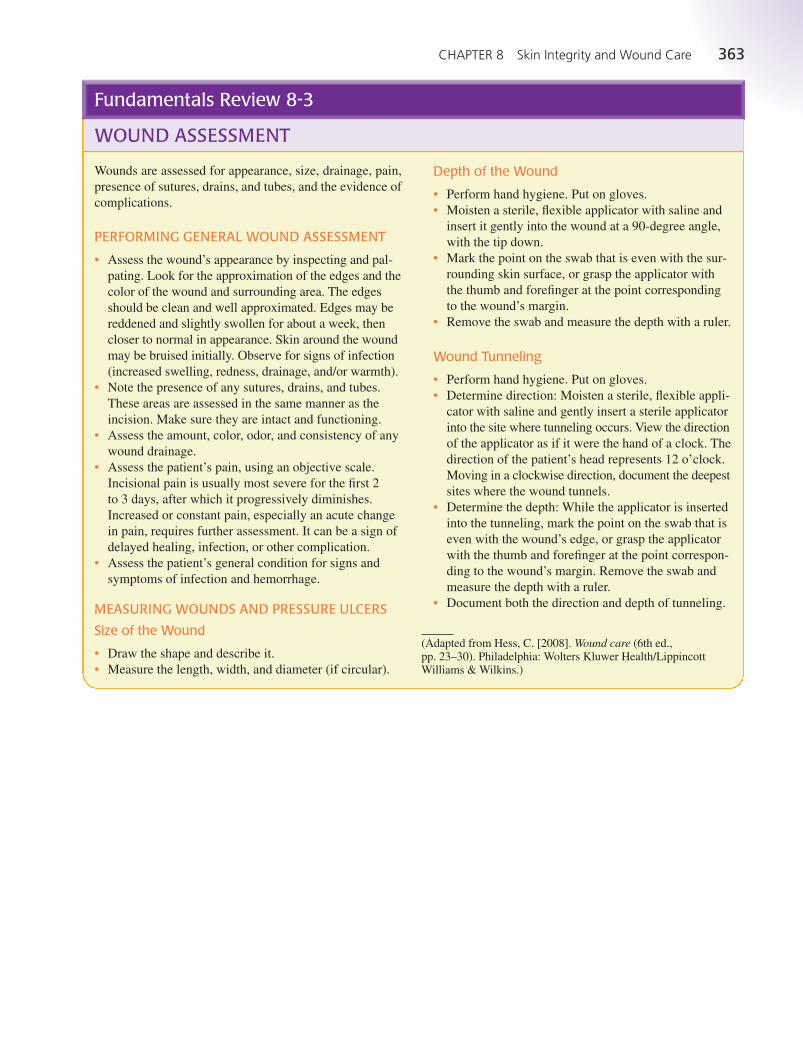
CHAPTER 8 Skin Integrity and Wound Care 363
Fundamentals Review 8-3
Wounds are assessed for appearance, size, drainage, pain,
presence of sutures, drains, and tubes, and the evidence of
complications.
PERFORMING GENERAL WOUND ASSESSMENT
• Assess the wound’s appearance by inspecting and pal-
pating. Look for the approximation of the edges and the
color of the wound and surrounding area. The edges
should be clean and well approximated. Edges may be
reddened and slightly swollen for about a week, then
closer to normal in appearance. Skin around the wound
may be bruised initially. Observe for signs of infection
(increased swelling, redness, drainage, and/or warmth).
• Note the presence of any sutures, drains, and tubes.
These areas are assessed in the same manner as the
incision. Make sure they are intact and functioning.
• Assess the amount, color, odor, and consistency of any
wound drainage.
• Assess the patient’s pain, using an objective scale.
Incisional pain is usually most severe for the first 2
to 3 days, after which it progressively diminishes.
Increased or constant pain, especially an acute change
in pain, requires further assessment. It can be a sign of
delayed healing, infection, or other complication.
• Assess the patient’s general condition for signs and
symptoms of infection and hemorrhage.
MEASURING WOUNDS AND PRESSURE ULCERS
Size of the Wound
• Draw the shape and describe it.
• Measure the length, width, and diameter (if circular).
Depth of the Wound
• Perform hand hygiene. Put on gloves.
• Moisten a sterile, flexible applicator with saline and
insert it gently into the wound at a 90-degree angle,
with the tip down.
• Mark the point on the swab that is even with the sur-
rounding skin surface, or grasp the applicator with
the thumb and forefinger at the point corresponding
to the wound’s margin.
• Remove the swab and measure the depth with a ruler.
Wound Tunneling
• Perform hand hygiene. Put on gloves.
• Determine direction: Moisten a sterile, flexible appli-
cator with saline and gently insert a sterile applicator
into the site where tunneling occurs. View the direction
of the applicator as if it were the hand of a clock. The
direction of the patient’s head represents 12 o’clock.
Moving in a clockwise direction, document the deepest
sites where the wound tunnels.
• Determine the depth: While the applicator is inserted
into the tunneling, mark the point on the swab that is
even with the wound’s edge, or grasp the applicator
with the thumb and forefinger at the point correspon-
ding to the wound’s margin. Remove the swab and
measure the depth with a ruler.
• Document both the direction and depth of tunneling.
(Adapted from Hess, C. [2008]. Wound care (6th ed., pp. 23–30). Philadelphia: Wolters Kluwer Health/LippincottWilliams & Wilkins.)
WOUND ASSESSMENT
LWBK545_C08_p358-435.qxd 08/07/2010 1:56 PM Page 363 Aptara
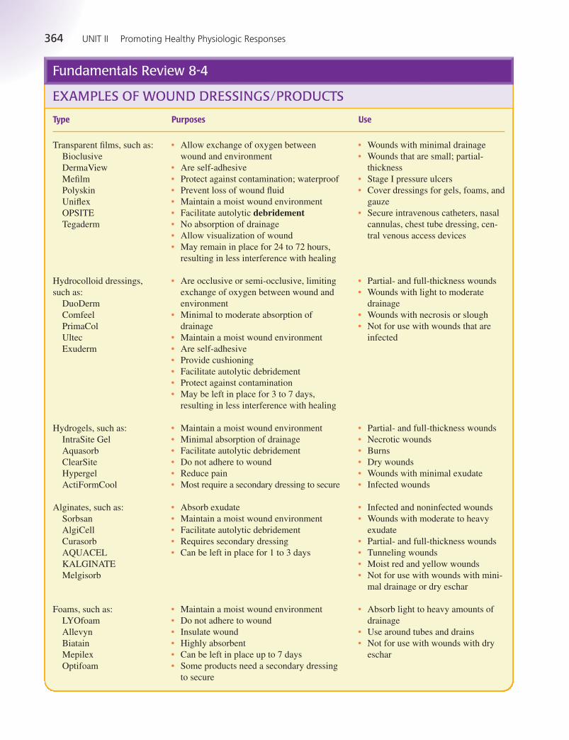
364 UNIT II Promoting Healthy Physiologic Responses
Fundamentals Review 8-4
EXAMPLES OF WOUND DRESSINGS/PRODUCTS
Type Purposes Use
Transparent films, such as:
Bioclusive
DermaView
Mefilm
Polyskin
Uniflex
OPSITE
Tegaderm
Hydrocolloid dressings,
such as:
DuoDerm
Comfeel
PrimaCol
Ultec
Exuderm
Hydrogels, such as:
IntraSite Gel
Aquasorb
ClearSite
Hypergel
ActiFormCool
Alginates, such as:
Sorbsan
AlgiCell
Curasorb
AQUACEL
KALGINATE
Melgisorb
Foams, such as:
LYOfoam
Allevyn
Biatain
Mepilex
Optifoam
• Allow exchange of oxygen between
wound and environment
• Are self-adhesive
• Protect against contamination; waterproof
• Prevent loss of wound fluid
• Maintain a moist wound environment
• Facilitate autolytic debridement• No absorption of drainage
• Allow visualization of wound
• May remain in place for 24 to 72 hours,
resulting in less interference with healing
• Are occlusive or semi-occlusive, limiting
exchange of oxygen between wound and
environment
• Minimal to moderate absorption of
drainage
• Maintain a moist wound environment
• Are self-adhesive
• Provide cushioning
• Facilitate autolytic debridement
• Protect against contamination
• May be left in place for 3 to 7 days,
resulting in less interference with healing
• Maintain a moist wound environment
• Minimal absorption of drainage
• Facilitate autolytic debridement
• Do not adhere to wound
• Reduce pain
• Most require a secondary dressing to secure
• Absorb exudate
• Maintain a moist wound environment
• Facilitate autolytic debridement
• Requires secondary dressing
• Can be left in place for 1 to 3 days
• Maintain a moist wound environment
• Do not adhere to wound
• Insulate wound
• Highly absorbent
• Can be left in place up to 7 days
• Some products need a secondary dressing
to secure
• Wounds with minimal drainage
• Wounds that are small; partial-
thickness
• Stage I pressure ulcers
• Cover dressings for gels, foams, and
gauze
• Secure intravenous catheters, nasal
cannulas, chest tube dressing, cen-
tral venous access devices
• Partial- and full-thickness wounds
• Wounds with light to moderate
drainage
• Wounds with necrosis or slough
• Not for use with wounds that are
infected
• Partial- and full-thickness wounds
• Necrotic wounds
• Burns
• Dry wounds
• Wounds with minimal exudate
• Infected wounds
• Infected and noninfected wounds
• Wounds with moderate to heavy
exudate
• Partial- and full-thickness wounds
• Tunneling wounds
• Moist red and yellow wounds
• Not for use with wounds with mini-
mal drainage or dry eschar
• Absorb light to heavy amounts of
drainage
• Use around tubes and drains
• Not for use with wounds with dry
eschar
LWBK545_C08_p358-435.qxd 08/07/2010 1:56 PM Page 364 Aptara
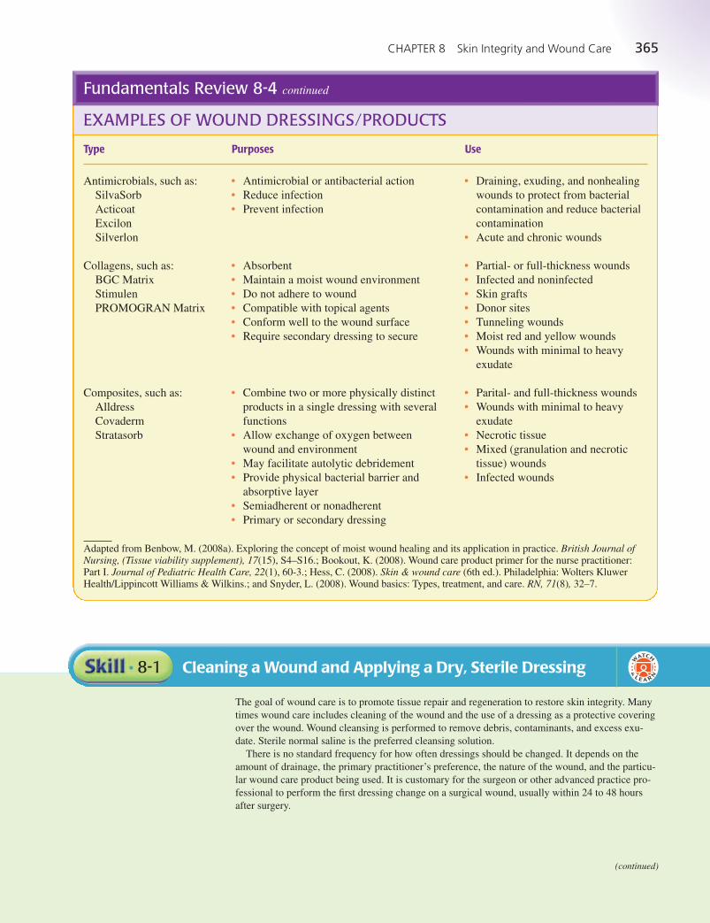
CHAPTER 8 Skin Integrity and Wound Care 365
Fundamentals Review 8-4 continued
EXAMPLES OF WOUND DRESSINGS/PRODUCTS
Type Purposes Use
Antimicrobials, such as:
SilvaSorb
Acticoat
Excilon
Silverlon
Collagens, such as:
BGC Matrix
Stimulen
PROMOGRAN Matrix
Composites, such as:
Alldress
Covaderm
Stratasorb
• Antimicrobial or antibacterial action
• Reduce infection
• Prevent infection
• Absorbent
• Maintain a moist wound environment
• Do not adhere to wound
• Compatible with topical agents
• Conform well to the wound surface
• Require secondary dressing to secure
• Combine two or more physically distinct
products in a single dressing with several
functions
• Allow exchange of oxygen between
wound and environment
• May facilitate autolytic debridement
• Provide physical bacterial barrier and
absorptive layer
• Semiadherent or nonadherent
• Primary or secondary dressing
• Draining, exuding, and nonhealing
wounds to protect from bacterial
contamination and reduce bacterial
contamination
• Acute and chronic wounds
• Partial- or full-thickness wounds
• Infected and noninfected
• Skin grafts
• Donor sites
• Tunneling wounds
• Moist red and yellow wounds
• Wounds with minimal to heavy
exudate
• Parital- and full-thickness wounds
• Wounds with minimal to heavy
exudate
• Necrotic tissue
• Mixed (granulation and necrotic
tissue) wounds
• Infected wounds
Adapted from Benbow, M. (2008a). Exploring the concept of moist wound healing and its application in practice. British Journal ofNursing, (Tissue viability supplement), 17(15), S4–S16.; Bookout, K. (2008). Wound care product primer for the nurse practitioner:Part I. Journal of Pediatric Health Care, 22(1), 60-3.; Hess, C. (2008). Skin & wound care (6th ed.). Philadelphia: Wolters KluwerHealth/Lippincott Williams & Wilkins.; and Snyder, L. (2008). Wound basics: Types, treatment, and care. RN, 71(8), 32–7.
Cleaning a Wound and Applying a Dry, Sterile Dressing• 8-1
The goal of wound care is to promote tissue repair and regeneration to restore skin integrity. Many
times wound care includes cleaning of the wound and the use of a dressing as a protective covering
over the wound. Wound cleansing is performed to remove debris, contaminants, and excess exu-
date. Sterile normal saline is the preferred cleansing solution.
There is no standard frequency for how often dressings should be changed. It depends on the
amount of drainage, the primary practitioner’s preference, the nature of the wound, and the particu-
lar wound care product being used. It is customary for the surgeon or other advanced practice pro-
fessional to perform the first dressing change on a surgical wound, usually within 24 to 48 hours
after surgery.
(continued)
LWBK545_C08_p358-435.qxd 08/07/2010 1:56 PM Page 365 Aptara
366 UNIT II Promoting Healthy Physiologic Responses
Cleaning a Wound and Applying a Dry, Sterile Dressing continued• 8-1
EQUIPMENT • Sterile gloves
• Clean disposable gloves
• Additional PPE, as indicated
• Gauze dressings
• Surgical or abdominal pads
• Sterile dressing set or suture set (for the sterile scissors and forceps)
• Sterile cleaning solution as ordered (commonly 0.9% normal saline solution, or a commercially
prepared wound cleanser)
• Sterile basin (may be optional)
• Sterile drape (may be optional)
• Plastic bag or other appropriate waste container for soiled dressings
• Waterproof pad and bath blanket
• Tape or ties
• Bath blanket or other linens for draping patient
• Additional dressings and supplies needed or required by the physician’s order
Assess the situation to determine the need for wound cleaning and a dressing change. Confirm any
medical orders relevant to wound care and any wound care included in the nursing plan of care.
Assess the patient’s level of comfort and the need for analgesics before wound care. Assess if the
patient experienced any pain related to prior dressing changes and the effectiveness of interven-
tions employed to minimize the patient’s pain. Assess the current dressing to determine if it is
intact. Assess for excess drainage, bleeding, or saturation of the dressing. Inspect the wound and
the surrounding tissue. Assess the appearance of the wound for the approximation of wound
edges, the color of the wound and surrounding area, and signs of dehiscence. Assess for the pres-
ence of sutures, staples, or adhesive closure strips. Note the stage of the healing process and char-
acteristics of any drainage. Also assess the surrounding skin for color, temperature, and edema,
ecchymosis, or maceration.
Determine the related factors for the nursing diagnoses based on the patient’s current status. Appro-
priate nursing diagnoses may include:
• Risk for Infection • Acute Pain
• Anxiety • Impaired Skin Integrity
• Disturbed Body Image • Deficient Knowledge
• Impaired Tissue Integrity • Delayed Surgical Recovery
The expected outcome to achieve when cleaning a wound and applying a dry, sterile dressing is that
the wound is cleaned and protected with a dressing without contaminating the wound area, without
causing trauma to the wound, and without causing the patient to experience pain or discomfort. Other
outcomes that are appropriate include: the wound continues to show signs of progression of healing,
and the patient demonstrates understanding of the need for wound care and dressing change.
ASSESSMENT
NURSING DIAGNOSIS
OUTCOME IDENTIFICATION AND PLANNING
IMPLEMENTATION
AA C T IC T I OO NN RR AA T IT I OO N A L EN A L E
1. Review the medical orders for wound care or the nursing plan
of care related to wound care.
2. Gather the necessary supplies and bring to the bedside stand
or overbed table.
3. Perform hand hygiene and put on PPE, if
indicated.
Reviewing the order and plan of care validates the correct patient
and correct procedure.
Preparation promotes efficient time management and organized
approach to the task. Bringing everything to the bedside con-
serves time and energy. Arranging items nearby is convenient,
saves time, and avoids unnecessary stretching and twisting of
muscles on the part of the nurse.
Hand hygiene and PPE prevent the spread of microorganisms.
PPE is required based on transmission precautions.
LWBK545_C08_p358-435.qxd 08/07/2010 1:56 PM Page 366 Aptara
CHAPTER 8 Skin Integrity and Wound Care 367
AA C T IC T I OO NN RR AA T IT I OO N A L EN A L E
4. Identify the patient.
5. Close curtains around bed and close door to room if possible.
Explain what you are going to do and why you are going to
do it to the patient.
6. Assess the patient for possible need for nonpharmacologic
pain-reducing interventions or analgesic medication before
wound care dressing change. Administer appropriate pre-
scribed analgesic. Allow enough time for analgesic to achieve
its effectiveness.
7. Place a waste receptacle or bag at a convenient location for
use during the procedure.
8. Adjust bed to comfortable working height, usually elbow
height of the caregiver (VISN 8, 2009).
9. Assist the patient to a comfortable position that provides easy
access to the wound area. Use the bath blanket to cover any
exposed area other than the wound. Place a waterproof pad
under the wound site.
10. Check the position of drains, tubes, or other adjuncts before
removing the dressing. Put on clean, disposable gloves and
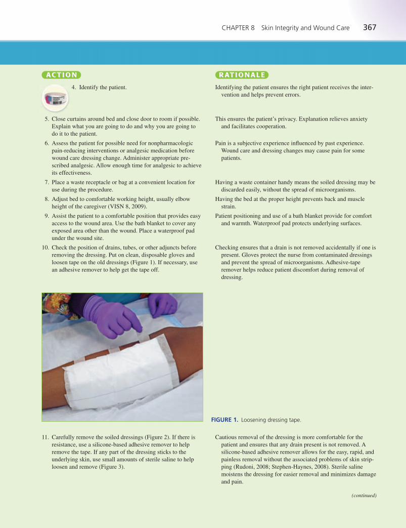
loosen tape on the old dressings (Figure 1). If necessary, use
an adhesive remover to help get the tape off.
Identifying the patient ensures the right patient receives the inter-
vention and helps prevent errors.
This ensures the patient’s privacy. Explanation relieves anxiety
and facilitates cooperation.
Pain is a subjective experience influenced by past experience.
Wound care and dressing changes may cause pain for some
patients.
Having a waste container handy means the soiled dressing may be
discarded easily, without the spread of microorganisms.
Having the bed at the proper height prevents back and muscle
strain.
Patient positioning and use of a bath blanket provide for comfort
and warmth. Waterproof pad protects underlying surfaces.
Checking ensures that a drain is not removed accidentally if one is
present. Gloves protect the nurse from contaminated dressings
and prevent the spread of microorganisms. Adhesive-tape
remover helps reduce patient discomfort during removal of
dressing.
FIGURE 1. Loosening dressing tape.
11. Carefully remove the soiled dressings (Figure 2). If there is
resistance, use a silicone-based adhesive remover to help
remove the tape. If any part of the dressing sticks to the
underlying skin, use small amounts of sterile saline to help
loosen and remove (Figure 3).
Cautious removal of the dressing is more comfortable for the
patient and ensures that any drain present is not removed. A
silicone-based adhesive remover allows for the easy, rapid, and
painless removal without the associated problems of skin strip-
ping (Rudoni, 2008; Stephen-Haynes, 2008). Sterile saline
moistens the dressing for easier removal and minimizes damage
and pain.
(continued)
LWBK545_C08_p358-435.qxd 08/07/2010 1:56 PM Page 367 Aptara
368 UNIT II Promoting Healthy Physiologic Responses
Cleaning a Wound and Applying a Dry, Sterile Dressing continued• 8-1
AA C T IC T I OO NN RR AA T IT I OO N A L EN A L E
12. After removing the dressing, note the presence, amount, type,
color, and odor of any drainage on the dressings (Figure 4).
Place soiled dressings in the appropriate waste receptacle.
Remove your gloves and dispose of them in an appropriate
waste receptacle (Figure 5).
The presence of drainage should be documented. Proper disposal
of soiled dressings and used gloves prevents spread of microor-
ganisms.
FIGURE 2. Removing dressing. FIGURE 3. Using saline to aid in removing dressing.
FIGURE 4. Assessing dressing that has been removed. FIGURE 5. Removing gloves.
13. Inspect the wound site for size, appearance, and drainage.
Assess if any pain is present. Check the status of sutures,
adhesive closure strips, staples, and drains or tubes, if present.
Note any problems to include in your documentation.
14. Using sterile technique, prepare a sterile work area andopen the needed supplies (Figure 6).
Wound healing or the presence of irritation or infection should be
documented.
Supplies are within easy reach and sterility is maintained.
LWBK545_C08_p358-435.qxd 08/07/2010 1:56 PM Page 368 Aptara
CHAPTER 8 Skin Integrity and Wound Care 369
AA C T IC T I OO NN RR AA T IT I OO N A L EN A L E
15. Open the sterile cleaning solution. Depending on the amount
of cleaning needed, the solution might be poured directly over
gauze sponges over a container for small cleaning jobs, or
into a basin for more complex or larger cleaning.
16. Put on sterile gloves (Figure 7).
Sterility of dressings and solution is maintained.
Use of sterile gloves maintains surgical asepsis and sterile tech-
nique and reduces the risk for spreading microorganisms.
17. Clean the wound. Clean the wound from top to bottom andfrom the center to the outside (Figure 8). Following this pat-tern, use new gauze for each wipe, placing the used gauze inthe waste receptacle. Alternately, spray the wound from topto bottom with a commercially prepared wound cleanser.
18. Once the wound is cleaned, dry the area using a gauze sponge
in the same manner. Apply ointment or perform other treat-
ments, as ordered (Figure 9).
Cleaning from top to bottom and center to outside ensures that
cleaning occurs from the least to most contaminated area and a
previously cleaned area is not contaminated again. Using a sin-
gle gauze for each wipe ensures that the previously cleaned area
is not contaminated again.
Moisture provides a medium for growth of microorganisms. The
growth of microorganisms may be inhibited and the healing
process improved with the use of ordered ointments or other
applications.
FIGURE 6. Setting up sterile field. FIGURE 7. Putting on sterile gloves.
FIGURE 8. Cleaning wound with dampened gauze. FIGURE 9. Applying antimicrobial ointment to wound with cotton applicator.
(continued)
LWBK545_C08_p358-435.qxd 08/07/2010 1:56 PM Page 369 Aptara
370 UNIT II Promoting Healthy Physiologic Responses
Cleaning a Wound and Applying a Dry, Sterile Dressing continued• 8-1
AA C T IC T I OO NN RR AA T IT I OO N A L EN A L E
19. If a drain is in use at the wound location, clean around the
drain. Refer to Skills 8-7, 8-8, 8-9, and 8-10.
20. Apply a layer of dry, sterile dressing over the wound (Figure 10).
Forceps may be used to apply the dressing.
21. Place a second layer of gauze over the wound site.
22. Apply a surgical or abdominal pad (ABD) over the gauze at
the site as the outermost layer of the dressing (Figure 11).
Cleaning the insertion site helps prevent infection.
Primary dressing serves as a wick for drainage. Use of forceps
helps ensure that sterile technique is maintained.
A second layer provides for increased absorption of drainage.
The dressing acts as additional protection for the wound against
microorganisms in the environment.
FIGURE 10. Applying dry dressing to site. FIGURE 11. Applying a surgical pad over dressing and securingwith tape.
23. Remove and discard gloves. Apply tape, Montgomery straps
or roller gauze to secure the dressings. Alternately, many
commercial wound products are self adhesive and do not
require additional tape.
24. After securing the dressing, label dressing with date and time.
Remove all remaining equipment; place the patient in a com-
fortable position, with side rails up and bed in the lowest
position.
25. Remove PPE, if used. Perform hand hygiene.
26. Check all wound dressings every shift. More frequent checks
may be needed if the wound is more complex or dressings
become saturated quickly.
Proper disposal of gloves prevents the spread of microorgan-
isms.Tape or other securing products are easier to apply after
gloves have been removed.
Recording date and time provides communication and demon-
strates adherence to plan of care. Proper patient and bed posi-
tioning promotes safety and comfort.
Removing PPE properly reduces the risk for infection transmis-
sion and contamination of other items. Hand hygiene prevents
the spread of microorganisms.
Checking dressings ensures the assessment of changes in patient
condition and timely intervention to prevent complications.
EVALUATION The expected outcome is met when the patient exhibits a clean, intact wound with a clean dress-
ing in place; the wound is free of contamination and trauma; the patient reports little to no pain
or discomfort during care; and the patient demonstrates signs and symptoms of progressive
wound healing.
LWBK545_C08_p358-435.qxd 08/07/2010 1:56 PM Page 370 Aptara
CHAPTER 8 Skin Integrity and Wound Care 371
DOCUMENTATIONGuidelines Document the location of the wound and that the dressing was removed. Record your assess-
ment of the wound including approximation of wound edges, presence of sutures, staples or
adhesive closure strips, and the condition of the surrounding skin. Note if redness, edema, or
drainage is observed. Document cleansing of the incision with normal saline and any applica-
tion of antibiotic ointment as ordered. Record the type of dressing that was reapplied. Note
pertinent patient and family education and any patient reaction to this procedure, including
patient’s pain level and effectiveness of nonpharmacologic interventions or analgesia if
administered.
9/8/12 0600 Dressing removed from left lateral calf incision. Scant purulent secretions notedon dressing. Incision edges approximately 1 mm apart, red, with ecchymosis and edemapresent. Small amount of purulent drainage from wound noted. Area cleansed with normalsaline, dried, antibiotic ointment applied per order. Surrounding tissue red and ecchymotic.Redressed with nonadhering dressing, gauze, and wrapped with stretch gauze. Patientreports adequate pain control after preprocedure analgesic; states pain is dull ache, 1/10 onpain scale.
—N. Joiner, RN
Sample Documentation
UNEXPECTEDSITUATIONS ANDASSOCIATEDINTERVENTIONS
• The previous wound assessment states that the incision was clean and dry and the wound edgeswere approximated, with the staples and surgical drain intact. The surrounding tissue was with-out inflammation, edema, or erythema. After the dressing is removed, the nurse notes the incisionedges are not approximated at the distal end, multiple staples are evident in the old dressing, thesurrounding skin tissue is red and swollen, and purulent drainage is on the dressing and leakingfrom the wound: Assess the patient for any other signs and symptoms, such as pain, malaise,
fever, and paresthesias. Place a dry sterile dressing over the wound site. Report the findings to
the physician and document the event in the patient’s record. Be prepared to obtain a wound
culture and implement any changes in wound care as ordered.
• After the nurse has put on sterile gloves, the patient moves too close to the edge of the bed andthe nurse must support her with his hands to prevent the patient from falling: If nothing else in
the sterile field was touched, remove the contaminated gloves and put on new sterile gloves. If
you did not bring a second pair, use the call bell to summon a coworker to provide a new pair
of gloves.
• The nurse has set up dressing supplies, removed the old dressing, and put on sterile gloves toclean the wound. The nurse then realizes that a necessary piece of dressing material has beenforgotten: Ask the patient to press the call bell to summon a coworker to provide the missing
supplies.
• Instruct the patient, if appropriate, and ancillary staff members to observe for excessive drainage
that may overwhelm the dressing. They should also report when dressings become soiled or loos-
ened from the skin.
• The skin of older adults is less elastic and more sensitive; use paper tape, Montgomery straps
(Refer to Skill 8-6), or roller gauze (on extremities) to prevent tearing of the skin.
SPECIALCONSIDERATIONSGeneral Considerations
Older Adult Considerations
LWBK545_C08_p358-435.qxd 08/07/2010 1:56 PM Page 371 Aptara
372 UNIT II Promoting Healthy Physiologic Responses
Applying a Saline-Moistened Dressing• 8-2
Gauze can be moistened with saline to keep the surface of open wounds moist. There are many
commercially prepared wound care products that are also available to maintain a moist wound envi-
ronment (see Fundamentals Review 8-4). This type of dressing promotes moist wound healing and
protects the wound from contamination and trauma. A moist wound surface enhances the cellular
migration necessary for tissue repair and healing. It is important that the dressing material be moist,
not wet, when placed in open wounds. Dressing materials are soaked in normal saline solution and
squeezed to remove excess saline so that the dressing is only slightly moist. The dressing can be
loosely packed in the wound bed if appropriate, and then covered with a secondary dressing to
absorb drainage.
Many commercially prepared dressing and wound care products are applied in a similar manner.
It is very important for the nurse to be aware of the products available in a particular facility and be
familiar with the indications for, and correct use of, each type of dressing and wound care product
(see Fundamentals Review 8-4).
• Clean disposable gloves
• Sterile gloves, if indicated
• Additional PPE, as indicated
• Sterile dressing set or suture set (for the sterile scissors and forceps)
• Sterile thin-mesh gauze dressing for packing, if ordered
• Sterile gauze dressings
• Surgical or abdominal pads
• Skin-protectant wipes
• Sterile basin
• Sterile cleaning solution as ordered (commonly 0.9% normal saline solution)
• Sterile saline
• Tape or ties
• Plastic bag or other appropriate waste container for soiled dressings
• Sterile cotton-tipped applicators
• Supplies for wound cleansing or irrigation, as necessary
• Waterproof pad and bath blanket
Assess the situation to determine the need for a dressing change. Confirm any medical orders rele-
vant to wound care and any wound care included in the nursing plan of care. Assess the patient’s
level of comfort and the need for analgesics before wound care. Assess if the patient experienced
any pain related to previous dressing changes and the effectiveness of interventions employed to
minimize the patient’s pain. Assess the current dressing to determine if it is intact. Assess for excess
drainage or bleeding or saturation of the dressing. Inspect the wound and the surrounding tissue.
Assess the location, appearance of the wound, wound stage (if appropriate), drainage, and types of
tissue present in the wound. Measure the wound. Note the stage of the healing process and charac-
teristics of any drainage. Also assess the surrounding skin for color, temperature, and edema, ecchy-
mosis, or maceration.
Determine the related factors for the nursing diagnoses based on the patient’s current status. An
appropriate nursing diagnosis is Impaired Skin Integrity. Other nursing diagnoses that may be
appropriate include:
• Anxiety • Disturbed Body Image
• Risk for Infection • Impaired Skin Integrity
• Chronic Pain • Acute Pain
• Deficient Knowledge • Impaired Tissue Integrity
The expected outcome to achieve when applying a saline-moistened dressing (or similar dressing)
is that the procedure is accomplished without contaminating the wound area, without causing
trauma to the wound, and without causing the patient to experience pain or discomfort. Other out-
comes that are appropriate include wound healing is promoted; the surrounding skin is without
signs of irritation, infection, and maceration; and the wound continues to show signs of progression
of healing.
EQUIPMENT
ASSESSMENT
NURSING DIAGNOSIS
OUTCOME IDENTIFICATION AND PLANNING
LWBK545_C08_p358-435.qxd 08/07/2010 1:57 PM Page 372 Aptara
CHAPTER 8 Skin Integrity and Wound Care 373
IMPLEMENTATION
AA C T IC T I OO NN RR AA T IT I OO N A L EN A L E
1. Review the medical orders for wound care or the nursing plan
of care related to wound care.
2. Gather the necessary supplies and bring to the bedside stand
or overbed table.
3. Perform hand hygiene and put on PPE, if
indicated.
4. Identify the patient.
5. Close curtains around bed and close door to room if possible.
Explain what you are going to do and why you are going to
do it to the patient.
6. Assess the patient for possible need for nonpharmacologic
pain-reducing interventions or analgesic medication before
wound care dressing change. Administer appropriate pre-
scribed analgesic. Allow enough time for analgesic to achieve
its effectiveness.
7. Place a waste receptacle or bag at a convenient location for
use during the procedure.
8. Adjust bed to comfortable working height, usually elbow
height of the caregiver (VISN 8, 2009).
9. Assist the patient to a comfortable position that provides easy
access to the wound area. Position the patient so the wound
cleanser or irrigation solution will flow from the clean end of
the wound toward the dirtier end, if being used (see Skill 8-1
for wound cleansing and Skill 8-4 for irrigation techniques).
Use the bath blanket to cover any exposed area other than the
wound. Place a waterproof pad under the wound site.
10. Put on clean gloves. Carefully and gently remove the soiled
dressings. If there is resistance, use a silicone-based adhesive
remover to help remove the tape. If any part of the dressing
sticks to the underlying skin, use small amounts of sterile
saline to help loosen and remove.
11. After removing the dressing, note the presence, amount, type,
color, and odor of any drainage on the dressings. Place soiled
dressings in the appropriate waste receptacle.
12. Assess the wound for appearance, stage, the presence of
eschar, granulation tissue, epithelialization, undermining,
tunneling, necrosis, sinus tract, and drainage. Assess the
appearance of the surrounding tissue. Measure the wound.
Refer to Fundamentals Review 8-3.
13. Remove your gloves and put them in the receptacle.
Reviewing the order and plan of care validates the correct patient
and correct procedure.
Preparation promotes efficient time management and organized
approach to the task. Bringing everything to the bedside con-
serves time and energy. Arranging items nearby is convenient,
saves time, and avoids unnecessary stretching and twisting of
muscles on the part of the nurse.
Hand hygiene and PPE prevent the spread of microorganisms.
PPE is required based on transmission precautions.
Identifying the patient ensures the right patient receives the inter-
vention and helps prevent errors.
This ensures the patient’s privacy. Explanation relieves anxiety
and facilitates cooperation.
Pain is a subjective experience influenced by past experience.
Wound care and dressing changes may cause pain for some
patients.
Having a waste container handy means the soiled dressing may be
discarded easily, without the spread of microorganisms.
Having the bed at the proper height prevents back and muscle
strain.
Patient positioning and use of a bath blanket provide for comfort
and warmth. Gravity directs the flow of liquid from the least
contaminated to the most contaminated area. Waterproof pad
protects underlying surfaces.
Gloves protect the nurse from handling contaminated dressings.
Cautious removal of the dressing is more comfortable for the
patient and ensures that any drain present is not removed. A
silicone-based adhesive remover allows for the easy, rapid, and
painless removal without the associated problems of skin strip-
ping (Rudoni, 2008; Stephen-Haynes, 2008). Sterile saline
moistens the dressing for easier removal and minimizes dam-
age and pain.
The presence of drainage should be documented. Discarding
dressings appropriately prevents the spread of microorganisms.
This information provides evidence about the wound healing
process and/or the presence of infection.
Discarding gloves prevents the spread of microorganisms.
(continued)
LWBK545_C08_p358-435.qxd 08/07/2010 1:57 PM Page 373 Aptara
374 UNIT II Promoting Healthy Physiologic Responses
Applying a Saline-Moistened Dressing continued• 8-2
AA C T IC T I OO NN RR AA T IT I OO N A L EN A L E
14. Using sterile technique, open the supplies and dressings. Place
the fine-mesh gauze into the basin and pour the ordered solu-
tion over the mesh to saturate it.
15. Put on the sterile gloves. Alternately, clean gloves (clean tech-
nique) may be used to clean a chronic wound.
16. Clean the wound. Refer to Skill 8-1. Alternately, irrigate the
wound, as ordered or required (see Skill 8-4).
17. Dry the surrounding skin with sterile gauze dressings.
18. Apply a skin protectant to the surrounding skin if needed.
19. If not already on, put on sterile gloves. Squeeze excess fluid
from the gauze dressing. Unfold and fluff the dressing.
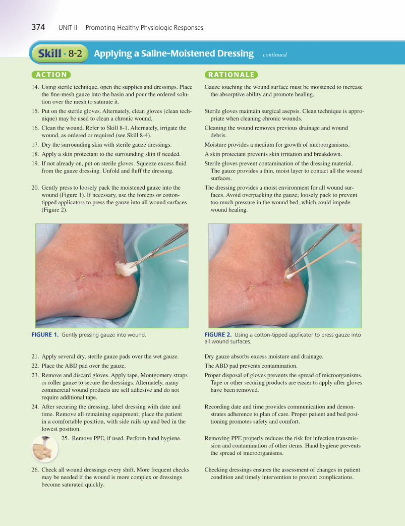
20. Gently press to loosely pack the moistened gauze into the
wound (Figure 1). If necessary, use the forceps or cotton-
tipped applicators to press the gauze into all wound surfaces
(Figure 2).
Gauze touching the wound surface must be moistened to increase
the absorptive ability and promote healing.
Sterile gloves maintain surgical asepsis. Clean technique is appro-
priate when cleaning chronic wounds.
Cleaning the wound removes previous drainage and wound
debris.
Moisture provides a medium for growth of microorganisms.
A skin protectant prevents skin irritation and breakdown.
Sterile gloves prevent contamination of the dressing material.
The gauze provides a thin, moist layer to contact all the wound
surfaces.
The dressing provides a moist environment for all wound sur-
faces. Avoid overpacking the gauze; loosely pack to prevent
too much pressure in the wound bed, which could impede
wound healing.
FIGURE 1. Gently pressing gauze into wound. FIGURE 2. Using a cotton-tipped applicator to press gauze intoall wound surfaces.
21. Apply several dry, sterile gauze pads over the wet gauze.
22. Place the ABD pad over the gauze.
23. Remove and discard gloves. Apply tape, Montgomery straps
or roller gauze to secure the dressings. Alternately, many
commercial wound products are self adhesive and do not
require additional tape.
24. After securing the dressing, label dressing with date and
time. Remove all remaining equipment; place the patient
in a comfortable position, with side rails up and bed in the
lowest position.
25. Remove PPE, if used. Perform hand hygiene.
26. Check all wound dressings every shift. More frequent checks
may be needed if the wound is more complex or dressings
become saturated quickly.
Dry gauze absorbs excess moisture and drainage.
The ABD pad prevents contamination.
Proper disposal of gloves prevents the spread of microorganisms.
Tape or other securing products are easier to apply after gloves
have been removed.
Recording date and time provides communication and demon-
strates adherence to plan of care. Proper patient and bed posi-
tioning promotes safety and comfort.
Removing PPE properly reduces the risk for infection transmis-
sion and contamination of other items. Hand hygiene prevents
the spread of microorganisms.
Checking dressings ensures the assessment of changes in patient
condition and timely intervention to prevent complications.
LWBK545_C08_p358-435.qxd 08/07/2010 1:57 PM Page 374 Aptara
CHAPTER 8 Skin Integrity and Wound Care 375
EVALUATION The expected outcome when applying a saline-moistened dressing is met when the procedure is
accomplished without contaminating the wound area, without causing trauma to the wound, and
without causing the patient to experience pain or discomfort. Other outcomes are met when sterile
technique is maintained (if appropriate); wound healing is promoted; the surrounding skin is with-
out signs of irritation, infection, and maceration; and the wound continues to show signs of progres-
sion of healing.
Document the location of the wound and that the dressing was removed. Record your assessment of
the wound, including evidence of granulation tissue, presence of necrotic tissue, stage (if appropriate),
and characteristics of drainage. Include the appearance of the surrounding skin. Document the cleans-
ing or irrigation of the wound and solution used. Record the type of dressing that was reapplied. Note
pertinent patient and family education and any patient reaction to this procedure, including patient’s
pain level and effectiveness of nonpharmacologic interventions or analgesia if administered.
DOCUMENTATIONGuidelines
11/20/11 1645 Healing abdominal incision with granulating tissue noted. Open area 2 cm �4 cm � 0.5 cm depth in center of incision. No evidence of necrosis or tunneling. Scant amountof serous drainage. Saline-moistened dressing applied to open wound; covered loosely withABD dressing. Patient denies pain from incision. Instructed patient that moist saline gauzewill facilitate the healing process and to notify nurse for any discomfort related to incision.
—R. Dobbins, RN
Sample Documentation
UNEXPECTEDSITUATIONS ANDRELATEDINTERVENTIONS
• When removing a patient’s dressing, the assessment reveals eschar in the wound: Notify the pri-
mary care provider or wound care specialist, as a different treatment modality and/or debride-
ment may be necessary. The presence of eschar in a wound precludes the staging of the wound.
The eschar must be removed for adequate pressure ulcer staging to be done. Stable (dry, adher-
ent, intact, without erythema or fluctuance) eschar on the heels serves as “the body’s natural
(biological) cover” and should not be removed (NPUAP, 2007a).
• The wound assessment reveals several depressions or crater-like areas on inspection of a wound:Notify the primary care provider or wound care specialist, who may order the wound to be
packed. Pack wound cavities loosely with dressing material. Overpacking may increase pressure
and interfere with tissue healing.
• The nurse notes that the wound dressing is dry upon removal: Reduce the time interval between
changes to prevent drying of the materials, which may disrupt healing tissue.
• Make sure ancillary staff understand the importance of reporting excessive drainage from the
dressing, and any soiled or loose dressings.
• Guidelines from the Wound, Ostomy, Continence Nurses Society (WOCN) and National Pres-
sure Ulcer Advisory Panel (NPUAP) recommend that clean gloves may be used to treat chronic
wounds and pressure ulcers as long as the infection-control procedures are followed. The no-touch technique may be used within these guidelines. Clean gloves are used to handle dressing
material. Irrigants and dressings are sterile. The wound is redressed by picking up dressing mate-
rials by the corner and placing the untouched side over the wound (NPUAP, 2007b; Wooten &
Hawkins, 2005).
• Many products are available to treat chronic wounds and pressure ulcers. Treatment varies based
on facility policy, nursing protocol, clinical specialist referrals, primary care provider orders, and
product in use.
Wooten, M., & Hawkins, K. (2005). WOCN position statement. Clean versus sterile: Management of chronicwounds. Available www.wocn.org/pdfs/WOCN_Library/Position_Statements/. Accessed January 14, 2009.
These guidelines are a collaborative effort of the Association for Professionals in Infection Con-
trol and Epidemiology (APIC) and the Wound, Ostomy, Continence Nurses Society (WOCN).
Approaches for chronic wound care management are presented, including the definitions of and
indications for ‘clean’ and ‘sterile’ technique. Cleansing of chronic wounds requires the use of
handwashing, clean (nonsterile) gloves, sterile cleansing solution, and irrigation with sterile device.
Routine dressing change without debridement requires the use of handwashing, clean (nonsterile)
gloves, sterile solutions, sterile dressing supplies, and sterile instruments.
SPECIALCONSIDERATIONS
EVIDENCE FOR PRACTICE
(continued)
LWBK545_C08_p358-435.qxd 08/07/2010 1:57 PM Page 375 Aptara
376 UNIT II Promoting Healthy Physiologic Responses
Applying a Saline-Moistened Dressing continued• 8-2
National Pressure Ulcer Advisory Panel (NPUAP). (2007b). Updated staging system. Wound infection andinfection control. Available www.npuap.org/pr2.htm. Accessed December 27, 2008.
The guidelines from the NPUAP state that clean, nonsterile dressings are acceptable for pressure
ulcer wound care. Pressure ulcers are nonsterile wounds; they are all contaminated with microor-
ganisms. There is no need to use sterile dressings on these wounds. Clean dressings should be
stored in their original packaging or other plastic wrap that protects them from moisture and dust.
Care providers should wash their hands before removing dressings from the package in order to not
contaminate the dressings by reaching into the package with soiled hands and/or gloves (NPUAP,
2007b, Question #309). Clean, nonsterile gloves can be used to treat multiple ulcers on the same
patient. If this is done, start with the cleaner appearing wounds and move to the larger and/or most
contaminated appearing wounds. When in doubt, change gloves between ulcers. Do not contami-
nate dressing supplies and wound care containers (e.g., solution bottles) with gloves that have been
in contact with the ulcer (NPUAP, 2007b, Question #310).
EVIDENCE FOR PRACTICE
Applying a Hydrocolloid Dressing• 8-3
Hydrocolloid dressings are wafer-shaped dressings that come in many shapes, sizes, and thick-
nesses. An adhesive backing provides adherence to the wound and surrounding skin. They absorb
drainage, maintain a moist wound surface, and decrease the risk for infection by covering the
wound surface (Refer to Fundamentals Review 8-4). Many commercially prepared dressing and
wound care products are applied in a similar manner. It is very important for the nurse to be aware
of the products available in a particular facility and be familiar with the indications for, and correct
use of, each type of dressing and wound care product.
• Hydrocolloid dressing
• Clean disposable gloves
• Sterile gloves, if indicated
• Additional PPE, as indicated
• Sterile dressing instrument set or suture set (for the scissors and forceps)
• Sterile cleaning solution as ordered (commonly 0.9% normal saline solution)
• Skin-protectant wipes
• Additional supplies needed for wound cleansing
• Sterile cotton-tipped applicators
• Waterproof pad
• Bath blanket
• Measuring tape or other supplies, such as sterile flexible applicator, for assessing wound meas-
urements, as indicated
Assess the situation to determine the need for a dressing change. Check the date when the current
dressing (if present) was placed. Confirm any medical orders relevant to wound care and any
wound care included in the nursing plan of care. Assess the current dressing to determine if it is
intact. Assess the patient’s level of comfort and the need for analgesics before wound care.
Assess if the patient experienced any pain related to prior dressing changes and the effectiveness
of interventions employed to minimize the patient’s pain. Assess the current dressing to determine
if it is intact. Assess for excess drainage or bleeding or saturation of the dressing. Inspect the wound
and the surrounding tissue. Assess the location, appearance of the wound, stage (if appropriate),
drainage, and types of tissue present in the wound. Measure the wound. Note the stage of the heal-
ing process and characteristics of any drainage. Also assess the surrounding skin for color, tempera-
ture, and edema, ecchymosis, or maceration.
EQUIPMENT
ASSESSMENT
LWBK545_C08_p358-435.qxd 08/07/2010 1:57 PM Page 376 Aptara
CHAPTER 8 Skin Integrity and Wound Care 377
Determine the related factors for the nursing diagnoses based on the patient’s current status. An
appropriate nursing diagnosis is Impaired Skin Integrity. Other nursing diagnoses that may be
appropriate include:
• Anxiety • Risk for Infection
• Disturbed Body Image • Chronic Pain
• Acute Pain • Impaired Tissue Integrity
The expected outcome to achieve when applying a hydrocolloid dressing is that the procedure is
accomplished without contaminating the wound area, without causing trauma to the wound, and
without causing the patient to experience pain or discomfort. Other outcomes that are appropriate
include sterile technique is maintained (if appropriate); wound healing is promoted; the surrounding
skin is without signs of irritation, infection, and maceration; and the wound continues to show signs
of progression of healing.
NURSING DIAGNOSIS
OUTCOME IDENTIFICATION AND PLANNING
IMPLEMENTATION
AA C T IC T I OO NN RR AA T IT I OO N A L EN A L E
1. Review the medical orders for wound care or the nursing plan
of care related to wound care.
2. Gather the necessary supplies and bring to the bedside stand
or overbed table.
3. Perform hand hygiene and put on PPE, if
indicated.
4. Identify the patient.
5. Close curtains around bed and close door to room if possible.
Explain what you are going to do and why you are going to
do it to the patient.
6. Assess the patient for possible need for nonpharmacologic
pain-reducing interventions or analgesic medication before
wound care dressing change. Administer appropriate pre-
scribed analgesic. Allow enough time for analgesic to achieve
its effectiveness before beginning procedure.
7. Place a waste receptacle or bag at a convenient location for
use during the procedure.
8. Adjust bed to comfortable working height, usually elbow
height of the caregiver (VISN 8, 2009).
9. Assist the patient to a comfortable position that provides
easy access to the wound area. Position the patient so the
wound cleanser or irrigation solution will flow from the
clean end of the wound toward the dirtier end, if being used
(See Skill 8-1 for wound cleansing and Skill 8-4 for irriga-
tion techniques). Use the bath blanket to cover any exposed
area other than the wound. Place a waterproof pad under the
wound site.
Reviewing the order and plan of care validates the correct patient
and correct procedure.
Preparation promotes efficient time management and organized
approach to the task. Bringing everything to the bedside con-
serves time and energy. Arranging items nearby is convenient,
saves time, and avoids unnecessary stretching and twisting of
muscles on the part of the nurse.
Hand hygiene and PPE prevent the spread of microorganisms.
PPE is required based on transmission precautions.
Identifying the patient ensures the right patient receives the inter-
vention and helps prevent errors.
This ensures the patient’s privacy. Explanation relieves anxiety
and facilitates cooperation.
Pain is a subjective experience influenced by past experience.
Wound care and dressing changes may cause pain for some
patients.
Having a waste container handy means the soiled dressing may be
discarded easily, without the spread of microorganisms.
Having the bed at the proper height prevents back and muscle
strain.
Patient positioning and use of a bath blanket provide for comfort
and warmth. Gravity directs the flow of liquid from the least
contaminated to the most contaminated area. Waterproof pad
protects underlying surfaces.
(continued)
LWBK545_C08_p358-435.qxd 08/07/2010 1:57 PM Page 377 Aptara
378 UNIT II Promoting Healthy Physiologic Responses
Applying a Hydrocolloid Dressing continued• 8-3
AA C T IC T I OO NN RR AA T IT I OO N A L EN A L E
10. Put on clean gloves. Carefully and gently remove the soiled
dressings. If there is resistance, use a silicone-based adhesive
remover to help remove the tape. If any part of the dressing
sticks to the underlying skin, use small amounts of sterile
saline to help loosen and remove.
11. After removing the dressing, note the presence, amount, type,
color, and odor of any drainage on the dressings. Place soiled
dressings in the appropriate waste receptacle.
12. Assess the wound for appearance, stage, the presence of
eschar, granulation tissue, epithelialization, undermining, tun-
neling, necrosis, sinus tract, and drainage. Assess the appear-
ance of the surrounding tissue. Measure the wound. Refer to
Fundamentals Review 8-3.
13. Remove your gloves and put them in the receptacle.
14. Set up a sterile field, if indicated, and wound cleaning sup-
plies. Put on sterile gloves. Alternately, clean gloves (clean
technique) may be used when cleaning a chronic wound.
15. Clean the wound. Refer to Skill 8-1. Alternately, irrigate the
wound, as ordered or required (see Skill 8-4).
16. Dry the surrounding skin with gauze dressings.
17. Apply a skin protectant to the surrounding skin.
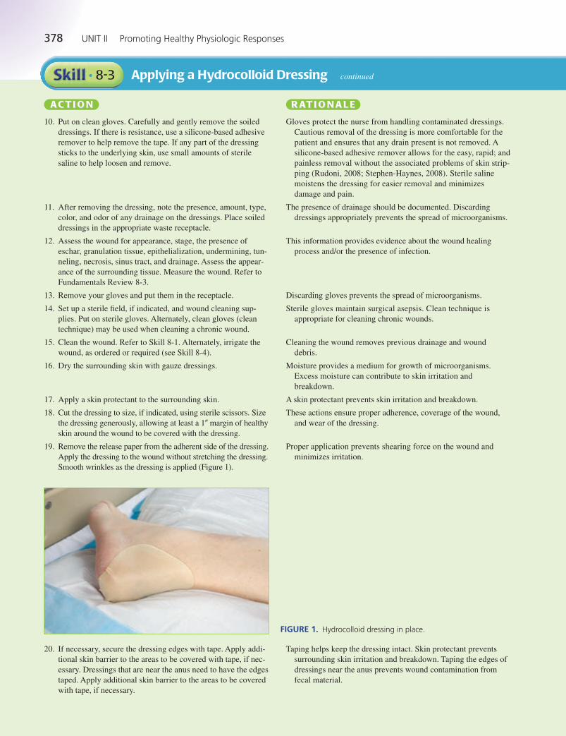
18. Cut the dressing to size, if indicated, using sterile scissors. Size
the dressing generously, allowing at least a 1� margin of healthy
skin around the wound to be covered with the dressing.
19. Remove the release paper from the adherent side of the dressing.
Apply the dressing to the wound without stretching the dressing.
Smooth wrinkles as the dressing is applied (Figure 1).
Gloves protect the nurse from handling contaminated dressings.
Cautious removal of the dressing is more comfortable for the
patient and ensures that any drain present is not removed. A
silicone-based adhesive remover allows for the easy, rapid; and
painless removal without the associated problems of skin strip-
ping (Rudoni, 2008; Stephen-Haynes, 2008). Sterile saline
moistens the dressing for easier removal and minimizes
damage and pain.
The presence of drainage should be documented. Discarding
dressings appropriately prevents the spread of microorganisms.
This information provides evidence about the wound healing
process and/or the presence of infection.
Discarding gloves prevents the spread of microorganisms.
Sterile gloves maintain surgical asepsis. Clean technique is
appropriate for cleaning chronic wounds.
Cleaning the wound removes previous drainage and wound
debris.
Moisture provides a medium for growth of microorganisms.
Excess moisture can contribute to skin irritation and
breakdown.
A skin protectant prevents skin irritation and breakdown.
These actions ensure proper adherence, coverage of the wound,
and wear of the dressing.
Proper application prevents shearing force on the wound and
minimizes irritation.
FIGURE 1. Hydrocolloid dressing in place.
20. If necessary, secure the dressing edges with tape. Apply addi-
tional skin barrier to the areas to be covered with tape, if nec-
essary. Dressings that are near the anus need to have the edges
taped. Apply additional skin barrier to the areas to be covered
with tape, if necessary.
Taping helps keep the dressing intact. Skin protectant prevents
surrounding skin irritation and breakdown. Taping the edges of
dressings near the anus prevents wound contamination from
fecal material.
LWBK545_C08_p358-435.qxd 08/07/2010 1:57 PM Page 378 Aptara
CHAPTER 8 Skin Integrity and Wound Care 379
AA C T IC T I OO NN RR AA T IT I OO N A L EN A L E
21. After securing the dressing, label dressing with date and
time. Remove all remaining equipment; place the patient in
a comfortable position, with side rails up and bed in the
lowest position.
22. Remove PPE, if used. Perform hand hygiene.
23. Check all wound dressings every shift. More frequent checks
may be needed if the wound is more complex or dressings
become saturated quickly.
Recording date and time provides communication and demon-
strates adherence to plan of care. Proper patient and bed posi-
tioning promotes safety and comfort.
Removing PPE properly reduces the risk for infection transmis-
sion and contamination of other items. Hand hygiene prevents
the spread of microorganisms.
Checking dressings ensures the assessment of changes in patient
condition and timely intervention to prevent complications.
EVALUATION The expected outcome when applying a hydrocolloid dressing is met when the procedure is accom-
plished without contaminating the wound area, without causing trauma to the wound, and without
causing the patient to experience pain or discomfort. Other outcomes are met when sterile tech-
nique is maintained (if appropriate); wound healing is promoted; surrounding skin is without signs
of irritation, infection, and maceration; and the wound continues to show signs of progression of
healing.
Document the location of the wound and that the dressing was removed. Record your assessment of
the wound, including evidence of granulation tissue, presence of necrotic tissue, stage (if appropri-
ate), and characteristics of drainage. Include the appearance of the surrounding skin. Document the
cleansing or irrigation of the wound and solution used. Record the type of hydrocolloid dressing
that was applied. Note pertinent patient and family education and any patient reaction to this proce-
dure, including patient’s pain level and effectiveness of nonpharmacologic interventions or analge-
sia if administered.
DOCUMENTATIONGuidelines
11/4/12 0930 Stage 3 wound on right hip area (3 � 2 � 2 cm) assessed. Granulation tissueabout 50%, no necrosis, undermining, or tunneling present. Minimal serous drainage onold dressing. Wound cleansed with normal saline. Hydrocolloid dressing applied. Due to bechanged in 5 days. Skin barrier applied to surrounding intact skin. Prior to dressing change,patient was medicated with Tylenol 650 mg PO for anticipated pain. Patient tolerated dress-ing change. Stated “pain not so bad,” about a “3.” Instructed patient to call for nurse forany discomfort related to dressing.
—M. Semet, RN
Sample Documentation
UNEXPECTED SITUATIONS AND RELATED INTERVENTION
• When removing a patient’s dressing, the assessment reveals eschar in the wound: Notify the pri-
mary care provider or wound care specialist, as a different treatment modality and/or debride-
ment may be necessary. The presence of eschar in a wound precludes the staging of the wound.
The eschar must be removed for adequate pressure ulcer staging to be done. Stable (dry, adher-
ent, intact, without erythema or fluctuance) eschar on the heels serves as “the body’s natural
(biological) cover” and should not be removed (NPUAP, 2007a).
• Guidelines from the Wound, Ostomy, Continence Nurses Society (WOCN) and National Pres-
sure Ulcer Advisory Panel (NPUAP) recommend that clean gloves may be used to treat chronic
wounds and pressure ulcers as long as the infection-control procedures are followed. The no-touch technique may be used within these guidelines. Clean gloves are used to handle dressing
material. Irrigants and dressings are sterile. The wound is redressed by picking up dressing mate-
rials by the corner and placing the untouched side over the wound (NPUAP, 2007b; Wooten &
Hawkins, 2005).
• Many products are available to treat chronic and pressure ulcers. Treatment varies based on facility
policy, nursing protocol, clinical specialist referrals, and physician orders.
SPECIAL CONSIDERATIONS
(continued)
LWBK545_C08_p358-435.qxd 08/07/2010 1:57 PM Page 379 Aptara
380 UNIT II Promoting Healthy Physiologic Responses
Applying a Hydrocolloid Dressing continued• 8-3
EVIDENCE FOR PRACTICE
Wooten, M., & Hawkins, K. (2005). WOCN position statement. Clean versus sterile: Management of
chronic wounds. Available www.wocn.org/pdfs/WOCN_Library/Position_Statements/. Accessed
January 14, 2009.
National Pressure Ulcer Advisory Panel (NPUAP). (2007b). Updated staging system. Wound infec-
tion and infection control. Available www.npuap.org/pr2.htm. Accessed December 27, 2008.
See Skill 8-2 for detailed information regarding these guidelines.
Performing Irrigation of a Wound• 8-4
Irrigation is a directed flow of solution over tissues. Wound irrigations are ordered to clean the area
of pathogens and other debris and to promote wound healing. Irrigation procedures may also be
ordered to apply heat or antiseptics locally. If the wound edges are approximated, clean technique
may be used; if the wound edges are unapproximated, sterile equipment and solutions are used for
irrigation. Normal saline is often the solution of choice when irrigating wounds.
• A sterile irrigation set, including a basin, irrigant container, and irrigation syringe
• Sterile irrigation solution as ordered by the physician, warmed to body temperature, commonly
0.9% normal saline solution
• Plastic bag or other waste container to dispose of soiled dressings
• Sterile gloves
• Sterile drape (may be optional)
• Clean disposable gloves
• Moisture-proof gown, mask, and eye protection
• Additional PPE, as indicated
• Sterile dressing set or suture set (for the sterile scissors and forceps)
• Waterproof pad and bath blanket as needed
• Sterile gauze dressings
• Sterile packing gauze as needed
• Tape or ties
• Skin-protectant wipes
Assess the situation to determine the need for wound irrigation. Confirm any medical orders rele-
vant to wound cape and any wound care included in the nursing plan of care. Assess the current
dressing to determine if it is intact. Assess the patient’s level of comfort and the need for analgesics
before wound care. Assess if the patient experienced any pain related to previous dressing changes
and the effectiveness of interventions employed to minimize the patient’s pain. Assess for excess
drainage or bleeding or saturation of the dressing. Inspect the wound and the surrounding tissue.
Assess the location, appearance of the wound, stage (if appropriate), drainage, and types of tissue
present in the wound. Measure the wound. Note the stage of the healing process and characteristics
of any drainage. Also assess the surrounding skin for color, temperature, and edema, ecchymosis, or
maceration.
Determine the related factors for the nursing diagnoses based on the patient’s current status. An
appropriate nursing diagnosis would be Risk for Infection. Other nursing diagnoses may include:
• Anxiety • Disturbed Body Image
• Acute Pain • Chronic Pain
• Deficient Knowledge • Impaired Skin Integrity
• Delayed Surgical Recovery • Impaired Tissue Integrity
• Risk for Trauma
EQUIPMENT
ASSESSMENT
NURSING DIAGNOSIS
LWBK545_C08_p358-435.qxd 08/07/2010 1:57 PM Page 380 Aptara
CHAPTER 8 Skin Integrity and Wound Care 381
The expected outcome to achieve when irrigating a wound is that the wound is cleaned without
contamination or trauma and without causing the patient to experience pain or discomfort. Other
outcomes that might be appropriate include: the wound continues to show signs of progression of
healing, and the patient demonstrates understanding about the need for wound irrigation.
OUTCOME IDENTIFICATION AND PLANNING
IMPLEMENTATION
AA C T IC T I OO NN RR AA T IT I OO N A L EN A L E
1. Review the medical orders for wound care or the nursing plan
of care related to wound care.
2. Gather the necessary supplies and bring to the bedside stand
or overbed table.
3. Perform hand hygiene and put on PPE, if
indicated.
4. Identify the patient.
5. Close curtains around bed and close door to room if possible.
Explain what you are going to do and why you are going to
do it to the patient.
6. Assess the patient for possible need for nonpharmacologic
pain-reducing interventions or analgesic medication before
wound care and/or dressing change. Administer appropriate
prescribed analgesic. Allow enough time for analgesic to
achieve its effectiveness before beginning procedure.
7. Place a waste receptacle or bag at a convenient location for
use during the procedure.
8. Adjust bed to comfortable working height, usually elbow
height of the caregiver (VISN 8, 2009).
9. Assist the patient to a comfortable position that provides easy
access to the wound area. Position the patient so the irrigation
solution will flow from the clean end of the wound toward the
dirtier end. Use the bath blanket to cover any exposed area
other than the wound. Place a waterproof pad under the
wound site.
10. Put on a gown, mask, and eye protection.
11. Put on clean gloves. Carefully and gently remove the soiled
dressings. If there is resistance, use a silicone-based adhesive
remover to help remove the tape. If any part of the dressing
sticks to the underlying skin, use small amounts of sterile
saline to help loosen and remove.
Reviewing the order and plan of care validates the correct patient
and correct procedure.
Preparation promotes efficient time management and organized
approach to the task. Bringing everything to the bedside con-
serves time and energy. Arranging items nearby is convenient,
saves time, and avoids unnecessary stretching and twisting of
muscles on the part of the nurse.
Hand hygiene and PPE prevent the spread of microorganisms.
PPE is required based on transmission precautions.
Identifying the patient ensures the right patient receives the inter-
vention and helps prevent errors.
This ensures the patient’s privacy. Explanation relieves anxiety
and facilitates cooperation.
Pain is a subjective experience influenced by past experience.
Wound care and dressing changes may cause pain for some
patients.
Having a waste container handy means the soiled dressing may be
discarded easily, without the spread of microorganisms.
Having the bed at the proper height prevents back and muscle
strain.
Patient positioning and use of a bath blanket provide for comfort
and warmth. Gravity directs the flow of liquid from the least
contaminated to the most contaminated area. Waterproof pad
protects underlying surfaces.
Using personal protective equipment such as gowns, masks, and
eye protection is part of Standard Precautions. A gown protects
clothes from contamination should splashing occur. Goggles
protect mucous membranes of eyes from contact with irrigant
fluid or wound drainage.
Gloves protect the nurse from handling contaminated dressings.
Cautious removal of the dressing is more comfortable for the
patient and ensures that any drain present is not removed. A
silicone-based adhesive remover allows for the easy, rapid, and
painless removal without the associated problems of skin strip-
ping (Rudoni, 2008; Stephen-Haynes, 2008). Sterile saline
moistens the dressing for easier removal and minimizes
damage and pain.
(continued)
LWBK545_C08_p358-435.qxd 08/07/2010 1:57 PM Page 381 Aptara
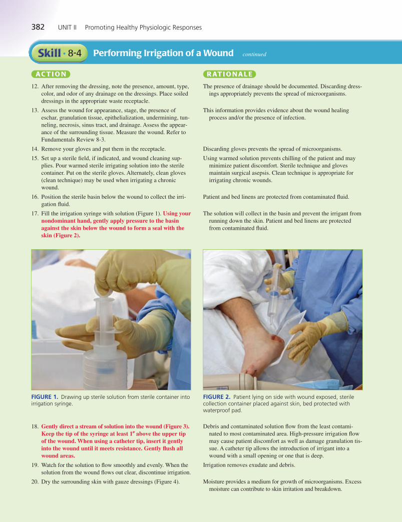
382 UNIT II Promoting Healthy Physiologic Responses
Performing Irrigation of a Wound continued• 8-4
AA C T IC T I OO NN RR AA T IT I OO N A L EN A L E
12. After removing the dressing, note the presence, amount, type,
color, and odor of any drainage on the dressings. Place soiled
dressings in the appropriate waste receptacle.
13. Assess the wound for appearance, stage, the presence of
eschar, granulation tissue, epithelialization, undermining, tun-
neling, necrosis, sinus tract, and drainage. Assess the appear-
ance of the surrounding tissue. Measure the wound. Refer to
Fundamentals Review 8-3.
14. Remove your gloves and put them in the receptacle.
15. Set up a sterile field, if indicated, and wound cleaning sup-
plies. Pour warmed sterile irrigating solution into the sterile
container. Put on the sterile gloves. Alternately, clean gloves
(clean technique) may be used when irrigating a chronic
wound.
16. Position the sterile basin below the wound to collect the irri-
gation fluid.
17. Fill the irrigation syringe with solution (Figure 1). Using yournondominant hand, gently apply pressure to the basinagainst the skin below the wound to form a seal with theskin (Figure 2).
The presence of drainage should be documented. Discarding dress-
ings appropriately prevents the spread of microorganisms.
This information provides evidence about the wound healing
process and/or the presence of infection.
Discarding gloves prevents the spread of microorganisms.
Using warmed solution prevents chilling of the patient and may
minimize patient discomfort. Sterile technique and gloves
maintain surgical asepsis. Clean technique is appropriate for
irrigating chronic wounds.
Patient and bed linens are protected from contaminated fluid.
The solution will collect in the basin and prevent the irrigant from
running down the skin. Patient and bed linens are protected
from contaminated fluid.
18. Gently direct a stream of solution into the wound (Figure 3).Keep the tip of the syringe at least 1� above the upper tipof the wound. When using a catheter tip, insert it gentlyinto the wound until it meets resistance. Gently flush allwound areas.
19. Watch for the solution to flow smoothly and evenly. When the
solution from the wound flows out clear, discontinue irrigation.
20. Dry the surrounding skin with gauze dressings (Figure 4).
Debris and contaminated solution flow from the least contami-
nated to most contaminated area. High-pressure irrigation flow
may cause patient discomfort as well as damage granulation tis-
sue. A catheter tip allows the introduction of irrigant into a
wound with a small opening or one that is deep.
Irrigation removes exudate and debris.
Moisture provides a medium for growth of microorganisms. Excess
moisture can contribute to skin irritation and breakdown.
FIGURE 1. Drawing up sterile solution from sterile container intoirrigation syringe.
FIGURE 2. Patient lying on side with wound exposed, sterilecollection container placed against skin, bed protected withwaterproof pad.
LWBK545_C08_p358-435.qxd 08/07/2010 1:57 PM Page 382 Aptara
CHAPTER 8 Skin Integrity and Wound Care 383
AA C T IC T I OO NN RR AA T IT I OO N A L EN A L E
21. Apply a skin protectant to the surrounding skin.
22. Apply a new dressing to the wound (see Skills 8-1, 8-2, 8-3)
(Figure 5).
A skin protectant prevents skin irritation and breakdown.
Dressings absorb drainage, protect the wound, and promote
healing.
FIGURE 3. Irrigating wound with a gentle stream of solution.Solution drains into collection container.
FIGURE 4. Drying around wound, not in wound, with sterilegauze pad.
FIGURE 5. Applying a new dressing.
23. Remove and discard gloves. Apply tape, Montgomery straps,
or roller gauze to secure the dressings. Alternately, many
commercial wound products are self adhesive and do not
require additional tape.
Tape or other securing products are easier to apply after gloves
have been removed. Proper disposal of gloves prevents the
spread of microorganisms.
(continued)
LWBK545_C08_p358-435.qxd 08/07/2010 1:57 PM Page 383 Aptara
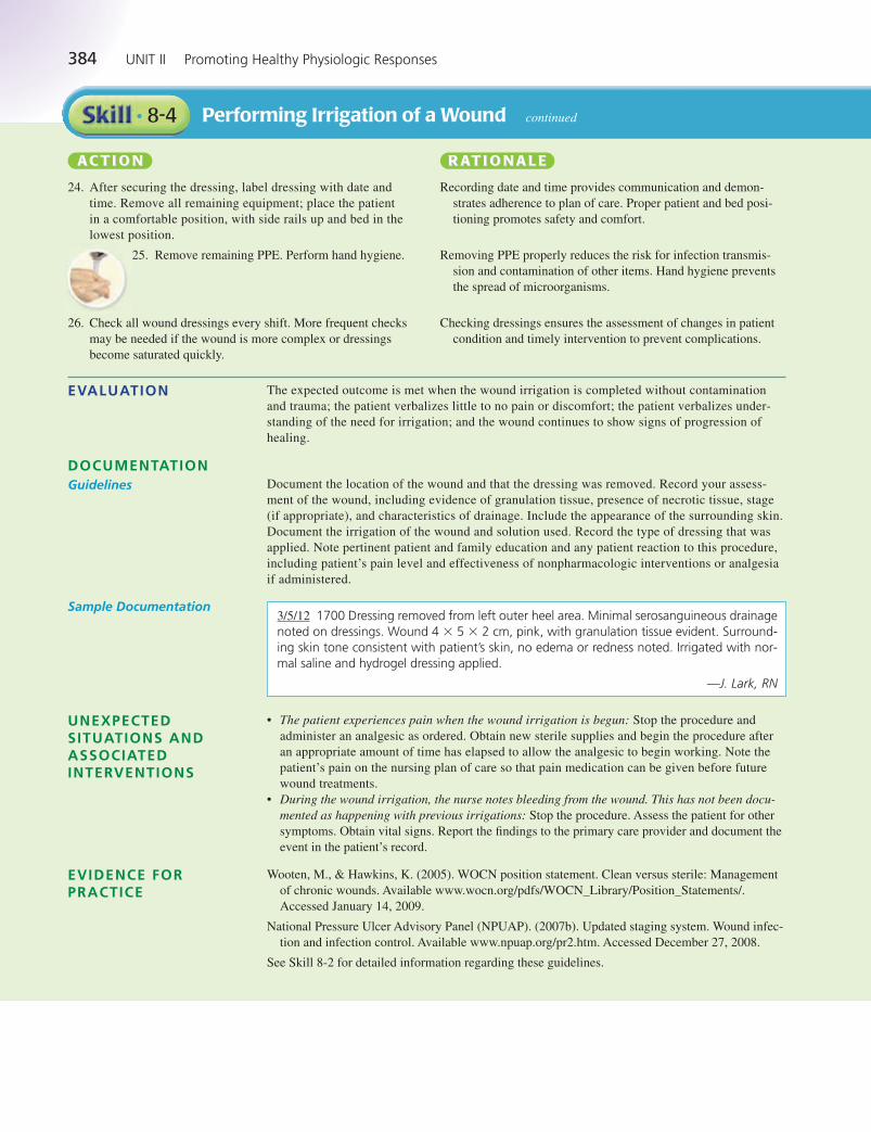
384 UNIT II Promoting Healthy Physiologic Responses
Performing Irrigation of a Wound continued• 8-4
AA C T IC T I OO NN RR AA T IT I OO N A L EN A L E
24. After securing the dressing, label dressing with date and
time. Remove all remaining equipment; place the patient
in a comfortable position, with side rails up and bed in the
lowest position.
25. Remove remaining PPE. Perform hand hygiene.
26. Check all wound dressings every shift. More frequent checks
may be needed if the wound is more complex or dressings
become saturated quickly.
Recording date and time provides communication and demon-
strates adherence to plan of care. Proper patient and bed posi-
tioning promotes safety and comfort.
Removing PPE properly reduces the risk for infection transmis-
sion and contamination of other items. Hand hygiene prevents
the spread of microorganisms.
Checking dressings ensures the assessment of changes in patient
condition and timely intervention to prevent complications.
EVALUATION The expected outcome is met when the wound irrigation is completed without contamination
and trauma; the patient verbalizes little to no pain or discomfort; the patient verbalizes under-
standing of the need for irrigation; and the wound continues to show signs of progression of
healing.
Document the location of the wound and that the dressing was removed. Record your assess-
ment of the wound, including evidence of granulation tissue, presence of necrotic tissue, stage
(if appropriate), and characteristics of drainage. Include the appearance of the surrounding skin.
Document the irrigation of the wound and solution used. Record the type of dressing that was
applied. Note pertinent patient and family education and any patient reaction to this procedure,
including patient’s pain level and effectiveness of nonpharmacologic interventions or analgesia
if administered.
DOCUMENTATIONGuidelines
3/5/12 1700 Dressing removed from left outer heel area. Minimal serosanguineous drainagenoted on dressings. Wound 4 � 5 � 2 cm, pink, with granulation tissue evident. Surround-ing skin tone consistent with patient’s skin, no edema or redness noted. Irrigated with nor-mal saline and hydrogel dressing applied.
—J. Lark, RN
Sample Documentation
UNEXPECTEDSITUATIONS ANDASSOCIATEDINTERVENTIONS
• The patient experiences pain when the wound irrigation is begun: Stop the procedure and
administer an analgesic as ordered. Obtain new sterile supplies and begin the procedure after
an appropriate amount of time has elapsed to allow the analgesic to begin working. Note the
patient’s pain on the nursing plan of care so that pain medication can be given before future
wound treatments.
• During the wound irrigation, the nurse notes bleeding from the wound. This has not been docu-mented as happening with previous irrigations: Stop the procedure. Assess the patient for other
symptoms. Obtain vital signs. Report the findings to the primary care provider and document the
event in the patient’s record.
Wooten, M., & Hawkins, K. (2005). WOCN position statement. Clean versus sterile: Management
of chronic wounds. Available www.wocn.org/pdfs/WOCN_Library/Position_Statements/.
Accessed January 14, 2009.
National Pressure Ulcer Advisory Panel (NPUAP). (2007b). Updated staging system. Wound infec-
tion and infection control. Available www.npuap.org/pr2.htm. Accessed December 27, 2008.
See Skill 8-2 for detailed information regarding these guidelines.
EVIDENCE FORPRACTICE
LWBK545_C08_p358-435.qxd 08/07/2010 1:57 PM Page 384 Aptara
CHAPTER 8 Skin Integrity and Wound Care 385
Collecting a Wound Culture• 8-5
A wound culture may be ordered to identify the causative organism of an infected wound. Identify-
ing the invading microorganism will provide useful information to select the most appropriate ther-
apy. A nurse or other primary health care provider can perform a wound culture. Maintaining strict
asepsis is crucial so that only the pathogen present in the wound is isolated. It is essential to use the
correct swab, based on the tests ordered, for collection of a specimen to isolate aerobic and/or
anaerobic organisms.
• A sterile Culturette kit (aerobic or anaerobic) with swab, or
a culture tube with individual sterile swabs
• Sterile gloves
• Clean disposable gloves
• Additional PPE, as indicated
• Plastic bag or appropriate waste receptacle
• Patient label for the sample tube
• Biohazard specimen bag
• Bath blanket (if necessary to drape the patient)
• Supplies to clean the wound and reapply a sterile dressing
after obtaining the culture (Refer to Skills 8-1 through 8-4)
Assess the situation to determine the need for wound culture. Confirm any medical orders relevant
to obtaining a wound culture, as well as wound care, and/or any wound care included in the nursing
plan of care. Assess the patient’s level of comfort and the need for analgesics before obtaining the
wound culture. Inspect the wound and the surrounding tissue. Assess the location, appearance of
the wound, stage (if appropriate), drainage, and types of tissue present in the wound. Measure the
wound. Note the stage of the healing process and characteristics of any drainage. Also assess the
surrounding skin for color, temperature, and edema, ecchymosis, or maceration.
Determine the related factors for the nursing diagnoses based on the patient’s current status. An
appropriate nursing diagnosis would be Risk for Infection. Other appropriate diagnoses may
include:
• Acute Pain • Disturbed Body Image
• Impaired Skin Integrity • Delayed Surgical Recovery
• Impaired Tissue Integrity
The expected outcome to achieve when collecting a wound culture is that the culture is obtained
without evidence of contamination, without exposing the patient to additional pathogens, and with-
out causing discomfort for the patient.
EQUIPMENT
ASSESSMENT
NURSING DIAGNOSIS
OUTCOME IDENTIFICATION AND PLANNING
IMPLEMENTATION
AA C T IC T I OO NN RR AA T IT I OO N A L EN A L E
1. Review the medical orders for obtaining a wound culture.
2. Gather the necessary supplies and bring to the bedside stand
or overbed table.
3. Perform hand hygiene and put on PPE, if
indicated.
4. Identify the patient.
Reviewing the order and plan of care validates the correct patient
and correct procedure.
Preparation promotes efficient time management and organized
approach to the task. Bringing everything to the bedside con-
serves time and energy. Arranging items nearby is convenient,
saves time, and avoids unnecessary stretching and twisting of
muscles on the part of the nurse.
Hand hygiene and PPE prevent the spread of microorganisms.
PPE is required based on transmission precautions.
Identifying the patient ensures the right patient receives the inter-
vention and helps prevent errors.
(continued)
LWBK545_C08_p358-435.qxd 08/07/2010 1:57 PM Page 385 Aptara
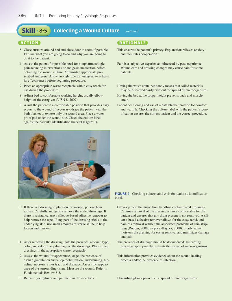
386 UNIT II Promoting Healthy Physiologic Responses
Collecting a Wound Culture continued• 8-5
AA C T IC T I OO NN RR AA T IT I OO N A L EN A L E
5. Close curtains around bed and close door to room if possible.
Explain what you are going to do and why you are going to
do it to the patient.
6. Assess the patient for possible need for nonpharmacologic
pain-reducing interventions or analgesic medication before
obtaining the wound culture. Administer appropriate pre-
scribed analgesic. Allow enough time for analgesic to achieve
its effectiveness before beginning procedure.
7. Place an appropriate waste receptacle within easy reach for
use during the procedure.
8. Adjust bed to comfortable working height, usually elbow
height of the caregiver (VISN 8, 2009).
9. Assist the patient to a comfortable position that provides easy
access to the wound. If necessary, drape the patient with the
bath blanket to expose only the wound area. Place a water-
proof pad under the wound site. Check the culture label
against the patient’s identification bracelet (Figure 1).
This ensures the patient’s privacy. Explanation relieves anxiety
and facilitates cooperation.
Pain is a subjective experience influenced by past experience.
Wound care and dressing changes may cause pain for some
patients.
Having the waste container handy means that soiled materials
may be discarded easily, without the spread of microorganisms.
Having the bed at the proper height prevents back and muscle
strain.
Patient positioning and use of a bath blanket provide for comfort
and warmth. Checking the culture label with the patient’s iden-
tification ensures the correct patient and the correct procedure.
FIGURE 1. Checking culture label with the patient’s identificationband.
10. If there is a dressing in place on the wound, put on clean
gloves. Carefully and gently remove the soiled dressings. If
there is resistance, use a silicone-based adhesive remover to
help remove the tape. If any part of the dressing sticks to the
underlying skin, use small amounts of sterile saline to help
loosen and remove.
11. After removing the dressing, note the presence, amount, type,
color, and odor of any drainage on the dressings. Place soiled
dressings in the appropriate waste receptacle.
12. Assess the wound for appearance, stage, the presence of
eschar, granulation tissue, epithelialization, undermining, tun-
neling, necrosis, sinus tract, and drainage. Assess the appear-
ance of the surrounding tissue. Measure the wound. Refer to
Fundamentals Review 8-3.
13. Remove your gloves and put them in the receptacle.
Gloves protect the nurse from handling contaminated dressings.
Cautious removal of the dressing is more comfortable for the
patient and ensures that any drain present is not removed. A sili-
cone-based adhesive remover allows for the easy, rapid, and
painless removal without the associated problems of skin strip-
ping (Rudoni, 2008; Stephen-Haynes, 2008). Sterile saline
moistens the dressing for easier removal and minimizes damage
and pain.
The presence of drainage should be documented. Discarding
dressings appropriately prevents the spread of microorganisms.
This information provides evidence about the wound healing
process and/or the presence of infection.
Discarding gloves prevents the spread of microorganisms.
LWBK545_C08_p358-435.qxd 08/07/2010 1:57 PM Page 386 Aptara
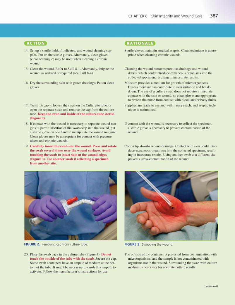
CHAPTER 8 Skin Integrity and Wound Care 387
AA C T IC T I OO NN RR AA T IT I OO N A L EN A L E
14. Set up a sterile field, if indicated, and wound cleaning sup-
plies. Put on the sterile gloves. Alternately, clean gloves
(clean technique) may be used when cleaning a chronic
wound.
15. Clean the wound. Refer to Skill 8-1. Alternately, irrigate the
wound, as ordered or required (see Skill 8-4).
16. Dry the surrounding skin with gauze dressings. Put on clean
gloves.
17. Twist the cap to loosen the swab on the Culturette tube, or
open the separate swab and remove the cap from the culture
tube. Keep the swab and inside of the culture tube sterile(Figure 2).
18. If contact with the wound is necessary to separate wound mar-
gins to permit insertion of the swab deep into the wound, put
a sterile glove on one hand to manipulate the wound margins.
Clean gloves may be appropriate for contact with pressure
ulcers and chronic wounds.
19. Carefully insert the swab into the wound. Press and rotatethe swab several times over the wound surfaces. Avoidtouching the swab to intact skin at the wound edges (Figure 3). Use another swab if collecting a specimen from another site.
Sterile gloves maintain surgical asepsis. Clean technique is appro-
priate when cleaning chronic wounds.
Cleaning the wound removes previous drainage and wound
debris, which could introduce extraneous organisms into the
collected specimen, resulting in inaccurate results.
Moisture provides a medium for growth of microorganisms.
Excess moisture can contribute to skin irritation and break-
down. The use of a culture swab does not require immediate
contact with the skin or wound, so clean gloves are appropriate
to protect the nurse from contact with blood and/or body fluids.
Supplies are ready to use and within easy reach, and aseptic tech-
nique is maintained.
If contact with the wound is necessary to collect the specimen,
a sterile glove is necessary to prevent contamination of the
wound.
Cotton tip absorbs wound drainage. Contact with skin could intro-
duce extraneous organisms into the collected specimen, result-
ing in inaccurate results. Using another swab at a different site
prevents cross-contamination of the wound.
FIGURE 2. Removing cap from culture tube. FIGURE 3. Swabbing the wound.
(continued)
20. Place the swab back in the culture tube (Figure 4). Do nottouch the outside of the tube with the swab. Secure the cap.
Some swab containers have an ampule of medium at the bot-
tom of the tube. It might be necessary to crush this ampule to
activate. Follow the manufacturer’s instructions for use.
The outside of the container is protected from contamination with
microorganisms, and the sample is not contaminated with
organisms not in the wound. Surrounding the swab with culture
medium is necessary for accurate culture results.
LWBK545_C08_p358-435.qxd 08/07/2010 1:57 PM Page 387 Aptara
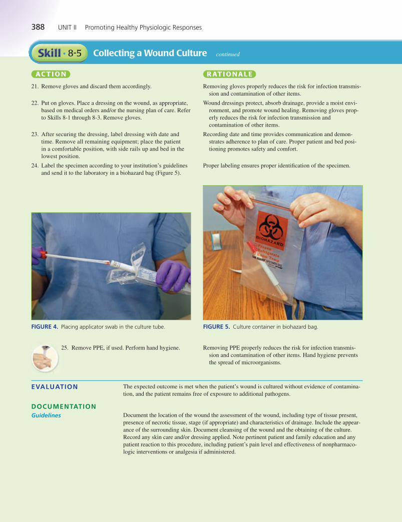
388 UNIT II Promoting Healthy Physiologic Responses
Collecting a Wound Culture continued• 8-5
AA C T IC T I OO NN RR AA T IT I OO N A L EN A L E
21. Remove gloves and discard them accordingly.
22. Put on gloves. Place a dressing on the wound, as appropriate,
based on medical orders and/or the nursing plan of care. Refer
to Skills 8-1 through 8-3. Remove gloves.
23. After securing the dressing, label dressing with date and
time. Remove all remaining equipment; place the patient
in a comfortable position, with side rails up and bed in the
lowest position.
24. Label the specimen according to your institution’s guidelines
and send it to the laboratory in a biohazard bag (Figure 5).
Removing gloves properly reduces the risk for infection transmis-
sion and contamination of other items.
Wound dressings protect, absorb drainage, provide a moist envi-
ronment, and promote wound healing. Removing gloves prop-
erly reduces the risk for infection transmission and
contamination of other items.
Recording date and time provides communication and demon-
strates adherence to plan of care. Proper patient and bed posi-
tioning promotes safety and comfort.
Proper labeling ensures proper identification of the specimen.
25. Remove PPE, if used. Perform hand hygiene. Removing PPE properly reduces the risk for infection transmis-
sion and contamination of other items. Hand hygiene prevents
the spread of microorganisms.
FIGURE 5. Culture container in biohazard bag.
EVALUATION The expected outcome is met when the patient’s wound is cultured without evidence of contamina-
tion, and the patient remains free of exposure to additional pathogens.
Document the location of the wound the assessment of the wound, including type of tissue present,
presence of necrotic tissue, stage (if appropriate) and characteristics of drainage. Include the appear-
ance of the surrounding skin. Document cleansing of the wound and the obtaining of the culture.
Record any skin care and/or dressing applied. Note pertinent patient and family education and any
patient reaction to this procedure, including patient’s pain level and effectiveness of nonpharmaco-
logic interventions or analgesia if administered.
DOCUMENTATIONGuidelines
FIGURE 4. Placing applicator swab in the culture tube.
LWBK545_C08_p358-435.qxd 08/07/2010 1:57 PM Page 388 Aptara
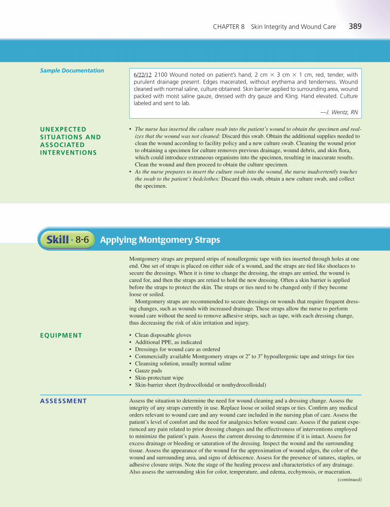
CHAPTER 8 Skin Integrity and Wound Care 389
6/22/12 2100 Wound noted on patient’s hand; 2 cm � 3 cm � 1 cm, red, tender, withpurulent drainage present. Edges macerated, without erythema and tenderness. Woundcleaned with normal saline, culture obtained. Skin barrier applied to surrounding area, woundpacked with moist saline gauze, dressed with dry gauze and Kling. Hand elevated. Culturelabeled and sent to lab.
—J. Wentz, RN
Sample Documentation
UNEXPECTEDSITUATIONS ANDASSOCIATEDINTERVENTIONS
• The nurse has inserted the culture swab into the patient’s wound to obtain the specimen and real-izes that the wound was not cleaned: Discard this swab. Obtain the additional supplies needed to
clean the wound according to facility policy and a new culture swab. Cleaning the wound prior
to obtaining a specimen for culture removes previous drainage, wound debris, and skin flora,
which could introduce extraneous organisms into the specimen, resulting in inaccurate results.
Clean the wound and then proceed to obtain the culture specimen.
• As the nurse prepares to insert the culture swab into the wound, the nurse inadvertently touchesthe swab to the patient’s bedclothes: Discard this swab, obtain a new culture swab, and collect
the specimen.
Applying Montgomery Straps• 8-6
Montgomery straps are prepared strips of nonallergenic tape with ties inserted through holes at one
end. One set of straps is placed on either side of a wound, and the straps are tied like shoelaces to
secure the dressings. When it is time to change the dressing, the straps are untied, the wound is
cared for, and then the straps are retied to hold the new dressing. Often a skin barrier is applied
before the straps to protect the skin. The straps or ties need to be changed only if they become
loose or soiled.
Montgomery straps are recommended to secure dressings on wounds that require frequent dress-
ing changes, such as wounds with increased drainage. These straps allow the nurse to perform
wound care without the need to remove adhesive strips, such as tape, with each dressing change,
thus decreasing the risk of skin irritation and injury.
• Clean disposable gloves
• Additional PPE, as indicated
• Dressings for wound care as ordered
• Commercially available Montgomery straps or 2� to 3� hypoallergenic tape and strings for ties
• Cleansing solution, usually normal saline
• Gauze pads
• Skin-protectant wipe
• Skin-barrier sheet (hydrocolloidal or nonhydrocolloidal)
Assess the situation to determine the need for wound cleaning and a dressing change. Assess the
integrity of any straps currently in use. Replace loose or soiled straps or ties. Confirm any medical
orders relevant to wound care and any wound care included in the nursing plan of care. Assess the
patient’s level of comfort and the need for analgesics before wound care. Assess if the patient expe-
rienced any pain related to prior dressing changes and the effectiveness of interventions employed
to minimize the patient’s pain. Assess the current dressing to determine if it is intact. Assess for
excess drainage or bleeding or saturation of the dressing. Inspect the wound and the surrounding
tissue. Assess the appearance of the wound for the approximation of wound edges, the color of the
wound and surrounding area, and signs of dehiscence. Assess for the presence of sutures, staples, or
adhesive closure strips. Note the stage of the healing process and characteristics of any drainage.
Also assess the surrounding skin for color, temperature, and edema, ecchymosis, or maceration.
EQUIPMENT
ASSESSMENT
(continued)
LWBK545_C08_p358-435.qxd 08/07/2010 1:57 PM Page 389 Aptara

390 UNIT II Promoting Healthy Physiologic Responses
Applying Montgomery Straps continued• 8-6
NURSING DIAGNOSIS Determine the related factors for the nursing diagnoses based on the patient’s current status. An
appropriate nursing diagnosis is Risk for Impaired Skin Integrity. Other nursing diagnoses that may
be appropriate include:
• Impaired Tissue Integrity • Risk for Infection
• Risk for Injury • Anxiety
• Acute Pain • Disturbed Body Image
• Deficient Knowledge • Impaired Skin Integrity
• Delayed Surgical Recovery
The expected outcome to achieve when applying Montgomery straps is that the patient’s skin is
free from irritation and injury. Other outcomes that may be appropriate include that the care is
accomplished without contaminating the wound area, without causing trauma to the wound, and
without causing the patient to experience pain or discomfort, and the wound continues to show
signs of progression of healing.
OUTCOME IDENTIFICATION AND PLANNING
IMPLEMENTATION
AA C T IC T I OO NN RR AA T IT I OO N A L EN A L E
1. Review the medical orders for wound care or the nursing plan
of care related to wound care.
2. Gather the necessary supplies and bring to the bedside stand
or overbed table.
3. Perform hand hygiene and put on PPE, if
indicated.
4. Identify the patient.
5. Close curtains around bed and close door to room if possible.
Explain what you are going to do and why you are going to
do it to the patient.
6. Assess the patient for possible need for nonpharmacologic
pain-reducing interventions or analgesic medication before
wound care dressing change. Administer appropriate pre-
scribed analgesic. Allow enough time for analgesic to achieve
its effectiveness before beginning procedure.
7. Place a waste receptacle at a convenient location for use dur-
ing the procedure.
8. Adjust bed to comfortable working height, usually elbow
height of the caregiver (VISN 8, 2009).
9. Assist the patient to a comfortable position that provides easy
access to the wound area. Use a bath blanket to cover any
exposed area other than the wound. Place a waterproof pad
under the wound site.
10. Perform wound care and a dressing change as outlined in
Skills 8-1 through 8-4, as ordered.
11. Put on clean gloves. Clean the skin on either side of the
wound with the gauze, moistened with normal saline.
Dry the skin.
Reviewing the order and plan of care validates the correct patient
and correct procedure.
Preparation promotes efficient time management and organized
approach to the task. Bringing everything to the bedside con-
serves time and energy. Arranging items nearby is convenient,
saves time, and avoids unnecessary stretching and twisting of
muscles on the part of the nurse.
Hand hygiene and PPE prevent the spread of microorganisms.
PPE is required based on transmission precautions.
Identifying the patient ensures the right patient receives the inter-
vention and helps prevent errors.
This ensures the patient’s privacy. Explanation relieves anxiety
and facilitates cooperation.
Pain is a subjective experience influenced by past experience.
Wound care and dressing changes may cause pain for some
patients.
Having a waste container handy means that the soiled dressing
may be discarded easily, without the spread of microorganisms.
Having the bed at the proper height prevents back and muscle
strain.
Patient positioning and use of a bath blanket provide for comfort
and warmth. Waterproof pad protects underlying surfaces.
Wound care aids in healing and provides protection for the wound.
Gloves prevent the spread of microorganisms. Cleaning and dry-
ing the skin prevents irritation and injury.
LWBK545_C08_p358-435.qxd 08/07/2010 1:57 PM Page 390 Aptara
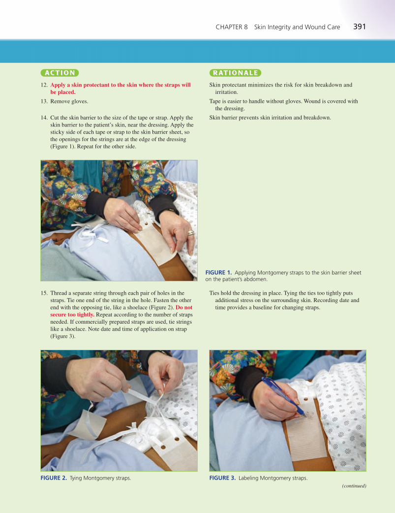
CHAPTER 8 Skin Integrity and Wound Care 391
AA C T IC T I OO NN RR AA T IT I OO N A L EN A L E
12. Apply a skin protectant to the skin where the straps willbe placed.
13. Remove gloves.
14. Cut the skin barrier to the size of the tape or strap. Apply the
skin barrier to the patient’s skin, near the dressing. Apply the
sticky side of each tape or strap to the skin barrier sheet, so
the openings for the strings are at the edge of the dressing
(Figure 1). Repeat for the other side.
Skin protectant minimizes the risk for skin breakdown and
irritation.
Tape is easier to handle without gloves. Wound is covered with
the dressing.
Skin barrier prevents skin irritation and breakdown.
FIGURE 1. Applying Montgomery straps to the skin barrier sheeton the patient’s abdomen.
FIGURE 2. Tying Montgomery straps. FIGURE 3. Labeling Montgomery straps.
15. Thread a separate string through each pair of holes in the
straps. Tie one end of the string in the hole. Fasten the other
end with the opposing tie, like a shoelace (Figure 2). Do notsecure too tightly. Repeat according to the number of straps
needed. If commercially prepared straps are used, tie strings
like a shoelace. Note date and time of application on strap
(Figure 3).
Ties hold the dressing in place. Tying the ties too tightly puts
additional stress on the surrounding skin. Recording date and
time provides a baseline for changing straps.
(continued)
LWBK545_C08_p358-435.qxd 08/07/2010 1:57 PM Page 391 Aptara
392 UNIT II Promoting Healthy Physiologic Responses
Applying Montgomery Straps continued• 8-6
AA C T IC T I OO NN RR AA T IT I OO N A L EN A L E
16. After securing the dressing, label dressing with date and
time. Remove all remaining equipment; place the patient
in a comfortable position, with side rails up and bed in the
lowest position.
17. Remove additional PPE, if used. Perform hand
hygiene.
18. Check all wound dressings every shift. More frequent checks
may be needed if the wound is more complex or dressings
become saturated quickly.
19. Replace the ties and straps whenever they are soiled, or every
2 to 3 days. Straps can be reapplied onto skin barrier. Skin
barrier can remain in place up to 7 days. Use a silicone-based
adhesive remover to help remove the skin barrier.
Recording date and time provides communication and demon-
strates adherence to plan of care. Proper patient and bed posi-
tioning promotes safety and comfort.
Removing PPE properly reduces the risk for infection transmis-
sion and contamination of other items. Hand hygiene prevents
the spread of microorganisms.
Checking dressings ensures the assessment of changes in patient
condition and timely intervention to prevent complications.
Replacing soiled ties and straps prevents growth of pathogens.
Minimizing removal of skin barrier prevents skin irritation
and breakdown. A silicone-based adhesive remover allows
for the easy, rapid, and painless removal without the associ-
ated problems of skin stripping (Rudoni, 2008; Stephen-
Haynes, 2008).
EVALUATION The expected outcome when applying Montgomery straps is met when the patient’s skin is clean,
dry, intact, and free from irritation and injury. Other outcomes are met when the patient exhibits a
clean wound area free of contamination and trauma. In addition, the patient verbalizes minimal
to no pain or discomfort, and the patient exhibits signs and symptoms indicative of progressive
wound healing.
Document the procedure, the patient’s response, and your assessment of the area before and after
application. Record a description of the wound, amount and character of the wound drainage, and
an assessment of the surrounding skin. Note the type of dressing that was applied, including the
application of skin protectant and a skin barrier. Document that Montgomery straps were applied
to secure the dressings. Record the patient’s response to the dressing care and associated pain
assessment. Include any pertinent patient and family education.
DOCUMENTATIONGuidelines
10/20/12 1930 Patient’s abdominal wound has large amounts of serosanguineous drainage,saturating multiple layers of gauze and ABDs, requiring dressing changes at least q 3 hours.Surrounding skin cleansed, skin protectant applied, and Montgomery straps applied tosecure wound dressings.
—D. Rightner, RN
Sample Documentation
UNEXPECTEDSITUATION ANDASSOCIATEDINTERVENTION
• A patient has had an abdominal wound for several weeks. Despite careful wound and skin care,the nurse observes signs of redness and irritation where the tape for the dressings has beenrepeatedly placed: Obtain the supplies listed in this skill. Apply Montgomery straps, being sure
to move the skin barrier sheet at least 1� away from the area of irritation.
LWBK545_C08_p358-435.qxd 08/07/2010 1:57 PM Page 392 Aptara
CHAPTER 8 Skin Integrity and Wound Care 393
Caring for a Penrose Drain• 8-7
Drains are inserted into or near a wound when it is anticipated that a collection of fluid in a
closed area would delay healing. A Penrose drain is a hollow, open-ended rubber tube. It allows
fluid to drain via capillary action into absorbent dressings. Penrose drains are commonly used
after a surgical procedure or for drainage of an abscess. After a surgical procedure, the surgeon
places one end of the drain in or near the area to be drained. The other end passes through the
skin, directly through the incision or through a separate opening referred to as a stab wound. A
Penrose drain is not sutured. A large safety pin is usually placed in the part outside the wound to
prevent the drain from slipping back into the incised area. This type of drain can be advanced or
shortened to drain different areas. The patency and placement of the drain are included in the
wound assessment.
• Sterile gloves
• Gauze dressings
• Sterile cotton-tipped applicators, if appropriate
• Sterile drain sponges
• Surgical or abdominal pads
• Sterile dressing set or suture set (for the sterile scissors and forceps)
• Sterile cleaning solution as ordered (commonly 0.9% normal saline solution)
• Sterile container to hold cleaning solution
• Clean safety pin
• Clean disposable gloves
• Plastic bag or other appropriate waste container for soiled dressings
• Waterproof pad and bath blanket
• Tape or ties
• Skin-protectant wipes if needed
• Additional dressings and supplies needed or as required for ordered wound care
Assess the situation to determine the necessity for wound cleaning and a dressing change. Confirm
any medical orders relevant to drain care and any drain care included in the nursing plan of care.
Assess the patient’s level of comfort and the need for analgesics before wound care. Assess if the
patient experienced any pain related to prior dressing changes and the effectiveness of interventions
employed to minimize the patient’s pain. Assess the current dressing to determine if it is intact, and
assess for the presence of excess drainage, bleeding, or saturation of the dressing. Assess the
patency of the Penrose drain.
Inspect the wound and the surrounding tissue. Assess the appearance of the wound for the
approximation of wound edges, the color of the wound and surrounding area, and signs of dehis-
cence. Note the stage of the healing process and the characteristics of any drainage. Assess the sur-
rounding skin for color, temperature, and the presence of edema, ecchymosis, or maceration.
Determine the related factors for the nursing diagnosis based on the patient’s current status. An
appropriate nursing diagnosis is Risk for Infection. Other nursing diagnoses may also be appropri-
ate, including:
• Anxiety • Acute Pain
• Disturbed Body Image • Impaired Skin Integrity
• Deficient Knowledge • Delayed Surgical Recovery
• Impaired Tissue Integrity
The expected outcome to achieve when performing care for a Penrose drain is that the Penrose
drain remains patent and intact; the care is accomplished without contaminating the wound area,
or causing trauma to the wound, and without causing the patient to experience pain or discom-
fort. Other outcomes that are appropriate may include: the wound shows signs of progressive
healing without evidence of complications, and the patient demonstrates understanding about
drain care.
EQUIPMENT
ASSESSMENT
NURSING DIAGNOSIS
OUTCOME IDENTIFICATION AND PLANNING
(continued)
LWBK545_C08_p358-435.qxd 08/07/2010 1:57 PM Page 393 Aptara
394 UNIT II Promoting Healthy Physiologic Responses
Caring for a Penrose Drain continued• 8-7
IMPLEMENTATION
AA C T IC T I OO NN RR AA T IT I OO N A L EN A L E
1. Review the medical orders for wound care or the nursing plan
of care related to wound/drain care.
2. Gather the necessary supplies and bring to the bedside stand
or overbed table.
3. Perform hand hygiene and put on PPE, if
indicated.
4. Identify the patient.
5. Close curtains around bed and close door to room if possible.
Explain what you are going to do and why you are going to
do it to the patient.
6. Assess the patient for possible need for nonpharmacologic
pain-reducing interventions or analgesic medication before
wound care dressing change. Administer appropriate pre-
scribed analgesic. Allow enough time for analgesic to achieve
its effectiveness before beginning procedure.
7. Place a waste receptacle at a convenient location for use
during the procedure.
8. Adjust bed to comfortable working height, usually elbow
height of the caregiver (VISN 8, 2009).
9. Assist the patient to a comfortable position that provides easy
access to the drain and/or wound area. Use a bath blanket to
cover any exposed area other than the wound. Place a water-
proof pad under the wound site.
10. Put on clean gloves. Check the position of the drain or drains
before removing the dressing. Carefully and gently remove
the soiled dressings. If there is resistance, use a silicone-based
adhesive remover to help remove the tape. If any part of the
dressing sticks to the underlying skin, use small amounts of
sterile saline to help loosen and remove.
11. After removing the dressing, note the presence, amount, type,
color, and odor of any drainage on the dressings. Place soiled
dressings in the appropriate waste receptacle.
12. Inspect the drain site for appearance and drainage. Assess if
any pain is present.
13. Using sterile technique, prepare a sterile work area and open
the needed supplies.
14. Open the sterile cleaning solution. Pour the cleansing solution
into the basin. Add the gauze sponges.
15. Put on sterile gloves.
Reviewing the order and plan of care validates the correct patient
and correct procedure.
Preparation promotes efficient time management and organized
approach to the task. Bringing everything to the bedside con-
serves time and energy. Arranging items nearby is convenient,
saves time, and avoids unnecessary stretching and twisting of
muscles on the part of the nurse.
Hand hygiene and PPE prevent the spread of microorganisms.
PPE is required based on transmission precautions.
Identifying the patient ensures the right patient receives the inter-
vention and helps prevent errors.
This ensures the patient’s privacy. Explanation relieves anxiety
and facilitates cooperation.
Pain is a subjective experience influenced by past experience.
Wound care and dressing changes may cause pain for some
patients.
Having a waste container handy means that the soiled dressing
may be discarded easily, without the spread of microorganisms.
Having the bed at the proper height prevents back and muscle
strain.
Patient positioning and use of a bath blanket provide for comfort
and warmth. Waterproof pad protects underlying surfaces.
Gloves protect the nurse from handling contaminated dressings.
Checking the position ensures that a drain is not removed acci-
dentally if one is present. Cautious removal of the dressing is
more comfortable for the patient and ensures that any drain
present is not removed. A silicone-based adhesive remover
allows for the easy, rapid, and painless removal without the
associated problems of skin stripping (Rudoni, 2008; Stephen-
Haynes, 2008). Sterile saline moistens the dressing for easier
removal and minimizes damage and pain.
The presence of drainage should be documented. Discarding
dressings appropriately prevents the spread of microorganisms.
The wound healing process and/or the presence of irritation or
infection must be documented.
Supplies are within easy reach and sterility is maintained.
Sterility of dressings and solution is maintained.
Sterile gloves help to maintain surgical asepsis and sterile tech-
nique and prevent the spread of microorganisms.
LWBK545_C08_p358-435.qxd 08/07/2010 1:57 PM Page 394 Aptara
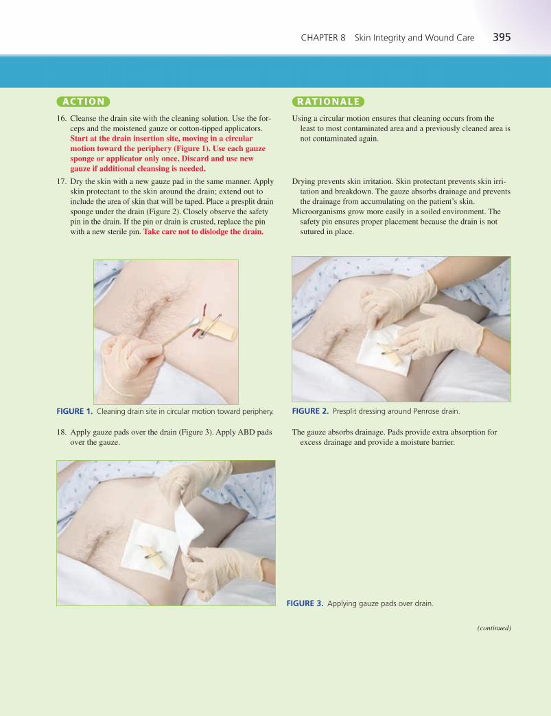
CHAPTER 8 Skin Integrity and Wound Care 395
16. Cleanse the drain site with the cleaning solution. Use the for-
ceps and the moistened gauze or cotton-tipped applicators.
Start at the drain insertion site, moving in a circularmotion toward the periphery (Figure 1). Use each gauzesponge or applicator only once. Discard and use newgauze if additional cleansing is needed.
17. Dry the skin with a new gauze pad in the same manner. Apply
skin protectant to the skin around the drain; extend out to
include the area of skin that will be taped. Place a presplit drain
sponge under the drain (Figure 2). Closely observe the safety
pin in the drain. If the pin or drain is crusted, replace the pin
with a new sterile pin. Take care not to dislodge the drain.
Using a circular motion ensures that cleaning occurs from the
least to most contaminated area and a previously cleaned area is
not contaminated again.
Drying prevents skin irritation. Skin protectant prevents skin irri-
tation and breakdown. The gauze absorbs drainage and prevents
the drainage from accumulating on the patient’s skin.
Microorganisms grow more easily in a soiled environment. The
safety pin ensures proper placement because the drain is not
sutured in place.
AA C T IC T I OO NN RR AA T IT I OO N A L EN A L E
FIGURE 2. Presplit dressing around Penrose drain.FIGURE 1. Cleaning drain site in circular motion toward periphery.
The gauze absorbs drainage. Pads provide extra absorption for
excess drainage and provide a moisture barrier.
FIGURE 3. Applying gauze pads over drain.
18. Apply gauze pads over the drain (Figure 3). Apply ABD pads
over the gauze.
(continued)
LWBK545_C08_p358-435.qxd 08/07/2010 1:57 PM Page 395 Aptara
396 UNIT II Promoting Healthy Physiologic Responses
Caring for a Penrose Drain continued• 8-7
19. Remove and discard gloves. Apply tape, Montgomery straps,
or roller gauze to secure the dressings.
20. After securing the dressing, label dressing with date and time.
Remove all remaining equipment; place the patient in a comfort-
able position, with side rails up and bed in the lowest position.
21. Remove additional PPE, if used. Perform hand
hygiene.
22. Check all wound dressings every shift. More frequent checks
may be needed if the wound is more complex or dressings
become saturated quickly.
Proper disposal of gloves prevents the spread of microorganisms.
Tape or other securing products are easier to apply after gloves
have been removed.
Recording date and time provides communication and demon-
strates adherence to plan of care. Proper patient and bed posi-
tioning promotes safety and comfort.
Removing PPE properly reduces the risk for infection transmis-
sion and contamination of other items. Hand hygiene prevents
the spread of microorganisms.
Checking dressings ensures the assessment of changes in patient
condition and timely intervention to prevent complications.
AA C T IC T I OO NN RR AA T IT I OO N A L EN A L E
EVALUATION The expected outcome is met when the patient exhibits a wound that is clean, dry, and intact, with a
patent, intact Penrose drain. Other outcomes that are appropriate may include: the patient remains
free of wound contamination and trauma; the patient reports minimal to no pain or discomfort; the
patient exhibits signs and symptoms of progressive wound healing; and the patient verbalizes an
understanding of the rationale for and/or the technique for drain care.
Document the location of the wound and drain, the assessment of the wound and drain site, and
intactness of the Penrose drain. Document the presence of drainage and characteristics on the old
dressing upon removal. Include the appearance of the surrounding skin. Document cleansing of the
drain site. Record any skin care and the dressing applied. Note pertinent patient and family educa-
tion and any patient reaction to this procedure, including patient’s pain level and effectiveness of
nonpharmacologic interventions or analgesia if administered.
DOCUMENTATIONGuidelines
UNEXPECTEDSITUATIONS ANDASSOCIATEDINTERVENTIONS
SPECIALCONSIDERATIONS
Sample Documentation3/13/12 1400 Patient medicated with morphine 3 mg IV as ordered prior to dressingchange. Dressing to right forearm removed. Dressings noted with small amount of serosan-guineous drainage. Forearm with gross edema and erythema. Penrose drain intact, withsafety pin in place. Incision edges approximated, staples intact. Area irrigated with normalsaline, dried, and redressed with gauze, ABD pads, and stretch gauze. Reinforced theimportance of keeping arm elevated on pillows, with patient verbalizing understanding.
—P. Towns, RN
• Assessment of the drain site reveals significantly increased edema, erythema, and drainage fromthe site, in addition to drainage via the drain: Cleanse the site as ordered or per the nursing plan
of care. Obtain vital signs, including the patient’s temperature. Document care and assessments.
Notify the primary care provider of the findings.
• Assessment of the drain site reveals that the drain has slipped back into the incision: Follow
facility policy and the medical orders related to advancing Penrose drains. Document assess-
ments and interventions. Notify the primary care provider of the findings and interventions.
• When preparing to change a dressing on a Penrose drain site, the nurse’s assessment reveals thatthe drain is completely out, lying in the dressing material: Assess the site and the patient for
symptoms of pain, increased edema/erythema/drainage. Provide site care as ordered. Notify the
primary care provider. Often, depending on the patient’s stage of recovery, the drain is left out.
Document the findings and interventions.
• Evaluate a sudden increase in the amount of drainage or bright red drainage and notify the
primary care provider of these findings.
• Wound care is often uncomfortable, and patients may experience significant pain. Assess the
patient’s comfort level and past experiences with wound care. Offer analgesics as ordered to
maintain the patient’s level of comfort.
LWBK545_C08_p358-435.qxd 08/07/2010 1:57 PM Page 396 Aptara
CHAPTER 8 Skin Integrity and Wound Care 397
Caring for a T-Tube Drain• 8-8
A biliary drain or T-tube (Figure 1) is sometimes placed in the common bile duct after removal of
the gallbladder (cholecystectomy) or a portion of the bile duct (choledochostomy). The tube drains
bile while the surgical site is healing. A portion of the tube is inserted into the common bile duct
and the remaining portion is anchored to the abdominal wall, passed through the skin, and con-
nected to a closed drainage system. Often, a three-way valve is inserted between the drain tube and
the drainage system to allow for clamping and flushing of the tube if necessary. The drainage
amount is measured every shift, recorded, and included in output totals.
(continued)
EQUIPMENT
ASSESSMENT
FIGURE 1. T-tube.
• Sterile gloves
• Clean disposable gloves
• Additional PPE, as indicated
• Sterile gauze pads
• Sterile drain sponges
• Cleansing solution, usually sterile normal saline
• Sterile cotton-tipped applicators (if appropriate)
• Transparent dressing
• Graduated collection container
• Waste receptacle
• Sterile basin
• Sterile forceps
• Tape
• Skin-protectant wipes
• Waterproof pad and bath blanket, if needed
Assess the situation to determine the need for wound cleaning, a dressing change, or emptying of
the drain. Confirm any medical orders relevant to drain care and any drain care included in the
nursing plan of care. Assess the patient’s level of comfort and the need for analgesics before wound
care. Assess if the patient experienced any pain related to prior dressing changes and the effective-
ness of interventions employed to minimize the patient’s pain. Assess the current dressing to deter-
mine if it is intact, and assess for evidence of excessive drainage or bleeding or saturation of the
dressing. Assess the patency of the T-tube and the drain site. Note the characteristics of the drainage
in the collection bag.
Inspect the wound and the surrounding tissue. Assess the appearance of the incision for the
approximation of wound edges, the color of the wound and surrounding area, and signs of dehis-
cence. Note the stage of the healing process and characteristics of any drainage. Assess the sur-
rounding skin for color, temperature, and edema, ecchymosis, or maceration.
LWBK545_C08_p358-435.qxd 08/07/2010 1:57 PM Page 397 Aptara
398 UNIT II Promoting Healthy Physiologic Responses
Caring for a T-Tube Drain continued• 8-8
Determine the related factors for the nursing diagnoses based on the patient’s current status. An
appropriate nursing diagnosis is Risk for Infection. Other nursing diagnoses may also be appropri-
ate, including:
• Acute Pain • Disturbed Body Image
• Anxiety • Impaired Skin Integrity
• Deficient Knowledge • Delayed Surgical Recovery
• Impaired Tissue Integrity
The expected outcome to achieve when performing care for a T-tube drain is that the drain remains
patent and intact; drain care is accomplished without contaminating the wound area and/or without
causing trauma to the wound; and the patient does not experience pain or discomfort. Other outcomes
that are appropriate may include: the wound continues to show signs of progression of healing; the
drainage amounts are measured accurately at the frequency required by facility policy and recorded
as part of the intake and output record; and the patient demonstrates understanding about drain care.
NURSING DIAGNOSIS
OUTCOME IDENTIFICATION AND PLANNING
IMPLEMENTATION
AA C T IC T I OO NN RR AA T IT I OO N A L EN A L E
1. Review the medical orders for wound care or the nursing plan
of care related to wound/drain care.
2. Gather the necessary supplies and bring to the bedside stand
or overbed table.
3. Perform hand hygiene and put on PPE, if
indicated.
4. Identify the patient.
5. Close curtains around bed and close door to room if possible.
Explain what you are going to do and why you are going to
do it to the patient.
6. Assess the patient for possible need for nonpharmacologic
pain-reducing interventions or analgesic medication before
wound care dressing change. Administer appropriate pre-
scribed analgesic. Allow enough time for analgesic to achieve
its effectiveness before beginning procedure.
7. Place a waste receptacle at a convenient location for use dur-
ing the procedure.
8. Adjust bed to comfortable working height, usually elbow
height of the caregiver (VISN 8, 2009).
9. Assist the patient to a comfortable position that provides easy
access to the drain and/or wound area. Use a bath blanket to
cover any exposed area other than the wound. Place a water-
proof pad under the wound site.
Emptying Drainage10. Put on clean gloves; put on mask or face shield if indicated.
11. Using sterile technique, open a gauze pad, making a sterile
field with the outer wrapper.
Reviewing the order and plan of care validates the correct patient
and correct procedure.
Preparation promotes efficient time management and organized
approach to the task. Bringing everything to the bedside con-
serves time and energy. Arranging items nearby is convenient,
saves time, and avoids unnecessary stretching and twisting of
muscles on the part of the nurse.
Hand hygiene and PPE prevent the spread of microorganisms.
PPE is required based on transmission precautions.
Identifying the patient ensures the right patient receives the inter-
vention and helps prevent errors.
This ensures the patient’s privacy. Explanation relieves anxiety
and facilitates cooperation.
Pain is a subjective experience influenced by past experience.
Wound care and dressing changes may cause pain for some
patients.
Having a waste container handy means that the soiled dressing
may be discarded easily, without the spread of microorganisms.
Having the bed at the proper height prevents back and muscle
strain.
Patient positioning and use of a bath blanket provide for comfort
and warmth. Waterproof pad protects underlying surfaces.
Gloves prevent the spread of microorganisms; mask reduces the
risk of transmission should splashing occur.
Using sterile technique deters the spread of microorganisms.
LWBK545_C08_p358-435.qxd 08/07/2010 1:57 PM Page 398 Aptara
CHAPTER 8 Skin Integrity and Wound Care 399
12. Place the graduated collection container under the outlet valve
of the drainage bag. Without touching the outlet, pull thecap off and empty the bag’s contents completely into thecontainer (Figure 2). Use the gauze to wipe the outlet, andreplace the cap (Figure 3).
Draining contents into container allows for accurate measurement
of the drainage. Touching the outlet with gloves or other surface
contaminates the valve, potentially introducing pathogens. Wip-
ing the outlet with gauze prevents contamination of the valve.
Recapping prevents the spread of microorganisms.
AA C T IC T I OO NN RR AA T IT I OO N A L EN A L E
FIGURE 2. Holding the collection container at the outlet valve. FIGURE 3. Resealing the outlet valve.
13. Carefully measure and note the characteristics of the drainage.
Discard the drainage according to facility policy.
14. Remove gloves and perform hand hygiene.
Cleaning the Drain Site15. Put on clean gloves. Check the position of the drain or drains
before removing the dressing. Carefully and gently remove
the soiled dressings. If there is resistance, use a silicone-
based adhesive remover to help remove the tape. If any part
of the dressing sticks to the underlying skin, use small
amounts of sterile saline to help loosen and remove. Do not
reach over the drain site.
16. After removing the dressing, note the presence, amount, type,
color, and odor of any drainage on the dressings. Place soiled
dressings in the appropriate waste receptacle. Remove gloves
and dispose of in appropriate waste receptacle.
17. Inspect the drain site for appearance and drainage. Assess if
any pain is present.
18. Using sterile technique, prepare a sterile work area and open
the needed supplies.
19. Open the sterile cleaning solution. Pour the cleansing solution
into the basin. Add the gauze sponges.
20. Put on sterile gloves.
Documentation promotes continuity of care and communication.
Appropriate disposal of biohazard material reduces the risk for
microorganism transmission.
Proper glove removal and performing hand hygiene prevent
spread of microorganisms.
Gloves protect the nurse from handling contaminated dressings.
Checking the position ensures that a drain is not removed acci-
dentally if one is present. Cautious removal of the dressing is
more comfortable for the patient and ensures that any drain
present is not removed. A silicone-based adhesive remover
allows for the easy, rapid, and painless removal without the
associated problems of skin stripping (Rudoni, 2008; Stephen-
Haynes, 2008). Sterile saline moistens the dressing for easier
removal and minimizes damage and pain.
The presence of drainage should be documented. Proper disposal
of gloves prevents spread of microorganisms.
Wound healing process and/or the presence of irritation or infec-
tion should be documented.
Preparing a sterile work area ensures that supplies are within easy
reach and sterility is maintained.
Sterility of dressings and solution is maintained.
Use of sterile gloves maintains surgical asepsis and sterile tech-
nique and reduces the risk of microorganism transmission.
(continued)
LWBK545_C08_p358-435.qxd 08/07/2010 1:57 PM Page 399 Aptara
400 UNIT II Promoting Healthy Physiologic Responses
Caring for a T-Tube Drain continued• 8-8
AA C T IC T I OO NN RR AA T IT I OO N A L EN A L E
21. Cleanse the drain site with the cleaning solution. Use the for-
ceps and the moistened gauze or cotton-tipped applicators.
Start at the drain insertion site, moving in a circular motiontoward the periphery. Use each gauze sponge only once.Discard and use new gauze if additional cleansing is needed.
22. Dry with new sterile gauze in the same manner. Apply skin
protectant to the skin around the drain; extend out to include
the area of skin that will be taped.
23. Place a presplit drain sponge under the drain. Apply gauze
pads over the drain. Remove and discard gloves.
24. Secure the dressings with tape as needed. Alternatively, before
removing gloves, place a transparent dressing over the tube
and insertion site. Be careful not to kink the tubing.
25. After securing the dressing, label dressing with date and time.
Remove all remaining equipment; place the patient in a comfort-
able position, with side rails up and bed in the lowest position.
26. Remove additional PPE, if used. Perform hand
hygiene.
27. Check drain status at least every four hours. Check all wound
dressings every shift. More frequent checks may be needed if
the wound is more complex or dressings become saturated
quickly.
Cleaning is done from the least to most contaminated area so that
a previously cleaned area is not contaminated again.
Drying prevents skin irritation. Skin protectant prevents skin irri-
tation and breakdown.
The gauze absorbs drainage and prevents the drainage from accu-
mulating on the patient’s skin. Proper disposal of gloves pre-
vents spread of microorganisms.
Kinked tubing could block drainage. Type of dressing used is
often determined by facility policy.
Recording date and time provides communication and demon-
strates adherence to plan of care. Proper patient and bed posi-
tioning promotes safety and comfort.
Removing PPE properly reduces the risk for infection transmis-
sion and contamination of other items. Hand hygiene prevents
the spread of microorganisms.
Checking drain ensures proper functioning and early detection
of problems. Checking dressings ensures the assessment of
changes in patient condition and timely intervention to prevent
complications.
EVALUATION The expected outcome is met when the patient exhibits a patent and intact T-tube drain with a
wound area that is free of contamination and trauma. The patient verbalizes minimal to no pain
or discomfort. Other outcomes that are appropriate may include: the patient exhibits signs and
symptoms of progressive wound healing, with drainage being measured accurately at the fre-
quency required by facility policy, and amounts recorded as part of the intake and output
record; and the patient verbalizes an understanding of the rationale for and/or the technique
for drain care.
Document the location of the wound and drain, the assessment of the wound and drain site, and
patency of the drain. Note if sutures are intact. Document the presence of drainage and character-
istics on the old dressing upon removal. Include the appearance of the surrounding skin. Docu-
ment cleansing of the drain site. Record any skin care and the dressing applied. Note pertinent
patient and family education and any patient reaction to this procedure, including patient’s pain
level and effectiveness of nonpharmacologic interventions or analgesia if administered. Docu-
ment the amount of bile drainage obtained from the drainage bag on the appropriate intake and
output record.
DOCUMENTATIONGuidelines
Sample Documentation8/9/12 1500 Dressing removed from T-tube site. No drainage noted on dressings. Drain sitewithout redness, edema, drainage, or ecchymosis. Suture intact. Exit site cleaned with nor-mal saline, dried, skin protectant applied, and redressed with dry dressing. Patient deniespain. Emptied collection bag of 20 mL bile-colored drainage.
—L. Saunders, RN
LWBK545_C08_p358-435.qxd 08/07/2010 1:57 PM Page 400 Aptara
CHAPTER 8 Skin Integrity and Wound Care 401
UNEXPECTEDSITUATIONS ANDASSOCIATEDINTERVENTIONS
SPECIALCONSIDERATIONS
• A patient’s T-tube has been consistently draining 30 to 50 mL a shift, but now there is no outputfor the current shift. You check the tubing and site and do not observe kinks or other exteriorobstructions: Assess for signs of obstructed bile flow, including chills, fever, tachycardia, nausea,
right upper quadrant fullness and pain, jaundice, dark foamy urine, and clay-colored stools.
Obtain vital signs. Notify the primary care provider of the situation and findings and document
the event in the patient’s record. Flushing of the tube with sterile saline via the three-way valve
may be ordered as part of the patient’s care.
• Patient had a T-tube placed after surgery. The surgeon has asked that the tube be clamped for 1 hour before and after meals: This diverts bile into the duodenum to aid in digestion and is
accomplished by turning the three-way access valve so the drain is closed to the drainage bag or
occluding the tube with a clamp. Monitor the patient’s response to clamping the tube. If the
patient reports new symptoms, such as right upper quadrant pain, nausea, or vomiting, unclamp
the tube. Assess for other symptoms and obtain vital signs. Report the findings to the surgeon and
document the intervention in the patient’s record.
• When the patient with a drain is ready to ambulate, empty and compress the drain before activity.
Secure the drain to the patient’s gown below the wound, making sure there is no tension on the
drainage tubing. This removes excess drainage, maintains maximum suction, and avoids strain
on the drain’s suture line.
Caring for a Jackson-Pratt Drain• 8-9
A Jackson-Pratt (J-P) or grenade drain collects wound drainage in a bulblike device that is compressed
to create gentle suction (Figure 1). It consists of perforated tubing connected to a portable vacuum unit.
After a surgical procedure, the surgeon places one end of the drain in or near the area to be drained. The
other end passes through the skin via a separate incision. These drains are usually sutured in place. The
site may be treated as an additional surgical wound, but often these sites are left open to air after the
first 24 hours after surgery. They are typically used with breast and abdominal surgery.
As the drainage accumulates in the bulb, the bulb expands and suction is lost, requiring recom-
pression. Typically, these drains are emptied every 4 to 8 hours, and when they are half full of
drainage or air. However, based on nursing assessment and judgment, the drain could be emptied
and recompressed more frequently.
FIGURE 1. Jackson-Pratt drain.(continued)
LWBK545_C08_p358-435.qxd 08/07/2010 1:58 PM Page 401 Aptara
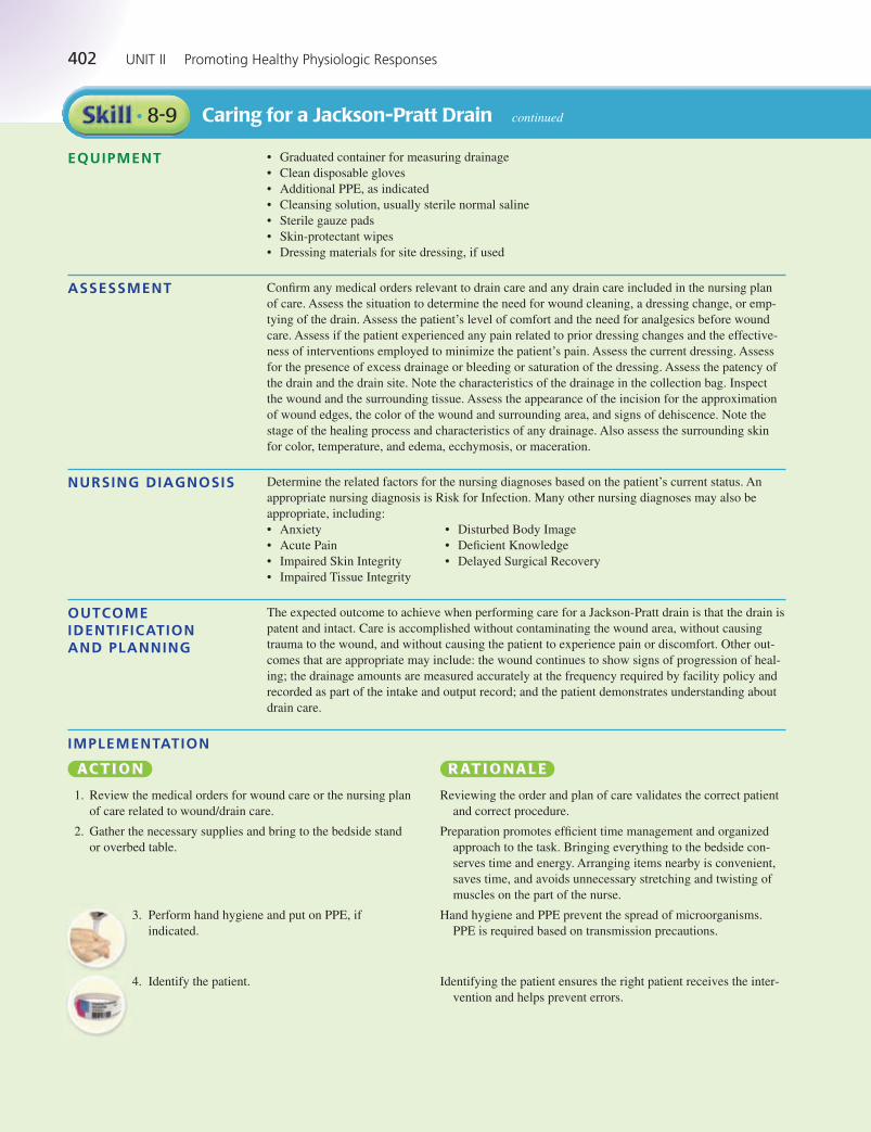
402 UNIT II Promoting Healthy Physiologic Responses
Caring for a Jackson-Pratt Drain continued• 8-9
EQUIPMENT • Graduated container for measuring drainage
• Clean disposable gloves
• Additional PPE, as indicated
• Cleansing solution, usually sterile normal saline
• Sterile gauze pads
• Skin-protectant wipes
• Dressing materials for site dressing, if used
Confirm any medical orders relevant to drain care and any drain care included in the nursing plan
of care. Assess the situation to determine the need for wound cleaning, a dressing change, or emp-
tying of the drain. Assess the patient’s level of comfort and the need for analgesics before wound
care. Assess if the patient experienced any pain related to prior dressing changes and the effective-
ness of interventions employed to minimize the patient’s pain. Assess the current dressing. Assess
for the presence of excess drainage or bleeding or saturation of the dressing. Assess the patency of
the drain and the drain site. Note the characteristics of the drainage in the collection bag. Inspect
the wound and the surrounding tissue. Assess the appearance of the incision for the approximation
of wound edges, the color of the wound and surrounding area, and signs of dehiscence. Note the
stage of the healing process and characteristics of any drainage. Also assess the surrounding skin
for color, temperature, and edema, ecchymosis, or maceration.
Determine the related factors for the nursing diagnoses based on the patient’s current status. An
appropriate nursing diagnosis is Risk for Infection. Many other nursing diagnoses may also be
appropriate, including:
• Anxiety • Disturbed Body Image
• Acute Pain • Deficient Knowledge
• Impaired Skin Integrity • Delayed Surgical Recovery
• Impaired Tissue Integrity
The expected outcome to achieve when performing care for a Jackson-Pratt drain is that the drain is
patent and intact. Care is accomplished without contaminating the wound area, without causing
trauma to the wound, and without causing the patient to experience pain or discomfort. Other out-
comes that are appropriate may include: the wound continues to show signs of progression of heal-
ing; the drainage amounts are measured accurately at the frequency required by facility policy and
recorded as part of the intake and output record; and the patient demonstrates understanding about
drain care.
ASSESSMENT
NURSING DIAGNOSIS
OUTCOME IDENTIFICATION AND PLANNING
IMPLEMENTATION
AA C T IC T I OO NN RR AA T IT I OO N A L EN A L E
1. Review the medical orders for wound care or the nursing plan
of care related to wound/drain care.
2. Gather the necessary supplies and bring to the bedside stand
or overbed table.
3. Perform hand hygiene and put on PPE, if
indicated.
4. Identify the patient.
Reviewing the order and plan of care validates the correct patient
and correct procedure.
Preparation promotes efficient time management and organized
approach to the task. Bringing everything to the bedside con-
serves time and energy. Arranging items nearby is convenient,
saves time, and avoids unnecessary stretching and twisting of
muscles on the part of the nurse.
Hand hygiene and PPE prevent the spread of microorganisms.
PPE is required based on transmission precautions.
Identifying the patient ensures the right patient receives the inter-
vention and helps prevent errors.
LWBK545_C08_p358-435.qxd 08/07/2010 1:58 PM Page 402 Aptara
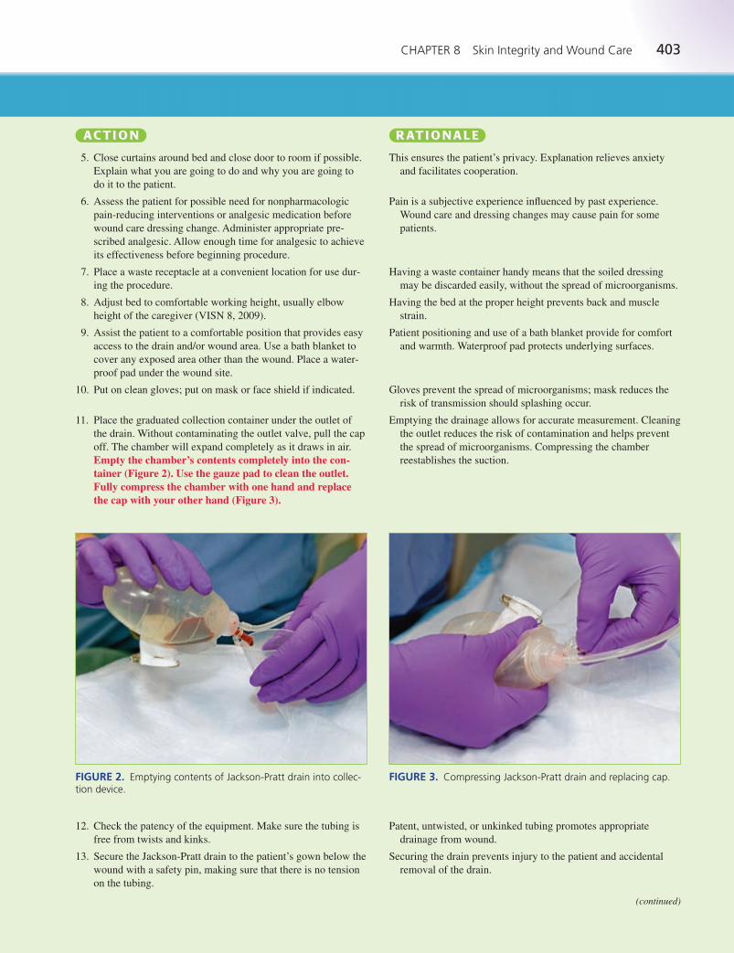
CHAPTER 8 Skin Integrity and Wound Care 403
AA C T IC T I OO NN RR AA T IT I OO N A L EN A L E
5. Close curtains around bed and close door to room if possible.
Explain what you are going to do and why you are going to
do it to the patient.
6. Assess the patient for possible need for nonpharmacologic
pain-reducing interventions or analgesic medication before
wound care dressing change. Administer appropriate pre-
scribed analgesic. Allow enough time for analgesic to achieve
its effectiveness before beginning procedure.
7. Place a waste receptacle at a convenient location for use dur-
ing the procedure.
8. Adjust bed to comfortable working height, usually elbow
height of the caregiver (VISN 8, 2009).
9. Assist the patient to a comfortable position that provides easy
access to the drain and/or wound area. Use a bath blanket to
cover any exposed area other than the wound. Place a water-
proof pad under the wound site.
10. Put on clean gloves; put on mask or face shield if indicated.
11. Place the graduated collection container under the outlet of
the drain. Without contaminating the outlet valve, pull the cap
off. The chamber will expand completely as it draws in air.
Empty the chamber’s contents completely into the con-tainer (Figure 2). Use the gauze pad to clean the outlet.Fully compress the chamber with one hand and replacethe cap with your other hand (Figure 3).
This ensures the patient’s privacy. Explanation relieves anxiety
and facilitates cooperation.
Pain is a subjective experience influenced by past experience.
Wound care and dressing changes may cause pain for some
patients.
Having a waste container handy means that the soiled dressing
may be discarded easily, without the spread of microorganisms.
Having the bed at the proper height prevents back and muscle
strain.
Patient positioning and use of a bath blanket provide for comfort
and warmth. Waterproof pad protects underlying surfaces.
Gloves prevent the spread of microorganisms; mask reduces the
risk of transmission should splashing occur.
Emptying the drainage allows for accurate measurement. Cleaning
the outlet reduces the risk of contamination and helps prevent
the spread of microorganisms. Compressing the chamber
reestablishes the suction.
FIGURE 2. Emptying contents of Jackson-Pratt drain into collec-tion device.
FIGURE 3. Compressing Jackson-Pratt drain and replacing cap.
12. Check the patency of the equipment. Make sure the tubing is
free from twists and kinks.
13. Secure the Jackson-Pratt drain to the patient’s gown below the
wound with a safety pin, making sure that there is no tension
on the tubing.
Patent, untwisted, or unkinked tubing promotes appropriate
drainage from wound.
Securing the drain prevents injury to the patient and accidental
removal of the drain.
(continued)
LWBK545_C08_p358-435.qxd 08/07/2010 1:58 PM Page 403 Aptara
404 UNIT II Promoting Healthy Physiologic Responses
Caring for a Jackson-Pratt Drain continued• 8-9
AA C T IC T I OO NN RR AA T IT I OO N A L EN A L E
14. Carefully measure and record the character, color, and amount
of the drainage. Discard the drainage according to facility pol-
icy. Remove gloves.
15. Put on clean gloves. If the drain site has a dressing, re-dress
the site as outlined in Skill 8-8. Include cleaning of the sutures
with the gauze pad moistened with normal saline. Dry sutures
with gauze before applying new dressing.
16. If the drain site is open to air, observe the sutures that secure
the drain to the skin. Look for signs of pulling, tearing,
swelling, or infection of the surrounding skin. Gently clean
the sutures with the gauze pad moistened with normal saline.
Dry with a new gauze pad. Apply skin protectant to the sur-
rounding skin if needed.
17. Remove and discard gloves. Remove all remaining equip-
ment; place the patient in a comfortable position, with side
rails up and bed in the lowest position.
18. Remove additional PPE, if used. Perform hand
hygiene.
19. Check drain status at least every four hours. Check all wound
dressings every shift. More frequent checks may be needed if
the wound is more complex or dressings become saturated
quickly.
Documentation promotes continuity of care and communication.
Appropriate disposal of biohazard material reduces the risk for
microorganism transmission. Proper disposal of gloves deters
transmission of microorganisms.
Dressing protects the site. Cleaning and drying sutures deters
growth of microorganisms.
Early detection of problems leads to prompt intervention and pre-
vents complications. Gentle cleaning and drying prevent the
growth of microorganisms. Skin protectant prevents skin irrita-
tion and breakdown.
Proper removal and disposal of gloves prevents spread of
microorganisms. Proper patient and bed positioning promotes
safety and comfort.
Removing PPE properly reduces the risk for infection transmis-
sion and contamination of other items. Hand hygiene prevents
the spread of microorganisms.
Checking drain ensures proper functioning and early detection of
problems. Checking dressings ensures the assessment of
changes in patient condition and timely intervention to prevent
complications.
EVALUATION The expected outcome is met when the patient exhibits a patent and intact Jackson-Pratt drain
with a wound area that is free of contamination and trauma. The patient verbalizes minimal to
no pain or discomfort. Other outcomes that are appropriate may include: the patient exhibits
signs and symptoms of progressive wound healing, with drainage being measured accurately at
the frequency required by facility policy, and amounts recorded as part of the intake and output
record; and the patient verbalizes an understanding of the rationale for and/or the technique for
drain care.
Document the location of the wound and drain, the assessment of the wound and drain site, and
patency of the drain. Note if sutures are intact. Document the presence of drainage and characteris-
tics on the old dressing upon removal. Include the appearance of the surrounding skin. Document
cleansing of the drain site. Record any skin care and the dressing applied. Note that the drain was
emptied and recompressed. Note pertinent patient and family education and any patient reaction to
this procedure, including patient’s pain level and effectiveness of nonpharmacologic interventions
or analgesia if administered. Document the amount and characteristics of drainage obtained on the
appropriate intake and output record.
DOCUMENTATIONGuidelines
2/7/12 2400 Right chest incision and drain open to air. Wound edges approximated, slightecchymosis, no edema, redness, or drainage. Steri-Strips intact. J-P drain patent andsecured with suture. Exit site without edema, drainage, or redness. Drain emptied andrecompressed. 40 mL sanguineous drainage recorded.
—Carol White, RN
Sample Documentation
LWBK545_C08_p358-435.qxd 08/07/2010 1:58 PM Page 404 Aptara
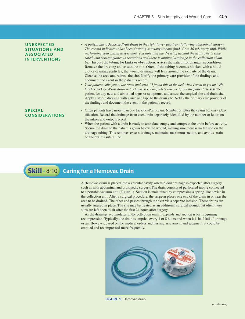
CHAPTER 8 Skin Integrity and Wound Care 405
UNEXPECTEDSITUATIONS ANDASSOCIATEDINTERVENTIONS
• A patient has a Jackson-Pratt drain in the right lower quadrant following abdominal surgery.The record indicates it has been draining serosanguineous fluid, 40 to 50 mL every shift. Whileperforming your initial assessment, you note that the dressing around the drain site is satu-rated with serosanguineous secretions and there is minimal drainage in the collection cham-ber: Inspect the tubing for kinks or obstruction. Assess the patient for changes in condition.
Remove the dressing and assess the site. Often, if the tubing becomes blocked with a blood
clot or drainage particles, the wound drainage will leak around the exit site of the drain.
Cleanse the area and redress the site. Notify the primary care provider of the findings and
document the event in the patient’s record.
• Your patient calls you to the room and says, “I found this in the bed when I went to get up.” Hehas his Jackson-Pratt drain in his hand. It is completely removed from the patient: Assess the
patient for any new and abnormal signs or symptoms, and assess the surgical site and drain site.
Apply a sterile dressing with gauze and tape to the drain site. Notify the primary care provider of
the findings and document the event in the patient’s record.
• Often patients have more than one Jackson-Pratt drain. Number or letter the drains for easy iden-
tification. Record the drainage from each drain separately, identified by the number or letter, on
the intake and output record.
• When the patient with a drain is ready to ambulate, empty and compress the drain before activity.
Secure the drain to the patient’s gown below the wound, making sure there is no tension on the
drainage tubing. This removes excess drainage, maintains maximum suction, and avoids strain
on the drain’s suture line.
SPECIALCONSIDERATIONS
Caring for a Hemovac Drain• 8-10
A Hemovac drain is placed into a vascular cavity where blood drainage is expected after surgery,
such as with abdominal and orthopedic surgery. The drain consists of perforated tubing connected
to a portable vacuum unit (Figure 1). Suction is maintained by compressing a spring-like device in
the collection unit. After a surgical procedure, the surgeon places one end of the drain in or near the
area to be drained. The other end passes through the skin via a separate incision. These drains are
usually sutured in place. The site may be treated as an additional surgical wound, but often these
sites are left open to air after the first 24 hours after surgery.
As the drainage accumulates in the collection unit, it expands and suction is lost, requiring
recompression. Typically, the drain is emptied every 4 or 8 hours and when it is half full of drainage
or air. However, based on the medical orders and nursing assessment and judgment, it could be
emptied and recompressed more frequently.
FIGURE 1. Hemovac drain.(continued)
LWBK545_C08_p358-435.qxd 08/07/2010 1:58 PM Page 405 Aptara
406 UNIT II Promoting Healthy Physiologic Responses
Caring for a Hemovac Drain continued• 8-10
EQUIPMENT • Graduated container for measuring drainage
• Clean disposable gloves
• Additional PPE, as indicated
• Cleansing solution, usually sterile normal saline
• Sterile gauze pads
• Skin-protectant wipes
• Dressing materials for site dressing, if used
Confirm any medical orders relevant to drain care and any drain care included in the nursing plan
of care. Assess the situation to determine the need for wound cleaning, a dressing change, or emp-
tying of the drain. Assess the patient’s level of comfort and the need for analgesics before wound
care. Assess if the patient experienced any pain related to prior dressing changes and the effective-
ness of interventions employed to minimize the patient’s pain. Assess the current dressing. Assess
for the presence of excess drainage or bleeding or saturation of the dressing. Assess the patency of
the drain and the drain site. Note the characteristics of the drainage in the collection bag. Inspect
the wound and the surrounding tissue. Assess the appearance of the incision for the approximation
of wound edges, the color of the wound and surrounding area, and signs of dehiscence. Note the
stage of the healing process and characteristics of any drainage. Also assess the surrounding skin
for color, temperature, and edema, ecchymosis, or maceration.
Determine the related factors for the nursing diagnoses based on the patient’s current status. An
appropriate nursing diagnosis is Risk for Infection. Many other nursing diagnoses may also be
appropriate, including:
• Anxiety • Acute Pain
• Disturbed Body Image • Deficient Knowledge
• Impaired Skin Integrity • Delayed Surgical Recovery
• Impaired Tissue Integrity
The expected outcome to achieve when performing care for a Hemovac drain is that the drain is
patent and intact. Care is accomplished without contaminating the wound area, without causing
trauma to the wound, and without causing the patient to experience pain or discomfort. Other out-
comes that are appropriate may include: the wound continues to show signs of progression of heal-
ing; the drainage amounts are measured accurately at the frequency required by facility policy and
recorded as part of the intake and output record; and the patient demonstrates understanding about
drain care.
ASSESSMENT
NURSING DIAGNOSIS
OUTCOME IDENTIFICATION AND PLANNING
IMPLEMENTATION
AA C T IC T I OO NN RR AA T IT I OO N A L EN A L E
1. Review the medical orders for wound care or the nursing plan
of care related to wound/drain care.
2. Gather the necessary supplies and bring to the bedside stand
or overbed table.
3. Perform hand hygiene and put on PPE, if
indicated.
4. Identify the patient.
Reviewing the order and plan of care validates the correct patient
and correct procedure.
Preparation promotes efficient time management and organized
approach to the task. Bringing everything to the bedside con-
serves time and energy. Arranging items nearby is convenient,
saves time, and avoids unnecessary stretching and twisting of
muscles on the part of the nurse.
Hand hygiene and PPE prevent the spread of microorganisms.
PPE is required based on transmission precautions.
Identifying the patient ensures the right patient receives the inter-
vention and helps prevent errors.
LWBK545_C08_p358-435.qxd 08/07/2010 1:58 PM Page 406 Aptara
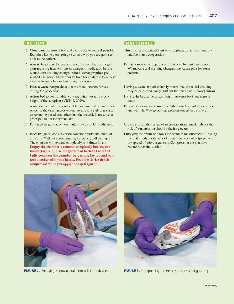
CHAPTER 8 Skin Integrity and Wound Care 407
AA C T IC T I OO NN RR AA T IT I OO N A L EN A L E
5. Close curtains around bed and close door to room if possible.
Explain what you are going to do and why you are going to
do it to the patient.
6. Assess the patient for possible need for nonpharmacologic
pain-reducing interventions or analgesic medication before
wound care dressing change. Administer appropriate pre-
scribed analgesic. Allow enough time for analgesic to achieve
its effectiveness before beginning procedure.
7. Place a waste receptacle at a convenient location for use
during the procedure.
8. Adjust bed to comfortable working height, usually elbow
height of the caregiver (VISN 8, 2009).
9. Assist the patient to a comfortable position that provides easy
access to the drain and/or wound area. Use a bath blanket to
cover any exposed area other than the wound. Place a water-
proof pad under the wound site.
10. Put on clean gloves; put on mask or face shield if indicated.
11. Place the graduated collection container under the outlet of
the drain. Without contaminating the outlet, pull the cap off.
The chamber will expand completely as it draws in air.
Empty the chamber’s contents completely into the con-tainer (Figure 2). Use the gauze pad to clean the outlet.Fully compress the chamber by pushing the top and bot-tom together with your hands. Keep the device tightlycompressed while you apply the cap (Figure 3).
This ensures the patient’s privacy. Explanation relieves anxiety
and facilitates cooperation.
Pain is a subjective experience influenced by past experience.
Wound care and dressing changes may cause pain for some
patients.
Having a waste container handy means that the soiled dressing
may be discarded easily, without the spread of microorganisms.
Having the bed at the proper height prevents back and muscle
strain.
Patient positioning and use of a bath blanket provide for comfort
and warmth. Waterproof pad protects underlying surfaces.
Gloves prevent the spread of microorganisms; mask reduces the
risk of transmission should splashing occur.
Emptying the drainage allows for accurate measurement. Cleaning
the outlet reduces the risk of contamination and helps prevent
the spread of microorganisms. Compressing the chamber
reestablishes the suction.
FIGURE 2. Emptying Hemovac drain into collection device. FIGURE 3. Compressing the Hemovac and securing the cap.
(continued)
LWBK545_C08_p358-435.qxd 08/07/2010 1:58 PM Page 407 Aptara
408 UNIT II Promoting Healthy Physiologic Responses
Caring for a Hemovac Drain continued• 8-10
AA C T IC T I OO NN RR AA T IT I OO N A L EN A L E
12. Check the patency of the equipment. Make sure the tubing is
free from twists and kinks.
13. Secure the Hemovac drain to the patient’s gown below the
wound with a safety pin, making sure that there is no tension
on the tubing.
14. Carefully measure and record the character, color, and amount
of the drainage. Discard the drainage according to facility
policy.
15. Put on clean gloves. If the drain site has a dressing, re-dress
the site as outlined in Skill 8-8. Include cleaning of the sutures
with the gauze pad moistened with normal saline. Dry sutures
with gauze before applying new dressing.
16. If the drain site is open to air, observe the sutures that secure
the drain to the skin. Look for signs of pulling, tearing,
swelling, or infection of the surrounding skin. Gently clean
the sutures with the gauze pad moistened with normal saline.
Dry with a new gauze pad. Apply skin protectant to the sur-
rounding skin if needed.
17. Remove and discard gloves. Remove all remaining equip-
ment; place the patient in a comfortable position, with side
rails up and bed in the lowest position.
18. Remove additional PPE, if used. Perform hand
hygiene.
19. Check drain status at least every four hours. Check all wound
dressings every shift. More frequent checks may be needed
if the wound is more complex or dressings become saturated
quickly.
Patent, untwisted, or unkinked tubing promotes appropriate
drainage from wound.
Securing the drain prevents injury to the patient and accidental
removal of the drain.
Documentation promotes continuity of care and communication.
Appropriate disposal of biohazard material reduces the risk for
microorganism transmission.
Dressing protects the site. Cleaning and drying sutures deters
growth of microorganisms.
Early detection of problems leads to prompt intervention and pre-
vents complications. Gentle cleaning and drying prevent the
growth of microorganisms. Skin protectant prevents skin irrita-
tion and breakdown.
Proper removal of gloves prevents spread of microorganisms.
Proper patient and bed positioning promotes safety and
comfort.
Removing PPE properly reduces the risk for infection transmis-
sion and contamination of other items. Hand hygiene prevents
the spread of microorganisms.
Checking drain ensures proper functioning and early detection
of problems. Checking dressings ensures the assessment of
changes in patient condition and timely intervention to prevent
complications.
EVALUATION The expected outcome is met when the patient exhibits a patent and intact Jackson-Pratt drain with
a wound area that is free of contamination and trauma. The patient verbalizes minimal to no pain or
discomfort. Other outcomes that are appropriate may include: the patient exhibits signs and symp-
toms of progressive wound healing, with drainage being measured accurately at the frequency
required by facility policy, and amounts recorded as part of the intake and output record; and the
patient verbalizes an understanding of the rationale for and/or the technique for drain care.
Document the location of the wound and drain, the assessment of the wound and drain site, and
patency of the drain. Note if sutures are intact. Document the presence of drainage and characteris-
tics on the old dressing upon removal. Include the appearance of the surrounding skin. Document
cleansing of the drain site. Record any skin care and any dressing applied. Note that the drain was
emptied and recompressed. Note pertinent patient and family education and any patient reaction to
this procedure, including patient’s pain level and effectiveness of nonpharmacologic interventions
or analgesia if administered. Document the amount and characteristics of drainage obtained on the
appropriate intake and output record.
DOCUMENTATIONGuidelines
1/18/12 1000 Hemovac drain in place in left lower extremity, site open to air. Suture intact;exit site slightly pink, without redness, edema, or drainage. Surrounding skin withoutedema, ecchymosis, or redness. Exit site and suture cleansed with normal saline. Hemovacemptied of 90 mL sanguineous secretions and recompressed.
—A. Smith, RN
Sample Documentation
LWBK545_C08_p358-435.qxd 08/07/2010 1:58 PM Page 408 Aptara
CHAPTER 8 Skin Integrity and Wound Care 409
UNEXPECTEDSITUATIONS ANDASSOCIATEDINTERVENTIONS
• A patient has a Hemovac drain placed in the left knee following surgery. The record indicates ithas been draining serosanguineous secretions, 40 to 50 mL every shift. While performing yourinitial assessment, you note that the collection chamber is completely expanded. The nurse emp-ties the device and compresses to resume suction. A short time later, the nurse observes that thechamber is completely expanded again: Inspect the tubing for kinks or obstruction. Inspect the
device, looking for breaks in the integrity of the chamber. Make sure the cap is in place and
closed. Assess the patient for changes in condition. Remove the dressing and assess the site.
Make sure the drainage tubing has not advanced out of the wound, exposing any of the perfora-
tions in the tubing. If you are not successful in maintaining the suction, notify the primary care
provider of the findings and interventions and document the event in the patient’s record.
• When the patient with a drain is ready to ambulate, empty and compress the drain before activity.
Secure the drain to the patient’s gown below the wound, making sure there is no tension on the
drainage tubing. This removes excess drainage, maintains maximum suction, and avoids strain
on the drain’s suture line.
SPECIALCONSIDERATIONS
Applying Negative Pressure Wound Therapy• 8-11
Negative-pressure wound therapy (NPWT) (or topical negative pressure [TNP]) promotes wound
healing and wound closure through the application of uniform negative pressure on the wound bed.
NPWT results in reduction in bacteria in the wound and the removal of excess wound fluid, while
providing a moist wound healing environment. The negative pressure results in mechanical tension
on the wound tissues, stimulating cell proliferation, blood flow to wounds, and the growth of new
blood vessels. An open-cell foam dressing is applied in the wound. A fenestrated tube is connected
to the foam, allowing the application of the negative pressure. The dressing and distal tubing are
covered by a transparent, occlusive, air-permeable dressing that provides a seal, allowing the appli-
cation of the negative pressure. Excess wound fluid is removed through tubing, and it also acts to
pull the wound edges together.
NPWT is used to treat a variety of acute or chronic wounds, wounds with heavy drainage,
wounds failing to heal, or wounds healing slowly. Examples of such wounds include pressure
ulcers, arterial, venous, and diabetic ulcers, dehisced surgical wounds, infected wounds, skin graft
sites, and burns. NPWT is not considered for use in the presence of active bleeding; wounds with
exposed blood vessels, organs, or nerves; malignancy in wound tissue; presence of dry/necrotic tis-
sue; or with fistulas of unknown origin (Hess, 2008; Preston, 2008; Thompson, 2008). Cautious use
is indicated in the presence of unrelieved pressure, anticoagulant therapy, poor nutritional status,
and immunosuppressant therapy (Preston). Candidates must be assessed for preexisting bleeding
disorders, use of anticoagulants and other medications, or use of supplements that prolong bleeding
times, such as aspirin or ginkgo biloba (Malli, 2005; Preston, 2008). NPWT dressings are changed
every 48 to 72 hours, depending on the manufacturer’s specifications and medical orders. Infected
wounds may require dressing changes every 12 to 24 hours.
The following Skill outlines the procedure for V.A.C. Therapy (KCl), as an example of NPWT.
There are many manufacturers of negative pressure wound therapy systems. The nurse should be
familiar with the components of, and procedures related to, the particular system in use.
• Negative pressure unit (V.A.C. ATS unit)
• Evacuation/collection canister
• V.A.C. Foam dressing
• V.A.C. drape
• T.R.A.C. Pad
• Skin-protectant wipes
• Sterile gauze sponge
• A sterile irrigation set, including a basin, irrigant container, and irrigation syringe
EQUIPMENT
(continued)
LWBK545_C08_p358-435.qxd 08/07/2010 1:58 PM Page 409 Aptara
410 UNIT II Promoting Healthy Physiologic Responses
Applying Negative Pressure Wound Therapy continued• 8-11
• Sterile irrigation solution as ordered by the physician, warmed to body temperature
• Waste receptacle to dispose of contaminated materials
• Sterile gloves (2 pairs)
• Sterile scissors
• Clean disposable gloves
• Gown, mask, eye protection
• Additional PPE, as indicated
• Sterile scissors
• Waterproof pad and bath blanket
Confirm the medical order for the application of NPWT. Check the patient’s chart and question the
patient about current treatments and medications that may make the application contraindicated.
Assess the situation to determine the need for a dressing change. Confirm any medical orders rele-
vant to wound care and any wound care included in the nursing plan of care. Assess the patient’s
level of comfort and the need for analgesics before wound care. Assess if the patient experienced
any pain related to prior dressing changes and the effectiveness of interventions employed to mini-
mize the patient’s pain. Assess the current dressing to determine if it is intact. Assess for excess
drainage or bleeding or saturation of the dressing. Inspect the wound and the surrounding tissue.
Assess the location, appearance of the wound, stage (if appropriate), drainage, and types of tissue
present in the wound. Measure the wound. Note the stage of the healing process and characteristics
of any drainage. Also assess the surrounding skin for color, temperature, and edema, ecchymosis,
or maceration.
Determine the related factors for the nursing diagnoses based on the patient’s current status. An
appropriate nursing diagnosis is Impaired Skin Integrity. Other nursing diagnoses that may be
appropriate or require the use of this skill include:
• Anxiety • Disturbed Body Image
• Acute Pain • Risk for Infection
• Risk for Injury • Deficient Knowledge
• Impaired Tissue Integrity
The expected outcome to achieve when applying negative pressure wound therapy is that the ther-
apy is accomplished without contaminating the wound area, without causing trauma to the wound,
and without causing the patient to experience pain or discomfort. Other outcomes that may be
appropriate include: the vacuum device functions correctly; the appropriate and ordered pressure is
maintained throughout therapy; and the wound exhibits progression in healing.
ASSESSMENT
NURSING DIAGNOSIS
OUTCOME IDENTIFICATION AND PLANNING
IMPLEMENTATION
AA C T IC T I OO NN RR AA T IT I OO N A L EN A L E
1. Review the medical order for the application of NPWT ther-
apy, including the ordered pressure setting for the device.
2. Gather the necessary supplies and bring to the bedside stand
or overbed table.
3. Perform hand hygiene and put on PPE, if
indicated.
4. Identify the patient.
Reviewing the order validates the correct patient and correct
procedure.
Preparation promotes efficient time management and organized
approach to the task. Bringing everything to the bedside con-
serves time and energy. Arranging items nearby is convenient,
saves time, and avoids unnecessary stretching and twisting of
muscles on the part of the nurse.
Hand hygiene and PPE prevent the spread of microorganisms.
PPE is required based on transmission precautions.
Identifying the patient ensures the right patient receives the inter-
vention and helps prevent errors.
LWBK545_C08_p358-435.qxd 08/07/2010 1:58 PM Page 410 Aptara
CHAPTER 8 Skin Integrity and Wound Care 411
AA C T IC T I OO NN RR AA T IT I OO N A L EN A L E
5. Close curtains around bed and close door to room if possible.
Explain what you are going to do and why you are going to
do it to the patient.
6. Assess the patient for possible need for nonpharmacologic
pain-reducing interventions or analgesic medication before
wound care dressing change. Administer appropriate pre-
scribed analgesic. Allow enough time for analgesic to
achieve its effectiveness before beginning procedure.
7. Adjust bed to comfortable working height, usually elbow
height of the caregiver (VISN 8, 2009).
8. Assist the patient to a comfortable position that provides
easy access to the wound area. Position the patient so the
irrigation solution will flow from the clean end of the wound
toward the dirty end. Expose the area and drape the patient
with a bath blanket if needed. Put a waterproof pad under
the wound area.
9. Have the disposal bag or waste receptacle within easy reach
for use during the procedure.
10. Using sterile technique, prepare a sterile field and add all
the sterile supplies needed for the procedure to the field.
Pour warmed, sterile irrigating solution into the sterile
container.
11. Put on a gown, mask, and eye protection.
12. Put on clean gloves. Carefully and gently remove the dress-
ing. If there is resistance, use a silicone-based adhesive
remover to help remove the drape. Note the number ofpieces of foam removed from the wound. Compare withthe documented number from the previous dressingchange.
13. Discard the dressings in the receptacle. Remove your gloves
and put them in the receptacle.
14. Put on sterile gloves. Using sterile technique, irrigate the
wound (see Skill 8-4).
15. Clean the area around the skin with normal saline. Dry the
surrounding skin with a sterile gauze sponge.
16. Assess the wound for appearance, stage, the presence of eschar,
granulation tissue, epithelialization, undermining, tunneling,
necrosis, sinus tract, and drainage. Assess the appearance of the
surrounding tissue. Measure the wound. Refer to Fundamentals
Review 8-3.
17. Wipe intact skin around the wound with a skin-protectantwipe and allow it to dry well.
18. Remove gloves if they become contaminated and discard
them into the receptacle.
This ensures the patient’s privacy. Explanation relieves anxiety
and facilitates cooperation.
Pain is a subjective experience influenced by past experience.
Wound care and dressing changes may cause pain for some
patients.
Having the bed at the proper height prevents back and muscle
strain.
Patient positioning and draping provide for comfort and warmth.
Gravity directs the flow of liquid from the least contaminated to
the most contaminated area. Waterproof pad protects the patient
and the bed linens.
Having the waste container handy means that soiled dressings
and supplies may be discarded easily, without the spread of
microorganisms.
Proper preparation ensures that supplies are within easy reach
and sterility is maintained. Warmed solution may result in less
discomfort.
Use of personal protective equipment is part of Standard Precau-
tions. A gown protects your clothes from contamination if
splashing should occur. Goggles protect mucous membranes
of your eyes from contact with irrigant fluid.
Gloves protect the nurse from handling contaminated dress-
ings. A silicone-based adhesive remover allows for the easy,
rapid, and painless removal without the associated problems
of skin stripping (Rudoni, 2008; Stephen-Haynes, 2008).
Counting the number of pieces of foam assures the removal
of all foam that was placed during the previous dressing
change.
Proper disposal of dressings and used gloves prevents the spread
of microorganisms.
Irrigation removes exudate and debris.
Moisture provides a medium for growth of microorganisms.
This information provides evidence about the wound healing
process and/or the presence of infection.
Skin protectant provides a barrier against irritation and breakdown.
Proper disposal of gloves prevents spread of microorganisms.
(continued)
LWBK545_C08_p358-435.qxd 08/07/2010 1:58 PM Page 411 Aptara
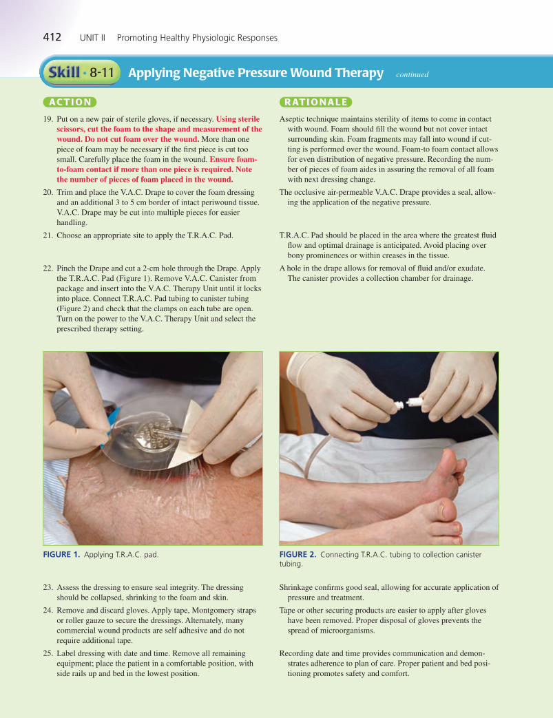
412 UNIT II Promoting Healthy Physiologic Responses
Applying Negative Pressure Wound Therapy continued• 8-11
AA C T IC T I OO NN RR AA T IT I OO N A L EN A L E
19. Put on a new pair of sterile gloves, if necessary. Using sterilescissors, cut the foam to the shape and measurement of thewound. Do not cut foam over the wound. More than one
piece of foam may be necessary if the first piece is cut too
small. Carefully place the foam in the wound. Ensure foam-to-foam contact if more than one piece is required. Notethe number of pieces of foam placed in the wound.
20. Trim and place the V.A.C. Drape to cover the foam dressing
and an additional 3 to 5 cm border of intact periwound tissue.
V.A.C. Drape may be cut into multiple pieces for easier
handling.
21. Choose an appropriate site to apply the T.R.A.C. Pad.
22. Pinch the Drape and cut a 2-cm hole through the Drape. Apply
the T.R.A.C. Pad (Figure 1). Remove V.A.C. Canister from
package and insert into the V.A.C. Therapy Unit until it locks
into place. Connect T.R.A.C. Pad tubing to canister tubing
(Figure 2) and check that the clamps on each tube are open.
Turn on the power to the V.A.C. Therapy Unit and select the
prescribed therapy setting.
Aseptic technique maintains sterility of items to come in contact
with wound. Foam should fill the wound but not cover intact
surrounding skin. Foam fragments may fall into wound if cut-
ting is performed over the wound. Foam-to foam contact allows
for even distribution of negative pressure. Recording the num-
ber of pieces of foam aides in assuring the removal of all foam
with next dressing change.
The occlusive air-permeable V.A.C. Drape provides a seal, allow-
ing the application of the negative pressure.
T.R.A.C. Pad should be placed in the area where the greatest fluid
flow and optimal drainage is anticipated. Avoid placing over
bony prominences or within creases in the tissue.
A hole in the drape allows for removal of fluid and/or exudate.
The canister provides a collection chamber for drainage.
FIGURE 1. Applying T.R.A.C. pad. FIGURE 2. Connecting T.R.A.C. tubing to collection canister tubing.
23. Assess the dressing to ensure seal integrity. The dressing
should be collapsed, shrinking to the foam and skin.
24. Remove and discard gloves. Apply tape, Montgomery straps
or roller gauze to secure the dressings. Alternately, many
commercial wound products are self adhesive and do not
require additional tape.
25. Label dressing with date and time. Remove all remaining
equipment; place the patient in a comfortable position, with
side rails up and bed in the lowest position.
Shrinkage confirms good seal, allowing for accurate application of
pressure and treatment.
Tape or other securing products are easier to apply after gloves
have been removed. Proper disposal of gloves prevents the
spread of microorganisms.
Recording date and time provides communication and demon-
strates adherence to plan of care. Proper patient and bed posi-
tioning promotes safety and comfort.
LWBK545_C08_p358-435.qxd 08/07/2010 1:58 PM Page 412 Aptara
CHAPTER 8 Skin Integrity and Wound Care 413
AA C T IC T I OO NN RR AA T IT I OO N A L EN A L E
26. Remove PPE, if used. Perform hand hygiene.
27. Check all wound dressings every shift. More frequent checks
may be needed if the wound is more complex or dressings
become saturated quickly.
Removing PPE properly reduces the risk for infection transmis-
sion and contamination of other items. Hand hygiene prevents
the spread of microorganisms.
Checking dressings ensures the assessment of changes in patient
condition and timely intervention to prevent complications.
EVALUATION The expected outcome is met when applying negative pressure wound therapy is accomplished
without contaminating the wound area, without causing trauma to the wound, and without causing
the patient to experience pain or discomfort. In addition, the vacuum device functions correctly; the
appropriate and ordered pressure is maintained throughout therapy; and the wound exhibits pro-
gression in healing.
Record your assessment of the wound, including evidence of granulation tissue, presence of
necrotic tissue, stage (if appropriate), and characteristics of drainage. Include the appearance of the
surrounding skin. Document the cleansing or irrigation of the wound and solution used. Document
the application of the NPWT, noting the pressure setting, patency, and seal of the dressing. Describe
the color and characteristics of the drainage in the collection chamber. Record pertinent patient and
family education and any patient reaction from this procedure, including the presence of pain and
effectiveness or ineffectiveness of pain interventions.
DOCUMENTATIONGuidelines
4/5/12 0800 NPWT dressing intact with good seal maintained, V.A.C. system patent, pres-sure setting 50 mm Hg. Purulent, sanguineous drainage noted in collection chamber andtubing. Surrounding tissue without edema, redness, ecchymosis, or signs of irritation.Patient verbalizes an understanding of movement limitations related to the system.
—B. Clark, RN
Sample Documentation
UNEXPECTEDSITUATIONS ANDASSOCIATEDINTERVENTIONS
• While assessing the patient, the nurse notes that the seal between the transparent dressing andthe foam and skin is not tight: Check the dressing seals, tubing connections, and canister inser-
tion, and ensure the clamps are open. If a leak in the transparent dressing is identified, the appro-
priate pressure is not being applied to the wound. Apply additional transparent dressing to reseal.
If this application does not correct the break, change the dressing.
• The patient complains of acute pain while NPWT is operating: Assess the patient for other symp-
toms, obtain vital signs, assess the wound, and assess the vacuum device for proper functioning.
Report your findings to the primary care provider and document the event in the patient’s record.
Administer analgesics as ordered. Continue or change the wound therapy as ordered.
• Change the wound dressing every 48 hours for noninfected wounds, or every 12 to 24 hours for
infected wounds. Time dressing changes to allow for wound assessment by the other members of
the healthcare team.
• Measure and record the amount of drainage each shift as part of the intake and output record.
• Be alert for audible and visual alarms on the vacuum device to alert you to problems, such as tip-
ping of the device greater than 45 degrees, a full collection canister, an air leak in the dressing, or
dislodgment of the canister.
• NPWT should operate for 24 hours. It should not be shut off for more than 2 hours in a 24-hour
period. When NPWT is restarted, irrigate the wound per medical order or facility policy, and
apply a new NPWT dressing.
• When maceration of the surrounding skin beneath the occlusive dressing occurs, this may be
treated by placing a barrier/wafer dressing beneath the transparent dressing to protect the skin.
Verify with facility policy as needed.
SPECIALCONSIDERATIONS
LWBK545_C08_p358-435.qxd 08/07/2010 1:58 PM Page 413 Aptara
414 UNIT II Promoting Healthy Physiologic Responses
Removing Sutures• 8-12
Skin sutures are used to hold tissue and skin together. Sutures may be black silk, synthetic material,
or fine wire. Sutures are removed when enough tensile strength has developed to hold the wound
edges together during healing. The time frame varies depending on the patient’s age, nutritional sta-
tus, and wound location. Frequently, after skin sutures are removed, adhesive wound closure strips
are applied across the wound to give additional support as it continues to heal. The removal of
sutures may be done by the primary care provider or by the nurse with a medical order.
• Suture removal kit or forceps and scissors
• Gauze
• Wound cleansing agent, according to facility policy
• Clean disposable gloves
• Additional PPE, as indicated
• Adhesive wound closure strips
• Skin protectant wipes
Inspect the surgical incision and the surrounding tissue. Assess the appearance of the wound for the
approximation of wound edges, the color of the wound and surrounding area, presence of wound
drainage noting color, volume, and odor, and for signs of dehiscence. Note the stage of the healing
process and characteristics of any drainage. Assess the surrounding skin for color, temperature, and
the presence of edema, maceration, or ecchymosis.
Determine the related factors for the nursing diagnoses based on the patient’s current status.
An appropriate nursing diagnosis is Risk for Infection. Other nursing diagnoses that may be
appropriate include:
• Anxiety • Delayed Surgical Recovery
• Acute Pain • Impaired Skin Integrity
• Deficient Knowledge
The expected outcome to achieve when removing surgical sutures is that the sutures are removed
without contaminating the incisional area, without causing trauma to the wound, and without caus-
ing the patient to experience pain or discomfort. In addition, other outcomes that are appropriate
include: the patient remains free of complications that would delay recovery; and the patient verbal-
izes an understanding of the procedure.
EQUIPMENT
ASSESSMENT
NURSING DIAGNOSIS
OUTCOME IDENTIFICATION AND PLANNING
IMPLEMENTATION
AA C T IC T I OO NN RR AA T IT I OO N A L EN A L E
1. Review the medical orders for suture removal.
2. Gather the necessary supplies and bring to the bedside stand
or overbed table.
3. Perform hand hygiene and put on PPE, if
indicated.
4. Identify the patient.
5. Close curtains around bed and close door to room if possible.
Explain what you are going to do and why you are going to
do it to the patient. Describe the sensation of suture removal
as a pulling or slightly uncomfortable experience.
Reviewing the order and plan of care validates the correct patient
and correct procedure.
Preparation promotes efficient time management and organized
approach to the task. Bringing everything to the bedside con-
serves time and energy. Arranging items nearby is convenient,
saves time, and avoids unnecessary stretching and twisting of
muscles on the part of the nurse.
Hand hygiene and PPE prevent the spread of microorganisms.
PPE is required based on transmission precautions.
Identifying the patient ensures the right patient receives the inter-
vention and helps prevent errors.
This ensures the patient’s privacy. Explanation relieves anxiety
and facilitates cooperation.
LWBK545_C08_p358-435.qxd 08/07/2010 1:58 PM Page 414 Aptara
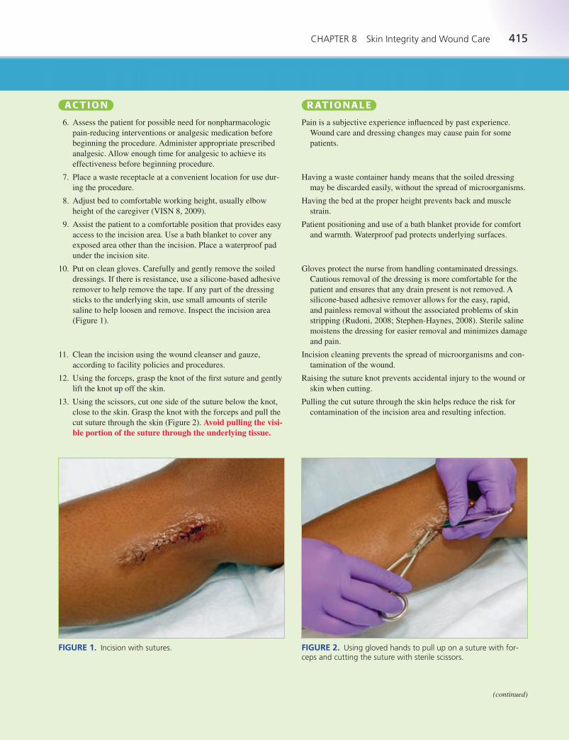
CHAPTER 8 Skin Integrity and Wound Care 415
AA C T IC T I OO NN RR AA T IT I OO N A L EN A L E
6. Assess the patient for possible need for nonpharmacologic
pain-reducing interventions or analgesic medication before
beginning the procedure. Administer appropriate prescribed
analgesic. Allow enough time for analgesic to achieve its
effectiveness before beginning procedure.
7. Place a waste receptacle at a convenient location for use dur-
ing the procedure.
8. Adjust bed to comfortable working height, usually elbow
height of the caregiver (VISN 8, 2009).
9. Assist the patient to a comfortable position that provides easy
access to the incision area. Use a bath blanket to cover any
exposed area other than the incision. Place a waterproof pad
under the incision site.
10. Put on clean gloves. Carefully and gently remove the soiled
dressings. If there is resistance, use a silicone-based adhesive
remover to help remove the tape. If any part of the dressing
sticks to the underlying skin, use small amounts of sterile
saline to help loosen and remove. Inspect the incision area
(Figure 1).
11. Clean the incision using the wound cleanser and gauze,
according to facility policies and procedures.
12. Using the forceps, grasp the knot of the first suture and gently
lift the knot up off the skin.
13. Using the scissors, cut one side of the suture below the knot,
close to the skin. Grasp the knot with the forceps and pull the
cut suture through the skin (Figure 2). Avoid pulling the visi-ble portion of the suture through the underlying tissue.
Pain is a subjective experience influenced by past experience.
Wound care and dressing changes may cause pain for some
patients.
Having a waste container handy means that the soiled dressing
may be discarded easily, without the spread of microorganisms.
Having the bed at the proper height prevents back and muscle
strain.
Patient positioning and use of a bath blanket provide for comfort
and warmth. Waterproof pad protects underlying surfaces.
Gloves protect the nurse from handling contaminated dressings.
Cautious removal of the dressing is more comfortable for the
patient and ensures that any drain present is not removed. A
silicone-based adhesive remover allows for the easy, rapid,
and painless removal without the associated problems of skin
stripping (Rudoni, 2008; Stephen-Haynes, 2008). Sterile saline
moistens the dressing for easier removal and minimizes damage
and pain.
Incision cleaning prevents the spread of microorganisms and con-
tamination of the wound.
Raising the suture knot prevents accidental injury to the wound or
skin when cutting.
Pulling the cut suture through the skin helps reduce the risk for
contamination of the incision area and resulting infection.
FIGURE 1. Incision with sutures. FIGURE 2. Using gloved hands to pull up on a suture with for-ceps and cutting the suture with sterile scissors.
(continued)
LWBK545_C08_p358-435.qxd 08/07/2010 1:58 PM Page 415 Aptara
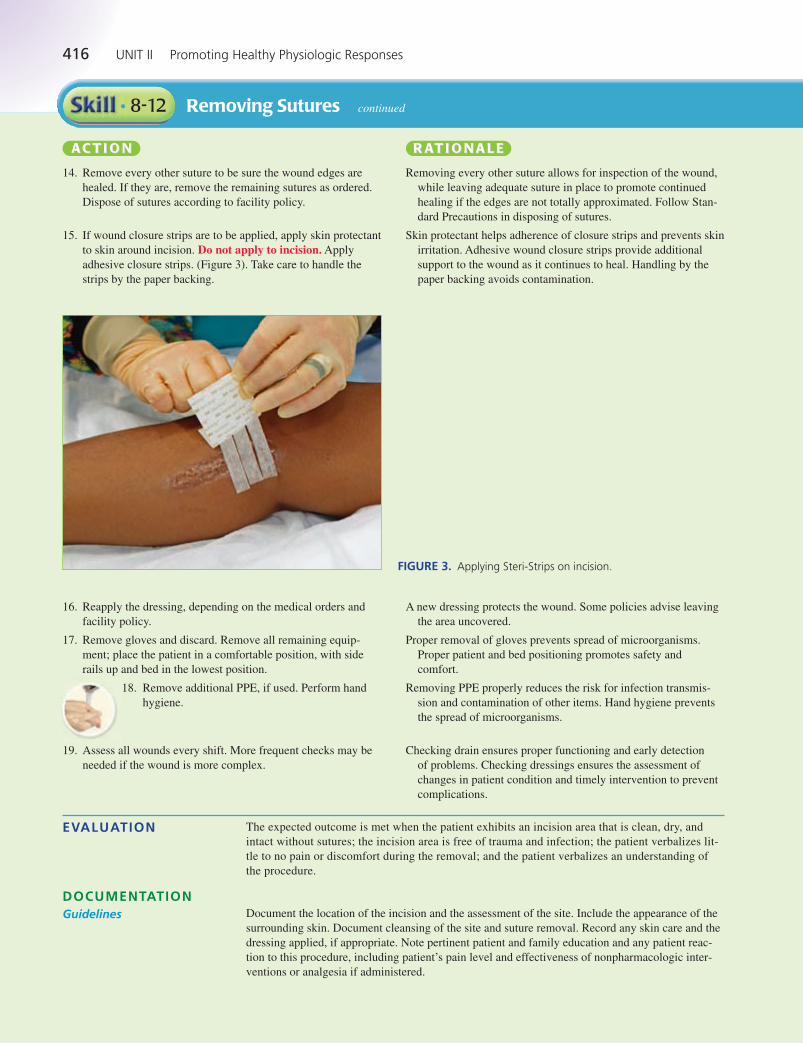
416 UNIT II Promoting Healthy Physiologic Responses
Removing Sutures continued• 8-12
AA C T IC T I OO NN RR AA T IT I OO N A L EN A L E
14. Remove every other suture to be sure the wound edges are
healed. If they are, remove the remaining sutures as ordered.
Dispose of sutures according to facility policy.
15. If wound closure strips are to be applied, apply skin protectant
to skin around incision. Do not apply to incision. Apply
adhesive closure strips. (Figure 3). Take care to handle the
strips by the paper backing.
Removing every other suture allows for inspection of the wound,
while leaving adequate suture in place to promote continued
healing if the edges are not totally approximated. Follow Stan-
dard Precautions in disposing of sutures.
Skin protectant helps adherence of closure strips and prevents skin
irritation. Adhesive wound closure strips provide additional
support to the wound as it continues to heal. Handling by the
paper backing avoids contamination.
FIGURE 3. Applying Steri-Strips on incision.
16. Reapply the dressing, depending on the medical orders and
facility policy.
17. Remove gloves and discard. Remove all remaining equip-
ment; place the patient in a comfortable position, with side
rails up and bed in the lowest position.
18. Remove additional PPE, if used. Perform hand
hygiene.
19. Assess all wounds every shift. More frequent checks may be
needed if the wound is more complex.
A new dressing protects the wound. Some policies advise leaving
the area uncovered.
Proper removal of gloves prevents spread of microorganisms.
Proper patient and bed positioning promotes safety and
comfort.
Removing PPE properly reduces the risk for infection transmis-
sion and contamination of other items. Hand hygiene prevents
the spread of microorganisms.
Checking drain ensures proper functioning and early detection
of problems. Checking dressings ensures the assessment of
changes in patient condition and timely intervention to prevent
complications.
EVALUATION The expected outcome is met when the patient exhibits an incision area that is clean, dry, and
intact without sutures; the incision area is free of trauma and infection; the patient verbalizes lit-
tle to no pain or discomfort during the removal; and the patient verbalizes an understanding of
the procedure.
Document the location of the incision and the assessment of the site. Include the appearance of the
surrounding skin. Document cleansing of the site and suture removal. Record any skin care and the
dressing applied, if appropriate. Note pertinent patient and family education and any patient reac-
tion to this procedure, including patient’s pain level and effectiveness of nonpharmacologic inter-
ventions or analgesia if administered.
DOCUMENTATIONGuidelines
LWBK545_C08_p358-435.qxd 08/07/2010 1:58 PM Page 416 Aptara
CHAPTER 8 Skin Integrity and Wound Care 417
3/4/12 1800 Right lower lateral leg surgical wound appears healed. Incision edges areapproximated, without erythema, edema, ecchymosis, or drainage. Skin warm and pink.Sutures removed without difficulty; skin protectant applied to skin surrounding incision andadhesive wound closure strips applied. Patient instructed in how to care for wound andexpectations regarding wound closure strips; patient and wife verbalized an understandingof information and asked appropriate questions.
—L. Downs, RN
Sample Documentation
UNEXPECTEDSITUATIONS ANDASSOCIATEDINTERVENTIONS
• Sutures are crusted with dried blood or secretions, making them difficult to remove: Moisten ster-
ile gauze with sterile saline and gently loosen crusts before removing sutures.
• Resistance is met when attempting to pull suture through the tissue: Use a gentle, continuous
pulling motion to remove the suture. If the suture still does not come out, do not use excessive
force. Report findings to the primary care provider and document the event in the patient’s record.
• Encourage the patient to splint chest and abdominal wounds during activity, such as changing
position, ambulation, coughing, and sneezing. This provides increased support for the skin and
underlying tissues and can decrease discomfort.
SPECIALCONSIDERATIONS
Removing Surgical Staples• 8-13
Surgical skin staples are made of stainless steel and are used to hold tissue and skin together. Sta-
ples decrease the risk of infection and allow faster wound closure. Surgical staples are removed
when enough tensile strength has developed to hold the wound edges together during healing. The
time frame for removal varies depending on the patient’s age, nutritional status, and wound loca-
tion. After skin staples are removed, adhesive wound closure strips are applied across the wound to
keep the skin edges approximated as it continues to heal. The removal of surgical staples may be
done by the primary care provider or by the nurse with a medical order.
• Staple remover
• Gauze
• Wound cleansing agent, according to facility policy
• Clean disposable gloves
• Additional PPE, as indicated
• Adhesive wound closure strips
• Skin protectant wipes
Inspect the surgical incision and the surrounding tissue. Assess the appearance of the wound for the
approximation of wound edges, the color of the wound and surrounding area, and signs of dehis-
cence. Note the stage of the healing process and the characteristics of any drainage. Assess the sur-
rounding skin for color, temperature, and the presence of edema or ecchymosis.
Determine the related factors for the nursing diagnoses based on the patient’s current status. An
appropriate nursing diagnosis is Risk for Infection. Other nursing diagnoses that may be appropri-
ate include:
• Anxiety • Acute Pain
• Impaired Skin Integrity • Delayed Surgical Recovery
• Deficient Knowledge
EQUIPMENT
ASSESSMENT
NURSING DIAGNOSIS
(continued)
LWBK545_C08_p358-435.qxd 08/07/2010 1:58 PM Page 417 Aptara
418 UNIT II Promoting Healthy Physiologic Responses
Removing Surgical Staples continued• 8-13
OUTCOME IDENTIFICATION AND PLANNING
The expected outcome to achieve when removing surgical staples is that the staples are removed
without contaminating the incisional area, without causing trauma to the wound, and without caus-
ing the patient to experience pain or discomfort. In addition, other outcomes that are appropriate
include: the patient remains free of complications that would delay recovery; and the patient verbal-
izes an understanding of the procedure.
IMPLEMENTATION
AA C T IC T I OO NN RR AA T IT I OO N A L EN A L E
1. Review the medical orders for staple removal.
2. Gather the necessary supplies and bring to the bedside stand
or overbed table.
3. Perform hand hygiene and put on PPE, if
indicated.
4. Identify the patient.
5. Close curtains around bed and close door to room if possible.
Explain what you are going to do and why you are going to
do it to the patient. Describe the sensation of staple removal
as a pulling experience.
6. Assess the patient for possible need for nonpharmacologic
pain-reducing interventions or analgesic medication before
beginning the procedure. Administer appropriate prescribed
analgesic. Allow enough time for analgesic to achieve its
effectiveness before beginning procedure.
7. Place a waste receptacle at a convenient location for use dur-
ing the procedure.
8. Adjust bed to comfortable working height, usually elbow
height of the caregiver (VISN 8).
9. Assist the patient to a comfortable position that provides easy
access to the incision area. Use a bath blanket to cover any
exposed area other than the incision. Place a waterproof pad
under the incision site.
10. Put on clean gloves. Carefully and gently remove the soiled
dressings. If there is resistance, use a silicone-based adhesive
remover to help remove the tape. If any part of the dressing
sticks to the underlying skin, use small amounts of sterile
saline to help loosen and remove. Inspect the incision area
(Figure 1).
11. Clean the incision using the wound cleanser and gauze,
according to facility policies and procedures.
12. Grasp the staple remover (Figure 2). Position the stapleremover under the staple to be removed. Firmly close thestaple remover. The staple will bend in the middle and theedges will pull up out of the skin.
Reviewing the order and plan of care validates the correct patient
and correct procedure.
Preparation promotes efficient time management and organized
approach to the task. Bringing everything to the bedside con-
serves time and energy. Arranging items nearby is convenient,
saves time, and avoids unnecessary stretching and twisting of
muscles on the part of the nurse.
Hand hygiene and PPE prevent the spread of microorganisms.
PPE is required based on transmission precautions.
Identifying the patient ensures the right patient receives the inter-
vention and helps prevent errors.
This ensures the patient’s privacy. Explanation relieves anxiety
and facilitates cooperation.
Pain is a subjective experience influenced by past experience.
Wound care and dressing changes may cause pain for some
patients.
Having a waste container handy means that the soiled dressing
may be discarded easily, without the spread of microorganisms.
Having the bed at the proper height prevents back and muscle
strain.
Patient positioning and use of a bath blanket provide for comfort
and warmth. Waterproof pad protects underlying surfaces.
Gloves protect the nurse from handling contaminated dressings.
Cautious removal of the dressing is more comfortable for the
patient and ensures that any drain present is not removed.
A silicone-based adhesive remover allows for the easy, rapid, and
painless removal without the associated problems of skin stripping
(Rudoni, 2008; Stephen-Haynes, 2008). Sterile saline moistens
the dressing for easier removal and minimizes damage and pain.
Incision cleaning prevents the spread of microorganisms and con-
tamination of the wound.
Correct use of staple remover prevents accidental injury to the
wound and contamination of the incision area and resulting
infection.
LWBK545_C08_p358-435.qxd 08/07/2010 1:58 PM Page 418 Aptara
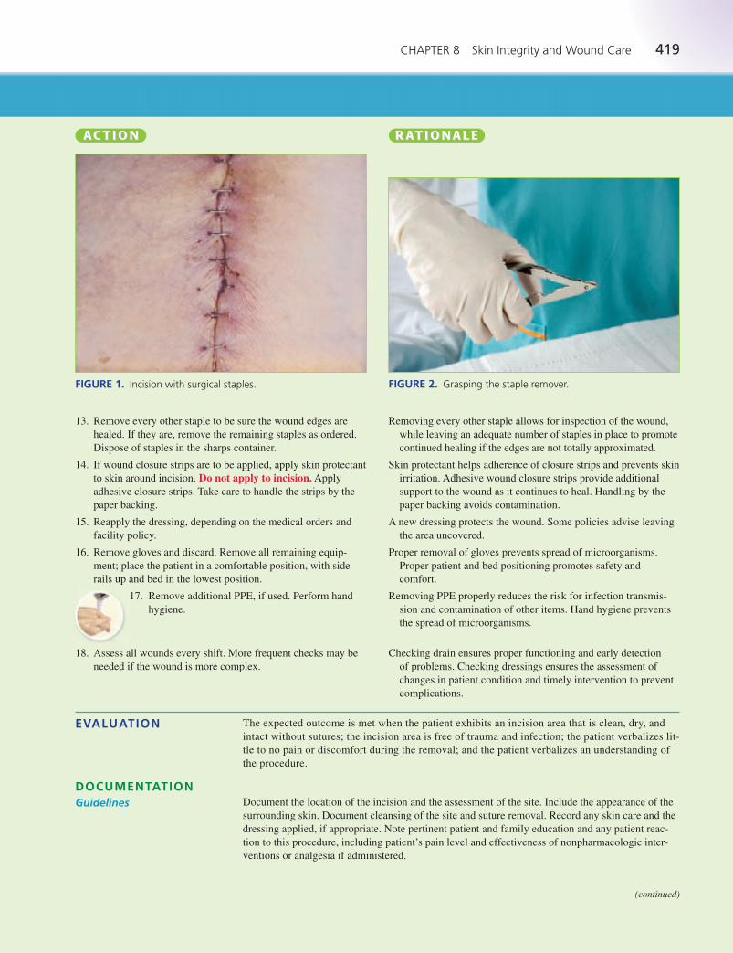
CHAPTER 8 Skin Integrity and Wound Care 419
AA C T IC T I OO NN RR AA T IT I OO N A L EN A L E
13. Remove every other staple to be sure the wound edges are
healed. If they are, remove the remaining staples as ordered.
Dispose of staples in the sharps container.
14. If wound closure strips are to be applied, apply skin protectant
to skin around incision. Do not apply to incision. Apply
adhesive closure strips. Take care to handle the strips by the
paper backing.
15. Reapply the dressing, depending on the medical orders and
facility policy.
16. Remove gloves and discard. Remove all remaining equip-
ment; place the patient in a comfortable position, with side
rails up and bed in the lowest position.
17. Remove additional PPE, if used. Perform hand
hygiene.
18. Assess all wounds every shift. More frequent checks may be
needed if the wound is more complex.
Removing every other staple allows for inspection of the wound,
while leaving an adequate number of staples in place to promote
continued healing if the edges are not totally approximated.
Skin protectant helps adherence of closure strips and prevents skin
irritation. Adhesive wound closure strips provide additional
support to the wound as it continues to heal. Handling by the
paper backing avoids contamination.
A new dressing protects the wound. Some policies advise leaving
the area uncovered.
Proper removal of gloves prevents spread of microorganisms.
Proper patient and bed positioning promotes safety and
comfort.
Removing PPE properly reduces the risk for infection transmis-
sion and contamination of other items. Hand hygiene prevents
the spread of microorganisms.
Checking drain ensures proper functioning and early detection
of problems. Checking dressings ensures the assessment of
changes in patient condition and timely intervention to prevent
complications.
FIGURE 1. Incision with surgical staples. FIGURE 2. Grasping the staple remover.
EVALUATION The expected outcome is met when the patient exhibits an incision area that is clean, dry, and
intact without sutures; the incision area is free of trauma and infection; the patient verbalizes lit-
tle to no pain or discomfort during the removal; and the patient verbalizes an understanding of
the procedure.
Document the location of the incision and the assessment of the site. Include the appearance of the
surrounding skin. Document cleansing of the site and suture removal. Record any skin care and the
dressing applied, if appropriate. Note pertinent patient and family education and any patient reac-
tion to this procedure, including patient’s pain level and effectiveness of nonpharmacologic inter-
ventions or analgesia if administered.
DOCUMENTATIONGuidelines
(continued)
LWBK545_C08_p358-435.qxd 08/07/2010 1:58 PM Page 419 Aptara
420 UNIT II Promoting Healthy Physiologic Responses
Removing Surgical Staples continued• 8-13
3/4/12 1800 Left upper lateral leg surgical wound appears healed. Incision edges areapproximated, without erythema, edema, ecchymosis, or drainage. Skin warm and pink.Staples removed without difficulty; skin protectant applied to skin surrounding incision andadhesive wound closure strips applied. Patient instructed in how to care for wound andexpectations regarding wound closure strips; patient and wife verbalized an understandingof information and asked appropriate questions.
—S. Hoffman, RN
Sample Documentation
UNEXPECTEDSITUATIONS ANDASSOCIATEDINTERVENTIONS
• The wound edges appear approximated before staple removal but pull apart afterward: Report
the findings to the primary care provider and document the event in the patient’s record. Apply
adhesive wound closure strips according to facility policy or medical order.
• The staples are stuck to the wound because of dried blood or secretions: Per facility policy or
medical order, apply moist saline compresses to loosen crusts before attempting to remove the
staples.
• Encourage the patient to splint chest and abdominal wounds (before and after removal) during
activity, such as changing position, ambulation, coughing, and sneezing. This provides increased
support for the skin and underlying tissues and can help decrease patient discomfort.
SPECIALCONSIDERATIONS
Applying an External Heating Pad• 8-14
Heat applications accelerate the inflammatory response, promoting healing. Heat is also used to
reduce muscle tension, relieve muscle spasm, and relieve joint stiffness. Heat also helps relieve
pain. It is used to treat infections, surgical wounds, inflammation, arthritis, joint pain, muscle pain,
and chronic pain.
Heat is applied by moist and dry methods. The medical order should include the type of applica-
tion, the body area to be treated, the frequency of application, and the length of time for the appli-
cations. Water used for heat applications needs to be at the appropriate temperature to avoid skin
damage: 115� to 125�F for older children and adults and 105� to 110�F for infants, young children,
older adults, and patients with diabetes or those who are unconscious.
Common types of external heating devices include Aquathermia pads (one brand) and crushable,
microwaveable hot packs. Aquathermia pads are used in healthcare agencies and are safer to use
than heating pads. The temperature setting for an Aquathermia pad should not exceed 105� to
109.4�F, depending on facility policy. Microwaveable packs are easy and inexpensive to use but
have several disadvantages. They may leak and pose a danger from burns related to improper use.
They are used most often in the home setting.
• Aquathermia heating pad (or other brand) with electronic unit
• Distilled water
• Cover for the pad, if not part of pad
• Gauze bandage or tape to secure the pad
• Bath blanket
• PPE, as indicated
Assess the situation to determine the appropriateness for the application of heat. Assess the patient’s
physical and mental status and the condition of the body area to be treated with heat. Confirm the
medical order for heat therapy, including frequency, type of therapy, body area to be treated, and
length of time for the application. Check the equipment to be used, including the condition of cords,
plugs, and heating elements. Look for fluid leaks. Once the equipment is turned on, make sure there
is a consistent distribution of heat and the temperature is within safe limits.
EQUIPMENT
ASSESSMENT
LWBK545_C08_p358-435.qxd 08/07/2010 1:58 PM Page 420 Aptara
CHAPTER 8 Skin Integrity and Wound Care 421
NURSING DIAGNOSIS Determine the related factors for the nursing diagnoses based on the patient’s current status. Nurs-
ing diagnoses that may be appropriate or require the use of this skill include:
• Chronic Pain • Acute Pain
• Impaired Skin Integrity • Risk for Impaired Skin Integrity
• Delayed Surgical Recovery • Impaired Tissue Integrity
• Risk for Injury
The expected outcome to achieve when applying an external heat source depends on the patient’s
nursing diagnosis. Outcomes that may be appropriate include the following: the patient experiences
increased comfort; the patient experiences decreased muscle spasms; the patient exhibits improved
wound healing; the patient demonstrates a reduction in inflammation; and the patient remains free
from injury.
OUTCOME IDENTIFICATION AND PLANNING
IMPLEMENTATION
AA C T IC T I OO NN RR AA T IT I OO N A L EN A L E
1. Review the medical order for the application of heat therapy,
including frequency, type of therapy, body area to be treated,
and length of time for the application.
2. Gather the necessary supplies and bring to the bedside stand
or overbed table.
3. Perform hand hygiene and put on PPE, if
indicated.
4. Identify the patient.
5. Close curtains around bed and close door to room if possible.
Explain what you are going to do and why you are going to
do it to the patient.
6. Adjust bed to comfortable working height, usually elbow
height of the caregiver (VISN 8, 2009).
7. Assist the patient to a comfortable position that provides easy
access to the area where the heat will be applied; use a bath
blanket to cover any other exposed area.
8. Assess the condition of the skin where the heat is to be
applied.
9. Check that the water in the electronic unit (Figure 1) is at
the appropriate level. Fill the unit two-thirds full or to the
fill mark, with distilled water, if necessary. Check the tem-
perature setting on the unit to ensure it is within the safe
range.
10. Attach pad tubing to electronic unit tubing (Figure 2).
11. Plug in the unit and warm the pad before use. Apply the heat-
ing pad to the prescribed area (Figure 3). Secure with gauze
bandage or tape.
Reviewing the order and plan of care validates the correct patient
and correct procedure.
Preparation promotes efficient time management and organized
approach to the task. Bringing everything to the bedside con-
serves time and energy. Arranging items nearby is convenient,
saves time, and avoids unnecessary stretching and twisting of
muscles on the part of the nurse.
Hand hygiene and PPE prevent the spread of microorganisms.
PPE is required based on transmission precautions.
Identifying the patient ensures the right patient receives the inter-
vention and helps prevent errors.
This ensures the patient’s privacy. Explanation relieves anxiety
and facilitates cooperation.
Having the bed at the proper height prevents back and muscle
strain.
Patient positioning and use of a bath blanket provide for comfort
and warmth.
Assessment supplies baseline data for post-treatment comparison
and identifies conditions that may contraindicate the application.
Sufficient water in the unit is necessary to ensure proper function
of the unit. Tap water leaves mineral deposits in the unit.
Checking the temperature setting helps to prevent skin or
tissue damage.
Allows flow of warmed water through heating pad.
Plugging in the pad readies it for use. Heat travels by conduction
from one object to another. Gauze bandage or tape holds the
pad in position; do not use pins, as they may puncture anddamage the pad.
(continued)
LWBK545_C08_p358-435.qxd 08/07/2010 1:58 PM Page 421 Aptara
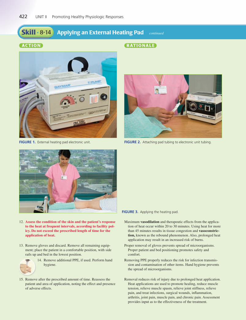
422 UNIT II Promoting Healthy Physiologic Responses
Applying an External Heating Pad continued• 8-14
AA C T IC T I OO NN RR AA T IT I OO N A L EN A L E
12. Assess the condition of the skin and the patient’s responseto the heat at frequent intervals, according to facility pol-icy. Do not exceed the prescribed length of time for theapplication of heat.
13. Remove gloves and discard. Remove all remaining equip-
ment; place the patient in a comfortable position, with side
rails up and bed in the lowest position.
14. Remove additional PPE, if used. Perform hand
hygiene.
15. Remove after the prescribed amount of time. Reassess the
patient and area of application, noting the effect and presence
of adverse effects.
Maximum vasodilation and therapeutic effects from the applica-
tion of heat occur within 20 to 30 minutes. Using heat for more
than 45 minutes results in tissue congestion and vasoconstric-tion, known as the rebound phenomenon. Also, prolonged heat
application may result in an increased risk of burns.
Proper removal of gloves prevents spread of microorganisms.
Proper patient and bed positioning promotes safety and
comfort.
Removing PPE properly reduces the risk for infection transmis-
sion and contamination of other items. Hand hygiene prevents
the spread of microorganisms.
Removal reduces risk of injury due to prolonged heat application.
Heat applications are used to promote healing, reduce muscle
tension, relieve muscle spasm, relieve joint stiffness, relieve
pain, and treat infections, surgical wounds, inflammation,
arthritis, joint pain, muscle pain, and chronic pain. Assessment
provides input as to the effectiveness of the treatment.
FIGURE 1. External heating pad electronic unit. FIGURE 2. Attaching pad tubing to electronic unit tubing.
FIGURE 3. Applying the heating pad.
LWBK545_C08_p358-435.qxd 08/07/2010 1:58 PM Page 422 Aptara
CHAPTER 8 Skin Integrity and Wound Care 423
EVALUATION The expected outcome is met when the patient exhibits increased comfort, decreased muscle spasm,
decreased pain, improved wound healing, and/or decreased inflammation. In addition, the patient
remains free of injury.
Document the rationale for application of heat therapy. If patient is receiving heat therapy for pain,
document the assessment of pain pre- and post- intervention. Specify the type of heat therapy and
location where it is applied, as well as length of time. Record the condition of the skin, noting any
redness or irritation before the heat application and after the application. Document the patient’s
reaction to the heat therapy. Record any appropriate patient or family education.
DOCUMENTATIONGuidelines
9/13/12 2300 Patient complaining of pain, rating it 5 out of 10. Aquathermia pad appliedto patient’s lower back for 30 minutes; now rating pain as 2 out of 10. Skin without signsof redness or irritation before and after application.
—M. Martinez, RN
Sample Documentation
UNEXPECTEDSITUATIONS ANDASSOCIATEDINTERVENTIONS
• When performing a periodic assessment of the site during the application of heat, the nurse notesexcessive swelling and redness at the site and the patient complains of pain that was not presentprior to the application of heat: Remove the heat source. Assess the patient for other symptoms
and obtain vital signs. Report your findings to the primary care provider and document the inter-
ventions in the patient’s record.
• Direct heat treatment is contraindicated for patients at risk for bleeding, patients with a sprained
limb in the acute stage, or patients with a condition associated with acute inflammation. Use
cautiously with children and older adults. Patients with diabetes, stroke, spinal cord injury,
and peripheral neuropathy are at risk for thermal injury, as are patients with very thin or
damaged skin. Be extremely careful when applying to heat-sensitive areas, such as scar tissue
and stomas.
• Instruct the patient not to lean or lie directly on the heating device, as this reduces air space and
increases the risk of burns.
• Check the water level in the Aquathermia unit periodically. Evaporation may occur. If the unit
runs dry, it could become damaged. Refill with distilled water periodically.
• A hot water bag or commercially prepared hot pack may be used in the home to apply heat. If
using a hot water bag, fill with hot tap water to warm the bag, then empty it to detect any leaks.
Check the temperature of the water with the bath thermometer or test on your inner wrist, adjust-
ing the temperature as ordered (usually 115�–125�F for adults). Checking the temperature
ensures that the heat applied is within the acceptable range of temperatures. Fill the bag one-half
to two-thirds full. Partial filling keeps the bag lightweight and flexible so that it can be molded to
the treatment area. Squeeze the bag until the water reaches the neck; this expels air, which would
make the bag inflexible and would reduce heat conduction. Fasten the top and cover the bag with
an absorbent cloth. The covering protects the skin from direct contact with the bag. If using a
commercially prepared hot pack, follow manufacturer’s directions and carefully assess skin
before and after heat application.
SPECIALCONSIDERATIONSGeneral Considerations
Home Care Considerations
LWBK545_C08_p358-435.qxd 08/07/2010 1:58 PM Page 423 Aptara
424 UNIT II Promoting Healthy Physiologic Responses
Applying a Warm Compress• 8-15
Warm moist compresses are used to help promote circulation, encourage healing, decrease edema,
promote consolidation of exudate, and decrease pain and discomfort. Moist heat softens crusted
material and is less drying to the skin. Moist heat also penetrates tissues more deeply than
dry heat.
The heat of a warm compress dissipates quickly, so the compresses must be changed frequently.
If a constant warm temperature is required, a heating device such as an Aquathermia pad (refer to
Skill 8-14) is applied over the compress. However, because moisture conducts heat, a low tempera-
ture setting is needed on the heating device. Many facilities have warming devices to heat the
dressing package to an appropriate temperature for the compress. These devices help reduce the
risk of burning or skin damage.
• Prescribed solution to moisten the compress material, warmed to 105� to 110�F
• Container for solution
• Gauze dressings or compresses
• Alternately, obtain the appropriate number of commercially packaged prewarmed dressings from
the warming device
• Clean disposable gloves
• Additional PPE, as indicated
• Waterproof pad and bath blanket
• Dry bath towel
• Tape or ties
• Aquathermia or other external heating device, if ordered or required to maintain the temperature
of the compress
Assess for circulatory compromise in the area where compress will be applied, including skin
color, pulses distal to the site, evidence of edema, and the presence of sensation. Assess the
situation to determine the appropriateness for the application of heat. Confirm the medical
order for the compresses, including the solution to be used, frequency, body area to be treated,
and length of time for the application. Assess the equipment to be used, if necessary, including
the condition of cords, plugs, and heating elements. Look for fluid leaks. Once the equipment
is turned on, make sure there is a consistent distribution of heat and the temperature is within
safe limits. Assess the application site frequently during the treatment, as tissue damage can
occur.
Determine the related factors for the nursing diagnoses based on the patient’s current status. An
appropriate nursing diagnosis is Risk for Injury. Many other nursing diagnoses may be appropriate,
including:
• Anxiety • Disturbed Body Image
• Acute Pain • Chronic Pain
• Impaired Skin Integrity • Risk for Impaired Skin Integrity
• Impaired Tissue Integrity • Deficient Knowledge
The expected outcome to achieve when applying warm compresses is that the patient shows signs
such as decreased inflammation, decreased muscle spasms, or decreased pain that indicate problems
have been relieved. Other outcomes that may be appropriate include: the patient experiences
improved healing, and the patient remains free from injury.
EQUIPMENT
ASSESSMENT
NURSING DIAGNOSIS
OUTCOME IDENTIFICATION AND PLANNING
IMPLEMENTATION
AA C T IC T I OO NN RR AA T IT I OO N A L EN A L E
1. Review the medical order for the application of a moist warm
compress, including frequency, and length of time for the
application.
Reviewing the order and plan of care validates the correct patient
and correct procedure.
LWBK545_C08_p358-435.qxd 08/07/2010 1:58 PM Page 424 Aptara
CHAPTER 8 Skin Integrity and Wound Care 425
AA C T IC T I OO NN RR AA T IT I OO N A L EN A L E
2. Gather the necessary supplies and bring to the bedside stand
or overbed table.
3. Perform hand hygiene and put on PPE, if
indicated.
4. Identify the patient.
5. Assess the patient for possible need for nonpharmacologic
pain-reducing interventions or analgesic medication before
beginning the procedure. Administer appropriate analgesic,
consulting physician’s orders, and allow enough time for
analgesic to achieve its effectiveness before beginning
procedure.
6. Close curtains around bed and close door to room if possible.
Explain what you are going to do and why you are going to
do it to the patient.
7. If using an electronic heating device, check that the water in
the unit is at the appropriate level. Fill the unit two-thirds full
with distilled water, or to the fill mark, if necessary. Check the
temperature setting on the unit to ensure it is within the safe
range (Refer to Skill 8-14).
8. Assist the patient to a comfortable position that provides easy
access to the area. Use a bath blanket to cover any exposed
area other than the intended site. Place a waterproof pad under
the site.
9. Place a waste receptacle at a convenient location for use
during the procedure.
10. Pour the warmed solution into the container and drop the
gauze for the compress into the solution. Alternately, if
commercially packaged pre-warmed gauze is used, open
packaging.
11. Put on clean gloves. Assess the application site for inflamma-
tion, skin color, and ecchymosis.
12. Retrieve the compress from the warmed solution,squeezing out any excess moisture (Figure 1). Alter-nately, remove pre-warmed gauze from open package.Apply the compress by gently and carefully molding it to the intended area. Ask patient if the application feelstoo hot.
Preparation promotes efficient time management and organized
approach to the task. Bringing everything to the bedside con-
serves time and energy. Arranging items nearby is convenient,
saves time, and avoids unnecessary stretching and twisting of
muscles on the part of the nurse.
Hand hygiene and PPE prevent the spread of microorganisms.
PPE is required based on transmission precautions.
Identifying the patient ensures the right patient receives the inter-
vention and helps prevent errors.
Pain is a subjective experience influenced by past experience.
Depending on the site of application, manipulation of the area
may cause pain for some patients.
This ensures the patient’s privacy. Explanation relieves anxiety
and facilitates cooperation.
Sufficient water in the unit is necessary to ensure proper function
of the unit. Tap water leaves mineral deposits in the unit.
Checking the temperature setting helps to prevent skin or tissue
damage.
Patient positioning and use of a bath blanket provide for comfort
and warmth. Waterproof pad protects underlying surfaces.
Having a waste container handy means that the used materials
may be discarded easily, without the spread of microorganisms.
Prepares compress for application.
Gloves protect the nurse from potential contact with microorgan-
isms. Assessment provides information about the area, the heal-
ing process and about the presence of infection and allows for
documentation of the condition of the area before the compress
is applied.
Excess moisture may contaminate the surrounding area and is
uncomfortable for the patient. Molding the compress to the skin
promotes retention of warmth around the site.
(continued)
LWBK545_C08_p358-435.qxd 08/07/2010 1:58 PM Page 425 Aptara
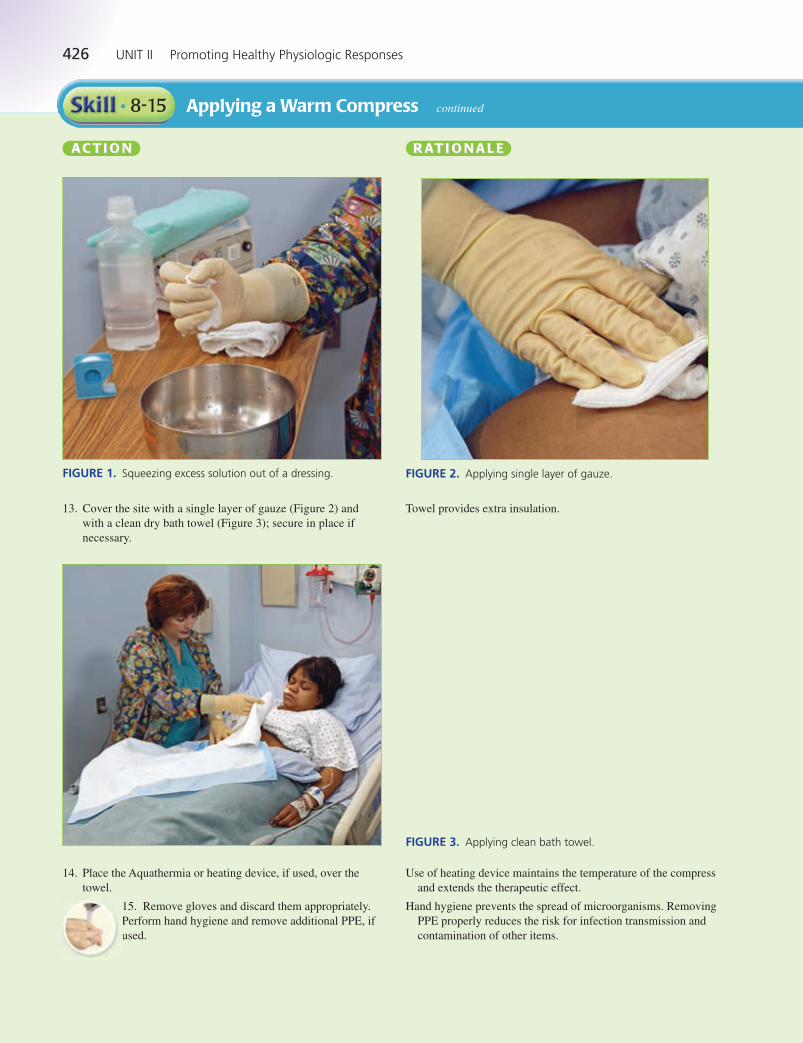
426 UNIT II Promoting Healthy Physiologic Responses
Applying a Warm Compress continued• 8-15
AA C T IC T I OO NN RR AA T IT I OO N A L EN A L E
FIGURE 2. Applying single layer of gauze.
FIGURE 3. Applying clean bath towel.
13. Cover the site with a single layer of gauze (Figure 2) and
with a clean dry bath towel (Figure 3); secure in place if
necessary.
14. Place the Aquathermia or heating device, if used, over the
towel.
15. Remove gloves and discard them appropriately.
Perform hand hygiene and remove additional PPE, if
used.
Use of heating device maintains the temperature of the compress
and extends the therapeutic effect.
Hand hygiene prevents the spread of microorganisms. Removing
PPE properly reduces the risk for infection transmission and
contamination of other items.
Towel provides extra insulation.
FIGURE 1. Squeezing excess solution out of a dressing.
LWBK545_C08_p358-435.qxd 08/07/2010 1:58 PM Page 426 Aptara
CHAPTER 8 Skin Integrity and Wound Care 427
AA C T IC T I OO NN RR AA T IT I OO N A L EN A L E
Extended use of heat results in an increased risk for burns from
the heat. Impaired circulation may affect the patient’s sensi-
tivity to heat.
Gloves protect the nurse from potential contact with microorgan-
isms.
Assessment provides information about the healing process; the
presence of irritation or infection should be documented.
Repositioning promotes patient comfort and safety.
Removing PPE properly reduces the risk for infection transmis-
sion and contamination of other items. Hand hygiene prevents
the spread of microorganisms.
EVALUATION The expected outcome is met when the patient reports relief of symptoms, such as decreased
inflammation, pain, or muscle spasms. In addition, the patient remains free of signs and symptoms
of injury.
Document the procedure, the length of time the compress was applied, including use of an
Aquathermia pad. Record the temperature of the Aquathermia pad and length of application time.
Include a description of the application area, noting any edema, redness, or ecchymosis. Document
the patient’s reaction to the procedure including pain assessment. Record any patient and family
education that was provided.
DOCUMENTATIONGuidelines
7/6/12 0900 Left forearm with positive radial pulse, sensation and movement within normallimits, skin pale with brisk capillary refill. Left medial forearm (IV access infiltration site) positive for redness, edema; no evidence of maceration or drainage. Moist saline compressapplied with Aquathermia pad set at 100ºF for 30 min. Site assessed every 10 min; no evidence of injury noted. Left arm elevated on pillows.
—S. Tran, RN
Sample Documentation
UNEXPECTEDSITUATIONS ANDASSOCIATEDINTERVENTIONS
• The nurse is monitoring a patient with a warm compress. Procedure requires that the nurse checkthe area of application every 5 minutes for tissue tolerance. The nurse notes excessive rednessand slight maceration of the surrounding skin, and the patient verbalizes increased discomfort:Stop the heat application. Remove the compress. Assess the patient for other symptoms. Obtain
vital signs. Report the findings to the primary care provider and document the event in the
patient’s record.
• Patients with diabetes, stroke, spinal cord injury, and peripheral neuropathy are at risk for
thermal injury, as are patients with very thin or damaged skin.
• Be extremely careful when applying to heat-sensitive areas, such as scar tissue and stomas.
SPECIALCONSIDERATIONS
16. Monitor the time the compress is in place to prevent burns and skin/tissue damage. Monitor the condition ofthe patient’s skin and the patient’s response at frequentintervals.
17. After the prescribed time for the treatment (up to 30 minutes),
remove the external heating device (if used) and put on
gloves.
18. Carefully remove the compress while assessing the skin con-
dition around the site and observing the patient’s response to
the heat application. Note any changes in the application area.
19. Remove gloves. Place the patient in a comfortable position.
Lower the bed. Dispose of any other supplies appropriately.
20. Remove additional PPE, if used. Perform hand
hygiene.
LWBK545_C08_p358-435.qxd 08/07/2010 1:58 PM Page 427 Aptara
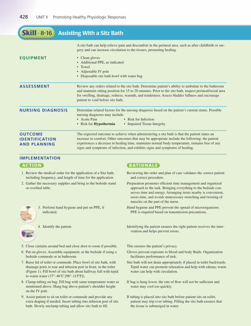
428 UNIT II Promoting Healthy Physiologic Responses
Assisting With a Sitz Bath• 8-16
A sitz bath can help relieve pain and discomfort in the perineal area, such as after childbirth or sur-
gery and can increase circulation to the tissues, promoting healing.
• Clean gloves
• Additional PPE, as indicated
• Towel
• Adjustable IV pole
• Disposable sitz bath bowl with water bag
Review any orders related to the sitz bath. Determine patient’s ability to ambulate to the bathroom
and maintain sitting position for 15 to 20 minutes. Prior to the sitz bath, inspect perineal/rectal area
for swelling, drainage, redness, warmth, and tenderness. Assess bladder fullness and encourage
patient to void before sitz bath.
Determine related factors for the nursing diagnosis based on the patient’s current status. Possible
nursing diagnoses may include:
• Acute Pain • Risk for Infection
• Risk for Hypothermia • Impaired Tissue Integrity
The expected outcome to achieve when administering a sitz bath is that the patient states an
increase in comfort. Other outcomes that may be appropriate include the following: the patient
experiences a decrease in healing time, maintains normal body temperature, remains free of any
signs and symptoms of infection, and exhibits signs and symptoms of healing.
EQUIPMENT
ASSESSMENT
NURSING DIAGNOSIS
OUTCOME IDENTIFICATION AND PLANNING
IMPLEMENTATION
AA C T IC T I OO NN RR AA T IT I OO N A L EN A L E
1. Review the medical order for the application of a Sitz bath,
including frequency, and length of time for the application.
2. Gather the necessary supplies and bring to the bedside stand
or overbed table.
3. Perform hand hygiene and put on PPE, if
indicated.
4. Identify the patient.
5. Close curtains around bed and close door to room if possible.
6. Put on gloves. Assemble equipment; at the bedside if using a
bedside commode or in bathroom.
7. Raise lid of toilet or commode. Place bowl of sitz bath, with
drainage ports to rear and infusion port in front, in the toilet
(Figure 1). Fill bowl of sitz bath about halfway full with tepid
to warm water (37�–46�C [98�–115�F]).
8. Clamp tubing on bag. Fill bag with same temperature water as
mentioned above. Hang bag above patient’s shoulder height
on the IV pole.
9. Assist patient to sit on toilet or commode and provide any
extra draping if needed. Insert tubing into infusion port of sitz
bath. Slowly unclamp tubing and allow sitz bath to fill.
Reviewing the order and plan of care validates the correct patient
and correct procedure.
Preparation promotes efficient time management and organized
approach to the task. Bringing everything to the bedside con-
serves time and energy. Arranging items nearby is convenient,
saves time, and avoids unnecessary stretching and twisting of
muscles on the part of the nurse.
Hand hygiene and PPE prevent the spread of microorganisms.
PPE is required based on transmission precautions.
Identifying the patient ensures the right patient receives the inter-
vention and helps prevent errors.
This ensures the patient’s privacy.
Gloves prevent exposure to blood and body fluids. Organization
facilitates performance of task.
Sitz bath will not drain appropriately if placed in toilet backwards.
Tepid water can promote relaxation and help with edema; warm
water can help with circulation.
If bag is hung lower, the rate of flow will not be sufficient and
water may cool too quickly.
If tubing is placed into sitz bath before patient sits on toilet,
patient may trip over tubing. Filling the sitz bath ensures that
the tissue is submerged in water.
LWBK545_C08_p358-435.qxd 08/07/2010 1:58 PM Page 428 Aptara
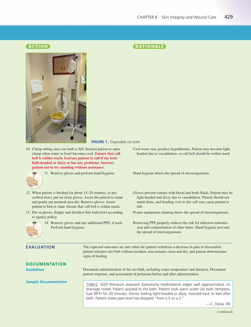
CHAPTER 8 Skin Integrity and Wound Care 429
AA C T IC T I OO NN RR AA T IT I OO N A L EN A L E
10. Clamp tubing once sitz bath is full. Instruct patient to open
clamp when water in bowl becomes cool. Ensure that callbell is within reach. Instruct patient to call if she feelslight-headed or dizzy or has any problems. Instructpatient not to try standing without assistance.
11. Remove gloves and perform hand hygiene.
12. When patient is finished (in about 15–20 minutes, or pre-
scribed time), put on clean gloves. Assist the patient to stand
and gently pat perineal area dry. Remove gloves. Assist
patient to bed or chair. Ensure that call bell is within reach.
13. Put on gloves. Empty and disinfect Sitz bath bowl according
to agency policy.
14. Remove gloves and any additional PPE, if used.
Perform hand hygiene.
Cool water may produce hypothermia. Patient may become light-
headed due to vasodilation, so call bell should be within reach.
Hand hygiene deters the spread of microorganisms.
Gloves prevent contact with blood and body fluids. Patient may be
light-headed and dizzy due to vasodilation. Patient should not
stand alone, and bending over to dry self may cause patient to
fall.
Proper equipment cleaning deters the spread of microorganisms.
Removing PPE properly reduces the risk for infection transmis-
sion and contamination of other items. Hand hygiene prevents
the spread of microorganisms.
FIGURE 1. Disposable sitz bath.
EVALUATION The expected outcomes are met when the patient verbalizes a decrease in pain or discomfort,
patient tolerates sitz bath without incident, area remains clean and dry, and patient demonstrates
signs of healing.
Document administration of the sitz bath, including water temperature and duration. Document
patient response, and assessment of perineum before and after administration.
DOCUMENTATIONGuidelines
7/30/12 1620 Perineum assessed. Episiotomy mediolateral; edges well approximated, nodrainage noted. Patient assisted to sitz bath. Patient took warm water sitz bath (tempera-ture 99�F) for 20 minutes. Denies feeling light-headed or dizzy. Assisted back to bed afterbath. Patient states pain level has dropped “from a 5 to a 2.”
—C. Stone, RN
Sample Documentation
(continued)
LWBK545_C08_p358-435.qxd 08/07/2010 1:58 PM Page 429 Aptara
430 UNIT II Promoting Healthy Physiologic Responses
Assisting With a Sitz Bath continued• 8-16
UNEXPECTEDSITUATIONS ANDASSOCIATEDINTERVENTIONS
• Patient complains of feeling light-headed or dizzy during sitz bath: Stop sitz bath. Do not attempt
to ambulate patient alone. Use call bell to summon help. Let patient sit on toilet until feeling sub-
sides or help has arrived to assist patient back to bed.
• Temperature of water is uncomfortable: The water may be too warm or cold, depending on the
patient’s preference. If this happens, clamp the tubing, disconnect the water bag, and refill it with
water that is comfortable for the patient, but no warmer than 115�F (46�C).
Applying Cold Therapy• 8-17
Cold constricts the peripheral blood vessels, reducing blood flow to the tissues and decreasing the
local release of pain-producing substances. Cold reduces the formation of edema and inflammation,
reduces muscle spasm, and promotes comfort by slowing the transmission of pain stimuli. The
application of cold therapy reduces bleeding and hematoma formation. The application of cold,
using ice, is appropriate after direct trauma, for dental pain, for muscle spasms, after muscle
sprains, and for the treatment of chronic pain. Ice can be used to apply cold therapy, usually in the
form of an ice bag or ice collar, or in a glove. Commercially prepared cold packs are also available.
For electronically-controlled cooling devices, see the accompanying Skill Variation.
• Ice
• Ice bag, ice collar, glove
• Commercially prepared cold packs
• Small towel or washcloth
• PPE, as indicated
• Disposable waterproof pad
• Gauze wrap or tape
• Bath blanket
Assess the situation to determine the appropriateness for the application of cold therapy. Assess the
patient’s physical and mental status and the condition of the body area to be treated with the cold
therapy. Confirm the medical order, including frequency, type of therapy, body area to be treated,
and length of time for the application. Assess the equipment to be used to make sure it will function
properly.
Determine the related factors for the nursing diagnoses based on the patient’s current status. An
appropriate nursing diagnosis is Acute Pain. Other nursing diagnoses that may be appropriate or
require the use of this skill include:
• Impaired Skin Integrity • Ineffective Tissue Perfusion
• Delayed Surgical Recovery • Chronic Pain
The expected outcome to achieve when applying an external cold source depends on the patient’s
nursing diagnosis. Outcomes that may be appropriate include the following: the patient experiences
increased comfort; the patient experiences decreased muscle spasms; the patient experiences decreased
inflammation; and the patient does not show signs of bleeding or hematoma at the treatment site.
EQUIPMENT
ASSESSMENT
NURSING DIAGNOSIS
OUTCOME IDENTIFICATION AND PLANNING
IMPLEMENTATION
AA C T IC T I OO NN RR AA T IT I OO N A L EN A L E
1. Review the medical order or nursing plan of care for the appli-
cation of cold therapy, including frequency, type of therapy,
body area to be treated, and length of time for the application.
Reviewing the order validates the correct patient and correct
procedure.
LWBK545_C08_p358-435.qxd 08/07/2010 1:58 PM Page 430 Aptara
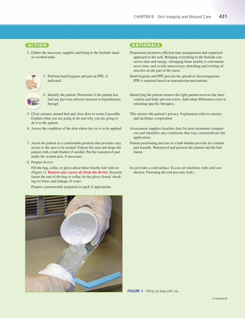
CHAPTER 8 Skin Integrity and Wound Care 431
AA C T IC T I OO NN RR AA T IT I OO N A L EN A L E
2. Gather the necessary supplies and bring to the bedside stand
or overbed table.
3. Perform hand hygiene and put on PPE, if
indicated.
4. Identify the patient. Determine if the patient has
had any previous adverse reaction to hypothermia
therapy.
5. Close curtains around bed and close door to room if possible.
Explain what you are going to do and why you are going to
do it to the patient.
6. Assess the condition of the skin where the ice is to be applied.
7. Assist the patient to a comfortable position that provides easy
access to the area to be treated. Expose the area and drape the
patient with a bath blanket if needed. Put the waterproof pad
under the wound area, if necessary.
8. Prepare device:
Fill the bag, collar, or glove about three-fourths full with ice
(Figure 1). Remove any excess air from the device. Securely
fasten the end of the bag or collar; tie the glove closed, check-
ing for holes and leakage of water.
Prepare commercially prepared ice pack if appropriate.
Preparation promotes efficient time management and organized
approach to the task. Bringing everything to the bedside con-
serves time and energy. Arranging items nearby is convenient,
saves time, and avoids unnecessary stretching and twisting of
muscles on the part of the nurse.
Hand hygiene and PPE prevent the spread of microorganisms.
PPE is required based on transmission precautions.
Identifying the patient ensures the right patient receives the inter-
vention and helps prevent errors. Individual differences exist in
tolerating specific therapies.
This ensures the patient’s privacy. Explanation relieves anxiety
and facilitates cooperation.
Assessment supplies baseline data for post-treatment compari-
son and identifies any conditions that may contraindicate the
application.
Patient positioning and use of a bath blanket provide for comfort
and warmth. Waterproof pad protects the patient and the bed
linens.
Ice provides a cold surface. Excess air interferes with cold con-
duction. Fastening the end prevents leaks.
FIGURE 1. Filling ice bag with ice.
(continued)
LWBK545_C08_p358-435.qxd 08/07/2010 1:58 PM Page 431 Aptara
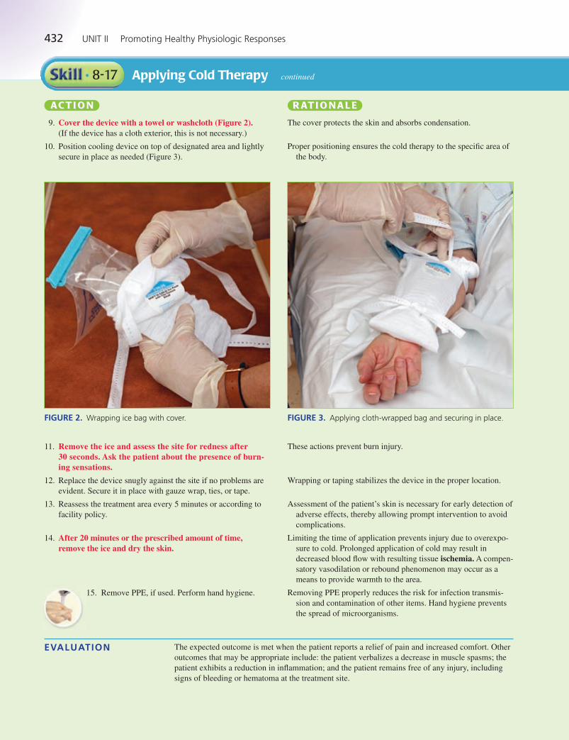
432 UNIT II Promoting Healthy Physiologic Responses
Applying Cold Therapy continued• 8-17
AA C T IC T I OO NN RR AA T IT I OO N A L EN A L E
9. Cover the device with a towel or washcloth (Figure 2).(If the device has a cloth exterior, this is not necessary.)
10. Position cooling device on top of designated area and lightly
secure in place as needed (Figure 3).
The cover protects the skin and absorbs condensation.
Proper positioning ensures the cold therapy to the specific area of
the body.
FIGURE 2. Wrapping ice bag with cover. FIGURE 3. Applying cloth-wrapped bag and securing in place.
11. Remove the ice and assess the site for redness after 30 seconds. Ask the patient about the presence of burn-ing sensations.
12. Replace the device snugly against the site if no problems are
evident. Secure it in place with gauze wrap, ties, or tape.
13. Reassess the treatment area every 5 minutes or according to
facility policy.
14. After 20 minutes or the prescribed amount of time,remove the ice and dry the skin.
15. Remove PPE, if used. Perform hand hygiene.
These actions prevent burn injury.
Wrapping or taping stabilizes the device in the proper location.
Assessment of the patient’s skin is necessary for early detection of
adverse effects, thereby allowing prompt intervention to avoid
complications.
Limiting the time of application prevents injury due to overexpo-
sure to cold. Prolonged application of cold may result in
decreased blood flow with resulting tissue ischemia. A compen-
satory vasodilation or rebound phenomenon may occur as a
means to provide warmth to the area.
Removing PPE properly reduces the risk for infection transmis-
sion and contamination of other items. Hand hygiene prevents
the spread of microorganisms.
EVALUATION The expected outcome is met when the patient reports a relief of pain and increased comfort. Other
outcomes that may be appropriate include: the patient verbalizes a decrease in muscle spasms; the
patient exhibits a reduction in inflammation; and the patient remains free of any injury, including
signs of bleeding or hematoma at the treatment site.
LWBK545_C08_p358-435.qxd 08/07/2010 1:58 PM Page 432 Aptara
CHAPTER 8 Skin Integrity and Wound Care 433
DOCUMENTATIONGuidelines Document the location of the application, time of placement and time of removal. Record the
assessment of the area where the cold therapy was applied, including the patient’s mobility, sensa-
tion, color, temperature, and any presence of numbness, tingling, or pain. Document the patient’s
response, such as any decrease in pain or change in sensation. Include any pertinent patient and
family education.
11/1/12 1430 Swelling noted on right lower extremity from mid-calf to foot. Toes warm,pink, positive sensation and movement, negative for numbness, tingling, and pain. Ice bagswrapped in cloth applied to right ankle and lower calf. Patient instructed to communicateany changes in sensation or pain; verbalizes an understanding of information.
—L. Semet, RN
11/1/12 1450 Ice removed from right lower extremity; neurovascular assessment unchanged.Right lower extremity elevated on two pillows.
—L. Semet, RN
Sample Documentation
UNEXPECTEDSITUATIONS ANDASSOCIATEDINTERVENTIONS
• When performing a skin assessment during therapy, the nurse notes increased pallor at the treat-ment site and sluggish capillary refill, and the patient reports alterations in sensation at theapplication site: Discontinue therapy, obtain vital signs, assess for other symptoms, notify the
primary care provider, and document the event in the patient’s record.
• The patient may experience a secondary defense reaction, vasodilation, that causes body temper-
ature to rebound, defeating the purpose of the therapy.
• Older adults are more at risk for skin and tissue damage because of their thin skin, loss of cold
sensation, decreased subcutaneous tissue, and changes in the body’s ability to regulate tempera-
ture. Check these patients more frequently during therapy.
SPECIALCONSIDERATIONSGeneral Considerations
Older Adult Considerations
Skill Variation Applying an Electronically-Controlled Cooling Device
4. Assess the involved extremity or body part.
5. Set the correct temperature on the device.
6. Wrap the cooling water-flow pad around the involved body
part.
7. Wrap Ace bandage or gauze pads around the water-flow
pads.
8. Assess to ensure that the cooling pads are functioning
properly.
9. Remove PPE, if used. Perform hand hygiene.
10. Recheck frequently to ensure proper functioning of
equipment.
11. Unwrap at intervals to assess skin integrity of the body
part.
Electronically controlled cooling devices are used in situations
to deliver a constant cooling effect. Postoperative orthopedic
patients as well as other patients with acute musculoskeletal
injuries may benefit from this therapy. A medical order is
required for use of this device. Initial assessment of the extrem-
ity is involved, as well as ongoing assessment throughout the
period of use. As with application of any electronic device,
ongoing monitoring for proper functioning and temperature
regulation is necessary.
1. Gather equipment and verify the medical order.
2. Perform hand hygiene. Put on PPE, as
indicated.
3. Identify the patient and explain the procedure.
LWBK545_C08_p358-435.qxd 08/07/2010 1:59 PM Page 433 Aptara
434 UNIT II Promoting Healthy Physiologic Responses
ENHANCE YOUR UNDERSTANDING
● Integrated Case Study Connection
The case studies in the back of the book are designed to focus
on integrating concepts. Refer to the following case studies to
enhance your understanding of the concepts related to the
skills in this chapter.
• Basic Case Studies: Tula Stillwater, page 965
• Intermediate Case Studies: Tula Stillwater, page 972
• Advanced Case Studies: Robert Espinoza, page 987
● Developing Critical Thinking Skills
1. While providing wound care for Lori Downs’ foot ulcer,
you note that the drainage, which was scant and yellow
yesterday, is now green and has saturated the old dressing.
Should you continue with the prescribed wound care?
2. Three days ago Tran Nguyen underwent a modified radi-
cal mastectomy. She has three Jackson-Pratt drains at
her surgical site. She has started asking questions about
her surgery and anticipated discharge home. Until this
morning, she has avoided looking at her surgical site.
You are helping her with her bathing and dressing. As
you help her remove her gown, she becomes visibly
upset and anxious and exclaims, “Oh no! What’s wrong?
I’m bleeding from the cuts!” You realize she is looking
at her drains. How should you respond?
3. Arthur Lowes has come to his surgeon’s office today for
a follow-up examination after a colon resection. After he
sees the physician, you, the treatment nurse, will remove
the surgical staples from the incision and apply adhesive
wound strips. As you prepare to remove the staples, Mr.
Lowes comments, “I hope my stomach doesn’t pop out
now!” What should you tell him?
● Suggested Answers for DevelopingCritical Thinking Skills
1. This is a significant change is the patient’s assessment.
Perform a thorough wound assessment and obtain vital
signs. Assess the patient for any new symptoms, such as
increased pain, chills, or abnormal sensation (such as
numbness, tingling). Report findings to the primary care
provider; a change in wound care, additional assess-
ments (such as diagnostic tests, laboratory tests), or
change/addition of medication may be required.
2. Reassure the patient regarding her wound status. Explain
what the drains are, how they work, and the intended
purpose. Provide information regarding wound care,
drain care, and recording of drainage amounts. Discuss
anticipated care requirements at home and potential
arrangements to ensure required care is performed,
either by the patient or significant other.
3. Reassure the patient regarding his wound status. Explain
the purpose of the staples, the process of wound healing,
and the purpose of adhesive wound strips. Discuss the
patient’s responsibilities for wound care at this point in
his healing.
Taylor Suite Resources
The Taylor Suite offers these additional resources to enhance
learning and facilitate understanding of this chapter:
• thePoint online resource, http://thepoint.lww.com/Lynn3e
• Student DVD-ROM included with the book
• Skill Checklists for Taylor’s Clinical Nursing Skills
• Taylor’s Video Guide to Clinical Nursing Skills: SkinIntegrity and Wound Care
• Fundamentals of Nursing: Chapter 32, Skin Integrity andWound Care
BIBLIOGRAPHYAndrews, M., & Boyle, J. (2008). Transcultural concepts in
nursing care. (5th ed.). Philadelphia: Wolters Kluwer Health/Lippincott Williams & Wilkins.
Association of Operating Room Nurses (AORN). (2006). Recom-mended practices for maintaining a sterile field. AORN Journal,83(2), 402–16.
Association for Professionals in Infection Control and Epidemiol-ogy (APIC). Guideline for prevention of surgical site infection.Source: Center for Disease Control and Prevention (CDC).Hospital Infection Control Practices Advisory Committee.Mangram, A., Horan, T., & Jarvise, W. (1999). American Journal of Infection Control, 27(2), 97–132. Availablewww.apic.org/AM/Template.cfm?Section�Guidelines-_and_Standards&template�/CM/ContentDisplay.cfm§ion�Topics1&ContentID�1148. Accessed January 14, 2009.
Benbow, M. (2008a). Exploring the concept of moist wound heal-ing and its application in practice. British Journal of Nursing,(Tissue viability supplement), 17(15), S4–S16.
Benbow, M. (2008b). Pressure ulcer prevention and pressure–relieving surfaces. British Journal of Nursing, 17(13), 830–35.
Bergstrom, N., Braden, B., Laguzza, A., & Holman, V. (1987).The Braden scale for predicting pressure sore risk. NursingResearch, 36(4), 205–210.
Bookout, K. (2008). Wound care product primer for the nurse prac-titioner: Part I. Journal of Pediatric Health Care, 22(1), 60–3.
Braden, B., & Maklebust, J. (2005). Preventing pressure ulcerswith the Braden scale. American Journal of Nursing, 105(6),70–72.
Brillhart, B. (2006). Preventive skin care for older adults. Geri-atrics & Aging, 9(5), 334–39.
Bulechek, G., Butcher, H., & McCloskey Dochterman, J. (Eds.).(2008). Nursing interventions classification (NIC) (5th ed.). St. Louis, MO: Mosby Elsevier.
Burr, S., & Penzer, R. (2005). Promoting skin health. NursingStandard, 19(36), 57–65.
Butler, C. (2006). Pediatric skin care: Guidelines for assessment,prevention, and treatment. Pediatric Nursing, 32(5), 443–50.
Carpenito-Moyet, L. (2008). Nursing diagnosis: Application toclinical practice. (12th ed.). Philadelphia: Wolters KluwerHealth/Lippincott Williams & Wilkins.
Edmonds, JU. (2007). Nutrition and wound healing: Putting the-ory into practice. Wound Care, 12(12), S31–S34.
LWBK545_C08_p358-435.qxd 08/07/2010 1:59 PM Page 434 Aptara
CHAPTER 8 Skin Integrity and Wound Care 435
Elliott, R., McKinley, S., & Fox, V. (2008). Quality improve-ment program to reduce the prevalence of pressure ulcers inan intensive care unit. American Journal of Critical Care,17(4), 328–35.
Ellis, J., & Bentz, P. (2007). Modules for basic nursing care (7thed). Philadelphia: Lippincott Williams & Wilkins.
Fischbach, F., & Dunning, M. (2009). A manual of laboratory anddiagnostic tests. (8th ed.). Philadelphia: Wolters Kluwer Health/Lippincott Williams & Wilkins.
Flores, A. (2008). Sterile versus non-sterile glove use and aseptictechnique. Nursing Standard, 23(6), 35–9.
Gray, M. (2007). Incontinence-related skin damage: Essentialknowledge. Ostomy Wound Management, 53(12), 28–32.
Halliday, K. (2005). Body piercing: Issues and challenges fornurses. Journal of Forensic Nursing, 1(2), 47–56.
Harvey, C. (2005). Wound healing. Orthopaedic Nursing, 24(2),143–157.
Hess, C. (2008). Skin & wound care (6th ed.). Philadelphia:Wolters Kluwer Health/Lippincott Williams & Wilkins.
Hockenberry, M. (2005). Wong’s essentials of pediatric nursing(7th ed.). St. Louis: Elsevier Mosby.
Jarvis, C. (2008). Physical Examination & Health Assessment. (5th ed.). St. Louis: Saunders/Elsevier.
Krasner, D. (1995). Wound care: How to use the red-yellow-blacksystem. American Journal of Nursing, 5(95), 44–47.
Kyle, T. (2008). Essentials of Pediatric Nursing. Philadelphia:Wolters Kluwer Health/Lippincott Williams & Wilkins.
Lindgren, M., Unosson, M., Krantz, A., & Ek, A. C. (2005). Pres-sure ulcer risk factors in patients undergoing surgery. Journalof Advanced Nursing, 50(6), 605–612.
Lloyd Jones, M. (2008). Assessing and managing wound painduring dressing changes. Nursing & Residential Care, 10(7),325–30.
Malli, S. (2005). Device safety. Keep a close eye on vacuum-assisted wound closure. Nursing, 35(7), 25.
Meltzer, D. (2005). Complications of body piercing. AmericanFamily Physician, 72(10), 2029–34.
Mendez-Eastman, S. (2005). Using negative-pressure for positiveresults. Nursing, 35(5), 48–50.
Moorhead, S., Johnson, M., Maas, M., et al. (Eds). (2008). Nurs-ing Outcomes Classification (NOC). (4th ed.). St. Louis:Mosby Elsevier.
National Pressure Ulcer Advisory Panel (NPUAP). (2001a). Pres-sure ulcer prevention: A competency-based curriculum. Avail-able www.npupa.org/PDF/prevcurr.pdf. Accessed January 14,2009.
National Pressure Ulcer Advisory Panel (NPUAP). (2001b). Pres-sure ulcer treatment: A competency-based curriculum. Avail-able www.npuap.org/PDF/treatment_curriculum.pdf. AccessedJanuary 14, 2009.
National Pressure Ulcer Advisory Panel (NPUAP). (2007a).Updated staging system. Pressure ulcer stages revised byNPUAP. Available www.npuap.org/pr2.htm. Accessed December 27, 2008.
National Pressure Ulcer Advisory Panel (NPUAP). (2007b).Updated staging system. Wound infection and infection control.Available www.npuap.org/pr2.htm. Accessed December 27,2008.
National Pressure Ulcer Advisory Panel (NPUAP). (2007c).Resources. Staging illustrations. Available www.npuap.org/resources.htm. Accessed January 9, 2009.
National Pressure Ulcer Advisory Panel (NPUAP). (2007d). Pres-sure ulcer prevention points. Available www.npuap.org/PU_Prev_Points.pdf. Accessed January 14, 2009.
Negative Pressure. (2008). British Journal of Nursing,2008–2009: Wound Care Handbook. Author. P. 164–8.CINAHL AN: 2010031958.
NANDA (2009). Nursing diagnoses: Definitions and classifica-tion 2009–2011. Philadelphia: Author.
Porth, C., & Matfin, G. (2009). Pathophysiology: Concepts ofaltered health states (8th ed.). Philadelphia: Wolters KluwerHealth/Lippincott Williams & Wilkins.
Preston, G. (2008). An overview of topical negative pressure ther-apy in wound care. Nursing Standard, 23(7), 62–8.
Preston, R. (2005). Aseptic technique: Evidence-based approachfor patient safety. British Journal of Nursing, 14(10), 540–2,544–6.
Reddy, M., Gill, S., & Rochon, P. (2006). Preventing pressureulcers: A systematic review. Journal of the American MedicalAssociation (JAMA), 296(8), 974–84, 1020.
Rudoni, C. (2008). A service evaluation of the use of silicone-based adhesive remover. British Journal of Nursing, StomaCare Supplement, 17(2), S4, S6, S8–9.
Saver, C. (2008). Cool kids: Children’s Hospital of Philadelphiauses therapeutic hypothermia in pediatric cases. Nursing Spec-trum, 17(22), 14–5.
Scardillo, J. (2005). Postoperative care of patients with surgicaldrains. Perspectives in Nursing, 4(4), 1, 4–6.
Scott, A. (2008). Wound care & cancer. Advance for Nurses,10(19), 29–30.
Smeltzer, S., Bare, B., Hinkle, J. H., & Cheever, K. H. (2010).Brunner & Suddarth’s textbook of medical-surgical nursing(12th ed.). Philadelphia: Wolters Kluwer Health/LippincottWilliams & Wilkins.
Snyder, L. (2008). Wound basics: Types, treatment, and care. RN,71(8), 32–7.
Stephen-Haynes, J. (2008). Skin integrity and silicone: APPEEL‘no-sting’ medical adhesive remover. British Journal of Nurs-ing, 17(12), 792–5.
Stephen-Haynes, J., & Thompson, G. (2007). The different meth-ods of wound debridement. British Journal of CommunityNursing, 12(6), Wound Care: S6, S8–10, S12–4.
Stotts, N. (1990). Seeing red, yellow and black: The three-colorconcept of wound care. Nursing, 20(2), 59–61.
Taylor, C., Lillis, C., LeMone, P., et al. (2011). Fundamentals ofNursing. (7th ed.). Philadelphia: Wolters Kluwer Health/Lippincott Williams & Wilkins.
Thompson, G. (2008). An overview of negative pressure woundtherapy (NPWT). Wound Care, 13(6), Wound Care: S23–4, S26, S28–30.
Vanderwee, K., Grypdonck, M., De Bacquer, D., et al. (2007).Effectiveness of turning with unequal time intervals on theincidence of pressure ulcer lesions. Journal of Advanced Nursing, 57(1), 59–68.
VISN 8 Patient Safety Center. (2009). Safe patient handling andmovement algorithms. Tampa, FL: Author. Available athttp://www.visn8.va.gov/patientsafetycenter/safePtHandling. Accessed April 23, 2010.
Voegeli, D. (2008). Care or harm: Exploring essential componentsin skin care regimens. British Journal of Nursing, 17(1), 24–9.
Walker, J. (2007). Patient preparation for safe removal of surgicaldrains. Nursing Standard, 21(49), 39–41.
Weber, J., & Kelley, J. (2007). Health assessment in nursing (3rded.). Philadelphia: Lippincott Williams & Wilkins.
Wooten, M., & Hawkins, K. (2005). WOCN position statement.Clean versus sterile: Management of chronic wounds. Availablewww.wocn.org/pdfs/WOCN_Library/Position_Statements/.Accessed January 14, 2009.
Worley, C. (2005a). So, what do I put on this wound? The wounddressing puzzle: Part I. Dermatology Nursing, 17(2), 143–144.
Worley, C. (2005b). So, what do I put on this wound? The wounddressing puzzle: Part II. Dermatology Nursing, 17(3), 204–205.
Worley, C. (2005c). So, what do I put on this wound? The wounddressing puzzle: Part III. Dermatology Nursing, 17(4), 299–300.
LWBK545_C08_p358-435.qxd 08/07/2010 1:59 PM Page 435 Aptara