Embed Size (px)
Citation preview

J. Anat. (1971), 109, 3, pp. 369-383 369With 11 figures
Printed in Great Britain
Patterns of lymphatic drainage in the adult laboratory rat
NICHOLAS L. TILNEY
Department of Surgery, Peter Bent Brigham Hospital, and HarvardMedical School, Boston, Massachusetts
(Accepted 27 April 1971)
INTRODUCTION
This study was undertaken to define and elucidate patterns of lymphatic drainagein the adult laboratory rat. The incentive for the work arose from investigations intothe role of regional lymphatics in the sensitization of the host by skin allografts. Ithas become clear that the response of rats to antigens, investigated increasingly inthe available inbred strains, requires an accurate knowledge of lymphoid anatomyand lymphatic drainage routes. Examinations of the lymphatics of specific bodyareas of the rat have appeared sporadically in the literature, but descriptions ofregional drainage patterns, especially of peripheral sites, are unavailable. Previousinvestigations by Job (1919), Greene (1935) and Sanders & Florey (1940) have con-centrated primarily upon the location of the lymphoid tissues. Miotti (1965) hasstressed visceral drainage, and Higgins (1925) has described the lymphatic system ofthe newborn rat. A more complete definition of both somatic and visceral lymphaticroutes is presented.
MATERIALS AND METHODS
One hundred and thirty normal adult rats of both sexes, each weighing between150 and 300 g, were studied. The animals came from five strains: each inbred -Oxford strains of the albino (AO), hooded (HO), agouti (DA), and F1 hybrid of theHO and DA strains - and 'stock' animals from a closed outbred albino colony.Under ether anaesthesia, the site for cutaneous injection was clipped or a serouscavity entered for visceral injection. The majority of animals were killed by etheroverdose and dissected within a few minutes because of the rapidity of uptake of theinjected material by lymphoid tissue. Several rats were studied between 48 h and6 weeks after injection to elucidate possible variations in the distribution of the dyewith time. Regional nodes and all secondary drainage sites were examined.
Three types of injected material were employed. The location of lymph nodes wasdemonstrated by the intraperitoneal injection of pontamine sky blue, 10-14 daysbefore dissection. This was administered as a sterile 5 00 solution in distilled waterat a dose of 1 ml per 100 g body weight. Macrophages coloured with dye remained inlymph nodes long after surrounding tissues have cleared and allowed the identificationof even minute lymphoid aggregations. The cutaneous areas were mapped by intra-dermal injections of 0-05-0d10 ml of colloidal carbon (Pelikan Ink, Guinther Wagner,Germany). This material filled lymphatic channels and readily stained draininglymph nodes. Similar injections of 1311 human serum albumin (Radiochemical Centre,
24 ANA 109

Amersham, Bucks, England) were used to confirm these patterns of drainage. Theregional lymph nodes were removed and their radioactive uptake measured in agamma scintillation spectrometer (Packard Series 410A).
Passive muscular motion was helpful in forcing the dye along peripheral lymph-atics. Subserosal injections of hollow viscera or subcapsular injections of solid organsusing a no. 30 needle satisfactorily demonstrated visceral lymphatic patterns. Thelymph trunks between lymph node groups were visualized by injections of minutevolumes of India ink directly into the nodal substance: gentle external pressure onthe node caused filling of the efferent lymphatics. Accurate localization of 'lymphatictaps' - the valved junctions of the major lymph ducts with the subclavian veins - wasfacilitated by total replacement of the circulating blood volume of the animal withsaline. Carbon particles could then easily be seen flowing from the lymph ducts intothe veins.Lymph node groups have been classified into somatic nodes, which drain the skin
and underlying musculature (Table 1), and visceral nodes, which drain primarily thethoracic, abdominal and pelvic organs (Table 2). Somatic nodes generally lie externalto skeletal muscle in subcutaneous or areolar tissue, or in fossae between musclemasses. These nodes have been divided functionally into a peripheral and centralgroup. The peripheral group of somatic nodes drains skin and musculo-skeletal sitesonly. The central group drains similar anatomical areas, but also receives lymphaticchannels from peripheral nodes.The nomenclature of the lymph nodes is that of Job (1915) and Sanders & Florey
(1940), although modifications have been made to emphasize specific drainagepatterns (Fig. 1). 'Lymph trunks' were defined as the large channels connectingvarious groups of nodes. 'Lymphatic ducts' drain particular groups of nodes andempty directly into the cisterna chyli, or into the subclavian veins through the'lymphatic taps'.
RESULTS
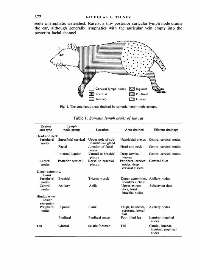
The location of lymphoid tissue and distribution of lymphatic channels was con-sistent in individual animals and among the strains of rats used in the study, despiteoccasional variation in size and number of nodes. Well-defined cutaneous areas weredrained by specific groups of nodes (Fig. 2). Nodes within these groups draineddistinct sites, although there was usually some overlap between adjacent nodes.Extensive lymphatic plexuses, present throughout the subdermis and in the sub-mucosa of hollow viscera, emptied into constant lymph channels which generallyfollowed veins toward the regional nodes. No peripheral channels entered any bodycavity with perforating blood vessels. Junctions between lymphatics and veins otherthan at the subclavian veins, or direct lymphatico-venous anastomoses within lymphnodes themselves, were not demonstrated.
Somatic nodesHead and neckThe peripheral lymph nodes draining the head and neck are the superficial
cervical, facial and internal jugular groups (Fig. 3). The superficial cervical nodes lieat the upper poles of the submandibular glands and drain the tongue and nasolabial
370 NICHOLAS L. TILNEY

Patterns of lymphatic drainage in the adult laboratory ratlymphatic plexus through channels running between the borders of the digastric andmasseter muscles. A small efferent lymphatic curls around the lateral aspect of eachsubmandibular gland to join efferent channels from the facial nodes, or to enter thisgroup directly.
Fig. 1. Lymph node groups in the dissected adult rat. Nodes lying dorsally are demonstrated byreflecting muscles and viscera. Adapted from Sanders & Florey (1940).
The facial nodes lie dorsal to the lower poles of the submandibular glands at thejunction of the anterior and posterior facial veins with the external jugular veins.They drain the skin of the head and the ventral aspect and sides of the neck throughlymph vessels running with the corresponding facial veins. A large efferent facialtrunk loops laterally around each sternocleidomastoid muscle to enter the posteriorcervical nodes. As has been noted by Higgins (1925) in studies of newborn rats, asmall area of the forehead has minimal lymphatic drainage and presumably repre-
24-2
371
372 NICHOLAS L. TILNEY
sents a lymphatic watershed. Rarely, a tiny posterior auricular lymph node drainsthe ear, although generally lymphatics with the auricular vein empty into theposterior facial channel.
/Cz Cervical lymph nodes E] Inguinal \9
5 Brachial m Popliteal= Axillary Gluteal
Fig. 2. The cutaneous areas drained by somatic lymph node groups.
Table 1. Somatic lymph nodes of the rat
Region Lymphand type node group Location Area drained Efferent drainage
Head and neckPeripheral Superficial cervical Upper pole of sub- Nasolabial plexus Central cervical nodesnodes mandibular gland
Facial Junction of facial Head and neck Central cervical nodesveins
Internal jugular Ventral to brachial Deep cervical Central cervical nodesplexus viscera
Central Posterior cervical Dorsal to brachial Peripheral cervical Cervical ductnodes plexus nodes, deep
cervical visceraUpper extremity,
TrunkPeripheral Brachial Triceps muscle Upper extremities, Axillary nodesnodes shoulders, chest
Central Axillary Axilla Upper extrem- Subclavian ductnodes ities, trunk,
brachial nodesHindquarters,
Lowerextremit yPeripheral Inguinal Flank Thigh, haunches, Axillary nodesnodes scrotum, lateral
tailPopliteal Popliteal space Foot, hind leg Lumbar, inguinal
nodesTail Gluteal Sciatic foramen Tail Caudal, lumbar,
inguinal, poplitealnodes

Patterns of lymphatic drainage in the adult laboratory ratThe internal jugular nodes lie close to the posterior cervical nodes, but belong
functionally to the peripheral group. They are ventral to each brachial plexus,immediately lateral to the carotid sheath, and drain the pharynx, larynx and theproximal part of the oesophagus through pharyngeal lymphatics running along thesurface of the deep cervical muscles. A short lymphatic from each internal jugularnode enters the adjacent posterior cervical node directly.The posterior cervical nodes lie dorsal to the brachial plexus on each side. These
single central nodes drain all peripheral nodes in the neck and may receive minortributaries from deep cervical structures. They empty into the posterior aspect ofeach subclavian vein through large cervical lymph ducts which run into the thoraxdorsal to the carotid sheaths.
Anterior facial lymphatic M ms.Nasolabial lymphatic Masseter ma.
Superficial cervical nodesDiatcm.
Parotid gland Submandibular gland
Internal jugular node Posterior facial lymphaticIS (I - ~~~~~Pharyngeal lymphatic
Facial nodes /Facial nodesJ, ~~~~~~~~~~~~Facialtrunk
Posterior cervical node v Brachial plexusCarotid a. -__________ External jugular v.
Sternocleidomastoid msavical duct
Fig. 3. Peripheral and deep cervical lymph nodes and lymphatic channels of the head and neck.The arrows demonstrate the direction of lymph flow. The cervical ducts run dorsal to thecarotid sheaths and enter the subclavian veins on either side.
Upper extremity and trunkThe peripheral group of brachial nodes lies within a fascial envelope high on each
triceps muscle. Usually three in number, they drain the forefoot and front leg,shoulders, neck and upper chest (Fig. 4). The dorsal member of the group drainsthe radial aspect of forefoot and distal foreleg through the afferent cephalic lymphvessel. This follows the cephalic vein and curves across the lateral aspect of theshoulder to enter the node. The intermediate brachial node drains the shoulder anddorsum of the neck through several small lymphatic branches. The ventral brachialnode drains the entire foot, foreleg and anterior chest through the brachial lymph-atic. This runs with the brachial vein, but divides to enter the ventral brachial andthe most rostral member of the group of axillary lymph nodes. A short radicle fromeach brachial node forms an efferent trunk which pierces the latissimus dorsi muscleand drains into the most caudal axillary node.The central somatic group of four axillary nodes lies along the chest wall at the
373

NICHOLAS L. TILNEY
junction of the lateral thoracic and cutaneous maximus veins. These drain the entiretrunk and foreleg, and receive efferent trunks from brachial and inguinal groups(Fig. 5). The caudal axillary node receives a large efferent channel from the inguinalnodes which follows the superficial epigastric vein cephalad along the milk line anddrains the nipples and the ventral aspects of chest and abdomen. The two inter-mediate axillary nodes drain the flanks and back through a major lymph vessel which
Fig. 4 Fig. 5
Fig. 4. Cutaneous lymphatic drainage of individual brachial lymph nodes.Fig. 5. Cutaneous lymphatic drainage of individual axillary lymph nodes.
joins them by traversing the panniculus carnosus muscle with the cutaneous maximusvein. A minor branch of this lymphatic continues in the deep dermis to the inter-mediate brachial node. The rostral axillary node receives the brachial lymph channelfrom the medial side of the arm and axilla. The large subclavian lymphatic ductruns from this node to enter the thorax with the axillary vein and join the subclavianvein with the other lymphatic taps.
374

Patterns of lymphatic drainage in the adult laboratory rat
Hind quartersThe paired inguinal nodes are embedded in subcutaneous fat in the flanks near the
superficial epigastric veins and drain the gluteal regions, thighs and lower abdomen(Fig. 6). The caudal member of the pair drains the hamstring, gluteal and perinealareas and receives channels which cross the groin from the lateral aspects of the tail,scrotum, or vagina. The rostral member drains the anterior thighs and flanks throughseveral small lymph channels. The large efferent inguinal lymphatic trunk coursescephalad along each nipple line to the axillary chains.
An nguinaliode
X,/.
Posterior inguinal nodes
Fig. 6. Cutaneous lymphatic drainage of individual inguinal lymph nodes.
The single popliteal node lies in the lateral aspect of the popliteal space near thesuperficial muscular vein (Fig. 7). It drains the footpad, foot and hind leg throughlymph vessels running with greater and lesser saphenous veins. Efferent poplitealtrunks follow each femoral vein to a retroperitoneal lymphatic plexus dorsal to theiliac vessels. The main trunk continues centrally to the iliac nodes, while smallertributaries travel with the superficial epigastric vessels to the inguinal nodes.
TailThe lymphatic drainage of the tail involves three pathways. Caudal channels along
its lateral aspect follow the superior gluteal veins to the gluteal lymph nodes. Thesesingle nodes lie deep to the gluteal muscles at each sciatic foramen (Fig. 7). Anefferent trunk drains to the lymphatic plexus dorsal to each iliac vein and ultimatelyto the para-aortic lymph chain. Other vessels from the tail course across the lateralscrotal wall and groin to the inguinal nodes. The ventral surface of the tail is drainedseparately by a channel which follows the middle sacral vein retroperitoneally throughthe pelvis to the caudal nodes. A minor efferent lymphatic trunk from each glutealnode runs with the inferior gluteal vein to the popliteal node, which serves as asecondary drainage site for the tail.
375

NICHOLAS L. TILNEY
Flexor caudi longus ms.Catudal lymphatics
Caudal v.(branch of superior gluteal v.)
N1
Fig. 7. Dissection of the gluteal area. The gluteal lymph node lies deep to the reflected gluteusmaximus muscle. The caudal lymphatic trunk enters the pelvis at the sciatic foramen and runsto the para-aortic lymph chain. A small efferent channel from the gluteal node runs with theinferior gluteal vein to the popliteal node.
Visceral drainageThoraxThe major lymphatic structures in the thorax are the thymus gland, and the para-
thymic and posterior mediastinal lymph nodes. The parathymic nodes are embeddedin fat on the lateral aspects of the thymic capsule, and drain the thymus gland, theperitoneal cavity, and the superior surface of the liver and liver capsule throughthe large internal thoracic lymph channels. These channels collect the effluent fromthe extensive lymphatic plexus on the pleural surface of the diaphragm and are joinedalong their course by small radicles from the anterior pericardium and parasternalarea (Fig. 8). MacCallum (1903) and Olin & Saldeen (1964) have shown that fluidor particles injected into the peritoneal cavity may pass directly through the fene-strated basement membrane of mesothelial cells on the peritoneal surface of thediaphragm, enter lymphatics between phrenic muscle bundles and empty into thediaphragmatic plexus.The posterior mediastinal nodes lie adjacent to the oesophagus on the right, and
to the superior vena cava on the left. The right posterior mediastinal node is usuallylarger and drains the right pleural space and lung, the base of the heart, and thethoracic portion of the oesophagus. The left node drains the left pleural space,thoracic viscera, and the thymus gland. These nodes receive minor paravertebrallymphatics from the diaphragmatic plexus, and are joined by intercostal lymphaticsand hilar radicles from the thoracic organs and pericardium. Although no suchanastomoses were found in this study, Kluge & Ongre (1968), using thorotrast,suggested that pericardial channels drain directly into rostral and caudal portionsof the thoracic duct. Small paravertebral nodes, present inconstantly behind the
376

Patterns of lymphatic drainage in the adult laboratory rat 377
pulmonary vessels, send efferent channels to the posterior mediastinal nodegroup.The thymus lies in the anterior mediastinum, drained directly by the parathymic
nodes. Other thymic lymphatics consistently empty into the left posterior medi-astinal node, although no drainage to the opposite node was ever demonstrated bythe techniques in this study.The mediastinal lymph ducts, major efferent channels from the thoracic nodes of
both sides, empty into the dorsum of the subclavian veins. The ducts from parathymicand posterior mediastinal nodes may merge as a single channel or enter the veinseparately. Histological examination of the 'mediastinal organ', mentioned bySanders & Florey (1940), showed only brown fat.
Table 2. Visceral lymph nodes of the rat
LymphRegion node group Location Area drained Efferent drainage
Thorax Parathymic
Posterior medi-astinal
Pelvis andRetroperi-toneum
Lateral to thymus
Paravertebralgutter
Paravertebral Dorsal to pul-monary vessels
Caudal Median sacral vein
Iliac
Para-aortic
Renal
External lumbar
Abdomen Splenic
Posterior gastric
Portal
Superiormesenteric
Inferiormesenteric
Aortic bifurcation
Para-aortic area
Dorsal to renalveins
Retroperitonealfat pad
Splenic vein
Gastroduodenalvein
Portal vein
Root ofmesentery
Mesentery ofdescending colon
Peritoneal cavity,liver, pericardium,thymus
Thoracic viscera,pleural space,pericardium,thymusDiaphragm, thoracicviscera
Ventral tail, anus,rectum, glutealnodesPelvic viscera, pop-liteal, gluteal,caudal nodes
Pelvic viscera,popliteal, gluteal,caudal nodes
Kidneys, supra-renals, lumbarlymphatics
Fat pad, psoasmuscles, pelvicviscera
Splenic capsule andtrabeculae
Distal oesophagus,stomach, pancreas,splenic node
Liver, splenic,posterior gastricnodesDuodenum, smallbowel, caecum,ascending, trans-verse colon
Descending andsigmoid colon
Mediastinal duct
Mediastinal duct
Posterior medi-astinal nodes
Iliac nodes
Renal nodes
Renal nodes
Renal duct tocisterna chyli
Lumbar lymphatics
Posterior gastricnodes
Portal nodes
Portal duct tocisterna chyli
Superiormesenteric ductto cisterna
Inferior mesentericduct to cisterna

378
Mediastinal ductsParathymic nodes
Posterior mediastinal nodesInternal mammary a.+v.
Internal mammary lymphatic
Sternum
NICHOLAS L. TILNEY
-Internal jugular v.
Subclavian v.
,vessels (ligated)
Intercostal lymphatics
- Diaphragm
Fig. 8. The sternum has been reflected to the right and the left anterior ribs removed. The dia-phragmatic lymphatic plexus drains into the large internal thoracic (mammary) lymphatics, whichenter the parathymic lymph nodes. Mediastinal ducts from the parathymic and posteriormediastinal nodes enter the subclavian veins.
Retroperitoneunm and pelvisThe nodes draining retroperitoneal and pelvic organs lie adjacent to the abdominal
aorta and inferior vena cava (Fig. 9). The small caudal nodes lie with the mediansacral vein at the aortic bifurcation, and drain the rectum, internal anal sphincterand ventral aspect of the tail through caudal channels running along the sacrum.Efferent trunks cross the iliac vessels and enter the iliac nodes. Other pelvic lymph-atics, especially from testis or ovary, may enter the caudal group or bypass it anddrain to nodes along the para-aortic lymph chain.The large iliac nodes lie along each side of the distal aorta, singly or in pairs. They
drain pelvic viscera through extensive lymphatic plexuses around the spermatic orovarian veins, and along branches of the internal iliac veins. They are importantsecondary drainage sites for caudal, gluteal and popliteal node groups. Efferentpara-aortic trunks lead to the renal nodes through an inconstant pair of para-aorticnodes near the lumbar veins.The renal nodes lie dorsal to each renal vein. They drain the kidneys and supra-
renal glands, and ultimately receive lymph from all pelvic and retroperitoneal viscera,hind limbs and tail. The right renal node drains into the cisterna chyli through therenal duct (Fig. 10). The left renal node may empty into the cisterna directly, butmore frequently an efferent lymph trunk runs across the great vessels to join the right

Patterns of lymphatic drainage in the adult laboratory ratrenal node. The cisternal group, an inconstant cluster of minute lymph nodes, liesrostral to the left renal vein. These may drain the suprarenal gland, retroperitoneumor diaphragm, and empty into either renal node. Free communication between bothpara-aortic lymph trunks is assured by one or more connecting lymphatics whichcross the great vessels. Engeset & Tjotta (1960) and Tilney (1970) have shown thatdye injected into a pelvic organ, the tail or a hind footpad, is taken up rapidly by allretroperitoneal nodes and drains into the cisterna chyli.
Fig. 9. The retroperitoneal lymph nodes drain the pelvic viscera and the caudal aspect of thebody. Channels from the hind limbs enter a lymphatic plexus lying dorsal to the iliac vessels.Efferent lymphatics run to the inguinal lymph nodes as well as to the retroperitoneal lymphaticchain. The renal nodes collect the efiluent from all retroperitoneal nodes and empty directlyinto the cisterna chyli through the renal lymphatic duct.
The small external lumbar nodes are frequently present in the retroperitoneal fatpads where the lumbar veins course toward the inferior vena cava. They drain theretroperitoneal space and psoas muscles, and act as accessory drainage sites forpelvic viscera. Afferent and efferent lymph channels follow the lumbar veins betweenthese minor nodes and the para-aortic trunk.
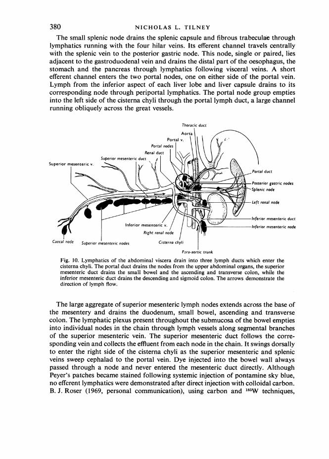
Abdominal visceraThe regional lymph nodes of the abdominal viscera are divided into three groups
which empty separately into the cisterna chyli through lymphatic ducts (Fig. 10).The splenic, posterior gastric and portal nodes draining the upper abdominal organs,the superior mesenteric chain draining the bowel, and the interior mesenteric chaindraining the descending colon, are considered as distinct functional entities.
379
NICHOLAS L. TILNEY
The small splenic node drains the splenic capsule and fibrous trabeculae throughlymphatics running with the four hilar veins. Its efferent channel travels centrallywith the splenic vein to the posterior gastric node. This node, single or paired, liesadjacent to the gastroduodenal vein and drains the distal part of the oesophagus, thestomach and the pancreas through lymphatics following visceral veins. A shortefferent channel enters the two portal nodes, one on either side of the portal vein.Lymph from the inferior aspect of each liver lobe and liver capsule drains to itscorresponding node through periportal lymphatics. The portal node group emptiesinto the left side of the cisterna chyli through the portal lymph duct, a large channelrunning obliquely across the great vessels.
Thoracic duct
Aorta
Portal v.l\ >
PrenalnductSuperior mesenteric duct
Superior mesenteric v.
Portal duct
Posterior gastric nodesSplenic node
Left renal node
Inferior mesenteric ductInferior mesenteric v. Inferior mesenteric node
Right renal node
Caccal node Superior mesenteric nodes Cisterna chyli
Para-aortic trunk
Fig. 10. Lymphatics of the abdominal viscera drain into three lymph ducts which enter thecisterna chyli. The portal duct drains the nodes from the upper abdominal organs, the superiormesenteric duct drains the small bowel and the ascending and transverse colon, while theinferior mesenteric duct drains the descending and sigmoid colon. The arrows demonstrate thedirection of lymph flow.
The large aggregate of superior mesenteric lymph nodes extends across the base ofthe mesentery and drains the duodenum, small bowel, ascending and transversecolon. The lymphatic plexus present throughout the submucosa of the bowel emptiesinto individual nodes in the chain through lymph vessels along segmental branchesof the superior mesenteric vein. The superior mesenteric duct follows the corre-sponding vein and collects the effluent from each node in the chain. It swings dorsallyto enter the right side of the cisterna chyli as the superior mesenteric and splenicveins sweep cephalad to the portal vein. Dye injected into the bowel wall alwayspassed through a node and never entered the mesenteric duct directly. AlthoughPeyer's patches became stained following systemic injection of pontamine sky blue,no efferent lymphatics were demonstrated after direct injection with colloidal carbon.B. J. Roser (1969, personal communication), using carbon and 185W techniques,
380

Patterns of lymphatic drainage in the adult laboratory rathas noted minute efferent channels running from Peyer's patches to regional mesen-teric nodes.The inferior mesenteric node lies in the mesentery of the descending colon where
the inferior mesenteric vein crosses the great vessels to join the superior mesentericvein. It drains the rectum and sigmoid colon through a lymph channel coursingbeside the vein. The efferent inferior mesenteric duct enters the cisterna on the leftside.
Internal jugular nodePosterior cervical node
Lymphatic tapsMediastinal ducts
Right superior vena cava
Parathymic nodes
Inferior vena cava
Cervical ductSubclavian v.Subclavian duct
cavaV.
chyli
Y/)Fig. 11. The cisterna lies dorsal to the great vessels and forms the thoracic duct. This duct runson the right of the great vessels in the thorax but crosses dorsal to them in the neck to enter theleft subclavian vein. The cervical, subclavian and mediastinal lymphatic ducts empty into thesubclavian veins through their lymphatic taps.
Lymphatic drainage of the omentum was not demonstrated in this study. Injectedparticulate dye was presumably ingested by macrophages which gradually migratedto omental 'milk spots' where they remained for prolonged periods. Casparis (1918)and Simer (1934) have described lymphatics in the fixed omentum of the rat, althoughnone has been demonstrated in vivo.
Cisterna chyli and thoracic ductThe cisterna chyli lies dorsal to the aorta and vena cava at the level of the renal
veins, and narrows within a few millimetres to form the thoracic duct (Fig. 11). Theduct runs on the left of the abdominal aorta but crosses deep to the great vessels at
381

NICHOLAS L. TILNEY
the diaphragm to continue its thoracic course to the right of the oesophagus. Itswings dorsal to the carotid arteries and trachea at the level of the clavicles andempties into the left subclavian vein with the other left lymphatic taps.
DISCUSSION
Particulate material such as colloidal carbon, when injected intradermally orsubserosally, readily fills lymphatic channels and drains to regional lymph nodes.This reinforces the observations of Hudack & McMaster (1932), who demonstratedthat local cutaneous trauma, however mild, causes an immediate increase in lymph-atic permeability which permits the entrance of large molecules. Similarly, labelledalbumin rapidly enters lymphatics and localizes in lymph nodes. The high concentra-tions of this antigen in the regional lymphoid tissue make it an accurate and effectivemeans of confirming studies with carbon.The location of lymphoid tissue was invariable among the animals examined,
although the numbers of lymph nodes in particular node groups varied slightly. Thismay be dependent in part upon the age of the animal. Fewer nodes were describedby Higgins (1925) in studies of the lymphoid system of the newborn rat, and distinctmorphological differences were found by Andrew & Andrew (1948) between thelymphoid tissues of young and old animals.Lymphatic channels were constant and often followed adjacent veins. All lymph-
atics ultimately drained to the lymphatic taps which emptied into the subclavianveins. Anatomical variations sometimes occurred at these major lymphatico-venousjunctions, and were also noted by Job (1915) in previous studies on the rat.
Somatic areas especially subject to environmental hazards possessed a diffuseregional drainage patttern. Mucocutaneous junctions and the footpads drained to atleast two separate nodal groups. The extensive lymphatic supply of the tail has beendelineated directly in this study and radiologically by Engeset & Tjotta (1960).Whether lymphatics from a particular site may bypass regional nodes and enter
major lymph trunks directly has been debated in the literature. Yoffey & Courtice(1956) were unable to document a complete lymphatic bypass of every node draininga structure, but Engeset (1959) has shown that lymphatics from the testes of rats anddogs do not necessarily enter any nodes on their course to the bloodstream. In thepresent study lymphatic drainage of the testis occasionally bypassed one or morenode groups, but inevitably entered nodes which themselves drained into the cisternachyli.
SUMMARY
Accurate knowledge of lymphoid anatomy and lymphatic routes in the laboratoryrat has become increasingly important as this animal is used more frequently inbiological investigations. Because of the unavailability of precise descriptions,lymphatic drainage patterns were studied in the adult rat using multiple injections ofcolloidal carbon and 1311 human serum albumin. The regional lymph drainage of theskin is described and visceral lymphatic anatomy defined. The anatomical constancyof lymphoid tissue and lymphatic channels between individual animals and betweenthe various strains examined is stressed.
382

Patterns of lymphatic drainage in the adult laboratory rat 383I should like to thank Professor J. L. Gowans and Dr B. J. Roser of the Sir
William Dunn School of Pathology, University of Oxford, and Professor D. W.Fawcett and Dr E. A. Edwards of the Department of Anatomy, Harvard MedicalSchool, for their encouragement and support. The illustrations were drawn byMiss C. Court, Department of Human Anatomy, University of Oxford, and werecorrected and lettered by Miss T. Holden, Peter Bent Brigham Hospital, Boston,Massachusetts, U.S.A.The study was performed while the author was a G. G. Peters Travelling Fellow
at the Sir William Dunn School of Pathology, University of Oxford.
REFERENCES
ANDREW, W. & ANDREW, N. V. (1948). Age changes in the deep cervical lymph nodes of 100 WistarInstitute rats. Am. J. Anat. 82, 105-166.
CASPARIS, H. R. (1918). Lymphatics of the omentum. Anat. Rec. 15, 93-99.ENGESET, A. (1959). The route of peripheral lymph to the blood stream. An X-ray study of the barrier
theory. J. Anat. 93, 96-100.ENGESET, A. & TJOTTA, E. (1960). Lymphatic pathways from the tail in rats and mice. Cancer Res. 20,
613-614.GREENE, E. C. (1935). Anatomy of the Rat. Philadelphia: American Philosophical Society.HIGGINS, G. M. (1925). On the lymphatic system of the newborn rat (Mus norvegicus albinus). Anat. Rec.
30, 243-258.HUDACK, S. & MCMASTER, P. D. (1932). The permeability of the wall of the lymphatic capillary. J. exp.Med. 56, 223-238.
JOB, T. T. (1915). The adult anatomy of the lymphatic system in the common rat (Epimys norvegicus).Anat. Rec. 9, 447-458.
KLUGE, T. & ONGRE, A. A. (1968). Pericardial absorption of thorium dioxide in rats. Acta path. microbiol.scand. 72, 87-102.
MACCALLUM, L. G. (1903). On the mechanism of absorption of granular materials from the peritoneum.Bull. Johns Hopkins Hosp. 14, 105-115.
Miorri, R. VAN (1965). Die Lymphknoten und Lymphgefasse der weissen Ratte (Rattus norvegicusBerkenhout, Epimys norvegicus). Acta anat. 62, 489-527.
OLIN, T. & SALDEEN, T. (1964). The lymphatic pathways from the peritoneal cavity: A lymphangio-graphic study in the rat. Cancer Res. 24, 1700-1711.
SANDERS, A. G. & FLOREY, H. W. (1940). The effects of the removal of lymphoid tissue. Br. J. exp. Path.21, 275-287.
SIMER, P. H. (1934). On the morphology of the omentum, with especial reference to its lymphatics.Am. J. Anat. 54, 203-228.
TILNEY, N. L. (1970). The systemic distribution of soluble antigen injected into the footpad of thelaboratory rat. Immunology 19, 181-184.
YOFFEY, J. M. & COURTICE, F. C. (1956). Lymphatics, Lymph, and Lymphoid Tissue, 2nd ed., London:E. Arnold.