Embed Size (px)
Citation preview
REVIEW
Lymphoid follicles in (very) severe COPD:
beneficial or harmful?G.G. Brusselle*, T. Demoor*, K.R. Bracke*, C-A. Brandsma# and W. Timens#
ABSTRACT: Inflammation is a main pathogenetic factor in the development and progression of
chronic obstructive pulmonary disease (COPD). Recently, it has become clear that not only the
innate, but also the specific immune response plays a role. A striking finding, in particular in lungs
of patients with severe COPD, often with a predominant emphysema phenotype, is the presence
of B-cell follicles. As seen in other tissues, these follicles are the result of lymphoid neogenesis.
The finding of oligoclonality in B-cell follicles in COPD suggests that they play a role in local
antigen specific immune responses. To date, it is not known which antigens may be involved;
microbial antigens, cigarette smoke-derived antigens and antigens from extracellular matrix
breakdown products have been suggested. Consequently, the pathogenetic role of this follicular
B-cell response is not yet clear. It might be protective against microbial colonisation and infection
of the lower respiratory tract and, therefore, beneficial, or it could be of a more harmful
(autoimmune) nature, directed against lung tissue components. It is necessary to determine the
specific antigen(s) and to explore the exact role of the COPD related B-cell response in order to
include modulation of this response and develop therapeutic options.
KEYWORDS: Chronic obstructive pulmonary disease, immune response, inflammatory response,
lymphocytes, review
Chronic obstructive pulmonary disease(COPD) is a chronic inflammatory dis-order in different compartments of the
lung, including a mixture of small airway disease(obstructive bronchiolitis) and parenchymaldestruction (emphysema). COPD is associatedwith a relentlessly progressive course andincreasing morbidity, disability and mortalityworldwide [1]. Smoking is known to be the majoraetiological factor, yet the exact pathogeneticmechanisms have not yet been elucidated.Smoking cessation is the only effective treatment,but it only partially attenuates further loss of lungfunction. In those COPD patients who have fullyceased smoking, pulmonary inflammation per-sists [2–4]. The mechanism that propagates thisinflammation is incompletely understood.
Since COPD is characterised by chronic airflowlimitation that is not fully reversible and usuallyprogressive, the Global Initiative for ChronicObstructive Lung Disease (GOLD) has introduceda five-stage classification for the severity of COPDbased on measurements of airflow limitationduring forced expiration, as measured by forcedexpiratory volume in 1 s [5]. The progression of
COPD is associated with infiltration of the wall ofsmall airways by innate and adaptive inflamma-tory immune cells. The inflammatory cells thatwere initially identified as being the most impor-tant in the pathogenesis of COPD were neutro-philic granulocytes and macrophages, which bothbelong to the innate immune system. The impor-tance of neutrophils and macrophages has beenconfirmed by animal studies showing the neces-sity of both these cells for the induction ofexperimental emphysema by cigarette smoke(CS) [6, 7]. It later became clear that the specificimmune system also plays a role; in particularCD8 positive T-lymphocytes were associated withsmoking and subsequent risk for COPD [8, 9].Recently, B-lymphocytes organised into peri-bronchiolar lymphoid follicles have been asso-ciated with severe COPD [10] and, in addition, inCOPD such follicles were also found in theparenchyma [11], whereas B-cells were increasedin large airways [12]. As the antigen inducing thisfollicular B-cell response is as yet unknown, it hasyet to be decided whether and to what extent thisresponse is of beneficial or harmful nature. Thisreview aims to shed light on the nature of thisresponse, the possible aetiological factors and to
AFFILIATIONS
*Dept of Respiratory Medicine, Ghent
University Hospital, Ghent, Belgium,
and#Dept of Pathology, University
Medical Center Groningen,
Groningen, The Netherlands.
CORRESPONDENCE
G.G. Brusselle
Dept of Respiratory Medicine
Ghent University Hospital
De Pintelaan 185
9000 Ghent
Belgium
E-mail: [email protected]
Received:
Oct 03 2008
Accepted after revision:
Jan 17 2009
European Respiratory Journal
Print ISSN 0903-1936
Online ISSN 1399-3003
EUROPEAN RESPIRATORY JOURNAL VOLUME 34 NUMBER 1 219
Eur Respir J 2009; 34: 219–230
DOI: 10.1183/09031936.00150208
Copyright�ERS Journals Ltd 2009
c

present our views on the possible role in the pathogenesis andprogression of COPD.
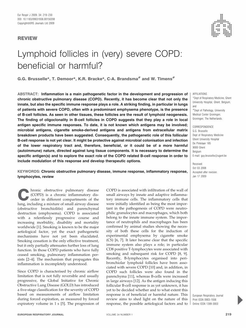
LYMPHOID FOLLICLES IN COPDWhereas one of the first reports on the presence of B-cellfollicles in COPD dates from 1992 [13], quite recently attentionhas been redrawn to a possible role of B-cells. An increase ofsmall airways containing B-cell follicles was found in patientswith severe COPD compared to normal subjects and patientswith mild-to-moderate COPD. Furthermore, the progression ofCOPD from GOLD stage 0 to GOLD stage 4 was indeed clearlyassociated with the number of small airways containing theselymphoid follicles [10, 14]. Furthermore, the presence of B-cellfollicles was demonstrated not only in relation to smallairways, but also in the lung parenchyma of COPD patients(fig. 1) and mice chronically exposed to CS [11]. An increase ofindividual B-cells in COPD was also observed, in particular inbronchial biopsy studies of large airways and, in addition,nonfollicular B-cell aggregates were identified [12, 15].
It is important to note that the original publication demon-strating lymphoid follicles in patients with severe and verysevere COPD [10] could encompass a selection bias, since alllung specimens of GOLD stage 3 and 4 COPD were obtainedfrom lung volume reduction surgery specimens of patientswith severe emphysema enrolled in the National EmphysemaTreatment Trial (NETT). However, in the study of VAN DER
STRATE et al. [11], the majority of the human lung tissuesamples (six out of eight patients with very severe COPD) wereobtained at lung transplantation (explant lungs). Nevertheless,more studies are needed to assess the role of lymphoid folliclesin different COPD disease stages and phenotypes, includingsevere COPD with predominant bronchiolitis.
With respect to B-cell function and development, a distinctionshould be made between B-cell aggregates without a specificfunctional architecture and B-cells organised in (primary orsecondary) follicles; this is also reflected in their supposedfunctional role. Lymphoid aggregates consist of mature,memory B-cells and/or T-cells without functional organisa-tion, thus mainly available for local priming and activation.The functional organisation (see below) of primary andsecondary lymphoid follicles (the latter containing germinalcentres) consists of a specific arrangement of memory andnaı̈ve B-cells, T-cells, dendritic cells (DCs) and folliculardendritic cells (FDC), which allows for B- and T-cell priming,clonal expansion, antigen retention (mostly as immune com-plexes), somatic hypermutation, affinity maturation andimmunoglobulin (Ig) class switching [16–18]. However, itshould be kept in mind that over time the distinction is notalways clear. When local circumstances and factors allow,lymphoid follicles can develop out of any lymphoid aggregate.Importantly, only the micro-anatomic organisation of alymphoid follicle, with the ability of germinal centre develop-ment, allows for its specific functions [16–18].
In COPD, the follicles in the lung [10, 11, 14] consist of largeaggregates of B-cells that are surrounded by lower numbers ofmainly CD4 (80–90%) and some CD8-positive T-cells. Theseaggregates can be considered lymphoid follicles, since FDCwere present in these aggregates, expressing CD21 and CD35(fig. 1). The presence of FDC, involved in affinity maturation
and isotype switching of B-cells, suggests antigen-drivenproliferation. The B-cells were mainly IgM-positive and IgD-negative, which suggests that these B-cells may have beenactivated to some extent. Moreover, a predominant part of theinfiltrate appeared to be CD27-positive, a marker for memoryB-cells [19]. Interestingly, CD138-positive (plasma) cells weredetectable in the direct vicinity of these follicles. Furthermore,CD40 and CD40 ligand, important co-stimulatory molecules,were expressed in these follicles. The Ki-67 antigen wasdetected on small central clusters of B-cells in the follicles,suggesting an early germinal centre reaction [17].
Most importantly, B-cells isolated from the B-cell follicles havebeen demonstrated to be oligoclonal in nature [11], suggestingantigen-specific induction of the B-cell follicles. In this study,10 lymphoid follicles were isolated by laser microdissectionfrom the lung tissue of eight patients with COPD [11].Sequence analysis of the Ig rearrangements revealed thepresence of 12 different B-cell contigs with one or moresequence variation in 10 of the contigs. In seven out of the eightpatients, clonal B-cell populations were observed, whereas inone patient only unrelated sequences were observed. Theseven patients with related Ig sequences demonstrated thepresence of ongoing mutations. The latter indicates oligoclonalB-cell proliferation in response to stimulation with as yetunknown antigen(s).
With respect to B-cell follicles in the lung, some authorsregarded these as part of the mucosal immune system and thusas so-called bronchus associated lymphoid tissue. However, inmost normal lungs no lymphoid tissue is observed at all [20].Lymphoid follicles have been observed in the human lung in(subclinical or clinical) lung disease, either associated withairways or with parenchyma. Consequently it seems mostlogical to regard this as so-called lymphoid neogenesis and,thus, as an ectopic lymphoid tissue [16, 21]. Such lymphoidtissues are also called tertiary lymphoid organs (TLOs), asopposed to primary (bone marrow and thymus) and secondarylymphoid organs (lymph nodes, Peyer’s patches and spleen).
The term lymphoid neogenesis refers to the development oforganised lymphoid structures which resemble secondarylymphoid organs in tissues that are targeted by chronicinflammatory processes, such as infection and autoimmunity[16, 22]. The purpose of immune responses is primarily theeradication of pathogens. However, certain antigens aredifficult to eradicate and result in sustained immune responsesleading to chronic inflammation. In tissues that harbour thetarget antigens of chronic adaptive immune responses,infiltration occurs by macrophages, DCs, T-cells, B-cells andplasma cells. These cells frequently organise themselves at ananatomic and functional level as in secondary lymphoidorgans, leading to the formation of B-cell follicles and T-cellareas [23]. This lymphoid neogenesis, also called TLOformation, is a dynamic process starting with sparse lympho-cytic infiltrates that evolve into aggregates and eventuallyorganise in B-cell follicles with germinal centres and distinct T-cell areas containing DCs and high endothelial venules (fig. 2).High endothelial venules regulate the extravasation of naı̈ve B-and T-cells. Thus, there are remarkable similarities between thestructure of secondary lymphoid organs and TLOs, includingthe compartmentalisation in distinct B-cell areas (follicles) and
REVIEW: COPD G.G. BRUSSELLE ET AL.
220 VOLUME 34 NUMBER 1 EUROPEAN RESPIRATORY JOURNAL

a) b)
d)
f)
c)
e)
FIGURE 1. Lymphoid follicles in chronic obstructive pulmonary disease. a) CD20: overview of emphysematous lung tissue with B-cell follicles apposing a small airway
(arrows) and a small B-cell aggregate in the parenchyma (arrowhead). b) CD20 staining of all B-cells. c) CD3 staining of apposing T-cells. d) CD21 (complement C3d-
receptor) showing delicate staining of follicular dendritic cells. e) CD35 (complement C3b-receptor) showing more intense staining of follicular dendritic cells and weak
staining of B-cells. f) CD83 staining of mature myeloid dendritic cells, showing subepithelial location (arrows), as well as presence in the rim (T-cell zone) of a lymphoid follicle
(arrowhead). a) Scale bars51,000 mm, b–f) scale bars5200 mm.
G.G. BRUSSELLE ET AL. REVIEW: COPD
cEUROPEAN RESPIRATORY JOURNAL VOLUME 34 NUMBER 1 221

T-cell areas (paracortex). However, in contrast to lymph nodes,TLOs are not supplied by afferent lymph vessels and are notencapsulated.
LYMPHOID NEOGENESIS IN MURINE MODELS OF COPDIn vivo animal models can help to unravel the cellular andmolecular mechanisms underlying the pathogenesis of COPDin general and of lymphoid neogenesis in particular [24–26].Mice represent the most favoured animal species with regardto the study of the immunological mechanisms of diseases, asseveral hundreds of inbred strains and specialised stocks (e.g.mutants) are available and since they offer the opportunity tomanipulate gene expression (e.g. transgenic and knock-outmice). Lymphoid follicles have been described not only in CS-based murine models of COPD, but also in pathogen-basedmodels of COPD and in transgenic mice over-expressingpro-inflammatory cytokines in the bronchial or alveolarepithelium.
CS-induced lymphoid folliclesWhen the time-course of CS-induced pulmonary inflammationis determined in wild-type mice, a progressive accumulation ofinnate inflammatory cells (i.e. neutrophils, macrophages andDCs) in the airways and lung parenchyma is demonstratedfrom day 3 onwards. In addition to the persistent accumulationof innate inflammatory cells upon prolonged exposure to CS, aclear and progressive infiltration of adaptive immune cells (i.e.B-cells and T lymphocytes, both CD4+ and CD8+ T-cells) hasbeen observed in bronchoalveolar lavage fluid and lungs ofmice exposed to CS for 3–6 months [11, 27]. Inflammatoryinfiltrates, with a predominantly lymphoid character, werefound to be present around bronchioles and in the lung
parenchyma of mice after chronic CS exposure, similar to thelymphoid follicles detected in lung sections of patients withCOPD. Lymphoid follicles consisting of B-cells and FDC withadjacent T-cells were detected in mice that had developedpulmonary inflammation after smoking for 6 months [11]. Theincrease in the number and the size of B-cell follicles wasprogressive with time and correlated with the (progressive)enlargement of alveolar airspaces, as evidenced by an increasein mean linear intercept. The development of emphysema inCS-exposed mice has been shown to be strain dependent.Although lymphoid follicles have been described in severalstrains of CS-exposed mice (e.g. BALB/c, C57BL/6 and C3H)[27–29], a formal head-to-head comparison of lymphoidneogenesis in different murine strains has not yet beenperformed. The number and size of peribronchial lymphoidfollicles was significantly attenuated in chronically CS-exposedchemokine receptor CCR5 and CCR6 knock-out mice com-pared with wild-type animals, indicating a prominent role ofchemokines in lymphoid neogenesis [28, 30].
Pathogen and lipopolysaccharide-induced lymphoidfolliclesSeveral bacteria, including nontypeable Haemophilus influenzae(NTHi), commonly colonise the lower airways of patients with(severe) COPD. Exposure of mice to an aerosolised NTHilysate every 8 weeks induced infiltration of macrophages,CD8+ T-cells and B-cells around airways and blood vessels,including rounded peribronchial lymphoid aggregates [31].Lipopolysaccharide (LPS) is a major pro-inflammatory glyco-lipid component of the bacterial cell wall of Gram-negativebacteria (such as NTHi). Interestingly, repeated intratrachealinstillation of LPS in mice resulted in persistent chronic
T-cellarea
HEV
FDC
Germinal centreB-cell
B-cell
Stromal cell
Blood vessel
LTα1β2
LTβR
DC CCL19
CCR7CCL21
CXCL12CXCR4
CXCR5
CXCL13
T-cellB-cellarea
FIGURE 2. Lymphoid neogenesis in chronic inflammation. a) In chronic inflammation, activated lymphocytes expressing lymphotoxin-a-b heterotrimer (LTa1b2) interact
with the lymphotoxin (LT)b receptor on neighbouring stromal cells. The stromal cell stimulation induces the expression of lymphoid chemokines (CC-chemokine ligands
(CCL)19, CCL21, CXC-chemokine ligands (CXCL)12 and CXCL13) along with adhesion molecules allowing the recruitment of naı̈ve lymphocytes and dendritic cells (DCs). b)
Prolonged inflammatory stimulation will lead to the organisation of lymphoid aggregates into lymphoid follicles. Germinal centres may form within the B-cell zone with highly
specialised follicular dendritic cells (FDC). High endothelial venules (HEV) expressing lymphoid chemokines and adhesion molecules ensure lymphocyte and DC recruitment.
REVIEW: COPD G.G. BRUSSELLE ET AL.
222 VOLUME 34 NUMBER 1 EUROPEAN RESPIRATORY JOURNAL

pulmonary inflammation, characterised by peribronchial andperivascular lymphocytic aggregates (consisting of CD4+ andCD8+ T lymphocytes and CD19+ B lymphocytes) [32]. These invivo studies demonstrate that some pathological changes ofCOPD, especially lymphoid aggregates, can be mimicked inmice after repeated exposure to either Gram-negative bacteriallysates or a component of the Gram-negative bacterial cell wall,i.e. LPS. Finally, MOYRON-QUIROZ et al. [33] demonstrated thatan infectious challenge with influenza virus in mice can triggerlymphoid chemokine expression and lymphoid neogenesis inthe lungs, even in lymphotoxin (LT)a-deficient mice that lacksecondary lymphoid organs [33].
Spontaneous development of lymphoid aggregates in lungsof transgenic miceTransgenic mice were generated in which human interleukin(IL)-1b is conditionally (with a doxycycline-inducible system)and specifically (by the Clara cell secretory protein promoter)expressed in the lung epithelium, especially in the bronchioli[34]. Induction of IL-1b expression in the lungs of adult micecaused pulmonary inflammation, distal airspace enlargement,airway wall fibrosis and lymphocytic aggregates in theairways. Human IL-6 and IL-6 receptor (IL-6R) doubletransgenic mice also developed mononuclear cell accumula-tion in peribronchovascular regions and lymphoid tissue-likestructures expressing the chemokine CXCL13, which isindispensable for lymphoid organogenesis [35]. In separatestudies, transgenic mice over expressing tumour-necrosis-factor (TNF)-a in the lung under the surfactant protein C(SPC) promoter have been shown to produce elements of bothemphysema and pulmonary fibrosis [36, 37]. Detailed histolo-gical analysis of SPC/TNF-a transgenic mice showed majorheterogeneous abnormalities of the lung parenchyma, includ-ing infiltration of macrophages and lymphocytes into thelungs, which was most prominent in the subpleural regions, aswell as distortion of alveolar structures due to septalthickening [38]. Since the pro-inflammatory cytokines IL-1b,IL-6 and TNF-a have pleiotropic effects and have beenimplicated in multiple inflammatory disorders, it is clear thatthe observed pathological abnormalities in these transgenicmice can be involved in several human diseases, including notonly COPD/emphysema but also lymphocytic fibrosingalveolitis, leading to pulmonary fibrosis.
LYMPHOID NEOGENESIS IN OTHER DISEASESLymphoid follicles are observed in various lung diseases otherthan COPD. Often, a long-term fibrosing process has beengoing on with, in due time, development of lymphoidaggregates and lymphoid follicles. This is observed in usualinterstitial pneumonia/end-stage pulmonary fibrosis and alsoin nonspecific interstitial pneumonia and bronchiectasis. Inbronchiectasis, follicles are not only observed in the airwaywall but also in the surrounding parenchyma, which oftenshows varying fibrosis and chronic inflammation. In most ofthe above conditions, stasis of cellular debris and mucus ispresent in distorted airspaces, in which all kinds of micro-organisms are present, often with clinical or subclinicalinfections. Therefore, an antimicrobial origin of the specificimmune response giving rise to the follicles is quite likely inthese diseases, although other origins cannot be excluded.
Lymphoid interstitial pneumonia (LIP) is an interstitial lungdisease characterised by an extensive proliferation of smallmature B- and T-cells; in general, mainly in the alveolarinterstitium. However, a form with follicular bronchiolitis isoften also considered to be part of this spectrum. Developmentof lymphoid follicles, most with florid germinal centres, is ageneral feature of LIP. At present LIP is considered apulmonary manifestation related to different autoimmunediseases; auto-antigens are also most likely underlying theimmune response giving rise to the lymphoid follicles. Withrespect to the airways, follicles are seen in bronchiectasis asdescribed above, but the most prominent airway manifestationof lymphoid follicles is in follicular bronchiolitis. This diseasecondition is quite rare, sometimes related to autoimmunedisease, and often considered as part of LIP (see above). As apathological diagnosis it has been observed in association withimmunodeficiency, such as in HIV, but also as an idiopathicdisease.
Lymphoid neogenesis has also been described outside thelungs in several chronically inflamed tissues from patientswith different underlying diseases, mainly autoimmune andinfectious diseases. Human chronic inflammatory diseaseswith lymphoid neogenesis encompass rheumatoid arthritis,Graves’ disease (hyperthyroidism), Hashimoto’s thyroiditis(hypothyroidism), myasthenia gravis, multiple sclerosis,Crohn’s disease, ulcerative colitis and chronic hepatitis C.The respective target tissues of the TLO formation are diverseand include the joints, the thyroid gland, the thymus, thecentral nervous system, the gut and the liver. In severalautoimmune diseases, the self-antigen, recognised by anti-bodies generated in ectopic germinal centres, has beendetected in lymphoid follicles (e.g. thyroglobulin and thyr-operoxidase in Hashimoto’s thyroiditis) [16].
CELLULAR AND MOLECULAR MECHANISMS OFLYMPHOID (NEO)GENESIS
Similarities in development of secondary lymphoid organsversus TLOsThe organisation of lymph nodes during organogenesis is theresult of a highly coordinated interplay between haematopoie-tic cells, mesenchymal stromal cells, adhesion molecules,cytokines and chemokines (fig. 3). The key players aremembers of the TNF family, mainly LTa1b2 and its receptorLTbR [39, 40]. Binding of membrane-bound LTa1b2, expressedby haematopoietic inducer cells to LTbR on the surface ofstromal organiser cells, induces the expression of adhesionmolecules, including intercellular adhesion molecule-1, vascu-lar cell-adhesion molecule-1, mucosal addressin cell-adhesionmolecule-1 and peripheral node addressin. The interactionbetween haematopoietic inducer cells and stromal cells via theLTa1b2–LTbR pathway also induces the expression of homeo-static lymphoid chemokines, such as CC-chemokine ligand(CCL)19 and CCL21, as well as CXC-chemokine ligand(CXCL)12 and CXCL13 [41]. These lymphoid chemokinesorchestrate the lymphocyte homing and compartmentalisationof the lymphoid organs. CCL19 and CCL21 are produced bystromal cells and regulate the homing of CCR7+ naive T-cellsand mature DCs to T-cell areas. CXCL12 and CXCL13 areproduced by FDC, thought to originate from mesenchymalstromal cells, and attract CXCR5+ B-cells into the follicles.
G.G. BRUSSELLE ET AL. REVIEW: COPD
cEUROPEAN RESPIRATORY JOURNAL VOLUME 34 NUMBER 1 223

Activated tissue fibroblasts (e.g. fibroblast-like synoviocytes)share several features with the stromal cells that form thereticular network of secondary lymphoid organs, including theproduction of lymphoid chemokines (such as CXCL12,formerly called stromal cell-derived factor 1).
DCsIn the respiratory tract, myeloid DCs form a dense network ofantigen-presenting cells in or just beneath the epithelium andsurvey the airways for exogenous and endogenous dangersignals [42]. Recently, a significant increase in Langerin+ DCs
has been demonstrated in the small airways of patients withCOPD compared with healthy smokers and never-smokers [43].As in the gastrointestinal tract, the accumulation of immatureLangerin+ DCs was associated with increased CCL20 levels ininduced sputum and lung tissue of COPD patients [43]. Uponantigen uptake and activation, DCs lose all chemokine receptorsexcept CCR7, which regulates the migration of DCs towards theregional lymph nodes as well as the influx of naı̈ve T-cells intothe paracortex. It is conceivable that similar mechanisms alsodirect the migration of DCs and T-cells towards the T-cell zoneof lymphoid follicles (fig. 3).
HEV
Lymphoid follicle
ECM
Elastin breakdownfragments
B-cell
T-cell
Macrophage
TNFIL-6IL-1β
Airway epithelium
Immature DC
Antibody
Plasma cell
Auto-antigen
LPS
Bacteria
Alveoli
FDC
Blood circulation
CCR6
MIP-3α
FIGURE 3. Development of lymphoid follicles in chronic obstructive pulmonary disease. In response to environmental insults, such as lipopolysaccharide (LPS),
cigarette smoke and bacteria, airway epithelium and macrophages express cytokines (including interleukin (IL)-1b, IL-6 and tumour necrosis factor (TNF)-a) and chemokines
(e.g. CCL20/macrophage inflammatory protein (MIP)-3a), leading to the accumulation of immature dendritic cells (DCs), T-cells and B-cells. When inflammation becomes
chronic due to persisting antigen exposure and/or tissue damage, lymphocyte aggregates will give rise to organised lymphoid follicles with separated B- and T-cell areas.
Mature lymphoid follicles may contain high endothelial venules (HEV), follicular dendritic cells (FDC) and germinal centres with the potential to produce plasma cells and
antibody responses against bacteria and/or auto-antigens, such as breakdown fragments from the extracellular matrix (ECM). Inhaled and systemic corticosteroids may
interfere with several mechanisms involved in the development and function of lymphoid follicles.
REVIEW: COPD G.G. BRUSSELLE ET AL.
224 VOLUME 34 NUMBER 1 EUROPEAN RESPIRATORY JOURNAL

PATHOPHYSIOLOGICAL ROLE OF LYMPHOIDFOLLICLES IN COPDWhat are the antigenic stimuli that trigger the formation oflymphoid follicles?There are several mechanisms that may underlie the increasednumbers of B-cell follicles in lung tissue of COPD patients. TheB-cell follicles could reflect, in part, a nonspecific, polyclonal,proliferation of B-cells, induced by local production of B-cellactivating mediators including cytokines (IL-4, IL-6, IL-13,TNF-a and lymphotoxin) . Alternatively, supported by thedemonstration of (oligo)clonality in follicular B-cells in thelung, they might well be the result of a specific humoralimmune reaction [11]. At present it is unclear against whichantigen(s) this B-cell proliferation could be directed. At leastthree potential sources of antigens should be considered:1) microbial; 2) CS components or derivatives; and 3) auto-antigens, encompassing epithelial (neo)antigens and degrada-tion products of extracellular matrix.
Chronic bacterial and viral colonisation and/or infectionThe increase in lymphocytes in the small airways and theirorganisation into bronchial lymphoid follicles are consistentwith an adaptive immune response, which is believed todevelop in relation to chronic bacterial and/or viral colonisa-tion and infection of the lower airways [44]. Several observa-tions plead in favour of this hypothesis.
First, colonisation of the lower airways by bacteria is acommon feature in COPD and, therefore, it is possible that atleast part of the reactivity of B- and T-cells in the lymphoidfollicles is directed against bacterial antigens [44]. Moreover,microbial colonisation and infection occur more frequently inthe later stages of COPD (severe and very severe COPD) thanin milder disease; this parallels the increasing frequency ofperibronchiolar lymphoid follicles in more severe COPD. Up to52% of patients with severe COPD are colonised in the stablestate with possible bacterial pathogens, including Haemophilusinfluenzae, Streptococcus pneumoniae, Moraxella catarrhalis andPseudomonas aeruginosa [45].
In addition, patients with COPD are prone to frequentexacerbations which are a significant cause of morbidity andmortality. The frequency of COPD exacerbations is modulatedby the degree of airway bacterial colonisation in the stable state,and by acquisition of new strains within a pathogenic bacterialspecies [44, 46, 47]. Using an in vivo model of airway infection inmice, strains of H. influenzae isolated from COPD patientsduring exacerbation induced more airway inflammation thancolonisers [48]. Importantly, exacerbation strains are strainswhich are not only isolated for the first time from sputum ofpatients experiencing clinical exacerbation symptoms, but arealso accompanied by the appearance of new bactericidalantibodies to the homologous infecting strain (compared withserum obtained 1 month before exacerbation). In a cohort studyof COPD patients investigating the dynamics of carriage andimmune responses to M. catarrhalis, MURPHY et al. [49]demonstrated that asymptomatic colonisation was associatedwith a greater frequency of a sputum IgA response thanexacerbation, whereas the intensity of the serum IgG responsewas greater after exacerbations than colonisation. Since devel-opment of new sputum IgA and serum IgG were not correlated,mucosal and systemic immune responses appear to occur
independently. Moreover, M. catarrhalis was cleared efficientlyfrom the respiratory tract after a short duration of carriage,indicating that COPD patients develop strain-specific protec-tion. In contrast, antibody responses to P. aeruginosa developedin 54% of COPD patients with persistent carriage, suggestingthat antibody responses to this bacterium do not mediateclearance, but are a marker for colonisation [50]. Thus, thefunctional role of the B-cell response to airway bacteria in COPDappears to be strain-dependent.
Secondly, in vivo studies in mice have demonstrated thatperibronchial lymphoid aggregates and/or follicles can beinduced by live infection with influenza virus or repeatedexposure to bacterial lysates of NTHi or bacterial cell wallcomponents (LPS) [31–33]. These animal studies indicate thatthe chronic exposure of the airways to bacteria and/orbacterial products may be sufficient to induce lymphoidneogenesis. However, VAN DER STRATE et al. [11] elegantlydemonstrated that this is not a prerequisite, since the mice thatwere chronically exposed to CS and developed B-cell follicleswere kept under specified pathogen free conditions, togetherwith the air-exposed littermates that did not have theselymphoid follicles. Extensive analysis of fresh frozen samplesof the lungs of these mice by RT-PCR did not reveal thepresence of microbial species (including Mycoplasma,Chlamydia, adenovirus or Pneumocystis).
Degradation products of extracellular matrixApart from microbial colonisation and infections as the triggerof the development of lymphoid follicles in the lung, an antigenspecific immune response against neo- or self antigens presentin lung tissue could also be the trigger. In the lungs of COPDpatients there is a chronic inflammatory response present withthe ongoing recruitment of inflammatory cells and concomitantlung tissue damage. This inflammatory environment canprovide the optimal conditions for B-cells to form aggregatesand start or sustain a local immune response. This immuneresponse can be directed against lung matrix proteins of thedestructed tissue, which can be recognised as neo-antigens. It isknown that degradation products of extracellular matrix, suchas hyaluronic acid, collagen and elastin, have a chemotacticeffect on neutrophils and monocytes [51, 52]. The effectsmediated by the inflammatory cells may lead to ongoingdegradation of extracellular matrix, which results in mainte-nance of the chemotactic gradient and therefore inflammation. Itis conceivable that B-cells are either locally present or attractedby this gradient, and start producing antibodies against theextracellular matrix degradation products. This is supported bythe recent findings of LEE et al. [53], showing the presence ofanti-elastin antibodies in plasma and anti-elastin antibodyproducing B-cells in lung tissue of emphysema patients. Theproduced antibodies subsequently bind to the fragments of theextracellular matrix, but also to the intact extracellular matrixand cause degradation of extracellular matrix by ‘‘frustratedphagocytosis’’. The follicles in COPD lung tissue resemble thedevelopment of similar B-cell follicles in the inflamed synovia inrheumatoid arthritis [54].
The immune response could also be directed against CScomponents or derivatives which are precipitated in lung tissue,since a low number of these follicles can be observed in ‘‘healthysmokers’’ [13]. CS contains ,4,500 different compounds [55], of
G.G. BRUSSELLE ET AL. REVIEW: COPD
cEUROPEAN RESPIRATORY JOURNAL VOLUME 34 NUMBER 1 225

which it is likely that several are immunogenic (e.g. tobaccoglycoprotein). These compounds will precipitate in the lung,possibly partially bind to the extracellular matrix and may elicitan antibody response. Subsequently, these newly producedantibodies will bind to their antigens, forming an immunecomplex. Phagocytic immune cells generally remove antigen–antibody complexes. If these complexes are either bound to theextracellular matrix, or in the vicinity of it, the extracellular matrixmay subsequently be broken down as an ‘‘innocent bystander’’due to the inflammatory reaction against the immune complexesby these phagocytic cells, similar to rheumatoid arthritis.
Both the mechanisms described for CS- or extracellular matrix-derived antigens may also explain the observation thatpulmonary inflammation continues in COPD patients aftersmoking cessation [2–4]. Even though the primary stimulus forinflammation (CS) has vanished, the inflammation continues: itis well known that at least some tobacco residues remainpresent in the lung for a long time. Direct or indirect antibody-mediated degradation of extracellular matrix results in newlyformed extracellular matrix-fragments, which may elicitfurther inflammation.
Hypothesis linking microbial and auto-antigen pathways
Notwithstanding the notion that the original response of theseB-cell follicles in COPD might be protective against bacteriacolonising or infecting the lower airways, continued presenceof these local immune responses in the lung and theconcomitant tissue destruction can promote the breaking ofimmune tolerance by, for instance, epitope spreading, mole-cular mimicry or bystander activation (see Appendix). Theseprocesses are described to occur after persistent microbialinfection or chronic inflammation with the presence of tissuedamage. The immune response then extends from the originalinducing antigen to similar, often self-reactive epitopes,leading to increased and perpetuated destruction of theinflamed organ and to the initiation of autoimmune responses.In several autoimmune diseases, where similar lymphoidfollicles have been shown to be present, these processes havebeen implicated to play a role in the initiation of theautoimmune response based on findings in animal models.However, there is no direct evidence yet for such anautoimmune mechanism in COPD. Nevertheless, the presenceof the chronic inflammatory response in the lungs of COPDpatients together with the occurrence of microbial colonisationand recurrent infections, and the presence of lung tissuedestruction might provide the circumstances for these pro-cesses to occur.
Are lymphoid follicles in COPD protective or harmful?Depending on the inducing factors of these lymphoid folliclesin the lung, they can be regarded as protective, e.g. protectionagainst infections, or harmful, e.g. by producing auto-antibodies and contributing to the perpetuation of the ongoinginflammatory response.
Protection against bacterial and viral colonisation and/or infection
Lymphoid neogenesis might have a protective function byinducing a local (and systemic) immune response against theinfectious agents. Indirect evidence in favour of a protectiverole of B-cell follicles in COPD comes from a randomised
controlled trial investigating the efficacy and safety ofrituximab in patients with COPD. Rituximab is a chimerichuman/mouse monoclonal antibody directed against theCD20 antigen, expressed on pre-B-cells and mature B-cells,but not on other cells (including plasma cells). It induces lysisand apoptosis of normal and malignant human B-cells.Rituximab has been long approved for treatment of CD20+B-cell non-Hodgkin’s lymphoma [56, 57], and has recentlybeen approved for treatment of rheumatoid arthritis in patientswho have failed TNF-inhibitor therapy [58, 59]. The study withrituximab in COPD has been terminated early by the datasafety monitoring board because of a significant increased riskof infectious complications (including lower respiratory tractinfections) in COPD patients treated with rituximab comparedwith placebo.
Harmful?
Recently, an increased presence of circulating memory B-cellsin peripheral blood of current smokers has been demonstrated,suggesting a smoke-induced specific immune response.Moreover, given the possibility of epitope spreading, mole-cular mimicry or bystander activation (see Appendix) inCOPD, the humoral immune response is likely to bepolyreactive and can thus be directed against several epitopesamong which are other cellular structures of the inflamed lung.Recently, a high prevalence of anti-Hep-2 epithelial cell auto-antibodies in COPD patients was shown, together with ahigher presence of antibodies against pulmonary airwayepithelial cells in COPD patients compared to healthy controls[60, 61]. Additionally, TARASEVICIENE-STEWART et al. [62]developed a model of autoimmune emphysema in rats byinjection of xenogeneic endothelial cells, and recently showedthe presence of anti-endothelial cell antibodies in patients withCOPD [63, 64]. Whether these antibody responses in COPD arepathogenetic and/or true autoimmune responses is still to bedetermined.
An important characteristic of a pathogenetic autoantibodywould be the induction of disease after passive transfer. In therat model of autoimmune emphysema this was proven byadoptive transfer of the anti-endothelial cell antibodies [62].For the anti-elastin and anti-epithelial antibodies detected inCOPD patients, however, this has not yet been proven andshould be addressed in similar animal models. Anothercharacteristic of a pathogenetic autoantibody is the presencein or near the disease specific lesion. This characteristic has notbeen directly proven in COPD. However, LEE et al. [53] showedthe presence of anti-elastin producing B-cells in lung tissue,implying that these antibodies are produced locally in thelung. Additionally, FEGHALI-BOSTWICK et al. [60] showed thedeposition of IgG immune complexes and complement (C3) inthe lungs of COPD patients and demonstrated in vitro thatantibodies from COPD patients can exert antibody-mediatedcellular cytotoxicity. Both studies are in support of apathogenetic role for these antibodies in COPD.
INFLUENCE OF PHARMACOTHERAPY ON LYMPHOIDFOLLICLESClinical data on the effect of pharmacotherapy on lymphoidfollicles are very scarce and indirect, due to the requirement ofinvasive procedures to obtain relatively large lung samples for
REVIEW: COPD G.G. BRUSSELLE ET AL.
226 VOLUME 34 NUMBER 1 EUROPEAN RESPIRATORY JOURNAL

pathological examination. In the NETT, patients with severe(GOLD 3) and very severe (GOLD 4) COPD were studiedlongitudinally after lung volume reduction surgery [65]. Whenthe pathological changes of the small airways in the resectedlung specimens were (cor)related with clinical outcomes aftersurgery, the investigators found that occlusion of the smallairways by inflammatory exudates containing mucus wasassociated with early death in these patients with severeemphysema [66]. Intriguingly, there was a strong trend towarda reduction in the number of airways containing lymphoidfollicles in COPD patients receiving oral and/or inhaledcorticosteroids. The observation that steroid therapy isassociated with a reduction in the numbers of follicle-contain-ing airways suggests that steroid-induced suppression of theadaptive immune responses in the peripheral airways mightincrease the probability of infection in the lower respiratorytract, and may account for the increase in pneumoniasobserved in recent clinical trials, including TORCH [67] andINSPIRE [68], as well as in observational studies [69].However, these observations should be interpreted withcaution due to the cross-sectional nature of the pathologicalevaluation in NETT.
We put forward the hypothesis that the increased risk ofpneumonias in corticosteroid-treated patients with COPD ispartially due to the steroid-induced suppression of adaptiveimmune responses in general, and of pulmonary B-celllymphoid follicle formation and function in particular.Several mechanisms might contribute to the suppression oflymphoid follicles by corticosteroids. First, inhaled corticoster-oids have profound effects on the airway epithelium, whichnormally facilitates adaptive immune responses by the synth-esis of a wide array of lipid mediators, cytokines andchemokines. Central among the anti-inflammatory propertiesof corticosteroids is the ability to blunt the infiltration ofinflammatory cells, including B- and T-cells, within theairways and lungs through the potent suppression ofepithelial-derived cytokines and chemoattractants [70](fig. 3). Importantly, recent insights into the mechanisms ofcorticosteroid action in airway epithelium have highlighted aconcomitant protective action of corticosteroids on innateimmune responses [71, 72] Secondly, corticosteroids alsoinhibit inflammatory cell migration to sites of inflammationby inhibiting the expression of adhesion molecules onleukocytes and endothelial cells. Thirdly, corticosteroidsinhibit epithelial-derived signals (e.g. granulocyte-macrophagecolony-stimulating factor and thymic stromal lymphopoietin),which normally enable recruitment, survival and activation ofDCs. Upon activation, DCs mature and migrate to regionallymph nodes and presumably also lymphoid follicles, indu-cing T-cell activation and differentiation (see above) which ismodulated by treatment with corticosteroids [73]. Finally,corticosteroids also inhibit T- and B-cell responses directly, bymultiple complex mechanisms, including induction of apop-tosis in lymphocytes, modulation of B-cell differentiation andinhibition of the expression of effector cytokines (in particularthe T-helper 2 cytokines IL-4, IL-5 and IL-13) in T-cells. Allthese mechanisms may contribute to the tentatively suppres-sive effects of corticosteroids on the development and functionof lymphoid follicles in COPD patients treated with inhaled orsystemic corticosteroids (fig. 3).
CONCLUSION AND FUTURE PROSPECTSPathogenesis and progression of severe-to-very severe COPD,in particular, is associated with the presence of peribronchialand parenchymal lymphoid follicles. Using in vivo models ofCOPD, lymphoid follicles have been described in the lungs ofmice which were either chronically exposed to CS orrepeatedly exposed to Gram-negative bacteria or LPS. Inhumans, pulmonary lymphoid neogenesis is not only observedin patients with COPD but also in other chronic diseases of theairways and lungs, including bronchiectasis, follicular bronch-iolitis, LIP and pulmonary fibrosis. Outside the lung, lymphoidfollicles have been demonstrated in several infectious andautoimmune diseases. Moreover, in some autoimmune dis-eases, the responsible auto-antigen has been detected withinthese ectopic lymphoid tissues. The cellular and molecularmechanisms involved in the development of lymphoid folliclesare similar to those responsible for the organisation of lymphnodes during organogenesis. The antigenic stimuli that triggerthe formation of B-cell follicles in COPD may include microbialantigens, CS components or derivatives, and auto-antigens,encompassing degradation products of extracellular matrixand epithelial (neo)antigens. Epitope spreading, molecularmimicry and bystander activation of B-cells might link theantimicrobial and autoimmune antibody responses observedin patients with severe COPD and emphysema. Therefore, thelymphoid follicles and their ensuing local immune responsesmight be regarded as either protective (e.g. protection againstcolonisation and/or infection of the lower respiratory tract) orharmful (e.g. by producing auto-antibodies and perpetuatingthe ongoing pulmonary inflammation).
We are convinced that the study of the development, structureand function of lymphoid follicles in COPD will provideimportant clues to a better understanding of the pathogenesisof COPD, including the aetiological agents involved and themechanisms responsible for the persistent airway inflamma-tion and the relentless progression of the disease. Importantquestions regarding the role of B-cell follicles and the ensuingantibody responses remain to be addressed. First, since COPDis a heterogeneous disease, encompassing chronic bronchitis,small airways disease (obstructive bronchiolitis) and pulmon-ary emphysema, does the contribution of lymphoid follicles tothe pathogenesis differ between the different compartmentsthey are found in? More research is needed to study therelative contribution of lymphoid follicles in the pathogenesisof the different (e.g. clinical, radiological, physiological andpathological) phenotypes of COPD [74–76]. Secondly, does thepresence of auto-antibodies implicate that COPD is really anautoimmune disease, where B-cell responses drive the patho-genetic mechanisms, or are these auto-antibodies only anepiphenomenon occurring late in the course of the disease (i.e.in end-stage COPD) and, thus, should they be regarded asinnocent bystanders? Are bacterial colonisation and/or infec-tion of the lower respiratory tract a prerequisite for thedevelopment of lymphoid follicles in patients with COPD? Isthere any link between the systemic manifestations of COPDand the development of lymphoid follicles? What is the effectof current and future pharmacotherapy for COPD on lym-phoid follicles? Finally and most importantly, are pulmonaryB-cell follicles and responses a worthwhile target in thetreatment of (specific subgroups of) COPD patients?
G.G. BRUSSELLE ET AL. REVIEW: COPD
cEUROPEAN RESPIRATORY JOURNAL VOLUME 34 NUMBER 1 227

SUPPORT STATEMENTThis work was supported by the Concerted Research Action of theUniversity of Ghent (Ghent, Belgium; BOF/GOA 01251504) and by theNetherlands Asthma Fund. K.R. Bracke is a postdoctoral fellowsponsored by the Fonds voor Wetenschappelijk Onderzoek–Vlaanderen (Brussels, Belgium).
STATEMENT OF INTERESTA statement of interest for G.G. Brusselle can be found at www.erj.ersjournals.com/misc/statements.dtl
REFERENCES1 Pauwels RA, Rabe KF. Burden and clinical features of chronic
obstructive pulmonary disease (COPD). Lancet 2004; 364: 613–620.
2 Rutgers SR, Postma DS, ten Hacken NH, et al. Ongoing airwayinflammation in patients with COPD who do not currently smoke.Thorax 2000; 55: 12–18.
3 Willemse BW, ten Hacken NH, Rutgers B, et al. Effect of 1-yearsmoking cessation on airway inflammation in COPD andasymptomatic smokers. Eur Respir J 2005; 26: 835–845.
4 Gamble E, Grootendorst DC, Hattotuwa K, et al. Airway mucosalinflammation in COPD is similar in smokers and ex-smokers: apooled analysis. Eur Respir J 2007; 30: 467–471.
5 Pauwels RA, Buist AS, Calverley PM, et al. Global strategy for thediagnosis, management, and prevention of chronic obstructivepulmonary disease. NHLBI/WHO Global Initiative for ChronicObstructive Lung Disease (GOLD) Workshop summary. Am J
Respir Crit Care Med 2001; 163: 1256–1276.
6 Hautamaki RD, Kobayashi DK, Senior RM, et al. Requirement formacrophage elastase for cigarette smoke-induced emphysema inmice. Science 1997; 277: 2002–2004.
7 D’hulst AI, Vermaelen KY, Brusselle GG, et al. Time course ofcigarette smoke-induced pulmonary inflammation in mice. Eur
Respir J 2005; 26: 204–213.
8 Saetta M, Di SA, Turato G, et al. CD8+ T-lymphocytes in peripheralairways of smokers with chronic obstructive pulmonary disease.Am J Respir Crit Care Med 1998; 157: 822–826.
9 Cosio MG, Guerassimov A. Chronic obstructive pulmonarydisease. Inflammation of small airways and lung parenchyma.Am J Respir Crit Care Med 1999; 160: S21–S25.
10 Hogg JC, Chu F, Utokaparch S, et al. The nature of small-airwayobstruction in chronic obstructive pulmonary disease. N Engl J
Med 2004; 350: 2645–2653.
11 van der Strate BW, Postma DS, Brandsma CA, et al. Cigarettesmoke-induced emphysema: a role for the B cell? Am J Respir Crit
Care Med 2006; 173: 751–758.
12 Gosman MM, Willemse BW, Jansen DF, et al. Increased number ofB-cells in bronchial biopsies in COPD. Eur Respir J 2006; 27: 60–64.
13 Bosken CH, Hards J, Gatter K, et al. Characterization of theinflammatory reaction in the peripheral airways of cigarettesmokers using immunocytochemistry. Am Rev Respir Dis 1992;145: 911–917.
14 Hogg JC. Pathophysiology of airflow limitation in chronicobstructive pulmonary disease. Lancet 2004; 364: 709–721.
15 Elliot JG, Jensen CM, Mutavdzic S, et al. Aggregations of lymphoidcells in the airways of nonsmokers, smokers, and subjects withasthma. Am J Respir Crit Care Med 2004; 169: 712–718.
16 Aloisi F, Pujol-Borrell R. Lymphoid neogenesis in chronicinflammatory diseases. Nat Rev Immunol 2006; 6: 205–217.
17 Kroese FGM, Timens W, Nieuwenhuis P. Germinal center reactionand B lymphocytes: morphology and function. Curr Top Pathol1990; 84: 103–148.
18 MacLennan IC. Germinal centers. Annu Rev Immunol 1994; 12:117–139.
19 Zubler RH. Naı̈ve and memory B cells in T-cell-dependent and T-independent responses. Springer Semin Immunopathol 2001; 23: 405–419.
20 Pabst R, Gehrke I. Is the bronchus-associated lymphoid tissue(BALT) an integral structure of the lung in normal mammals,including humans? Am J Respir Cell Mol Biol 1990; 3: 131–135.
APPENDIX Glossary of terms
B-cell aggregates Rather diffuse accumulations of B- and T-cells without functional organisation,
possibly preceding the development of lymphoid follicles
B-cell follicles Part of functionally organised lymphoid tissue with segregated B- and T-cell areas,
which may contain FDC networks with germinal centres
Bystander activation Activation of B cells via soluble mediators present in the (inflammatory) micro-
environment of the B-cell without the help of a cognate T-cell (i.e. a T-cell primed
by the same antigen)
Dendritic cells (DCs) Bone marrow-derived cells that take up antigen in peripheral tissues and present
antigenic peptides to T-cells
Epitope spreading The fact that antibody responses to auto-antigens tend to become more diverse as
the response persists, due to responses being made to epitopes other than the
original epitope (antigenic determinant)
Follicular dendritic cells (FDCs) Highly specialised cells of mesenchymal origin which form networks in lymphoid
follicles and present antigen to B-cells, mostly as immune complexes
Lymphoid neogenesis The process that gives rise to tertiary lymphoid organs
Molecular mimicry The induction of antibodies and T-cells that react against a pathogen but also
cross-react with self antigens
Mucosa-associated lymphoid tissue (MALT) Lymphoid tissue diffusely present in the different mucosae, including tonsils, Peyer’s
patches and (in some species) bronchus-associated lymphoid tissue
Primary lymphoid organ The thymus and bone marrow, where lymphocytes are generated
Secondary lymphoid organs The lymph nodes, spleen and MALT
Tertiary lymphoid organs Ectopic lymphoid tissue with distinct B- and T-cell zones, which may contain FDCs,
germinal centres and specialised high endothelial venules
REVIEW: COPD G.G. BRUSSELLE ET AL.
228 VOLUME 34 NUMBER 1 EUROPEAN RESPIRATORY JOURNAL

21 Carragher DM, Rangel-Moreno J, Randall TD. Ectopic lymphoidtissues and local immunity. Semin Immunol 2008; 20: 26–42.
22 Hjelmstrom P. Lymphoid neogenesis: de novo formation oflymphoid tissue in chronic inflammation through expression ofhoming chemokines. J Leukoc Biol 2001; 69: 331–339.
23 Drayton DL, Liao S, Mounzer RH, et al. Lymphoid organdevelopment: from ontogeny to neogenesis. Nat Immunol 2006; 7:344–353.
24 Brusselle GG, Bracke KR, Maes T, et al. Murine models of COPD.Pulm Pharmacol Ther 2006; 19: 155–165.
25 Shapiro SD. Transgenic and gene-targeted mice as models for chronicobstructive pulmonary disease. Eur Respir J 2007; 29: 375–378.
26 Churg A, Cosio M, Wright JL. Mechanisms of cigarette smoke-induced COPD: insights from animal models. Am J Physiol LungCell Mol Physiol 2008; 294: L612–L631.
27 D’hulst AI, Maes T, Bracke KR, et al. Cigarette smoke-inducedpulmonary emphysema in scid-mice. Is the acquired immunesystem required? Respir Res 2005; 6: 147.
28 Bracke KR, D’hulst AI, Maes T, et al. Cigarette smoke-inducedpulmonary inflammation and emphysema are attenuated inCCR6-deficient mice. J Immunol 2006; 177: 4350–4359.
29 Maes T, Bracke KR, Vermaelen KY, et al. Murine TLR4 isimplicated in cigarette smoke-induced pulmonary inflammation.Int Arch Allergy Immunol 2006; 141: 354–368.
30 Bracke KR, D’hulst AI, Maes T, et al. Cigarette smoke-inducedpulmonary inflammation, but not airway remodelling, is attenu-ated in chemokine receptor 5-deficient mice. Clin Exp Allergy 2007;37: 1467–1479.
31 Moghaddam SJ, Clement CG, De la Garza MM, et al. Haemophilus
influenzae lysate induces aspects of the chronic obstructivepulmonary disease phenotype. Am J Respir Cell Mol Biol 2008; 38:629–638.
32 Vernooy JH, Dentener MA, van Suylen RJ, et al. Long-termintratracheal lipopolysaccharide exposure in mice results inchronic lung inflammation and persistent pathology. Am J RespirCell Mol Biol 2002; 26: 152–159.
33 Moyron-Quiroz JE, Rangel-Moreno J, Kusser K, et al. Role ofinducible bronchus associated lymphoid tissue (iBALT) inrespiratory immunity. Nat Med 2004; 10: 927–934.
34 Lappalainen U, Whitsett JA, Wert SE, et al. Interleukin-1b causespulmonary inflammation, emphysema, and airway remodeling inthe adult murine lung. Am J Respir Cell Mol Biol 2005; 32: 311–318.
35 Goya S, Matsuoka H, Mori M, et al. Sustained interleukin-6signalling leads to the development of lymphoid organ-likestructures in the lung. J Pathol 2003; 200: 82–87.
36 Miyazaki Y, Araki K, Vesin C, et al. Expression of a tumor necrosisfactor-alpha transgene in murine lung causes lymphocytic andfibrosing alveolitis. A mouse model of progressive pulmonaryfibrosis. J Clin Invest 1995; 96: 250–259.
37 Fujita M, Shannon JM, Irvin CG, et al. Overexpression of tumornecrosis factor-alpha produces an increase in lung volumes andpulmonary hypertension. Am J Physiol Lung Cell Mol Physiol 2001;280: L39–L49.
38 Lundblad LK, Thompson-Figueroa J, Leclair T, et al. Tumornecrosis factor-alpha overexpression in lung disease: a singlecause behind a complex phenotype. Am J Respir Crit Care Med2005; 171: 1363–1370.
39 De Togni P, Goellner J, Ruddle NH, et al. Abnormal developmentof peripheral lymphoid organs in mice deficient in lymphotoxin.Science 1994; 264: 703–707.
40 Futterer A, Mink K, Luz A, et al. The lymphotoxin beta receptorcontrols organogenesis and affinity maturation in peripherallymphoid tissues. Immunity 1998; 9: 59–70.
41 Mebius RE. Organogenesis of lymphoid tissues. Nat Rev Immunol
2003; 3: 292–303.
42 Vermaelen K, Pauwels R. Pulmonary dendritic cells. Am J Respir
Crit Care Med 2005; 172: 530–551.
43 Demedts IK, Bracke KR, Van PG, et al. Accumulation of dendritic
cells and increased CCL20 levels in the airways of patients withchronic obstructive pulmonary disease. Am J Respir Crit Care Med
2007; 175: 998–1005.
44 Sethi S, Murphy TF. Infection in the pathogenesis and course of
chronic obstructive pulmonary disease. N Engl J Med 2008; 359:2355–2365.
45 Patel IS, Seemungal TA, Wilks M, et al. Relationship between
bacterial colonisation and the frequency, character, and severity ofCOPD exacerbations. Thorax 2002; 57: 759–764.
46 Sethi S, Evans N, Grant BJ, et al. New strains of bacteria andexacerbations of chronic obstructive pulmonary disease. N Engl J
Med 2002; 347: 465–471.
47 Sethi S, Sethi R, Eschberger K, et al. Airway bacterial concentra-tions and exacerbations of chronic obstructive pulmonary disease.
Am J Respir Crit Care Med 2007; 176: 356–361.
48 Chin CL, Manzel LJ, Lehman EE, et al. Haemophilus influenzae from
patients with chronic obstructive pulmonary disease exacerbationinduce more inflammation than colonizers. Am J Respir Crit Care
Med 2005; 172: 85–91.
49 Murphy TF, Brauer AL, Grant BJ, et al. Moraxella catarrhalis inchronic obstructive pulmonary disease: burden of disease and
immune response. Am J Respir Crit Care Med 2005; 172: 195–199.
50 Murphy TF, Brauer AL, Eschberger K, et al. Pseudomonas aeruginosa
in chronic obstructive pulmonary disease. Am J Respir Crit Care
Med 2008; 177: 853–860.
51 McKee CM, Penno MB, Cowman M, et al. Hyaluronan (HA)
fragments induce chemokine gene expression in alveolar macro-phages. The role of HA size and CD44. J Clin Invest 1996; 98:2403–2413.
52 Horton MR, Shapiro S, Bao C, et al. Induction and regulation of
macrophage metalloelastase by hyaluronan fragments in mousemacrophages. J Immunol 1999; 162: 4171–4176.
53 Lee SH, Goswami S, Grudo A, et al. Antielastin autoimmunity intobacco smoking-induced emphysema. Nat Med 2007; 13: 567–569.
54 Magalhaes R, Stiehl P, Morawietz L, et al. Morphological and
molecular pathology of the B cell response in synovitis ofrheumatoid arthritis. Virchows Arch 2002; 441: 415–427.
55 Sopori M. Effects of cigarette smoke on the immune system. Nat
Rev Immunol 2002; 2: 372–377.
56 Cvetkovic RS, Perry CM. Rituximab: a review of its use in non-
Hodgkin’s lymphoma and chronic lymphocytic leukaemia. Drugs
2006; 66: 791–820.
57 Cheson BD, Leonard JP. Monoclonal antibody therapy for B-cellnon-Hodgkin’s lymphoma. N Engl J Med 2008; 359: 613–626.
58 Browning JL. B cells move to centre stage: novel opportunities for
autoimmune disease treatment. Nat Rev Drug Discov 2006; 5:564–576.
59 Smolen JS, Aletaha D, Koeller M, et al. New therapies for treatmentof rheumatoid arthritis. Lancet 2007; 370: 1861–1874.
60 Feghali-Bostwick CA, Gadgil AS, Otterbein LE, et al.
Autoantibodies in patients with chronic obstructive pulmonarydisease. Am J Respir Crit Care Med 2008; 177: 156–163.
61 Bonarius HP, Timens W, Brandsma CA, et al. Elevated anti-epithelial autoantibodies titer and increased cationic amino acids
in IgH-CDR3 in chronic obstructive pulmonary disease. Am J
Respir Crit Care Med 2008; 177: A657.
62 Taraseviciene-Stewart L, Scerbavicius R, Choe KH, et al. An animal
model of autoimmune emphysema. Am J Respir Crit Care Med 2005;171: 734–742.
63 Taraseviciene-Stewart L, Douglas IS, Nana-Sinkam PS, et al. Isalveolar destruction and emphysema in chronic obstructive
pulmonary disease an immune disease? Proc Am Thorac Soc 2006;3: 687–690.
64 Taraseviciene-Stewart L, Voelkel NF. Molecular pathogenesis of
emphysema. J Clin Invest 2008; 118: 394–402.
G.G. BRUSSELLE ET AL. REVIEW: COPD
cEUROPEAN RESPIRATORY JOURNAL VOLUME 34 NUMBER 1 229

65 Fishman A, Martinez F, Naunheim K, et al. A randomized trialcomparing lung-volume-reduction surgery with medical therapyfor severe emphysema. N Engl J Med 2003; 348: 2059–2073.
66 Hogg JC, Chu FS, Tan WC, et al. Survival after lung volume reductionin chronic obstructive pulmonary disease: insights from small airwaypathology. Am J Respir Crit Care Med 2007; 176: 454–459.
67 Calverley PM, Anderson JA, Celli B, et al. Salmeterol andfluticasone propionate and survival in chronic obstructivepulmonary disease. N Engl J Med 2007; 356: 775–789.
68 Wedzicha JA, Calverley PM, Seemungal TA, et al. The preventionof chronic obstructive pulmonary disease exacerbations bysalmeterol/fluticasone propionate or tiotropium bromide. Am JRespir Crit Care Med 2008; 177: 19–26.
69 Ernst P, Gonzalez AV, Brassard P, et al. Inhaled corticosteroid use inchronic obstructive pulmonary disease and the risk of hospitalizationfor pneumonia. Am J Respir Crit Care Med 2007; 176: 162–166.
70 Stellato C. Glucocorticoid actions on airway epithelial responses inimmunity: functional outcomes and molecular targets. J Allergy
Clin Immunol 2007; 120: 1247–1263.
71 Schleimer RP. Glucocorticoids suppress inflammation but spareinnate immune responses in airway epithelium. Proc Am Thorac
Soc 2004; 1: 222–230.72 Zhang N, Truong-Tran QA, Tancowny B, et al. Glucocorticoids
enhance or spare innate immunity: effects in airway epitheliumare mediated by CCAAT/enhancer binding proteins. J Immunol
2007; 179: 578–589.73 Georas SN. Inhaled glucocorticoids, lymphocytes, and dendritic
cells in asthma and obstructive lung diseases. Proc Am Thorac Soc2004; 1: 215–221.
74 Dransfield MT, Washko GR, Foreman MG, et al. Genderdifferences in the severity of CT emphysema in COPD. Chest2007; 132: 464–470.
75 Makita H, Nasuhara Y, Nagai K, et al. Characterisation ofphenotypes based on severity of emphysema in chronic obstruc-tive pulmonary disease. Thorax 2007; 62: 932–937.
76 Omori H, Fujimoto K, Katoh T. Computed-tomography findingsof emphysema: correlation with spirometric values. Curr Opin
Pulm Med 2008; 14: 110–114.
REVIEW: COPD G.G. BRUSSELLE ET AL.
230 VOLUME 34 NUMBER 1 EUROPEAN RESPIRATORY JOURNAL