Embed Size (px)
Citation preview

Lymphoid System
T and B Cell Development
Prof. Dr. Zahid Shakoor
MBBS, Dip. Med. Immunology (UK), Ph D (London)
King Saud University

Blood Cells Blood cell precursors originate in
yolk sac and liver In postnatal life the stem cells
reside in the bone marrow Stem cells differentiate into cells of
erythroid, myeloid or lymphoid series
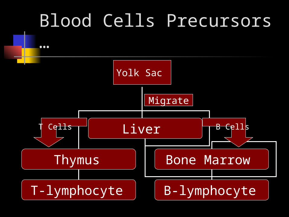
Blood Cells Precursors …
Yolk Sac
Migrate
Liver
Thymus Bone Marrow
B-lymphocyte T-lymphocyte
B CellsT Cells

Primary Lymphoid Organs

Primary Lymphoid Organs …
Primary Lymphoid Organs Either :
Disapear Lose Their Function After Birth
Primary Lymphoid Organs Are :Thymus – Liver – Bone Marrow - Yolk Sac

Secondary Lymphoid Organs

T Cells First , Immature T cells Aggregate
in the cortex of the thymus . Then it goes to the Medulla To
become Maturet . From there it leaves the medulla to
blood stream .
So, in short ..Cortex Medulla (mature) Blood stream

T-Lymphocyte Differentiation
Stem cells lack antigen receptors and CD3, CD4, CD8 surface markers
All T cells should have CD3 ! During their passage through thymus
they differentiate into T cells expressing these markers , In other words …
•Initially , the stem cell will have CD3 – which is the marker for T cells – as well as the 2 receptors ( CD4 & CD8 ) and that is Called Double Positive .-Then it will lose one of the cell receptors and become either :CD4 Positive OR CD8 Positive cell .All T cells should have CD3 ! Stem cells initially do not express CD4 and CD8 (Double Negative)

T-Lymphocyte Differentiation ( cont)
CD8 +ve cell ( Cytotoxic cell )
if the double +ve contacts with MHC I and loses its CD4 receptor . MHC I is present in All Cells having Nucleus .
CD4 +ve ( T helper cells )
if the double +ve contacts with MHC II and
loses CD8 receptor . MHC II present in
Macrophages , Monocytes , Dyndrit cells
…. Etc
T Cells could be …

T-Lymphocyte Differentiation
Thymic Education – teaching the cell to recognize the body cells from foreign , invading and abnormal cells .
Involves two processes CD4 and CD8 positive bearing receptors for “self”
proteins are killed (clonal deletion). This type of removal is called “negative selection”- tolerance to self proteins
CD4 and CD8 positive cells bearing antigen receptors that do not react with self MHC proteins are also killed this process is called “positive selection”
In Other Words

Thymic Education In the thymus , MHC is going to
teach the T cells to :1. Recognize the body cells from
foregin bodies .2. Kill The body cells if they look
abnormal .
Cont

Thymic Education Negative Selection – Killing of T
cells That recognize the body cells as foreign bodies and attack it ( so , they have the capacity to kill your own cells )
Positive Selection – Killing of T cells that doesn’t have the ability to recognize the body cells .

Mature Lymphocyte

T-Lymphocytes Effector Functions
CD8+ve cells ( cytotoxic ) will kill virus- infected cell , tumor cells and allografts (tissue transplant from one human to another )
T cell precursors differentiate under the influence of thymic hormones (thymocins and thymopoetins) into T cell subpopulations ie, CD3, CD4 and CD8.

T-Lymphocytes
All T cells have CD3 proteins on their cell surface in association with T cell receptor(CD4 – CD8 ) , that transmits information - that T cell receptor is occupied – to the inside of the cell .
Mature T cells have either CD4 or CD8 proteins but not both

Functions of Helper Lymphocytes
CD4 Lymphocytes (T Helper ) Functions
Help B cells to develop into antibody producing plasma cells
Help CD8 cells to become activated cytotoxic T cells
Help macrophages in delayed ( Chronic ) type of hypersensitivity
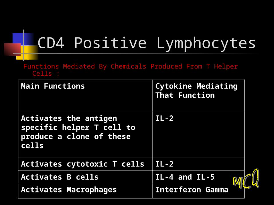
CD4 Positive LymphocytesFunctions Mediated By Chemicals Produced From T Helper Cells :
Main Functions Cytokine Mediating That Function
Activates the antigen specific helper T cell to produce a clone of these cells
IL-2
Activates cytotoxic T cells IL-2
Activates B cells IL-4 and IL-5
Activates Macrophages Interferon Gamma

CD8 positive cells About 35% of peripheral blood T
cells Perform cytotoxic functions They kill virus-infected cell, tumor
and allograft cells Perforins ( a chemical that makes wholes in the
attacked cell )
Programmed cell death (apoptosis)

B cells Origin During embryogenesis – fetal liver Migrate to bone marrow – final destination They do not require thymus for maturation Maturation of B cells involves two phases:
Antigen independent phase consists of stem cells, pre-B cells and B cells
Antigen dependent phase consists of activated B cells and plasma cells
The difference Between
B cells – the antibody that it produces is present on its surface
Plasma Cells – it will send its antibody to the circulation

B cells B cells display surface IgM which serves
as antigen receptor
Surface IgD on some B cells also serves as and antigen receptor
Pre B cells are found in bone marrow and mature B cells are found circulating in bloodstream

Important Point ! Once the B lymphocyte recognizes
the Special Pathogen , or foreign body and became activated , it will be special for that pathogen only for the rest of Its Life .
So , if the lymphocyte come across a Myobacterium TB and became activated to kill it , it will never fight another pathogen Except This Myo.TB for the rest of its life .

B cells B cells constitute about 30% of
circulating small lymphocyte
Their life span is short ie, days or weeks
Location: lymph nodes – germinal centers, spleen – white pulp and Peyer’s patches
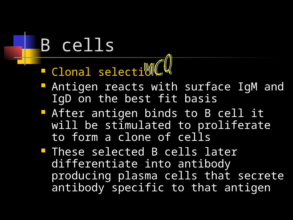
B cells Clonal selection Antigen reacts with surface IgM and IgD
on the best fit basis After antigen binds to B cell it will be
stimulated to proliferate to form a clone of cells
These selected B cells later differentiate into antibody producing plasma cells that secrete antibody specific to that antigen

/B cell is located in the lymph node in: a)cortex. b)paracotex. c)medulla d)trabeculae.
/T lymphocytes"school" is:a)thymus.
b)bone marrow.c)fetal liver.
d)tonsill.e)thyroid.

Thank you