Embed Size (px)
Citation preview
DISEASES OF AQUATIC ORGANISMS Dis Aquat Org
Published March 27
Lysosomal changes in the hemocytes of the freshwater mussel Dreissena polymorpha experimentally exposed to lead and zinc
Laure Giamberini*, Jean Claude Pihan
Centre de Recherches Ecologiques de 1'Universite de Metz, Equipe d'Ecotoxicologie, BP 4116, F-57040 Metz, France
ABSTRACT: This study examines the structural changes of the lysosomal system of the hemocytes of the zebra mussel Dreissena polymorpha experimentally exposed to lead and zinc. A cytochemical tech- nique which demonstrated acid phosphatase activity as a lysosomal marker was used on blood cell monolayers. The results indicate that the effects of both metals on hemocytic lysosomes were variable and that no marked linear relationship between lysosomal changes and metal concentration and expo- sure time was observed. Hemocytes of exposed zebra mussels exhibited enlarged and/or more numer- ous lysosomes. The use of the lysosomal changes in the hemocytes as a biomarker of contaminant expo- sure is discussed.
KEY WORDS: Freshwater mussel . Heavy metals . Hemocytes . Lysosomal changes . Biomarker
INTRODUCTION
Aquatic organisms are able to concentrate in their soft tissues a wide range of contaminants occurring in their environment. Therefore, they have been used as sentinel organisms in the monitoring of environmental pollution. In these studies, particular attention has been given to marine filter-feeding bivalves (Phillips & Rainbow 1988, Browler 1989, Cossa 1989) and, most often, only the xenobiotic body burdens have been taken into account. During the last decade, several authors have shown the importance of assessing the biological effects of pollutants in biomonitoring pro- grams (Bayne 1989, Livingstone et al. 1989, Widdows & Donkin 1989, Narbonne et al. 1991, Veldhuizen- Tsoerkan et al. 1991, Depledge et al. 1995). In this con- text, numerous markers of pollution effects and/or exposure have been developed at different levels of the biological organization (Bayne 1985, Galgani et al. 1992, Regoli 1992, Cajaraville et al. 1995a, Regoli & Principato 1995, Sole et al. 1995). Cellular markers can offer rapid and sensitive indicators of adaptive
responses to environmental contamination, before pol- lutant-induced injury manifests at the level of the whole organism (Moore 1985, Cajaraville et al. 1995b). In many cases, the earliest detectable changes are associated with subcellular organelles such as lyso- somes (Moore 1985). Lysosomes are involved in a broad spectrum of both physiological and pathological functions (Holtzman 1976, Moore 1982, Cheng 1983). Lysosomes are also involved in accumulation and detoxication of both metallic and organic xenobiotics (George 1980, George & Viarengo 1985, Viarengo et al. 1985, 1988). Many nlicroanalytical investigations have demonstrated the occurrence of trace metals in the lysosomal-vacuolar system of different cell types in bivalve molluscs (Schulz-Baldez 1977, George et al. 1978, Lowe et al. 1979, Ballan-Dufranqais et al. 1985, Nigro et al. 1992). In n~olluscs, the digestive gland represents one of the main target organs of pollutant accumulation. Therefore, using various technical approaches, the responses of the digestive cell lysoso- mal system have received the principal attention in pollution assessment in marine ecosystems. Numerous field and experimental studies have shown that various environmental stressors (metallic and organic xeno- biotics, thermal, hypoxia and salinity stress, oxygen-
O Inter-Research 1997 Resale of full article not permitted
222 Dis Aquat Org
free radicals) caused 2 main types of general stress responses in mollusc digestive gland cells: reduction in lysosomal membrane stability and enlargement of lysosomes (Moore 1976, Lowe et al. 1981, Patel & Patel 1985, Viarengo et al. 1987, Lowe 1988, Moore 1988, Cajaraville et al. 1989, Lowe & Clarke 1989, Mari- gomez et al. 1989, Winston et al. 1991, Regoli 1992, Etxeberria et al. 1994, Krishnakumar et al. 1994, Lowe & Pipe 1994, Svenden & Weeks 1995).
On the other hand, the biological tools to assess the health condition of organisms are lacking for the mon- itoring of freshwater ecosystems. Previous investiga- tions have reported the use of the freshwater mussel Dreissena polymorpha as a bioindicator of water pollu- tion (Kraak et al. 1991, Mersch & Pihan 1993). In zebra mussel, as in other bivalve species, the hemocytes are rich in lysosomal hydrolases and are involved in metal transport and detoxication (Giamberini 1993). In this context, the purpose of the present study was to evalu- ate cytochemically the lysosomal responses in the hemocytes of D. polymorpha experimentally exposed to high concentrations of an essential metal (Zn) or of a non-essential metal (Pb).
MATERIALS AND METHODS
Zebra mussel exposure. Mature specimens of Dreis- sena polymorpha (18 to 22 mm in length) were col- lected at a reference location and acclimatized to the experimental conditions for 1 wk. The intoxication experiment was conducted at 14 & 1°C on a 16 h light: 8 h dark cycle. Fifty zebra mussels were placed on tiles to which they could become attached, kept in 9 l tanks, and exposed to lead chloride (PbCl*) at concentrations of 400 and 800 pg 1-' or to zinc chloride (ZnC12) at con- centrations of 500 and 1000 pg 1-' for 5 wk. Lethal con- centrations have been established for D. polymorpha populations in The Netherlands (Kraak et al. 1994). These are for lead: LCS0 (8 wk) = 2330 pg I-', LCso (10 wk) = 497 pg 1-l; and for zinc: (3 wk) = 4293 pg I-', LCS0 (10 wk) = 1065 pg 1-l. The control group received only dechlorinated tap water. Turnover of the tank volume was achieved by means of a peristaltic pump. PbC12 and ZnC1, stock solutions were contlnu- ously introduced into the tanks by a second pump to yield the desired concentration of metals. Food was continuously added in the form of a monospecific sus- pension of the algae Chlamydomonas variabilis, which is readily assimilated by D. polymorpha (Ten Winkel &
Davids 1982). Four zebra mussels were sampled after 7, 21 and 35 treatment days from each experimental group.
Hemolymph colIection and hemocyte monolayers. Hemolymph was obtained by making a notch near the
posterior margin of the mussel shell in the dorsal por- tion. A needle was inserted between the valves, and the hemolymph was withdrawn from the posterior adductor muscle sinus. Portions of hemolymph sam- ples were placed onto a cleaned glass slide, in a moist chamber, and the hemocytes were allowed to settle for 15 min at room temperature. Subsequently, the hemo- cytes were fixed for 5 min with 1.25% glutaraldehyde (grade I; Sigma Chemical Co., St. Louis, MO, USA) in cacodylate buffer solution (0.025 M, pH 7 .4 ) and rinsed with buffer solution.
Enzyme cytochemistry. Acid phosphatases (ACP) (E.C.3.1.3.2) were demonstrated by the azo-dye cou- pling method (Diagnostic kit 386, Sigma Chemical Co.) using naphthol AS-B1 phosphate as substrate and Fast Garnet GBC as coupler (Goldberg & Barka 1962). The change in the presence and relative amounts of ACP following the exposure to metals was assessed accord- ing to the method of Moore & Gelder (1985) modified for the present study. To indicate the relative degree of reactivity, 200 hemocytes were examined on each slide and assigned to 1 of 4 categories: class 1 (0 to 25% of the endoplasm filled with positive granules), class 2 (25 to 50% of the endoplasm filled with positive granules), class 3 (50 to 75 % of the endoplasm filled with positive granules) and class 4 (75 to 100% of the endoplasm filled with positive granules). The degree of reactivity reflects the relative number and/or size of positively staining granules in hemocytes and not the amount of final reaction per granule. The data were then ex- pressed as percentages.
Statistical analysis. Data of the control and experi- mental groups were compared using the Mann-Whit- ney U-test.
RESULTS
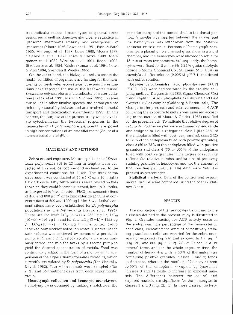
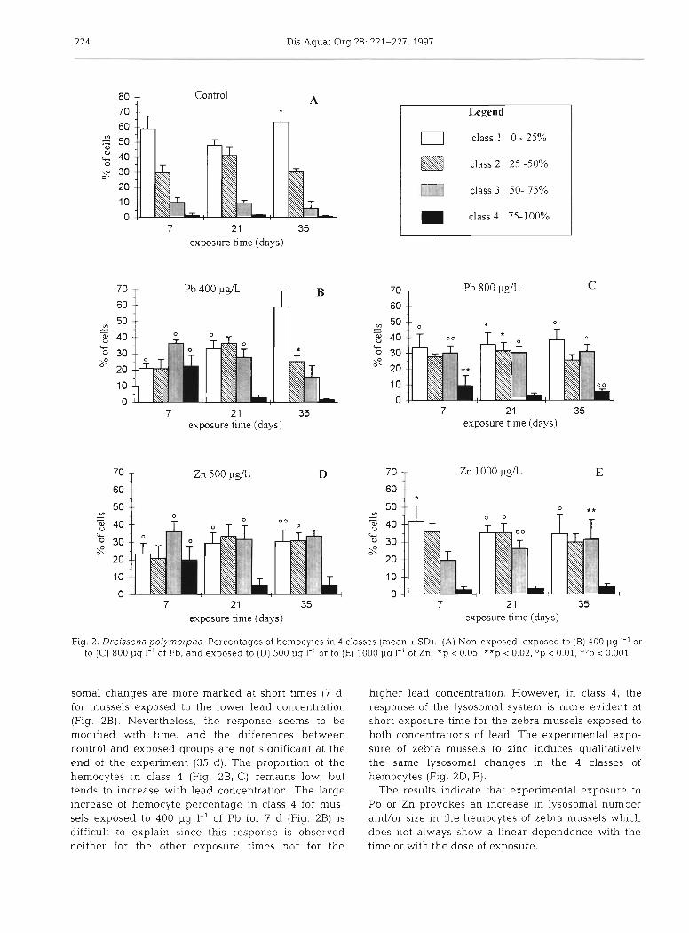
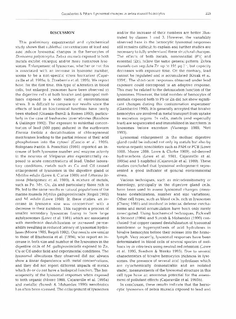
The morphology of the hemocytes belonging to the 4 classes defined in the present study is illustrated in Fig. 1. Granules reacting for ACP activity occur in the endoplasm. The percentage of the hemocytes in each class, indicating the amount of positively stain- ing granules in cells, are reported for the zebra mus- sels non-exposed (Fig. 2A) and exposed to 400 pg I-' (Fig. 2B) and 800 pg 1-' (Fig. 2C) of Pb for 35 d. In general terms and for the whole exposure time, the number of hemocytes with ~ 5 0 % of the endoplasm containing positive granules (classes 1 and 2) tends to decrease, whereas the number of hemocytes with >>50% of the endoplasm occupied by lysosomes (classes 3 and 4) tends to increase in exposed mus- sels. The differences between the control and exposed mussels are significant for the hemocytes in classes 1 and 3 (Fig. 2B, C) . In these cla.sses, the lyso-
Giamberini & Pihan: Lysosomal changes in zebra mussel hemocytes 223
Fig. 1. Dreissena poly- morpha. Acid phospha- tase reactivity (small curved arrows) in the endoplasmic granules in the 4 classes of hemo- cytes. (A) Hemocytes of class 1 (B) Hemocytes of class 2 (double arrow- heads) and of class 3 (simple arrowheads). (C) Hemocytes of class 4. Abbreviations: e = ectoplasm; g = endo- plasmic granules; n = nucleus; p = pseudopod
224 Dis Aquat Org 28: 221-227, 1997
Control A T
7 21 35 exposure time (days)
7 2 1 35 exposure h ~ n e (days)
Legend
0 class l 0 - 25%
class 2 25 -50%
class 3 50- 75%
class 4 75-100%
7 21 35 exposure hlne (days)
7 21 35 exposure time (days)
7 21 35 exposure time (days)
Fig. 2. Dre~ssena polymorpha Percentages of hemocytes In 4 classes (mean * SD). (A) Non-exposed, exposed to (B) 400 pg I-' or to (C) 800 pg I-' of Pb, and exposed to (D) 500 pg 1.' or to (E) 1000 pg 1-' of Zn. *p < 0.05, **p < 0.02; Op < 0.01, OOp < 0.001
somal changes are more marked at short times (7 d ) for mussels exposed to the lower lead concentration (Fig. 2B). Nevertheless, the response seems to be modified with time, and the differences between control and exposed groups are not significant at the end of the experiment (35 d) . The proportion of the hemocytes in class 4 (Fig. 2B, C) remains low, but tends to increase with lead concentration. The large increase of hemocyte percentage in class 4 for mus- sels exposed to 400 pg I-' of Pb for 7 d (Fig. 2B) is difficult to explain since this response is observed neither for the other exposure times nor for the
higher lead concentration. However, in class 4 , the response of the lysosomal system is more evident at short exposure time for the zebra mussels exposed to both concentrations of lead. The experimental expo- sure of zebra mussels to zinc induces qualitatively the same lysosomal changes in the 4 classes of hemocytes (Fig. ZD, E).
The results indicate that experimental exposure to Pb or Zn provokes an increase in lysosomal number and/or size in the hemocytes of zebra mussels whlch does not always show a linear dependence with the time or with the dose of exposure.
Giamberini & Pihan: Lysosomal changes in zebra mussel hemocytes 225
DISCUSSION
This preliminary experimental and cytochemical study shows that sublethal concentrations of lead and zinc induce lysosomal changes in the hemocytes of Dreissena polymorpha. Zebra mussels exposed to both metals exhibit enlarged and/or more numerous lyso- somes. Enlargement of lysosomes, whether or not this is associated with an increase in lysosome number, seems to be a non-specific stress biomarker (Cajar- aville et al. 1995a, b, Etxeberria et al. 1995). We report here, for the first time, this type of alteration in blood cells, but enlarged lysosomes have been observed in the digestive cells of both bivalve and gastropod mol- luscs exposed to a wide variety of environmental stress. It is difficult to compare our results since the effects of lead on biochemical functions have rarely been studied (Gnassia-Barelli & Romeo 1993), particu- larly in the case of freshwater invertebrates (Rainbow & Dallinger 1992). The exposure to sublethal concen- tration of lead (500 ppm) induced in the earthworm Eisenia foetida a destabilisation of chloragosomal membranes leading to the partial release of their acid phosphatases into the cytosol (Cancio et al. 1995). Bolognani-Fantin & Franchini (1991) reported an in- crease of both lysosome number and enzyme activity in the neurons of Viviparus ater experimentally ex- posed to acute concentrations of lead. Under labora- tory conditions, metals such as Cu and Cd caused enlargement of lysosomes in the digestive gland of Mytilus edulis (Lowe & Clarke 1989) and Littorina lit- torea (Marigomez et al. 1989). A mixture of metals, such as Fe, Mn, Cu, Zn and particularly those rich in Pb, led to the same results in natural populations of the marine mussels Mytilus galloprovincialjs (Regoli 1992) and M. edulis (Lowe 1988). In these studies, an in- crease in lysosome size was concomitant with a decrease in their numbers. This suggests a process of smaller secondary lysosomes fusing to form large autolysosomes (Lowe et al. 1981) which are associated with membrane destabilization or increased perme- ability resulting in reduced latency of lysosomal hydro- lases (Moore 1985, Regoli 1992). Our results are similar to those of Etxeberria et al. (1994), who report an in- crease in both size and number of the lysosomes in the digestive cells of M. galloprovincialis exposed to Zn, Cu or Cd under field and experimental conditions. The lysosomal alterations they observed did not always show a linear dependence with metal concentrations, and they did not report different impacts of metals which do or do not have a biological function. The het- erogeneity of the lysosomal responses when exposed to both organic (Moore 1988, Cajaraville et al. 1995a) and metallic (Suresh & Mohandas 1990) xenobiotics has often been stressed. The enlargement of lysosomes
and/or the increase of their numbers are better illus- trated by classes 1 and 3. However, the variability observed here in the hemocytic lysosomal responses still remains difficult to explain and further studies are necessary to fully understand these structural changes. The effects of both metals, nonessential (Pb) and essential (Zn), follow the same general pattern. Zebra mussels can regulate Zn up to 191 pg 1-l, but capacity decreases with exposure time. On the contrary, lead cannot be regulated and is accumulated (Kraak et al. 1994). The short-term responses observed under lead exposure could correspond to an adaptive response. This may be related to the detoxication function of the lysosomes. However, the total number of hemocytes of animals exposed both to Pb or Zn did not show signifi- cant changes during this contamination experiment (Giamberini 1993). It is generally accepted that bivalve hemocytes are involved in metal transport from uptake to excretion organs. In cells, metals (and especially lead) are sequestrated and immobilized in inert form in lysosomes before excretion (Viarengo 1989, Nott 1993).
Lysosomal enlargement in the mollusc digestive gland could be induced not only by metals but also by various organic xenobiotics such as PAH or PCB (Lowe 1988, Moore 1988, Lowe & Clarke 1989), oil-derived hydrocarbons (Lowe et al. 1981, Cajaraville et al. 1995a) and l-naphthol (Cajaraville et al. 1989). These studies concluded that lysosomal enlargement repre- sented a good indicator of general environmental stress.
Various techniques, such as microdensitometry or stereology, principally in the digestive gland cells, have been used to assess lysosomal changes (mem- brane destabilization, number and size variations). Other cell types, such as blood cells, rich in lysosomes (Cheng 1981) and involved in internal defense mecha- nisms and metal accumulation have been only rarely investigated. Using biochemical techniques, Pickwell & Steinert (1984) and Suresh & Mohandas (1990) con- cluded that copper caused destabilization of lysosomal membrane or hypersynthesis of acid hydrolases in bivalve hemocytes before their release into the hemo- lymph. Very recently, lysosomal responses have been determinated in blood cells of several species of mol- luscs by in vitro tests using neutral red retention (Lowe et al. 1995, Svedsen & Weeks 1995). Due to several characteristics of bivalve hemocytes (richness in lyso- somes, the presence of several acid hydrolases which are cytochemically demonstrable and an isolated stade), measurements of the lysosomal structure in this cell type have an enormous potential for the assess- ment of pollutant effects (Cajaraville et al. 1995b).
In conclusion, these results indicate that the hemo- cytic lysosomes of zebra mussels exposed to lead and
226 Dis Aquat Org 28: 221-227, 1997
zinc exhibit structural changes . Thus , w e could con- sider t h e response of the vacuolar-lysosomal system in the hemocytes of Dreissena p o l y n ~ o r p h a a s a potential biomarker for exposure to freshwater pollution. How- ever , several fur ther investigations a r e necessary before validation of this biomarker. First, a n au tomated i m a g e analysis will improve t h e objectivity a n d t h e precision of the method . Moreover, parameters such a s size a n d n u m b e r c a n b e identified separately a n d more rapidly. In addition, experimental approaches with dif- ferent a n d more realistic metal concentrations will b e useful to precisely measure t h e sensitivity of the response. T h e sensitivity of this potential biological marker mus t b e tested i n natural conditions w h e r e mixtures of contaminants a n d various environmental stresses occur. Finally, to obtain a n integrated evalua- tion of organism heal th, t h e lysosomal changes will b e extremely useful i n concert with other biomarkers a t different levels of t h e biological organization, such a s the gametogene t ic index, a n d t h e scope-for-growth a n d body condition index.
Acknowledgements. The authors are grateful to Dr Daniel P. Molloy, New York State Museum, for comments on the man- uscript translation.
LITERATURE CITED
Ballan-Dufranqais C, Jeantet AY, Feghali C, Halpern S (1985) Physiological features of heavy metal storage in bivalves digestive cells and amoebocytes: EMPA and factor analy- sis of correspondences. Blol Cell 53:283-292
Bayne BL (1985) Ecological consequences of stress. In: Bayne BL, Brown DA, Burns K, Dixon DR, Ivanovici A, Llving- stone DR, Lowe DM, Moore MN. Stebbing ARD, Widdows J (eds) The effects of stress and pollution on marine ani- mals. Praeger Publishers. New York, p 141-160
Bayne BL (1989) Measuring the biological effects of pollution: The mussel watch approach. Water Sci Technol 21: 1089-1100
Bolognani-Fantin AM, Franchini A (1991) Ultracytochemical modifications in ganglia of Viviparus ater subsequent experimental 1.ead intoxication Proc 10th Int Malacol Congr (Tiibingen 1989):181- 185
Browler JA (1989) Introduction: aquatic organisms as indica- tors of environmental pollution Water Resour Bull 24: 927-929
Cajaraville MP, Abascal I , Etxeberria M, Marigomez I (1995a) Lysosomes as cellular markers of environmental pollution: time and dose-dependent responses of the digestive lyso- somal system of mussels after petroleum hydrocarbon exposure Environ Toxicol Wat Qual 10: 1-8
Cajaravllle MP, Marigomez JA, Angulo E (1989) A stereolog- ical survey of lysosomal structure alterations in Littor~na littorea exposed to l-naphthol. Comp Biochem Physlol 93C (2):231-237
Cajaraville MP, Robledo Y, Etxeberria M, Marigomez I (1995b) C:ellular biomarkers as useful tools in the blologi- cal monitoring of environmental pollution molluscan digestive lysosomes. In: Cajaraville MP (ed) Cell biology
in environmental toxicology. University of Basque Coun- try Press Service, Bilbo, p 29-55
Cancio I, Gwynn I , lreland MP, Cajaraville MP (1995) The effects of sublethal lead exposure on the ultrastructure and on the distribution of acid phosphatase activity in the chloragocytes of earthworms (Annelida. Oligochaeta). Histochem J 27:965-973
Cheng TC (1981) Bivalves. In: Ratcliffe NA. Rowley AF (eds) Invertebrate blood cells. Academic Press, London, p 231-300
Cheng TC (1983) The role of lysosomes in molluscan inflam- mation. Am Zoo1 23:129-144
Cossa D (1989) A review of the use of Mytdus spp. as quanti- tative indicators of cadmium and mercury contamination in coastal waters. Oceanol Acta 12:417-423
Depledge MH, Aagaard A, Gyorkijs P (1995) Assessment of trace metal toxicity using molecular, physiological and behavioural biomarkers. Mar Pollut Bull 31:19-27
Etxeberria M, Cajaraville MP, Marigomez I (1995) Changes in digestive cell lysosomal structure in mussels as biomark- ers of environmental stress in the Urdaibai estuary (Biscay coast, Iberian peninsula). Mar Pollut Bull 30(9):599-603
Etxeberria M, Sastre I , Cajaraville MP, Marigomez I (1994) Digestive lysosome enlargement induced by experimental exposure to metals (Cu, Cd and Zn) in mussels collected from a zinc-polluted site. Arch Environ Contam Toxicol 27: 338-345
Galgani F, Bocquene G, Truquet P, Burgeot T, Chiffoleau JF. Claisse D (1992) Monitoring of pollutant biochemical effects on marine organisms of the French coasts. Oceanol Acta 15 (4):355-364
George SG (1980) Correlation of metal accumulation in mus- sels with the mechanisms of uptake, metabolism and detoxification: a revlew. Thalassia Jugosl 16:347-365
George SG, Pine BJS, Cheyne AR, Coombs TL, Grant PT (1978) Detoxication of metals by marine bivalves: an ultra- structural study of the compartmentation of copper and zinc in the oyster Ostrea edulis. Mar Biol 45:147-156
George SG, Viarengo A (1985) A model for heavy metal homeostasis and detoxification in mussels. In: Vernberg FG, Thurberg FP, Calabrese A , Vernberg WB (eds) Marine pollution and physiology. Recent advances. University of South Carollna Press, Columbia, p 125-143
Giamberini L (1993) Etude des mecanismes de transport et detoxication des metaux lourds chez la moule d'eau douce Dreissena polymorpha. R61e des hemocytes et des organes du systeme excreteur (histologie, ultrastructure, micro- analyse). PhD thesis, University of Metz
Gnassia-Barelli M, Romeo M (1993) Some aspects of lead eco- toxicology in the marine environment. Aquat Toxicol 26: 163-170
Goldberg AF, Barka T (1962) Acid phosphatase activity in human blood cells. Nature 189:297
Holtzman E (1976) Lysosomes: a survey. Springer-Verlag, Wien
Kraak MSH, Scholten MCT, Peeters WHM, de Kock WC (1991) Biomonitoring of heavy metals in the western Euro- pean rivers Rhine and Meuse using the freshwater mussel Dreissena polymorpha. Environ Pollut 74: 101 -1 14
Kraak MSH, Wlnk YA, Stuijfzand SC, Buckert-de Jong MC, de Groot CJ, Admlraal W (1994) Chronic ecotoxicity of Zn and Pb to the zebra mussel Dreissena polymorpha. Aquat Toxic01 30:77-89
Krishnakumar PK, Casillas E, Varanasi U (1994) Effect of environmental contaminants on the health of Mytilus edulis from Puget Sound, Washington. USA. I . Cytochem- ical measures of lysosomal responses in the digestive cells
Giamberini & Pihan: Lysosomal changes 111 zebra lnussel heinocytes 227
using automatic image analysis. Mar Ecol Prog Ser 106: 249-261
Livingstone DR, Moore MN, Widdows J (1989) Ecotoxicology: b~ological effects measurements on molluscs and their use in impact assessment. Pollution of the North Sea: an assessment. Springer-yerlag. Berlin, p 624-637
Lowe DM (1988) Alterations in cellular structure of Mytilus edulis resulting from exposure to environmental contami- nants under field and experimental conditions Mar Ecol Prog Ser 46:91-100
Lowe DM, Clarke KR (1989) Contaminant-induced changes in the structure of the digestive epithelium of Mytilus edulis. Aquat Toxicol 15345-358
Lowe DM, Moore MN, Clarke KR (1979) The cytochemical distributions of zinc (Zn 11) and iron (Fe 111) in the common mussel, Mytilus edulis, and their relationship with lyso- somes. J Mar Biol Assoc UK 59:851-858
Lowe DM, Moore MN, Clarke KR (1981) Effects of oil on d~gestive cells in mussels: quantitative alterations In cellu- lar and lysosomal structure. Aquat Toxicol 1 213-226
Lowe DM. Pipe RK (1994) Contaminant induced lysosomal membrane damage in marine mussel digestive cells: an in vitro study. Aquat Toxicol 30:357-365
Lowe DM. Soverchia C, Moore MN (1995) Lysosomal mem- brane responses in the blood and digestive cells of mussels experimentally exposed to fluoranthene Aquat Toxicol 33:105-112
Marigonlez JA, Vega MM, Calaraville MP, Angulo E (1989) Quantitative responses of the digestive-lysosomal system of winkles to sublethal concentrations of cadmium. Cell Mol Biol35:555-562
Mersch J, Pihan JC (1993) Simultaneous assessment of envi- ronmental impact on condition and trace metal availability in zebra mussels Dreissena polyrnorpha transplanted into the Wiltz River, Luxembourg. Comparison with the aquatic moss. Arch Environ Contam Toxicol 25.353-364
Moore CA. Gelder SR (1985) Demonstration of lysosomal enzymes in hemocytes of Mercenaria mercenaria (Mol- lusca: Bivalvia). Trans Am Microsc Soc 104(3):242-249
Moore MN (1976) The quantitative cytochemical effects of three metal ions on a lysosomal hydrolase of a hydroid. J Mar B~ol Assoc UK 56:995-1005
Moore M N (1982) Lysosomes and environmental stress. Mar Pollut Bull 13 (2):42-43
Moore MN (1985) Cellular responses to pollutants. Mar Pollut Bull 16:134-139
Moore MN (1988) Cytochemical responses of the lysosomal system and NADPH-ferrihemoprotein reductase in mol- luscan digestive cells to environmental and experimental exposure to xenobiotics. Mar Ecol Prog Ser 46:81-89
Narbonne JF, Ribera D, M~chel X, Raoux C, Garrigues P, Monod JL, Lemaire P, Galgani F, Rolneo M, Salaiin JP, Lafaurie M (1991) lndicateurs biochimiques de contami- nation de l'environnement marin: etude comparative e n mer Mediterrannee. Oceanis 17(3):257-275
Nigro M, Orlando E, Regoli F (1992) Ultrastructural localiza- t ~ o n of metal binding sites in the kidney of the Antarctic scallop Adamussiuln colbecki. Mar Biol 113:637-643
Nott JA (1993) Cytology of pollutant metals in marine inverte- brates: a review of microanalytical applications. Scanning Microsc 5(1): 191-205
Patel S, Patel B (1985) Effect of environmental parameters on lysosomal marker enzymes in the tropical blood clam Anadara granosa. Mar Biol 85:245-252
Phill~ps DJH, Rainbow PS (1988) Barnacles and mussels as
Responsible Subject Editor: D. E. Hinton, Davis, Californja, USA
biomonitors of trace elements: a comparative study. Mar Ecol Prog Ser 49.83-93
Pickwell GV, Steinert SA (1984) Serum blochemica1 and cell- ular responses to experimental cupnc Ion challenge in mussels. Mar Environ Res 14:245-265
Ra~nbow PS, Dallinger R (1992) Metal uptake, regulation and excretion in freshwater invertebrates. In: Dallinger R. Rainbow PS (eds) Ecotoxicology of metals in invertebrates, Chap 7. SETAC Special Publications Series, Lewis Pub- lishers, Roca Katon, FL, p 119-132
Regoli F (1992) Lysosomal responses as a sensitive stress Index in biomonitor~ng heavy metal pollution. Mar Ecol Prog Ser 83:63-69
Regoli F. Principato G (1995) Glutathione, glutathione-depen- dent and antioxidant enzymes in mussel, Mytilus gallo- provincialis, exposed to metals under field and laboratory conditions: implications for the use of biochemical bio- markers. Aquat Toxicol 31:143-164
Schulz-Baldez M (1977) Lead transport in the common mussel Mytilus edulis. In. MC Lusky DS, Berry AJ (eds) Physiology and behavior of marine organisms. Pergamon Press, Oxford, p 211-219
Sole M, Porte C, Albaiges J (1995) The use of biomarkers for assessing the effects of organic pollution in mussels. Sci Total Environ 1591147-153
Suresh K, Mohandas A (1990) Effect of sublethal concentra- tions of copper on he~nocyte number in bivalves. J Inver- tebr Path01 55:325-331
Svendsen C, Weeks JM (1995) The use of a lysosome assay for the rapid assessment of cellular stress from copper to the freshwater snail Viviparus contectus (Millet). Mar Pollut Bull 31(1-3):139-142
Ten Winkel EH, Davids C (1982) Food selection by Dreissena polymorpha Pallas Freshwat Biol 12:553-558
Veldhuizen-Tsoerkan MB, Holwerda DA, de Bont AMT, Smaal AC, Zandee D1 (1991) A field study on stress indices in the sea mussel, Mytllus edulis: application of the 'stress approach' in biomonitoring. Arch Environ Contam Toxicol 21:497-504
Viarengo A (1989) Heavy metals in marine invertebrates: mechanisms of regulation and toxicity at the cellular level. Rev Aquat Sci 1.295-317
Viarengo A, Canes1 L, Pert~ca M, Mancinelli G , Orunesu M, Mazzucotelli A, Bouquegneau JM (1988) Biochemical characterization of a copper-thionein Involved in Cu accu- mulation in the lysosomes of the digestive gland of mus- sels exposed to the metal. Mar Environ Res 24:163-166
Viarengo A, Moore MN, Mancinelli G, Mazzucotelli A, Pipe RK, Farrar SV (1987) Metallothioneins and lysosomes in metal toxicity and accumulation in marine mussels: the effect of cadmium in the presence and absence of phenan- threne. Mar Biol 94.251-257
Vlarengo A, Moore MN, Pertica M, Manc~nelli G , Zanicchi G (1985) Detoxification of copper in the cells of the digestive gland of mussel: the role of lysosomes and thioneins. Sci Total Environ 44:135-145
Widdows J , Donkin P (1989) The application of combined tis- sue residue chemistry and physiological measurement of mussels [Mytilus edulis) for the assessment of environ- mental pollution. Hydrobiologia 188/189.455-461
Winston GW, Moore MN, Straatsburg I , Klrchin MA (1991) Decreased stability of digestive gland lysosomes from the common mussel Mytilus edulis L. by in vitro generation of oxygen-free radicals. Arch Environ Contam Toxicol 21: 401-408
Manuscript first received: February 8, 1996 Revised version accepted: November 22, 1996