Embed Size (px)
Citation preview
Linköping University Postprint
Lysosome-targeted stress reveals increased stability of lipofuscin-
containing lysosomes
Yuri Stroikin, Hanna Mild, Uno Johansson, Karin Roberg and Karin Öllinger Original publication: Yuri Stroikin, Hanna Mild, Uno Johansson, Karin Roberg and Karin Öllinger, Lysosome-targeted stress reveals increased stability of lipofuscin-containing lysosomes, 2008, AGE http://dx.doi.org/10.1007/S11357-007-9045-9. Copyright: The original publication is available at http://www.springerlink.com Postprint available free at: Linköping University E-Press:
http://urn.kb.se/resolve?urn=urn:nbn:se:liu:diva-11051

2
Lysosome-targeted stress reveals increased stability of
lipofuscin-containing lysosomes
Yuri Stroikina*
, Hanna Milda, Uno Johansson
a, Karin Roberg
b and Karin Öllinger
a
aDivision of Experimental Pathology, Department of Neuroscience and Locomotion,
Faculty of Health Sciences, Linköping University, 581 85 Linköping, Sweden
bDivision of Oto-rhino-laryngology,
Faculty of Health Sciences, Linköping University, 581 85 Linköping, Sweden
*Correspondence:
Y. Stroikin, Division of Experimental Pathology, Linköping University, 581 85
Linköping, Sweden; Phone: +46 13 221525; Fax: +46 13 221529; E-mail:
Word counts for the text: 3815
Word counts for the abstract: 124
Number of figures: 7
3
Abstract
Cellular ageing is associated with accumulation of undegradable intralysosomal
material, called lipofuscin. In order to accelerate the lipofuscin-accumulation,
confluent, growth arrested human fibroblasts were cultured under hyperoxic
conditions. To provide a better insight into the effects of lipofuscin on cellular
functions, we compared lysosomal stability in control and lipofuscin-loaded human
fibroblasts under conditions of lysosome-targeted stress induced by exposure to either
the lysosomotropic detergent MSDH or the redox-cycling quinone naphthazarin. We
show that lysosomal damage, assessed by acridine-orange relocation, translocation of
cathepsin D to the cytosol, and alkalinization of lysosomes is more pronounced in
control than in lipofuscin-loaded fibroblasts. Finding that lysosomal integrity was less
affected or even preserved in case of lipofuscin-loaded cells enables us to suggest that
lipofuscin exerts lysosome-stabilizing properties.
Keywords: alkalinization, autophagolysosomes, bafilomycin A1, cathepsin D, MSDH,
naphthazarin quinone

4
Introduction
Membrane-bound acidic organelles, generally termed lysosomes, are considered a
major site for degradation of both extra- and endogenously derived material (De Duve
and Wattiaux 1966). In the latter case, degradation of the cell’s own constituents, so-
called autophagy, not only serves nutritional purposes but also secures intracellular
homeostasis through the removal of damaged and potentially hazardous biomolecules
and organelles (Klionsky 2005). Along with the ability to provide for intracellular
degradation and repair, the lysosomal system is also involved in different signal
transduction pathways (Miaczynska et al. 2004), including that of programmed cell
death (Ferri and Kroemer 2001; Yin et al. 2005). It has been shown that lysosome-
targeted stress, induced by either the lysosomotropic detergent O-methyl-serine
dodecylamide hydrochloride (MSDH; Li et al. 2000) or the redox-cycling quinone
5,8-dihydroxy-1,4-naphthoquinone (NzQ; Roberg et al. 1999), results in
permeabilisation of lysosomal membrane, relocation of lysosomal constituents to the
cytosol and, finally, apoptosis.
Another distinctive feature of lysosomal compartment is that it serves as a site for
storage of non-degraded material. Such material is collectively called lipofuscin,
when age-related, and ceroid when its accumulation is caused by pathological
conditions (Porta 2002; Seehafer and Pearce 2006). According to the free-radical
theory of ageing (Harman 1956), formation of oxidatively damaged intracellular
structures is an inevitable side effect of aerobic life. Imperfect degradation of such
damaged cellular components results in an accumulation of so-called “biological
garbage” of which lipofuscin is an example (Brunk and Terman 2002a). A suitable
model of induced cellular senescence is established by culturing cells under

5
conditions of chronic oxidative stress, which accelerates age-related changes and
results in premature lipofuscin-accumulation (Grune et al. 2005; Terman and Brunk
1998). For the sake of simplicity and due to the similarity of the mechanisms behind
age-related and oxidative stress-induced accumulation (Brunk and Terman 2002a), we
here refer to such intralysosomal pigment as lipofuscin.
Physiological effects of lipofuscin are generally viewed as deleterious due to the
suggested implication of lipofuscin in the formation of free radicals (Brunk and
Terman 2002b). It has been shown that lipofuscin acts as a photosensitizer,
compromising the integrity of lysosomal membrane and finally resulting in cell death
(Wihlmark et al. 1997). The age-related decline in the efficacy of certain proteases
can also be explained in terms of lipofuscin effects. Thus, it has been suggested that
newly produced lysosomal enzymes are misplaced to the lipofuscin-loaded lysosomes
in a futile attempt to degrade lipofuscin instead of performing a useful function within
autophagolysosomes (Terman and Brunk 2004). On the other hand, we recently
found, that moderate levels of lipofuscin are protective and increase the resistance of
ageing fibroblasts to cell death (Stroikin et al. 2007). Beneficial effect of lipofuscin in
this case is explained in terms of hormesis - an adaptation to low doses of otherwise
harmful agents (Rattan 2004).
As has been recently shown by our group, complete starvation causes activation
of programmed cell death through the destabilization of lysosomal compartment.
Such a destabilization was suppressed in lipofuscin-loaded fibroblasts (Stroikin et al.
2007). The presented study is a continuation of the previous one in order to further
investigate the possible hormetic effect of lipofuscin on lysosomal function. Instead of
investigating the apoptotic response, which has been already found disturbed in
lipofuscin-loaded cells, we focus on the stability of lysosomal compartment,
6
considered one of the major regulators of programmed cell death. For this purpose we
compared the integrity of lysosomes in lipofuscin-loaded and control growth-arrested
human fibroblasts under conditions of lysosome-targeted stress induced either by a
lysosomotropic detergent (MSDH) or acute oxidative stress (NzQ). Here we
demonstrate that lysosome-targeted stress results in deleterious changes of lysosomal
compartment that are significantly more pronounced in control fibroblasts than in
lipofuscin-loaded cells.
Materials and methods
Culture conditions and experimental design
AG-1518 human fibroblasts (obtained from Coriell Institute, Camden, NJ, USA)
were cultured in Eagle’s minimum essential medium supplemented with 10% foetal
bovine serum, 2 mM glutamine, 100 IU/ml penicillin-G and 100 µg/ml streptomycin
in an atmosphere of 8% O2, 87% N2 and 5% CO2, at 37°C (normal conditions). The
cells were sub-cultivated at a 1:2 ratio until they reached passage 22-23, and were
then allowed to grow until confluency. These cultures are referred to as control. Some
confluent fibroblast cultures were exposed to 40% O2, 55% N2 and 5% CO2
(hyperoxia) for two months to induce lipofuscin accumulation (Terman and Brunk
1998) and are referred to as lipofuscin-loaded. The culture medium was changed
twice a week. Evaluation of lipofuscin-accumulation was performed by flow
cytometric estimation (Becton Dickinson Biosciences, San Jose, CA, USA) of cellular
autofluorescence and was found 2-3 fold higher in lipofuscin-loaded cells compared
to controls (data not shown).

7
Both controls and lipofuscin-loaded cells were exposed to three different agents
presumably affecting lysosomal integrity: (i) MSDH at a concentration of 25 µM for
15, 30 or 60 minutes; (ii) NzQ at a concentration of 0.75 µM for 15, 30 or 60 minutes;
(iii) the vacuolar ATPase inhibitor bafilomycin A1 (Baf A1; Bowman et al. 1988) at a
concentration of 20 nM for 15 or 30 minutes. Treatment with Baf A1 was used as a
positive control during lysosomal pH assessment.
Lysosomal stability assessment by acridine orange (AO)
Lysosomal stability was assessed by the AO-relocation method (Olsson et al.
1989). AO is a lysosomotropic weak base with metachromatic features. Oligomeric
form of highly concentrated and protonated AO (AOH+) exhibits red fluorescence, as
is the case in intact lysosomes. Lysosomal alkalinization and translocation of
lysosomal content to the cytosol during lysosomal stress results in the formation of
the monomeric deprotonated form of AO with green fluorescence.
Cells on cover-slips were briefly stained with 5 µg/ml AO for 15 minutes under
normal culture conditions, rinsed in complete medium and exposed to one of the
lysosomal-stress-inducing agents as described in previous section. Live cultures were
examined in an Axiovert S100TV microscope (Carl Zeiss, Jenna, Germany) equipped
with a Hamamatsu digital camera C4742-95 (Hamamatsu, Hamamatsu City, Japan)
using 60 x /1.4 oil lens, a halogen lamp and a blue-excitation filter. Emission was
detected using a high pass filter above 520 nm. Phase-contrast and fluorescent images
were obtained using OpenLab software (Improvision, Coventry, UK). Measurements
of lysosomal red AO-fluorescence were performed using the National Institute of
Health Image program (http://rsb.info.hih.gov/nih-image/). Fluorescence intensity was

8
expressed in arbitrary units (a. u.) being a product of average pixel value per lysosome
and the lysosome area.
Assessment of lysosomal pH
The lysosomal pH was measured by flow cytometry as described elsewhere
(Nilsson et al. 2003). Briefly, cells were exposed to 40,000 MW FITC-dextran
(Molecular Probes, Eugene, OR, USA) at a concentration of 0.1 mg/ml for 3 days at
37°C. Loading with FITC-dextran was followed by incubation in complete medium
for another 24 hours. After that, cells were trypsinized, centrifuged at ~300 x g for 5
minutes, re-suspended in the culture medium, filtered through a 70 µm cell strainer
and analyzed by flow cytometry (Becton Dickinson Biosciences, San Jose, CA,
USA). A 488 nm argon laser was used for the FITC excitation, and emission was
detected in the FL1 and FL2 channels using a 530 ± 28 nm and a 610 ± 20 nm barrier
filters, respectively. Data from 10,000 cells was analyzed using the CellQuest
program (Becton Dickinson Immunocytometry systems, San Jose, CA, USA).
Modified Britton-Robinson buffers (pH 4.0–7.0), containing sodium azide and 2-
deoxyglucose at a final concentration of 50 mM each and nigericin at a final
concentration of 10 µM were used for the preparation of a standard curve. The
FL1/FL2 ratios were used to calculate the pH employing the standard curve.
Immunocytochemical detection of cathepsin D
Cells plated on cover-slips were fixed in 4% formaldehyde in phosphate-buffered
saline (PBS) for 20 minutes in 4ºC, rinsed in PBS, and exposed to incubation buffer
containing 0.1% saponin and 5% foetal bovine serum in PBS for another 20 minutes
at room temperature. The cells were then incubated with 1:100 diluted polyclonal

9
rabbit anti-human antibodies to cathepsin D (DAKO, Roskilde, Denmark) for 1 hour
in a humidifier at room temperature. After rinsing in the incubation buffer (2 x 5
minutes), the specimens were exposed to 1:100 diluted goat anti-rabbit IgG-Alexa 594
conjugate (Molecular Probes, Eugene, OR, USA) for 1 hour at room temperature;
rinsed in PBS and distilled water, and mounted in Vectashield (Vector Laboratories,
Burlingame, CA, USA). Images were obtained using a Microphot-SA fluorescence
microscope equipped with an ORCA 100 Hamamatsu color digital camera
(Hamamatsu, Hamamatsu City, Japan).
Extraction of cytosol
Disturbed lysosomal integrity was additionally assessed by estimation of the
amount of cathepsin D translocated to the cytosol during lysosomal stress. Cytosolic
extraction was performed using the cholesterol-solubilizing agent digitonin as
described elsewhere (Johansson et al. 2003). Briefly, digitonin at low concentrations
permeabilizes cholesterol-rich membranes such as the plasma membrane, leaving
cholesterol-poor membrane of lysosomes and mitochondria more or less intact. Cells
were exposed to an extraction buffer (250 mM sucrose, 20 mM Hepes, 10 mM KCl,
1.5 mM MgCl2, 1 mM EDTA and 1 mM EGTA) containing digitonin at concentration
of 25 µg/ml on ice for 12 minutes. The extraction buffer was collected and proteins
were precipitated by addition of 5% trichloric acid on ice for 10 minutes and
centrifuged at ~ 20,800 x g for 15 minutes to obtain the protein pellet.
Immunoblotting is described below.

10
Western blot analysis
The pellet was dissolved in 25 µl of lysis buffer containing 6 M urea, 63 mM
Tris-HCl (pH 6.8), 10% glycerol and 2% SDS. Samples were then neutralized using 4
µl of 1 M NaOH per sample. Consequently, 50 µM ditiotreitol (DTT) and 0.05%
bromphenol blue were added and 30 µl aliquots of the cell lysate were fractionated by
15% SDS-PAGE. The proteins were then blotted on a nitrocellulose membrane,
which was subsequently incubated in 5% skimmed milk in Tris-buffered saline (50
mM Tris, 0.15 M NaCl, pH 7.5) with 0.1% Tween-20 (TBS-T) for 90 minutes at room
temperature and then washed in TBS-T. The membrane was then exposed over night
at 4ºC to a monoclonal mouse anti-human cathepsin D antibody (Oncogene Research
Products, San Diego, CA, USA) diluted 1:1000 in 0.1% skimmed milk in TBS-T. The
membrane was then washed and incubated for 1 hour at room temperature with
horseradish peroxidase (HRP)-conjugated goat anti-mouse secondary antibody
(DAKO, Roskilde, Denmark) diluted 1:1500. Bands were visualized using Western
blotting Luminol Reagent (Santa Cruz Biotechnology, Santa Cruz, CA, USA). Equal
loading was verified by reprobing the membrane with a mouse anti-glyceraldehyde-3-
phosphate dehydrogenase (GAPDH; Biogenesis, Poole, Dorset, U.K.) antibody
diluted 1:400.
Transmission electron microscopy
Cells were fixed by adding 2% glutaraldehyde (Agar Scientic, Essex, UK) in 0.1
M sucrose-sodium cacodylate-HCl buffer (pH 7.2; Sigma, St Louis, MO, USA) and
post-fixed in osmium (Johnson Matthey Chemicals, Royston, UK). Specimens were
stained en bloc with 2% uranyl acetate (Sigma) in 50% ethanol, dehydrated in a
graded series of ethanol, embedded in Epon-812 (Fluka AG, Buchs, Switzerland) and
11
polymerized at 60ºC for two days. Thin sections were cut with a diamond knife
(DIATOME, Bienne, Switzerland), stained with lead citrate, and then examined and
photographed in a JEOL 1230-EX electron microscope (JEOL, Tokyo, Japan) at 100
kV.
Statistics
For the estimation of AO-fluorescence, 100 to 140 lysosomes from each
specimen were analyzed. All experiments were repeated at least three times. Values
are given as means ± SD. Data obtained by the acridine orange relocation method
were analyzed using the ANOVA and post hoc LSD test. The lysosomal pH data were
analyzed using Kruskal-Wallis and post hoc Mann Whitney test. P-values < 0.05 were
considered significant.
Results
Phase-contrast microscopy of MSDH- and NzQ-treated fibroblasts reveals
significant morphological differences between control and lipofuscin-loaded cells
(Figure 1a). Thus, MSDH-induced cytosolic vacuolization (arrows in Figure 1a),
albeit present in both control and lipofuscin-loaded fibroblasts, is more pronounced in
control cells. Relatively higher optical density of AO-loaded lysosomes (dark dots on
phase-contrast images) enables their distinction from newly formed vacuoles. During
exposure to NzQ, there is a noticeable agglomeration of lysosomes in control cells;
while in lipofuscin-loaded fibroblasts this phenomenon is less obvious (arrowheads in
Figure 1a).

12
Figure 1a. Lysosomal stress causes more prominent alterations in control cells than in lipofuscin-loaded. Phase-contrast images of fibroblasts cultured under normal conditions (controls) or pre-exposed to hyperoxia in order to accelerate accumulation of lipofuscin, and then exposed to either
25 µM MSDH or 0.75 µM naphthazarin (NzQ) and stained with acridine orange. Arrows mark cytosolic vacuoles and arrowheads indicate agglomeration of acridine orange-positive lysosomes.
Scale bar, 20 µm.
Non-confocal fluorescent imaging of AO-stained cells shows that the number of
lysosomes does not obviously differ between non-treated control and lipofuscin-
loaded fibroblasts (Figure 1b). Both MSDH- and NzQ-treatment result in decrease of
the number of lysosomes in control cells, whereas neither of the treatments shows any
effect on lysosomal number in lipofuscin-loaded fibroblasts (Figure 1b).

13
Figure 1b. The number of lysosomes decreases during MSDH- and naphthazarin-treatment in control but not in lipofuscin-loaded fibroblasts. Non-confocal fluorescent images of fibroblasts cultured under normal conditions (controls) or pre-exposed to hyperoxia in order to accelerate
accumulation of lipofuscin, and then exposed to either 25 µM MSDH or 0.75 µM naphthazarin (NzQ) and stained with acridine orange. Scale bar, 20 µm.
Ultrastructural changes of control fibroblasts exposed to 25 µM MSDH for 60
minutes (Figure 2) are characterized by advanced autophagy represented by
accumulation of autophagolysosomes (Figure 2b). Moreover, the number of
organelles with typical lysosomal morphology, observed in untreated cells (Figure
2a), decreases during MSDH-treatment (Figure 2b).

14
Figure 2. Ultrastructural changes of MSDH-treated fibroblasts include pronounced autophagic vacuolization. (a) Untreated control fibroblast and (b) fibroblasts exposed to 25 µM MSDH for 60 minutes. Autophagolysosomes (AL) found in MSDH-treated cells represent the advanced stage of
autophagy. L: lysosome; Mt: mitochondria; N: nucleus. Scale bar, 2 µm.
Relocation of AO to the cytosol in MSDH- and NzQ-treated fibroblasts is
estimated by the decrease of red-fluorescence-intensity of lysosomes. During
exposure to MSDH, the fluorescence intensity becomes significantly reduced already
after 15 minutes in control fibroblasts but only after 60 minutes in lipofuscin-loaded
cells (Figure 3a). NzQ-treatment results in a significant decrease (after 15 minutes)
and consequent recovery (observed after 30 and 60 minutes) of AO-associated red
fluorescence intensity in control cells. Lipofuscin-loaded fibroblasts, on the other
hand, do not show any significant changes of AO-fluorescence during exposure to
NzQ (Figure 3b).

15
Figure 3. More pronounced acridine orange-relocation in control than in lipofuscin-loaded fibroblasts during lysosomal stress. Fibroblasts were cultured under normal conditions (control) and at hyperoxia in order to accumulate lipofuscin. Acridine orange accumulates in undamaged lysosomes and generates red fluorescence. Decrease of red fluorescence intensity denotes relocation of acridine
orange to the cytosol. MSDH-treatment (a) causes significant acridine orange-relocation after 15 minutes in control cells and only after 60 minutes in lipofuscin-loaded. Exposure to naphthazarin
(NzQ) for 15 minutes results in significant relocation of acridine orange in control cells with a consequent tendency toward recovery (b). Naphthazarin-treatment does not have any significant effects
on lipofuscin-loaded cells. Values are mean ± SD, n = 3 with 100-140 assessed lysosomes in each sample (* = P < 0.05 compared to corresponding non-treated cells [0 min]).
Intact lysosomes of non-treated cells are characterized by a grainy pattern of
cathepsin D immunostaining (Figure 4). During MSDH-treatment, staining pattern
becomes diffuse, as observed in control cells after 15 minutes and even more
pronounced after 60 minutes. In lipofuscin-loaded fibroblasts, diffuse pattern of
cathepsin D staining becomes evident after 60 minutes of MSDH-treatment and is less
prominent than in control cells. NzQ-treated control fibroblast cultures initially show
diffuse lysosomal staining (observed after 15 minutes of exposure), which later
returns to the grainy one (observed after 60 minutes of exposure to NzQ). In

16
lipofuscin-loaded fibroblasts cathepsin D staining remains lysosomal during NzQ-
treatment (Figure 4).
Figure 4. Lysosomal stress is associated with cathepsin D translocation, which is more obvious in control than in lipofuscin-loaded fibroblasts. Fibroblasts cultured under normal conditions
(control) or pre-exposed to hyperoxia in order to accumulate lipofuscin were treated with either 25 µM MSDH or 0.75 µM naphthazarin (NzQ). Cells were fixed and immunostained for cathepsin D and an Alexa 594-conjugated secondary antibody was used. The change from a grainy (lysosome-like) to a
diffuse (cytosolic) pattern of cathepsin D immunostaining during MSDH-treatment is more evident in control (after 15 minutes) than lipofuscin-loaded fibroblasts (after 60 minutes). Lipofuscin-loaded cells do not show any changes in cathepsin D staining during naphthazarin-treatment, while in control cells
diffuse staining appeared after 15 minutes of treatment with a consequent tendency toward recovery and acquisition of a grainy pattern of staining after 60 minutes. Scale bar, 20 µm.
Digitonin-extraction of cytosol enables the estimation of the amount of cathepsin
D released from the lysosomes during lysosomal stress. Western blot analysis
17
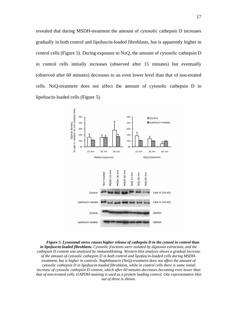
revealed that during MSDH-treatment the amount of cytosolic cathepsin D increases
gradually in both control and lipofuscin-loaded fibroblasts, but is apparently higher in
control cells (Figure 5). During exposure to NzQ, the amount of cytosolic cathepsin D
in control cells initially increases (observed after 15 minutes) but eventually
(observed after 60 minutes) decreases to an even lower level than that of non-treated
cells. NzQ-treatment does not affect the amount of cytosolic cathepsin D in
lipofuscin-loaded cells (Figure 5).
Figure 5. Lysosomal stress causes higher release of cathepsin D to the cytosol in control than in lipofuscin-loaded fibroblasts. Cytosolic fractions were isolated by digitonin extraction, and the
cathepsin D content was analyzed by immunoblotting. Western blot analysis shows a gradual increase of the amount of cytosolic cathepsin D in both control and lipofuscin-loaded cells during MSDH-treatment, but is higher in controls. Naphthazarin (NzQ)-treatment does not affect the amount of cytosolic cathepsin D in lipofuscin-loaded fibroblasts, while in control cells there is some initial
increase of cytosolic cathepsin D content, which after 60 minutes decreases becoming even lower than that of non-treated cells. GAPDH-staining is used as a protein loading control. One representative blot
out of three is shown.

18
MSDH-treatment is characterized by a gradual increase of lysosomal pH in both
control and lipofuscin-loaded cells (Figure 6a). Control fibroblasts show earlier and
more pronounced lysosomal alkalinization than lipofuscin-loaded cells (after 15 and
60 minutes in controls and lipofuscin-loaded cells, respectively). NzQ-treatment does
not have any apparent effect on lysosomal pH of lipofuscin-loaded fibroblasts, while
in control cells the increase in lysosomal pH is observed after 15 minutes, becomes
significant after 30 minutes, and is followed by lysosomal acidification, observed
after 60 minutes of exposure to NzQ (Figure 6b). Treatment with Baf A1, used as a
positive control for lysosomal alkalinization, results in a significant increase of
lysosomal pH in both control and lipofuscin-loaded cells, but is considerably higher in
controls (Figure 6c). Regardless of treatment used, the difference in pH between
controls and lipofuscin-loaded cells remains significant at all time points (Figure 6a-
c).
Discussion
First suggested by de Duve and Wattiaux (1966), the idea of lysosomal
involvement in cell death has been gaining more evidence. During lysosome-targeted
stress, the type of cell death depends on the degree of lysosomal damage. Thus,
extensive release of lysosomal content results in necrosis, while partial lysosomal
rupture mediates apoptosis (Brunk et al. 2001; Zhao et al. 2003). Lysosomal
destabilization is an initial event in programmed cell death induced by oxidative
stress, radiation, and exposure to MSDH and oxidized low-density lipoprotein (Brunk
and Svensson 1999; Li et al. 2000; Persson et al. 2005; Yuan et al. 1997). It has been
shown that permeabilisation of lysosomal membrane leading to translocation of

19
lysosomal enzymes to the cytosol precedes mitochondrial release of cytochrome c and
subsequent caspase-activation (Roberg et al. 1999).
Figure 6. Treatments of fibroblasts with MSDH, naphthazarin or bafilomycin A1 result in lysosomal alkalinization that is more pronounced in control than in lipofuscin-loaded cells.
Fibroblasts were allowed to endocytose FITC-dextran for three days and the lysosomal pH was assessed by ratiometric calculation. MSDH-treatment (a) causes significant lysosomal alkalinization
observed in control fibroblasts already after 15 minutes of exposure. Alkalinization of lipofuscin-loaded lysosomes becomes significant only after 60 minutes. Increase in lysosomal pH during naphthazarin (NzQ)-treatment (b) becomes significant after 30 minutes in control cells with a
consequent tendency toward acidification. No effects of naphthazarin-treatment on pH of lipofuscin-loaded lysosomes are detected. Inhibition of the lysosomal proton pump using bafilomycin A1 (Baf A1) causes significant lysosomal alkalinization in both control and lipofuscin-loaded cells (c). Values are mean ± SD, n = 3 (* = P < 0.05 compared to corresponding non-treated cells [0 min]). Difference
between control and lipofuscin-loaded cells during all three types of treatment remains significant at all time-points.
The current study is a continuation of the recently presented work of our group,
showing that lipofuscin-loaded cells are more resistant to cell death (Stroikin et al.
2007). Here, instead of studying the apoptotic response, we focus on the effects of

20
lipofuscin on lysosomal stability, proven to be crucial for the initiation of apoptosis
(Brunk et al. 2001).
The presented MSDH-induced vacuolization (Figures 1a, b; 2b) can be
considered an adaptive cellular response as an attempt to limit the damage (Henics
and Wheatly 1999), by means of autophagic sequestration. Since lysosomal enzymes
translocated to the cytosol jeopardize survival of the cell, autophagy is primarily
focused on sequestration of the released lysosomal content. It has also been suggested
that damaged entire lysosomes can be autophagocytozed (Stroikin et al. 2004; Kiffin
et al. 2006). Regardless of the mechanism of autophagic sequestration, it does not
prevent apoptosis of MSDH-treated cells (Li et al. 2000), since the acidic interior of
autophagolysosomes is attracting MSDH, which at low pH becomes protonated and
acquires properties of a detergent (Firestone et al. 1979).
During MSDH-treatment, the number of vacuoles (Figure 1a), corresponding to
autophagolysosomes (Figure 2b), becomes extensive, occupying the entire cell. This
suggests that activated autophagy is no longer a repairing mechanism, but rather an
executioner of cellular demise (Gozuacik and Kimchi 2004), which is in agreement
with both the decrease of the amount of AO-positive lysosomes (Figure 1b) and
ultrastructural findings of the decreased number of morphologically typical lysosomes
(Figure 2a, b). These morphological features of lysosomal deterioration during
MSDH-exposure are consistent with findings of gradual (i) decrease of lysosomal-
associated AO-fluorescence; (ii) increase of diffuse cathepsin D immunostaining; (iii)
increase of cathepsin D in the cytosolic fraction and, (iv) lysosomal alkalinization.
Although these changes are present in both control and lipofuscin-loaded fibroblasts,
the deleterious effect of MSDH-treatment is significantly less pronounced in
lipofuscin-loaded cells, which indicates reduced lysosomal sensitivity. The difference
21
of susceptibility toward MSDH-induced lysosomal stress between control and
lipofuscin-loaded fibroblasts cannot be explained in terms of decreased tropism of
MSDH to lipofuscin containing lysosomes, since lysosomal pH, responsible for both
tropism and protonation of MSDH, is practically equally low in both control (4.24 ±
0.26) and lipofuscin-loaded (4.11 ± 0.17) cells. On the other hand, lipofuscin-
containing lysosomes, when extensively overloaded, are excluded from physiological
functioning as sites of degradation (Terman and Brunk 2004), causing formation of
new, lipofuscin-free lysosomes. Such an increase of the overall volume of lysosomal
compartment can be considered a possible modulator of reactivity towards
lysosomotropic detergents. In this light, lipofuscin could be viewed as an indirect
factor of increasing lysosomal stability. But since the number of lysosomes in non-
treated cells does not markedly differ between control and lipofuscin-loaded
fibroblasts (Figure 1b), the volume of the lysosomal compartment cannot be rendered
as a factor responsible for the increased resistance to MSDH-treatment in the
presented study.
During NzQ-treatment of control cells, the agglomeration of lysosomes without
apparent cytosolic vacuolization (Figure 1a) might represent successful reparative
autophagy. Lysosomal damage revealed by the initial decrease of lysosomal
fluorescence (Figure 3b), translocation of cathepsin D to the cytosol (Figures 4, 5) and
lysosomal alkalinization (Figure 6b) triggers the mechanisms leading to the
accomplished control over the damage. The idea of effective autophagic repair is
consistent with the results showing the eventual (i) regaining of lysosomal-associated
AO-fluorescence (Figure 3b); (ii) recovery of the grainy pattern of cathepsin D
immunostaining (Figure 4); (iii) decrease of the cytosolic fraction of cathepsin D
(Figure 5) and (iv) lysosomal acidification (Figure 6b) of control fibroblasts after 60

22
minutes of exposure to NzQ. Logically, not all cells manage to accomplish a
successful damage-control. Some cells die due to the extensive lysosomal damage.
Considering that one fraction of cells die, measurements performed on NzQ-exposed
cells might represent cellular resistance rather than recovery from cell damage. On the
other hand, during continuous exposure to NzQ, lysosomal agglomeration (Figure 1a),
coexisting with a decrease of the number of lysosomes (Figure 1b) suggests an
ongoing intracellular process by which cells cope with unfavorable conditions. Even
if some cells are lost during NzQ-treatment, conclusions drawn from the
measurements made on the surviving population of cells still emphasize the
mechanism of cell survival.
The absence of changes in lysosomal compartment of lipofuscin-loaded cells
suggests that NzQ-treatment does not cause significant lysosomal damage in these
cells. According to recent theories, high content of iron associated with lipofuscin
should have increased the lysosomal sensitivity to oxidative stress (Yu et al. 2003).
Alternatively, the resistance of lipofuscin-loaded cells can be due to their pre-
exposure to hyperoxic conditions. The possibility of up-regulation of anti-oxidative
defense during chronic oxidative stress still remains to be investigated.
Physiological effects of lipofuscin on cellular functions in general and on
lysosomal integrity in particular, have been a matter of controversial opinions. The
increased sensitivity of lipofuscin-accumulating cells to lysosomal breach and
apoptosis has been explained in terms of redox-active iron content, which promotes
formation of free radicals under conditions of oxidative stress (Terman and Brunk
2004). In opposition, some researchers doubt that a convincing evidence of
deleterious effects of lipofuscin has ever been demonstrated (Porta et al. 2002).
Moreover, we recently showed that moderate levels of lipofuscin have protective

23
effects on cell survival during nutritional deprivation (Stroikin et al. 2007). The
previously suggested idea, that lipofuscin permanently occupies active sites of
lysosomal enzymes, preventing their translocation to the cytosol and engagement in
programmed cell death during lysosomal stress, is consistent with the present findings
of decreased cytosolic translocation of cathepsin D in lipofuscin-loaded cells. In
addition, lipofuscin exhibits some proton-trapping properties, considering that
decrease of the proton gradient upon treatment with Baf A1 is significantly lower in
lipofuscin-loaded cells (Figure 6c). Positive correlation between cellular lipofuscin
content and resistance to oxidative stress can also be indicative of lipofuscin
functioning as a trap for free radicals, explaining the high resistance of lipofuscin-
loaded cells to NzQ-treatment. While the exact mechanism of lipofuscin influence on
cellular functions remains to be elucidated, we suggest that lipofuscin has lysosome-
stabilizing properties, making these organelles less sensitive and diminishing their
influence on cellular functioning.
In conclusion, increased autophagy following MSDH-treatment can be viewed as
an adaptive cellular response in order to limit the damage caused by the leakage of
protons and proteolytic enzymes into the cytosol because of impairment of the
lysosomal membrane. Relocation of lysosomal content to the cytosol and decrease of
intralysosomal pH are indicators of lysosome-targeted stress induced either by
exposure to the lysosomotropic detergent MSDH or the redox-cycling quinone
naphthazarin. Such a destabilization is significantly hampered in lysosomes, which
contain the ageing-associated pigment lipofuscin. Increased stability of lipofuscin-
containing lysosomes can be related to the decreased inducibility of apoptosis in
ageing cells.

24
Acknowledgements
We thank Linda Vainikka for technical assistance. This study was financially
supported by Lions Research Foundation and by grant from the Medical Branch of the
Swedish Research Council (Vetenskapsrådet).

25
References
Bowman EJ, Siebers A and Altendorf K (1988) Bafilomycins: a class of inhibitors of
membrane ATPases from microorganisms, animal cells, and plant cells. Proc
Natl Acad Sci USA 85:7972-7976
Brunk UT and Svensson I (1999) Oxidative stress, growth factor starvation and Fas
activation may all cause apoptosis through lysosomal leak. Redox Rep 4:3-11
Brunk UT, Neuzil J and Eaton JW (2001) Lysosomal involvement in apoptosis.
Redox Rep 6:91-97
Brunk UT and Terman A (2002a) Lipofuscin: Mechanisms of age-related
accumulation and influence on cell functions. Free Radic Biol Med 33:611-
619
Brunk UT and Terman A (2002b) The mitochondrial-lysosomal axis theory of aging:
Accumulation of damaged mitochondria as a result of imperfect
autophagocytosis. Eur J Biochem 269:1996-2002
De Duve C and Wattiaux R (1966) Functions of lysosomes. Annu Rev Physiol
28:435-492
Ferri KF and Kroemer G (2001) Organelle-specific initiation of cell death pathways.
Nat Cell Biol 3:255-263
Firestone RA, Pisano JM and Bonney RJ (1979) Lysosomotropic agents. 1. Synthesis
and cytotoxic action of lysosomotropic detergents. J Med Chem 22:1130-1133
Gozuacik D and Kimchi A (2004) Autophagy as a cell death and tumor suppressor
mechanism. Oncogene 23:2891-2906

26
Grune T, Merker K, Jung T, Sitte N and Davies K.J (2005) Protein oxidation and
degradation during postmitotic senescence. Free Radic Biol Med 39:1208-
1215
Harman D (1956) Aging: a theory based on free radical and radiation chemistry. J
Gerontol 211:298-300
Henics T and Wheatley DN (1999) Cytoplasmic vacuolation, adaptation and cell
death: a view on new perspectives and features. Biol Cell 91:485-498
Johansson AC, Steen H, Öllinger K and Roberg K (2003) Cathepsin D mediates
cytochrome c release and caspase activation in human fibroblast apoptosis
induced by staurosporine. Cell Death Differ 10:1253-1259
Kiffin R, Bandyopadhyay U and Cuervo AM (2006) Oxidative stress and autophagy.
Antioxid Redox Signal 8:152-162
Klionsky DJ (2005) The molecular machinery of autophagy: unanswered questions. J
Cell Sci 118:7-18
Li W; Yuan X, Nordgren G, Dalen H, Dubowchik GM, Firestone RA and Brunk UT
(2000) Induction of cell death by the lysosomotropic detergent MSDH. FEBS
Lett 470:35-39
Miaczynska M, Pelkmans L and Zerial M (2004) Not just a sink: endosomes in
control of signal transduction. Curr Opin Cell Biol 16:400-406
Nilsson C, Johansson U and Öllinger K (2003) Analysis of cytosolic and lysosomal
pH in apoptotic cells by flow cytometry. Methods Cell Sci 25:185-194
Olsson GM, Rungby J, Rundquist I and Brunk UT (1989) Evaluation of lysosomal
stability in living cultured macrophages by cytofluorometry. Effect of silver
lactate and hypotonic conditions. Virchows Arch B Cell Pathol Incl Mol
Pathol 56:263-269

27
Persson HL, Kurz T, Eaton JW and Brunk UT (2005) Radiation-induced cell death:
importance of lysosomal destabilization. Biochem J 389:877-884
Porta EA (2002) Pigments in aging: an overview. Ann N Y Acad Sci 959:57-65
Porta EA, Berra A, Monserrat AJ and Benavides SH (2002) Differential lectin
histochemical studies on lipofuscin (age-pigment) and on selected ceroid
pigments. Arch Gerontol Geriatr 34:193-203
Rattan SI (2004) Aging, anti-aging, and hormesis. Mech Ageing Dev 125:285-289
Roberg K, Johansson U and Öllinger K (1999) Lysosomal release of cathepsin D
precedes relocation of cytochrome c and loss of mitochondrial transmembrane
potential during apoptosis induced by oxidative stress. Free Radic Biol Med
27:1228-1237
Seehafer SS and Pearce DA (2006) You say lipofuscin, we say ceroid: defining
autofluorescent storage material. Neurobiol Aging 27:576-588
Stroikin Y, Dalen H, Lööf S and Terman A (2004) Inhibition of autophagy with 3-
methyladenine results in impaired turnover of lysosomes and accumulation of
lipofuscin-like material. Eur J Cell Biol 83:583-590
Stroikin Y, Johansson U, Asplund S and Öllinger K (2007) Increased resistance of
lipofuscin-loaded prematurely senescent fibroblasts to starvation-induced
programmed cell death. Biogerontology 8:43-53
Terman A and Brunk UT (1998) Ceroid/lipofuscin formation in cultured human
fibroblasts: The role of oxidative stress and lysosomal proteolysis. Mech
Ageing Dev 104:277-291
Terman A and Brunk UT (2004) Lipofuscin. Int J Biochem Cell Biol 36:1400-1404

28
Wihlmark U, Wrigstad A, Roberg K, Nilsson SE and Brunk UT (1997) Lipofuscin
accumulation in cultured retinal pigment epithelial cells causes enhanced
sensitivity to blue light irradiation. Free Radic Biol Med 22:1229-1234
Yin L, Stearns R and Gonzalez-Flecha B (2005) Lysosomal and mitochondrial
pathways in H2O2-induced apoptosis of alveolar type II cells. J Cell Biochem
94:433-445
Yu Z, Persson HL, Eaton JW and Brunk UT (2003) Intralysosomal iron: a major
determinant of oxidant-induced cell death. Free Radic Biol Med 34:1243-1245
Yuan XM, Li W, Olsson AG and Brunk UT (1997) The toxicity to macrophages of
oxidized low-density lipoprotein is mediated through lysosomal damage.
Atherosclerosis 133:153-161
Zhao M, Antunes F, Eaton JW and Brunk UT (2003) Lysosomal enzymes promote
mitochondrial oxidant production, cytochrome c release and apoptosis. Eur J
Biochem 270:3778-3786