Embed Size (px)
Citation preview
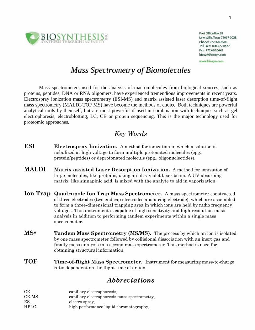
1
MMaassss SSppeeccttrroommeettrryy ooff BBiioommoolleeccuulleess
Mass spectrometers used for the analysis of macromolecules from biological sources, such as
proteins, peptides, DNA or RNA oligomers, have experienced tremendious improvements in recent years.
Electrospray ionization mass spectrometry (ESI-MS) and matrix assisted laser desorption time-of-flight
mass spectrometry (MALDI-TOF MS) have become the methods of choice. Both techniques are powerful
analytical tools by themself, but are most powerful if used in combination with techniques such as gel
electrophoresis, electroblotting, LC, CE or protein sequencing. This is the major technology used for
proteomic approaches.
Key Words
ESI Electrospray Ionization. A method for ionization in which a solution is
nebulized at high voltage to form multiple protonated molecules (epg.,
protein/peptides) or deprotonated moleculs (epg., oligonucleotides).
MALDI Matrix assisted Laser Desorption Ionization. A method for ionization of
large molecules, like proteins, using an ultraviolet laser beam. A UV-absorbing
matrix, like sinnapinic acid, is mixed with the analyte to aid in vaporization.
Ion Trap Quadrupole Ion Trap Mass Spectrometer. A mass spectrometer constructed
of three electrodes (two end cap electrodes and a ring electrode), which are assembled
to form a three-dimensional trapping area in which ions are held by radio frequency
voltages. This instrument is capable of high sensitivity and high resolution mass
analysis in addition to performing tandem experiments within a single mass
spectrometer.
MSn Tandem Mass Spectrometry (MS/MS). The process by which an ion is isolated
by one mass spectrometer followed by collisional dissociation with an inert gas and
finally mass analysis in a second mass spectrometer. This method is used for
obtaining structural information.
TOF Time-of-flight Mass Spectrometer. Instrument for measuring mass-to-charge
ratio dependent on the flight time of an ion.
Abbreviations
CE capillary electrophoresis,
CE-MS capillary electrophoresis mass spectrometry,
ES electro spray,
HPLC high performance liquid chromatography,
2
HV high voltage,
kD kilodalton,
MS mass spectrometry,
MW molecular weight,
PAGE polyacrylamide gel electrophoresis,
SDS-PAGE sodium dodecylsulfate polyacrylamide gel electrophoresis,
TOF time of flight,
MALDI-TOF-MS Matrix assisted laser desorption/ionization time-of-flight mass spectrometry
UV ultra violet
Introduction
Since 1988, two new methods have emerged for getting proteins into the gas phase as intact
molecular species bearing integral excess charges:
1. Matrix-assisted laser desorption MS and
2. Electrospray ionization MS.
The laser desorption experiment is optimally combined with TOF mass measurements (price
range: $100,000.00 to 500,000.00 for a new instrument), whereas the electrospray method is optimally
combined with a quadrupole mass filter (price range: $450,000.00 to 2,000,000.00). Both methods give
mass accuracy of up to 1 part in 10,000 for proteins with MWs less than 30 to 40 kD and somewhat
reduced mass accuracy for larger proteins.
Proteins with molecular masses of up to more than 100 kilodaltons can be analyzed at picomole
sensitivities to give simple mass spectra corresponding to the intact molecule. Accurate measurements of
the molecular weights (MWs) of biopolymers are necessary for this analytical technique. Most of the
techniques developed to date for the measurements of the masses of proteins have accuracies limited to 5
to 10%. The most widely used of these techniques is sodium dodecylsulfate-polyacrylamide gel
electrophoresis (SDS-PAGE). SDS-PAGE has assumed a pivotal role in biological research because of
the power of simple visualization of the total protein content of a sample, together with crude information
on the relative MWs and approximate amounts of the proteins present. A technique with high accuracy is
matrix-assisted laser desorption/ionization time-of-flight (TOF) mass spectrometry (MS). This technique
provides a measure of the mass of a protein with an accuracy of ~0.01%.
Matrix-Assisted Laser Desorption MS
All mass spectrometers designed to analyze proteins consist of two essential components, the ion
source and the mass analyzer. In the ion source, a seemingly unlikely phase transition is effected: proteins
introduced as solids or in solution are converted into intact, naked ionized molecules in the gas phase.
Subsequently, in the mass analyzer, the mass-to-charge (m/z) ratios of the naked protein molecule ions are
determined.
3
Methodologies have been developed whereby intact protein ions could be generated in large
numbers by laser photon bombardment of protein-containing samples. An intense production of intact,
naked ionized protein molecules can be achieved when dilute proteins imbedded in a solid matrix are
bombarded with intense, short duration bursts or pulse of focused ultraviolet (UV) laser light, often 337
nm from a N2 laser. The solid matrix consists of low-MW organic molecules that strongly absorb the UV
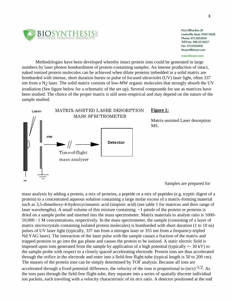
irradiation (See figure below for a schematic of the set up). Several compounds for use as matrices have
been studied. The choice of the proper matrix is still semi-empirical and may depend on the nature of the
sample studied.
Figure 1:
Matrix-assisted Laser desorption
MS.
Samples are prepared for
mass analysis by adding a protein, a mix of proteins, a peptide or a mix of peptides (e.g. tryptic digest of a
protein) to a concentrated aqueous solution containing a large molar excess of a matrix-forming material
such as 3,5-dimethoxy-4-hydroxycinnamic acid (sinpinic acid) (see table 1 for matrices and their range of
laser wavelengths). A small volume of this mixture containing ~1 pmole of the protein or proteins is
dried on a sample probe and inserted into the mass spectrometer. Matrix materials to analyte ratio is 1000-
10,000 : 1 M concentrations, respectively. In the mass spectrometer, the sample (consisting of a layer of
matrix microcrystals containing isolated protein molecules) is bombarded with short duration (1 to 10 ns)
pulses of UV laser light (typically, 337 nm from a nitrogen laser or 355 nm from a frequency-tripled
Nd:YAG laser). The interaction of the laser pulse with the sample causes a fraction of the matrix and
trapped proteins to go into the gas phase and causes the protein to be ionized. A static electric field is
imposed upon ions generated from the sample by application of a high potential (typically +- 30 kV) to
the sample probe with respect to a closely spaced accelerating electrode. Protein ions are thus accelerated
through the orifice in the electrode and enter into a field-free flight-tube (typical length is 50 to 200 cm).
The masses of the protein ions can be simply determined by TOF analysis. Because all ions are
accelerated through a fixed potential difference, the velocity of the ions is proportional to (m/z)-1/2. As
the ions pass through the field-free flight-tube, they separate into a series of spatially discrete individual
ion packets, each traveling with a velocity characteristic of its m/z ratio. A detector positioned at the end
MATRIX-ASSITED LASER DESORPTION
MASS SPECTROMETER
Time-of-flight
mass analyzer
Laser
HV
Detector
IONS
4
of the field-free flight-tube produces a signal as each ion packet strikes it. A recording of the detector
signal as a function of time yields a TOF spectrum. The difference between the start time, set by the
occurrence of the laser pulse and common to all ions, and the arrival time of an individual ion at the
detector is proportional to (m/z)+1/2 and can be used to calculate the ion's m/z ratio. Such a calculation
can be used to convert the x-axis of the spectrum (TOFs) into a m/z ratio axis (a conventional mass
spectrum). All ions of different m/z ratios arising from a single laser shot are measured; they simply arrive
at the ion detector at different times. The MALDI systems are calibrated by measuring the ion arrival
times of known reference standards. This calibration may be performed either externally for routine
analysis or with an internal standard for higher accuracy analysis. The technique is primarily a qualitative
technique, so the relative peak heights or areas may not accurately represent the ratios of components
present in a sample. Additionally some components have higher desorption/ionization yields than others.
This can be often observed in the MALDI analysis of protein digest mixtures where most, but not
necessarily all, of the expected fragments are observed. Recent work of several investigators suggests that
the technique can be used for semiquantitative analysis runs of mixtures of defined components.
Performance specifications of MALDI-TOF-MS for a high-quality commercial instrument include
a resolution of 400 (mass range is 200 to 800 kilodaltons), a mass determination accuracy up to 1 part in
104, a sensitivity of better than 1 pmole, and a spectrum acquisition time of 1 min. The method appears
almost universal for proteins that can be dissolved in appropriate solvents, such as a volume/volume ratio
of 2:1 of 0.1% triflouroacetic acid-acetonitrile or 100% hexafluoro-isopropanol (for proteins with
hydrophobic character). The technique has the ability to analyze complex mixtures of peptides and
proteins in the presence of large molar excesses of salts, buffers, lipids, and other species. The limitations
that need to be considered include the occurrence of adduct artifacts that limit the mass accuracy for
masses greater than 30 to 40 kD, the requirement that both the protein and the matrix material be soluble
in the solvent mixture used, and the poisoning effect on the mass spectra of traces of ionic detergent (such
as SDS) or involatile additives (such as glycerol and dimethyl sulfoxide).
The physicochemical events leading to the transfer of proteins to the gas phase and their ionization
in matrix-assisted laser desorption/ionization have not yet been fully explained. The matrix is believed to
serve several functions, including absorption of energy from the laser light and the isolation of individual
protein molecules within the large molar excess of the solid matrix. The protein-matrix mixture typically
forms a microcrystalline layer spontaneously upon drying the sample on the insertion probe tip. Upon
irradiation with a short duration pulse of laser light, one model for the mechanism assumes that the upper-
most layer of matrix is induced to undergo a phase transition from the solid to the gas phase. The
subsequent expansion of these matrix molecules into the vacuum drags the matrix-isolated protein
molecules into the gas phase. During the transfer to the gas phase, the proteins undergo ionization through
proton transfer reactions with the matrix by reaction processes that are not yet understood.
5
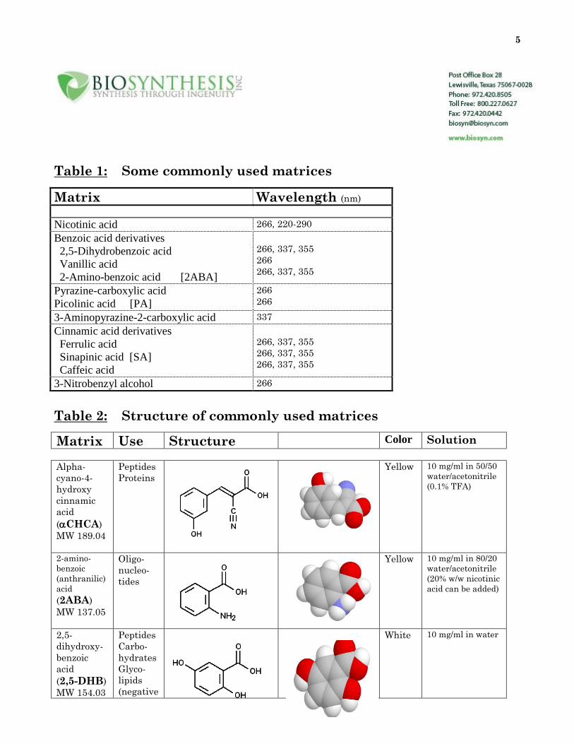
Table 1: Some commonly used matrices
Matrix Wavelength (nm)
Nicotinic acid 266, 220-290
Benzoic acid derivatives
2,5-Dihydrobenzoic acid
Vanillic acid
2-Amino-benzoic acid [2ABA]
266, 337, 355
266
266, 337, 355
Pyrazine-carboxylic acid
Picolinic acid [PA]
266
266
3-Aminopyrazine-2-carboxylic acid 337
Cinnamic acid derivatives
Ferrulic acid
Sinapinic acid [SA]
Caffeic acid
266, 337, 355
266, 337, 355
266, 337, 355
3-Nitrobenzyl alcohol 266
Table 2: Structure of commonly used matrices
Matrix Use Structure Color Solution
Alpha-
cyano-4-
hydroxy
cinnamic
acid
(CHCA)
MW 189.04
Peptides
Proteins
Yellow 10 mg/ml in 50/50
water/acetonitrile
(0.1% TFA)
2-amino-
benzoic
(anthranilic)
acid
(2ABA)
MW 137.05
Oligo-
nucleo-
tides
Yellow 10 mg/ml in 80/20
water/acetonitrile
(20% w/w nicotinic
acid can be added)
2,5-
dihydroxy-
benzoic
acid
(2,5-DHB)
MW 154.03
Peptides
Carbo-
hydrates
Glyco-
lipids
(negative
White 10 mg/ml in water
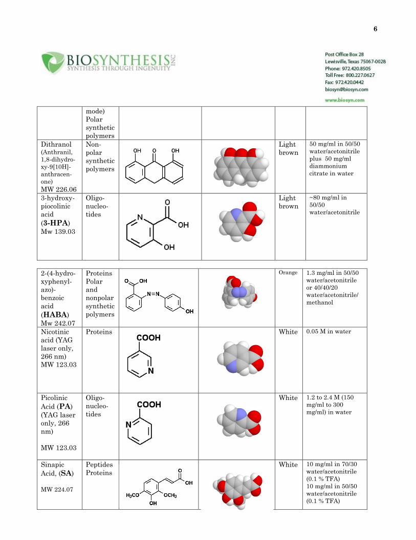
6
mode)
Polar
synthetic
polymers
Dithranol (Anthranil,
1,8-dihydro-
xy-9[10H]-
anthracen-
one)
MW 226.06
Non-
polar
synthetic
polymers
Light
brown
50 mg/ml in 50/50
water/acetonitrile
plus 50 mg/ml
diammonium
citrate in water
3-hydroxy-
piocolinic
acid
(3-HPA)
Mw 139.03
Oligo-
nucleo-
tides
Light
brown
~80 mg/ml in
50/50
water/acetonitrile
2-(4-hydro-
xyphenyl-
azo)-
benzoic
acid
(HABA)
Mw 242.07
Proteins
Polar
and
nonpolar
synthetic
polymers
Orange 1.3 mg/ml in 50/50
water/acetonitrile
or 40/40/20
water/acetonitrile/
methanol
Nicotinic
acid (YAG
laser only,
266 nm)
MW 123.03
Proteins White 0.05 M in water
Picolinic
Acid (PA)
(YAG laser
only, 266
nm)
MW 123.03
Oligo-
nucleo-
tides
White 1.2 to 2.4 M (150
mg/ml to 300
mg/ml) in water
Sinapic
Acid, (SA)
MW 224.07
Peptides
Proteins
White 10 mg/ml in 70/30
water/acetonitrile
(0.1 % TFA)
10 mg/ml in 50/50
water/acetonitrile
(0.1 % TFA)
7
Correlation of Processed Proteins with their Genes
Once the cDNA sequence of a gene has been determined accurate, measurement of the MW of the
corresponding protein can provide valuable information. If the measured mass of the protein agrees with
that calculated from the gene sequence, it is likely that the deduced sequence is correct, the amino and
carboxyl terminals of the mature protein have been correctly assigned, and the protein contains no post-
translationally modified amino acid residues. A difference between the measured and predicted MWs
implies either an error in the cDNA deduced sequence or a post-translational modification or processing
of the protein. Sometimes, differences are observed between the measured and calculated MWs that are
more difficult to interpret. In such cases, a useful strategy involves degradation of the protein by chemical
or enzymatic means and measurement by MALDI-TOF-MS of the total mixture of peptide products so
generated. Comparison of the accurately measured masses of the degradation products with those
predicted from the cDNA sequence yields information on the sites and natures of modifications and
errors.
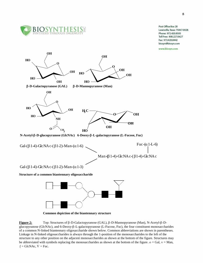
Glycoprotein Analysis
The determination of the carbohydrate portion of glycoproteins provides an analytical challenge to
researchers, especially when only small amounts of sample are available. The reason for this is the very
large number of isomers that are possible when these structures are built up from their constituent
monosaccharides. Unlike peptides and oligonucleotides, which are composed of linear head-to-tail
combinations of different amino acids and nucleotides, respectively, oligosaccharides contain many
isobaric monosaccharides that not only can be linked through different hydroxy groups, but also can form
complex branching patterns. See Figure 2 below for an example.
Monosaccharide composition and sequence analysis alone is not sufficient to determine the
detailed primary structure. Only nuclear magnetic resonance (NMR) has the capability of identifying an
oligosaccharide structure, but it lacks the sensitivity to address many biological problems. Mass
spectrometry is much more sensitive but requires that the molecules be made volatile before analysis.
MALDI-TOF-MS has been introduced for the ionization of large peptides and proteins. However, it has
been shown that other compounds, including oligosaccharides, could be ionized (Mock et al., 1991).
All methods and strategies developed so far may be applied without or, if necessary, with the
needed modifications to enable the use of the techniques mentioned above for mass and structure
elucidation when glycoproteins are studied. Combination of HPLC or CE with MS peptide mapping of a
protein is ideal for evaluating the presence of modifications, including those labile to the conditions of the
Edman degradation. To obtain the best signals for oligosaccharides, the dried mixture of sample and
matrix can be redissolved on the target with ethanol and allowed to recrystallize.
8
O
OH
OH
HO
HOOH
O
OH
OHHO
HO
OH
-D-Galactopyranose (GAL) -D-Mannopyranose (Man)
O
OH
OHHO
HONH
O CH3
O OH
OH
H C
HOOH
3
N-Acetyl--D-glucopyranose (GlcNAc) 6-Deoxy--L-galactopyranose (L-Fucose, Fuc)
Gal-(1-4)-GlcNAc-(1-2)-Man-(1-6)
Gal-(1-4)-GlcNAc-(1-2)-Man-(1-3)
Man-(1-4)-GlcNAc-(1-4)-GlcNAc
Fuc-(1-L-6)
Structure of a common biantennary oligosaccharide
Common depiction of the biantennary structure
Figure 2: Top: Structures of -D-Galactopyranose (GAL), -D-Mannopyranose (Man), N-Acetyl--D-
glucopyranose (GlcNAc), and 6-Deoxy--L-galactopyranose (L-Fucose, Fuc), the four constituent monosaccharides
of a common N-linked biantennary oligosaccharide shown below. Common abbreviations are shown in parentheses.
Linkage in N-linked oligosaccharides is always through the 1-position of the monosaccharides to the left of the
structure to any other position on the adjacent monosaccharides as shown at the bottom of the figure. Structures may
be abbreviated with symbols replacing the monosaccharides as shown at the bottom of the figure. = Gal, = Man,
= GlcNAc, = Fuc.
9
Phosphoprotein Analysis
Phosphoproteins play a central role in many intracellular processes, including signal transduction
and regulation of cell division. The site and extent of phosphorylation of key proteins are believed to play
an important regulatory role in many intracellular signaling pathways.
The following is an example to demonstrate the analytical capabilities of the technique: The
enzyme "cAMP-dependent protein kinase" is a complex with a mass of 178 kD and is made up of two
regulatory and two catalytic subunits (Knighton et al., 1991, Chrivia et al., 1988). If cAMP is present,
which binds to the regulatory subunit, the complex dissociates and releases the enzymatically active
catalytic subunits. The 3-D molecular structure of the catalytic subunit of cAMP-dependent protein kinase
has been determined by x-ray crystallography. The protein consists of 350 amino acids with a molecular
mass of 40,440 daltons.
The alpha catalytic subunit of cAMP-dependent protein kinase from the mouse was cloned and
expressed in Escherichia coli. The recombinant protein was isolated as a mixture of molecular species, all
containing the same peptide chain but differing from each other in the degree of phosphorylation at
specific residues. Three isoforms of the recombinant enzyme were prepared in highly purified form for
structural analysis. Each was believed to be a homogeneous molecular species. The final stage of
purification resulted in the protein samples being in a high salt buffer. The use of on-line reverse-phase
HPLC-electrospray MS allowed the determination of the MWs of the three isoforms. The experiment
simultaneously evaluated the purity of the proteins and desalted them in a form suitable to electrospray
MS analysis.
The isoforms differed from one another by the mass of a single-PO3H group (80 daltons). The
predicted sequence of the mature form of the recombinant polypeptide chain asks for a calculated mass of
40,440 daltons. Isoform I had a molecular mass of 40,759 daltons, 319 daltons above the calculated mass
and therefore containing four phosphate groups (319/80). Isoform II had a mass of 40,678 daltons (that is,
-81 daltons with respect to I and +238 daltons with respect to the parent polypeptide chain) and thus
contained three phosphate groups. Isoform III had a mass of 40,600 daltons (that is, -78 daltons with
respect to II, -159 daltons with respect to I, and +160 daltons with respect to the parent) and had two
phosphate groups. The data confirmed that the isoforms were of high purity and were homogeneous
molecular species. The differences in phosphate content of the three isoforms were consistent with prior
data.
Although the above work was done using electrospray MS similar results may be obtained using
MALDI-TOF-MS. The results showed that phosphoproteins can be analyzed by MS. The accuracy of
MW measurement is well within the limits for useful determination of the degree of phosphorylation of
intact proteins of typical size. The combination with enzymatic digestion and HPLC- or CE-MS peptide
mapping is feasible to allow for a powerful technique for the structural characterization of
phosphoproteins.
10
PROTEIN SEQUENCE DETERMINATION
Determination of the amino acid sequence of a protein molecule plays a central role in much
biological research. Typically, the biological researcher's first direct observation of a protein would be by
1-D or 2-D PAGE. A critical step in the study of many biologically important proteins is the
determination of limited stretches of amino acid sequence data from 5 to 100 picomole amounts of the
natural protein isolated from a biological source. These limited sequence data are frequently the key
information used to identify and clone the gene corresponding to the protein of interest. The nucleic acid
sequence of the gene is then determined and translated to afford the complete amino acid sequence of the
translation product. After cloning and expression of the identified gene, amino acid sequence data are
used to confirm the structure of the protein produced. At the present time, amino acid sequence data are
almost invariably generated by automated Edman degradation of a protein from the amino terminal, either
of the intact protein or of peptides separated after proteolytic digestion of the protein.
Protein ladder sequencing with one-step MS readout.
Chait and Kent (1992) and others have described a new approach to determine the amino acid
sequence of picomole amounts of a protein which takes advantage of the ability of MALDI-TOF-MS to
accurately and rapidly measure protein mixtures. Manual Edman chemistry is used prior to mass analysis
to generate, in a controlled fashion, a family of sequence-defined fragments from a polypeptide chain. The
sequence-defined fragments are analyzed and read out using laser desorption MS to simultaneously
generate the complete data set in a single operation as a protein ladder. Mass differences between
consecutive peaks define the identity of a particular amino acid, based on the distinctive mass of each
genetically coded amino acid. Problems arise in the differentiation between leucine and isoleucine (same
mass) and glutamine and lysine (Gln and Lys; 0.04 D mass difference). The family of fragments found
defines the sequence of amino acids in the original peptide chain. One way of generating the sequence-
defined set of fragments from a peptide or protein is to carry out the Edman degradation in the presence of
a terminating agent (This is not automated yet. The reactions are done in a volume containing > 5
picomoles. Even so, only picomole amounts are needed for the experiment. A terminating reagent may
not be needed if reaction times and temperature are optimized.). The protein ladder sequencing method
relies on the capabilities of matrix-assisted laser desorption MS to measure the MWs of proteins and
peptide with high accuracy. This method is as yet unproved for very small amounts of proteins of
unknown sequence isolated from biological sources, but the potential exists for speeding up and
simplifying protein sequencing.
Future Developments
There is a need for further improvement of methods for sample preparation for Edman chemistry
sequencing as well as for ion production for matrix-assisted laser desorption MS. It is desirable to be able
to use water as a matrix, which would allow the direct examination of biological specimens.
11
The resolution for TOF instrumentation for proteins is constantly increasing towards the resolution
imposed by the envelope of the isotope distribution over the full mass range of interest. Furthermore, the
speed for data collection and analysis is also in a state of constant improvement. Many researchers are
working towards this goal at present. This will lead to further improvement of the described techniques in
regard to resolutiona and through-put in the near future.
Experimental
MALDI-MS matrices
4-hydroxy--cyano-cinnamic acid (HCCA) (Sigma, St. Louis, Missouri), sinapic acid (SA) (Fluka
Chemicals, Buchs, Switzerland), 2,5-dihydrobenzoic acid (DHB).
Preparation of MALDI-MS matrices
HCCA (10 microgram/microliter) dissolved in 70% aqueous acetonitrile, 0.1% TFA was used to analyze
proteins. A saturated solution of HCCA in 45% aqueous acetonitrile, 0.1% TFA or DHB dissolved in
30% aqueous acetonitrile, 0.1% TFA was used to analyze protein digests. Prior to use, 100 microliter
DHB was lyophilized in a Speed Vac (Savant, Farmingdale, New York) to remove all traces of organic
solvent and redissolved in an equal volume of 30% aqueous acetonitrile, 0.1% TFA. Sinapinic acid (~0.1
M or saturated solution) was dissolved in 50% acetonitrile, 50% water and 0.1% trifluoroacetic acid. A
fresh matrix solution was prepared daily.
Preparation of MALDI-MS samples (Bennet et al., 2000)
Prior to MALDI-MS analysis, all protein or peptide samples were desalted and concentgrated on
disposable microcolumns packed with POROS R2 50 (protein) or POROS R2 20 (peptides) reversed-
phase media (Perseptive Biosystems, Cambridge, Massachusets) or with ZipTipTM
s or SuproTipTM
s C4
(protein) and SuproTipTM
s C8 (peptides) (The Nest Group, Inc.). Insert a brief description of the protocol
here!!! Peptide samples from the in-situ digests were desalted and concentrated sd described above. The
peptides bound to the reversed-phase media were washed with 10 microL 0.1% TFA, and eluted onto the
MALDI-MS target with matrix solution (two to three droplets of ~ 100 nanoL).
Alternatively:
For Proteins: Sample solutions were prepared without glycerol or SDS in sample buffer at
concentrations between 1 to 10 mg/ml. Aliquots of each sample were mixed with matrix solution to make
a series of dilutions (1:2, 1:4, 1:8, -> 1/20).
MALDI-TOF-MS analysis
12
A Perseptive VoyagerTM
(BioSpectrometryTM
Workstation) benchtop matrix assisted laser desorption
ionization (MALDI) mass spectrometer was used to obtain the mass spectra shown. It employs a N2 laser,
1.2 meter flight tube and 30 kV accelerating voltage. The manufacturer’s data analysis package (GRAMS
based) was used for data analysis. Generally, 15 to 30 spectra were averaged to obtain spectra. Calibration
was done every day or when a new set of samples were analyzed using a freshly prepared calibration mix.
Peaks reflecting oligomers of alpha-Lactalbumin or beta-Lactoglobulin were used to calibrate mass ranges
>50 kDa.
Mass spectra were acquired on a time-of-flight mass spectrometer (Voyager; Perseptive Biosystems now
PE/Biosystems). It was operated in linear mode with an acceleration potential of 30 k.V. Spectra were
aquired in either positive or negative mode as shown on spectra. Ions were desorbed by irradiation with
the build-in … laser ( nm, .. ns). The equivalent flight path length was 1.2 m. The base pressure was 10-4
Pa. Up to 20 to 40 single shot spectra were averaged for each spectra shown.
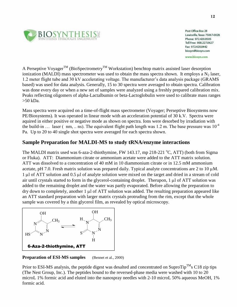
Sample Preparation for MALDI-MS to study tRNA/enzyme interactions
The MALDI matrix used was 6-aza-2-thiothymine, FW 143.17, mp 218-221 oC, ATT) (both from Sigma
or Fluka). ATT: Diammonium citrate or ammonium acetate were added to the ATT matrix solution.
ATT was dissolved to a concentration of 40 mM in 10 diammonium citrate or in 12.5 mM ammonium
acetate, pH 7.0. Fresh matrix solution was prepared daily. Typical analyte concentrations are 2 to 10 M.
1 l of ATT solution and 0.5 l of analyte solution were mixed on the target and dried in a stream of cold
air until crystals started to form in the glycerol-containing droplet. Therupon, 1 l of ATT solution was
added to the remaining droplet and the water was partly evaporated. Before allowing the preparation to
dry down to completely, another 1 l of ATT solution was added. The resulting preparation appeared like
an ATT standard preparation with larger matrix crystals protruding from the rim, except that the whole
sample was covered by a thin glycerol film, as revealed by optical microscopy.
References
Preparation of ESI-MS samples (Bennet et al., 2000)
Prior to ESI-MS analysis, the peptide digest was desalted and concentrated on SuproTipTM
s C18 zip tips
(The Nest Group, Inc.). The peptides bound to the reversed-phase media were washed with 10 to 20
microL 1% formic acid and eluted into the nanospray needles with 2-10 microL 50% aqueous MeOH, 1%
formic acid.
N
NN
CH3
HS
OH
N
NN
CH3
S
OH
H
H
6-Aza-2-thiothymine, ATT
13
References
Bennet, K.L., Kussmann, M., Bjoerk, P., Godzwon, M., Mikkelsen, M., Soerensen, P., and Roepstorff, P., 2000, Chemical
cross-linking with thiol-cleavable reagents combined with differential mass spectrometric peptide mapping-A noval approach
to assess intermolecular protein contacts. Protein Science 9:1503-1518.
Chait, Brian T., and Kent, Stephen B., 1992: Weighing Naked Proteins: Practical, High-Accuracy Mass Measurements of Peptides and
Proteins in Science 257, 1885-1894.
Chrivia, J.C., Uhler, M.D., and McKnight, G.S., J. Biol. Chem. 263, 5739 (1988)
Knighton, D.R., et al., Science 253, 407 (1991)
Harvey, D.J.; Matrix-assisted laser desorption/ionization mass spectrometry of oligosaccharides 1994 in American Laboratory December
1994 pp.22-28.
Juhasz and Biemann, 1995, "Utility of non-covalent complexes in the matrix-assisted laser desorption ionization mass
spectometry of heparin-derived oligosaccharides" Carbohydrate Research 270 (1995) 131-147. This article describes the use
of synthetic basic peptides for complexing to heparin oligosaccharides to facilitate more efficient ionization of the
oliogosaccharides. They also describe the use of angiogenin, a heparin binding protein, for the same purpose. Notes: Good results for oligosaccharides with gentisic acid and ferrulic acid depending on the compound (saturated solutions in 50 %
ethanol). 6-Aza-2-thiothymine (make a saturated solution in 50% acetonitrile, then take the supernatant and add 5 %
ammonium citrate). This last one works well with any negatively charged compound.
For a brief description of methods on how to prepare the matrices go to Brian Chait's web site:
http://prowl.rockefeller.edu/recipes/contents.htm
You will find the following definitions for matrix preparation there:
Dried droplet
Slow crystallization
Polycrystalline thin films
Seeded films
14
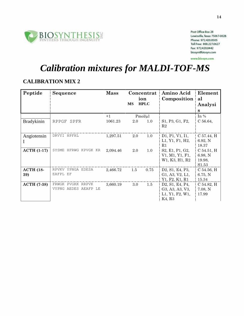
Calibration mixtures for MALDI-TOF-MS
CALIBRATION MIX 2
Peptide Sequence Mass Concentrat
ion
MS HPLC
Amino Acid
Composition
Element
al
Analysi
s
+1 Pmol/l In %
Bradykinin RPPGF SPFR 1061.23 2.0 1.0 S1, P3, G1, F2,
R2
C 56.64,
Angiotensin
I
DRVYI HPFHL 1,297.51 2.0 1.0 D1, P1, V1, I1,
L1, Y1, F1, H2,
R1
C 57.44, H
6.92, N
18.37
ACTH (1-17) SYSME HFRWG KPVGK KR 2,094.46 2.0 1.0 S2, E1, P1, G2,
V1, M1, Y1, F1,
W1, K3, H1, R2
C 54.51, H
6.98, N
19.98,
S1.53
ACTH (18-
39)
RPVKV YPNGA EDESA
EAFPL EF
2,466.72 1.5 0.75 D2, S1, E4, P3,
G1, A3, V2, L1,
Y1, F2, K1, R1
C 54.56, H
6.75, N
15.34
ACTH (7-38) FRWGK PVGKK RRPVK
VYPNG AEDES AEAFP LE
3,660.19 3.0 1.5 D2, S1, E4, P4,
G3, A3, A3, V3,
L1, Y1, F2, W1,
K4, R3
C 54.82, H
7.08, N
17.99
15
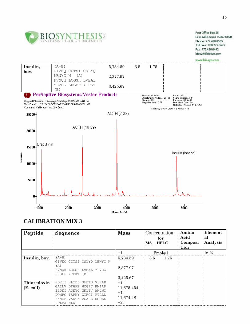
Insulin,
bov.
(A+B)
GIVEQ CCTSI CSLYQ
LENYC N (A)
FVNQH LCGSH LVEAL
YLVCG ERGFF YTPKT
(B)
5,734.59
2,377.97
3,425.67
3.5 1.75
CALIBRATION MIX 3
Peptide Sequence Mass Concentration for
MS HPLC
Amino
Acid
Composi
tion
Element
al
Analysis
+1 Pmol/l In %
Insulin, bov. (A+B)
GIVEQ CCTSI CSLYQ LENYC N
(A)
FVNQH LCGSH LVEAL YLVCG
ERGFF YTPKT (B)
5,734.59
2,377.97
3,425.67
3.5 1.75
Thioredoxin
(E. coli)
SDKII HLTDD SFDTD VLKAD
GAILV DFWAE WCGPC KMIAP
ILDEI ADEYQ GKLTV AKLNI
DQNPG TAPKY GIRGI PTLLL
FKNGE VAATK VGALS KGQLK
EFLDA NLA
+1;
11,675.454
+1;
11,674.48
+2;
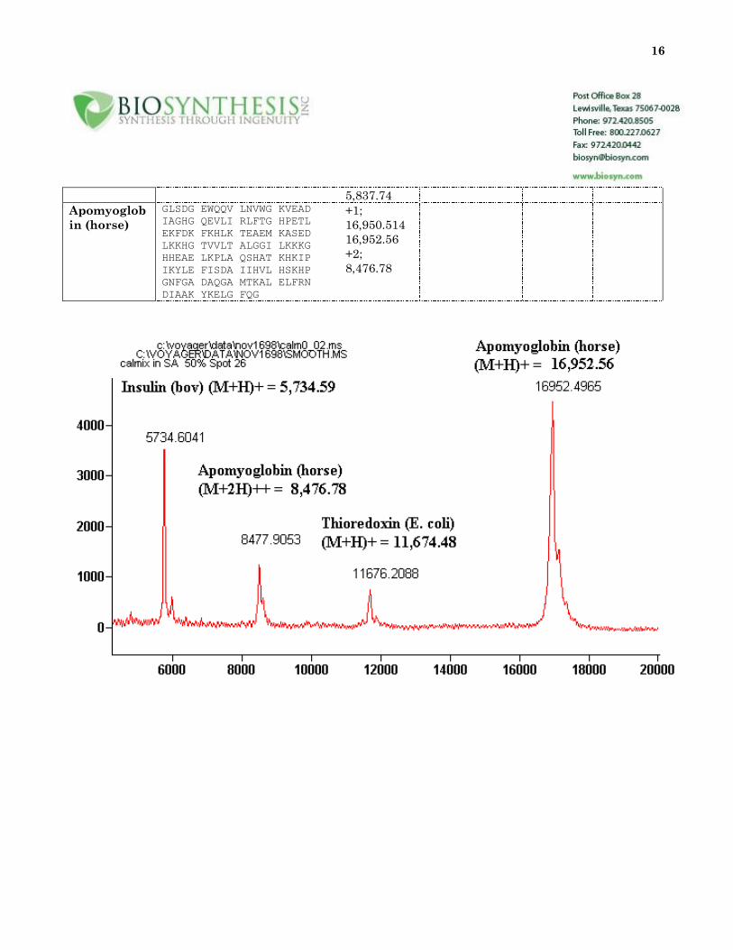
16
5,837.74
Apomyoglob
in (horse)
GLSDG EWQQV LNVWG KVEAD
IAGHG QEVLI RLFTG HPETL
EKFDK FKHLK TEAEM KASED
LKKHG TVVLT ALGGI LKKKG
HHEAE LKPLA QSHAT KHKIP
IKYLE FISDA IIHVL HSKHP
GNFGA DAQGA MTKAL ELFRN
DIAAK YKELG FQG
+1;
16,950.514
16,952.56
+2;
8,476.78
17
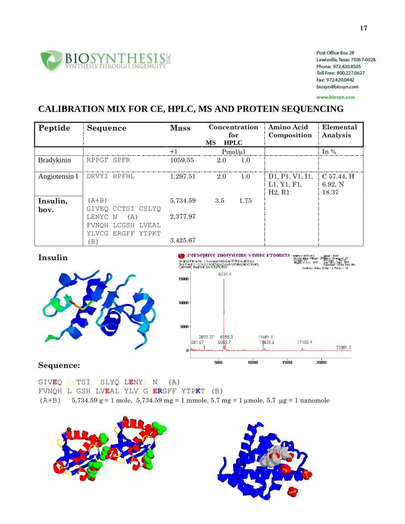
CALIBRATION MIX FOR CE, HPLC, MS AND PROTEIN SEQUENCING
Peptide Sequence Mass Concentration
for MS HPLC
Amino Acid
Composition
Elemental
Analysis
+1 Pmol/l In %
Bradykinin RPPGF SPFR 1059.55 2.0 1.0
Angiotensin I DRVYI HPFHL 1,297.51 2.0 1.0 D1, P1, V1, I1,
L1, Y1, F1,
H2, R1
C 57.44, H
6.92, N
18.37
Insulin,
bov.
(A+B)
GIVEQ CCTSI CSLYQ
LENYC N (A)
FVNQH LCGSH LVEAL
YLVCG ERGFF YTPKT
(B)
5,734.59
2,377.97
3,425.67
3.5 1.75
Insulin
Sequence:
GIVEQ CCTSI CSLYQ LENYC N (A)
FVNQH LCGSH LVEAL YLVCG ERGFF YTPKT (B)
(A+B) 5,734.59 g = 1 mole, 5,734.59 mg = 1 mmole, 5.7 mg = 1 mole, 5.7 g = 1 nanomole

18
Thioredoxin, dimer Myoglobin
19
Mass Spectrometry – Definitions
Standard Definitions of Terms Relating to Mass Spectrometry, a Report from the Committee on
Measurements and Standards of ASMS", JASMS Vol. 2 #4, July/August 1991, web site: http://www.asms.org/.
Collected Terms Suggestions, grouped by category:
Analyzers
Double focusing - A combination of direction and velocity focusing in sector instruments
used to achieve high resolution.
Tandem mass spectrometry (MS/MS) - Two-stage mass analysis experiment, used to
study the chemistry of selected ions or individual components in mixtures.
Note from a reader: I despise "MS/MS spectra" -- Who has ever heard of an MS spectrum (a mass
spectrometry spectrum). Prefer "tandem mass spectrum" or better "product-ion spectrum" or "precursor-
ion spectrum". How do you handle a "metastable-ion spectrum"--please don't sanction a "metastable
spectrum".
Ion trap analyzer - type of mass analyzer whereby ions are confined in a region of space
and analyzed, as opposed to a dispersive mass analyzer (See Paul Ion Trap and Penning Ion
Trap)
Note from a reader: The definition of "ion trap analyzer" is too limited. I realize that
99% of the mass spec community refers to a Paul, or Quadrupole Ion Trap simply as "an Ion
Trap" thanks to Finnigan, but an FTICR/MS is also a type of ion trap based mass analyzer.
Paul ion trap - A type of mass analyzer in which ions are confined in space by means of a
three dimensional, rotationally symmetric quadrupolar electric field. Sorting of ions is
performed by changing the field conditions appropriately to destabilize an ion of a
particular m/z. The destabilized ion is then detected when it exits the trap and strikes a
collection device, e.g. an electron multiplier or conversion dynode.
Penning ion trap - An ion trap that confines ions by placing them in a static magnetic
field. Inside the field, the ions are subject to the Lorentz force which causes ions of a
particular m/z to cyclotron at a specific frequency (cyclotron frequency).
Fourier transform ion cyclotron resonance (FTICR) analyzer - A type of mass
spectrometer that uses a Penning ion trap to confine ions for mass analysis. Ions of all m/z
values are excited by applying RF energy over a range of frequencies corresponding to the
cyclotron frequencies of the ions to be detected. After cessation of the applied RF
20
energy, all ions are detected simultaneously by measuring the current induced on the
"detect" electrodes by the confined ions. The mass spectrum is obtained by application of
the Fourier transform to the measured signal to extract the cyclotron frequencies of the
ions. Once the cyclotron frequencies are known, the m/z values are calculated via the
cyclotron equation.
Another reader’s definition: Fourier transform ICR - A method of obtaining data from an
ion cyclotron resonance (Penning) trap, whereby all ions are translationally excited within a
time much shorter than the ion/neutral collision time; the image current of the combined
ions’ signal is detected; and the resulting time-domain signal is converted to a frequency
(reciprocal mass) - domain signal by the Fourier Transform mathematical method. "FT
Mass Spectrometry" is inappropriate. The FT method is not limited to ICR; FT time-of-
flight has been demonstrated, and FT methods for other analyzers are possible.
Note from a reader: In the definition of "Ion cyclotron resonance analyzer," no mention
is made of a magnet, a (in my humble opinion) grave omission. If you decide to incorporate
our suggestions, you could have a note directing readers to "Fourier transform ion cyclotron
resonance analyzer" and "Penning ion trap".
Double resonance - In ICR, the irradiation of one ion at or near its cyclotron frequency,
and observation of the effect on the intensities of other ions in the spectrum.
Cyclotron motion - Cyclic rotation of an ion in a fixed magnetic field
Magnetron motion - Slow circular drift of an ion along a path of constant electrostatic
potential; magnetron motion occurs in ICR as a result of the crossed radial electric field and
axial magnetic field.
Time-of-flight mass spectrometer - An analytical instrument in which ions are formed
and accelerated and their flight times measured to determine their mass. Time-of-flight
mass spectrometers can be distinguished by those that accelerate ions to constant
momentum (in which case flight times are linear with mass) and constant energy (in which
case flight times are proportional to the square root of mass). Time-of-flight mass
spectrometers were originally known as velocitrons.
Reflectron - A device used in a time-of-flight mass spectrometer that retards and then
reverses ion velocities in order to correct for the flight times of ions having different kinetic
energies. The reflectron is sometimes known as an ion mirror.
Single-stage reflectron - A reflectron in which a single retarding field is used to retard
and then reverse ion velocities. Generally, a retarding field is used that is constant through
21
the depth of the reflectron, and achieved by retarding voltages that increase linearly with
reflectron depth. Such reflectrons provide first-order correction for differences in ion kinetic
energy.
Dual-stage reflectron. - A reflectron in which two retarding fields are used to reverse ion
velocities. Generally, both retarding regions are constant field, each achieved by retarding
voltages that increase linearly with reflectron depth. In the most common embodiment,
ions are retarded by two-thirds to three-quarters of their mean kinetic energies within the
first retarding regions which comprises approximately 10% of the total reflectron depth.
Isotopic peak(ion) - Due to other isotopes of the same chemical but different isotopic
composition.
Isobaric peak(ion) - of the same normal mass (integral).
Mass spectrum - Plot of ion abundance vs. mass-to-charge ratio normalized to most
abundant ion.
Note from a reader: molecular weight - The text in the current document could be updated slightly to
more clearly reflect some of the values relating to high mass (> 3,000) distributions, particularly when
observed at less than unit resolution.
Note from a reader: mass unit - Should there be a comment on "µ " vs "amu" vs the
'biomedical term' "Dalton"? IUPAC currently defines "m = unified atomic mass (1/12 of the
mass of an atom of nuclide 12C)".
Mass-to-charge ratio (m/z) - Daltons/electronic charge.
Note from a reader: on Thomson - the fluid dynamics people have already used that one; it
is listed in the CRC Handbook and IUPAC documents. ASMS should be doing things in
addition to or clarifying points mentioned (or not) in IUPAC. However, we should be
cautious about doing anything that actually opposes or conflicts with IUPAC documents.
Mass spectrometry - This is the study of mass spectra obtained by using a mass
spectrometer. The term "mass spectroscopy" should be avoided, because this implies optical
dispersion. Possible exceptions might be in the case of photoplate or various optical
methods of detection.
Mass Selective Detector - a detector that only monitors ion currents at certain m/z
values.
22
Mathieu stability diagram - Diagram showing the solutions to the Mathieu equation
which correspond to stable ion trajectories and displayed as a function of parameters
related to operating voltages, mass and charge of the trapped ions.
Secondary ion mass spectrometry - Mass spectrometry based on analysis of particles
that are emitted when a surface, usually a solid, although sometimes a liquid, is bombarded
by energetic (~ keV) primary particles (e.g. Ar+ and Cs+).
Flowing Afterglow - a reactor for observing ion-molecule reactions, in which ions are
introduced to a bath gas containing a neutral reactant, and flowing rapidly down a vacuum
system, where neutral pressure and distance become the reaction variables. Detection of
the ions is by mass spectrometry through a leak at the product end of the system.
High Pressure Pulsed MS - a combined reactor and detector for observing ion-molecule
reactions. A chemical ionization (CI) source is operated in a time-resolved fashion, where
the ions are generated in one point in time, allowed to react in the CI source, and extracted
and analyzed by MS at some later time.
Sample Introduction
Ion source - Device used to generate sample ions by electron impact, chemical ionization,
etc.
Gas chromatography-mass spectrometry (GC-MS) - combined technique for mixture
analysis in which the separated GC components are passed continuously into the MS.
Desorption ionization - Method used to ionize non-volatile solid samples by impact of energetic particles or
photon beams.
Spray ionization - Method used to ionize liquid samples directly by electrical, thermal, or
pneumatic energy through formation of a spray of fine droplets.
Liquid chromatography-mass spectrometry (LC-MS) - combined technique for
mixture analysis.
CE/MS - The combining of capillary electrophoresis with mass spectrometry.
CEC/MS - The combining of capillary electrokinetic chromatography (or capillary
"electrochromatography"?) with mass spectrometry.
23
CE/MS Interface - The interface used between the capillary electrophoresis (CE) and the
mass spectrometer to provide a continuous introduction of effluent from the CE into the
MS. Common interfaces include: (1) layer flow interface, (2) liquid junction interface, and
(3) direct interface. Another opinion: Instead of "layer flow interface" it should be "sheath-
flow" interface.
Layer Flow Interface - An approach using coaxial concentric tubes to add solvent and/or
solvent modifiers post-column (outer tube) to the column effluent (inner tube) for improved
CE/MS or LC/MS operation.
Liquid Junction Interface - An interface used to combine CE with MS in which a
reservoir supplies the additional solvent flow to the CE effluent in order to achieve stable
electrospray operation.
Ion Molecule reactions
Collision-induced dissociation - Process whereby a mass-selected ion is excited and
caused to fragment by collision with a target gas, especially in MS/MS.
Inelastic collision - Collision in which internal energy is not conserved.
Ionization Nomenclature
Radical ion (odd-electron ion) - Charged open-shell molecule with at least one unpaired
electron.
Odd-electron ion - See radical ion.
Parent ion (m1+) - Any ion (including negatively and doubly charged ions) that gives
fragments.
Product ion (m2+) - Ion generated by fragmentation of any parent ion.
Metastable ion - Ion that fragments slowly after emergence from the ion source but before
it reaches the detector; in sector instruments, metastable ions give rise to signals which
appear at unique m/z values related to the parent and product ion masses.
Surface-induced dissociation - Process whereby a mass-selected ion is excited and
caused to fragment by collision with a target surface.
24
Ionization energy(IE) - Minimum energy required to remove an electron from a molecule.
Endothermicity of the process M ® M.+ .
Glow discharge - Method used to ionize solid samples for elemental analysis by applying
an electric field to create an energetic plasma.
Inductively coupled plasma - Method used to ionize solution samples for elemental
analysis by a plasma.
Electron affinity - Enthalpy change for the process M- ® M + e-.
Proton affinity - Enthalpy change for the process MH+ ® M + H+.
Electron capture - Ionization process in which a molecule or atom captures a thermal
energy electron typically in a CI
source, and generates the molecular radical anion.
Electron impact - Ionization method in which molecules are ionized directly by energetic
electrons (usually 70 eV) at low pressure (<10-5 torr).
Even-electron ion - Ion with even number of electrons commonly with a closed shell
electronic configuration.
Distribution of internal energy - Analogue of Boltzmann distribution for molecules not
in thermal equilibrium.
Resonance electron capture of thermal electrons - Should be referred to as "electron
capture ionization" or "ECI". The processes which ECI describes (Chemical Ionization Mass
Spectrometry, Harrison, 2nd ed., 1992, p. 24-25):
1) MX + e- ® MX- electron capture
2) MX + e- ® M. + X- dissociative electron capture
Case (1) leads to formation and detection of a molecular anion, while case (2) leads to
detection of a fragment with loss of a radical, for example, pentafluorobenzyl ester
fragmentation.
{ Note from submittor: Whether or not ECI/MS is acceptable to the committee, I hope
that some consensus can be reached to describe the two processes I have described above. I
25
have seen them described as NCI, NICI, ECCI, ECNI and ECNICI. I think its time to pick
one. By the way, my second choice would be ECNI (electron capture negative ion).}
The term ECI should be separate from the "chemical ionization" section of nomenclature
and the term "electron attachment" should be dropped or changed to "electron capture" to
avoid confusion. This description should be limited to only those processes which involve
thermal electron capture as the principle mode of ionization. Other negative ionization
procedures which are the result of ion/molecule interactions, such as chloride attachment,
should still be referred to as negative ion chemical ionization. Under electron capture
conditions, the description of the "reagent" gas should be changed to "moderating gas" or
"buffer gas". As the gas is not acting as a reagent, these terms should be more description
of the process involved.
Appearance energy - Endothermicity of process AB+ ® A+ + B
Atmospheric pressure chemical ionization - A variant of chemical ionization
performed at atmospheric pressure.
A reader’s comment: Atmospheric pressure ionization (API) should remain a general term,
for any form of ionization at atmosphere. The definition given is specific for chemical
ionization at atmosphere. The definition given should be for a separate term, atmospheric
pressure chemical ionization (APCI). This was merely the first atmospheric pressure
ionization means that was commercialized. API would then properly include, electrospray
(ESI), APCI, Ionspray (a coined term, and more generally this is pneumatically-assisted
electrospray), and flame ionization (in some of the early papers on API this was the mode of
ionization). Lots of others have been tried including microwave, etc.
Atmospheric Pressure Chemical Ionization (APCI) - The formation of ionized species
when gaseous molecules interact with ions (reagent ions) at atmospheric pressure. The
reagent ions are formed by a corona discharge of the vaporized solvent introduced into the
system.
Atmospheric Pressure Ionization (API) - Ionization technique(s) that occur at
atmospheric pressure. Specific API ionization techniques include electrospray,
pneumatically assisted electrospray and atmospheric pressure chemical ionization and is
often used to couple LC to MS.
Chemical ionization - Method in which neutral molecules are ionized by ion-molecule
reactions to generate a parent ion at a pressure of about 1 torr.
26
Breakdown curve - Plot of ion abundance vs. ion internal energy normalized at each
energy; shows mass spectrum as a function of internal energy.
Charge exchange - Process whereby one particle transfers an electron to another,
e.g. M + A+. ® M+. + A, used in chemical ionization.
Spray ionization - Methods used to ionize liquid samples directly by electrical, thermal, or
pneumatic energy through formation of a spray of fine droplets.
Electrospray Ionization (ESI) - A technique used to produce gas phase ions from
molecules in solution. The process makes use of strong electric fields for both nebulization
and charging of a liquid, drying to reduce the size of the charged droplets to increase the
field strength, and ion evaporation. Ionization occurs when the field strength in the
droplets exceeds the solvation energy of the molecule in solution.
Another opinion: I object to the use of the term "electrospray ionization". However, I think
everyone will use it anyway. Technically, electrospray is a spray process which eventually
produces gas phase ions via ion evaporation. The latter is the ionization process. However,
I think the former term will continue to be used.
Another opinion: Electrospray - Generation of a fine mist of droplets by spraying solutions
through an electrically biased capillary. In mass spectrometric applications ("electrospray
MS"), provision is made for solvent evaporation from the sprayed droplets, and resulting
ions are sampled for mass analysis via a differential pumping system. In some instances,
the ions are formed as a result of electrochemistry driven by the capillary bias
("electrospray ionization" or ESI), but in most cases the ions are present in the solution a
priori (ES).
Ion Evaporation - A desorption ionization process which brings ions from a liquid to the
gas phase when the electric field strength exceeds the solvation energy of an ion in solution.
Ion evaporation processes are found in the techniques of electrospray, pneumatically
assisted electrospray and thermospray.
Pneumatically Assisted Electrospray - An electrospray process where pneumatic
nebulization (e.g., N2) assists in the initial nebulization of the liquid introduced into the
electrospray system (vs. nebulization through charging of the liquid). The process is often
used to obtain electrospray spectra at higher flow rates (e.g., >0.2 mL/min) and with
solvents with high surface tensions (e.g., water).
Ion Spray - Manufacturer’s trade name for pneumatically assisted electrospray.
27
Ion Spray - Also known as "pneumatically assisted electrospray," a variation on ES in
which the droplet formation process is facilitated by incorporation of a sheath nebulizing
gas passing through a second capillary concentric with and larger than the ES capillary.
The nebulizing gas helps accommodate larger flow rates and/or use of liquids of relatively
high surface tension (e.g., aqueous solutions without co-solvents, which would otherwise
require spray potentials above the threshold for electric discharge). In common usage, the
distinction between ES and ion spray has blurred. Most commercial "ES" sources use
nebulizing gas, but do not use the "ion spray" terminology [which may therefore be
superfluous].
Thermospray - A process where liquid is thermally vaporized in a capillary and the ions in
the resulting aerosol are transferred from the liquid to the gas phase. The ionization
process can involve ion evaporation or chemical ionization (the reagent ions are formed by a
filament or discharge) ionization of the solvent. The technique is often used in LC/MS.
Particle Beam - An interface often used for LC/MS where the sample is separated from
the solvent and then introduced into the MS for ionization. The process involves: (1)
solution nebulization, (2) vaporization of the solvent to obtain unsolvated sample molecules
(particles), (3) momentum separation of the particles of sample from solvent gas, and (4)
introduction of the particles into the MS for ionization (EI, CI, or desorption technique).
Thermobeam (or Thermabeam) - A trade name for particle beam which uses thermal
nebulization vs. pneumatic nebulization to form the initial aerosol. Particle beam should be
used in place of this term.
MAGIC (Mono disperse aerosol generation interface for combined LC/MS) - A trade name
for a particle beam approach in which the initial droplet formation is carefully controlled
to generate a uniform aerosol. Particle beam should be used in place of this term.
LC/MS Interfaces - It has been suggested by several ASMS members that there are three
ways to make a spray or aerosol for LC/MS: with a gas, heat, or electricity. Hence the
terms: (1) aerospray; (2) thermospray; and (3) electrospray. Under this model:
MAGIC ® aerospray
Particle Beam ® aerospray
Thermobeam ® aero-thermospray
ion spray ® aero-electrospray
All this may be a bit "extreme", since it does not closely follow what is in the literature,
and excludes APCI. However, this whole issue should be addressed, and a few terms
settled on.
28
EH - A methodology by which ions in solution can be desorbed from a liquid meniscus
directly into an evacuated chamber by application of a suitable electrical bias to a metal
capillary in which the solution is contained. In contrast to ES, droplet spraying is avoided
in EH MS applications (by using very low flow rates), since there is no mechanism (other
than metastable or collision-induced dissociation) for solvent removal subsequent to ion
desorption. This latter feature lends to EH the possibility for studying condensed-phase ion
solvation less invasively than other MS methods. Flow rates are restricted by using small
capillaries and/or viscous solvents. In applications as a source of heavy primary ions for
SIMS, flow rates are increased and/or solution compositions are adjusted to promote
emission of massive clusters.
Thermal ionization - Method used to ionize solid samples on a hot surface of a metal
filament.
Unimolecular rate constant k - in s-1, dependent upon internal energy (epsilon) , which
is shown explicitly.
Scanning of Spectra
Reconstructed ion Chromatogram (RIC) - Sometimes also referred to as "extracted ion
current profiles" (EICP) - This is a chromatographic plot of the intensity of a single m/z (or
range, or selected values) versus scan number, or time. This plot is produced by re-
processing scanned data.
Another reader’s opinion: Please make it clear in the definition of terms that "MRM" is not
the correct term for SRM. I am frustrated by manufacturers that invent their own terms.
Selected ion monitoring (SIM) - experiment in which mass analyzer is used to detect one
or a few ions as a function of time.
Base peak - The most intense peak in the mass spectrum (only in the mass range plotted?),
hence 100% relative abundance.
Relative abundance(RA) - Normalization relative to the base peak.
Neutral loss scan - An MS/MS experiment which records all parent ions which lose a
particular neutral fragment.
Parent ion - Any ion that fragments to a product ion.
29
Parent scan - An MS/MS experiment which records all parent ions which produce a
particular product ion.
Product ion scan - An MS/MS experiment which records all product ions derived from a
single parent ion.
Types of Ions, Ion Structure
Distonic ion - Radical ion in which the charge and radical sites are formally located on
different atoms in the molecule.
Fragmentation pattern - Set of reactions leading from the molecular ion to fragment
ions.
Fragment ion - ion not generated by direct ionization of a neutral molecule.
Ion internal energy - Total electronic, vibrational, and rotational energy referenced to
ground state of the ion.
Molecular ion - Ion derived from the neutral molecule by loss or gain of an electron or
other simple unit e.g., (M+H)+, (M+Cl)-, (M-H)-.
Multiple-charged ions - Ion bearing more than a single charge and having
correspondingly reduced mass/charge ratios.
Photodissociation - Process in which an ion fragments by absorption of one or more
photons.
Charge-remote fragmentation (remote site fragmentation) - Decompositions that occur
without any obvious involvement of the charge site. These reactions may be of closed-shell
species and have thermal analogies or be of radical ions and be radical-site induced. One
requirement is a stable charge site, which is usually closed shell, stable, and localized (e.g.,
-COO-, -COOLi2+, -OHNa+, -SO3-, etc.). The reactions are particularly useful in locating
functional groups in aliphatic chains such as in fatty acids, surfactants, lipids, steroids but
also occur for peptides and other biomolecules. Many charge-remote fragmentations
require high-energy collisional activation but others have low-energy requirements and are
seen at metastable-ion decompostions or under low-energy collisional conditions.
Resolution of the Confusion on Peak Separation
30
Mass resolving power and mass resolution have been used interchangeably throughout the
literature, so the confusion surrounding their exact meaning is understandable. In his
forthcoming book, "Guide to Mass Spectrometry," Ken Busch advocates definitions that are
consistent these proposed terminologies for mass resolution and mass resolving power. In
most disciplines, resolution is understood to be the smallest observable change in a
quantity, whereas resolving power, i.e. the ability to distinguish two closely spaced
quantities, is inversely proportional to resolution.
Proposed definitions:
mass resolution - the mass (actually, m/z) difference, Dmx that exists between two adjacent
peaks in a mass spectrum that are of equal size and shape (Gaussian, Lorentzian,
triangular) with a specified amount of overlap, where the subscript "x" denotes the overlap
criterion (10% valley, Full Width at Half Height [FWHH], etc.). See Usage Note for mass
resolving power and theoretical mass resolving power mass resolving power - m/Dmx,
where Dmx is the mass resolution. See Usage Note for theoretical mass resolving power
Usage note: Although the definition of mass resolution is contingent upon two adjacent,
mass spectral peaks of equal size and shape, which is almost never the case experimentally,
it is acceptable to calculate the mass resolving power or mass resolution from a single peak.
An assumption is made about the peak shape, whereby the peak width at 5% height for a
single peak would be approximately equivalent to the distance between the apexes of two
peaks with a 10% valley between them. This assumption is not unreasonable for most
common peak shapes encountered in mass spectrometry. Therefore, the mass resolving
power that is obtained by dividing the mass (m/z) value at the apex of a peak by the peak
width at 5% of the peak height could be indicated as m/Dm10%V theoretical mass resolving
power -
Usage note: Theoretical mass resolving power is useful for determining the relative
difficulty in separating two peaks in a mass spectrum. The "masses" are actually m/z
values, and the subscript "d" indicates that the criterion used to determine Dm is simply
the difference in mass between the two peaks. One should be careful to notice the subtle
distinction between Dmd, a quantity that is independent of instrumental performance, and
Dmx, a quantity that is determined by instrumental performance. It is important to realize
that the theoretical mass resolving power makes no peak shape assumptions. Therefore,
the choice of overlap criterion, i.e., 10% valley, full width half height, etc. is the link
between the theoretical mass resolving power and the experimentally measured "mass
resolving power." For an instrument to be capable of separating two particular ions, the
instrument must possess a mass resolving power (over the range m + Dm) that is greater
than the theoretical mass resolving power calculated for the ions in question. For example,
31
if it is desired to determine whether or not a particular mass spectrometer is capable of
resolving 41K+ from 40Ar1H+, determine the theoretical mass resolving power:
Next, the instrumental mass resolving power of the instrument at m/z = 41 is compared
with the theoretical mass resolving power. For a quadrupole based instrument, a 10%
valley overlap would correspond to a Dm of approximately 1 Da, assuming typical scan
rates are used. For a peak at m/z = 41, this corresponds to a "mass resolving power" = 41.
Therefore, this particular instrument does not possess mass resolving power capable of
separating these two species. >From the preceding discussion, it is apparent that even
greater mass resolving power would be required for a separation if two adjacent peaks if the
peaks are not of equal size and shape. The lesser peak could be lost in the "wings" of the
larger peak.
Another comment: Note that resolving power is dimensionless, but when defined as
peakwidth, it usually has units of "parts-per-million" (of mass). Thus, a resolution of 10,000
corresponds to 100 ppm.
The Committee is indebted to Kenneth E. Milgram and John R. Eyler for extensive work on
the Resolution section.
The following section contains a variety of terms and subjects that need clear definitions:
Initial time distribution
Initial spatial distribution
Space focus plane
Dual-stage extraction
Initial kinetic energy distribution
Time-lag focusing
Gridless reflectron
Quadratic reflectron
Post-source decay
Delayed extraction
32
In-source decay
Note from a reader: MS/MS scans - The MS/MS scanning definitions in the ASMS
document are stated in terms of sector instruments. This should be updated to include
hybrids, quadrupoles, traps, TOF, and FT/MS. Both Richard Martinez (Rapid
Communications in MS, Vol. 3, #12, pp427-31 (1989)) and Graham Cooks have had specific
suggestions.
abbreviations - Several people have commented about a need for an "accepted & official
list" of MS abbreviations, acronyms, and syntax (GC-MS vs. GC/MS, GC/MS/MS, etc. Some
of these are covered in the ASMS document, more work could be done. IUPAC Guidelines
clearly (and correctly) state that "An acronym, abbreviation, or invented jargon should only
be used after a full explanation of its meaning has been given in the text".
The issue of syntax/-punctuation was one that was avoided by the committee during the
initial editing process, but something that should be addressed. Aside from esthetics, it has
the potential to affect computer searches of abbreviations. The collected thoughts at the
time were:
Slash indicates the connection of two techniques or instruments. It connotes some sort
of physical or temporal connection. Thus GC/MS is a GC connected to an MS. MS/MS is
more subjective, but represents one mass spectrometer (or separation process) connected in
tandem to another. "FT-MS" should probably used, since "FT" modifies how the "MS" is
done; an "FT" isn't connected or attached to an "MS". But, try to convince someone that
uses an FTMS or FT/MS.
A dash is the conventional English Grammar punctuation used for "compound words"
and "unit modifiers" (see pp.31-34 of the ACS Style Guide, 1986). Thus "frit-FAB MS"
would be similar the ACS example of "water-soluble polymer".
However, what do you do about LSIMS, SIMS, FABMS, and all the others that have been
"out there" for so long? What do you do with something complicated like: "high-resolution
capillary GC/MS"? How does one best clarify whether the technique being described is
high GC resolution or high MS resolution?
The questions:
- when does one put a slash, as in MS/MS?
- when does one put a dash, as in frit-FAB?
- when does one run it all together, as in ITMS?
33
ICR & FTMS - There are very few "ICR related" terms.
TOF - There are few terms relating to time-of-flight MS.
inorganic MS - Should ASMS deal with microwave induced plasma MS, glow discharge,
etc.?
ion abundances - One reviewer commented on the awkward terms often used to describe
ion abundances in a spectrum. "Strong" and "weak" should probably refer to GC peaks.
"Abundant" is OK, but "scarce" is strange.
Ionization nomenclature - FAB, LSIMS, dynamic SIMS, static SIMS - all need attention.
laser ionization - A suggestion was made that there is insufficient treatment of this area,
particularly addressing multi-photon phenomena. Related terms to define might include
RIMS and SNMS. Are there any recommendations? There are no entries relating to
MALDI.
Other chromatography/MS - CZE/MS, CE/MS, GPC/MS, SFC/MS (or SCF/MS), etc.
survivor ion MS - See article by Frantisek Turecek (OMS, V. 27, 1335-6 (1992)).
isotopomers & isotopologs - See letter to C. & En. News, p. 2, Dec. 7, 1992.
Biochemistry & biotechnology - Given the ever-increasing influence of these disciplines on
MS, to what extent should we get into these issues? One possibility would be to cite some
specific reference (as was done on "vacuum terminology" by citing a document by the
American Vacuum Society).
A reader’s comment: Data System - This section of the document is terribly out-of-date. Maybe some
terms should be dropped, but many new terms could be added.
sensitivity - Should this definition be modified to include image current detectors, array
detectors, etc.? Should the definitions relating to GC/MS - sensitivity, S/N, internal
standards etc. be considered? (see paper by Bob Boyd – Rapid Communications in MS V. 7,
257-71 (1993)).
"EPA-related" terms - Things like MDL, PQL, etc. These are "in common use", yet are
often only poorly understood.
"FDA-related" terms - Things relating to GLP and GALP.
34
List of web sites on mass spectrometry:
http://firewall.protana.dk/
http://www.asms.org/
http://masspec.scripps.edu/massspec.HTML
http://www.thermoquest.com/
http://www.finnigan.com/
http://www.daltonics.bruker.com/
http://www.micromass.co.uk/
http://www.sequenom-san.com/
http://www.ciphergen.com/index1.html
http://www.inficad.com/~ibi/
http://www.matrixscience.com/cgi/index.pl?page=/home.html
http://www.nist.gov/
http://www.nist.gov/srd/
http://base-peak.wiley.com/msi/mscom.html
http://www.acdlabs.com/
http://www.maspec.demon.co.uk/
http://www.garvan.unsw.edu.au/public/corthals/index.html
http://members.aol.com/msmssoft/
http://www.hdscience.com/
http://www.chemistry.gatech.edu/stms/
http://www.maldi.com/
http://sx102a.niddk.nih.gov/smass.html
http://medlib.med.utah.edu/masspec/