Embed Size (px)
Citation preview
1
Magnetic Particle Spectroscopy: A Short Review
of Applications
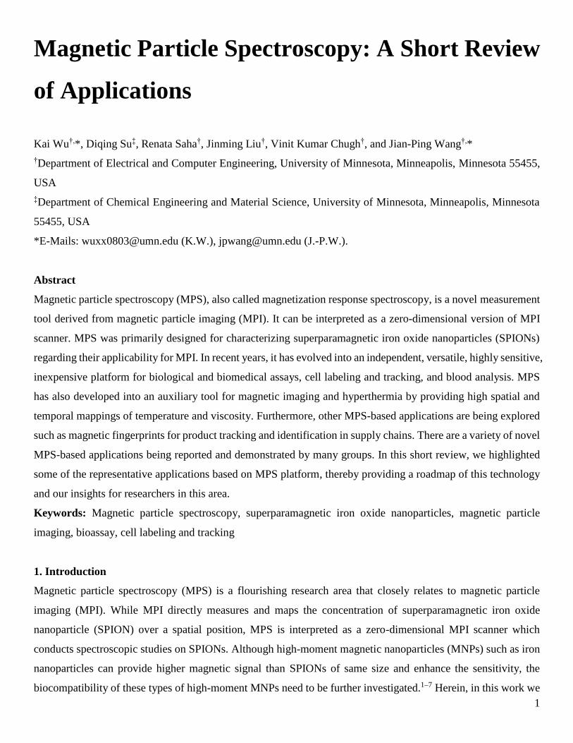
Kai Wu†,*, Diqing Su‡, Renata Saha†, Jinming Liu†, Vinit Kumar Chugh†, and Jian-Ping Wang†,*
†Department of Electrical and Computer Engineering, University of Minnesota, Minneapolis, Minnesota 55455,
USA
‡Department of Chemical Engineering and Material Science, University of Minnesota, Minneapolis, Minnesota
55455, USA
*E-Mails: [email protected] (K.W.), [email protected] (J.-P.W.).
Abstract
Magnetic particle spectroscopy (MPS), also called magnetization response spectroscopy, is a novel measurement
tool derived from magnetic particle imaging (MPI). It can be interpreted as a zero-dimensional version of MPI
scanner. MPS was primarily designed for characterizing superparamagnetic iron oxide nanoparticles (SPIONs)
regarding their applicability for MPI. In recent years, it has evolved into an independent, versatile, highly sensitive,
inexpensive platform for biological and biomedical assays, cell labeling and tracking, and blood analysis. MPS
has also developed into an auxiliary tool for magnetic imaging and hyperthermia by providing high spatial and
temporal mappings of temperature and viscosity. Furthermore, other MPS-based applications are being explored
such as magnetic fingerprints for product tracking and identification in supply chains. There are a variety of novel
MPS-based applications being reported and demonstrated by many groups. In this short review, we highlighted
some of the representative applications based on MPS platform, thereby providing a roadmap of this technology
and our insights for researchers in this area.
Keywords: Magnetic particle spectroscopy, superparamagnetic iron oxide nanoparticles, magnetic particle
imaging, bioassay, cell labeling and tracking
1. Introduction
Magnetic particle spectroscopy (MPS) is a flourishing research area that closely relates to magnetic particle
imaging (MPI). While MPI directly measures and maps the concentration of superparamagnetic iron oxide
nanoparticle (SPION) over a spatial position, MPS is interpreted as a zero-dimensional MPI scanner which
conducts spectroscopic studies on SPIONs. Although high-moment magnetic nanoparticles (MNPs) such as iron
nanoparticles can provide higher magnetic signal than SPIONs of same size and enhance the sensitivity, the
biocompatibility of these types of high-moment MNPs need to be further investigated.1–7 Herein, in this work we
2
only focus on the MPS-based applications using SPIONs. Primarily the MPS platform was dedicated to assessing
the performance of the SPION magnetic tracers regarding their applicability for MPI.8–13 Over the years, it has
developed into a highly sensitive, fast and versatile sensing platform for a wide variety of biological and
biomedical assays.14–18 In MPS, sinusoidal magnetic fields are applied to periodically magnetized SPIONs. The
magnetic moments of SPIONs tend to align with the applied fields through relaxation processes (Néel and
Brownian processes), which are countered by the thermal fluctuation. In addition, the relaxation processes are
directly linked to the physical conditions such as the viscosity and temperature of SPION aqueous medium as
well as the bound states of target analytes (chemicals/biological compounds) to the SPIONs.16,17,19–29 On the other
hand, SPIONs, with their size comparable to chemical/biological compounds, have been the subject of increasing
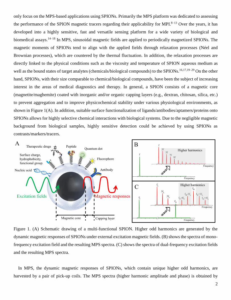
interest in the areas of medical diagnostics and therapy. In general, a SPION consists of a magnetic core
(magnetite/maghemite) coated with inorganic and/or organic capping layers (e.g., dextran, chitosan, silica, etc.)
to prevent aggregation and to improve physicochemical stability under various physiological environments, as
shown in Figure 1(A). In addition, suitable surface functionalization of ligands/antibodies/aptamers/proteins onto
SPIONs allows for highly selective chemical interactions with biological systems. Due to the negligible magnetic
background from biological samples, highly sensitive detection could be achieved by using SPIONs as
contrasts/markers/tracers.
Figure 1. (A) Schematic drawing of a multi-functional SPION. Higher odd harmonics are generated by the
dynamic magnetic responses of SPIONs under external excitation magnetic fields. (B) shows the spectra of mono-
frequency excitation field and the resulting MPS spectra. (C) shows the spectra of dual-frequency excitation fields
and the resulting MPS spectra.
In MPS, the dynamic magnetic responses of SPIONs, which contain unique higher odd harmonics, are
harvested by a pair of pick-up coils. The MPS spectra (higher harmonic amplitude and phase) is obtained by
3
Fourier transform of the detected time domain signal, as shown in Figure 1(B) & (C). Since the harmonic
amplitude is largely dependent on the quantity of SPIONs in the testing sample, the MPS results could be biased
by the deviations of SPION quantities in each sample, especially for the scenarios of detecting very low
abundancy of target analytes.14 Due to this concern, other SPION quantity-independent metrics have been
reported and demonstrated for bioassays, such as magnetic susceptibility, harmonic phase angle (or phase lag),
and harmonic amplitude ratios (the 3rd over the 5th harmonic ratio R35 or the 5th over the 3rd harmonic ratio
R53).17,29–34 In recent years, MPS has shown a strong capability of sensing the subtle changes in any property
influencing the Néel and/or Brownian processes of the SPIONs within seconds.
MPS bears the superior advantages over conventional magnetometers by providing fast and simple
measurement procedures for SPION systems without the need of cooling the device. The working mechanisms
and theories of MPS platform have been extensively reported by several papers.8,14,18,35 Herein, in this review, we
focus on the MPS derived applications. We firstly reviewed the MPS-based application for biological and
biomedical assays using surface functionalized SPIONs as magnetic markers, followed by MPS-based cell
analysis where the SPIONs are used as contrast agents to monitor cell uptake, passage, and cell vitality.17,29,36–39
With superior biocompatibility, biodegradability and superparamagnetic nature, SPIONs are much desired
magnetic markers/tracers for the aforementioned applications. In addition, MPS has also been exploited as an
auxiliary tool for applications such as magnetic imaging and hyperthermia by providing real-time viscosity and
temperature mapping, as well as mechanical force sensor in ball mills.15,21,40,41 Furthermore, other MPS-based
applications that are sometimes overlooked but owns great market potentials such as the characterization of
SPIONs from aqueous medium, the marking/tracking of products in the complex global resource and supply
chains, the determination of total blood volume and the evaluation of blood clot progression.15,16,42–46 In this short
review, we are only able to list some of the representative applications and the MPS-based applications are not
limited to this paper. We hope this review could provide insights into the great potential of MPS and serve as a
roadmap for researchers in this area.
2. MPS-based Biological and Biomedical Assays
2.1 MPS-based Immunoassays
As is aforementioned, the magnetic moments of SPIONs relax to align with the external magnetic field through
the joint Néel and Brownian processes. Néel process is the internal flipping of magnetic moment inside a stational
SPION while Brownian process is the physical rotation of the entire SPION along with its magnetic moment.
While both processes are countered by the thermal fluctuation, Néel process is also affected by the crystal
anisotropy and volume of the magnetic core. On the other hand, Brownian process is affected by the liquid
medium viscosity and hydrodynamic volume of the SPION (volume of magnetic core and surface attachments).
For free and non-interacting SPIONs with magnetic core size below 20 nm, Néel process guides the dynamic
4
magnetic responses of SPIONs, while Brownian process becomes the dominating factor when magnetic core size
of SPION is above 25 nm.42,47 For the applications of MPS-based immunoassays, Brownian process-dominated
SPIONs are most sensitive to the binding events of target analytes, thus, are frequently used for this purpose.
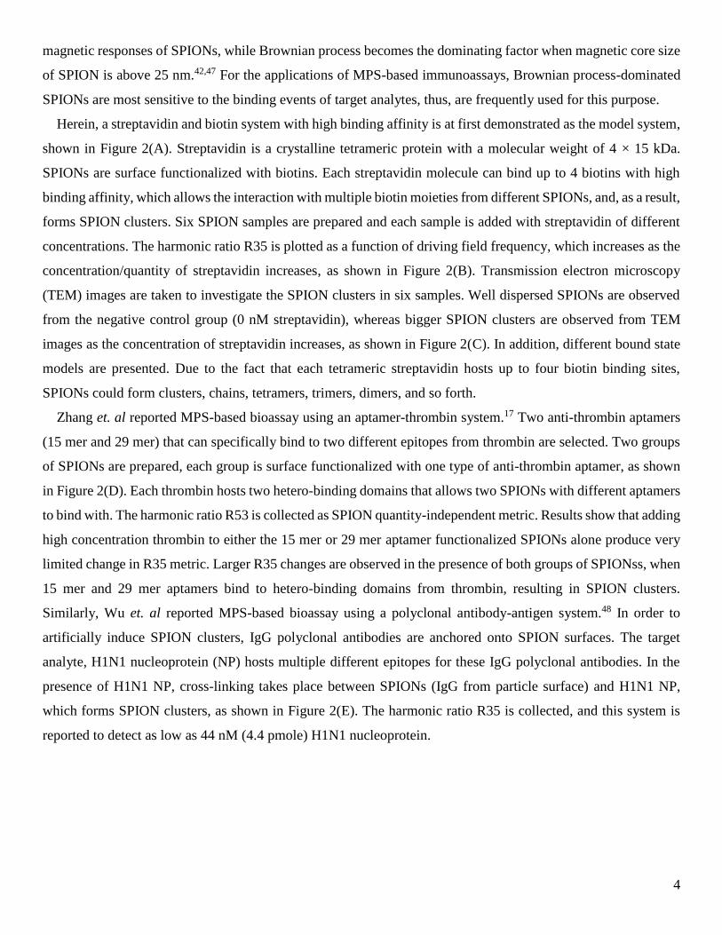
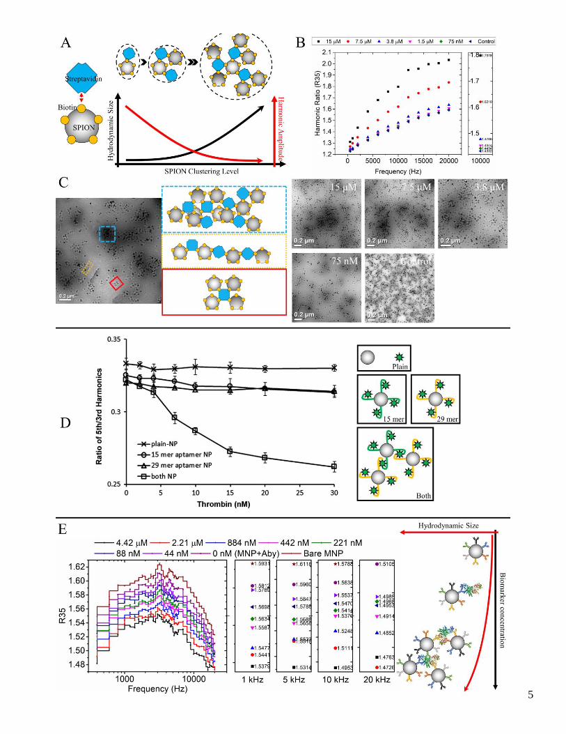
Herein, a streptavidin and biotin system with high binding affinity is at first demonstrated as the model system,
shown in Figure 2(A). Streptavidin is a crystalline tetrameric protein with a molecular weight of 4 × 15 kDa.
SPIONs are surface functionalized with biotins. Each streptavidin molecule can bind up to 4 biotins with high
binding affinity, which allows the interaction with multiple biotin moieties from different SPIONs, and, as a result,
forms SPION clusters. Six SPION samples are prepared and each sample is added with streptavidin of different
concentrations. The harmonic ratio R35 is plotted as a function of driving field frequency, which increases as the
concentration/quantity of streptavidin increases, as shown in Figure 2(B). Transmission electron microscopy
(TEM) images are taken to investigate the SPION clusters in six samples. Well dispersed SPIONs are observed
from the negative control group (0 nM streptavidin), whereas bigger SPION clusters are observed from TEM
images as the concentration of streptavidin increases, as shown in Figure 2(C). In addition, different bound state
models are presented. Due to the fact that each tetrameric streptavidin hosts up to four biotin binding sites,
SPIONs could form clusters, chains, tetramers, trimers, dimers, and so forth.
Zhang et. al reported MPS-based bioassay using an aptamer-thrombin system.17 Two anti-thrombin aptamers
(15 mer and 29 mer) that can specifically bind to two different epitopes from thrombin are selected. Two groups
of SPIONs are prepared, each group is surface functionalized with one type of anti-thrombin aptamer, as shown
in Figure 2(D). Each thrombin hosts two hetero-binding domains that allows two SPIONs with different aptamers
to bind with. The harmonic ratio R53 is collected as SPION quantity-independent metric. Results show that adding
high concentration thrombin to either the 15 mer or 29 mer aptamer functionalized SPIONs alone produce very
limited change in R35 metric. Larger R35 changes are observed in the presence of both groups of SPIONss, when
15 mer and 29 mer aptamers bind to hetero-binding domains from thrombin, resulting in SPION clusters.
Similarly, Wu et. al reported MPS-based bioassay using a polyclonal antibody-antigen system.48 In order to
artificially induce SPION clusters, IgG polyclonal antibodies are anchored onto SPION surfaces. The target
analyte, H1N1 nucleoprotein (NP) hosts multiple different epitopes for these IgG polyclonal antibodies. In the
presence of H1N1 NP, cross-linking takes place between SPIONs (IgG from particle surface) and H1N1 NP,
which forms SPION clusters, as shown in Figure 2(E). The harmonic ratio R35 is collected, and this system is
reported to detect as low as 44 nM (4.4 pmole) H1N1 nucleoprotein.
5
6
Figure 2. (A) Streptavidin and biotin functionalized SPION. As the quantity of streptavidin increases in the SPION
liquid medium, the SPIONs are likely to form clusters. The dashed lines represent the hydrodynamic sizes of
SPIONs due to the clustering. As the SPION clustering level increases, the hydrodynamic size increases, and the
harmonic amplitude decreases. (B) Harmonic ratio of the 3rd over the 5th (R35) collected from six SPION samples
added with different concentrations (quantities) of streptavidin, plotted as a function of driving field frequency.
The inset figure summarizes the R35 under a drive field frequency of 10 kHz. (C) Left: bright-field TEM images
of six SPION samples. Different SPION cluster models are given. Right: TEM images of SPIONs samples (15
µM, 7.5 µM, 3.8 µM, 75 nM, control (0 nM)) showing that the degree of particle clustering increases as the
quantity (concentration) of streptavidin increases. (D) Harmonic ratio of the 5th over the 3rd (R53) from two
populations of SPIONs, conjugated with 15 mer and 29 mer anti-thrombin aptamer, as a function of thrombin
concentration. (E) Harmonic ratio of the 3rd over the 5th (R35) collected from nine SPION samples added with
different concentrations (quantities) of H1N1 NP, plotted as a function of driving field frequency. Insets highlight
the R35 measured at 1 kHz, 5 kHz, 10 kHz and 20 kHz, respectively. (A-C) reprinted with permission from 29,
Copyright (2019) American Chemical Society. (D) reprinted with permission from 17, Copyright (2013) Elsevier.
(E) reprinted with permission from 48, Copyright (2020) American Chemical Society.
It is already known that the binding events of target analytes affects the Brownian process and dynamic
magnetic responses of SPIONs under external fields. While this detection mechanism yields detectable changes
in harmonic amplitudes, phases, and harmonic ratios, larger changes in these metrics can be achieved by designing
multiple ligands/antibodies/aptamers/proteins on SPIONs that could bind to hetero binding domains on the same
biomarker, assembling the SPIONs into clusters or aggregates. This kind of detection mechanism greatly
increased the hydrodynamic sizes of SPIONs, resulting in increased relaxation time, which can be sensed by the
MPS system.
The nonspecific binding (NSB) between other molecules/chemicals to the SPIONs is a critical problem in the
design of MPS-based immunoassays as the purpose of MPS bioassays is to measure only the binding events
between the target analytes and the SPIONs, instead of the background. Some frequently used methods to prevent
NSB are suggested herein as below: 1) adjust the pH of SPION liquid medium; 2) use protein blockers such as
bovine serum albumin (BSA) to block spare binding sites on SPIONs; 3) use surfactants such as Tween 20 to
disrupt hydrophobic interactions between target analytes and SPIONs; 4) adjust salt concentration in SPION
liquid medium to prevent charges on the target analytes from interacting with charges on the SPIONs.
2.2 MPS-based Cell Analysis
SPIONs have been used for a wide variety of applications such as molecular and cell separation, drug delivery,
hyperthermia, magnetic resonance imaging (MRI), and MPI, etc.9,49–62 The need for quantitative methods to
7
evaluate the SPION (or iron) content from contrast media solutions and biological matrixes is thus obvious. There
are various tools reported to measure SPION (or iron) content such as spectrophotometric elemental analysis
techniques (e.g., Perls’ Prussian blue colorimetry), fluorophores/radionuclides labeled SPIONs, and R1
relaxometry.63–66 Despite the extensive work done to quantify iron in an aqueous matrix, these techniques suffer
from: inability to distinguish between exogenous SPIONs and endogenous iron, narrow detectable range, and
nonlinearity.36,63 Recently, MPS is reported as an alternative to measure iron content (or to quantify SPIONs)
from aqueous matrix, where MPS utilizes the non-linear magnetic responses from SPIONs exposed to excitation
magnetic field.
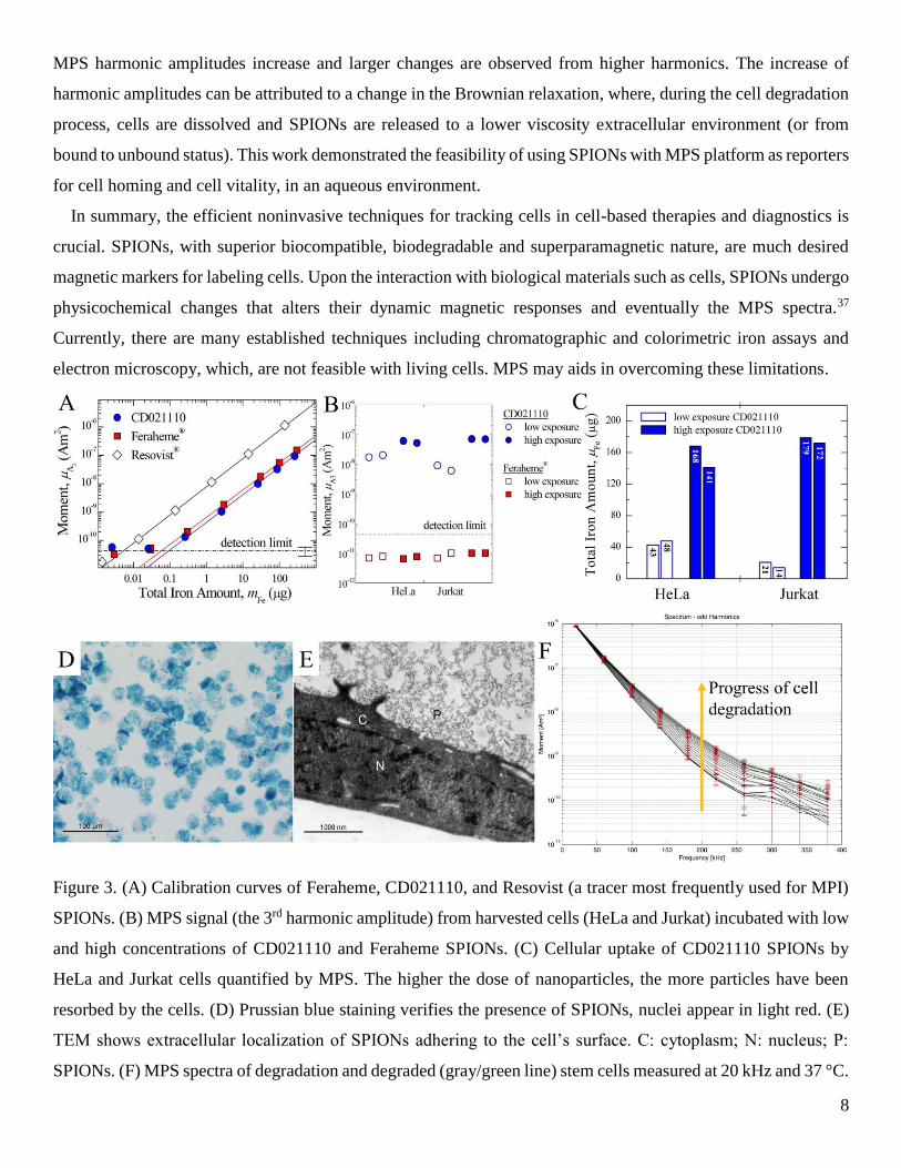
Loewa et. al reported the quantification of SPIONs uptaken in cells using MPS platform,36 where they took the
3rd harmonic amplitude as a measure for the SPION content. To this end, the harmonic amplitudes of known
SPION (iron) content under the same excitation field condition are recorded. As shown in Figure 3(A), the
calibration curves are highly linear for all types of SPIONs. Two tumor cell lines, HeLa and Jurkat, were incubated
with SPIONs of different concentrations, for 30 h, followed by harvesting ~106 cells for MPS measurements. The
3rd harmonic amplitudes from these harvested cells were recorded in Figure 3(B), indicating the SPIONs were up
taken by cells. Using the calibration curves, the calculated iron content up taken by cells are given in Figure 3(C).
Although this is a preliminary work without considering the impact of SPION aqueous matrix (viscosity, binding,
aggregations, etc.) and the influence of size selective cellular uptake of SPIONs, this method provides the basis
of a practical implementation of in vivo studies of SPION (iron) content from tissue samples. Besides the harmonic
amplitude, the harmonic ratio R53 is also reported to be an indicator to reveal distinct changes in the magnetic
behaviors of SPIONs in response to cellular uptake.37
Gräfe et. al established an in vitro testing system to investigate SPION transport across cellular layers as well
as examining MPS for reliable SPION quantification.38 A blood-brain barrier (BBB) representing human brain
microvascular endothelial cells (HBMEC) model was chosen. Their experiment confirms the excellent suitability
of MPS for sensitive SPION quantification at different stages of particles passing cellular layers, indicating a
promising future method to pass SPIONs loaded drugs across BBB without disturbing its integrity. As a result,
the drug loading and release efficiency can be monitored by MPS approach.
Fidler et. al reported MPS for in vitro cell vitality monitoring, where human mesenchymal stem cells (hMSCs)
are labeled with SPIONs and tracked.39 The hMSCs are a promising tool in regenerative medicine and it’s able to
repair damaged tissue. However, the tissue healing using hMSCs will only be possible if cells can be homed to
their target and are still vital. This challenge calls for a long-term, non-invasive method to label and monitor the
cells. Fidler et. al labeled hMSCs with SPIONs and the verification of SPION-labeled hMSCs by Prussian blue,
TEM, and MPS, the light and TEM analysis is shown in Figure 3(D) & (E). For cell vitality assessment, the MPS
spectra was continuously monitored during a cell degradation process initiated by adding Sodium Dodecyl (lauryl)
Sulfate (SDS, which dissolves the cells). As shown in Figure 3(F), during the dissolution of the cells by SDS, the
8
MPS harmonic amplitudes increase and larger changes are observed from higher harmonics. The increase of
harmonic amplitudes can be attributed to a change in the Brownian relaxation, where, during the cell degradation
process, cells are dissolved and SPIONs are released to a lower viscosity extracellular environment (or from
bound to unbound status). This work demonstrated the feasibility of using SPIONs with MPS platform as reporters
for cell homing and cell vitality, in an aqueous environment.
In summary, the efficient noninvasive techniques for tracking cells in cell-based therapies and diagnostics is
crucial. SPIONs, with superior biocompatible, biodegradable and superparamagnetic nature, are much desired
magnetic markers for labeling cells. Upon the interaction with biological materials such as cells, SPIONs undergo
physicochemical changes that alters their dynamic magnetic responses and eventually the MPS spectra.37
Currently, there are many established techniques including chromatographic and colorimetric iron assays and
electron microscopy, which, are not feasible with living cells. MPS may aids in overcoming these limitations.
Figure 3. (A) Calibration curves of Feraheme, CD021110, and Resovist (a tracer most frequently used for MPI)
SPIONs. (B) MPS signal (the 3rd harmonic amplitude) from harvested cells (HeLa and Jurkat) incubated with low
and high concentrations of CD021110 and Feraheme SPIONs. (C) Cellular uptake of CD021110 SPIONs by
HeLa and Jurkat cells quantified by MPS. The higher the dose of nanoparticles, the more particles have been
resorbed by the cells. (D) Prussian blue staining verifies the presence of SPIONs, nuclei appear in light red. (E)
TEM shows extracellular localization of SPIONs adhering to the cell’s surface. C: cytoplasm; N: nucleus; P:
SPIONs. (F) MPS spectra of degradation and degraded (gray/green line) stem cells measured at 20 kHz and 37 °C.
9
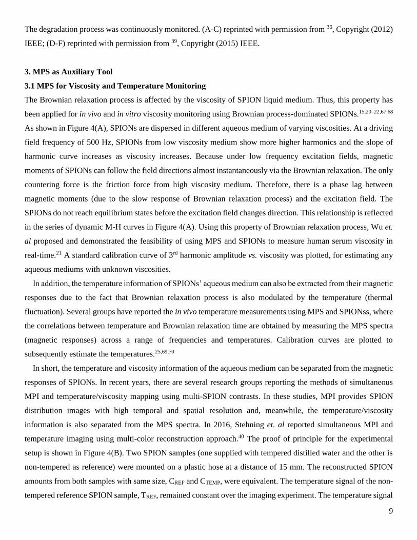
The degradation process was continuously monitored. (A-C) reprinted with permission from 36, Copyright (2012)
IEEE; (D-F) reprinted with permission from 39, Copyright (2015) IEEE.
3. MPS as Auxiliary Tool
3.1 MPS for Viscosity and Temperature Monitoring
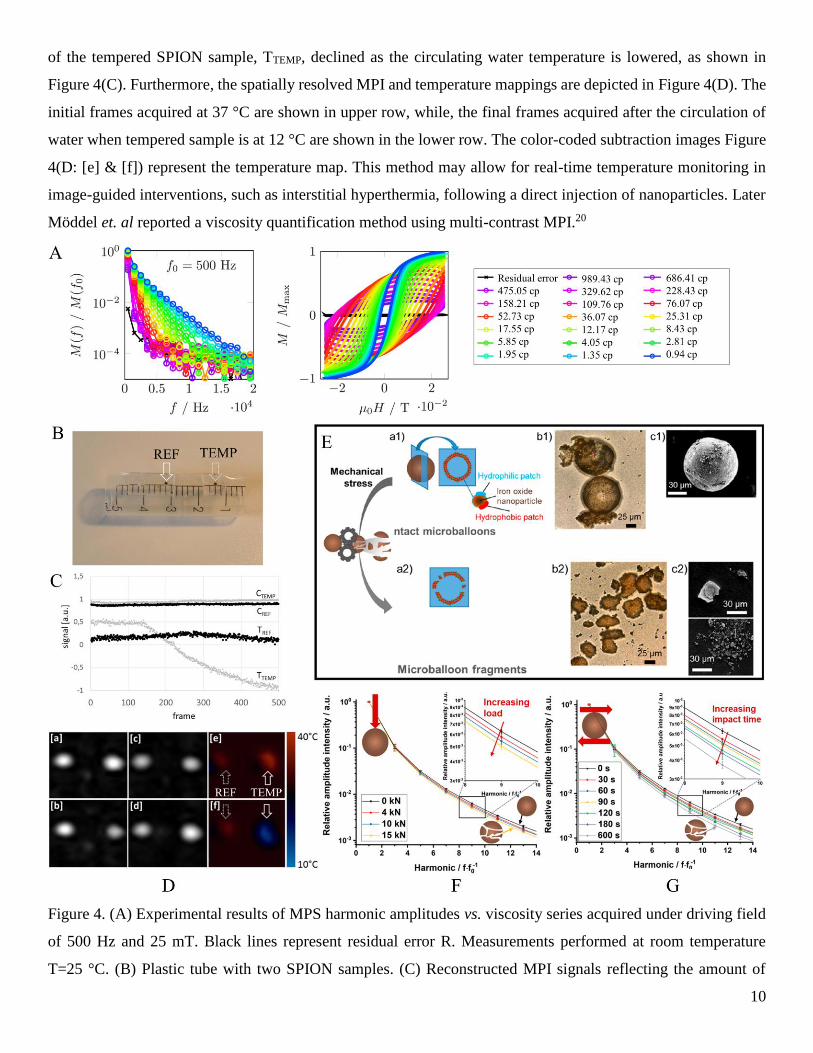
The Brownian relaxation process is affected by the viscosity of SPION liquid medium. Thus, this property has
been applied for in vivo and in vitro viscosity monitoring using Brownian process-dominated SPIONs.15,20–22,67,68
As shown in Figure 4(A), SPIONs are dispersed in different aqueous medium of varying viscosities. At a driving
field frequency of 500 Hz, SPIONs from low viscosity medium show more higher harmonics and the slope of
harmonic curve increases as viscosity increases. Because under low frequency excitation fields, magnetic
moments of SPIONs can follow the field directions almost instantaneously via the Brownian relaxation. The only
countering force is the friction force from high viscosity medium. Therefore, there is a phase lag between
magnetic moments (due to the slow response of Brownian relaxation process) and the excitation field. The
SPIONs do not reach equilibrium states before the excitation field changes direction. This relationship is reflected
in the series of dynamic M-H curves in Figure 4(A). Using this property of Brownian relaxation process, Wu et.
al proposed and demonstrated the feasibility of using MPS and SPIONs to measure human serum viscosity in
real-time.21 A standard calibration curve of 3rd harmonic amplitude vs. viscosity was plotted, for estimating any
aqueous mediums with unknown viscosities.
In addition, the temperature information of SPIONs’ aqueous medium can also be extracted from their magnetic
responses due to the fact that Brownian relaxation process is also modulated by the temperature (thermal
fluctuation). Several groups have reported the in vivo temperature measurements using MPS and SPIONss, where
the correlations between temperature and Brownian relaxation time are obtained by measuring the MPS spectra
(magnetic responses) across a range of frequencies and temperatures. Calibration curves are plotted to
subsequently estimate the temperatures.25,69,70
In short, the temperature and viscosity information of the aqueous medium can be separated from the magnetic
responses of SPIONs. In recent years, there are several research groups reporting the methods of simultaneous
MPI and temperature/viscosity mapping using multi-SPION contrasts. In these studies, MPI provides SPION
distribution images with high temporal and spatial resolution and, meanwhile, the temperature/viscosity
information is also separated from the MPS spectra. In 2016, Stehning et. al reported simultaneous MPI and
temperature imaging using multi-color reconstruction approach.40 The proof of principle for the experimental
setup is shown in Figure 4(B). Two SPION samples (one supplied with tempered distilled water and the other is
non-tempered as reference) were mounted on a plastic hose at a distance of 15 mm. The reconstructed SPION
amounts from both samples with same size, CREF and CTEMP, were equivalent. The temperature signal of the non-
tempered reference SPION sample, TREF, remained constant over the imaging experiment. The temperature signal
10
of the tempered SPION sample, TTEMP, declined as the circulating water temperature is lowered, as shown in
Figure 4(C). Furthermore, the spatially resolved MPI and temperature mappings are depicted in Figure 4(D). The
initial frames acquired at 37 °C are shown in upper row, while, the final frames acquired after the circulation of
water when tempered sample is at 12 °C are shown in the lower row. The color-coded subtraction images Figure
4(D: [e] & [f]) represent the temperature map. This method may allow for real-time temperature monitoring in
image-guided interventions, such as interstitial hyperthermia, following a direct injection of nanoparticles. Later
Möddel et. al reported a viscosity quantification method using multi-contrast MPI.20
Figure 4. (A) Experimental results of MPS harmonic amplitudes vs. viscosity series acquired under driving field
of 500 Hz and 25 mT. Black lines represent residual error R. Measurements performed at room temperature
T=25 °C. (B) Plastic tube with two SPION samples. (C) Reconstructed MPI signals reflecting the amount of
11
SPIONs (CTEMP and CREF) and temperature information (TTEMP and TERF) of the tempered and reference probe.
(D) Projection images of 3D reconstructed signal channels [a]-[d], and color-coded difference signal representing
temperature [e], [f] acquired at the beginning [top row] and end [bottom row] of the experiment. (E) Hollow
microballoons composed of SPIONs. Drop of the MPS signal curves of microballoons during the application of
quasi-static compression (F) or of dynamic shear and impact forces in a ball mill-like setup (G) increases
significantly with increasing load or milling time, respectively. (A) reprinted with permission from 15, Copyright
(2019) American Chemical Society; (B-D) reprinted with permission from 40, Copyright (2016) Infinite Science
Publishing. (E-G) reprinted with permission from 41, Copyright (2019) American Chemical Society.
3.2 MPS for Mechanical Force Monitoring
Wintzheimer et. al reported a novel application of MPS and SPIONs as sensors for mechanical stress.41 The as-
assembled SPIONs yield hollow microballoons (as shown in Figure 4(E)). This kind of structure is continuously
fragmented when subjected to mechanical forces. By using MPS spectra, this structure deformation can be readily
detected and, enables quantification of the applied mechanical forces in ball mills. Figure 4(F) & (G) depict the
MPS harmonic amplitudes of the microballoons after the application of quasi-static compression or dynamic shear
and impact forces, respectively. In both cases, the relative amplitude intensities drop with the increasing
mechanical energy, which proves that the MPS spectra of these hollow microballoon supraparticles are clearly
distinguishable in the course of mechanical forces. This type of mechanical force sensor does not necessarily have
to be removed from the processed material for measurements, furthermore, its magnetic property allows easy
removal after milling.
4. Other applications
4.1 MPS for SPION Characterization
As the sinusoidal magnetic fields excite SPIONs into their nonlinear saturation magnetization. The dynamic
magnetic responses, represented by higher harmonics in frequency domain, provide information about the
physical and magnetic properties of the SPIONs. It has been reported that the harmonic ratio R35 is inversely
proportional to the saturation magnetization Ms and magnetic core diameter D of the SPION.15,42,67,71 The
harmonic phase angle (lag) of magnetic moment to the excitation field carries the information of hydrodynamic
size of SPION. Numerical simulations were carried out to reveal the correlation between harmonic ratio and Ms
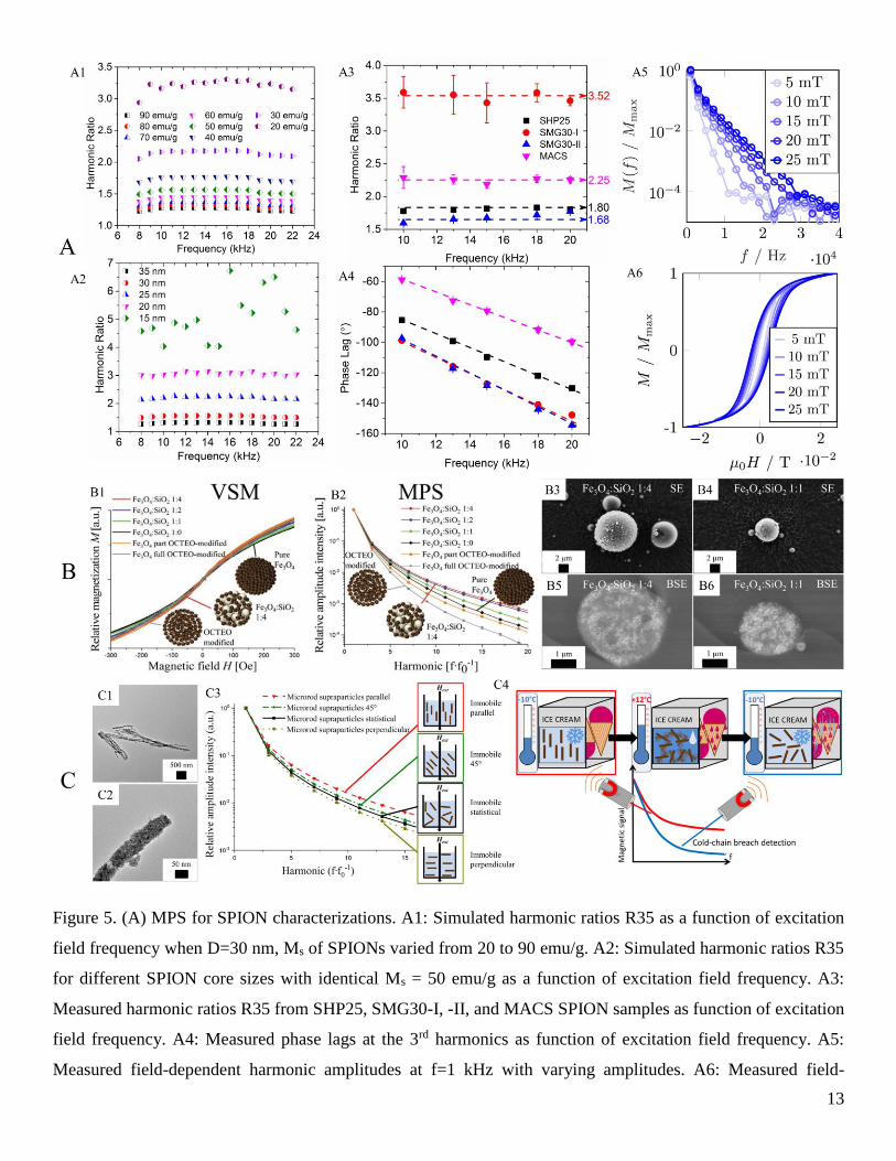
as well as D. As shown in Figure 5(A: A1 & A2), a SPION system is assumed, with log-normal size distribution,
effective anisotropy constant Keff = 1.8 × 10⁵ erg/cm3 (1.8 × 10⁴ J/m3), Ms and D are varied. The results show that
for SPIONs with identical core sizes, a smaller harmonic ratio R35 corresponds to a higher Ms, while, for SPIONs
with identical Ms, a smaller core size yields a larger harmonic ratio R35. This conclusion is further proved by
experimentally measuring the harmonic ratios from four commercially avaible SPION samples, as shown in
12
Figure 5(A: A3). For SMG30-I and -II samples, with identical magnetic core size, a smaller harmonic ratio R35
from SMG30-II indicates a higher Ms over SMG30-I, which is further proved by the Vibrating Sample
Magnetometer (VSM) results and high-angle annular dark-field scanning transmission electron microscopy
energy-dispersive X-ray spectroscopy (HAADF–STEM–EDS) mapping images.42 In addition, the 3rd harmonic
phase lag to excitation field recorded in Figure 5(A: A4) shows that SMG30-I and -II SPIONs have similar
hydrodynamic size, and are larger than SHP25. On the other hand, for the multicore MACS SPIONs, the SPIONss
are embedded in a polymer and Néel process is dominating, which is a different scenario compared to the single
core SPIONs (SHP25, SMG30-I and -II).
In addition to the magnetic and physical properties of SPIONs that affect the MPS spectra (harmonic amplitudes
and phases), the excitation fields should also be taken into consideration. Draack et. al reported the field-
dependent MPS spectra by measuring SPIONs under different exaction field amplitudes and frequencies.15 The
field-dependent harmonic amplitudes are measured at a constant excitation frequency, f=1 kHz, with varying
amplitudes from 5 mT to 25 mT, as shown in Figure 5(A: A5). The dynamic M-H curves are plotted in Figure
5(A: A6). A higher magnetic field strength results in more saturated magnetizations and more pronounced
hysteresis of the magnetization loops.
4.2 MPS for Magnetic Fingerprint
Based on the aforementioned phenomenon, each type of SPIONs with different magnetic core size D,
hydrodynamic size, saturation magnetization Ms, shows unique MPS spectra, which could be used as magnetic
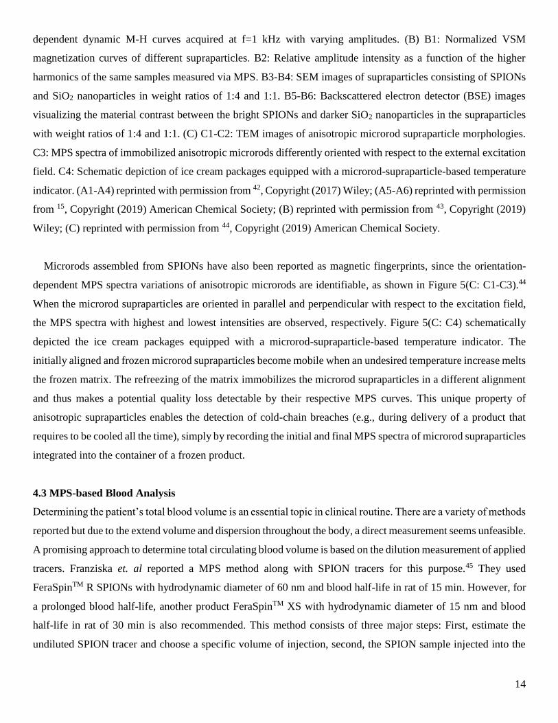
fingerprints for product tracking and identification. Müssig et. al reported the specially engineered magnetic
supraparticles that exhibit unique magnetic signature in MPS, proving its potential for marker applications.43 In
their work, the supraparticles are formed by assembling SPIONs with SiO2 nanoparticles with different ratios, as
shown in Figure 5(B: B3-B6). They prepared different kinds of supraparticles by mixing SPIONs (~10 nm) and
SiO2 nanoparticles (~20 nm) in weight ratios of 1:0, 1:1, 1:2, and 1:4. The M-H curves measured by VSM are
plotted in Figure 5(B: B1), yet, the differences with respect to hysteresis loops can hardly be quantified. However,
clearly distinguishable MPS spectra are observed in Figure 5(B: B2). In contrast to the VSM, MPS spectra of
various supraparticles are easily quantified by determining the amplitudes of higher harmonics. In addition to
mixing SPIONs with non-magnetic SiO2 nanoparticles, changing the interparticle distance by surface
modification of SPIONs with OCTEO can also modify MPS spectra (as shown in Figure 5(B: B1-B2), the “Fe3O4
part/full OCTEO-modified” curves). This work shows such spraparticles along with MPS could enable easy and
versatile tracking and identification for products. By mixing different amount of SiO2 nanoparticles with SPIONs
or structurally adjusting the SPIONs, it is possible to change the magnetic properties (MPS spectra) of
supraparticles and generate countless unique magnetic fingerprints.
13
Figure 5. (A) MPS for SPION characterizations. A1: Simulated harmonic ratios R35 as a function of excitation
field frequency when D=30 nm, Ms of SPIONs varied from 20 to 90 emu/g. A2: Simulated harmonic ratios R35
for different SPION core sizes with identical Ms = 50 emu/g as a function of excitation field frequency. A3:
Measured harmonic ratios R35 from SHP25, SMG30-I, -II, and MACS SPION samples as function of excitation
field frequency. A4: Measured phase lags at the 3rd harmonics as function of excitation field frequency. A5:
Measured field-dependent harmonic amplitudes at f=1 kHz with varying amplitudes. A6: Measured field-
14
dependent dynamic M-H curves acquired at f=1 kHz with varying amplitudes. (B) B1: Normalized VSM
magnetization curves of different supraparticles. B2: Relative amplitude intensity as a function of the higher
harmonics of the same samples measured via MPS. B3-B4: SEM images of supraparticles consisting of SPIONs
and SiO2 nanoparticles in weight ratios of 1:4 and 1:1. B5-B6: Backscattered electron detector (BSE) images
visualizing the material contrast between the bright SPIONs and darker SiO2 nanoparticles in the supraparticles
with weight ratios of 1:4 and 1:1. (C) C1-C2: TEM images of anisotropic microrod supraparticle morphologies.
C3: MPS spectra of immobilized anisotropic microrods differently oriented with respect to the external excitation
field. C4: Schematic depiction of ice cream packages equipped with a microrod-supraparticle-based temperature
indicator. (A1-A4) reprinted with permission from 42, Copyright (2017) Wiley; (A5-A6) reprinted with permission
from 15, Copyright (2019) American Chemical Society; (B) reprinted with permission from 43, Copyright (2019)
Wiley; (C) reprinted with permission from 44, Copyright (2019) American Chemical Society.
Microrods assembled from SPIONs have also been reported as magnetic fingerprints, since the orientation-
dependent MPS spectra variations of anisotropic microrods are identifiable, as shown in Figure 5(C: C1-C3).44
When the microrod supraparticles are oriented in parallel and perpendicular with respect to the excitation field,
the MPS spectra with highest and lowest intensities are observed, respectively. Figure 5(C: C4) schematically
depicted the ice cream packages equipped with a microrod-supraparticle-based temperature indicator. The
initially aligned and frozen microrod supraparticles become mobile when an undesired temperature increase melts
the frozen matrix. The refreezing of the matrix immobilizes the microrod supraparticles in a different alignment
and thus makes a potential quality loss detectable by their respective MPS curves. This unique property of
anisotropic supraparticles enables the detection of cold-chain breaches (e.g., during delivery of a product that
requires to be cooled all the time), simply by recording the initial and final MPS spectra of microrod supraparticles
integrated into the container of a frozen product.
4.3 MPS-based Blood Analysis
Determining the patient’s total blood volume is an essential topic in clinical routine. There are a variety of methods
reported but due to the extend volume and dispersion throughout the body, a direct measurement seems unfeasible.
A promising approach to determine total circulating blood volume is based on the dilution measurement of applied
tracers. Franziska et. al reported a MPS method along with SPION tracers for this purpose.45 They used
FeraSpinTM R SPIONs with hydrodynamic diameter of 60 nm and blood half-life in rat of 15 min. However, for
a prolonged blood half-life, another product FeraSpinTM XS with hydrodynamic diameter of 15 nm and blood
half-life in rat of 30 min is also recommended. This method consists of three major steps: First, estimate the
undiluted SPION tracer and choose a specific volume of injection, second, the SPION sample injected into the
15
subject, third, a small amount of blood is draw after a mixing time. From the measured concentration, the total
circulating blood volume is calculated.
Another work reported by Hafsa et. al proposing the evaluation of blood clot progression using SPION tracers
and MPS.16 By measuring the SPIONs’ relaxation time in the clot vicinity, the clot age, clot stiffness, and the
amount of SPIONs bound to the clot can be estimated. It is reported that during the clot formation, SPIONs
become trapped in the clot mesh, which, restricts their Brownian relaxation process, causing an increase of their
relaxation time.46 In this work, the SPIONs are surface functionalized with ATP15 and ATP29 aptamers that bind
to hetero epitopes on thrombin. SPIONs bound to thrombus are identified by their increased relaxation time using
MPS. The harmonic ratio R35 is reported as metric for the Brownian process, which in turn, reflects the bound
state of SPIONs.
16
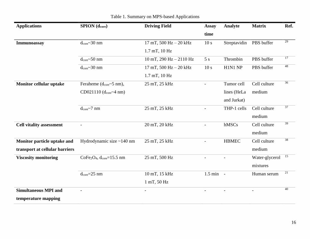
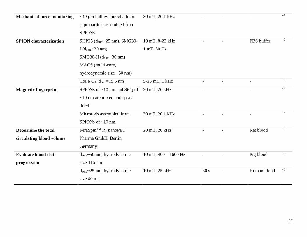
Table 1. Summary on MPS-based Applications
Applications SPION (dcore) Driving Field Assay
time
Analyte Matrix Ref.
Immunoassay dcore~30 nm 17 mT, 500 Hz – 20 kHz
1.7 mT, 10 Hz
10 s Streptavidin PBS buffer 29
dcore~50 nm 10 mT, 290 Hz – 2110 Hz 5 s Thrombin PBS buffer 17
dcore~30 nm 17 mT, 500 Hz – 20 kHz
1.7 mT, 10 Hz
10 s H1N1 NP PBS buffer 48
Monitor cellular uptake Feraheme (dcore~5 nm),
CD021110 (dcore~4 nm)
25 mT, 25 kHz - Tumor cell
lines (HeLa
and Jurkat)
Cell culture
medium
36
dcore~7 nm 25 mT, 25 kHz - THP-1 cells Cell culture
medium
37
Cell vitality assessment - 20 mT, 20 kHz - hMSCs Cell culture
medium
39
Monitor particle uptake and
transport at cellular barriers
Hydrodynamic size ~140 nm 25 mT, 25 kHz - HBMEC Cell culture
medium
38
Viscosity monitoring CoFe2O4, dcore=15.5 nm 25 mT, 500 Hz - - Water-glycerol
mixtures
15
dcore=25 nm 10 mT, 15 kHz
1 mT, 50 Hz
1.5 min - Human serum 21
Simultaneous MPI and
temperature mapping
- - - - - 40
17
Mechanical force monitoring ~40 μm hollow microballoon
supraparticle assembled from
SPIONs
30 mT, 20.1 kHz - - - 41
SPION characterization SHP25 (dcore~25 nm), SMG30-
I (dcore~30 nm)
SMG30-II (dcore~30 nm)
MACS (multi-core,
hydrodynamic size ~50 nm)
10 mT, 8-22 kHz
1 mT, 50 Hz
- - PBS buffer 42
CoFe2O4, dcore=15.5 nm 5-25 mT, 1 kHz - - - 15
Magnetic fingerprint SPIONs of ~10 nm and SiO2 of
~10 nm are mixed and spray
dried
30 mT, 20 kHz - - - 43
Microrods assembled from
SPIONs of ~10 nm.
30 mT, 20.1 kHz - - - 44
Determine the total
circulating blood volume
FeraSpinTM R (nanoPET
Pharma GmbH, Berlin,
Germany)
20 mT, 20 kHz - - Rat blood 45
Evaluate blood clot
progression
dcore~50 nm, hydrodynamic
size 116 nm
10 mT, 400 – 1600 Hz - - Pig blood 16
dcore~25 nm, hydrodynamic
size 40 nm
10 mT, 25 kHz 30 s - Human blood 46
18
5. Conclusion
In this review, we summarized the recent advances in MPS-based applications and shown in Table 1. For
biological and biomedical assays, by surface functionalizing suitable ligands/antibodies/aptamers/proteins on
SPIONs that could specifically bind to target analytes, SPIONs are explored as magnetic markers and their
dynamic magnetic responses are monitored by MPS. The binding events of target analytes affect the Brownian
process and dynamic magnetic responses of SPIONs and cause detectable changes in harmonic amplitudes, phases,
and harmonic ratios. Due to the negligible magnetic background from biological samples, highly sensitive
detection could be achieved by using SPIONs as biomarkers. This detection mechanism based on a MPS platform
allows simple, fast, inexpensive, and high sensitivity bioassays. In the areas of cell labeling, tracking and cell-
based theragnostics (therapy and diagnostics), MPS provides a quantitative method to evaluate the SPION (or
iron) content from contrast biological matrixes, and even in living cells. Upon the interaction with biological
materials such as cells, SPIONs undergo physicochemical changes that alter their MPS spectra. In addition to
these stand-alone applications, MPS has also developed into a mature auxiliary tool supporting the magnetic
imaging and hyperthermia by providing real-time, high spatial and temporal resolution viscosity/temperature
mapping. Other novel applications based on MPS such as SPION characterizations, magnetic fingerprints, and
blood analysis are also reviewed.
In summary, many good and novel works have been reported in recent years based on MPS platform and this
process is still ongoing. With the unique nonlinear magnetic responses of SPIONs, facile surface chemical
modifications, flexible structural design of SPION assemblies (supraparticles) with novel magnetic/physical
properties, MPS has found its position in more and more new areas.
ASSOCIATED CONTENT
ORCID
Kai Wu: 0000-0002-9444-6112
Diqing Su: 0000-0002-5790-8744
Renata Saha: 0000-0002-0389-0083
Jinming Liu: 0000-0002-4313-5816
Jian-Ping Wang: 0000-0003-2815-6624
Notes
The authors declare no conflict of interest.
ACKNOWLEDGMENTS
19
This study was financially supported by the Institute of Engineering in Medicine of the University of Minnesota
through FY18 IEM Seed Grant Funding Program, the Distinguished McKnight University Professorship, the
Centennial Chair Professorship, and the Robert F Hartmann Endowed Chair from the University of Minnesota.
REFERENCES
(1) Liu, J.; Su, D.; Wu, K.; Wang, J.-P. High-Moment Magnetic Nanoparticles. J. Nanoparticle Res. 2020, 22
(3), 1–16.
(2) Wu, K.; Su, D.; Liu, J.; Saha, R.; Wang, J.-P. Magnetic Nanoparticles in Nanomedicine: A Review of Recent
Advances. Nanotechnology 2019, 30 (50), 502003.
(3) Liu, J.; Schliep, K.; He, S.-H.; Ma, B.; Jing, Y.; Flannigan, D. J.; Wang, J.-P. Iron Nanoparticles with Tunable
Tetragonal Structure and Magnetic Properties. Phys. Rev. Mater. 5, 2 (5), 054415.
https://doi.org/10.1103/PhysRevMaterials.2.054415.
(4) Srinoi, P.; Chen, Y.-T.; Vittur, V.; Marquez, M. D.; Lee, T. R. Bimetallic Nanoparticles: Enhanced Magnetic
and Optical Properties for Emerging Biological Applications. Appl. Sci. 2018, 8 (7), 1106.
(5) Li, Y.; Wang, N.; Huang, X.; Li, F.; Davis, T. P.; Qiao, R.; Ling, D. Polymer-Assisted Magnetic Nanoparticle
Assemblies for Biomedical Applications. ACS Appl. Bio Mater. 2019.
(6) Mora, B.; Perez-Valle, A.; Redondo, C.; Boyano, M. D.; Morales, R. Cost-Effective Design of High-Magnetic
Moment Nanostructures for Biotechnological Applications. ACS Appl. Mater. Interfaces 2018, 10 (9),
8165–8172.
(7) Liu, J.; Wu, K.; Wang, J.-P. Magnetic Properties of Cubic FeCo Nanoparticles with Anisotropic Long Chain
Structure. AIP Adv. 2016, 6 (5), 056126.
(8) Biederer, S.; Knopp, T.; Sattel, T.; Lüdtke-Buzug, K.; Gleich, B.; Weizenecker, J.; Borgert, J.; Buzug, T.
Magnetization Response Spectroscopy of Superparamagnetic Nanoparticles for Magnetic Particle Imaging.
J. Phys. Appl. Phys. 2009, 42, 205007.
(9) Arami, H.; Khandhar, A. P.; Tomitaka, A.; Yu, E.; Goodwill, P. W.; Conolly, S. M.; Krishnan, K. M. In Vivo
Multimodal Magnetic Particle Imaging (MPI) with Tailored Magneto/Optical Contrast Agents.
Biomaterials 2015, 52, 251–261.
(10) Dhavalikar, R.; Rinaldi, C. On the Effect of Finite Magnetic Relaxation on the Magnetic Particle Imaging
Performance of Magnetic Nanoparticles. J. Appl. Phys. 2014, 115 (7), 074308.
(11) Gleich, B.; Weizenecker, J. Tomographic Imaging Using the Nonlinear Response of Magnetic Particles.
Nature 2005, 435 (7046), 1214–1217.
(12) Deissler, R. J.; Martens, M. A. Dependence of the Magnetization Response on the Driving Field Amplitude
for Magnetic Particle Imaging and Spectroscopy. Magn. IEEE Trans. On 2015, 51, 1–4.
20
(13) Du, Y.; Lai, P. T.; Leung, C. H.; Pong, P. W. Design of Superparamagnetic Nanoparticles for Magnetic
Particle Imaging (MPI). Int. J. Mol. Sci. 2013, 14 (9), 18682–18710.
(14) Wu, K.; Su, D.; Saha, R.; Wong, D.; Wang, J.-P. Magnetic Particle Spectroscopy-Based Bioassays: Methods,
Applications, Advances, and Future Opportunities. J. Phys. Appl. Phys. 2019, 52, 173001.
(15) Draack, S.; Lucht, N.; Remmer, H.; Martens, M.; Fischer, B.; Schilling, M.; Ludwig, F.; Viereck, T.
Multiparametric Magnetic Particle Spectroscopy of CoFe2O4 Nanoparticles in Viscous Media. J. Phys.
Chem. C 2019, 123 (11), 6787–6801.
(16) Khurshid, H.; Shi, Y.; Berwin, B. L.; Weaver, J. B. Evaluating Blood Clot Progression Using Magnetic
Particle Spectroscopy. Med. Phys. 2018, 45 (7), 3258–3263.
(17) Zhang, X.; Reeves, D. B.; Perreard, I. M.; Kett, W. C.; Griswold, K. E.; Gimi, B.; Weaver, J. B. Molecular
Sensing with Magnetic Nanoparticles Using Magnetic Spectroscopy of Nanoparticle Brownian Motion.
Biosens. Bioelectron. 2013, 50, 441–446.
(18) Paysen, H.; Wells, J.; Kosch, O.; Steinhoff, U.; Trahms, L.; Schaeffter, T.; Wiekhorst, F. Towards
Quantitative Magnetic Particle Imaging: A Comparison with Magnetic Particle Spectroscopy. AIP Adv.
2018, 8 (5), 056712.
(19) Yu, K.; Zhang, M.; Li, Y.; Wang, R. Increased Whole Blood Viscosity Associated with Arterial Stiffness in
Patients with Non‐alcoholic Fatty Liver Disease. J. Gastroenterol. Hepatol. 2014, 29 (3), 540–544.
(20) Möddel, M.; Meins, C.; Dieckhoff, J.; Knopp, T. Viscosity Quantification Using Multi-Contrast Magnetic
Particle Imaging. New J. Phys. 2018, 20 (8), 083001.
(21) Wu, K.; Liu, J.; Wang, Y.; Ye, C.; Feng, Y.; Wang, J.-P. Superparamagnetic Nanoparticle-Based Viscosity
Test. Appl. Phys. Lett. 2015, 107, 053701.
(22) Wu, K.; Ye, C.; Liu, J.; Wang, Y.; Feng, Y.; Wang, J.-P. In Vitro Viscosity Measurement on
Superparamagnetic Nanoparticle Suspensions. IEEE Trans. Magn. 2016, 52 (7).
(23) Draack, S.; Viereck, T.; Kuhlmann, C.; Schilling, M.; Ludwig, F. Temperature-Dependent MPS
Measurements. Int. J. Magn. Part. Imaging 2017, 3 (1).
(24) Perreard, I.; Reeves, D.; Zhang, X.; Kuehlert, E.; Forauer, E.; Weaver, J. Temperature of the Magnetic
Nanoparticle Microenvironment: Estimation from Relaxation Times. Phys. Med. Biol. 2014, 59, 1109.
(25) Weaver, J. B.; Rauwerdink, A. M.; Hansen, E. W. Magnetic Nanoparticle Temperature Estimation. Med.
Phys. 2009, 36 (5), 1822–1829.
(26) Wells, J.; Paysen, H.; Kosch, O.; Trahms, L.; Wiekhorst, F. Temperature Dependence in Magnetic Particle
Imaging. AIP Adv. 2018, 8 (5), 056703.
(27) Orlov, A. V.; Bragina, V. A.; Nikitin, M. P.; Nikitin, P. I. Rapid Dry-Reagent Immunomagnetic Biosensing
Platform Based on Volumetric Detection of Nanoparticles on 3D Structures. Biosens. Bioelectron. 2016,
79, 423–429.
21
(28) Nikitin, M. P.; Orlov, A.; Sokolov, I.; Minakov, A.; Nikitin, P.; Ding, J.; Bader, S.; Rozhkova, E.; Novosad,
V. Ultrasensitive Detection Enabled by Nonlinear Magnetization of Nanomagnetic Labels. Nanoscale 2018,
10 (24), 11642–11650.
(29) Wu, K.; Liu, J.; Su, D.; Saha, R.; Wang, J.-P. Magnetic Nanoparticle Relaxation Dynamics-Based Magnetic
Particle Spectroscopy for Rapid and Wash-Free Molecular Sensing. ACS Appl. Mater. Interfaces 2019, 11
(26), 22979–22986. https://doi.org/10.1021/acsami.9b05233.
(30) Hong, C.-Y.; Wu, C.; Chiu, Y.; Yang, S.-Y.; Horng, H.-E.; Yang, H.-C. Magnetic Susceptibility Reduction
Method for Magnetically Labeled Immunoassay. Appl. Phys. Lett. 2006, 88, 212512.
(31) Yang, C.; Yang, S.-Y.; Chen, H.; Weng, W.; Horng, H.-E.; Chieh, J.-J.; Hong, C.; Yang, H.-C. Effect of
Molecule-Particle Binding on the Reduction in the Mixed-Frequency Alternating Current Magnetic
Susceptibility of Magnetic Bio-Reagents. J. Appl. Phys. 2012, 112, 024704.
(32) Hong, C.-Y.; Chen, W.; Jian, Z.; Yang, S.-Y.; Horng, H.-E.; Yang, L.; Yang, H.-C. Wash-Free
Immunomagnetic Detection for Serum through Magnetic Susceptibility Reduction. Appl. Phys. Lett. 2007,
90, 074105.
(33) Rauwerdink, A. M.; Weaver, J. B. Harmonic Phase Angle as a Concentration-Independent Measure of
Nanoparticle Dynamics. Med. Phys. 2010, 37, 2587–2592.
(34) Tu, L.; Jing, Y.; Li, Y.; Wang, J.-P. Real-Time Measurement of Brownian Relaxation of Magnetic
Nanoparticles by a Mixing-Frequency Method. Appl. Phys. Lett. 2011, 98, 213702.
(35) Wawrzik, T.; Schilling, M.; Ludwig, F. Perspectives of Magnetic Particle Spectroscopy for Magnetic
Nanoparticle Characterization. In Magnetic Particle Imaging; Springer, 2012; pp 41–45.
(36) Loewa, N.; Wiekhorst, F.; Gemeinhardt, I.; Ebert, M.; Schnorr, J.; Wagner, S.; Taupitz, M.; Trahms, L.
Cellular Uptake of Magnetic Nanoparticles Quantified by Magnetic Particle Spectroscopy. IEEE Trans.
Magn. 2012, 49 (1), 275–278.
(37) Poller, W. C.; Löwa, N.; Wiekhorst, F.; Taupitz, M.; Wagner, S.; Möller, K.; Baumann, G.; Stangl, V.;
Trahms, L.; Ludwig, A. Magnetic Particle Spectroscopy Reveals Dynamic Changes in the Magnetic
Behavior of Very Small Superparamagnetic Iron Oxide Nanoparticles during Cellular Uptake and Enables
Determination of Cell-Labeling Efficacy. J. Biomed. Nanotechnol. 2016, 12 (2), 337–346.
(38) Gräfe, C.; Slabu, I.; Wiekhorst, F.; Bergemann, C.; von Eggeling, F.; Hochhaus, A.; Trahms, L.; Clement,
J. H. Magnetic Particle Spectroscopy Allows Precise Quantification of Nanoparticles after Passage through
Human Brain Microvascular Endothelial Cells. Phys. Med. Biol. 2016, 61 (11), 3986.
(39) Fidler, F.; Steinke, M.; Kraupner, A.; Grüttner, C.; Hiller, K.-H.; Briel, A.; Westphal, F.; Walles, H.; Jakob,
P. M. Stem Cell Vitality Assessment Using Magnetic Particle Spectroscopy. IEEE Trans. Magn. 2015, 51
(2), 1–4.
22
(40) Stehning, C.; Gleich, B.; Rahmer, J. Simultaneous Magnetic Particle Imaging (MPI) and Temperature
Mapping Using Multi-Color MPI. Int. J. Magn. Part. Imaging 2016, 2 (2).
(41) Wintzheimer, S.; Müssig, S.; Wenderoth, S.; Prieschl, J.; Granath, T.; Fidler, F.; Haddad, D.; Mandel, K.
Hollow Superparamagnetic Nanoparticle-Based Microballoons for Mechanical Force Monitoring by
Magnetic Particle Spectroscopy. ACS Appl. Nano Mater. 2019, 2 (10), 6757–6762.
(42) Wu, K.; Schliep, K.; Zhang, X.; Liu, J.; Ma, B.; Wang, J. Characterizing Physical Properties of
Superparamagnetic Nanoparticles in Liquid Phase Using Brownian Relaxation. Small 2017, 13, 1604135.
(43) Müssig, S.; Fidler, F.; Haddad, D.; Hiller, K.-H.; Wintzheimer, S.; Mandel, K. Supraparticles with a
Magnetic Fingerprint Readable by Magnetic Particle Spectroscopy: An Alternative beyond Optical Tracers.
Adv. Mater. Technol. 2019, 4 (9), 1900300.
(44) Müssig, S.; Granath, T.; Schembri, T.; Fidler, F.; Haddad, D.; Hiller, K.-H.; Wintzheimer, S.; Mandel, K.
Anisotropic Magnetic Supraparticles with a Magnetic Particle Spectroscopy Fingerprint as Indicators for
Cold-Chain Breach. ACS Appl. Nano Mater. 2019, 2 (8), 4698–4702.
(45) Weigl, F.; Seifert, A.; Kraupner, A.; Jakob, P. M.; Hiller, K.-H.; Fidler, F. Determination of the Total
Circulating Blood Volume Using Magnetic Particle Spectroscopy. Int. J. Magn. Part. Imaging 2017, 3 (1).
(46) Starmans, L. W.; Moonen, R. P.; Aussems-Custers, E.; Daemen, M. J.; Strijkers, G. J.; Nicolay, K.; Grüll,
H. Evaluation of Iron Oxide Nanoparticle Micelles for Magnetic Particle Imaging (MPI) of Thrombosis.
PLOS One 2015, 10 (3).
(47) Tu, L.; Wu, K.; Klein, T.; Wang, J.-P. Magnetic Nanoparticles Colourization by a Mixing-Frequency
Method. J. Phys. Appl. Phys. 2014, 47, 155001.
(48) Wu, K.; Liu, J.; Saha, R.; Su, D.; Krishna, V. D.; Cheeran, M. C.; Wang, J.-P. Magnetic Particle
Spectroscopy (MPS) for Detection of Influenza A Virus Subtype H1N1. ACS Appl. Mater. Interfaces 2020.
(49) Chen, Y.; Xianyu, Y.; Wang, Y.; Zhang, X.; Cha, R.; Sun, J.; Jiang, X. One-Step Detection of Pathogens
and Viruses: Combining Magnetic Relaxation Switching and Magnetic Separation. ACS Nano 2015, 9 (3),
3184–3191.
(50) Gong, J.-L.; Liang, Y.; Huang, Y.; Chen, J.-W.; Jiang, J.-H.; Shen, G.-L.; Yu, R.-Q. Ag/SiO 2 Core-Shell
Nanoparticle-Based Surface-Enhanced Raman Probes for Immunoassay of Cancer Marker Using Silica-
Coated Magnetic Nanoparticles as Separation Tools. Biosens. Bioelectron. 2007, 22 (7), 1501–1507.
(51) Herrmann, I.; Schlegel, A.; Graf, R.; Stark, W. J.; Beck-Schimmer, B. Magnetic Separation-Based Blood
Purification: A Promising New Approach for the Removal of Disease-Causing Compounds? J.
Nanobiotechnology 2015, 13 (1), 49.
(52) He, J.; Huang, M.; Wang, D.; Zhang, Z.; Li, G. Magnetic Separation Techniques in Sample Preparation for
Biological Analysis: A Review. J. Pharm. Biomed. Anal. 2014, 101, 84–101.
23
(53) Hola, K.; Markova, Z.; Zoppellaro, G.; Tucek, J.; Zboril, R. Tailored Functionalization of Iron Oxide
Nanoparticles for MRI, Drug Delivery, Magnetic Separation and Immobilization of Biosubstances.
Biotechnol. Adv. 2015, 33 (6), 1162–1176.
(54) Inglis, D. W.; Riehn, R.; Austin, R.; Sturm, J. Continuous Microfluidic Immunomagnetic Cell Separation.
Appl. Phys. Lett. 2004, 85 (21), 5093–5095.
(55) Iranmanesh, M.; Hulliger, J. Magnetic Separation: Its Application in Mining, Waste Purification, Medicine,
Biochemistry and Chemistry. Chem. Soc. Rev. 2017, 46 (19), 5925–5934.
(56) Tamer, U.; Boyaci, I. H.; Temur, E.; Zengin, A.; Dincer, I.; Elerman, Y. Fabrication of Magnetic Gold
Nanorod Particles for Immunomagnetic Separation and SERS Application. J. Nanoparticle Res. 2011, 13
(8), 3167–3176. https://doi.org/10.1007/s11051-010-0213-y.
(57) Yang, K.; Hu, Y.; Dong, N. A Novel Biosensor Based on Competitive SERS Immunoassay and Magnetic
Separation for Accurate and Sensitive Detection of Chloramphenicol. Biosens. Bioelectron. 2016, 80, 373–
377.
(58) Huang, J.; Bu, L.; Xie, J.; Chen, K.; Cheng, Z.; Li, X.; Chen, X. Effects of Nanoparticle Size on Cellular
Uptake and Liver MRI with Polyvinylpyrrolidone-Coated Iron Oxide Nanoparticles. ACS Nano 2010, 4
(12), 7151–7160.
(59) Saritas, E. U.; Goodwill, P. W.; Croft, L. R.; Konkle, J. J.; Lu, K.; Zheng, B.; Conolly, S. M. Magnetic
Particle Imaging (MPI) for NMR and MRI Researchers. J. Magn. Reson. 2013, 229, 116–126.
(60) Wang, Y.-X. J. Superparamagnetic Iron Oxide Based MRI Contrast Agents: Current Status of Clinical
Application. Quant. Imaging Med. Surg. 2011, 1 (1), 35.
(61) Löwa, N.; Radon, P.; Kosch, O.; Wiekhorst, F. Concentration Dependent MPI Tracer Performance. Int. J.
Magn. Part. Imaging 2016, 2 (1).
(62) Zanganeh, S.; Aieneravaie, M.; Erfanzadeh, M.; Ho, J.; Spitler, R. Magnetic Particle Imaging (MPI). In Iron
Oxide Nanoparticles for Biomedical Applications; Elsevier, 2018; pp 115–133.
(63) Boutry, S.; Forge, D.; Burtea, C.; Mahieu, I.; Murariu, O.; Laurent, S.; Vander Elst, L.; Muller, R. N. How
to Quantify Iron in an Aqueous or Biological Matrix: A Technical Note. Contrast Media Mol. Imaging
2009, 4 (6), 299–304.
(64) Maxwell, D. J.; Bonde, J.; Hess, D. A.; Hohm, S. A.; Lahey, R.; Zhou, P.; Creer, M. H.; Piwnica‐Worms,
D.; Nolta, J. A. Fluorophore‐conjugated Iron Oxide Nanoparticle Labeling and Analysis of Engrafting
Human Hematopoietic Stem Cells. Stem Cells 2008, 26 (2), 517–524.
(65) Luo, X. F.; Xie, X. Q.; Cheng, S.; Yang, Y.; Yan, J.; Zhang, H.; Chai, W. M.; Schmidt, B.; Yan, F. H. Dual-
Energy CT for Patients Suspected of Having Liver Iron Overload: Can Virtual Iron Content Imaging
Accurately Quantify Liver Iron Content? Radiology 2015, 277 (1), 95–103.
24
(66) Riemer, J.; Hoepken, H. H.; Czerwinska, H.; Robinson, S. R.; Dringen, R. Colorimetric Ferrozine-Based
Assay for the Quantitation of Iron in Cultured Cells. Anal. Biochem. 2004, 331 (2), 370–375.
(67) Wu, K.; Tu, L.; Su, D.; Wang, J.-P. Magnetic Dynamics of Ferrofluids: Mathematical Models and
Experimental Investigations. J. Phys. Appl. Phys. 2017, 50 (8), 085005.
(68) Utkur, M.; Muslu, Y.; Saritas, E. U. Relaxation-Based Viscosity Mapping for Magnetic Particle Imaging.
Phys. Med. Biol. 2017, 62 (9), 3422.
(69) Perreard, I.; Reeves, D.; Zhang, X.; Weaver, J. B. Magnetic Nanoparticles Temperature Measurements; 2013.
(70) Rauwerdink, A. M.; Hansen, E. W.; Weaver, J. B. Nanoparticle Temperature Estimation in Combined Ac
and Dc Magnetic Fields. Phys. Med. Biol. 2009, 54, L51.
(71) Wu, K.; Su, D.; Liu, J.; Wang, J.-P. Estimating Saturation Magnetization of Superparamagnetic
Nanoparticles in Liquid Phase. J. Magn. Magn. Mater. 2019, 471, 394–399.
TOC