Embed Size (px)
Citation preview
ORIGINAL ARTICLE
Magnetic phase evolution and particle size estimationstudy on nanocrystalline Mg–Mn ferrites
K. B. Modi • N. H. Vasoya • V. K. Lakhani •
T. K. Pathak
Received: 28 October 2013 / Accepted: 12 December 2013 / Published online: 27 December 2013
� The Author(s) 2013. This article is published with open access at Springerlink.com
Abstract The nanocrystalline spinel ferrite compositions
of MgxMn1-xFe2O4 (x = 0.0, 0.2, 0.4 and 0.5) system have
been synthesized by the chemical co-precipitation route.
The structural and magnetic properties have been studied
by means of X-ray powder diffraction (XRD), transmission
electron microscopy (TEM) and Mossbauer spectroscopic
measurements. 57Fe Mossbauer spectra of three specimens,
as prepared, annealed at 200 �C and sintered at 1,100 �C,
of the studied compositions are recorded and analyzed to
study the magnetic phase evolution. The Mossbauer spectra
of as-prepared samples show a paramagnetic doublet,
annealed samples exhibit simultaneous presence of a cen-
tral paramagnetic doublet superimposed on two broad
magnetic sextets while spectra for sintered samples show
two well-resolved Zeeman split sextets corresponding to
the Fe3? ions at the tetrahedral sites and the other due to
the Fe3? ions at the octahedral sites of the spinel lattice
along with presence of central doublet. The particle size
estimated from the probability versus hyperfine magnetic
field distribution curve is in agreement with those
determine from XRD and TEM analysis, validates the
method employed.
Keywords Nano ferrite materials � Magnetic properties �Mossbauer spectroscopy
Introduction
In recent years, magnetic nanoparticles of ferrites have
attracted much attention because of their importance in the
understanding of the fundamental physical properties as
well as their applications in various fields (Singhal et al.
2012). Due to the very small sizes (1–100 nm) novel and/or
improved magnetic characteristics are observed for the
nanosized magnetic particles when compared to that of the
coarse-grained bulk counterpart (Sharma et al. 2005).
Nanocrystalline materials can be synthesized either by
consolidating small clusters (bottom-up approach) or
breaking down the polycrystalline bulk materials into
crystalline units with dimensions of nanometer (top-down
approach) (Pathak et al. 2010; Vasoya et al. 2010). It is also
known that the high temperature sintering of nanoparticle
ferrite precursors transforms them into coarse-grained
ceramics ferrite materials (Pandya et al. 1991).
In order to characterize nanophase materials Mossbauer
spectroscopy can successfully employed to observe the
important phenomena of superparamagnetism and collec-
tive magnetic excitations. The shape of Mossbauer spec-
trum depends strongly on the relaxation time (s). In the
case of 57Fe Mossbauer spectroscopy a magnetically split
spectrum with six lines is observed when s[10-7 s, where
as for s\10-10 s, the spectrum consists of one or two sharp
lines. But in the intermediate range, the spectra are com-
plex with broad lines. In solid state physics, the analysis of
K. B. Modi (&)
Department of Physics, Saurashtra University,
Rajkot 360 005, India
e-mail: [email protected]
N. H. Vasoya
Sanjaybhai Rajguru College of Engineering, Morvi Road,
Rajkot 360 003, India
V. K. Lakhani
Department of Physics, Bahauddin Science College,
Junagadh 362 001, India
T. K. Pathak
Government Engineering College, Kalavad Road,
Rajkot 360 005, India
123
Appl Nanosci (2015) 5:11–17
DOI 10.1007/s13204-013-0287-9
the properties of surface layer of nanoparticles is an
important but complicated issue. The investigation tech-
niques should make it possible to observe the surface and
bulk layers of atoms constituting nanoparticles. Mossbauer
spectroscopy provides quantitative information not only on
the properties of surface layers but also on the size of
nanoparticles (Nikolaev et al. 2001).
Manganese ferrite, MnFe2O4, is considered as a most
controversial spinel ferrite or a complex system (Chen
et al. 1996). The distribution of cations among the available
tetrahedral (A-) site and octahedral (B-) site, degree of
inversion, oxidation state of Fe3? and Mn2? and particu-
larly its magnetic properties are still a subject of interest.
On the other hand, magnesium ferrite, MgFe2O4, is a
partially inverse spinel and it can be considered as a col-
linear ferrimagnet whose degree of inversion depends on
the thermal history of the sample, microstructure and
preparation methodology (Modi et al. 1996). The system
under study, MgxMn1-xFe2O4 (x = 0.0, 0.2, 0.4 and 0.5),
belongs to a large class of compounds having a general
formula A2?B23?O4
2- and crystallize in the spinel struc-
ture. Due to very high electrical resistivity and low
dielectric losses manganese –magnesium ferrites are
widely used in microwave devices (Lakshman et al. 2005).
The Mg–Mn ferrites are quite versatile from the point of
view of their applications and simplicity of their prepara-
tion. These ferrites have a rectangular hysteresis loop
characteristics making them suitable for the use in memory
and switching circuits of digital computers and as a phase
shifter (Gagan et al. 2006).
The present work deals with the nanoparticles synthesis
of MgxMn1-xFe2O4 spinel ferrite system using the chem-
ical co-precipitation technique, study of magnetic phase
evolution as a function of heat treatment and particle size
estimation by Mossbauer spectral analysis.
Experimental details
Nanoparticles of mixed spinel ferrite system: MgxMn1-x
Fe2O4, with variable compositions, x = 0.0, 0.2, 0.4 and
0.5 were prepared by the chemical co-precipitation route.
Manganese sulfate (MnSO4�H2O), magnesium sulfate
(MgSO4�7H2O) and ferrous sulfate (FeSO4�7H2O) of ana-
lytical reagent (AR) grade were procured from Thomas
Baker, Mumbai.
The starting solution was prepared by mixing 50 ml of
aqueous solutions of MnSO4.H2O (0.25 M), MgSO4�7H2O
(0.25 M) and FeSO4�7H2O (0.5 M) in stoichiometric pro-
portions. Deionized double-distilled water was used as a
solvent to avoid the production of impurities in the final
product. 2 M solution of sodium hydroxide (NaOH) was
prepared as a precipitant. The starting salt solution with pH
*3.5 was added dropwise to the precipitant because the
solubility product constant (Ksp) for the hydroxides of the
cations is exceeded and sequential precipitation of the
hydroxides can be avoided. The pH of the solution was
constantly monitored. The reactants were constantly stirred
using a magnetic stirrer until a pH level of 10–11 was
reached. A suspension (pH 10.5) thus formed, containing
dark green intermediate precipitates, was then heated and
kept at 60 �C. During heating, hydrogen peroxide (H2O2)
was added to promote oxidation reaction until all the
intermediate precipitation changed into dark brownish
precipitation of the spinel ferrites. The precipitate was then
filtered, washed twice with water and then with acetone to
remove impurities if any. The precipitate was then dried
overnight. The acquired substance was then grinded into a
fine powder (as-prepared samples). At this stage the
product contains some associated water (up to 10 wt%),
which was then removed by heating at 200 �C for 6 h
under vacuum (annealed samples). In order to carry out the
structural and magnetic phase evolution study, one set of
the as-prepared samples was sintered in air at 1,100 �C for
18 h and slowly cooled to room temperature (sintered
samples).
X-ray diffraction data were collected using a Philips (PW
1710) automated X-ray powder diffractometer with CuKaradiation at room temperature (27 �C). TEM images of the
samples were taken using TECNAIK 20 (Philips) micro-
scope operated at 200 kV. For the TEM observations, the
powder was first dispersed in amyl acetate by ultra sonica-
tion and then the suspensions were dropped on a copper grid
with a carbon film. The grain sizes and shapes were deter-
mined from the TEM pictures. Mossbauer spectrometer of
electromechanical type with a constant acceleration trans-
ducer and a 256 multi-channel analyzer operating in time
mode was used to obtain the spectra of the samples at room
temperature (27 �C) in transmission geometry and 14.4 keV
gamma rays were detected with Xenon–methane filled pro-
portional counter. The radiation source of a 10 mCi: 57Co
isotope in a Rh-matrix was used where the absorber thick-
ness was ideally thin (*0.15 mg 57Fe/cm2).
Results and discussion
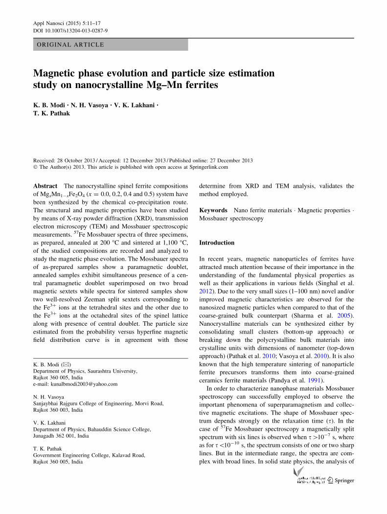
The X-ray powder diffraction patterns recorded at room
temperature (27 �C) for the as-prepared (x = 0.0, 0.2 and
0.4), annealed at 200 �C (x = 0.2 and 0.5) and sintered at
1,100 �C (x = 0.5) samples are shown in Fig. 1. The
spectra for the as-prepared particles have two broad peaks
without any structure. The background noise and broadness
of the peaks are characteristic of particles with nanometer
dimensions. The X-ray diffractometry showed that
annealed samples were single phase spinel. The significant
12 Appl Nanosci (2015) 5:11–17
123
line broadening is seen due to nanosized particle formation
in annealed samples. This is expected because in the
nanosized particles there are insufficient diffraction cen-
ters, which causes the line broadening. A representative
XRD pattern of sintered sample of x = 0.5 composition
shows sharp Bragg reflections corresponding to the fcc
spinel structure; thus, exhibiting no signature of evolution
of any extra phase after high temperature sintering, con-
firming good quality of wet chemically prepared ferrite
samples with expected stoichiometry. The sharp Bragg
reflections indicate that the fine particle nature is lost due to
high temperature sintering and the ferrite materials are akin
to the coarse-grained ceramically prepared products.
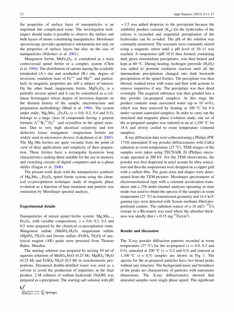
The cell edge parameter for annealed samples was
found to decrease with Mg2?-substitution (Table 1) which
is attributed to the replacement of larger cation Mn2?
having Pauling ionic radius of 0.83 A by smaller cation
Mg2? (0.66 A) in the system (Weast 1980–1981). Typical
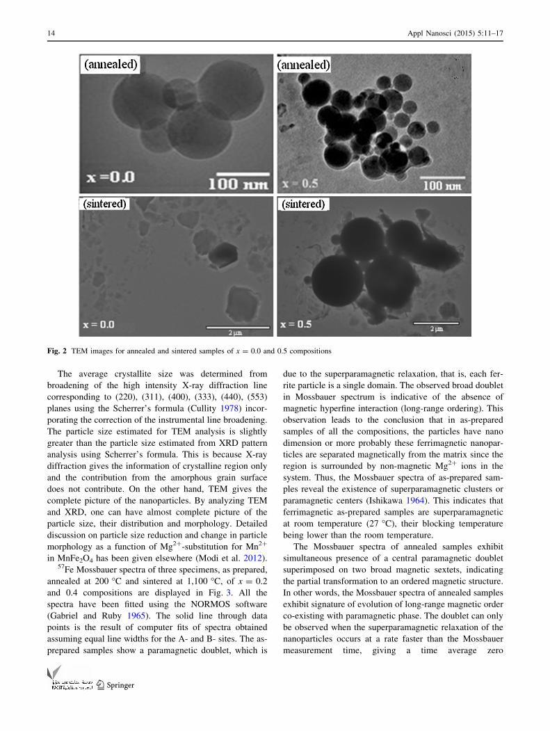
TEM images for the samples annealed at 200 �C with
x = 0.0 and 0.5 compositions are shown in Fig. 2. It can
be seen that particles are quite well dispersed and not
much agglomeration is present. The mean particle size for
MnFe2O4 (x = 0.0) is found to be *40 nm, which
decreases with increasing Mg-content (x) and becomes
*27 nm for Mg0.5Mn0.5Fe2O4 (x = 0.5) composition. It
is well known that materials with a cubic crystal structure
are prone to grow into a spherical shape to minimize the
surface tension (Parekh et al. 2006). TEM images for the
typical x = 0.0 and 0.5 compositions of sintered(w)
product are also shown in Fig. 2 for comparison purpose.
However, for sintered samples particles appear in an
irregular shape for x = 0.0 composition, while for
x = 0.5 composition, along with spherically shaped par-
ticles with mean particle size of 1.5 lm, few particles are
of irregular shape.
Fig. 1 X-ray powder diffractograms for as-prepared (x = 0.0, 0.2 and 0.4), annealed (x = 0.2 and 0.5) and sintered (S) (x = 0.5) samples of
Mg–Mn ferrites
Table 1 Lattice constant (a) and average particle size (D) for
annealed samples of Mg–Mn ferrite compositions
Mg-content (x) a (nm) ± 0.0002 nm D (nm) ± 1 nm
XRD TEM Mossbauer
0.0 0.8479 38 40 42
0.2 0.8448 33 36 33
0.4 0.8427 28 31 30
0.5 0.8408 25 27 22
Appl Nanosci (2015) 5:11–17 13
123
The average crystallite size was determined from
broadening of the high intensity X-ray diffraction line
corresponding to (220), (311), (400), (333), (440), (553)
planes using the Scherrer’s formula (Cullity 1978) incor-
porating the correction of the instrumental line broadening.
The particle size estimated for TEM analysis is slightly
greater than the particle size estimated from XRD pattern
analysis using Scherrer’s formula. This is because X-ray
diffraction gives the information of crystalline region only
and the contribution from the amorphous grain surface
does not contribute. On the other hand, TEM gives the
complete picture of the nanoparticles. By analyzing TEM
and XRD, one can have almost complete picture of the
particle size, their distribution and morphology. Detailed
discussion on particle size reduction and change in particle
morphology as a function of Mg2?-substitution for Mn2?
in MnFe2O4 has been given elsewhere (Modi et al. 2012).57Fe Mossbauer spectra of three specimens, as prepared,
annealed at 200 �C and sintered at 1,100 �C, of x = 0.2
and 0.4 compositions are displayed in Fig. 3. All the
spectra have been fitted using the NORMOS software
(Gabriel and Ruby 1965). The solid line through data
points is the result of computer fits of spectra obtained
assuming equal line widths for the A- and B- sites. The as-
prepared samples show a paramagnetic doublet, which is
due to the superparamagnetic relaxation, that is, each fer-
rite particle is a single domain. The observed broad doublet
in Mossbauer spectrum is indicative of the absence of
magnetic hyperfine interaction (long-range ordering). This
observation leads to the conclusion that in as-prepared
samples of all the compositions, the particles have nano
dimension or more probably these ferrimagnetic nanopar-
ticles are separated magnetically from the matrix since the
region is surrounded by non-magnetic Mg2? ions in the
system. Thus, the Mossbauer spectra of as-prepared sam-
ples reveal the existence of superparamagnetic clusters or
paramagnetic centers (Ishikawa 1964). This indicates that
ferrimagnetic as-prepared samples are superparamagnetic
at room temperature (27 �C), their blocking temperature
being lower than the room temperature.
The Mossbauer spectra of annealed samples exhibit
simultaneous presence of a central paramagnetic doublet
superimposed on two broad magnetic sextets, indicating
the partial transformation to an ordered magnetic structure.
In other words, the Mossbauer spectra of annealed samples
exhibit signature of evolution of long-range magnetic order
co-existing with paramagnetic phase. The doublet can only
be observed when the superparamagnetic relaxation of the
nanoparticles occurs at a rate faster than the Mossbauer
measurement time, giving a time average zero
Fig. 2 TEM images for annealed and sintered samples of x = 0.0 and 0.5 compositions
14 Appl Nanosci (2015) 5:11–17
123
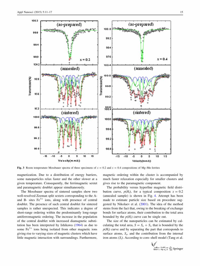
magnetization. Due to a distribution of energy barriers,
some nanoparticles relax faster and the other slower at a
given temperature. Consequently, the ferrimagnetic sextet
and paramagnetic doublet appear simultaneously.
The Mossbauer spectra of sintered samples show two
well-resolved Zeeman split sextets corresponding to the A-
and B- sites Fe3? ions, along with presence of central
doublet. The presence of such central doublet for sintered
samples is rather unexpected. This indicates a degree of
short-range ordering within the predominantly long-range
antiferromagnetic ordering. The increase in the population
of the central doublet with increased diamagnetic substi-
tution has been interpreted by Ishikawa (1964) as due to
some Fe3? ions being isolated from other magnetic ions
giving rise to varying sizes of magnetic clusters which have
little magnetic interaction with surroundings. Furthermore,
magnetic ordering within the cluster is accompanied by
much faster relaxation especially for smaller clusters and
gives rise to the paramagnetic component.
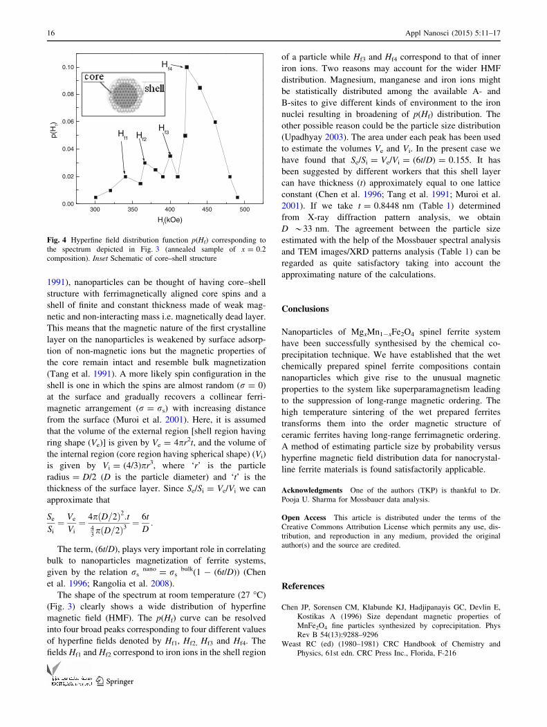
The probability versus hyperfine magnetic field distri-
bution curve, p(Hf), for a typical composition x = 0.2
(annealed sample) is shown in Fig. 4. Attempt has been
made to estimate particle size based on procedure sug-
gested by Nikolaev et al. (2001). The idea of the method
stems from the fact that, owing to the breaking of exchange
bonds for surface atoms, their contribution to the total area
bounded by the p(Hf) curve can be single out.
The size of the nanoparticles can be estimated by cal-
culating the total area, S = Se ? Si, that is bounded by the
p(Hf) curve and by separating the part that corresponds to
surface atoms, Se, and the contribution from the internal
iron atoms (Si). According to core–shell model (Tang et al.
Fig. 3 Room temperature Mossbauer spectra of three specimens of x = 0.2 and x = 0.4 compositions of Mg–Mn ferrites
Appl Nanosci (2015) 5:11–17 15
123
1991), nanoparticles can be thought of having core–shell
structure with ferrimagnetically aligned core spins and a
shell of finite and constant thickness made of weak mag-
netic and non-interacting mass i.e. magnetically dead layer.
This means that the magnetic nature of the first crystalline
layer on the nanoparticles is weakened by surface adsorp-
tion of non-magnetic ions but the magnetic properties of
the core remain intact and resemble bulk magnetization
(Tang et al. 1991). A more likely spin configuration in the
shell is one in which the spins are almost random (r = 0)
at the surface and gradually recovers a collinear ferri-
magnetic arrangement (r = rs) with increasing distance
from the surface (Muroi et al. 2001). Here, it is assumed
that the volume of the external region [shell region having
ring shape (Ve)] is given by Ve = 4pr2t, and the volume of
the internal region (core region having spherical shape) (Vi)
is given by Vi = (4/3)pr3, where ‘r’ is the particle
radius = D/2 (D is the particle diameter) and ‘t’ is the
thickness of the surface layer. Since Se/Si = Ve/Vi we can
approximate that
Se
Si
¼ Ve
Vi
¼ 4pðD=2Þ2:t43pðD=2Þ3
¼ 6t
D:
The term, (6t/D), plays very important role in correlating
bulk to nanoparticles magnetization of ferrite systems,
given by the relation rsnano = rs
bulk(1 - (6t/D)) (Chen
et al. 1996; Rangolia et al. 2008).
The shape of the spectrum at room temperature (27 �C)
(Fig. 3) clearly shows a wide distribution of hyperfine
magnetic field (HMF). The p(Hf) curve can be resolved
into four broad peaks corresponding to four different values
of hyperfine fields denoted by Hf1, Hf2, Hf3 and Hf4. The
fields Hf1 and Hf2 correspond to iron ions in the shell region
of a particle while Hf3 and Hf4 correspond to that of inner
iron ions. Two reasons may account for the wider HMF
distribution. Magnesium, manganese and iron ions might
be statistically distributed among the available A- and
B-sites to give different kinds of environment to the iron
nuclei resulting in broadening of p(Hf) distribution. The
other possible reason could be the particle size distribution
(Upadhyay 2003). The area under each peak has been used
to estimate the volumes Ve and Vi. In the present case we
have found that Se/Si = Ve/Vi = (6t/D) = 0.155. It has
been suggested by different workers that this shell layer
can have thickness (t) approximately equal to one lattice
constant (Chen et al. 1996; Tang et al. 1991; Muroi et al.
2001). If we take t = 0.8448 nm (Table 1) determined
from X-ray diffraction pattern analysis, we obtain
D *33 nm. The agreement between the particle size
estimated with the help of the Mossbauer spectral analysis
and TEM images/XRD patterns analysis (Table 1) can be
regarded as quite satisfactory taking into account the
approximating nature of the calculations.
Conclusions
Nanoparticles of MgxMn1-xFe2O4 spinel ferrite system
have been successfully synthesised by the chemical co-
precipitation technique. We have established that the wet
chemically prepared spinel ferrite compositions contain
nanoparticles which give rise to the unusual magnetic
properties to the system like superparamagnetism leading
to the suppression of long-range magnetic ordering. The
high temperature sintering of the wet prepared ferrites
transforms them into the order magnetic structure of
ceramic ferrites having long-range ferrimagnetic ordering.
A method of estimating particle size by probability versus
hyperfine magnetic field distribution data for nanocrystal-
line ferrite materials is found satisfactorily applicable.
Acknowledgments One of the authors (TKP) is thankful to Dr.
Pooja U. Sharma for Mossbauer data analysis.
Open Access This article is distributed under the terms of the
Creative Commons Attribution License which permits any use, dis-
tribution, and reproduction in any medium, provided the original
author(s) and the source are credited.
References
Chen JP, Sorensen CM, Klabunde KJ, Hadjipanayis GC, Devlin E,
Kostikas A (1996) Size dependant magnetic properties of
MnFe2O4 fine particles synthesized by coprecipitation. Phys
Rev B 54(13):9288–9296
Weast RC (ed) (1980–1981) CRC Handbook of Chemistry and
Physics, 61st edn. CRC Press Inc., Florida, F-216
300 350 400 450 5000.00
0.02
0.04
0.06
0.08
0.10 Hf4
Hf3Hf2
Hf1p(
Hf)
Hf(kOe)
Fig. 4 Hyperfine field distribution function p(Hf) corresponding to
the spectrum depicted in Fig. 3 (annealed sample of x = 0.2
composition). Inset Schematic of core–shell structure
16 Appl Nanosci (2015) 5:11–17
123
Cullity BD (1978) Elements of X-ray diffraction. 2nd ed. Addison-
Wesley, Reading. 99
Gabriel JR, Ruby SL (1965) Computation of mossbauer spectra. Nucl
Inst Meth 36:23–28
Gagan K, Kanthwal M, Chauhan BS, Sing M (2006) Cation
distribution in mixed Mg–Mn ferrite systems from X-ray
diffraction technique and saturation magnetization. Ind J Pure
Appl Phys 44:930–934
Ishikawa Y (1964) Superparamagnetism in magnetically dilute
systems. J Appl Phys 35(3):1054–1059
Lakshman A, Subba Rao PSV, Rao BP, Rao KH (2005) Electrical
properties of In3? and Cr3? substituted magnesium–manganese
ferrites. J Phys D Appl Phys 38(5):673–678
Modi KB, Joshi HH, Kulkarni RG (1996) Magnetic and electrical
properties of Al3? substituted MgFe2O4. J Mater Sci 31:1311–1317
Modi KB, Vasoya NH, Lakhani VK, Pathak TK (2012) Spherical to
Needle Shaped Particles Transformation Study on Nanocrystal-
line Mg–Mn Ferrites. J Adv Microsc Res 7(1):40–43
Muroi M, Street R, McCormiok PG, Amighian J (2001) Magnetic
properties of ultrafine MnFe2O4 powders prepared by mechano-
chemical processing. Phys Rev B 63:184414–184420
Nikolaev VI, Shipilin AM, Zakharova IN (2001) On estimating
nanoparticles size with the help of Mossbauer effect. Phys Solid
State 43(8):1455–1457
Pandya PB, Joshi HH, Kulkarni RG (1991) Bulk magnetic properties
of Co–Zn ferrites prepared by co-precipitation method. J Mater
Sci 26:5509–5512
Parekh K, Upadhyay RV, Belova L, Rao KV (2006) Ternary
monodispersed Mn0.5Zn0.5Fe2O4 ferrite nanoparticles:
preparation and magnetic characterization. Nanotechnology
17:5970–5975
Pathak TK, Vaoya NH, Lakhani VK, Modi KB (2010) Structural and
magnetic phase evolution study on needle shaped nanoparticles
of magnesium ferrites. Ceram Int 36(1):275–281
Rangolia MK, Chhantbar MC, Tanna AR, Modi KB, Baldha GJ, Joshi
HH (2008) Magnetic behaviour of nano-sized and coarse
powders of Cd–Ni ferrites synthesized by wet- chemical route.
Ind J Pure Appl Phys 46:60–64
Sharma RK, Suwalka O, Lakshmi N, Venugopalan K, Banerjee A,
Joy PA (2005) Synthesis of Chromium substituted nanoparticles
of cobalt zinc ferrites by coprecipitation. Mater Lett
59(27):3402–3405
Singhal S, Jauhar S, Singh J, Chandra K, Bansal S (2012)
Investigation of structural, magnetic, electrical and Optical
properties of chromium substituted cobalt ferrites (CoCrx-
Fe2-xO4, 0 B x B 1) synthesized using sol gel auto combustion
method. J Mol Struct 1012:182–188
Tang ZX, Sorensen CM, Klabunde KJ, Hadjipanayis GC (1991) Size-
dependent Curie temperature in nanoscale MnFe2O4 particles.
Phys Rev Lett 67(25):3602–3605
Upadhyay C (2003) Controlled formation of nanosize spinel ferrites
(4–15 nm) and their magnetic and structural studies. Ph.D.
thesis, IIT—Kanpur, India. 102
Vasoya NH, Vanpariya LH, Sakariya PN, Timbadiya MD, Pathak TK,
Lakhani VK, Modi KB (2010) Synthesis of nanostructured
material by mechanical milling and study on structural property
modification in Ni0.5Zn0.5Fe2O4. Ceram Int 36(3):947–954
Appl Nanosci (2015) 5:11–17 17
123